
Acute Cholecystitis and Choledocholithiasis · choledocholithiasis, intraoperative anatomy,...
25
Acute Cholecystitis and Choledocholithiasis Eric Rellinger April 3, 2013
Transcript of Acute Cholecystitis and Choledocholithiasis · choledocholithiasis, intraoperative anatomy,...
Acute Cholecystitis and Choledocholithiasis
Eric Rellinger April 3, 2013
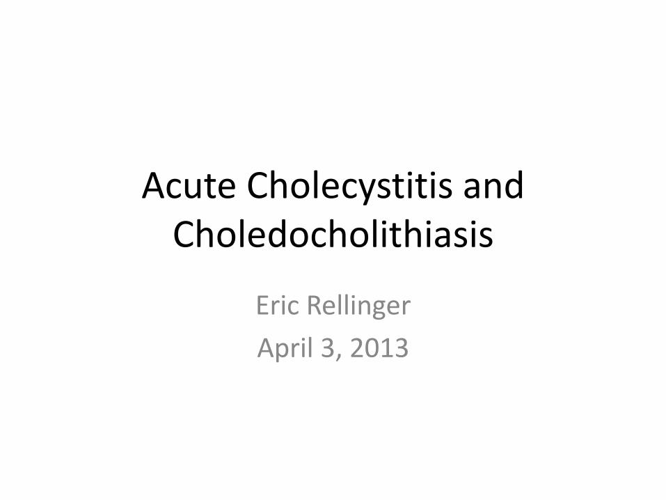
Overview • Biliary Anatomy • Acute cholecystitis
– Diagnosis • Physical Exam • Laboratory • Imaging
– Management • Choledocholithiasis
– Diagnosis • Laboratory • Imaging
– Management
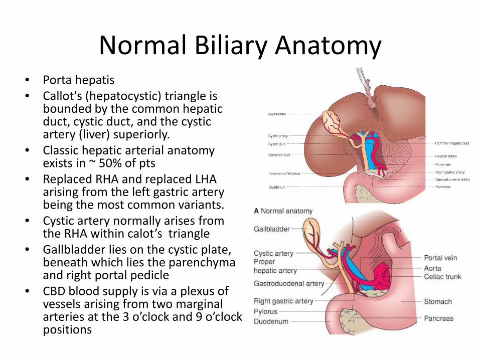
Normal Biliary Anatomy • Porta hepatis • Callot's (hepatocystic) triangle is
bounded by the common hepatic duct, cystic duct, and the cystic artery (liver) superiorly.
• Classic hepatic arterial anatomy exists in ~ 50% of pts
• Replaced RHA and replaced LHA arising from the left gastric artery being the most common variants.
• Cystic artery normally arises from the RHA within calot’s triangle
• Gallbladder lies on the cystic plate, beneath which lies the parenchyma and right portal pedicle
• CBD blood supply is via a plexus of vessels arising from two marginal arteries at the 3 o’clock and 9 o’clock positions
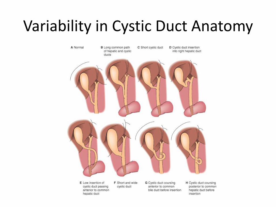
Variability in Cystic Duct Anatomy
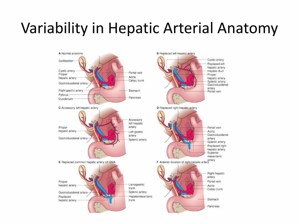
Variability in Hepatic Arterial Anatomy
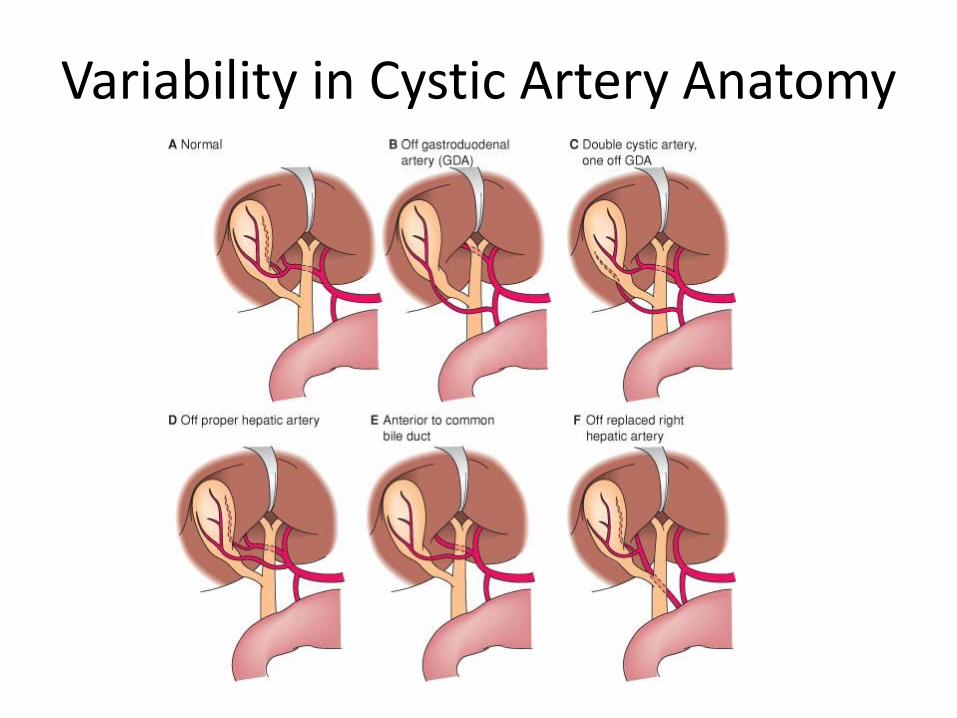
Variability in Cystic Artery Anatomy
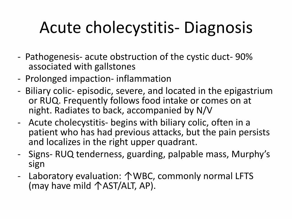
Acute cholecystitis- Diagnosis - Pathogenesis- acute obstruction of the cystic duct- 90%
associated with gallstones - Prolonged impaction- inflammation - Biliary colic- episodic, severe, and located in the epigastrium
or RUQ. Frequently follows food intake or comes on at night. Radiates to back, accompanied by N/V
- Acute cholecystitis- begins with biliary colic, often in a patient who has had previous attacks, but the pain persists and localizes in the right upper quadrant.
- Signs- RUQ tenderness, guarding, palpable mass, Murphy’s sign
- Laboratory evaluation: ↑WBC, commonly normal LFTS (may have mild ↑AST/ALT, AP).
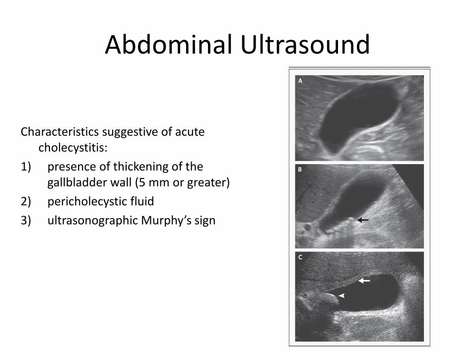
Abdominal Ultrasound
Characteristics suggestive of acute cholecystitis:
1) presence of thickening of the gallbladder wall (5 mm or greater)
2) pericholecystic fluid 3) ultrasonographic Murphy’s sign
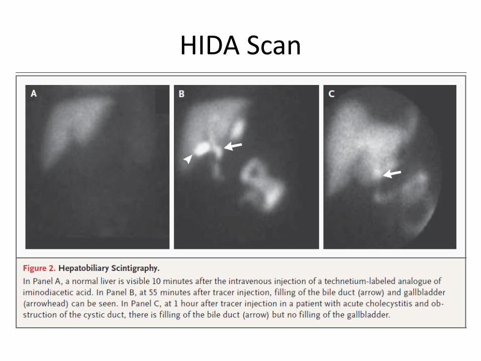
HIDA Scan
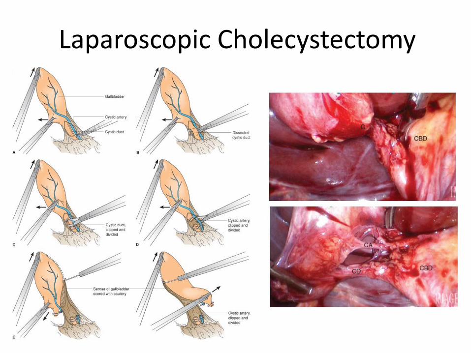
Laparoscopic Cholecystectomy
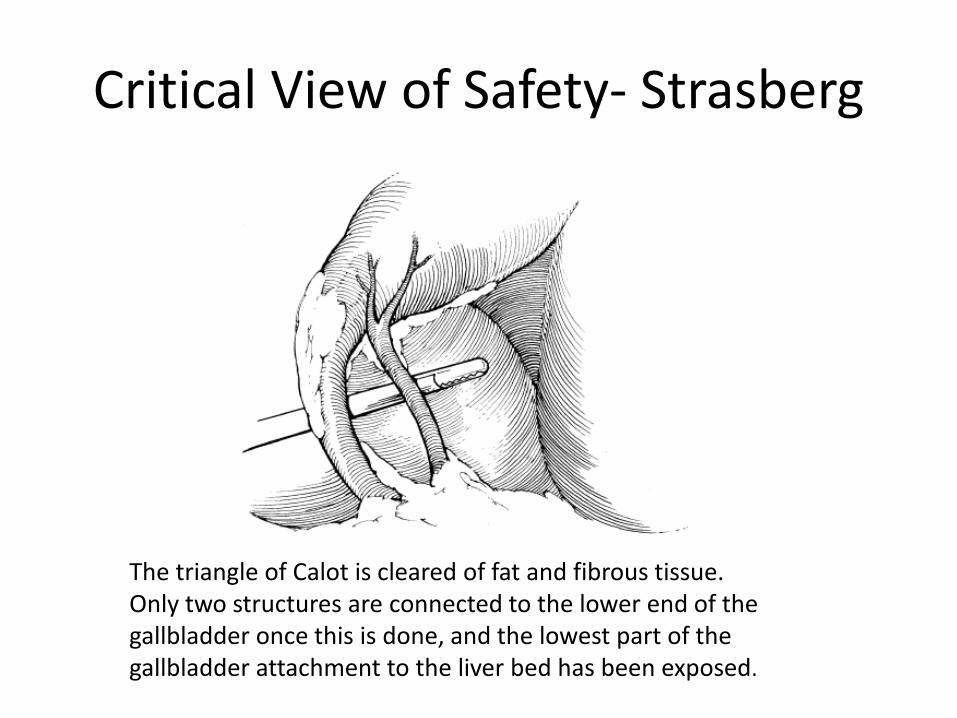
Critical View of Safety- Strasberg
The triangle of Calot is cleared of fat and fibrous tissue. Only two structures are connected to the lower end of the gallbladder once this is done, and the lowest part of the gallbladder attachment to the liver bed has been exposed.
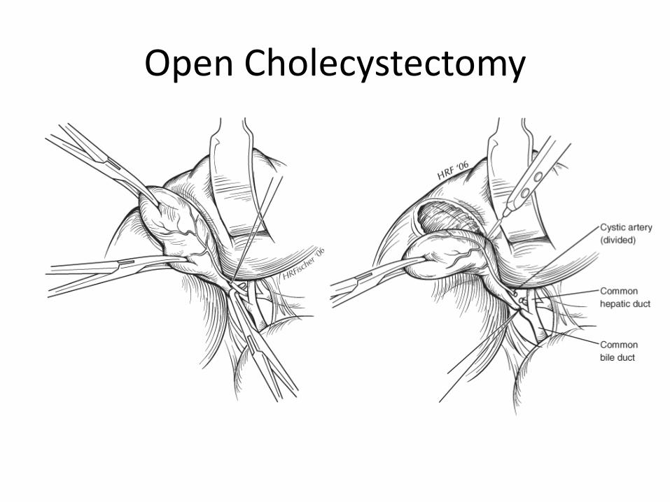
Open Cholecystectomy

Subtotal/Partial cholecystectomy • Used for portal hypertension or
difficult anatomy within the triangle of calot
• Partial cholecystecotmy- gallbladder not in contact with liver bed is removed with cautery-cystic duct orifice is suture ligated vs. drain left in place
• Subtotal cholecystectomy- gallbladder is transected at the infundibulum
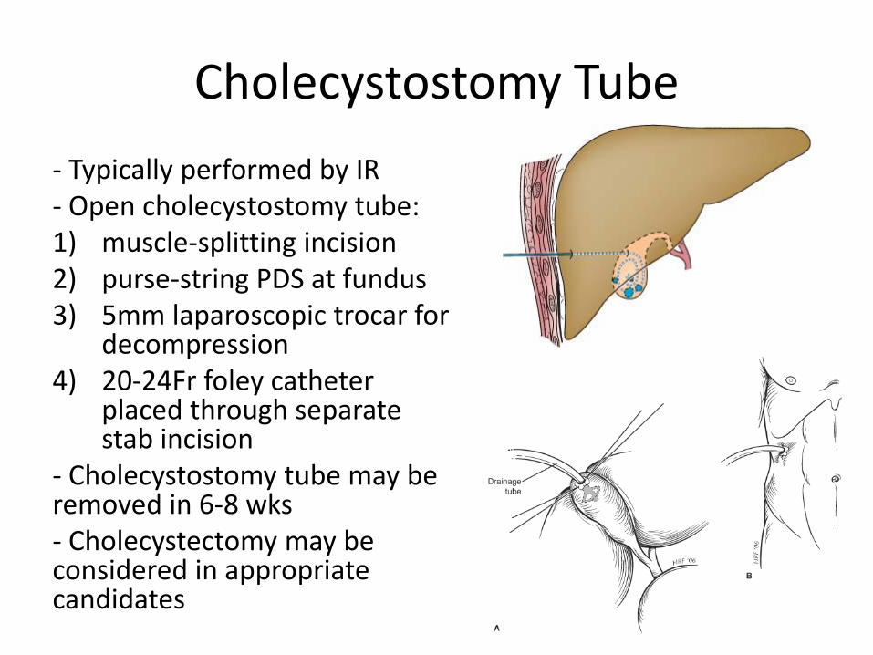
Cholecystostomy Tube - Typically performed by IR - Open cholecystostomy tube: 1) muscle-splitting incision 2) purse-string PDS at fundus 3) 5mm laparoscopic trocar for
decompression 4) 20-24Fr foley catheter
placed through separate stab incision
- Cholecystostomy tube may be removed in 6-8 wks - Cholecystectomy may be considered in appropriate candidates
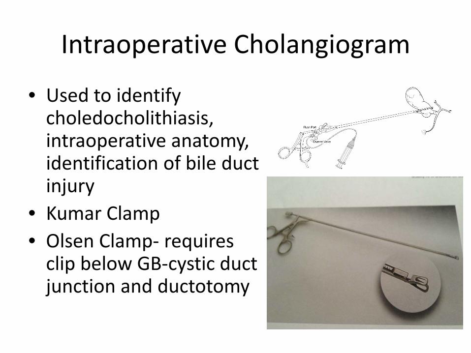
Intraoperative Cholangiogram
• Used to identify choledocholithiasis, intraoperative anatomy, identification of bile duct injury
• Kumar Clamp • Olsen Clamp- requires
clip below GB-cystic duct junction and ductotomy
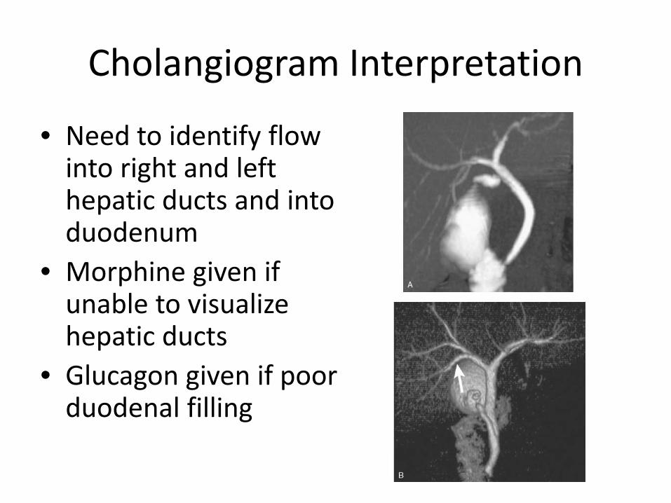
Cholangiogram Interpretation
• Need to identify flow into right and left hepatic ducts and into duodenum
• Morphine given if unable to visualize hepatic ducts
• Glucagon given if poor duodenal filling
Choledocholithiasis
• Evidence of obstructive jaundice • Common laboratory evaluation- Elevated Tbili,
Cbili, Alkaline phosphatase, LDH • Imaging criteria- enlarged CBD (or imaging of
a CBD stone • Gallstone pancreatitis
Treatment options
• ERCP vs. Lap CBDE vs. Open CBDE • Post-operative ERCP vs. common bile duct exploration • ERCP- reported successful ductal clearance- 80-90%,
normal exam found in 40% • Laparoscopic CBDE- 80-90% success rate • Single-stage laparoscopic cholecystectomy and CBDE is
preferred- less expensive, shorter hospital stay • Key limitations to lap CBDE- insufficient experience,
time constraints, and inadequate equipment
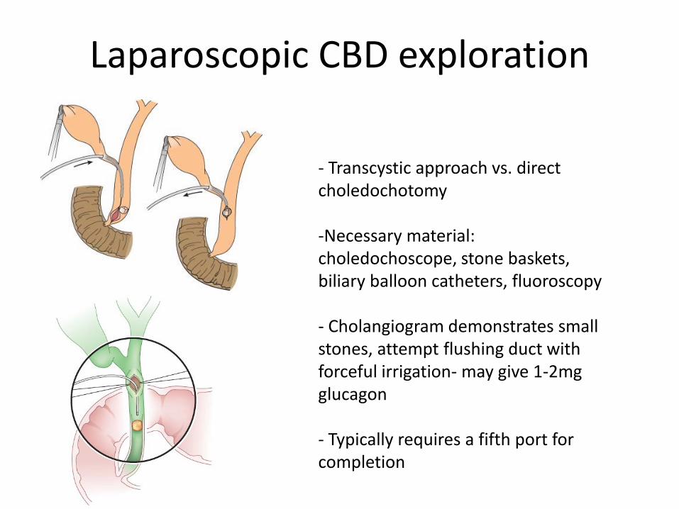
Laparoscopic CBD exploration
- Transcystic approach vs. direct choledochotomy -Necessary material: choledochoscope, stone baskets, biliary balloon catheters, fluoroscopy
- Cholangiogram demonstrates small stones, attempt flushing duct with forceful irrigation- may give 1-2mg glucagon
- Typically requires a fifth port for completion
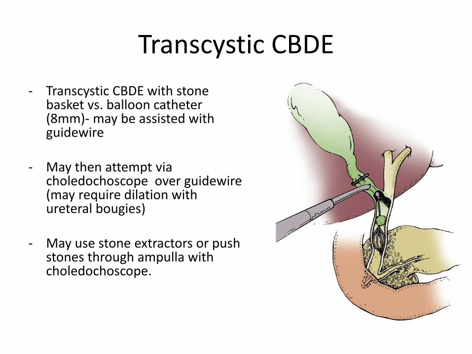
Transcystic CBDE - Transcystic CBDE with stone
basket vs. balloon catheter (8mm)- may be assisted with guidewire
- May then attempt via choledochoscope over guidewire (may require dilation with ureteral bougies)
- May use stone extractors or push stones through ampulla with choledochoscope.
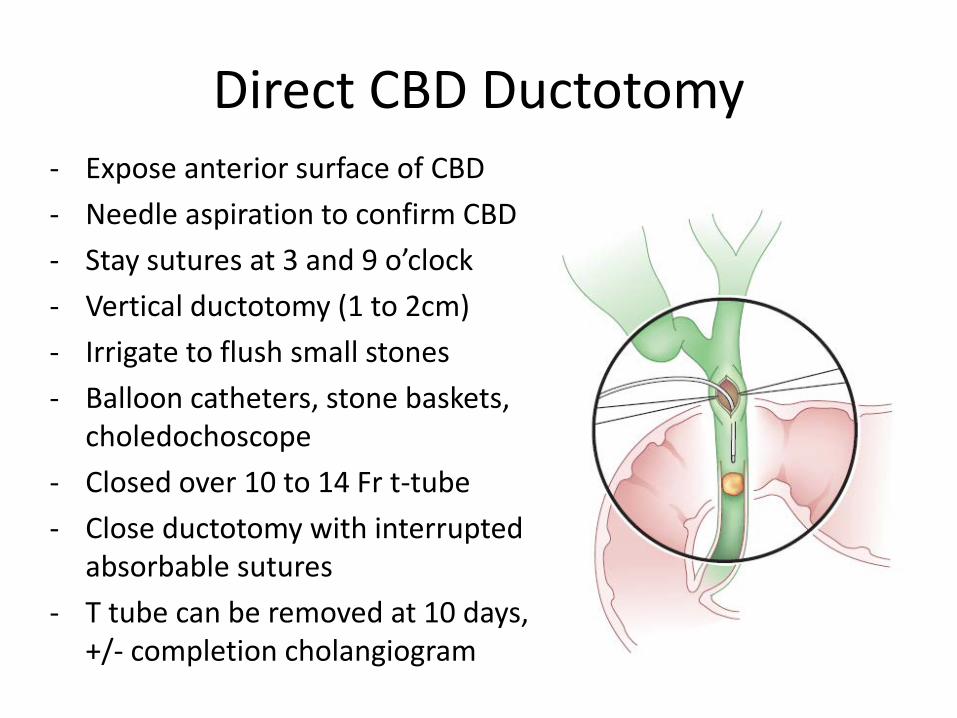
Direct CBD Ductotomy - Expose anterior surface of CBD - Needle aspiration to confirm CBD - Stay sutures at 3 and 9 o’clock - Vertical ductotomy (1 to 2cm) - Irrigate to flush small stones - Balloon catheters, stone baskets,
choledochoscope - Closed over 10 to 14 Fr t-tube - Close ductotomy with interrupted
absorbable sutures - T tube can be removed at 10 days,
+/- completion cholangiogram

Laparoscopic CBD exploration
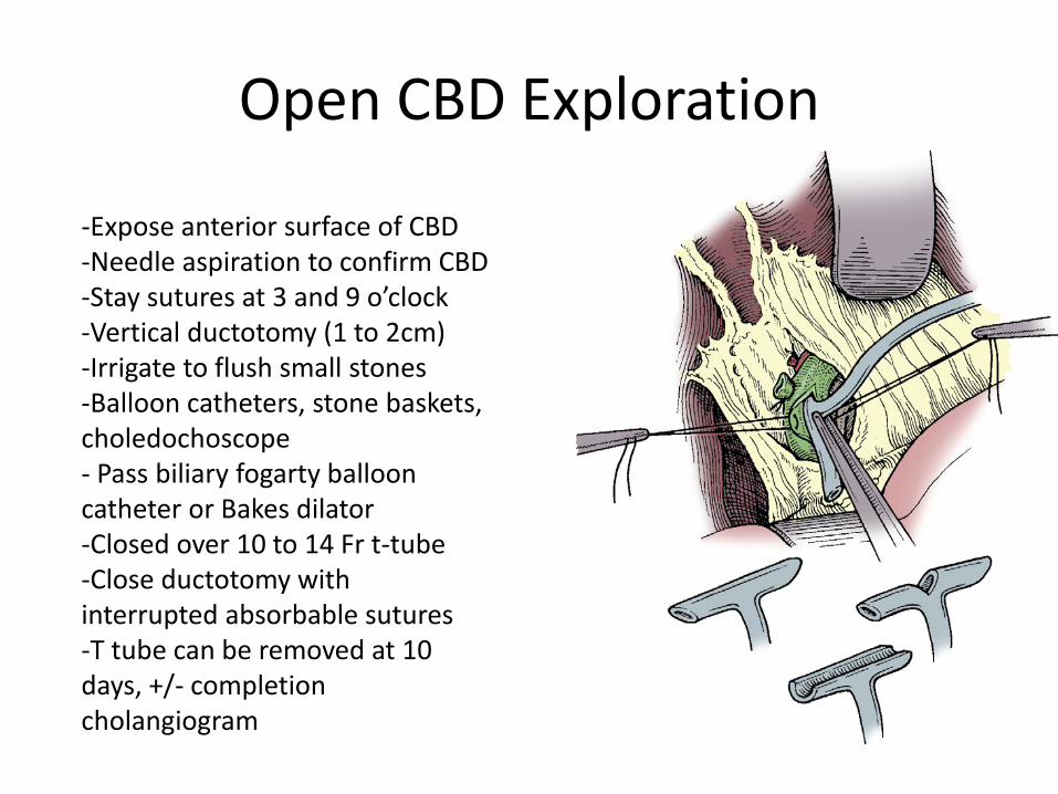
Open CBD Exploration
-Expose anterior surface of CBD -Needle aspiration to confirm CBD -Stay sutures at 3 and 9 o’clock -Vertical ductotomy (1 to 2cm) -Irrigate to flush small stones -Balloon catheters, stone baskets, choledochoscope - Pass biliary fogarty balloon catheter or Bakes dilator -Closed over 10 to 14 Fr t-tube -Close ductotomy with interrupted absorbable sutures -T tube can be removed at 10 days, +/- completion cholangiogram