Elective/Special Course: MYCOLOGY and PLANT PATHOLOGY
189
Elective/Special Course: MYCOLOGY and PLANT PATHOLOGY 4102 [Special paper – I (Theoretical)]
Transcript of Elective/Special Course: MYCOLOGY and PLANT PATHOLOGY

Elective/Special Course: MYCOLOGY and PLANT PATHOLOGY
4102 [Special paper – I (Theoretical)]

1. Fungal diversity in different ecosystems
The structure and composition of fungal cell, effect of
environment on fungal growth and behavior

Diversity spectrum• THE number of fungi recorded in India exceeds 27,000 species, the largest biotic
community after insects1. The true fungi belong to kingdom Eukaryota which hasfour phyla, 103 orders, 484 families and 4979 genera. The eighth edition ofDictionary of the Fungi2 has recognized eleven phyla.
• The Deuteromycotina is not accepted as a formal taxonomic category. The number offungal genera reported from the world and that from India between 1905 and 1995,are shown in Table 1. About 205 new genera have been described from India, ofwhich 32% were discovered by C. V. Subramanian of the University of Madras. Ofthese, approximately 27,000 species are reported to colonize diversified habitats1.This indicates a ten-fold increase in the last 70 years.
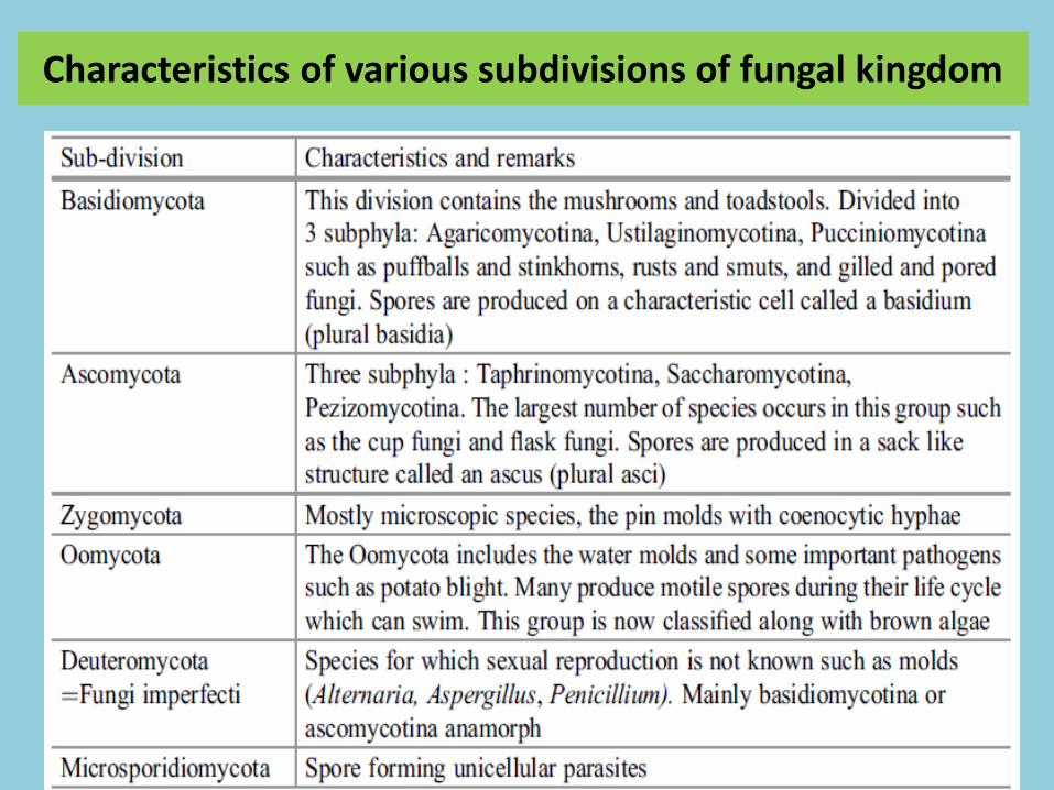
Characteristics of various subdivisions of fungal kingdom

Fungal cell wall• Fungal cell wall is very different from insect exoskeletons
or a plant cell walls in metabolic point of view.
• The cell wall is made up of:
– 1) chitin (polymers of acetylated amino sugar N-acetyl-glucosamine)
– 2) glucan (polymers of glucose)
– 3) proteins (polymers of amino acids).
• Glucan and chitin are components of the primary wall.
• Proteins are components of the secondary wall.

Basic component of fungal cell wall

• Other components include chitosan, melanins and lipids.
• Enzymes include cellulase which acts on cellulose of plants.
• The outermost surface of the cell wall
– 1) provides a medium between the cell and the environment
– 2) a site where antigen and agglutinin gets attached to the substrate, host and other cells.

Chitin
• Chained polymer β(1-4) N-acetyl-glucosamine.
• Found naturally as structural polysacharides in most fungal cell wall as cell wall components.
• Gives strength where each molecule contains a unit of sugar that is bonded by hydrogen bond to give it rigidity.
• Each microfibril of chitin gives the shape of the cell and gives strength to mature cell walls.
• Microfibrils can be of various shapes:
– in yeast: short and thick
– in hyphal wall: long and interwoven

• Septa is rich in chitin
• Septa can be stained with “calcoflour white”.
• Saccharomyces cerevisiae has low amount of chitin.

Chitosan
• Chained β(1-4)glucosamine.
• Result of continuousdeacetylation of chitin.
• An importantcomponent in wall ofZygomycetes and canbe found in ascosporewalls of Saccharomycescerevisiae.


Glucan
• Most fungal walls contain β- chained glucan.
• Glucan are polysaccharides that contain onlyglucose as structural components, and are linked
with β-( 1-3 ) glycosidic bonds.
• Walls of Ascomycetes and Basidiomycetes contain
glucan with branching of β(1-6) glucan.
• There are some fungi with a(1-4) glucan.

Glycoprotein and Protein
• Glycoproteins are protein that contain oligosaccharide chains(glycans) covalently attached to polypeptide side- chains.
• Glycoproteins (include mannoprotein, galactoprotein andxyloprotein) are important components of the matrixs of cellwalls.
• Glycoprotein contain glucosamine and/or N-acetylglucosamine.
• In parasitic fungi such as Candida albicans and Aspergillusfumigatus, the glycoproteins are antigens.
• Mannoprotein in Saccharomyces cerevisiae are largemolecules.
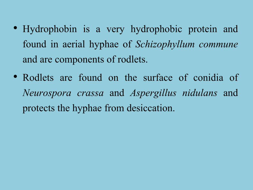
• Hydrophobin is a very hydrophobic protein andfound in aerial hyphae of Schizophyllum communeand are components of rodlets.
• Rodlets are found on the surface of conidia ofNeurospora crassa and Aspergillus nidulans andprotects the hyphae from desiccation.

Characteristics of Fungal Cell Wall
1. Gives shape to fungi.
2. Gives strength to fungi.
3. Provides protection for the protoplasm from ultraviolet rays for the presence of melanins
4. Ability to resist lysis by organic solvents such as enzymes, toxins, osmotic integrity.
5. Ability to bind with metal ions.
6. Secretes enzymes from their walls (invertase hydrolyses sucrose to glucose and fructose) and so assisting in nutrition.
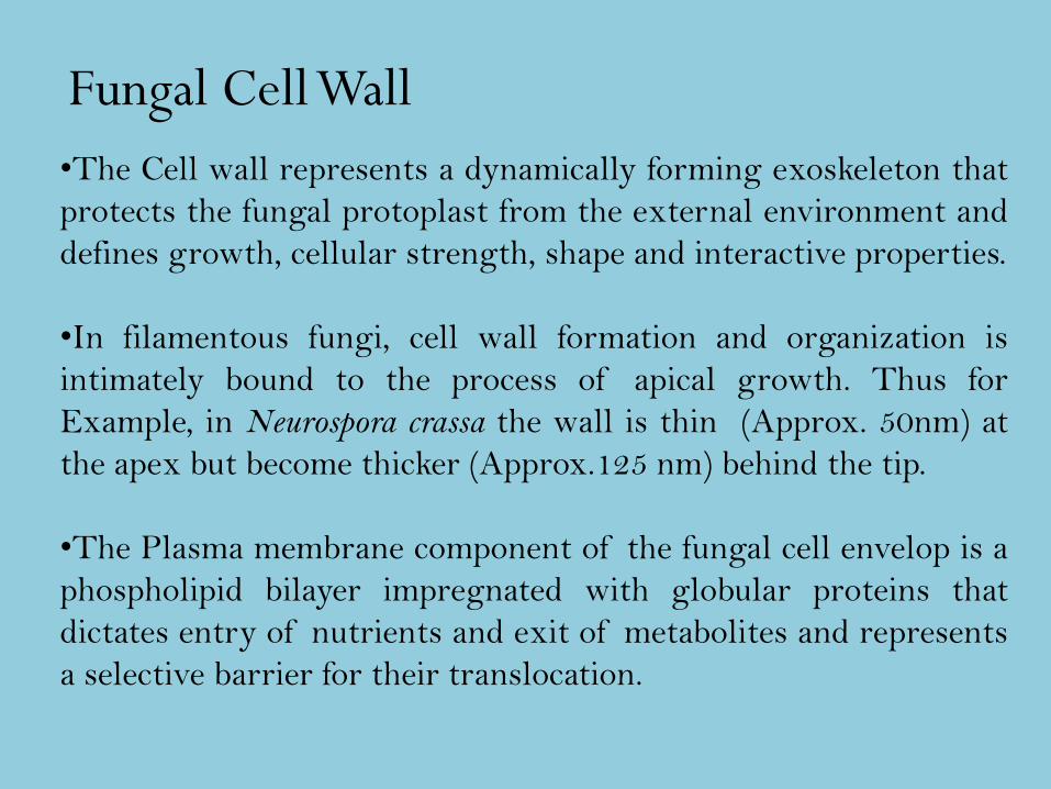
•The Cell wall represents a dynamically forming exoskeleton that
protects the fungal protoplast from the external environment and
defines growth, cellular strength, shape and interactive properties.
•In filamentous fungi, cell wall formation and organization is
intimately bound to the process of apical growth. Thus for
Example, in Neurospora crassa the wall is thin (Approx. 50nm) at
the apex but become thicker (Approx.125 nm) behind the tip.
•The Plasma membrane component of the fungal cell envelop is a
phospholipid bilayer impregnated with globular proteins that
dictates entry of nutrients and exit of metabolites and represents
a selective barrier for their translocation.
Fungal Cell Wall

•Ergosterol is the major sterol found in the membranes of fungi, in contrast to
the cholessterol found in the membranes of animals and phytosterols in plants.
This distinction is exploited during the use of certain antifungal agents used
to treat some fungal infections and can be used as an assay tool to quantify
fungal growth.
•The periplasm or periplasmic space is the region external to the
plasmamembrane and internal to the cell wall. In yeast cell, it consist of
secreted proteins (mannoproteins) and enzymes ( such as invertase and acid
phosphatase) that are unable to enter cell wall. In filamentous fungi, the cell
membrane and wall may be intimately bound to form a compact hyphae. So, it
is resistant to plasmolysis.
•Ultrastructural analysis of fungal cell wall reveals a thick, complex fibrillar
network. The cell wall of filamentous fungi are mainly compost of different
polysaccharides according to taxonomic groups.
•Generally, the semi-crytalline microfibrillar components are organized in a
network mainly in the central of cell wall region and are embedded within an
amorphous matrix.

Division Semi-crytalline microfibrillar
network of POLYMER in the
Central of Cell Wall
Gel-like
AMORPHOUS
MATRIX
COMPONENT
Perforated
Septa Present
or AbsentOomycetes β-(1-3),β-(1-6) Glucan cellulose Glucan
AbsentChytridiomycet
es
Chitin microfibrils [ß(1-4)-linked
polymer of N-acetylglucosamine]
, Glucan
Glucan
AbsentZygomycota Chitin, Chitosan [ß(1-4)-linked
polymer of glucosamine]
Polyglucuronic acid,
Glucuronomannoprotei
ns, AbsentAscomycota/
Deuteromyces
Chitin, β -(1-3), β-(1-6) Glucan
[ß(1-3) and ß(1-6)] alkali insoluble
Galactomannoproteins,
α (1-3) Glucan Present mostly
simple with large
central poreBasidiomycota Chitin β -(1-3), β-(1-6) Glucan Xylomannoproteins,
α (1-3) Glucan Present mostly
Dolipore
Mejor polymer found in different taxonomic division of fungi with septation (adapted from Gooday in Gow & Gadd, 1995).

• Approximately 80% of the fungal cell wall consists of
Polysaccharides.
• Most fungi have a fibrillar structure built-up with chitin,
chitosan (Zygomycota only), and ß-glucans, and a variety of
heteropolysaccharides.
• Proteins constitute a small fraction of wall bound material,
rarely more than 20%, and often as glycoprotein, which
function as mating, recognition, wall modification andnutrition. A Class of hydrophobic proteins called hydrophobins are
localized within the aerial growth or appresoria of certain fungi,
Hydrophobins (may constitute up to 10% of total wall protein)
are expressed constitutively, and become bound to the wall when
the hyphae emerge in air.
Fungal cell wall components

• Lipids are found in walls, usually in very small
concentrations.
• Walls also contain a range of other minor components,
including pigments, melanin and salts. Melanin is
important for protecting the hyphae and spores against
biotic and abiotic stress (UV light),
• The hyphae of higher fungi extend via tip growth followed
by cross wall formation or septation, whereas the lower
fungi remain aseptate. Septa may offer some structural
support to hyphae.
• EXTRA LAYERS of wall material are deposited in the
lateral walls behind the extending apex - strengthening the
wall as the hypha matures.

• In the oldest parts of the hyphae (and in many fungal
spores) LIPIDS and PIGMENTS may be deposited in the
wall:
• LIPIDS serve as a nutrient reserve and help prevent
desiccation of PIGMENTS, such as MELANIN, help
protect the protoplast against the damaging effects of UV
radiation.
• Antigenic glycoproteins, agglutinans- adhesions—on cell
wall surface.

In the filamentous fungi, the wall of HYPHAL TIP is thinner
and simpler in structure, consisting of only TWO LAYERS –
1. - an inner layer of fibrils (polymeric) embedded in
protein and
2. - outer layer of mainly protein.
-an inner layer
- outer layer

(a) Outermost layer of amorphous ß-1,3-glucans and ß-1,6-
glucans. (b) glycoprotein reticulum embedded in protein. (c) a
more or less discrete protein layer, (d) chitin microfibrils
embedded in protein, (e) plasma membrane. Based on Hunsley &
Burnett, 1970. [© Jim Deacon]
Diagram to illustrate the wall architecture in a ‘mature’
(subapical) region of a hypha of Neurospora crassa as evidenced
by sequential enzymatic digestion.

PROTECTS the underlying protoplasm;
Determines and MAINTAINS THE SHAPE of the fungal cell
or hypha; if you remove the wall the resulting protoplast will
always assume a spherical shape;
Acts as an INTERFACE between the fungus and its
environment;
Acts as a BINDING SITE for some enzymes;
Possesses ANTIGENIC properties - which allow interactions
with other organisms.
Cell wall Functions:

FUNCTION IN RELATION TO
STRUCTURE
• Glycoproteins in Wall
• Hydrophobins in Walls
• Glomalin in Walls

Different but compatible glycoproteins, called
agglutinins, in the walls of each complementary
hypha fuse to form a complex structure binding the
cells together with release of hormone-like
compounds.
Adhesion is also mediated by fibrillar glyco-proteins
embedded in a gel-like matrix. The fibrils are
highly specific and a complementary protein on
the surface of the partner in pathogenic and
mutualistic interactions.
Glycoproteins in Wall:

Hydrophobins in Walls:
Hydrophobins may constitute up to 10% of total wall
protein. Each molecule consists of a hydrophobic
domain and a hydrophilic domain.
The protein is attached to the fungal wall by the
hydrophilic end. The hydrophobic domain is exposed.
Hydrophobins reduces movement of water through
the wall of the hypha and providing some protection
from desiccation. It may also increase the strength of
the wall.

Glomalin in Walls
Some members of the Glomeromycota produce a
putative glycoprotein in the walls. The compound has
been called glomalin. Recent work suggests that the
compound is related to the Heat Shock Proteins
(HSPs) in group 60.
However, the structure and characterization of the
function of glomalin are incomplete.

Septum of Ascomycota has spherical, membrane-bound organelles
called WORONIN BODIES. First reported by the Russian
mycologist Mikhail Stepanovich Woronin in the euascomycete
Ascobolus pulcherrimus in 1864.
Not all fungi belonging to the Ascomycota possess Woronin
bodies .
– those often possess LARGE HEXAGONAL CRYSTALS OF
PROTEIN (LHCPs) in the cytoplasm that are capable of serving
the same function, i.e. they can seal the septal pores of damaged or
ageing hyphae.
WORONIN BODIES and
LARGE HEXAGONAL CRYSTALS OF PROTEIN

Associated with each septum are spherical, membrane-bound organelles called WORONIN BODIES

THE FORMATION OF WORONIN BODY
AS ADAPTATION
A Woronin body (named after the Russian botanist
Mikhail Stepanovich Woronin) is a peroxisome
derived, dense core microbody with a unit membrane
found near the septae that divide hyphal compartments
in filamentous Ascomycota. One established function of
Woronin bodies is the plugging of the septal pores after
hyphal wounding, which restricts the loss of cytoplasm
to the sites of injury

Fungal HYPHAE
Cylindrical, branching filaments composed of a tubular
cell wall filled with cytoplasm and organelles. Most
fungal hyphae are 2-10 µm diameter. Each HYPHA
(singular) is essentially a tube - consisting of a rigid
wall and containing protoplasm tapered at its tip -
this is the region of active growth (i.e. the extension
zone). Hyphae tend to form a larger network of cells
called a MYCELIUM.

Fungal hyphae have a number of
unique features:
1. SEPTA (cross-walls), possess one of more PORES - such septa
divide up the hyphae into a series of interconnected HYPHAL
COMPARTMENTS, rather than separate, discrete cells.
2. Each hyphal cell or compartment normally contains one or more
NUCLEI able to pass between adjacent compartments, via the
central septal pore.
3. The GROWING TIP is structurally and functionally very
different from the rest of the hypha, Its cytoplasm appears more
dense there are no major organelles at the extreme tip

4. At the extreme tip there is an accumulation of membrane-
bound vesicles - the APICAL VESICULAR CLUSTER
(COMPLEX) (AVC) - which plays an important role in
apical growth.
5. VACUOLES may be visible in sub-apical hyphal
compartments - although small at first, they grow larger
and merge with one another; they store and recycle
cellular metabolites, e.g. enzymes and nutrients.
6. Mycorrhizal species store phosphate in the form of
polyphosphate. Calcium storage for intracellular
signaling. Proteases for breakdown of cellular proteins
and recycling of amino acids. Regulation of cellular pH.

APICAL VESICULAR CLUSTER (AVC):
The growing hyphal tip is structurally and functionally different
from the rest of the hypha with thinner and simpler in
structure than the mature lateral wall of the hypha and the
presence of vesicles which form the APICAL VESICULAR
CLUSTER (AVC):
When a hypha stops growing, these vesicles disappear, when
growth of the hypha resumes, the vesicles reappear.
When a hypha is growing straight ahead, the vesicles are
positioned in the centre of the hyphal tip, movement of the
vesicles to the left or right side of the hyphal tip is accompanied
by a change in direction of growth of the hypha.

Vesicles of the AVC contain:
• Wall PRECURSORS - the sub-units or building
blocks of the wall polymers - e.g. uridine diphosphate
N-acetylglucosamine, the sub-unit of chitin wall.
• LYTIC ENZYMES - which help breakdown and
separate wall components - e.g. chitinase, glucanase.
• wall SYNTHASE ENZYMES - which help assemble
new wall components and so increase the size of the
wall - e.g. chitin synthase, glucan synthase.

• The AVC is found at the actively growing hyphal
tip (the apex) and consists of a mass of vesicles
surrounding the Spitzenkörper, an opaque
structure which can be seen with phase contrast
microscopy. Further back from the centre of the
AVC, several mitochondria are seen.
• The AVC can be seen in ascomycetes,
basidiomycetes and oomycetes; in fact, in all the
fungal species which grow as mycelia.
Vesicles of the AVC contain:

NUCLEI
Nuclei are always present in living cells.
In the fungi nuclei are 1 - 3 μm in diameter,
somewhat smaller than most other
eukaryotic organisms, where they range from
3 - 10 μm.
Contain 3--40 chromosomes which are
small and difficult to visualize in stained and
squashed preparations.

Up to 13--40 Mb (million base pairs) DNA coding for
6,000 to 13,000 genes
Usually haploid [Schizophyllum commune (6)
Neurospora crassa (7),
Emericella (Aspergillus nidulans(8),
Saccharomyces cerevisiae (16),
Ustilago maydis (20);
Naturally diploid - Candida albicans and members of
Oomycota or exist
as polyploid species - Allomyces and several sp. of
Phytophthora

Nuclear membrane persists during division, unlike
plants and animals. The process of formation of 2 sister
nuclei by constriction in the middle of the nuclear
membrane-Karyochoresis)
No clear metaphase plate, chromosomes randomly
dispersed, at anaphase daughter chromatids pull apart
along two tracks on spindle fibres of different lengths.
Various types of spindle pole bodies; significance of
the differences unclear, needed to ensure that
chromosomes separate correctly during nuclear division.
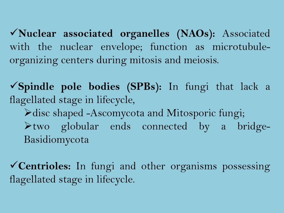
Nuclear associated organelles (NAOs): Associated
with the nuclear envelope; function as microtubule-
organizing centers during mitosis and meiosis.
Spindle pole bodies (SPBs): In fungi that lack a
flagellated stage in lifecycle,
disc shaped -Ascomycota and Mitosporic fungi;
two globular ends connected by a bridge-
Basidiomycota
Centrioles: In fungi and other organisms possessing
flagellated stage in lifecycle.

ENDOPLASMIC RETICULUM

ENDOPLASMIC RETICULUM
ER is a single interconnected membrane system
subdivided into a no. of functional systems.
It is continuous with the nuclear envelope and
intracellulary forms a contiguous system of both non-
fenestrated sheets and variously branched tubules. The
latter predominates at the cell periphery where the
tubules are closely associated with and may possess
molecular links to the plasma membrane.
ER persists throughout the cell cycle.

The type of ER network varies in yeast and
filamentous fungi (tubular & distributed towards the
periphery vs. cisternal sheets often stacked into parallel
arrays and found throughout the radius.
Prominent peripheral ER network in rust fungus),
during stages of specific development (tubular
vesicular network) mostly rough with polyribosomes.
•The ER is highly motile. In Ustilago maydis
cytoplasmic dynein and microtubules were shown to
be required for ER motility but not for maintaining
basic ER organization.

Essential role in both Protein and Lipid pathways as the
site of biosynthesis of nearly all cellular membrane and
trans membrane proteins regardless of ultimate
destination.
Luminal proteins destined for Golgi apparatus and
Vacoules are co-translationally delivered into the ER
lumen
ER lumen stores calcium ions to be released into cytosol
upon induction via appropriate signalling.
FUNCTION

GOLGI APPARATUSA- Assemblages of tubular
cisternae and a lack of
cistarnal stacks are the
hallmarks of Golgi in
filamentous fungi, observed
interspersed with vesicle in
hyphae of Aspergillus
nidulans. The width of
tubules of a single
cisternum is fairly uniform.
Golgibody of
basidiomycetes are
characterized by Swollen
peripheral terminals

In true fungi, golgi bodies are morphologically very simple.
Lacks the stacks of flattened cisternae or dictyosomes and
consists of single cisternal elements.
Golgi equivalents are individual organelles, consisting of
(i) Fenestrated sheets with tubular extensions that are
dispersed throughout the hyphae
OR
(ii)Fenestrated hollow spheres or sheets wrapped closely around
mitochondria. The width of cisternal tubules is generally
uniform within such a sheet or sphere,
GOLGI APPARATUS

TUBULES, VESICLES/MULTIVESICULAR BODIES & VACOULES
A- Multivesicular bodies are
thought to be intermeadiate
compartment between early and
late endosome and can be very
abundant in hyphae in Giberella
persicaria. They are inter-spread
with microtubules and vesicles
that exhibit bidirectional
movement.
B- Fungal vacuoles are an
assemblage of pleiomorphic
entities that can exist as
elongated tubular structure.

HYDROGENOSOMES:
In some obligately anaerobic organisms, including
fungi, an organelle is observed which appears to
produce hydrogenase and pyruvate oxidoreductase. The
enzymes function in the anaerobic conversion of
organic carbon to energy. The organelle is called a
hydrogenosome. Hydrogenosomes have been observed
in the Chytridiomycota found in the rumen of
herbivores; these Chytrids lack mitochondria.

Relatively little is known about them. Appear as tiny
vesicles that are coated with a dense filamentous
material. They are numerous in tips of actively
growing hyphae (peripheral subapical region) where
they are characteristically found in close association
with the plasma membrane.
FILASOMES:

Plasmalemmasomes are the various membrane
configurations which are external to the
plasmalemma, often in a pocket projecting into the
cytoplasm, and less obviously embedded in wall
material.
Lomasome has been defined as membranous vesicular
material embedded in the wall external to the line of
the plasmalemma. There is a gradation between these
two structures.
PLASMALEMMASOMES AND LOMASOMES

The cytoplasm is typical is all respects of a eukaryotic cell. Of
particular interest is the presence of plasmids. These have been
characterized in yeasts.
As many as one hundred plasmids are found in yeast cells.
Plasmids are also found in filamentous fungi, where some are
associated with disease virulence.
Typical fungal cytoplasmic constituents are Multivesicular
bodies (MVBs), Woronin bodies, Filasomes, Glycogen
storage particles, microbodies, Golgibodies (Golgi equivalents)
strands of Endoplasmic Reticulum and the microtubules and
microfilaments that comprise the fungal cytoskeleton.
CYTOPLASM

•Aside from Nuclei it is the most conspicuous structure.
Barely visible in light microscopy as tiny thread or rod
like structures, at ultrastructural level appear as
electron-dense structures.
•The mitochondria of fungi are clearly recognizable.
They have a double bilayer membrane and contain
complex internal membranes.
•They differ from other eukaryotic organisms in that
the mitochondria are commonly elongate, oriented
along the hyphal axis.
MITOCHONDRIA

The flattened or plate-like mitochondrial cristae in Fungi
is similar to that of animals. Contrast with Oomycota-
Tubular.
•The membranes are organised as parallel lamellae
usually oriented along the long axis. This orientation is
particularly common in older regions of the hypha where
vacuoles comprise a large proportion of each
compartment, and the cytoplasm is between the vacuole
and the wall.
Giant, branched mitochondria have been observed in
yeasts, and intermediate forms occur in cells
transforming from yeast to filamentous growth.

The flagellum
Among the true fungi only chytrids have flagella. The
motile zoospores have one posterior flagellum
(opisthokont) with no tinsel (whiplash). The flagellum
has 9+2 microtubules arranged in the typical eukaryotic
pattern. The chytrid rumen fungi have posteriorly
multiflagellate (up to 16 whiplash flagella) zoospores.
The organization of the flagellar apparatus at the base
of the flagellum is a characteristic feature of different
groups of the chytrids.

Principal factors influencing fungal growth and mycotoxin production

2. Fermentation technology
Feedstock for fermentation process, fermentor design and operation, solid substrate fermentations.

What is Fermentation?
• The word Fermentation is derived from Latin wordfervere which means to boil.
• But the conventional definition of Fermentation is tobreak down of larger molecules into smaller and simplemolecules using microorganisms.
• In Biotechnology, Fermentation means any process bywhich microorganisms are grown in large quantities toproduce any type of useful materials.

Microorganisms used in Fermentation
• Microorganisms used in Fermentation include bacteria,fungi, algae and actinomycetes. The commonly usedspecies are-
• Bacteria: Acetobacter lacti, Acetobacter woodi, Bacillussubtilis, Bacillus polymyxa, Clostridium etc.
• Algae: Spirulina maxima, Chlorella sorokiniana etc.
• Fungi: Aspergillus oryzae, Aspergillus niger,Saccharomyces cervisae, Saccharomyces lipolytica etc.
• Actinomycetes: Streptomyces griseus, Streptomycesnoursei etc.

Culturing the Microorganisms• After isolation of microorganisms they are grown in culture
medium. Different types of microbial cultures are used fordifferent purposes. Some of the common types of cultures are-– 1. Batch culture
– 2. Continuous culture
– 3. Fed-Batch culture
Batch Culture:
• It is the simplest method of culturing the microorganisms inwhich the microorganisms are grown on a limited amount ofmedium until essential nutrients are exhausted to toxicbyproducts inhibit the growth. In a batch culture, the microbespass through a number of stages during their growth.

A. Lag phase: The growth of microorganisms will not occur immediatelyafter inoculation. They take some time to adjust or adapt to themedium. This time is called Lag phase. The Lag phase can be reducedby using relatively large amount of exponentially growing inoculumwhich is grown in a medium having similar composition as that usedin the fermentation.
B. Exponential or Log phase: In this phase, the microbes grow in anexponential manner consuming the nutrients present in the medium.
C. Stationary or Deceleration phase: As soon as the level of nutrients isreduced or exhausted in the medium, the growth of culture graduallyslows down. This may also occur due to accumulation of toxicmetabolites which inhibits the growth. During this phase, themicroorganisms can not grow and hence their biomass can notincrease.
D. Death or Decline phase: In this phase the nutrients in the medium exhaust completely and there will be accumulation of toxic materials which leads to death of microbial cells.

Growth Curve

Continuous culture
• If the culture medium is designed such that the cessationof growth is due to depletion of nutrients rather than byaccumulation of toxins, the exponential growth in thebatch culture can be prolonged by the addition of freshmedium to the culture vessel. If the addition of freshmedium displaces an equal amount of culture, thencontinuous production of cells can be achieved. If themedium is added continuously to such a system at asuitable rate, the displacement of culture can be balancedby the production of new biomass and a steady state canbe achieved.

Fed-Batch Culture
• It is also the batch culture which is fed continuously withfresh medium with fresh medium without the removal oforiginal culture from the fermenter. The volume ofmedium in the fermenter increases continuously.

Improvement of industrial strains of microorganisms
• Improvement of industrial strains means improvement ofthe productivity of the microorganisms those are used inindustrial production of fermented products.
• This can be done by two techniques-
– 1. Mutation
– 2. Recombination

• 1. Mutation: The changes those occur in the nucleotidesequence of DNA are called as mutation. This change isinheritable. The strain that exhibits altered characters dueto mutation is called mutant.
• 2. Recombination: The process of recombination helpsto generate new combination of genes from differentindividuals. The process of recombination is applicable tofungi and bacteria. Recombination can be done by twotechniques-
– Protoplast fusion
– In vitro rDNA technology.

Common features of typical fermenter:• 1. They should be strong enough to withstand the pressure exerted by
large volume of the medium.• 2. The materials used for the construction of fermenter should not be
corroded by the fermentation product and it should not yield toxic ionto the medium.
• 3. The fermenter should have provision for the control andprevention of the growth of contaminating microorganisms becauseindustrial fermentation requires pure culture.
• 4. If aerobic organisms are used in the process, there should beprovision for rapid incorporation of sterile air into the medium so thatthe oxygen is immediately dissolved in the medium and available tothe microorganisms.
• 5. The Carbon dioxide produced by the microorganisms should beremoved from the medium.

• 6. Stirring is necessary to mix the organisms with themedium and to make nutrients and oxygen available toindividual microbe.
• 7. The fermenter should provide provision for the additionof antifoaming agents intermittently depending on thefoaming status of the medium.
• 8. Thermostatic system should be available to maintainconstant temperature in the fermenter.
• 9. There should be provision for aseptic withdrawal ofculture during fermentation and also for the asepticintroduction of inoculum at the starting of the fermentationprocess.
• 10. A system should be available for detection of pH of theculture medium and also for its adjustment.

A typical fermenter.

Biologicals obtained from fermentation
• 1. Antibiotics– Penicillin– Streptomycin– Tetracycline
• 2. Solvents– Ethanol– Glycerol
• 3. Vitamins– Riboflavin– Cyanocobalamine
• 4. Organic acids– Lactic acid– Citric acid

Production of Penicillin by Fermentation
Formerly, Penicillium notatum was used for penicillin fermentation. Now-a-days, the principal organism for the commercial production is Penicillium chrysogenum
• Penicillin is a beta-lactum thiozolidine ring. Different penicillins are produced in presence of different precursors.

Production• Initially, the strain of microorganism is grown in a sporulation
medium and subsequently, the inoculum is transferred into thefermentation medium. After inoculation, incubation is done for 5-7days to allow spore production. Later, the spores are transferred to theliquid medium in shake flask to allow vegetative growth.
• Finally, appropriate volume of inoculum is added to final fermentercontaining the sterile medium of which consists of lactose, glucose,NaNO3, ZnSO4, CaCO3, phenylacetic acid and vegetable oil etc.
• The pH is maintained at 7.0 using CaCO3 which is optimum for theproduction of penicillin. The medium is maintained at a temperatureof 22-25*C.
• After 48-96 hours of cultivation when optimum production ofpenicillin is obtained, the fermented broth containing about 1% ofpenicillin is processed. The crude antibiotic is subjected to theextraction procedure.

Solid-State Fermentation Technology
• Solid-state fermentation has long been applied to the food industry.SSFs are processes carried out with microbes growing on nutrientimpregnated solid substrate with little or no free water. The growth ofkoji, an enzyme-rich mold grown on shallow trays of steamed rice, is aclassical example of SSF. Solid state fermentation (SSF) can be directlycarried out with abundant low-cost biomaterials (starch, cellulose,lignin, hemicellulose, chitin, etc.) with minimal or no pretreatment, andthus is relatively simple, uses less energy than submerged fermentation(SmF), and can provide unique micro environments conducive tomicrobial growth and metabolic activities. Currently, SSF is undergoinga renewed surge of interest, primarily because of the opportunities thatSSF affords for increased productivity and product concentration ascompared to SmF new product possibilities, cheaper product recovery,and the prospect of using a wide range of agri-industry commodities andwaste streams as substrates.

• Large amounts of excess plant biomass are produced by theagri-industry. It is desirable to use this as a renewableresource for sustainable chemical production via microbialcultivation. If not used to generate a value-added product, thebiomass would remain in the waste stream and requireexpensive disposal or treatments. The major reason thatWestern industry is reluctant to use SSF is a lack ofknowledge and of scalable bioreactor technologies. There arevery few data on growth and product formation kinetics,reactor design, or process control in SSF available in theliterature. However, with the increased interest in SSF withthe goal of developing industrially applicable SSF systems,progress is being made. This chapter will review the recentadvances in SSF system research and development.

Advantages and Disadvantages of Traditional Solid-State Fermentation Technology
• These are the advantages of solid-state fermentation:• 1.The medium is cheap. Cereals, wheat bran, and other agricultural
products can be used.• 2.Special products can be produced. For example, red pigments,
whose production is enhanced by solid-state fermentation.• 3.The purification, recovery, and disposal of downstream processes
in solid-state fermentation are usually simpler than in liquid fermentation.
• 4.Solid-state fermentation can produce food with a special flavor and improve nutritional value. For example, tempeh can be used as a substitute for meat, and its amino acid and fatty acids can be easily digested.
• 5.There is no wastewater discharge in solid-state fermentation.

3. Enzyme technology
Fungal enzymes of commercial importance, production of fungal
enzymes, free and immobilized cells and enzymes.

Fungal enzymes• Enzymes are the biocatalysts synthesized by living cells. They are
complex protein molecules that bring about chemical reactionsconcerned with life. It is fortunate that enzymes continue to function(bring out catalysis) when they are separated from the cells i.e. in vitro.
• Basically, enzymes are nontoxic and biodegradable. They can beproduced in large amounts by microorganisms for industrialapplications.
• The role of industrially produced enzymes increases every day withthe development of native and recombinant proteins in modernbiotechnology.
• Enzyme technology broadly involves production, isolation,purification and use of enzymes (in soluble or immobilized form) forthe ultimate benefit of humankind. In addition, recombinant DNAtechnology and protein engineering involved in the production of moreefficient and useful enzymes are also a part of enzyme technology.

Applications of Enzymes:
• Microbial enzymes have been utilized for many centurieswithout knowing them fully. The first enzyme producedindustrially was taka-diastase (a fungal amylase) in 1896,in United States. It was used as a pharmaceutical agent tocure digestive disorders.
• Enzymes have wide range of applications. These includetheir use in food production, food processing andpreservation, washing powders, textile manufacture, leatherindustry, paper industry, medical applications, andimprovement of environment and in scientific research.
• As per recent estimates, a great majority of industriallyproduced enzymes are useful in processes related to foods(45%), detergents (35%), textiles (10%) and leather (3%).For details on the applications of individual enzymes.
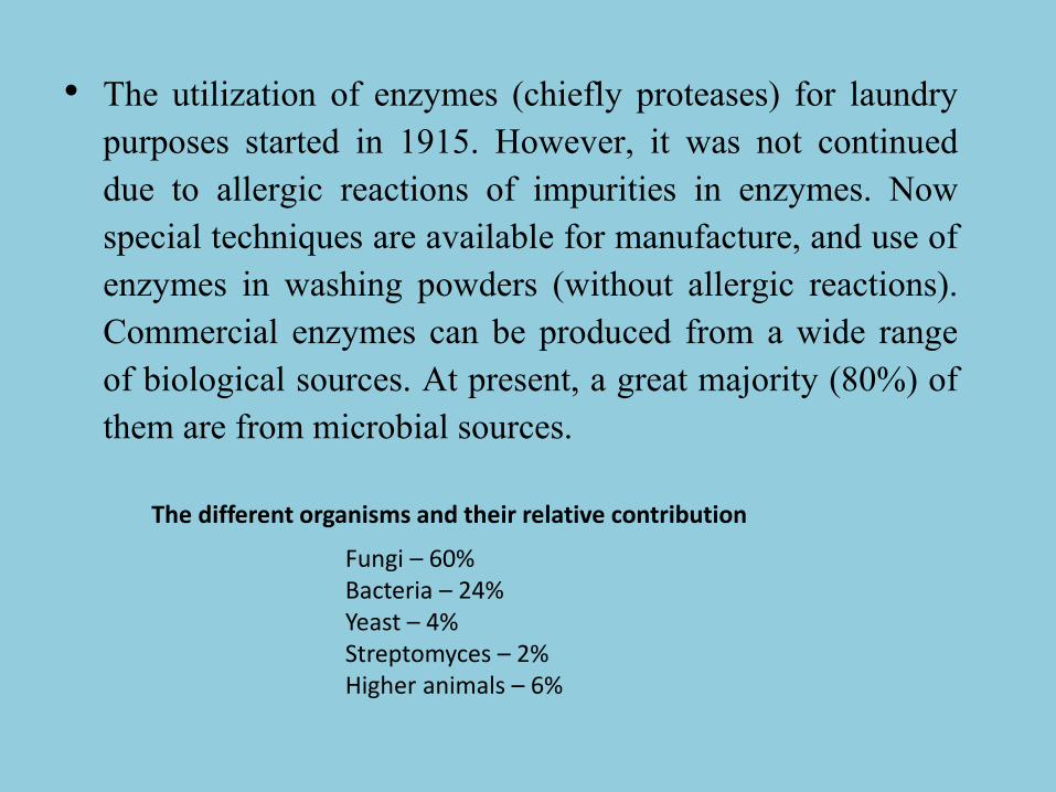
• The utilization of enzymes (chiefly proteases) for laundrypurposes started in 1915. However, it was not continueddue to allergic reactions of impurities in enzymes. Nowspecial techniques are available for manufacture, and use ofenzymes in washing powders (without allergic reactions).Commercial enzymes can be produced from a wide rangeof biological sources. At present, a great majority (80%) ofthem are from microbial sources.
Fungi – 60%Bacteria – 24%Yeast – 4%Streptomyces – 2%Higher animals – 6%
The different organisms and their relative contribution

Enzymes from microbial sources• Microorganisms are the most significant and convenient sources of
commercial enzymes. They can be made to produce abundantquantities of enzymes under suitable growth conditions.Microorganisms can be cultivated by using inexpensive media andproduction can take place in a short period.
• In addition, it is easy to manipulate microorganisms in geneticengineering techniques to increase the production of desiredenzymes. Recovery, isolation and purification processes are easywith microbial enzymes than that with animal or plant sources.
• In fact, most enzymes of industrial applications have beensuccessfully produced by microorganisms. Various fungi, bacteriaand yeasts are employed for this purpose

Various fungi, bacteria and yeasts are employed for enzyme
production


Aspergillus niger— A unique organism for production of bulk enzymes
• Among the microorganisms, A. niger (afungus) occupies a special position for themanufacture of a large number of enzymes ingood quantities. There are well over 40commercial enzymes that are convenientlyproduced by A. niger. These include a-amylase, cellulase, protease, lipase, pectinase,phytase, catalase and insulinase.

Commercial recombinant enzymes
• Recombinant fungi are one of the mainsources of enzymes for industrialapplications. The industrial enzyme marketreached $1.6 billion in 1998 for the followingapplication areas (excluding diagnostic andtherapeutic enzymes).

Aspartic proteinase
• A recombinant strain of Aspergillus oryzae
producing an aspartic proteinase from
Rhizomucor miehei has been approved by
Food and Drug Administration (FDA) for
cheese production.

Lipases
• Lipases are extremely important in the detergentindustry. They are extensively used in householddetergents, industrial cleaners, and leather processing,where they can be combined with proteases, oxidases,and peroxidases. To be suitable, lipases should bealkalophilic, able to work at temperatures above 45Degree C and at pH values of about 10, and capable offunctioning in the presence of the various componentsof wash-product formulations, such as oxidants andsurfactants. In 1994, Novo Nordisk introducedLipolase, the first commercial recombinant lipase foruse in a detergent, by cloning the Humicola lanuginoselipase gene into the A. oryzae genome

Microbial lipases
• Microbial lipases have a huge potential in areas such as food
technology, biomedical sciences, and chemical industries
since they are: (1) stable in organic solvents, (2) possess broad
substrate specificity, (3) do not require cofactors, and (4)
exhibit high enantioselectivity.
• In the food industry, lipases are commonly used in the
production of fruit juices, baked foods, desirable flavors in
cheeses, and interesterification of fats and oils to produce
modified acylglycerols. There are three fungal recombinant
lipases currently used in the food industry, Rhizomucor miehi,
Thermomyces lanuginosus and Fusarium oxysporum, all of
which are produced in A. oryzae

The Technology of Enzyme Production
• In general, the techniques employed for
microbial production of enzymes are
comparable to the methods used for
manufacture of other industrial products .The
salient features are briefly described.
– 1. Selection of organisms
– 2. Formulation of medium
– 3. Production process
– 4. Recovery and purification of enzymes.

An outline of the flow chart for enzyme production by
microorganisms

Selection of organism
• The most important criteria for selecting themicroorganism are that the organism should produce themaximum quantities of desired enzyme in a short timewhile the amounts of other metabolite produced areminimal. Once the organism is selected, strainimprovement for optimising the enzyme production canbe done by appropriate methods (mutagens, UV rays).From the organism chosen, inoculum can be prepared in aliquid medium.

Formulation of medium:
• The culture medium chosen should contain all thenutrients to support adequate growth of microorganismsthat will ultimately result in good quantities of enzymeproduction. The ingredients of the medium should bereadily available at low cost and are nutritionally safe.Some of the commonly used substrates for the mediumare starch hydrolysate, molasses, corn steep liquor, yeastextract, whey, and soy bean meal. Some cereals (wheat)and pulses (peanut) have also been used. The pH of themedium should be kept optimal for good microbial growthand enzyme production.

Production process:• Industrial production of enzymes is mostly carried out by submerged
liquid conditions and to a lesser extent by solid-substratefermentation. However, solid substrate fermentation is historicallyimportant and still in use for the production of fungal enzymes e.g.amylases, cellulases, proteases and pectinases.
• The medium can be sterilized by employing batch or continuoussterilization techniques. The fermentation is started by inoculating themedium. The growth conditions (pH, temperature, O2 supply, nutrientaddition) are maintained at optimal levels.
• The bioreactor system must be maintained sterile throughout thefermentation process. The duration of fermentation is variable around2-7 days, in most production processes. Besides the desiredenzyme(s), several other metabolites are also produced. Theenzyme(s) have to be recovered and purified.

Recovery and purification of enzymes:• The desired enzyme produced may be excreted into the culture medium
(extracellular enzymes) or may be present within the cells (intracellularenzymes). Depending on the requirement, the commercial enzyme may becrude or highly purified. Further, it may be in the solid or liquid form. Thesteps involved in downstream processing i.e. recovery and purification stepsemployed will depend on the nature of the enzyme and the degree of puritydesired.
• In general, recovery of an extracellular enzyme which is present in the brothis relatively simpler compared to an intracellular enzyme. For the release ofintracellular enzymes, special techniques are needed for cell disruption byphysical means (sonication, high pressure, glass beads). The cell walls ofbacteria can be lysed by the enzyme lysozyme. For yeasts, the enzyme β-glucanase is used. However, enzymatic methods are expensive.
• The recovery and purification steps will be the same for both intracellularand extracellular enzymes, once the cells are disrupted and intracellularenzymes are released. The most important consideration is to minimise theloss of desired enzyme activity.

Recovery and purification of enzymes
• Removal of cell debris- Filtration or centrifugation can be used to remove celldebris.
• Removal of nucleic acids- Nucleic acids interfere with the recovery and purificationof enzymes. They can be precipitated and removed by adding poly-cations such aspolyamines, streptomycin and polyethyleneimine.
• Enzyme precipitation-Enzymes can be precipitated by using salts (ammoniumsulfate) organic solvents (isopropanol, ethanol, and acetone). Precipitation isadvantageous since the precipitated enzyme can be dissolved in a minimal volume toconcentrate the enzyme.
• Liquid-liquid partition- Further concentration of desired enzymes can be achievedby liquid-liquid extraction using polyethylene glycol or polyamines.
• Separation by chromatography-There are several chromatographic techniques forseparation and purification of enzymes. These include ion-exchange, size exclusion,affinity, hydrophobic interaction and dye ligand chromatography .Among these, ion-exchange chromatography is the most commonly used for enzyme purification.
• Drying and packing-The concentrated form of the enzyme can be obtained bydrying. This can be done by film evaporators or freeze dryers (lyophilizers). Thedried enzyme can be packed and marketed. For certain enzymes, stability can beachieved by keeping them in ammonium sulfate suspensions.
• All the enzymes used in foods or medical treatments must be of high grade purity,and must meet the required specifications by the regulatory bodies. These enzymesshould be totally free from toxic materials, harmful microorganisms and should notcause allergic reactions.

Immobilized enzyme• An immobilized enzyme is an enzyme attached to an inert, insoluble
material—such as calcium alginate (produced by reacting a mixtureof sodium alginate solution and enzyme solution with calcium chloride).This can provide increased resistance to changes in conditions such as pHor temperature. It also lets enzymes be held in place throughout the reaction,following which they are easily separated from the products and may beused again - a far more efficient process and so is widely used in industryfor enzyme catalysed reactions. An alternative to enzyme immobilizationis whole cell immobilization.
• Immobilized enzymes are very important for commercial uses as theypossess many benefits to the expenses and processes of the reaction ofwhich include:
• Economy: The immobilized enzyme is easily removed from the reactionmaking it easy to recycle the biocatalyst. This is particularly useful inprocesses such as the production of Lactose Free Milk, as the milk can bedrained from a container leaving the enzyme (Lactase) inside ready for thenext batch.
• Stability: Immobilized enzymes typically have greater thermaland operational stability than the soluble form of the enzyme.

Immobilization of an Enzyme• There are various ways by which one can immobilize an enzyme. Various methods
used for immobilization of enzymes are adsorption, covalent binding, entrapment,
Affinity-tag binding and Cross-linkage :
• Affinity-tag binding: Enzymes may be immobilized to a surface, e.g. in a porous
material, using non-covalent or covalent Protein tags. This technology has been
established for protein purification purposes.
• Adsorption on glass, alginate beads or matrix: Enzyme is attached to the outside
of an inert material. In general, this method is the slowest among those listed here
as the active site of the immobilized enzyme may be blocked by the matrix or bead,
greatly reducing the activity of the enzyme.
• Entrapment: The enzyme is trapped in insoluble beads or microspheres, such
as calcium alginate beads.
• Cross-linkage: Enzyme molecules are covalently bonded to each other to create a
matrix consisting of almost only enzyme. The reaction ensures that the binding site
does not cover the enzyme's active site, the activity of the enzyme is only affected
by immobility.
• Covalent bond: The enzyme is bound covalentely to an insoluble support (such as
silica gel or macroporous polymer beads with epoxide groups). This approach
provides the strongest enzyme/support interaction, and so the lowest protein
leakage during catalysis.
Immobilization of enzymes
• Various types of gels are used for entrapment of enzymes.The enzyme may be entrapped within polymeric meshsuch as agar, polyacrylamide gel or calcium alginate bycarrying out the polymerization reaction and /or cross –linking reaction in the presence of enzyme.

Polyacrylamide
• Polyacrylamide gel is the most commonly used material forentrapment. For the preparation of 15% gel, 7.5 gacrylamide, 0.5 g bisacrylamide, 50 mg ammoniumpersulfate was added to 25 ml of phosphate buffer, pH 6.8and mixed to dissolve these solids. Then 25 ml of amylase
solution added. Mixed properly and added 50 μl of TEMED.Mixed gently and poured into glass Petri dishes, or gelcasting vertical electrophoresis unit in order to get the gel ofuniform and desired thickness. Polymerization was done atroom temperature for 1 h. The gel was cut into small piecesand suspended in 0.1 M phosphate buffer till further use.

Entrapment in calcium alginate gel
• For calcium alginate beads, 25 ml of 0.5, 1, 2, 3 and 4%solutions of sodium alginate were prepared and mixedwith equal volume of amylase solution to get the finalconcentration of sodium alginate 0.25, 0.5, 1, 1.5 and2%, respectively. Entrapment of enzyme in calciumalginate gel was done by modifying the method ofKierstan and Bucke. Different CaCl2 concentrations(0.1, 0.2, 0.3, 0.4 and 0.5 M) were used to optimize thebest concentration. For 50 ml sodium alginate- enzymemixture, 500 ml of CaCl2 solution was used.

Immobilized whole cell• Immobilized whole cell system is an alternative to enzyme
immobilization. Unlike enzyme immobilization, where theenzyme is attached to a solid support (such as calcium alginate)in immobilized whole cell systems, the target cell isimmobilized. Such methods may be implemented when theenzymes required are difficult or expensive to extract, anexample being intracellular enzymes. Also, if a series ofenzymes are required in the reaction; whole cell immobilizationmay be used for convenience.This is only done on acommercial basis when the need for the product is morejustified.

4. Fungal toxinsMycotoxicoses‐ fungi in dermatomycosis, aspergillosis and fungi
allergenic to man and animal.

Mycotoxicoses
Mycotoxicoses is defined as an illness of man or animal due toingestion of pre-formed substances produced by the action ofcertain molds or filamentous fungi on particular foodstuff.
MYCOTOXINS
– Secondary metabolites produced by food-borne filamentous fungi
– Vary in their severity :Carcinogen/allergen
– Non-volatile & low molecular weight
– Some are lethal
– Cause identifiable diseases
– Weaken the immune system without producing symptoms

Feeds Most Susceptible to Fungi producing Mycotoxins
Cottonseed
Peanut meal
Rye
Bread
Corn
Wheat
Oats
Barley
Sorghum

Fungi responsible for mycotoxicoses
• Most of the significant fungi producing mycotoxicosesmainly belong to toxigenic species of 3 genera:

Dermatophytes• Dermatophytes are aerobic fungi that can invade and infect the
keratinized layers of skin, hair, and nails. Three genera of fungi,Trichophyton, Microsporum, and Epidermophyton, account for mostdermatophytic infections.
• These fungi are found worldwide and infection is acquired by contactwith infected humans or animals, or from exposure to contaminatedsoil or fomites (e.g., combs, brushes). Dermatophytes often areclassified by their host preferences – anthropophilic and zoophilicspecies primarily infect human and animal species, respectively;geophilic species reside in soil.
• Direct contact can result in transmission of zoophilic or geophilicdermatophytes to humans. Dermatophytes do not cause invasivedisease except in immunocompromised hosts. The clinical diseaseattributable to dermatophytes varies by organism, site of infection,and host immunologic responses.

MEDICALLY SIGNIFICANT MYCOTOXINS
1.Aflatoxins
2.Fumonisins
3.Trichothecenes
4.Ochratoxins
5.Cyclopiazonic Acid
6.Zearalenone
7.Patulin


AFLATOXINS(Aflatoxicosis)• These toxins were discovered as a cause of mysterious disease termed as Turkey-X-
disease during 1960s in England.• Killed approx.1,00,000 birds and ruined turkey industry.• Aflatoxins most commonly produced by A. flavus are B1 and B2.• A.parasiticus produces G1and G2.• These are designated as B & G on the basis of their metabolites which exhibits
blue(B) and green(G) fluorescence under U.V. light on TLC plates.• Aflatoxin B1 is the most important biologically active mycotoxin because of its
common occurrence in food items and is highly toxic and carcinogenic.• Poisoning that results from ingesting aflatoxins 2 forms:
– Acute severe intoxication: Result in severe liver damage, And subsequent illness or death.
– Chronic subsymptomatic aflatoxicosis: signs and symptoms are lethargy, anorexia and muscle weakness followed by spasm.

Reye’s syndrome
• It is an acute aflatoxicosisin which patient presentswith signs and symptomsof encephalopathy and fattydegeneration of viscera.
• This is an endemic diseaseof children in developingcountries.

FUMONOSINS• Secondary metabolites produced by various species of fusarium.
• They are toxic and carcinogenic
• Common contaminants of maize ,corn and their products.
• Cause fatal illness in animals like equineleukoencephalomalacia(ELEM)fatal disease in horses,Porcinepulmonary edema(PPE) in pigs,and hepatotoxic and carcinogeniceffects in rats.
• Cause toxicity by blocking ceramide synthetase which converts sphingosine & acetyl CoA to ceramide.
• An outbreak has been reported in 1999 in poultry due to consumption of fumonisin-contaminated feed containing rain –damaged maize.

OCHRATOXINS• Naturally occuring mycotoxin and is produced by various species of
Aspergillus and Pencillium.• Originally isolated from Aspergillus ochraceous hence named
ochratoxin.• Out of many ochratoxins, ochratoxin A is medically significant.• Natural occurrence of these toxins in grains and other plant
products.• Human exposure to ochratoxin• Direct – consumption of contaminated plant food• Indirect – consumption of animal tissues exposed to contaminated
materials.• It produces fatal renal disease called as endemic nephropathy and
urinary tract tumors.

CYCLOPIAZONIC ACID
• Produced by genous Aspergillus and Pencillium
• Occurs naturally in agriculture products such as groundnut and corn.
– Co-contaminant with aflatoxin
– Clinical symptoms are: loss of weight,weakness,vomiting,diarrhoea,dehydration,convulsions and death.
• Causing symptoms of kodua poisonin (consumed kodomillet seed as staple food )

ZEARALENONE• Produced by Fusarium species
• Found in variety of infected cereals like maize, barley,wheat grains and sorghum
• Cause genital disorders in domestic animals

PATULIN• Derived from Pencillium patulum
• It was initially thought to be the antiviral drug whichrelieve the symptoms of common cold Subsequentlyrealized as mycotoxin

Toxin hypothesis
1. A toxin should produce all symptoms characteristic
of the disease.
2. Sensitivity to toxin will be correlated with
susceptibility to pathogen.
3. Toxin production by the pathogen will be directly
related to its ability to cause disease.
– Except, victorin, the toxic metabolite of Cochliobolusvictoriae, the vast majority of toxins associated with plant diseases fail to exhibit all the above characters.
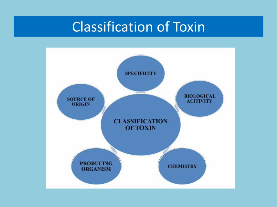
Classification of Toxin

Source of Origin

1. Pathotoxins (Wheeler and Luke, 1963)
-Tab-toxin; HMT toxin
-causal role in the disease
2. Vivo toxins (Diamond & Waggoner, 1953)
-partial role in disease
-Fusaric acid, pyricularin
3. Phytotoxins
-Role is suspected in disease
-Alternaric aid; cochliobolin.

Pathotoxins (Wheeler and Luke, 1963)
Those toxins which plays an important causal role in disease
and are produced by the pathogen or interaction between host
and pathogen
Criteria for any toxin to be a pathotoxin:
-When applied on a susceptible host in low concconcentration,
should produce all or nearly all symptoms of the disease
-Toxin and the pathogen should have same host range; same
resistance/ susceptibility spectrum
-The pathogenicity of the pathogen should be correlated with
its capacity to produce the toxin.
e.g. Tab-toxin: Pseudomonas tabaci; HMT toxin: Dreschlera
maydis race T

Vivotoxins (Diamond & Waggoner, 1953)
Those toxins produced in the infected plant /host by the pathogen that function in the production of the partial disease.
Criteria/ characteristics of vivotoxins
•---Reproducible separation of the toxin from the diseased host
•---Purification and chemical characterization and
•---Induction of atleast a part of disease syndrome when applied on healthy plant
These are generally non-specific
•Fusaric acid: Fusarium spp.
•Pyricularin: Pyricularia oryzae

Phytotoxins
Phytotoxin are toxic substances produced by living organism and whose role in disease is merely suspected rather than established.
• Role is suspected in disease
• Alternaric acid; cochliobolin.

Based on producing organism
Sheffer & Briggs, 1981

Based on Chemistry

Based on Biological Activity
Scheffer and Briggs, 1981

Based on Specificity

Effect of toxins on host tissue
Changes cell permeability Disruption of normal metabolic activity Loss of salts from protoplasm which increases
– Respiration – tab toxinine– Uncoupling of oxidative phosphorylation – victorin– Inhibition of host enzyme - tab toxin inhibit
normal host enzymes – Affect cellular transport system e.g. H+/K+
exchange at the cell membrane
Other mechanisms – Interfere with growth regulatory system e.g.
inhibition of root growth ( F. moniliforme)

Host selective(specific) toxins

Host-Specific (selective) Toxins
Produced by fungi
Almost all are produced by loculoascomycetes-Alternaria spp. and Cochliobolus spp. primarily
First toxins characterized – AKT-toxin (Japan 1933),Victorin (USA,1947), HC-toxin (USA,1965)
Toxins reproduce all disease symptoms when applied to asusceptible host.
Scheffer & Nelson – 1960’s – single genetic loci control toxinproduction in Cochliobolus spp. E.g.
-Cochliobolus heterostrophus – T-toxin – TOX1
-C. carbonum – HC-toxin – TOX2
-C. victoriae – Victorin – TOX3

Specific/ selective toxins
Are those toxins which adversely affect the specific host
of the pathogen
Are normally essential for pathogenicity
20 such toxins has been recognized mainly produced by
Alternaria and Cochliobolus spp.
–Victorin
–T-toxin,
–HC-toxin,
–AAL-toxin

VICTORIN
Victoria blight of oats -Cochliobolus victoriae
A host-specific toxin – victorin: a cyclic pentapeptides
Toxin reproduces all disease symptoms when applied to a susceptible host
Susceptibility to toxin conferred by dominant allele at Vb locus
Victorin causes premature senescence of leaves

T-toxin •Produced by Helminthosporium maydis race T
(Cochliobolus heterostrophus race T)
•Southern Corn Leaf Blight
•Toxin is a polyketide
• Toxin causes swelling, uncoupling of oxidative phosphorylation, stimulation of respiration, leakage of Ca2+ and NAD in mitochondria
•single genetic locus is involved in toxin production
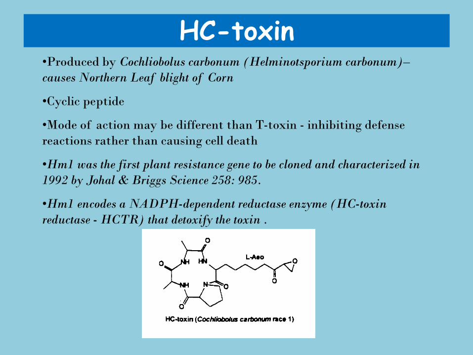
HC-toxin •Produced by Cochliobolus carbonum (Helminotsporium carbonum)–
causes Northern Leaf blight of Corn
•Cyclic peptide
•Mode of action may be different than T-toxin - inhibiting defense
reactions rather than causing cell death
•Hm1 was the first plant resistance gene to be cloned and characterized in
1992 by Johal & Briggs Science 258: 985.
•Hm1 encodes a NADPH-dependent reductase enzyme (HC-toxin
reductase - HCTR) that detoxify the toxin .
Alternaria toxins • Produced by closely related Alternaria spp. Alternaria
alternata.
• All are low molecular weight, secondary metabolites and are
structurally similar
• Toxins affects
-Plasma membrane of susceptible hosts
- electrolyte loss, membrane invaginations (Kohmoto et al.
1993)
-One specific genotype of each host is susceptible to toxin
- very narrow host range
• Tangerine pathotype – ACT-toxin
• Strawberry pathotype – AF-toxin
• Japanese pear pathotype – AKT-toxin


Non host selective (non-specific) toxins

Non-Specific/ selective
Are those toxins that affect the protoplast ofmany unrelated plant species in addition tothe main host of the pathogen producing thetoxins
•E.g. Tab toxin, phseolotoxin, tentoxibCercosporin
•These toxins affect virulence of the pathogenbut are not essential for pathogenicity.

Tab toxin (Non-Selective)Produced by Pseudomonas syringae pv. tabaci causes wild fire disease in
tobacco.
• Toxin producing strains cause necrotic spots on leaves surrounded byyellow halo.
• Tab toxin is a dipeptide composed of amino acid threonine andtabtoxinine
• The toxin as such is not toxic but in the cell it get hydrolysed and releaseaminoacid tabtoxinine which is toxic
• Act by
-inhibiting / inactivating the enzyme glutamine synthetase
-Uncoupling of phosphorylation and photorespiration,
-destroy the thylakoid membrane of the chloroplast thus causeschlorosis and then necrosis.
Fig.: Structure of Tabtoxin

Phseolotoxin
Produced by P. syringae pv. phaseolicola causes halo blight of bean
• Toxin causes growth reduction.
• Disrupt apical dominance and accumulation of amino acid ornithine
• Phaseolotoxin is a modified ornithine-alanine -arginine tripeptidewith phosphosulfinyl group.
• Toxin act by – inhibiting pyrimidine nucleotide biosynthesis, reduce activity of
ribosomes, interfere with lipid synthesis
– Changes cell permeability

Tentoxin
Produced by Alternaria alternata (A. tenuis) causes leaf spots and chlorosis.
• It is cyclic tetrapeptide that bind to and inactivate the protein (chloroplast – coupling factor) involved in energy transfer into chloroplast.
• Also inhibits phosphorylation of ADP to ATP
• Leading to disruption of chlorophyll synthesis

Cercosporin
• Produced by Cercospora spp. And other fungi causes leaf spot disease
• This toxin is activated by light and become toxic to plants by generating activated species if oxygen (single O); the activated O destroy the host membrance and provide nutrients to pathogen,
• It is photosensitizing perylenequinone that absorb light energy
Fig. 12: Structure of Cercosporin

Fusarium toxins
Marticin: pathotoxin produced by Fusariumoxysporum f sp. pisi – pea wilt; have nepthazarin; redpigmented compounds
Fusric acid: Vivotoxin, chemically 5-n- butyl-picolinicaacid produced by many spp of Fusairim: Fusariumoxysporum f sp. Batatis (sweet potato); cubense(banana); lini (Linseed); lycopersici (Tomato);vasinfectum (Cotton)
Lycomarasmin: Fusarium oxysporum f sp. lycopersici

Pyricularin
Pyricularia oryzae – rice blast
Exist in two froms:
-α-picolinic acid
-pyricularin
Toxic to conidial germination, however, the fungus produces a pyricualrin binding protein (copper oxidase) that binds with pyricularic and destroy the fungitoxicitybut not the phytotoxicity.
It affect respiration and growth at low conc. But inhibit at high concentration.

Ergot alkaloid
• The most prominent member of this group isClaviceps purpurea.
• This fungus grows on rye and related plants andproduces alkaloids.
• Cause ergotisms in humans and other mammals whoconsume grains contaminated with its fruitingstructure called ergot sclerotium

Aflatoxin• Aspergillus flavus, Aspergillus nomius and Aspergillus parasiticus.
• There are four kinds of aflatoxins such as aflatoxin B1, B2, G1and G2, in which aflatoxin B1 (AFB1) is highly toxic andcarcinogenic (Leontopoulos et al., 2003).
• Aflatoxins are known to be potent carcinogenic agents that poseserious hazards to human and animal health (Sidhu et al., 2009).
• In addition, aflatoxin also has an impact on agricultural economythrough the loss of crop production (Wu, 2004).
• Food and Agriculture Organization of United Nations (CAST,2003) shows that 25% feedstuffs is polluted by mycotoxin in theworld, and it results in over 1 billion dollars loss for poultryindustry annually .

Other non specific toxins include
Fumaric acid : Rhizopus spp. Oxalic acid: Sclerotinia, Sclerotium spp. Alternaric acid: Alternaira spp. Ophiobolin: Cochliobolus spp. Pyricularin: Magnaporthe griseaLycomarasmin: F. oxysporum in tomato

Non-host selective fungal toxin

Non host selective bacterial toxin

Toxin produced by fungal plant pathogens


Structures of selected mycotoxins

5. Fungi as food and beverage and food processing
Alcoholic beverage, mushrooms and other macro fungi, edible biomass from yeast and moulds, single cell proteins (SCP) Bread,
soybean products, cheese and fermented milk, other fermented foods
.

• Among the tombs, stone and wooden sculptures and other artifacts of the ancient Egyptiancivilizations are numerous depictions of life there almost 6000 years ago. Among these depictionsare scenes of bakeries, breweries, vineyards, and wine presses. This is perhaps some of the firstsolid evidence of the use of fungi in large-scale food and beverage production. Archaeologicaldata, however, are still unraveling the mysteries of “how long has man been doing this?” Currentdiscovery of the ice man in the foothills of the Alps with cereal grain and mushrooms in hispouch suggest that bread making and mushroom use may go back 10,000 years. And, thediscovery of wine residues in clay urns in Iran date back to 7,000 BC. Yet the discovery of theorganism responsible of fermentation that goes on in bread and wine making was not until 1680when Antoine van Leeuwenhoek first discovered yeast cells under his microscope. It was morethan 200 years later before Louis Pasteur concluded that through anaerobic respiration(fermentation) sugar was converted into carbon dioxide and alcohol by such tiny yeast. Whileyeast are central to the baking and brewing industries, many filamentous fungi are utilized in theproduction of cheeses and a wide array of exotic foods. We will examine first the use of fungi inbrewing and baking; then in the production of cheeses and other exotic fermented foods andbeverages.
Production of Foods and Beverages:

• There are several types of bakers yeast (Saccharomycescerevisiae). Compressed yeasts: What we commonly refer toas “yeast cakes” or wet yeast. It contains about 70% moistureand has a shelf life of about 3-4 weeks. Such yeast is perishableand should be stored under refrigeration (4-5º C). Crumbledyeast forms are similar to yeast cakes but with less water and ina crumbled condition. This type is often sold in 50 lb bags.Active Dry Yeast: Dry yeast is prepared similar to compressedyeast but is dried under specially controlled conditions. Themoisture content is 6-8%, which enables them to be stored for afew months. Dry yeast should be revived in water at 105-110ºF. Instant Dry Yeast: Through a quick drying process undercontrolled conditions, more porous yeast can be produced thatbecome active immediately on rehydration. This is probably themost popular yeast used in home baking.

Fermented Beverages
• Ethanol Production by Yeasts: For large-scale production ofethanol by yeast, an organism that is tolerant to highconcentrations of ethanol and highly osmotic substrates isessential. Species of Saccharomyces are the most tolerant toethanol. Wine yeasts can tolerate levels up to 20%, whereas,bakers yeast used in beer making and bakeries can toleratelevels at from 4 to 6%. Most organisms are inactivated inconcentrations of ethanol above 15%. Extensive research hasbeen done to produce strains that will be effective in utilizingvarious substrates and tolerate high levels of glucose orsucrose. They must also be resistant to certain metabolites thatmay be toxic. Ethanol tolerance is genetically controlled by alarge number of genes (Ismail & Ali, 1971. Fol. Microbiol.16:300-359).

• Distilled Alcohol: Distillation involves the conversion of a substance intoa vapor that is subsequently condensed to the liquid state. The process goesback to the time of Aristotle (around 350-380 BC). Distillation is used inparticular to separate mixtures of liquids in which the boiling points of theliquids differ. In the production of distilled alcoholic beverages, thefermented substrates contain, water, alcohol, oils and other liquids. Theboiling point of alcohol is 79.3° C and that of water is 100° C; thus thealcohol will vaporize at a much lower temperature. Its vapor can becondensed in a cooling coil from the still and thus separated from the otherliquids. Species of Saccharomyces and Schizosaccharomyces are used toproduce distilled alcoholic beverages.
• Industrial alcohol is produced in the U.S. with corn as the primarysubstrate. In Brazil, sugarcane is used in large-scale alcohol production forautomobiles. With the ever-increasing depletion of fossil fuel reserves,production of ethanol from plant products is more attractive. With industrialethanol, flavor and other qualities are not as important as consumableproducts. Therefore, the yeast strains used are selected for their efficiencyto digest substrates and in their tolerance to high levels of ethanol.

Alcoholic beverages• Alcoholic beverages are made from a large variety of starchy or sugary products.
Whiskey is made largely from barley, but sometimes from other grains. They areessentially aged grain alcohol that start out as clear but becomes colored when agedinside of charred barrels. Bourbon is a form of whiskey made from fermented corn,or occasionally other grains. Vodka is produced largely from potatoes in Russia andother east European countries because they are cheap. They often use grains as well.Rum is a distilled spirit made from sugar-containing substrates like molasses andcane juice. It is initially clear, but like bourbon, it is darkened by storage in woodenbarrels; or in recent years adding caramel after distillation. It is popular in tropical andsubtropical countries where sugarcane is grown. Cognac is made from distilled grapewine and gets its name from Conacais, France where it was first made. British andDutch merchants began to distill wine to prevent spoilage in shipment. Brandy ismade in a similar way as cognac, initially from grape wine but later in North Americafrom fermented juices of pears and peaches. Tequila is a briny liquor madeexclusively in Mexico from fermented juice of the Agave cactus. The Mexicans weremaking a fermented drink 1000 years before they were invaded by Spain. TheSpaniards brought to Mexico with them the art of distillation; hence, tequila came intoexistence.

Wine• Wine in the strict sense is the fermented juice of grapes. However, wines are now
made of many juices. The making of wine is known from the earliest history of man;one of the earliest written is the Biblical account of Noah (perhaps 5000 BC). Toquote a recent report (Gainesville Sun, June 5, 1996), “Talk about a vintage!Scientists say they have found the oldest evidence of wine residue at the bottom of asquat, 7,000-year old pottery jar. Traces of two chemicals in the jar, found in theZagros Mountains of Iran, extend the known history of wine-making 2,000 years.”The jar came from a time when people were first establishing permanent settlementsand were likely already cultivating the grapes from which the wine was made. Forcenturies wine was stored in flasks of animal skin or in clay vessels. During the riseof the Roman Empire, wooden vessels as well as clay urns were common. The use ofbottles and corks became common towards the end of the 17th century.
• While wine making has gone on for centuries, the process was brought to thiscountry by the early settlers. Perhaps the earliest wine making in the U.S. was donein Florida by early Spanish explorers. Yet today, only a few wineries occur in Floridawhere they use native muscadine grapes.

• Wines are named after the type of grapes or the geographic area or specificvillage where they were first produced. For example, Burgandy, Bordeaux,Champagne, and Alsacs are important wines of France. During the 17thcentury, wine makers in the Rhine valley found that grapes allowed to rot onthe vine gave the wine a sweeter taste. There are three basic types of wines,(1) table wines, (2) fortified wines, and (3) sparkling wines. Table wines aremade from pressed grapes fermented in vats with the addition of sugar,yeasts, sulfur dioxide. Saccharomyces ellipsoideus is the common yeast usedin the fermentation process. The alcohol content is around 12-15%. Sulfurdioxide is used to keep down the vinegar producing bacterium Acetobacter.Wine may be chilled in vats to cause sedimentation and the “free run” wine isdecanted. Fortified wines receive an addition of alcohol, brandy or otheralcoholic beverages, and the final alcohol level is from 16-23%. A commonfortified wine is port wine which gets it name because sailors who used tocome into port would purchase wine spiked with brandy or liquors to give agreater alcohol level. Sparkling wines such as champagne go through adouble fermentation in which the alcoholic content reaches 20%. Somesparkling wines have a natural effervescence, others are made effervescent bybubbling them with carbon dioxide. All natural wines are below 20% alcoholbecause beyond that level, the fermenting yeast would be killed.

• There are red, white, and pink (rosé) wines, Red wines aremade from black grapes in which the husks (mush or skins)are left in contact with the juice throughout fermentation.Most white wines are made from green colored grapes orfrom black grapes in which the husks are removed soon afterpressing. Leaving the husks of black grapes in the fermenterfor a short period of time produces a pink wine.
• European wine production was almost devastated when in the19th century imported American rootstock infested with alouse, Phylloxera, which feeds on grape roots, destroyed morethan 2,500,000 acres throughout France and surroundingcountries. Many growers never recovered. Others importedresistant rootstock and the industry was soon reestablished.

BEER• Beer is a beverage obtained by the alcoholic fermentation by yeasts of a malted
cereal, usually barley malt, with or without other starchy materials, and to whichhops have been added . (For more information on beer-making, see Hardwick, 1983.Biotech.5:166-229). Brewing seems to have originated in the Babylonian Empire(Mesopotamia) before 6000 BC. By 2000 BC, many types of beer existed inBabylon. There is evidence that the brewing of beer developed independently inEgypt. Some have suggested that the Hebrews learned to use hops to flavor beerwhile they were in captivity in Babylon during the 8th and 9th century BC. TheGreeks learned to make beer from the Egyptians and the Romans in turn learnedfrom the Greeks.
• There are two types of beers, lager and ales. Lager beer employs bottomfermentation in which the “spent yeast” settles to the bottom and the green beer isaged. With aging, it mellows and is carbonated with C02. In the brewing of ales, theyeast selected is a top fermenter, forming a foam that can be removed. Enzymes areoften used in the early stages of brewing, i.e. the “mashing” stage, and later in thebrewing process. Amylase produced by other fungi increases starch digestion andresults in low carbohydrate, or “lite” beers. Several other carefully monitoredenzymes are added during the brewing process. Without protease, for example, beerswould become hazy, and glucoamylases produced by certain species of Aspergillusare used to sweeten beers.

• Four things are vital to brewing beer, barley malt, hops, water, and yeast.In the making of barley malt, barley is allowed to go into dormancy afterharvest. During malting certain enzymes are formed within the grain.Before malting the barley is soaked in water to initiate germination, afterwhich grains are dried to about 2% moisture level. Timing of germination,temperature and duration of drying, and other factors affect the kind of maltthat is produced. Corn, rice, and other grains may be added as adjuncts toadd texture and flavor. These are closely guarded industry secrets. Hops arenot essential for the manufacture of beer. The flowers of the female hopsplant are used, and only from unfertilized flowers. Pollinated hops will givethe beer a bitter flavor. Hop growing in the U.S. is chiefly in Oregon,California, Idaho, and New York. The best hops for brewing come fromSlovakia and the Czech Repuplic (formerly Czechoslovakia). Hops areadded to mashed malt solution (wort) during boiling. Water quality is vitalto beer production, especially pH. Alkaline or “hard” water gives poorresults and water with sulphur, iron, or bacteria should be avoided.Apparently, there must be ideal water in the mountains around Golden,Colorado! The yeast used in beer brewing is Saccharomyces cerevisiae.

• The main stages of brewing are mashing, boiling, andfermenting. Prior to mashing, malt is crushed between rollersto form flour. Mashing involves adding water (100-150F),solubilizing the starch by enzyme action, and separating outthe husk. After mashing, the “wort” is boiled in large coppertanks or kettles, stopping enzyme action. The wort is filteredand passed into a high-speed centrifuge to achieve clarity. Itthen passes into large coolers. Fermentation begins when yeastis added to the cooled wort at 1 lb/barrel. After about 8 daysmost all fermentable substrate has been converted into alcohol.The beer is then bottled, canned, or stored in large kegs formarketing. The following images show the various types ofcookers, fermentation tanks, and sediment tanks utilized in themanufacture of beer. The manufacture, distribution, and sale ofbeer are major industries in the U.S. and in Florida.

OTHER TYPES OF FERMENTED PRODUCTS
• Rice Beer: Rice beer is called sake in Japan, murcha or pachwai in India.It is made with rice plus species of Mucor or Aspergillus oryzea and sugar.
• Kanji: A beerlike beverage made with carrots and beets fermented withHansenula. Taste much like cherry wine.
• Toddy: Sweet fermented sap from tropical palms using Saccharomycescervisiae. Many substitute organisms can be used.
• Others fermented products include: Shoyu (=soy sauce): This is onefermented product well recognized throughout the US and Europe. Shoyu,referred to as Ketjap in Indonesia, has been produced in Japan for over1000 years. The average Japanese consumes more than 3 gallons of soysauce per year. Shoyu is prepared by soaking soybeans for 15 hr andautoclaving them for 1 hr. Clean, roasted wheat powder is added. Aftercooling, a starter, tane koji composed of Aspergillus oryzae or A. soyae isused as the inoculum. After fermentation for 3-4 months, or up a year, thematerial is combined with equal parts of brine and placed in tanks. Theresulting mash is compressed and the liquid supernatant is retrieved,sterilized and bottled.

• Tea Fungus: Perhaps one of the strangest fermentationsencountered is the so called “tea fungus” of eastern Europeand Asia sometimes referred to as Hongo. This drink is madefrom dry tea leaves steeped in a liter of water, and the leavesremoved and 100 gm of sugar added. The solution isautoclaved and a “starter” culture is added and the material isallowed to ferment. Two yeasts and Acetobacter have beenisolated from the starter.
• Anchu is an alcoholic drink in Taiwan made from red rice.Awamori is an alcoholic drink prepared from sweet potatoesin Japan. Braga is a fermented drink made from millet inRomania.

Cheese Making:• We do not know when and where cheese was first made; its discovery
was probably an accident.• In the Judeo-Christian Bible, II Samuels 17:27-29 says that cheese of
the herd was given to King David (approx. 1000 BC). So, cheese hasbeen around for a while! The production of cheese is a multimilliondollar industry in the US and is also a dominant industry in othercountries. It involves the fermentation of milk from different animals,using various organisms, and processing them under rigidly controlledconditions. Milk is coagulated, the liquid whey removed, and thesolid curd is preserved. Curd may be made in three ways: acidcoagulation, heat coagulation, and rennet coagulation. There areblock cheeses, powdered cheeses, soft cheeses, and various processedcheeses. Ricotta and similar cheeses are made from heat coagulatedcurd .
• Two of the most common cheeses in which filamentous fungi areused in fermentation are Camembert and Roquefort.

• Camembert Cheese originated in the town of Camembert in theNormandy district of France. Legend attributes its discovery to amilkmaid, Maria Harel, who first shared it locally with neighbors.This blue cheese is now marketed worldwide and is manufactured ina number of countries. Camembert cheese, also called Brie, obtainsits flavor and texture from lipolytic and proteolytic activity of moldsbelonging to Penicillium camemberti and P. casicolum.
• The production of camembert cheese requires rigidly controlledconditions or else other fungi, particularly Scopulariopsisbrevicaulis, will ruin the cheese. For this reason, it has been verydifficult to establish a successful industry in other countries.Camembert cheese has not gained wide popularity in the US.
• Roquefort Cheese is made from the fermentation of milk curd byPenicillium roqueforti, a species first isolated in 1904 and describedby Thom (1906, USDA, Bur. Animal Ind. Bull. 82).

• There are apparently many strains of this fungus, some of them beingdescribed as distinct species. Thom and Raper (ibid.) discuss a dozen suchstrains. The different enzymes produced by P. roqueforti results in variouscheese flavors. P. roqueforti is a common fungus on the surface layer ofsilage. This is a likely source of the original clones that were discovered inthe production of cheese. Charles Thom, USDA, Peoria, IL, introduced theproduction of roquefort cheese in the US in 1906. Thom and co-workersdiscovered the lipolytic and proteolytic enzymes of the mold releasedcaproic, caprylic, and capric acids which imparted flavor to roquefortcheese. Unlike Camembert which has only surface growth of the fungus,the veins one sees in blue cheeses are zones of Penicillium growth within.Other blue-veined cheeses similar to roquefort are Dolce Verdi producedby Penicillium expansum and Ellischour, with a red pigment, produced byP. nalgiovensis.
• Other blue cheeses include fromage blue cheese in central France (fromsheep and cow milk); gorgonzola in northern Italy, stilton in England,cammelost in Norway, and neufchatal in Germany.

Fungi used in Food & Beverage Production

Fungi used in Food & Beverage Production

Single Cell ProteinDuring World War II, when there were stortages in proteins and vitamins in
the diet, the Germans produced yeasts and a mould (Geotrichumcandidum) in some quantity for food; this led to the idea to produce edibleproteins on a large scale by means of microorganisms during 1970s.
Several industrial giants investigated the possibility of converting cheaporganic materials into protein using microorganism.
Single-Cell Protein (SCP) is a term coined at Massachusetts Institute ofTechnology by Prof C.L. Wilson (1966) and represents microbial cells(primary) grown in mass culture and harvested for use as protein sourcesin foods or animal feeds.
Single Cell Protein (SCP) is not a pure protein but refers to whole cells ofbacteria, yeasts, filamentous fungi or algae.
Contains carbohydrates, lipids, nucleic acids, mineral salts and vitamins.Carbon substrates: major substrates used in commercial SCP production is
alcohols, n-alkanes, molasses, sulphite liquor and whey.

Yeasts Fermentation in Bread-making.• Bread is a baked product made of dough that has been raised by yeasts or other gas forming
organisms. Why or when man first ate seeds or grasses, learned to grind them into flour,mix the flour with water or milk and bake it into bread is not known. Remains in thesediment of a Swiss Lake dweller show that humans were baking bread about 10,000 yearsago. Archeological data reveal that bread making has been a central part of almost allsubsequent cultures of mankind. The Egyptians appear to be the first to discover that lettingdough ferment would result in light, flavorsome bread. Until the last two centuries,however, light bread was more or less restricted to nobility. Today, we all enjoy the nobleuse of light bread. At the peak of the Greek Empire, several types of bread were producedand baking later became a vital business in most medieval towns (Sanderson et al. 1983.Yeast fermentation in bread making.
• How to make a lot of dough? In the early years, bread making relied on naturalfermentation. During the late 1700’s, commercially available yeasts were used in large-scale production of bread. Charles Fleischman of the U.S. introduced in 1886 a new typeof compressed yeast for distillers and bakers. An average baking formula for white breadrequires 100 lbs of white flour, 2 ½ lb of yeast, 2 lbs of salt, 6-7 lbs of sugar, 4 lbs of drymilk, and enzymes when sometimes needed. Modern mixers can handle more than 1600 lbsof dough. Larger US bakers have continuous mixers. By the mid 1960’s, more than 40% ofbread making was done in continuous mixers. Close to 50,000,000 loaves of white breadare sold in the U.S. annually; it is a $5 billion dollar industry!

• Yeast is the essential ingredient in many bakery products,giving it lightness and a delicious flavor. The common baker’syeast is Saccharomyces cerevisiae, a cellular, budding yeast.The use of fermentation in producing bakery products datesback to beyond 2000 BC where Egyptian hieroglyphics clearlydepicts the practice. It was only in the 19th century that ascientific basis for the process was understood when scientistssuch as Charles Hansen of the Carlsburg Labs in Denmarkdeveloped methods of isolating and culturing pure strains ofthe yeast. The commercial production was a tremendous boostto the baking and brewing industries. Today, large tanks orfermentors are used to culture yeast. Water and molasses arecommonly used and a supply of oxygen is admitted. It isestimated that approximately 1.4 million tons of compressedyeast is produced each year.
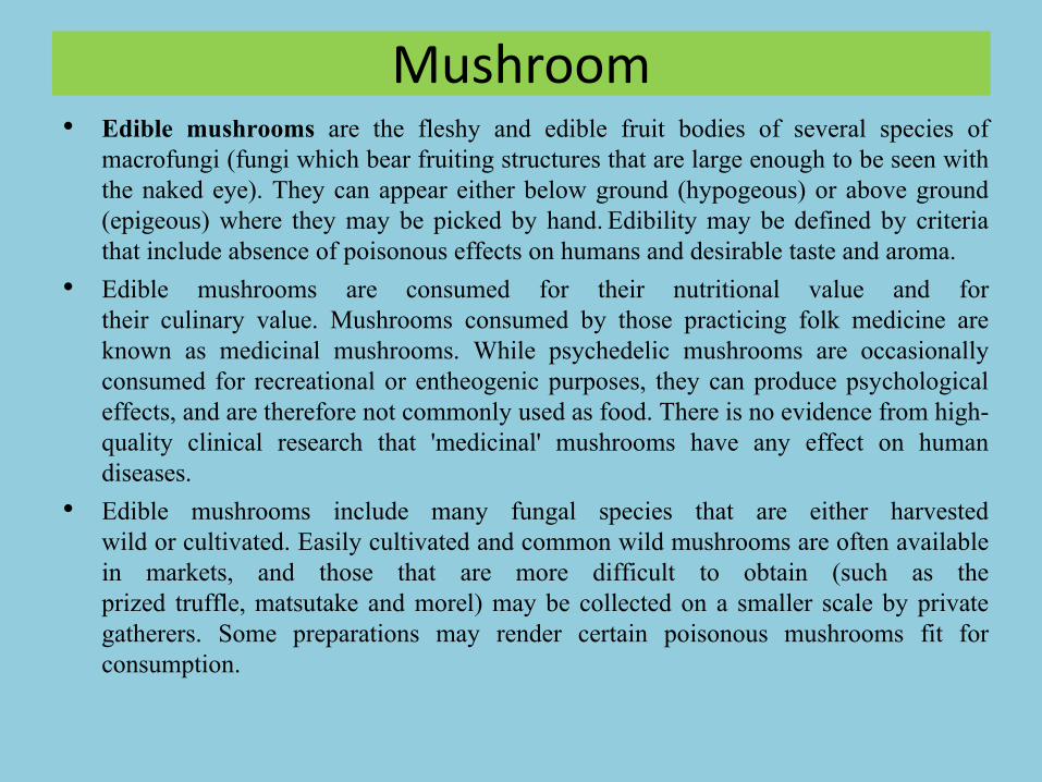
Mushroom• Edible mushrooms are the fleshy and edible fruit bodies of several species of
macrofungi (fungi which bear fruiting structures that are large enough to be seen withthe naked eye). They can appear either below ground (hypogeous) or above ground(epigeous) where they may be picked by hand. Edibility may be defined by criteriathat include absence of poisonous effects on humans and desirable taste and aroma.
• Edible mushrooms are consumed for their nutritional value and fortheir culinary value. Mushrooms consumed by those practicing folk medicine areknown as medicinal mushrooms. While psychedelic mushrooms are occasionallyconsumed for recreational or entheogenic purposes, they can produce psychologicaleffects, and are therefore not commonly used as food. There is no evidence from high-quality clinical research that 'medicinal' mushrooms have any effect on humandiseases.
• Edible mushrooms include many fungal species that are either harvestedwild or cultivated. Easily cultivated and common wild mushrooms are often availablein markets, and those that are more difficult to obtain (such as theprized truffle, matsutake and morel) may be collected on a smaller scale by privategatherers. Some preparations may render certain poisonous mushrooms fit forconsumption.

• Before assuming that any wild mushroom is edible, it shouldbe identified. Accurate determination and proper identification of aspecies is the only safe way to ensure edibility, and the only safeguardagainst possible accident. Some mushrooms that are edible for mostpeople can cause allergic reactions in some individuals, and old orimproperly stored specimens can cause food poisoning. Great care shouldtherefore be taken when eating any fungus for the first time, and onlysmall quantities should be consumed in case of individual allergies.Deadly poisonous mushrooms that are frequently confused with ediblemushrooms and responsible for many fatal poisonings include severalspecies of the genus Amanita, in particular, Amanita phalloides, the deathcap. It is therefore better to eat only a few, easily recognizable species,than to experiment indiscriminately. Moreover, even normally ediblespecies of mushrooms may be dangerous, as mushrooms growing inpolluted locations can accumulate pollutants such as heavy metals.

7. Fungal metabolites
Primary metabolites of economic importance, secondary metabolites inmedicine and agriculture

Beneficial and harmful effects of fungi

Biosynthesis of different metabolites by fungi using building blocks. Buildingblocks are photosynthates; green fonts-primary metabolites, blue fonts-secondary metabolites

Metabolic pathwaysThe pathways are usually named after enzymes or intermediates involved and are
also commonly used to classify secondary metabolites. Three most commonpathways studied are
• (1) the mevalonic acid pathway (synthesize terpenoids, steroids, etc).
• (2) the shikimic acid pathway (synthesize aromatic amino acids, alkaloids, etc).
• (3) the acetate pathway (synthesize polyketides, fatty acids, etc).
– The enzymes associated with these pathways are nonribosomal peptidesynthetases (NRPSs), polyketide synthases (PKSs), terpene cyclases (TCs),dimethylallyl tryptophan synthetases (DMATs), and geranylgeranyldiphosphate synthases (GGPPs), etc.
– These enzymes utilize building blocks like acetyl-coA, amino acids,mevalonate, and their different counterparts for the production of differentfungal metabolites

Difference Between Primary Metabolites and Secondary Metabolites
• The metabolites which are required for the growth andmaintenance of cellular function are called primarymetabolites, while such metabolites which are not requiredfor the growth and maintenance of the cellular functions andare the end products of the primary metabolism are called assecondary metabolites.
• The primary metabolites consist of the vitamins, amino acids,nucleosides and organic acids, which are necessary at the timeof logarithmic phase of microbial growth. But the productslike alkaloids, steroids, antibiotics, gibberellins, toxins are thesecondary metabolite compound produced during thestationary phase of the cell growth.
• The microorganisms possess the tremendous capacity tosynthesize the variety of products that are commercially used.

Primary Metabolites• The metabolism products that are produced during the growth phase
of an organisms in order to perform the physiological functions andsupports in overall development of the cell are called primarymetabolites.
• It occurs at the Growth phase
• These are produced in large quantities, and their extraction is easy.
• Same in every species, which means they produce the same products.
• These products are used in industries for various purpose.
• Primary products play the significant role in the cell growth,reproduction and development.
• E.g. Vitamins, carbohydrates, proteins and lipids are some of theexamples.

Secondary Metabolite• The end products of primary metabolism that are synthesized after the
growth phase has been completed and are important in ecological andother activities of the cell are known as secondary metabolites.
• It occurs at the Stationary phase.
• These are produced in small quantities, and their extraction is difficult.
• Varies in different species.
• Secondary metabolites such as antibiotics, gibberellins are alsoimportant.
• They also indirectly support the cell, in sustaining their life for longduration.
• E.g. Phenolics, steroids, essential oils, alkaloids, steroids are fewexamples.

Key Differences Between Primary and Secondary Metabolites
• The primary metabolites are considered as the products that are produced during thegrowth phase of organisms and are primarily involved in the growth and developmentof an organism. On the other hand, secondary metabolites are said to be as the endproducts of the primary metabolites, involved in the stationary phase during the growthof a microorganism and play a role in ecological functions.
• Primary metabolism pathway occurs at the growth phase and is also known astrophophase while the secondary metabolism pathway occurs at the stationary phaseand is also known as idiophase.
• Primary metabolites are produced in large quantities, and their extraction is easy, infact, the products are same in every species, whereas secondary metabolites areproduced in small quantities, and their extraction is difficult, even their products aredifferent for different species.
• The products manufactured during the primary metabolism pathway are useful inindustries for various purpose and also plays very important role in the cell growth,reproduction and development.Whereas secondary metabolites such as antibiotics,gibberellins are also important and they also indirectly support the cell, in theirsurvival for a long time.
• Examples of primary metabolites are vitamins, carbohydrates, proteins, and lipidswhile secondary metabolites examples are phenolics, steroids, essential oils, alkaloids,steroids are few examples.

Recent examples of bioactive molecules of fungi and their biological activities

Recent examples of bioactive molecules of fungi and their biological activities

Recent examples of bioactive molecules of fungi and their biological activities

Secondary metabolites(Pharmaceutical preparations)
Fungal source Active Ingredient Medicinal properties
Ganoderma lucidum
Ganoderic acid, Beta-
glucan
Liver protection, Antibiotic properties, Inhibits
cholesterol synthesis
Lentinula edodes Eritadenine, Lentinan Lower cholesterol, Anti-cancer agent
A. bisporous Lectins Enhance insulin secretion
P. sajor-caju Lovastatin Lower cholesterol
G. frondosa Polysaccharide, Lectins
Increases insulin secretion, Decrease blood
glucose
Auricularia auricula Acidic, polysaccharides Decrease blood glucose
Flammulina velutipes Ergothioneine, Proflamin Antioxidant, Anti cancer activity
Trametes versicolor Polysaccharide-K (Kresin) Decrease immune system depression
Cordyceps sinensis Cordycepin
Cure lung infections, Hypoglycemic activity,
Cellular health properties, Anti-depressant
activity

Secondary metabolites(Pharmaceutical preparations)

8. Future of fungal biotechnology
Production of mammalian proteins by fungi, other
applications of gene cloning in fungi and their importance

Recombinant DNA technology:
Manipulation of industrially important fungi, edible
mushroom and bio‐control agents