ELAINE N. MARIEB -...
13
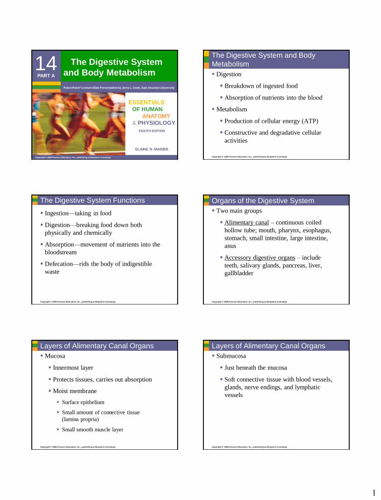
1 ELAINE N. MARIEB EIGHTH EDITION 14 Copyright © 2006 Pearson Education, Inc., publishing as Benjamin Cummings PowerPoint ® Lecture Slide Presentation by Jerry L. Cook, Sam Houston University ESSENTIALS OF HUMAN ANATOMY & PHYSIOLOGY PART A The Digestive System and Body Metabolism Copyright © 2006 Pearson Education, Inc., publishing as Benjamin Cummings The Digestive System and Body Metabolism Digestion Breakdown of ingested food Absorption of nutrients into the blood Metabolism Production of cellular energy (ATP) Constructive and degradative cellular activities Copyright © 2006 Pearson Education, Inc., publishing as Benjamin Cummings The Digestive System Functions Ingestion—taking in food Digestion—breaking food down both physically and chemically Absorption—movement of nutrients into the bloodstream Defecation—rids the body of indigestible waste Copyright © 2006 Pearson Education, Inc., publishing as Benjamin Cummings Organs of the Digestive System Two main groups Alimentary canal – continuous coiled hollow tube; mouth, pharynx, esophagus, stomach, small intestine, large intestine, anus Accessory digestive organs – include teeth, salivary glands, pancreas, liver, gallbladder Copyright © 2006 Pearson Education, Inc., publishing as Benjamin Cummings Layers of Alimentary Canal Organs Mucosa Innermost layer Protects tissues, carries out absorption Moist membrane Surface epithelium Small amount of connective tissue (lamina propria) Small smooth muscle layer Copyright © 2006 Pearson Education, Inc., publishing as Benjamin Cummings Layers of Alimentary Canal Organs Submucosa Just beneath the mucosa Soft connective tissue with blood vessels, glands, nerve endings, and lymphatic vessels
Transcript of ELAINE N. MARIEB -...
1
ELAINE N. MARIEB
EIGHTH EDITION
14
Copyright © 2006 Pearson Education, Inc., publishing as Benjamin Cummings
PowerPoint® Lecture Slide Presentation by Jerry L. Cook, Sam Houston University
ESSENTIALS
OF HUMAN
ANATOMY
& PHYSIOLOGY
PART A
The Digestive System
and Body Metabolism
Copyright © 2006 Pearson Education, Inc., publishing as Benjamin Cummings
The Digestive System and Body
Metabolism
Digestion
Breakdown of ingested food
Absorption of nutrients into the blood
Metabolism
Production of cellular energy (ATP)
Constructive and degradative cellular
activities
Copyright © 2006 Pearson Education, Inc., publishing as Benjamin Cummings
The Digestive System Functions
Ingestion—taking in food
Digestion—breaking food down both
physically and chemically
Absorption—movement of nutrients into the
bloodstream
Defecation—rids the body of indigestible
waste
Copyright © 2006 Pearson Education, Inc., publishing as Benjamin Cummings
Organs of the Digestive System
Two main groups
Alimentary canal – continuous coiled
hollow tube; mouth, pharynx, esophagus,
stomach, small intestine, large intestine,
anus
Accessory digestive organs – include
teeth, salivary glands, pancreas, liver,
gallbladder
Copyright © 2006 Pearson Education, Inc., publishing as Benjamin Cummings
Layers of Alimentary Canal Organs
Mucosa
Innermost layer
Protects tissues, carries out absorption
Moist membrane
Surface epithelium
Small amount of connective tissue
(lamina propria)
Small smooth muscle layer
Copyright © 2006 Pearson Education, Inc., publishing as Benjamin Cummings
Layers of Alimentary Canal Organs
Submucosa
Just beneath the mucosa
Soft connective tissue with blood vessels,
glands, nerve endings, and lymphatic
vessels
2
Copyright © 2006 Pearson Education, Inc., publishing as Benjamin Cummings
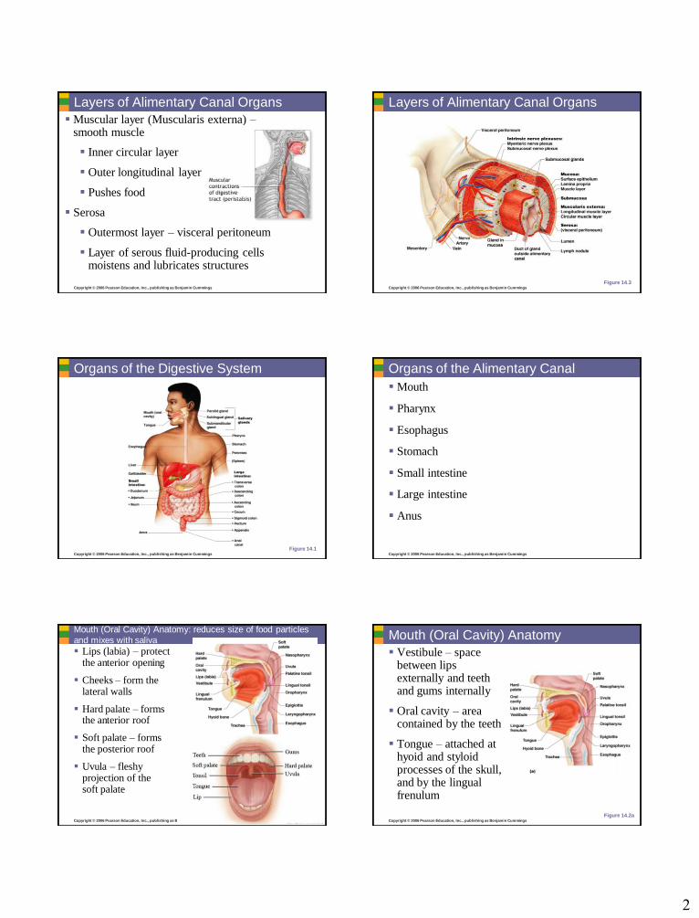
Layers of Alimentary Canal Organs
Muscular layer (Muscularis externa) –smooth muscle
Inner circular layer
Outer longitudinal layer
Pushes food
Serosa
Outermost layer – visceral peritoneum
Layer of serous fluid-producing cells moistens and lubricates structures
Copyright © 2006 Pearson Education, Inc., publishing as Benjamin Cummings
Layers of Alimentary Canal Organs
Figure 14.3
Copyright © 2006 Pearson Education, Inc., publishing as Benjamin Cummings
Organs of the Digestive System
Figure 14.1Copyright © 2006 Pearson Education, Inc., publishing as Benjamin Cummings
Organs of the Alimentary Canal
Mouth
Pharynx
Esophagus
Stomach
Small intestine
Large intestine
Anus
Copyright © 2006 Pearson Education, Inc., publishing as Benjamin Cummings
Mouth (Oral Cavity) Anatomy: reduces size of food particles
and mixes with saliva
Lips (labia) – protect the anterior opening
Cheeks – form the lateral walls
Hard palate – forms the anterior roof
Soft palate – forms the posterior roof
Uvula – fleshy projection of the soft palate
Figure 14.2aCopyright © 2006 Pearson Education, Inc., publishing as Benjamin Cummings
Mouth (Oral Cavity) Anatomy
Vestibule – space between lips externally and teeth and gums internally
Oral cavity – area contained by the teeth
Tongue – attached at hyoid and styloid processes of the skull, and by the lingual frenulum
Figure 14.2a
3
Copyright © 2006 Pearson Education, Inc., publishing as Benjamin Cummings
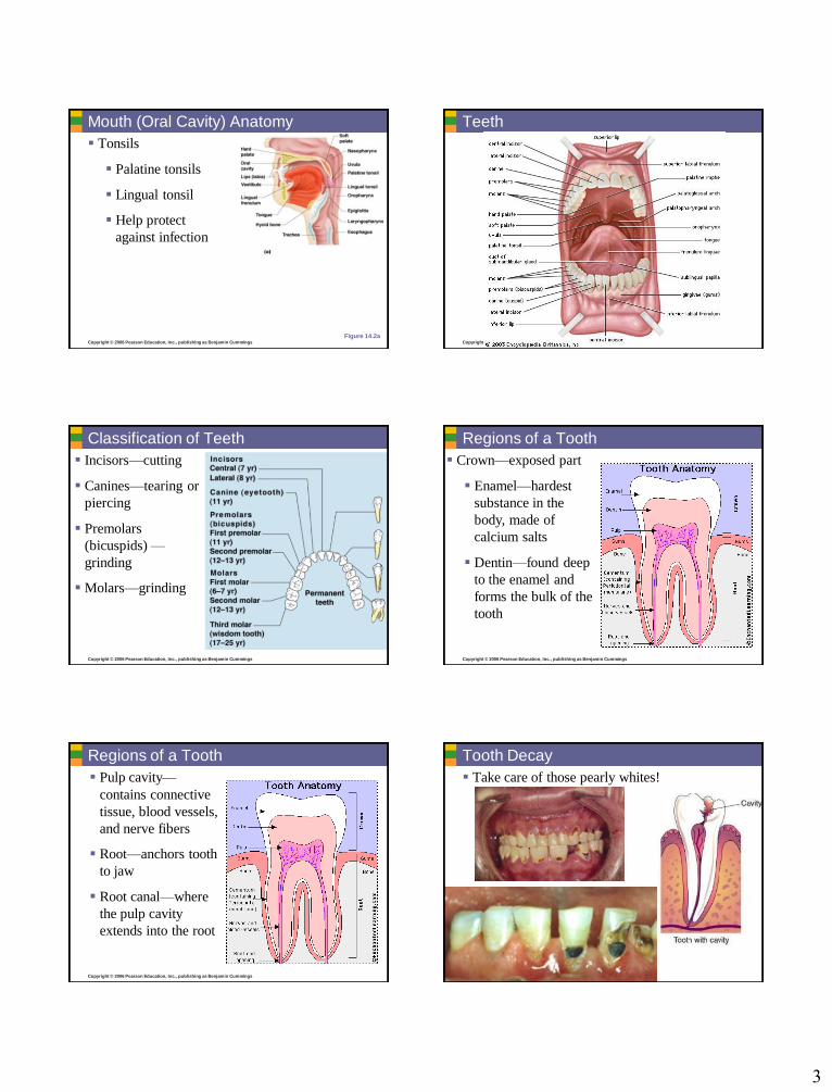
Mouth (Oral Cavity) Anatomy
Tonsils
Palatine tonsils
Lingual tonsil
Help protect
against infection
Figure 14.2aCopyright © 2006 Pearson Education, Inc., publishing as Benjamin Cummings
Teeth
Copyright © 2006 Pearson Education, Inc., publishing as Benjamin Cummings
Classification of Teeth
Incisors—cutting
Canines—tearing or
piercing
Premolars
(bicuspids) —
grinding
Molars—grinding
Copyright © 2006 Pearson Education, Inc., publishing as Benjamin Cummings
Regions of a Tooth
Crown—exposed part
Enamel—hardest
substance in the
body, made of
calcium salts
Dentin—found deep
to the enamel and
forms the bulk of the
tooth
Copyright © 2006 Pearson Education, Inc., publishing as Benjamin Cummings
Regions of a Tooth
Pulp cavity—
contains connective
tissue, blood vessels,
and nerve fibers
Root—anchors tooth
to jaw
Root canal—where
the pulp cavity
extends into the root
Copyright © 2006 Pearson Education, Inc., publishing as Benjamin Cummings
Tooth Decay
Take care of those pearly whites!
4
Copyright © 2006 Pearson Education, Inc., publishing as Benjamin Cummings
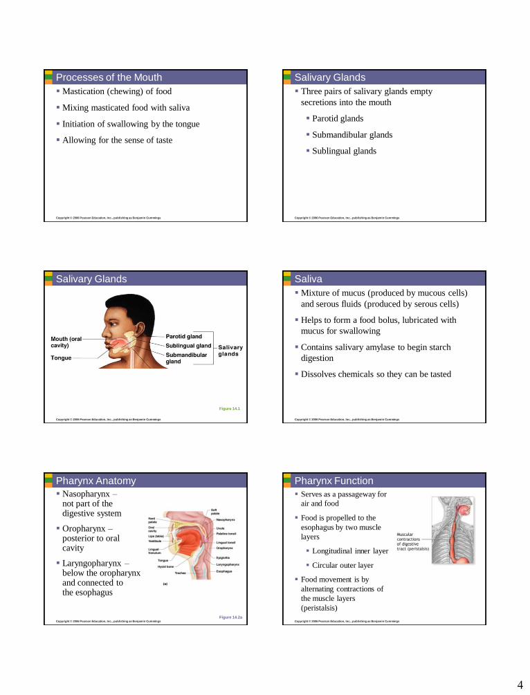
Processes of the Mouth
Mastication (chewing) of food
Mixing masticated food with saliva
Initiation of swallowing by the tongue
Allowing for the sense of taste
Copyright © 2006 Pearson Education, Inc., publishing as Benjamin Cummings
Salivary Glands
Three pairs of salivary glands empty
secretions into the mouth
Parotid glands
Submandibular glands
Sublingual glands
Copyright © 2006 Pearson Education, Inc., publishing as Benjamin Cummings
Figure 14.1
Salivary Glands
Copyright © 2006 Pearson Education, Inc., publishing as Benjamin Cummings
Saliva
Mixture of mucus (produced by mucous cells)
and serous fluids (produced by serous cells)
Helps to form a food bolus, lubricated with
mucus for swallowing
Contains salivary amylase to begin starch
digestion
Dissolves chemicals so they can be tasted
Copyright © 2006 Pearson Education, Inc., publishing as Benjamin Cummings
Pharynx Anatomy
Nasopharynx –not part of the digestive system
Oropharynx –posterior to oral cavity
Laryngopharynx –below the oropharynx and connected to the esophagus
Figure 14.2aCopyright © 2006 Pearson Education, Inc., publishing as Benjamin Cummings
Pharynx Function
Serves as a passageway for
air and food
Food is propelled to the
esophagus by two muscle
layers
Longitudinal inner layer
Circular outer layer
Food movement is by
alternating contractions of
the muscle layers
(peristalsis)
5
Copyright © 2006 Pearson Education, Inc., publishing as Benjamin Cummings
Esophagus
Runs from pharynx to stomach through the
diaphragm
Conducts food by peristalsis
(slow rhythmic squeezing)
Passageway for food only (respiratory system
branches off after the pharynx)
Copyright © 2006 Pearson Education, Inc., publishing as Benjamin Cummings
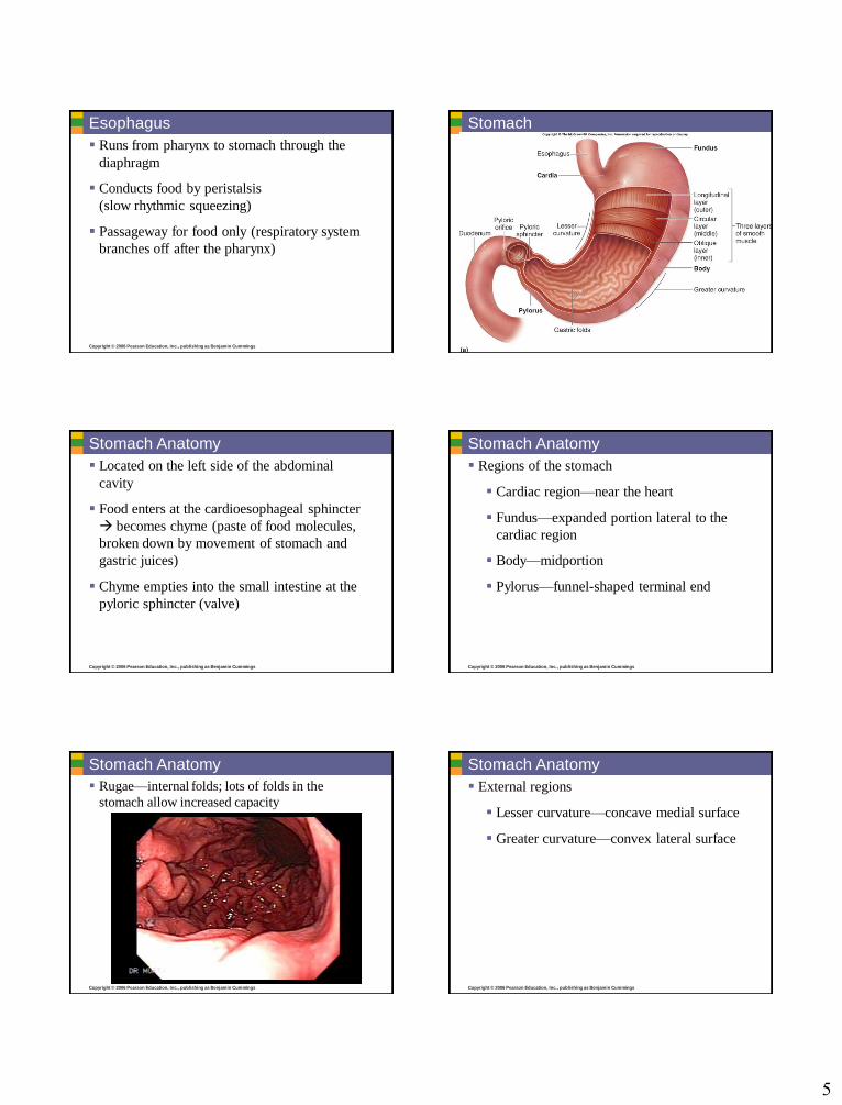
Stomach
Copyright © 2006 Pearson Education, Inc., publishing as Benjamin Cummings
Stomach Anatomy
Located on the left side of the abdominal
cavity
Food enters at the cardioesophageal sphincter
becomes chyme (paste of food molecules,
broken down by movement of stomach and
gastric juices)
Chyme empties into the small intestine at the
pyloric sphincter (valve)
Copyright © 2006 Pearson Education, Inc., publishing as Benjamin Cummings
Stomach Anatomy
Regions of the stomach
Cardiac region—near the heart
Fundus—expanded portion lateral to the
cardiac region
Body—midportion
Pylorus—funnel-shaped terminal end
Copyright © 2006 Pearson Education, Inc., publishing as Benjamin Cummings
Stomach Anatomy
Rugae—internal folds; lots of folds in the
stomach allow increased capacity
Copyright © 2006 Pearson Education, Inc., publishing as Benjamin Cummings
Stomach Anatomy
External regions
Lesser curvature—concave medial surface
Greater curvature—convex lateral surface

6
Copyright © 2006 Pearson Education, Inc., publishing as Benjamin Cummings
Stomach Physiology
Temporary storage tank for food
Site of food breakdown
Chemical breakdown of protein begins
Delivers chyme (processed food) to the small
intestine
Copyright © 2006 Pearson Education, Inc., publishing as Benjamin Cummings
Structure of the Stomach Mucosa Mucosa is simple columnar epithelium
Mucous neck cells —produce a sticky alkaline mucus; prevents stomach from digesting itself
Gastric glands —situated in gastric pits and secrete gastric juice
Chief cells —produce protein-digesting enzymes (pepsinogens)
Parietal cells —produce hydrochloric acid (acidic conditions activate enzymes)
Enteroendocrine cells —produce gastrin (hormone important to digestive activities)
Copyright © 2006 Pearson Education, Inc., publishing as Benjamin Cummings
Stomach Anatomy
Figure 14.4a
Copyright © 2006 Pearson Education, Inc., publishing as Benjamin Cummings
Pancreas Produces a wide spectrum of digestive enzymes
that break down all categories of food
Enzymes are secreted into the duodenum
Alkaline fluid introduced with enzymes neutralizes acidic chyme
Endocrine products of pancreas
Insulin
Glucagons
Note: endocrine (secretes directly into bloodstream) and exocrine (uses pancreatic duct) functions!
Copyright © 2006 Pearson Education, Inc., publishing as Benjamin Cummings Copyright © 2006 Pearson Education, Inc., publishing as Benjamin Cummings
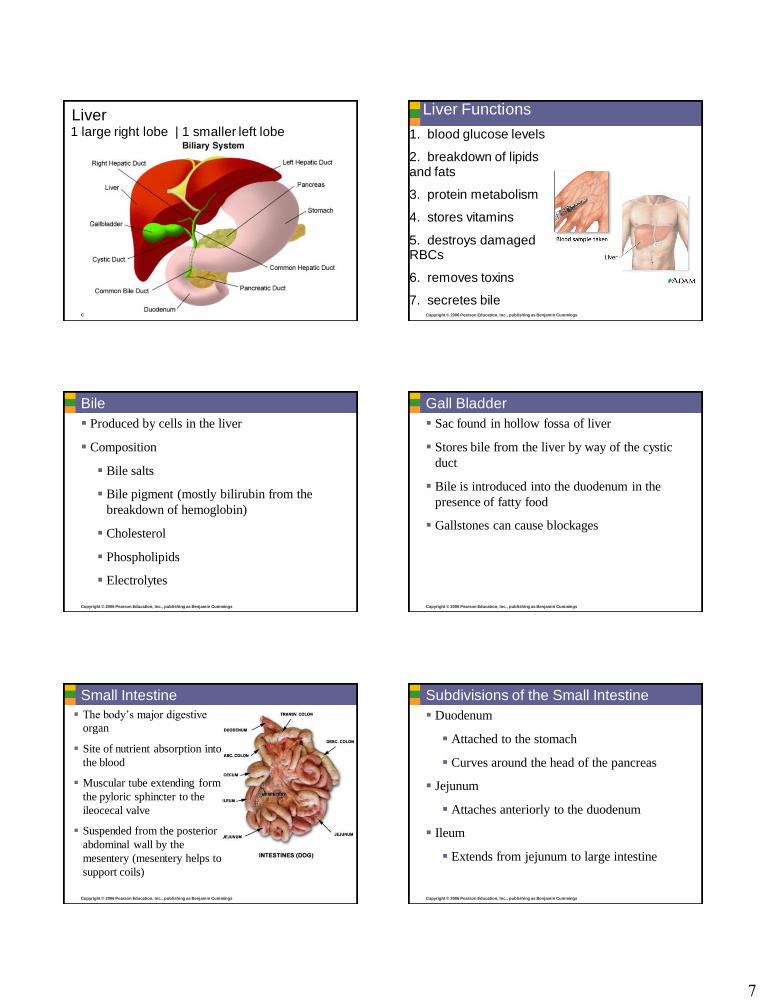
Liver
Largest gland in the body
Located on the right side of the body under
the diaphragm
Consists of four lobes suspended from the
diaphragm and abdominal wall by the
falciform ligament
Connected to the gall bladder via the common
hepatic duct
Hepatic portal vein delivers blood
7
Copyright © 2006 Pearson Education, Inc., publishing as Benjamin Cummings
Liver1 large right lobe | 1 smaller left lobe
Copyright © 2006 Pearson Education, Inc., publishing as Benjamin Cummings
Liver Functions
1. blood glucose levels
2. breakdown of lipids and fats
3. protein metabolism
4. stores vitamins
5. destroys damaged RBCs
6. removes toxins
7. secretes bile
Copyright © 2006 Pearson Education, Inc., publishing as Benjamin Cummings
Bile
Produced by cells in the liver
Composition
Bile salts
Bile pigment (mostly bilirubin from the
breakdown of hemoglobin)
Cholesterol
Phospholipids
Electrolytes
Copyright © 2006 Pearson Education, Inc., publishing as Benjamin Cummings
Gall Bladder
Sac found in hollow fossa of liver
Stores bile from the liver by way of the cystic
duct
Bile is introduced into the duodenum in the
presence of fatty food
Gallstones can cause blockages
Copyright © 2006 Pearson Education, Inc., publishing as Benjamin Cummings
Small Intestine
The body’s major digestive
organ
Site of nutrient absorption into
the blood
Muscular tube extending form
the pyloric sphincter to the
ileocecal valve
Suspended from the posterior
abdominal wall by the
mesentery (mesentery helps to
support coils)
Copyright © 2006 Pearson Education, Inc., publishing as Benjamin Cummings
Subdivisions of the Small Intestine
Duodenum
Attached to the stomach
Curves around the head of the pancreas
Jejunum
Attaches anteriorly to the duodenum
Ileum
Extends from jejunum to large intestine
8
Copyright © 2006 Pearson Education, Inc., publishing as Benjamin Cummings
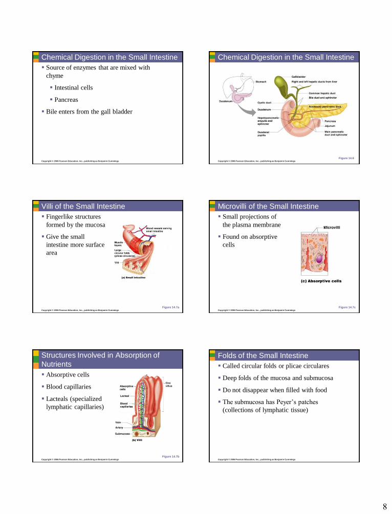
Chemical Digestion in the Small Intestine
Source of enzymes that are mixed with
chyme
Intestinal cells
Pancreas
Bile enters from the gall bladder
Copyright © 2006 Pearson Education, Inc., publishing as Benjamin Cummings
Chemical Digestion in the Small Intestine
Figure 14.6
Copyright © 2006 Pearson Education, Inc., publishing as Benjamin Cummings
Villi of the Small Intestine
Fingerlike structures
formed by the mucosa
Give the small
intestine more surface
area
Figure 14.7aCopyright © 2006 Pearson Education, Inc., publishing as Benjamin Cummings
Microvilli of the Small Intestine
Small projections of
the plasma membrane
Found on absorptive
cells
Figure 14.7c
Copyright © 2006 Pearson Education, Inc., publishing as Benjamin Cummings
Structures Involved in Absorption of
Nutrients
Absorptive cells
Blood capillaries
Lacteals (specialized
lymphatic capillaries)
Figure 14.7bCopyright © 2006 Pearson Education, Inc., publishing as Benjamin Cummings
Folds of the Small Intestine
Called circular folds or plicae circulares
Deep folds of the mucosa and submucosa
Do not disappear when filled with food
The submucosa has Peyer’s patches
(collections of lymphatic tissue)
9
Copyright © 2006 Pearson Education, Inc., publishing as Benjamin Cummings
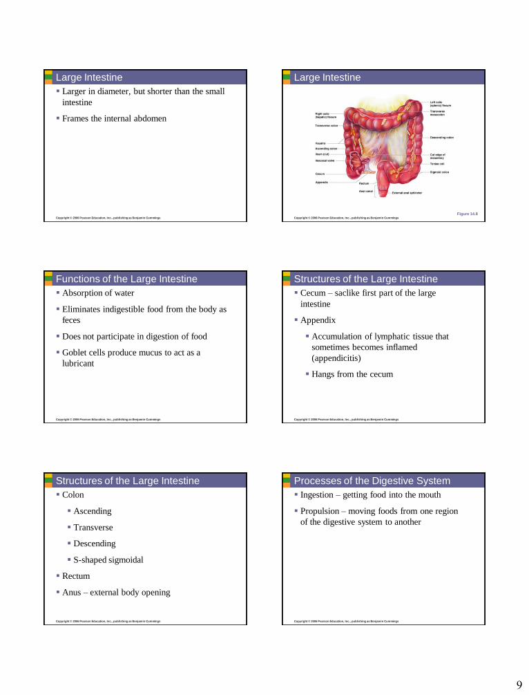
Large Intestine
Larger in diameter, but shorter than the small
intestine
Frames the internal abdomen
Copyright © 2006 Pearson Education, Inc., publishing as Benjamin Cummings
Large Intestine
Figure 14.8
Copyright © 2006 Pearson Education, Inc., publishing as Benjamin Cummings
Functions of the Large Intestine
Absorption of water
Eliminates indigestible food from the body as
feces
Does not participate in digestion of food
Goblet cells produce mucus to act as a
lubricant
Copyright © 2006 Pearson Education, Inc., publishing as Benjamin Cummings
Structures of the Large Intestine
Cecum – saclike first part of the large
intestine
Appendix
Accumulation of lymphatic tissue that
sometimes becomes inflamed
(appendicitis)
Hangs from the cecum
Copyright © 2006 Pearson Education, Inc., publishing as Benjamin Cummings
Structures of the Large Intestine
Colon
Ascending
Transverse
Descending
S-shaped sigmoidal
Rectum
Anus – external body opening
Copyright © 2006 Pearson Education, Inc., publishing as Benjamin Cummings
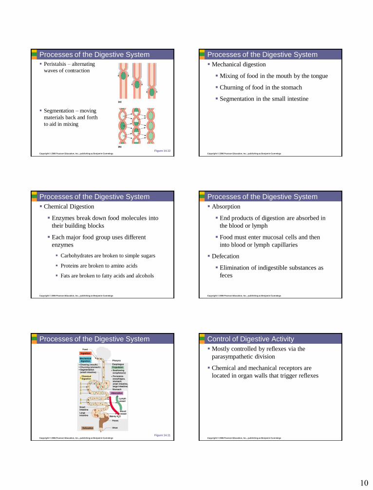
Processes of the Digestive System
Ingestion – getting food into the mouth
Propulsion – moving foods from one region
of the digestive system to another
10
Copyright © 2006 Pearson Education, Inc., publishing as Benjamin Cummings
Processes of the Digestive System
Peristalsis – alternating
waves of contraction
Segmentation – moving
materials back and forth
to aid in mixing
Figure 14.12Copyright © 2006 Pearson Education, Inc., publishing as Benjamin Cummings
Processes of the Digestive System
Mechanical digestion
Mixing of food in the mouth by the tongue
Churning of food in the stomach
Segmentation in the small intestine
Copyright © 2006 Pearson Education, Inc., publishing as Benjamin Cummings
Processes of the Digestive System
Chemical Digestion
Enzymes break down food molecules into
their building blocks
Each major food group uses different
enzymes
Carbohydrates are broken to simple sugars
Proteins are broken to amino acids
Fats are broken to fatty acids and alcohols
Copyright © 2006 Pearson Education, Inc., publishing as Benjamin Cummings
Processes of the Digestive System
Absorption
End products of digestion are absorbed in
the blood or lymph
Food must enter mucosal cells and then
into blood or lymph capillaries
Defecation
Elimination of indigestible substances as
feces
Copyright © 2006 Pearson Education, Inc., publishing as Benjamin Cummings
Processes of the Digestive System
Figure 14.11Copyright © 2006 Pearson Education, Inc., publishing as Benjamin Cummings
Control of Digestive Activity
Mostly controlled by reflexes via the
parasympathetic division
Chemical and mechanical receptors are
located in organ walls that trigger reflexes
11
Copyright © 2006 Pearson Education, Inc., publishing as Benjamin Cummings
Control of Digestive Activity
Stimuli include:
Stretch of the organ
pH of the contents
Presence of breakdown products
Reflexes include:
Activation or inhibition of glandular secretions
Smooth muscle activity
Copyright © 2006 Pearson Education, Inc., publishing as Benjamin Cummings
Digestive Activities of the Mouth
Mechanical breakdown
Food is physically broken down by
chewing
Chemical digestion
Food is mixed with saliva
Breaking of starch into maltose by salivary
amylase
Copyright © 2006 Pearson Education, Inc., publishing as Benjamin Cummings
Activities of the Pharynx and Esophagus
These organs have no digestive function
Serve as passageways to the stomach
Copyright © 2006 Pearson Education, Inc., publishing as Benjamin Cummings
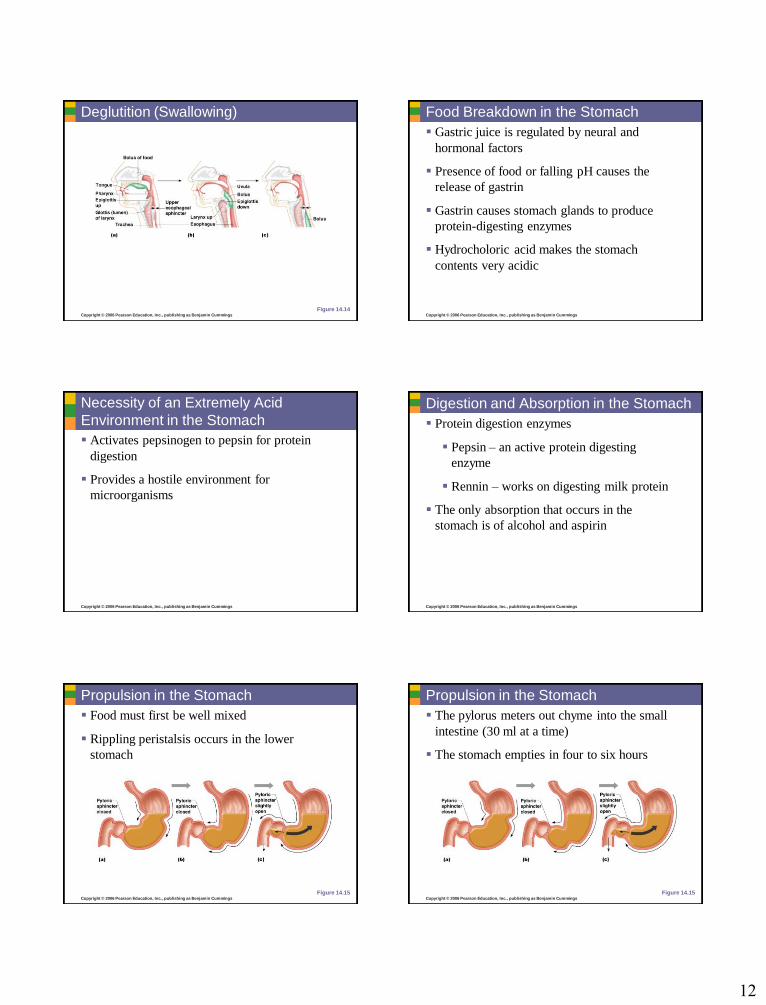
Deglutition (Swallowing)
Buccal phase
Voluntary
Occurs in the mouth
Food is formed into a bolus
The bolus is forced into the pharynx by the
tongue
Copyright © 2006 Pearson Education, Inc., publishing as Benjamin Cummings
Deglutition (Swallowing)
Pharyngeal-esophageal phase
Involuntary transport of the bolus
All passageways except to the stomach are
blocked
Tongue blocks off the mouth
Soft palate (uvula) blocks the nasopharynx
Epiglottis blocks the larynx
Copyright © 2006 Pearson Education, Inc., publishing as Benjamin Cummings
Deglutition (Swallowing)
Pharyngeal-esophogeal phase (continued)
Peristalsis moves the bolus toward the
stomach
The cardioesophageal sphincter is opened
when food presses against it
12
Copyright © 2006 Pearson Education, Inc., publishing as Benjamin Cummings
Deglutition (Swallowing)
Figure 14.14Copyright © 2006 Pearson Education, Inc., publishing as Benjamin Cummings
Food Breakdown in the Stomach
Gastric juice is regulated by neural and
hormonal factors
Presence of food or falling pH causes the
release of gastrin
Gastrin causes stomach glands to produce
protein-digesting enzymes
Hydrocholoric acid makes the stomach
contents very acidic
Copyright © 2006 Pearson Education, Inc., publishing as Benjamin Cummings
Necessity of an Extremely Acid
Environment in the Stomach
Activates pepsinogen to pepsin for protein
digestion
Provides a hostile environment for
microorganisms
Copyright © 2006 Pearson Education, Inc., publishing as Benjamin Cummings
Digestion and Absorption in the Stomach
Protein digestion enzymes
Pepsin – an active protein digesting
enzyme
Rennin – works on digesting milk protein
The only absorption that occurs in the
stomach is of alcohol and aspirin
Copyright © 2006 Pearson Education, Inc., publishing as Benjamin Cummings
Propulsion in the Stomach
Food must first be well mixed
Rippling peristalsis occurs in the lower
stomach
Figure 14.15Copyright © 2006 Pearson Education, Inc., publishing as Benjamin Cummings
Propulsion in the Stomach
The pylorus meters out chyme into the small
intestine (30 ml at a time)
The stomach empties in four to six hours
Figure 14.15
13
Copyright © 2006 Pearson Education, Inc., publishing as Benjamin Cummings
Digestion in the Small Intestine
Enzymes from the brush border
Break double sugars into simple sugars
Complete some protein digestion
Pancreatic enzymes play the major digestive function
Help complete digestion of starch (pancreatic amylase)
Carry out about half of all protein digestion (trypsin, etc.)
Copyright © 2006 Pearson Education, Inc., publishing as Benjamin Cummings
Digestion in the Small Intestine
Pancreatic enzymes play the major digestive
function (continued)
Responsible for fat digestion (lipase)
Digest nucleic acids (nucleases)
Alkaline content neutralizes acidic chyme
Copyright © 2006 Pearson Education, Inc., publishing as Benjamin Cummings
Absorption in the Small Intestine
Water is absorbed along the length of the
small intestine
End products of digestion
Most substances are absorbed by active
transport through cell membranes
Lipids are absorbed by diffusion
Substances are transported to the liver by the
hepatic portal vein or lymph
Copyright © 2006 Pearson Education, Inc., publishing as Benjamin Cummings
Propulsion in the Small Intestine
Peristalsis is the major means of moving food
Segmental movements
Mix chyme with digestive juices
Aid in propelling food
Copyright © 2006 Pearson Education, Inc., publishing as Benjamin Cummings
Food Breakdown and Absorption in the
Large Intestine
No digestive enzymes are produced
Resident bacteria digest remaining nutrients
Produce some vitamin K and B
Release gases
Water and vitamins K and B are absorbed
Remaining materials are eliminated via feces
Copyright © 2006 Pearson Education, Inc., publishing as Benjamin Cummings
Propulsion in the Large Intestine
Sluggish peristalsis
Mass movements
Slow, powerful movements
Occur three to four times per day
Presence of feces in the rectum causes a defecation
reflex
Internal anal sphincter is relaxed
Defecation occurs with relaxation of the
voluntary (external) anal sphincter


![Elaine N. Marieb - whs-hs.weatherfordisd.comwhs-hs.weatherfordisd.com/ourpages/auto/2013/11/18/36665686/Ch10... · Title: Ch10_EHAP-Lect [Compatibility Mode] Author: tcomstoc Subject:](https://static.fdocuments.net/doc/165x107/5b9450f209d3f252738c732b/elaine-n-marieb-whs-hs-title-ch10ehap-lect-compatibility-mode-author.jpg)