Diagnosis and Treatment of Hemorrhagic Stroke · – Tumor – Coagulopathy, underlying disorder,...
51
Diagnosis and Treatment of Hemorrhagic Stroke Robert E. Replogle M.D. Rochester Neurosurgical Partners Stroke Treatment Alliance of Rochester Wednesday, March 28, 2012
Transcript of Diagnosis and Treatment of Hemorrhagic Stroke · – Tumor – Coagulopathy, underlying disorder,...
Diagnosis and Treatment of Hemorrhagic Stroke
Robert E. Replogle M.D.Rochester Neurosurgical Partners
Stroke Treatment Alliance of Rochester
Wednesday, March 28, 2012
Stroke in the U.S.• Stroke is the #1 cause of disability• Stroke is the #3 cause of death• 700,000 strokes annually -Approximately 80% ischemic • Up to 40% due to large vessel occlusions Every 45 seconds someone in the U.S.has a stroke Stroke survivors have a 10X greater risk of having a
repeat stroke than the general population
Wednesday, March 28, 2012
Etiology of Stroke
• Ischemic- 80% – Occlusive • Intracerebral- large vessel vs small vessel – Embolic
– Cervical carotid – Cardiac, arterial
• Hemorrhagic- 20% – Aneurysm- subarachnoid hemorrhage (SAH)
– Arterial-venous malformation (AVM)- intracerebral hemorrhage (ICH)
Wednesday, March 28, 2012
Classification
• Primary (78% ~ 88%)– Spontaneous rupture of small vessels damaged
by chronic hypertension or amyloid angiopathy• Secondary
– Vascular abnormalities (AVM, aneurysm, vasculitis, dural sinus thrombosis)
– Tumor– Coagulopathy, underlying disorder,
anticoagulants, antiplatelets, lytics– Drug abuse-cocaine
Wednesday, March 28, 2012
Guideline for the management of spontaneous intracranial hemorrhage
1. Intracerebral hemorrhage is more than twice as common as subarachnoid hemorrhage(SAH) and is much more likely to result in death or major disability than cerebral infarction or SAH.
2. Advancing age and hypertension are the most important risk factor for ICH.
Stroke, 1999, 30:905~915Wednesday, March 28, 2012
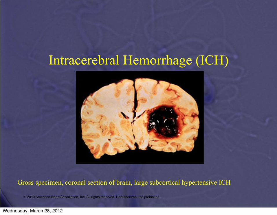
Intracerebral Hemorrhage (ICH)
Gross specimen, coronal section of brain, large subcortical hypertensive ICH
© 2010 American Heart Association, Inc. All rights reserved. Unauthorized use prohibited.
Wednesday, March 28, 2012
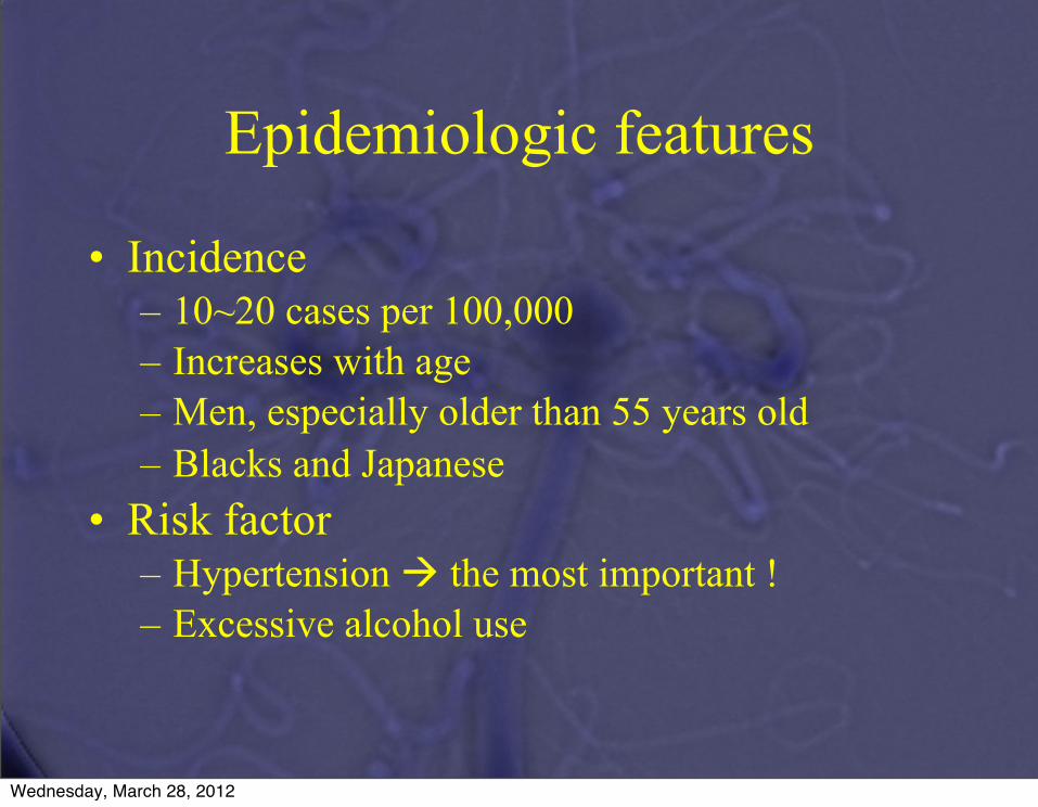
Epidemiologic features
• Incidence– 10~20 cases per 100,000– Increases with age– Men, especially older than 55 years old– Blacks and Japanese
• Risk factor– Hypertension the most important !– Excessive alcohol use
Wednesday, March 28, 2012
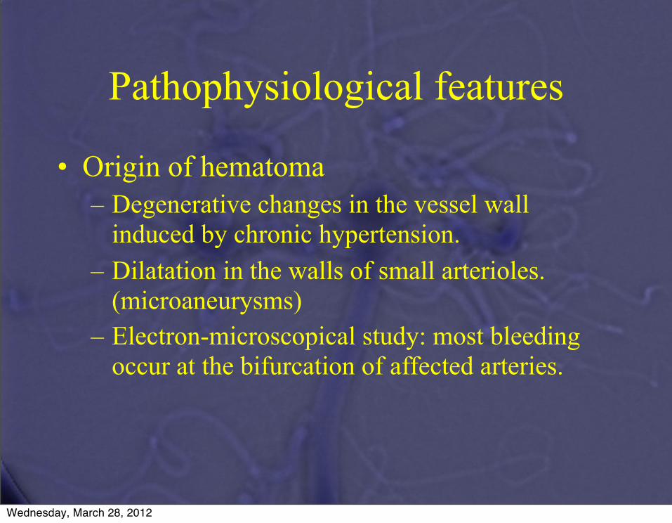
Pathophysiological features
• Origin of hematoma– Degenerative changes in the vessel wall
induced by chronic hypertension.– Dilatation in the walls of small arterioles.
(microaneurysms)– Electron-microscopical study: most bleeding
occur at the bifurcation of affected arteries.
Wednesday, March 28, 2012
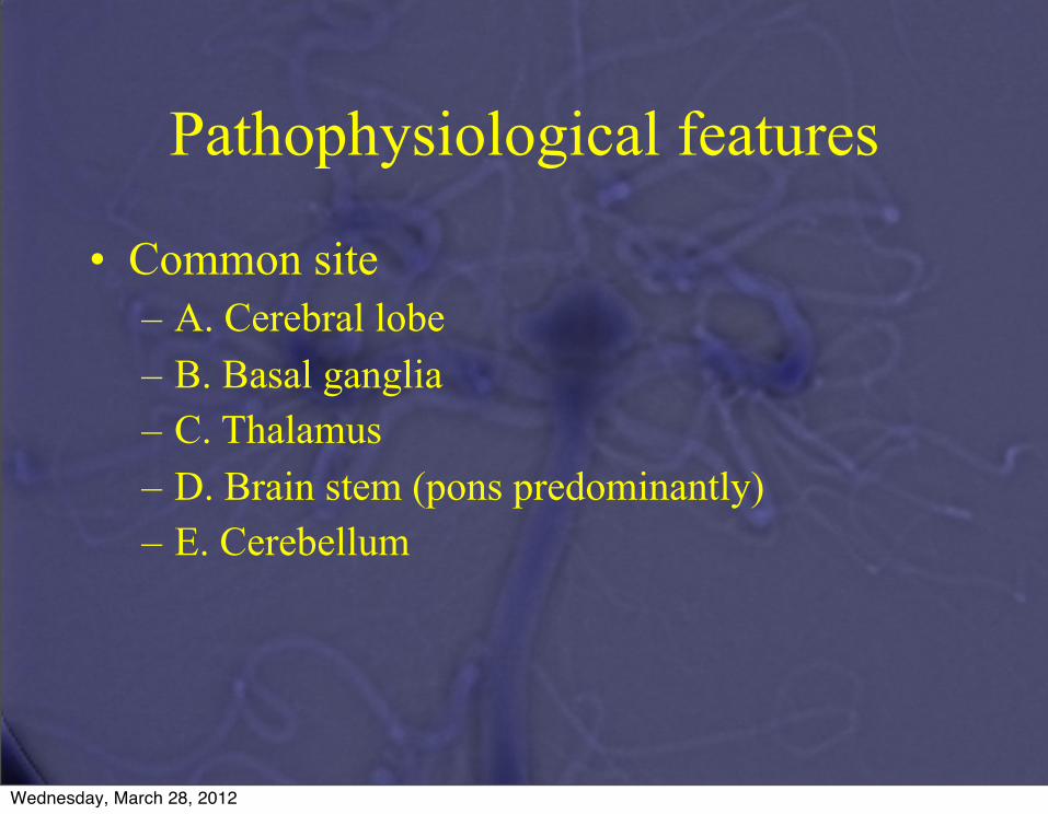
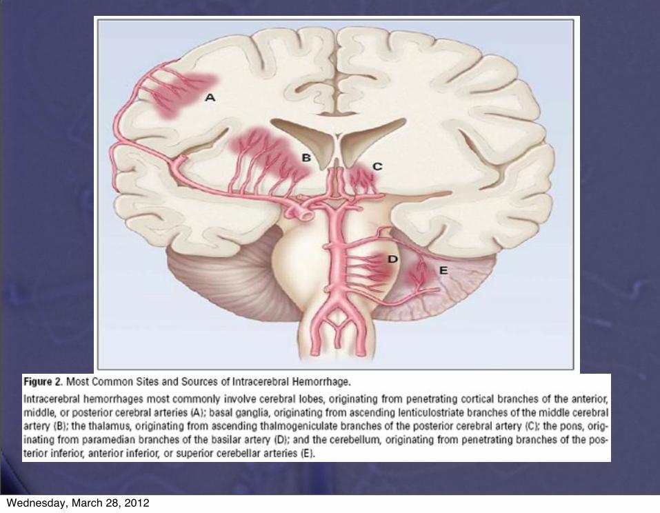
Pathophysiological features
• Common site– A. Cerebral lobe– B. Basal ganglia– C. Thalamus– D. Brain stem (pons predominantly)– E. Cerebellum
Wednesday, March 28, 2012
Wednesday, March 28, 2012
Wednesday, March 28, 2012
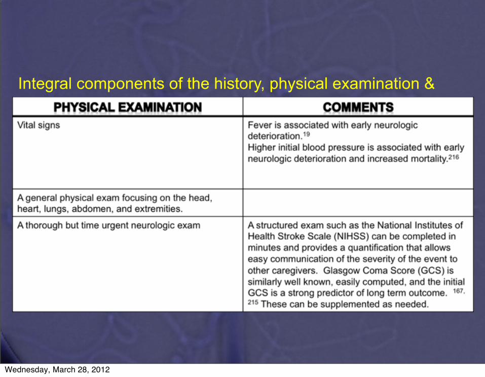
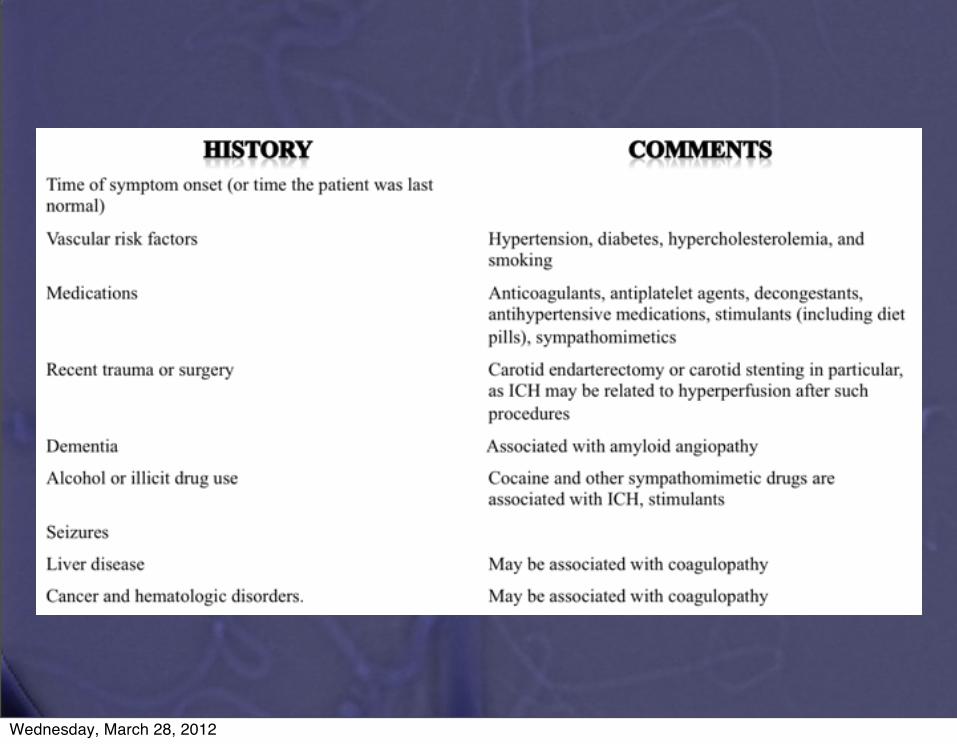
Integral components of the history, physical examination &
work up of the ICH patient in the emergency department
Wednesday, March 28, 2012
Integral components of the history, physical examination &
work up of the ICH patient in the emergency department
© 2010 American Heart Association, Inc..
Wednesday, March 28, 2012
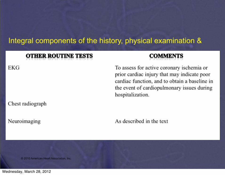
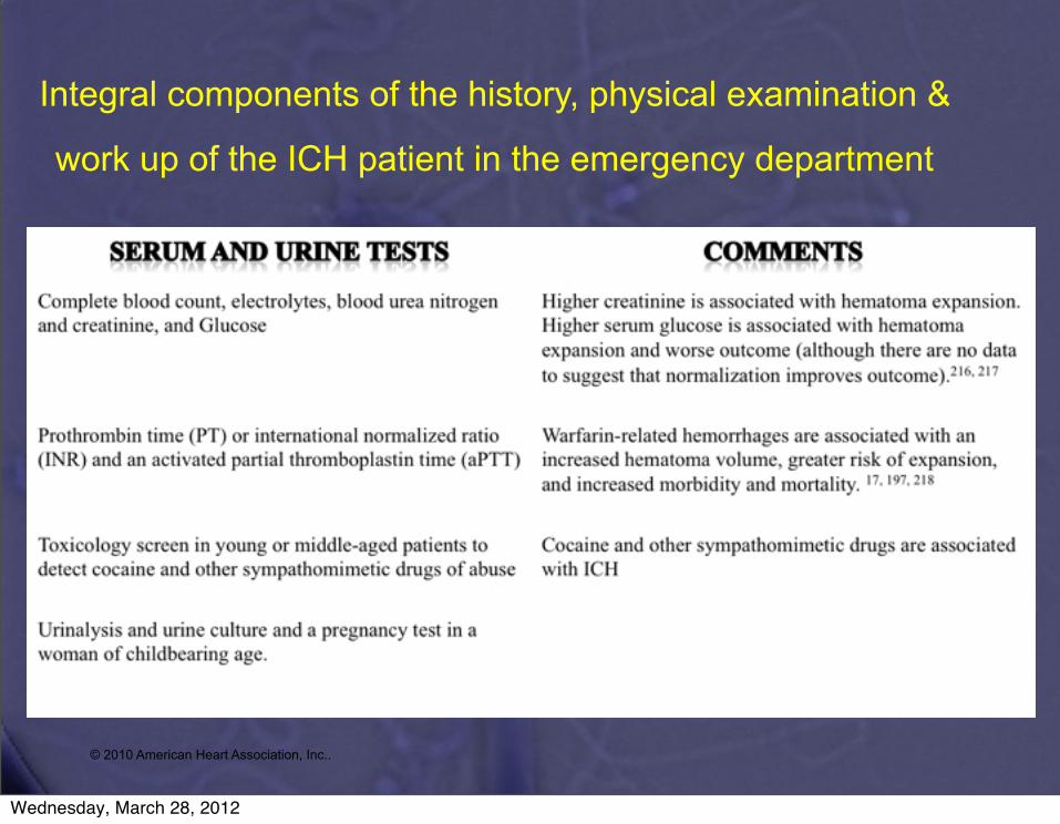
Integral components of the history, physical examination &
work up of the ICH patient in the emergency department
© 2010 American Heart Association, Inc.
Wednesday, March 28, 2012
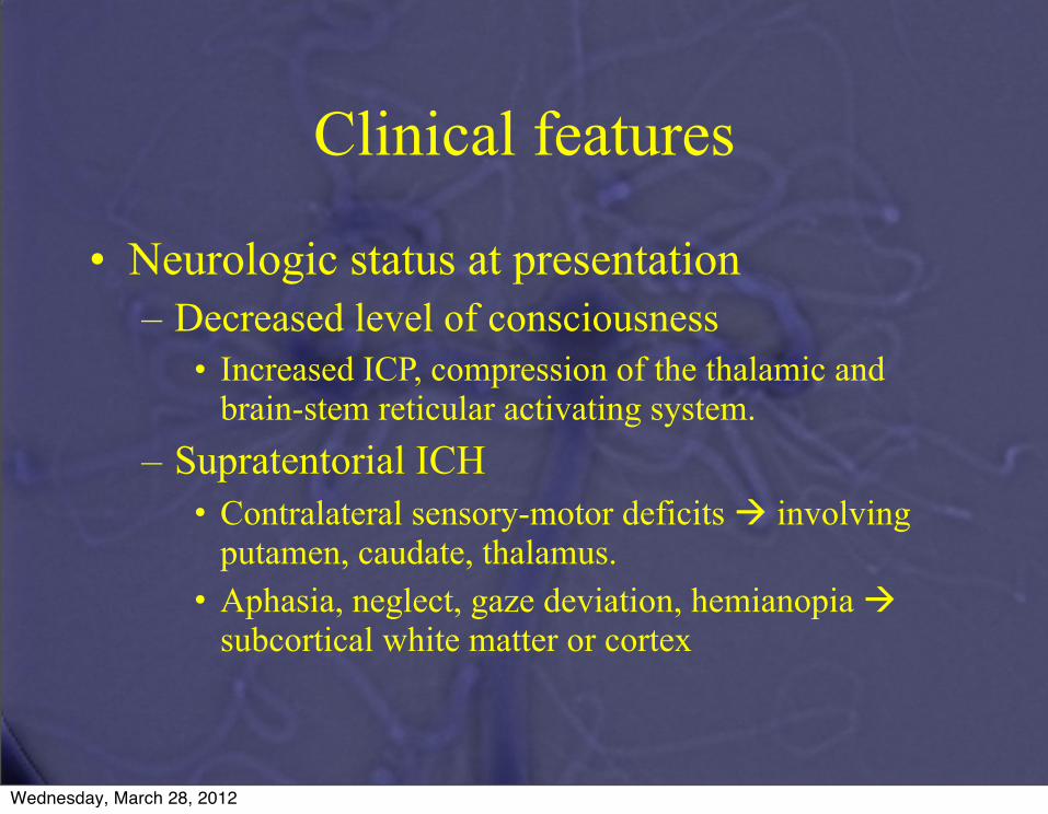
Clinical features
• Neurologic status at presentation– Decreased level of consciousness
• Increased ICP, compression of the thalamic and brain-stem reticular activating system.
– Supratentorial ICH • Contralateral sensory-motor deficits involving
putamen, caudate, thalamus.• Aphasia, neglect, gaze deviation, hemianopia
subcortical white matter or cortex
Wednesday, March 28, 2012
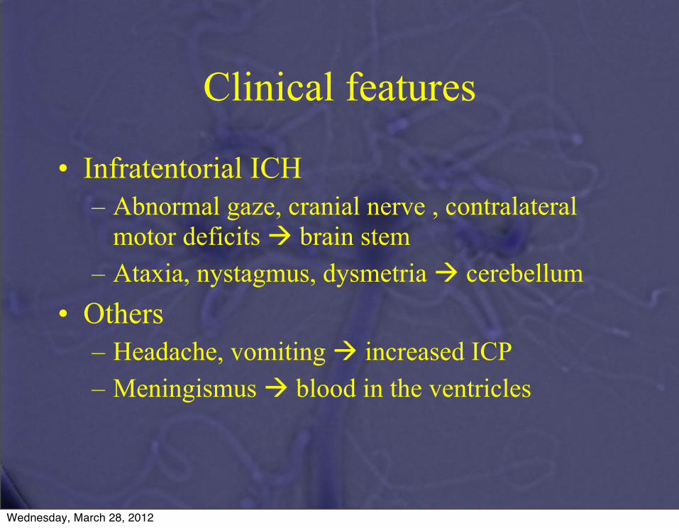
Clinical features
• Infratentorial ICH– Abnormal gaze, cranial nerve , contralateral
motor deficits brain stem– Ataxia, nystagmus, dysmetria cerebellum
• Others– Headache, vomiting increased ICP– Meningismus blood in the ventricles
Wednesday, March 28, 2012
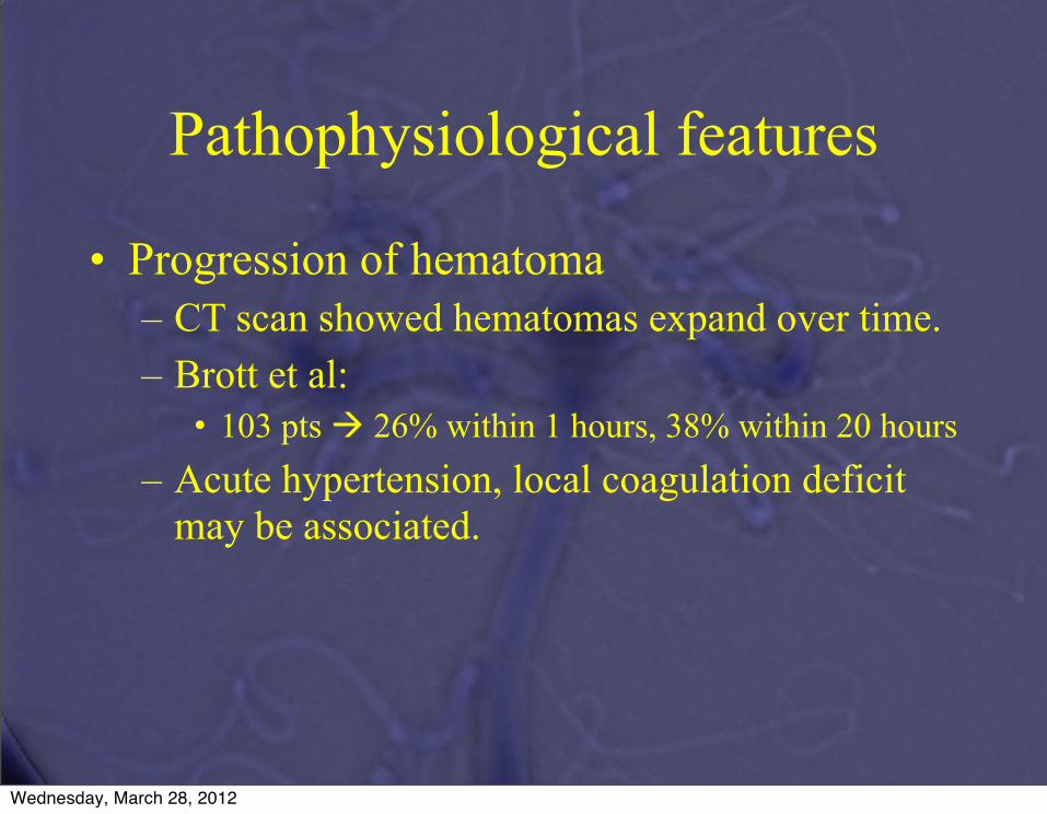
Pathophysiological features
• Progression of hematoma– CT scan showed hematomas expand over time.– Brott et al:
• 103 pts 26% within 1 hours, 38% within 20 hours– Acute hypertension, local coagulation deficit
may be associated.
Wednesday, March 28, 2012
Pathophysiological features
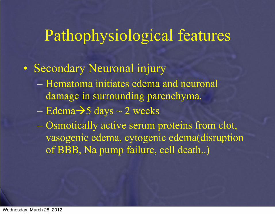
• Secondary Neuronal injury– Hematoma initiates edema and neuronal
damage in surrounding parenchyma.– Edema5 days ~ 2 weeks– Osmotically active serum proteins from clot,
vasogenic edema, cytogenic edema(disruption of BBB, Na pump failure, cell death..)
Wednesday, March 28, 2012
Clinical features
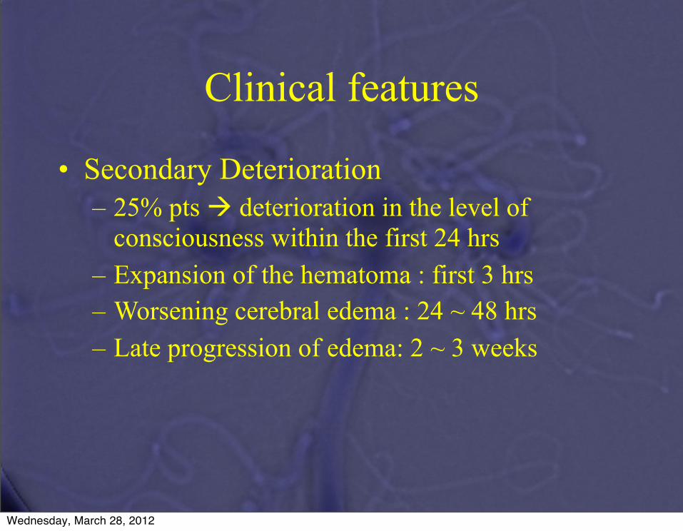
• Secondary Deterioration– 25% pts deterioration in the level of
consciousness within the first 24 hrs– Expansion of the hematoma : first 3 hrs– Worsening cerebral edema : 24 ~ 48 hrs– Late progression of edema: 2 ~ 3 weeks
Wednesday, March 28, 2012
Clinical features
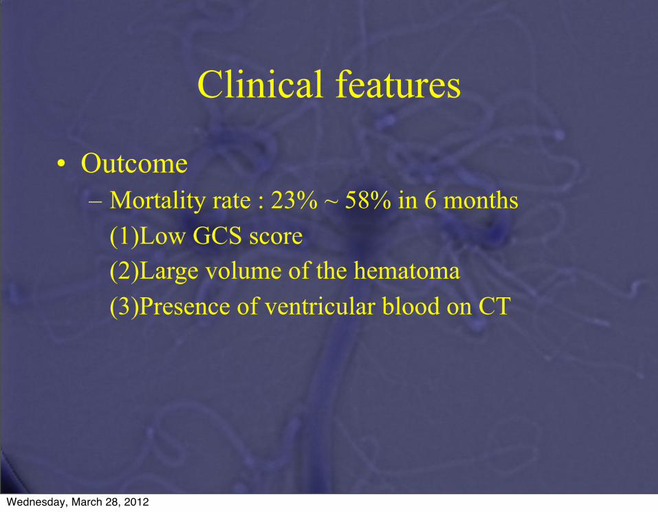
• Outcome– Mortality rate : 23% ~ 58% in 6 months (1)Low GCS score (2)Large volume of the hematoma (3)Presence of ventricular blood on CT
Wednesday, March 28, 2012
Clinical features
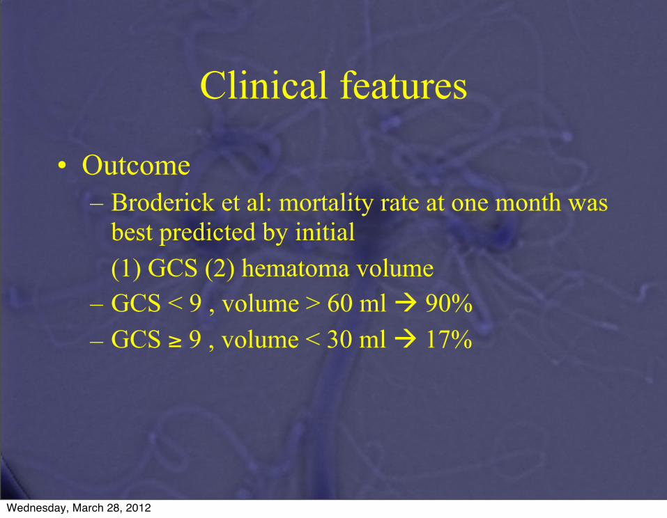
• Outcome– Broderick et al: mortality rate at one month was
best predicted by initial (1) GCS (2) hematoma volume– GCS < 9 , volume > 60 ml 90%– GCS ≥ 9 , volume < 30 ml 17%
Wednesday, March 28, 2012
Diagnosis
• CT scan infarction or hemorrhage– Location and size of the hematoma– Presence of ventricular blood– Hydrocephalus
Wednesday, March 28, 2012
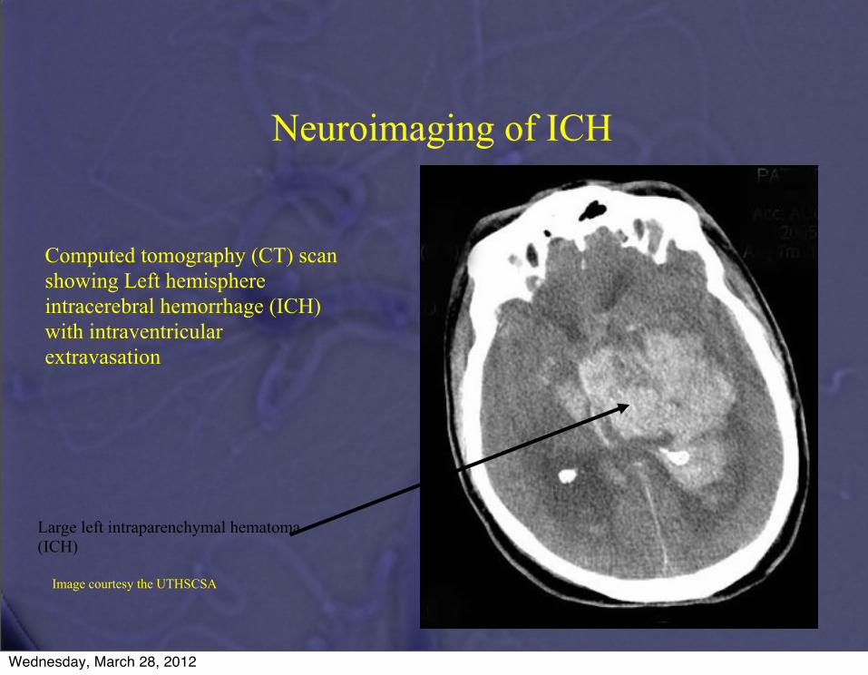
Large left intraparenchymal hematoma (ICH)
Computed tomography (CT) scan showing Left hemisphere intracerebral hemorrhage (ICH) with intraventricular extravasation
Neuroimaging of ICH
Image courtesy the UTHSCSA
Wednesday, March 28, 2012
and dural venous sinus thrombosis (DVST)–ie, secondary ICH(SICH).1-3 Timely and accurate identification of patients withSICH is important. These patients may benefit from promptsurgical or endovascular intervention once an underlying vascularabnormality has been identified, because rates of rehemorrhage,with increased morbidity and mortality, can be as high as 18% to50% per year.1,2,4-6 A recent literature review and multinationalphysician survey conducted at several leading European medicalinstitutions highlighted the need to develop a standardizedapproach to the diagnostic work-up of patients with nontraumaticICH in order to identify those patients with underlying vascularetiologies.7
The SICH score, derived from a retrospective cohort of623 patients with ICH and validated in a prospective cohort of222 patients with ICH examined with computed tomographicangiography (CTA) at the Massachusetts General Hospital, usesclinical and non-contrast CT (NCCT) characteristics at present-ation to predict the likelihood that a given patient with ICH harborsa vascular etiology for the ICH (Table 1).8,9 This scoring system wasdeveloped in a retrospective review of CTA data; however, if theSICH score is a robust scoring system, then it should be applicableto an independent patient population examined with a differentneurovascular imaging modality, such as catheter angiography.Although in recent years CTA has gained increasing acceptance as
a valuable examination in the initial evaluation of patients withICH for the detection of underlying vascular lesions, catheter an-giography and intraoperative findings are considered the goldstandard in the evaluation of this patient population.
OBJECTIVE
The objective of the present study was to validate the SICHscore in an independent data set using catheter angiography andintraoperative findings, rather than CTA, to establish the pres-ence or absence of an underlying vascular etiology for the ICH.
METHODS
Study Overview
Our study was a retrospective analysis of all consecutive adult patientswith ICH who were examined with either catheter angiography oremergent hematoma evacuation at our institution over a 5.4-year period.We assigned each patient with ICH in our cohort a SICH score based onreview of the admission NCCT and baseline clinical characteristics(Table 1). The performance of the SICH score as a predictor of anunderlying vascular etiology for the ICH in this independent populationwas analyzed using receiver operating characteristic (ROC) analysis. Wethen compared the performance of the SICH score in the presentpopulation examined with catheter angiography and intraoperativefindings with the original population examined with CTA at theMassachusetts General Hospital. Finally, we determined the perfor-mance of the SICH score in the pooled patient population examined atboth institutions using ROC analysis.
Patient Selection
Our study was approved by our hospital’s institutional review boardand conducted in compliance with the Health Insurance Portability andAccountability Act. We conducted a retrospective review of all adultpatients who presented to our institution from January 1, 2005, throughJune 1, 2010, with nontraumatic ICH on a NCCT examination of thehead and were evaluated with either catheter angiography or emergenthematoma evacuation with intraoperative inspection of the hematomacavity.Patient exclusion criteria were (1) the presence of associated sub-
arachnoid hemorrhage (SAH) in the basal cisterns; (2) evidence of recentischemic infarction, such as loss of gray-white matter differentiation ina vascular territory, suggesting hemorrhagic transformation as the causeof the ICH; (3) a known intracranial vascular abnormality or mass lesion;or (4) probable cerebral amyloid angiopathy according to the Bostoncriteria.10
We assigned each patient in our independent cohort a SICH scorebased on review of the admission NCCT and baseline clinical charac-teristics (Table 1).
Image Acquisition
NCCT acquisitions were performed according to standard protocolson 16-, 40-, or 64-slice helical CT scanners (Somaton Sensation, Sie-mens AG, Munich, Germany). The NCCT examination was performedusing axial technique with 120 kVp, 400 mAs, 10-second scan time, and4.8-mm collimation. Catheter angiography was performed using a ded-icated biplanar neuroangiographic unit (Axiom Artis, Siemens AG,
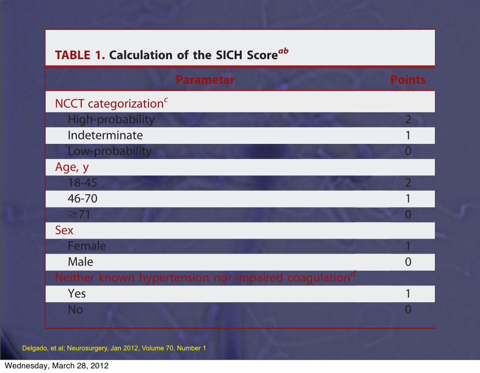
TABLE 1. Calculation of the SICH Scoreab
Parameter Points
NCCT categorizationc
High-probability 2Indeterminate 1Low-probability 0
Age, y18-45 246-70 1$71 0
SexFemale 1Male 0
Neither known hypertension nor impaired coagulationd
Yes 1No 0
aaPTT, activated partial thromboplastin time; ICH, intracerebral hemorrhage; INR,international normalized ratio; NCCT, non-contrast computed tomography; SICH,secondary intracerebral hemorrhage.bThe SICH score is calculated by adding the total number of points for a givenpatient.cHigh-probability NCCT: an examination with either (1) enlarged vessels or calci-fications along the margins of the ICH, or (2) hyperdensity within a dural venoussinus or cortical vein along the presumed venous drainage path of the ICH. Low-probability NCCT: an examination in which neither (1) nor (2) is present and the ICHis located in the basal ganglia, thalamus, or brainstem. Indeterminate NCCT: anexamination that does not meet criteria for either high- or low-probability NCCT.dImpaired coagulation is defined as admission INR .3, aPTT .80 seconds, plateletcount per microliter of blood ,50 000, or daily antiplatelet therapy.
DELGADO ALMANDOZ ET AL
132 | VOLUME 70 | NUMBER 1 | JANUARY 2012 www.neurosurgery-online.com
Copyright © Congress of Neurological Surgeons. Unauthorized reproduction of this article is prohibited.
Delgado, et al; Neurosurgery, Jan 2012, Volume 70, Number 1
Wednesday, March 28, 2012
performed selectively and primarily with catheter angiography,the SICH score could be used as a guide to select patients forneurovascular evaluation. For example: (1) patients with a SICHscore of 0 would not merit neurovascular evaluation; (2) patientswith SICH scores 1 and 2 could be initially screened with
a noninvasive technique such as CTA or MR angiography andonly undergo evaluation with catheter angiography if the initialtest is either positive or equivocal; and (3) patients with SICHscores of 3 or greater should be evaluated directly with catheterangiography. Conversely, at institutions where neurovascular
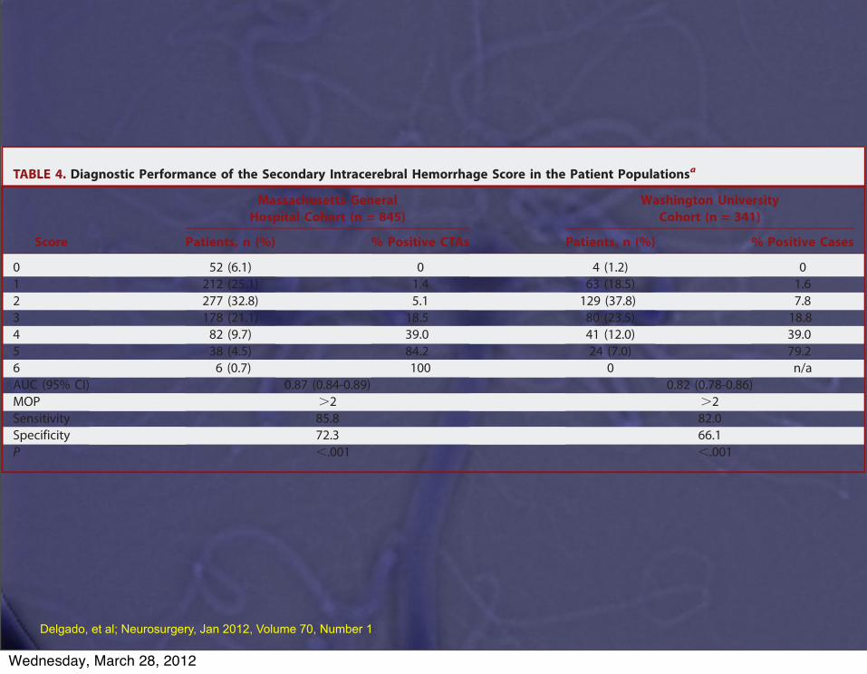
TABLE 4. Diagnostic Performance of the Secondary Intracerebral Hemorrhage Score in the Patient Populationsa
Massachusetts GeneralHospital Cohort (n = 845)
Washington UniversityCohort (n = 341)
Score Patients, n (%) % Positive CTAs Patients, n (%) % Positive Cases
0 52 (6.1) 0 4 (1.2) 01 212 (25.1) 1.4 63 (18.5) 1.62 277 (32.8) 5.1 129 (37.8) 7.83 178 (21.1) 18.5 80 (23.5) 18.84 82 (9.7) 39.0 41 (12.0) 39.05 38 (4.5) 84.2 24 (7.0) 79.26 6 (0.7) 100 0 n/aAUC (95% CI) 0.87 (0.84-0.89) 0.82 (0.78-0.86)MOP .2 .2Sensitivity 85.8 82.0Specificity 72.3 66.1P ,.001 ,.001
aCTA, computed tomographic angiogram; n/a, not applicable; AUC, area under the curve; CI, confidence interval; MOP, maximum operating point.
FIGURE 1. A 54-year-old man with a history of hypertension and with intact coagulation presented with acute onset ofheadache, vomiting, and left-sided weakness. A, low-probability non-contrast CT (NCCT) study demonstrates an acute rightbasal ganglia hemorrhage with associated intraventricular hemorrhage (SICH score 1). B, frontal right common carotidangiogram demonstrates a 5-mm aneurysm arising from a lenticulostriate branch of the right middle cerebral artery (arrowhead).
VALIDATION OF THE SICH SCORE
NEUROSURGERY VOLUME 70 | NUMBER 1 | JANUARY 2012 | 135
Copyright © Congress of Neurological Surgeons. Unauthorized reproduction of this article is prohibited.
Delgado, et al; Neurosurgery, Jan 2012, Volume 70, Number 1
Wednesday, March 28, 2012
Diagnosis
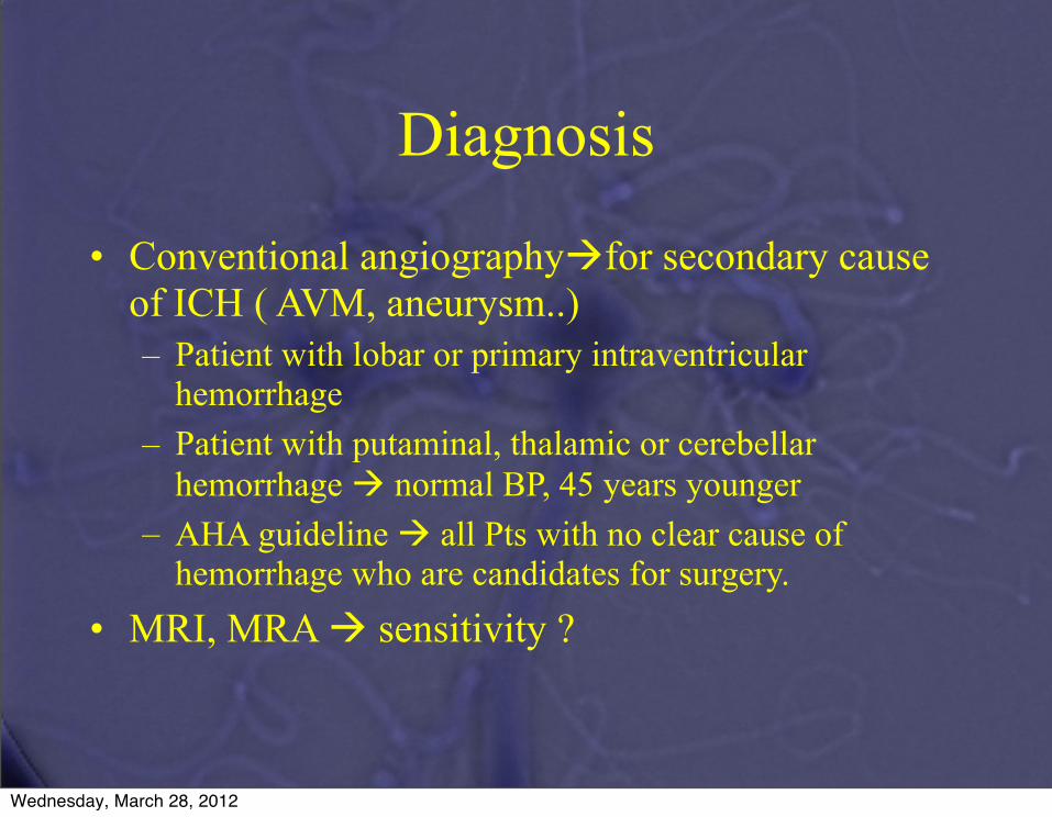
• Conventional angiographyfor secondary cause of ICH ( AVM, aneurysm..)– Patient with lobar or primary intraventricular
hemorrhage– Patient with putaminal, thalamic or cerebellar
hemorrhage normal BP, 45 years younger– AHA guideline all Pts with no clear cause of
hemorrhage who are candidates for surgery.• MRI, MRA sensitivity ?
Wednesday, March 28, 2012
Role of CTA & CTP in Acute StrokeACEP Scientific Assembly 2009
Andrew W. Asimos, MD, FACEP
15
CTP versus CTA Source Images for CTP versus CTA Source Images for Detecting Infarct CoreDetecting Infarct Core
Tan JC et al. Ann Neurol 2007; 61:533-543.
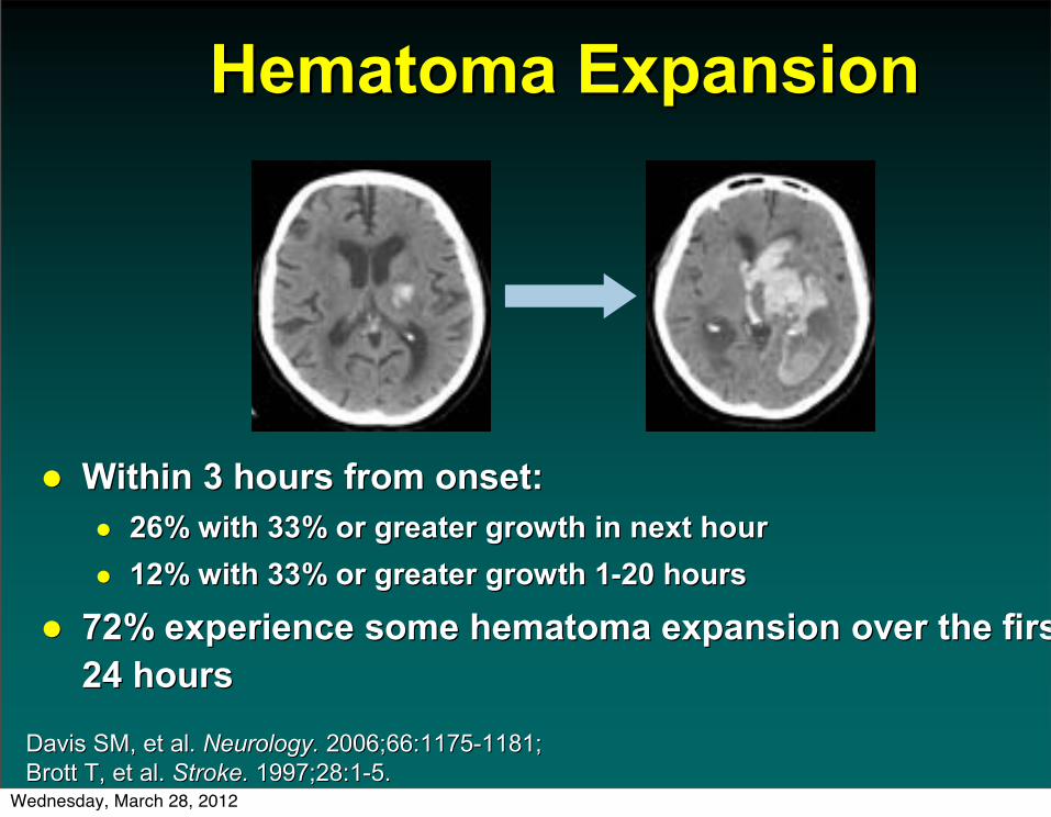
Hematoma ExpansionHematoma Expansion
Within 3 hours from onset:Within 3 hours from onset:26% with 33% or greater growth in next hour26% with 33% or greater growth in next hour12% with 33% or greater growth 112% with 33% or greater growth 1--20 hours20 hours
72% experience some hematoma expansion over the first 72% experience some hematoma expansion over the first 24 hours24 hours
Davis SM, et al. Davis SM, et al. Neurology.Neurology. 2006;66:11752006;66:1175--1181;1181;Brott T, et al.Brott T, et al. Stroke. Stroke. 1997;28:11997;28:1--5.5.
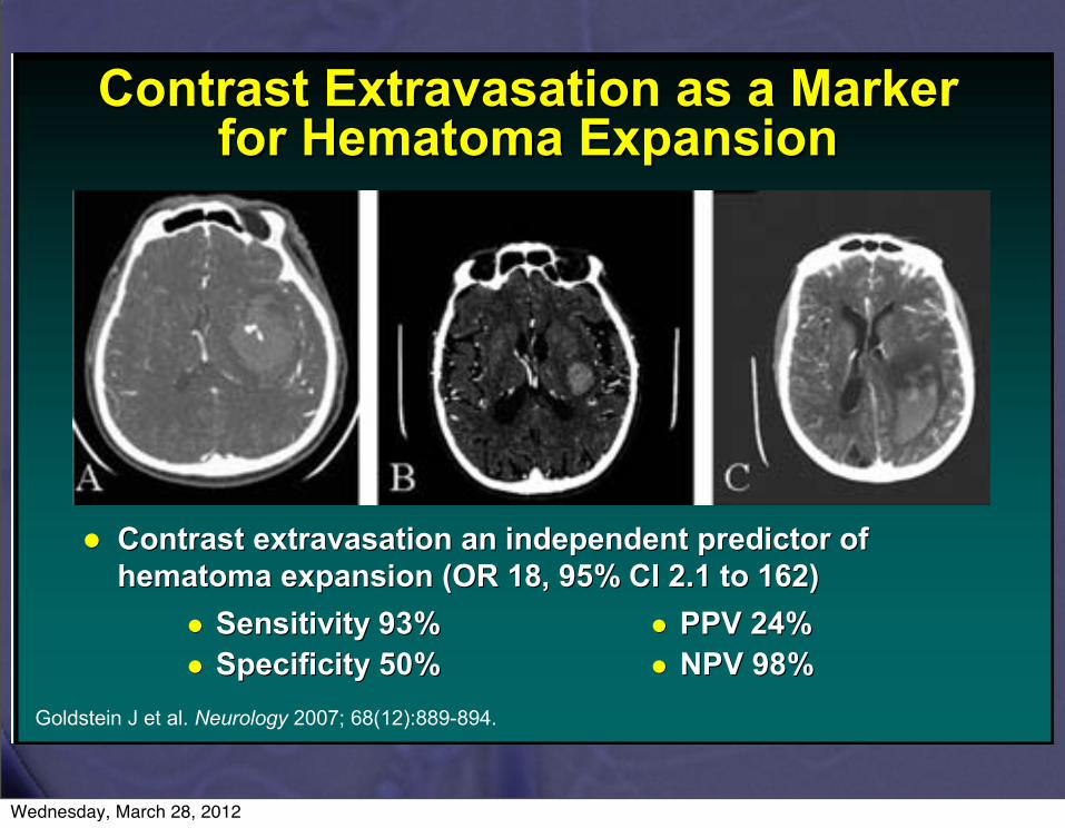
Contrast Contrast ExtravasationExtravasation as a Marker as a Marker for Hematoma Expansionfor Hematoma Expansion
Contrast Contrast extravasationextravasation an independent predictor of an independent predictor of hematoma expansion (OR 18, 95% CI 2.1 to 162)hematoma expansion (OR 18, 95% CI 2.1 to 162)
Goldstein J et al. Neurology 2007; 68(12):889-894.
Sensitivity 93%Sensitivity 93%Specificity 50%Specificity 50%
PPV 24%PPV 24%NPV 98%NPV 98%
Wednesday, March 28, 2012

CTA and ICH: • CT contrast
extravasates into hematoma– Spot sign, white
arrows• May predict
hematoma expansion
Delgado Almandoz et al. Stroke, 40 (9): 2994. 2009© 2010 American Heart Association, Inc. All rights reserved. Unauthorized use prohibited.
Wednesday, March 28, 2012
Role of CTA & CTP in Acute StrokeACEP Scientific Assembly 2009
Andrew W. Asimos, MD, FACEP
15
CTP versus CTA Source Images for CTP versus CTA Source Images for Detecting Infarct CoreDetecting Infarct Core
Tan JC et al. Ann Neurol 2007; 61:533-543.
Hematoma ExpansionHematoma Expansion
Within 3 hours from onset:Within 3 hours from onset:26% with 33% or greater growth in next hour26% with 33% or greater growth in next hour12% with 33% or greater growth 112% with 33% or greater growth 1--20 hours20 hours
72% experience some hematoma expansion over the first 72% experience some hematoma expansion over the first 24 hours24 hours
Davis SM, et al. Davis SM, et al. Neurology.Neurology. 2006;66:11752006;66:1175--1181;1181;Brott T, et al.Brott T, et al. Stroke. Stroke. 1997;28:11997;28:1--5.5.
Contrast Contrast ExtravasationExtravasation as a Marker as a Marker for Hematoma Expansionfor Hematoma Expansion
Contrast Contrast extravasationextravasation an independent predictor of an independent predictor of hematoma expansion (OR 18, 95% CI 2.1 to 162)hematoma expansion (OR 18, 95% CI 2.1 to 162)
Goldstein J et al. Neurology 2007; 68(12):889-894.
Sensitivity 93%Sensitivity 93%Specificity 50%Specificity 50%
PPV 24%PPV 24%NPV 98%NPV 98%
Wednesday, March 28, 2012
Role of CTA & CTP in Acute StrokeACEP Scientific Assembly 2009
Andrew W. Asimos, MD, FACEP
16
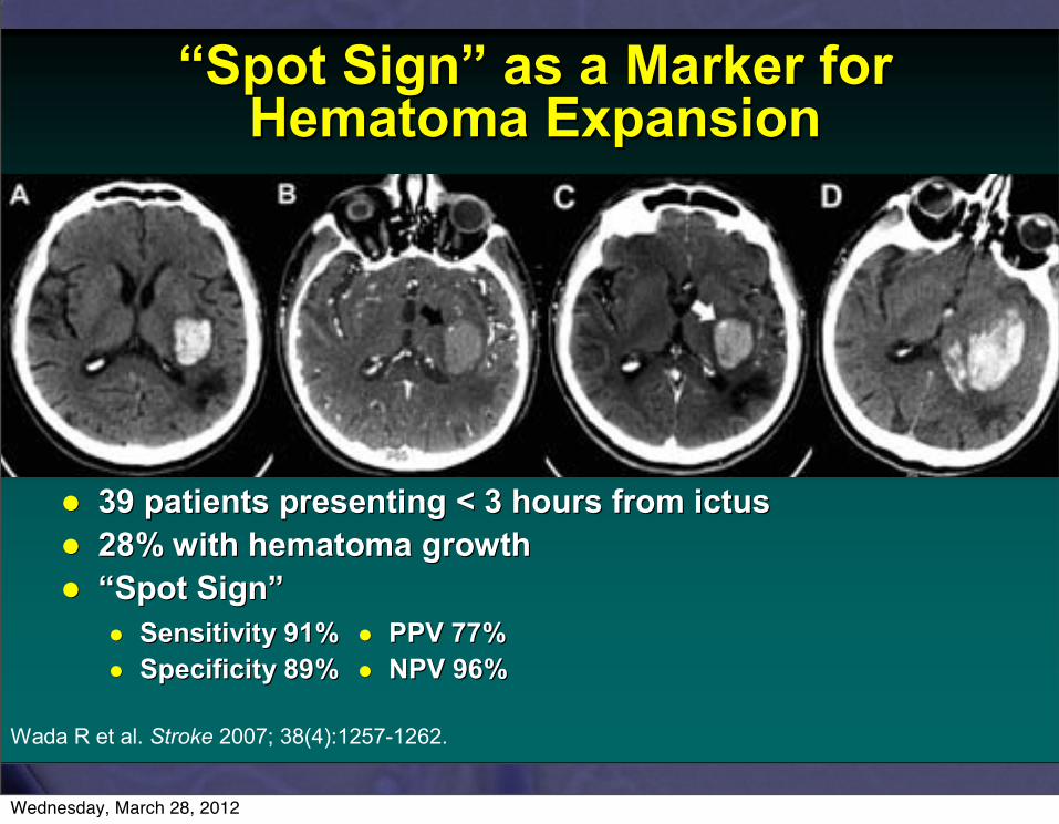
““Spot SignSpot Sign”” as a Marker for as a Marker for Hematoma ExpansionHematoma Expansion
39 patients presenting < 3 hours from ictus39 patients presenting < 3 hours from ictus28% with hematoma growth28% with hematoma growth““Spot SignSpot Sign””
Wada R et al. Stroke 2007; 38(4):1257-1262.
Sensitivity 91%Sensitivity 91%Specificity 89%Specificity 89%
PPV 77%PPV 77%NPV 96%NPV 96%
The The SSPOPOTT Sign fSign foor r PPredicting and Treating redicting and Treating IICH GrowCH Growtth Study (STOPh Study (STOP--IT Study)IT Study)
Randomize ICH patients within 6 hours of onset Randomize ICH patients within 6 hours of onset with a spot sign to treatment with with a spot sign to treatment with rFVIIarFVIIa vsvsplaceboplaceboOutcome measuresOutcome measures
Sensitivity and specificity of the spot sign for Sensitivity and specificity of the spot sign for predicting hematoma growthpredicting hematoma growthAccuracy of site investigators for correct identification Accuracy of site investigators for correct identification of the spot sign as compared to a blinded study of the spot sign as compared to a blinded study neuroradiologistneuroradiologistRate of hematoma growth among spotRate of hematoma growth among spot--positive positive subjects at 24 hourssubjects at 24 hours
rFVIIarFVIIa versus placeboversus placeboMortality at 90 daysMortality at 90 daysGood (Good (mRSmRS 00--3) versus poor (3) versus poor (mRSmRS 44--6) functional 6) functional outcomeoutcome
Should CTA replace CT/LP for Should CTA replace CT/LP for Evaluation of SAH?Evaluation of SAH?
116 pts at tertiary center studied116 pts at tertiary center studiedPts evaluated with concern for SAHPts evaluated with concern for SAH
““Worst headache of lifeWorst headache of life””Thunderclap headacheThunderclap headacheSudden onset headache with AMS, LOC, or Sudden onset headache with AMS, LOC, or neurologic deficitsneurologic deficits
Carstairs et al. Acad Emerg Med 2006;3(5):486-92.
Wednesday, March 28, 2012
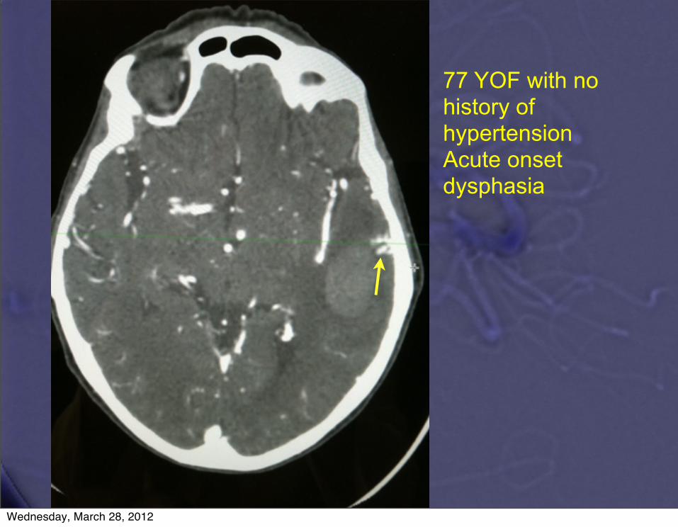
77 YOF with no history of hypertensionAcute onset dysphasia
Wednesday, March 28, 2012
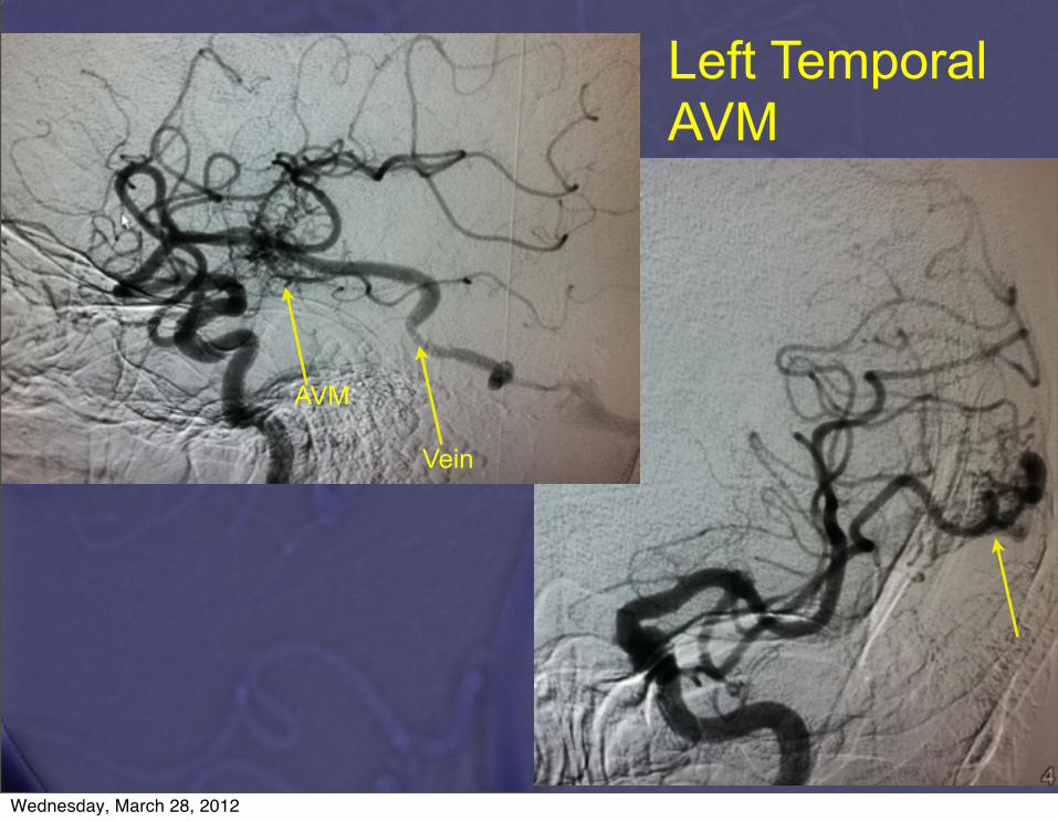
Left Temporal AVM
Vein
AVM
Wednesday, March 28, 2012
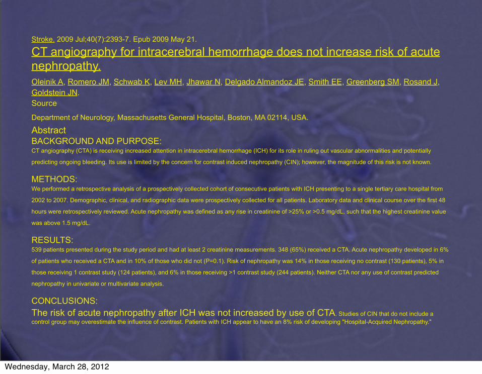
Stroke. 2009 Jul;40(7):2393-7. Epub 2009 May 21.
CT angiography for intracerebral hemorrhage does not increase risk of acute nephropathy.Oleinik A, Romero JM, Schwab K, Lev MH, Jhawar N, Delgado Almandoz JE, Smith EE, Greenberg SM, Rosand J, Goldstein JN.Source
Department of Neurology, Massachusetts General Hospital, Boston, MA 02114, USA.
AbstractBACKGROUND AND PURPOSE:CT angiography (CTA) is receiving increased attention in intracerebral hemorrhage (ICH) for its role in ruling out vascular abnormalities and potentially
predicting ongoing bleeding. Its use is limited by the concern for contrast induced nephropathy (CIN); however, the magnitude of this risk is not known.
METHODS:We performed a retrospective analysis of a prospectively collected cohort of consecutive patients with ICH presenting to a single tertiary care hospital from
2002 to 2007. Demographic, clinical, and radiographic data were prospectively collected for all patients. Laboratory data and clinical course over the first 48
hours were retrospectively reviewed. Acute nephropathy was defined as any rise in creatinine of >25% or >0.5 mg/dL, such that the highest creatinine value
was above 1.5 mg/dL.
RESULTS:539 patients presented during the study period and had at least 2 creatinine measurements. 348 (65%) received a CTA. Acute nephropathy developed in 6%
of patients who received a CTA and in 10% of those who did not (P=0.1). Risk of nephropathy was 14% in those receiving no contrast (130 patients), 5% in
those receiving 1 contrast study (124 patients), and 6% in those receiving >1 contrast study (244 patients). Neither CTA nor any use of contrast predicted
nephropathy in univariate or multivariate analysis.
CONCLUSIONS:The risk of acute nephropathy after ICH was not increased by use of CTA. Studies of CIN that do not include a control group may overestimate the influence of contrast. Patients with ICH appear to have an 8% risk of developing "Hospital-Acquired Nephropathy."
Wednesday, March 28, 2012
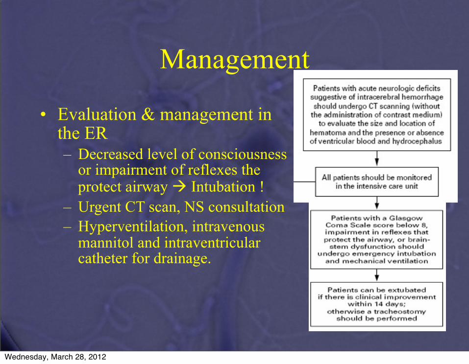
Management
• Evaluation & management in the ER– Decreased level of consciousness
or impairment of reflexes the protect airway Intubation !
– Urgent CT scan, NS consultation– Hyperventilation, intravenous
mannitol and intraventricular catheter for drainage.
Wednesday, March 28, 2012
Need to control BP and coagulation issues ASAP!
Wednesday, March 28, 2012
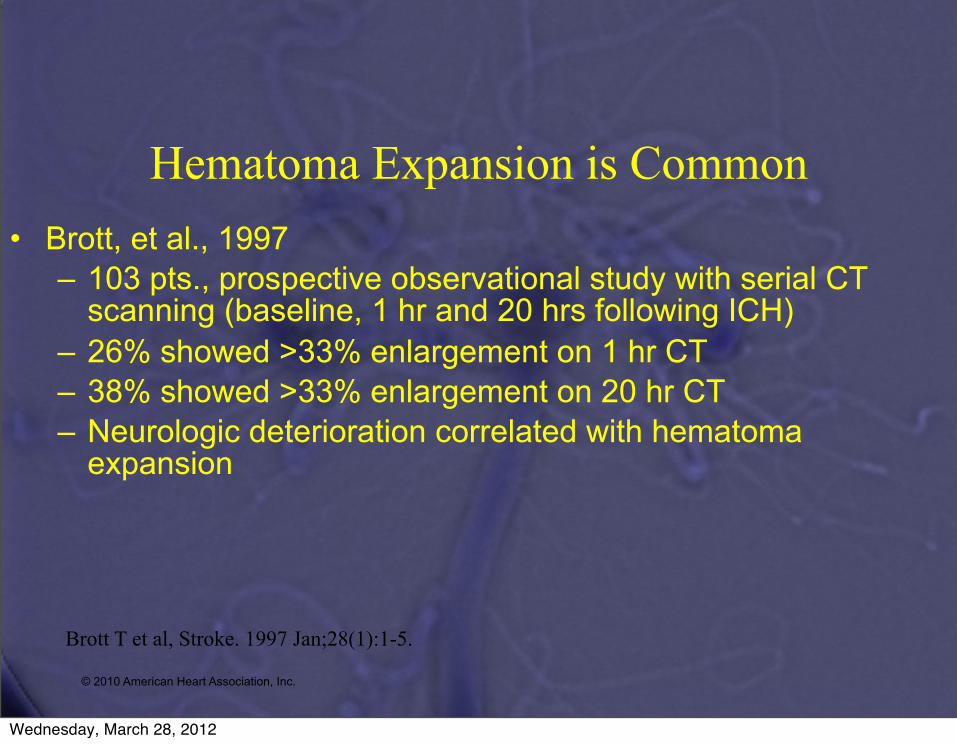
Hematoma Expansion is Common• Brott, et al., 1997
– 103 pts., prospective observational study with serial CT scanning (baseline, 1 hr and 20 hrs following ICH)
– 26% showed >33% enlargement on 1 hr CT– 38% showed >33% enlargement on 20 hr CT– Neurologic deterioration correlated with hematoma
expansion
Brott T et al, Stroke. 1997 Jan;28(1):1-5.
© 2010 American Heart Association, Inc.
Wednesday, March 28, 2012
© 2010 American Heart Association, Inc. All rights reserved. Unauthorized use prohibited.
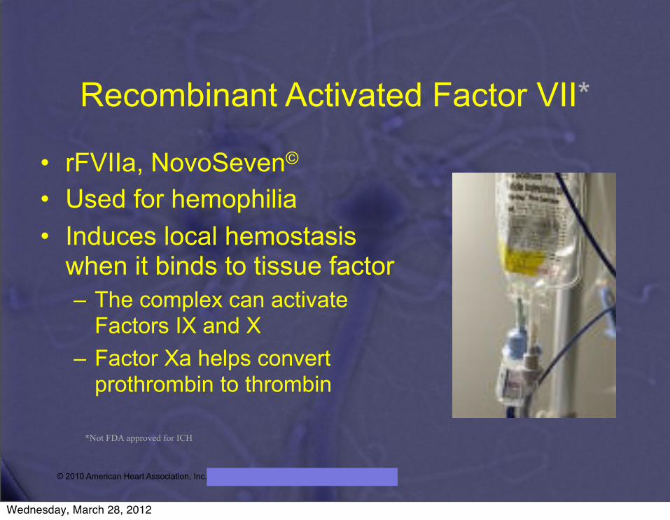
Recombinant Activated Factor VII*
• rFVIIa, NovoSeven©
• Used for hemophilia• Induces local hemostasis
when it binds to tissue factor– The complex can activate
Factors IX and X– Factor Xa helps convert
prothrombin to thrombin
*Not FDA approved for ICH
Wednesday, March 28, 2012
© 2010 American Heart Association, Inc. All rights reserved. Unauthorized use prohibited.
“FAST” Trials• A phase II randomized trial showed that treatment with rFVIIa
within four hours after ICH onset – limited hematoma growth – improved clinical outcomes relative to placebo – increased frequency of thromboembolic events (7% vs. 2%)
• A subsequent phase III study comparing placebo to 20 µg/kg and 80 µg/kg of rFVIIa:
– both doses diminished hematoma enlargement– failed to show differences in clinical outcome– Overall serious thromboembolic adverse event rates were similar, the higher
rFVIIa (80 µg/kg) group had significantly more arterial events than placebo.
• The authors noted imbalances in treatment groups, particularly Mayer SA, et al for the FAST Trial Investigators., N Engl J Med. 2008 May 15;358(20):2127-37.Mayer SA for the FAST Trial Investigators. N Engl J Med. 2005 Feb 24;352(8):777-85.
Wednesday, March 28, 2012
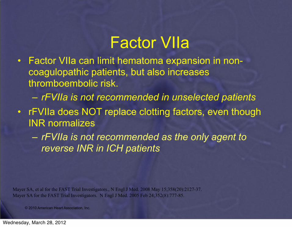
Factor VIIa• Factor VIIa can limit hematoma expansion in non-
coagulopathic patients, but also increases thromboembolic risk.– rFVIIa is not recommended in unselected patients
• rFVIIa does NOT replace clotting factors, even though INR normalizes– rFVIIa is not recommended as the only agent to
reverse INR in ICH patients
Mayer SA, et al for the FAST Trial Investigators., N Engl J Med. 2008 May 15;358(20):2127-37.Mayer SA for the FAST Trial Investigators. N Engl J Med. 2005 Feb 24;352(8):777-85.
© 2010 American Heart Association, Inc.
Wednesday, March 28, 2012
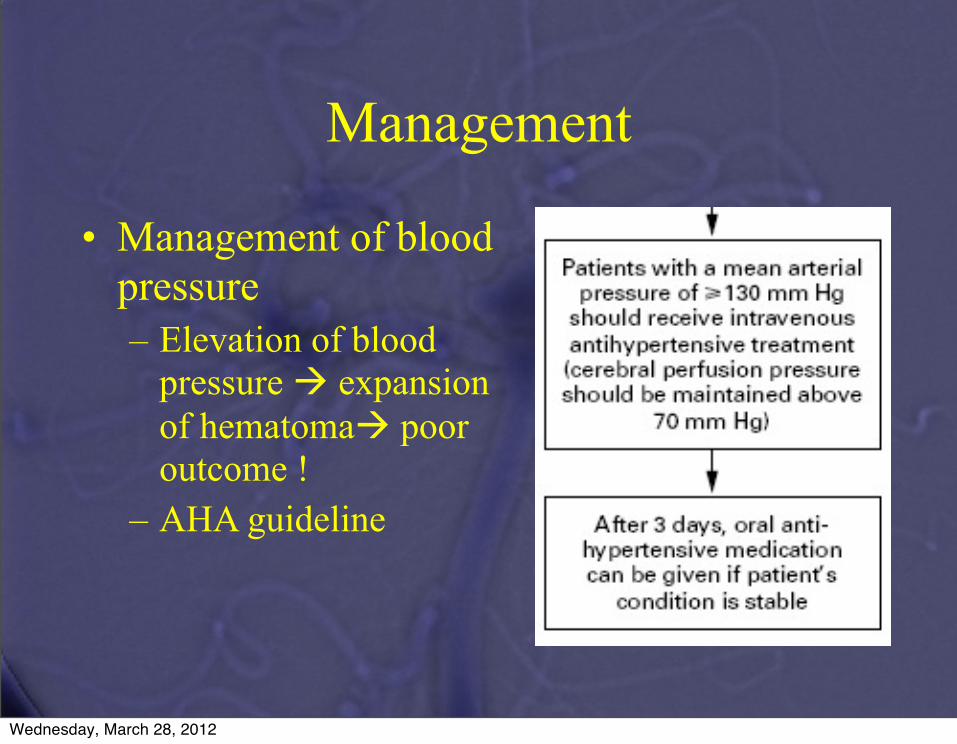
Management
• Management of blood pressure– Elevation of blood
pressure expansion of hematoma poor outcome !
– AHA guideline
Wednesday, March 28, 2012
ICH: Secondary Effects
• Profound transitory elevation of BP– Secondary to increased ICP
• Cerebral autoregulation may be lost• Goal is controlled reduction of hypertension
– Low BP risks ischemia– High BP risks increased hemorrhage– Nitroprusside or nitroglycerin not first-line agents
• Increase ICP• Impair cerebrovascular reactivity to changes in carbon dioxide partial pressure
(PCO2 ), critically problematic with elevated ICP• Exacerbate a decrease in CPP
– Nicardipine Minimal ICP effect Rapid control of BP
Wednesday, March 28, 2012
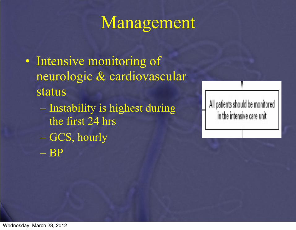
Management
• Intensive monitoring of neurologic & cardiovascular status– Instability is highest during
the first 24 hrs– GCS, hourly– BP
Wednesday, March 28, 2012
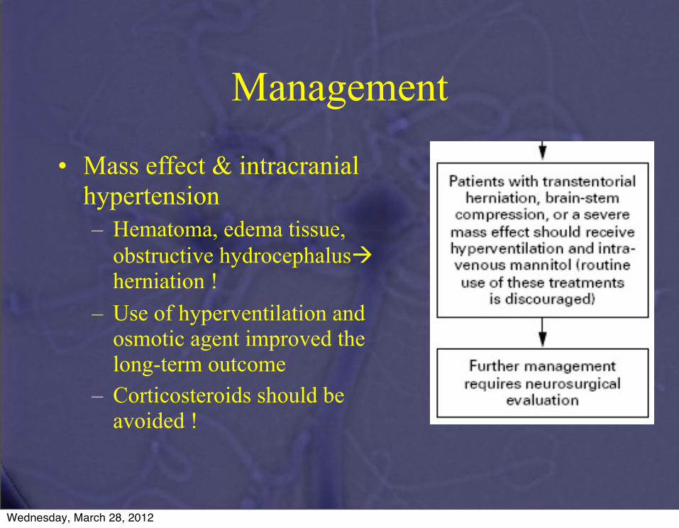
• Mass effect & intracranial hypertension– Hematoma, edema tissue,
obstructive hydrocephalus herniation !
– Use of hyperventilation and osmotic agent improved the long-term outcome
– Corticosteroids should be avoided !
Management
Wednesday, March 28, 2012

Management
• Ventricular blood and hydrocephalus– Blood in ventricles
obstructive hydrocephalus high mortality rate !
– External drainage– Clots in the catheter and
infection
Wednesday, March 28, 2012
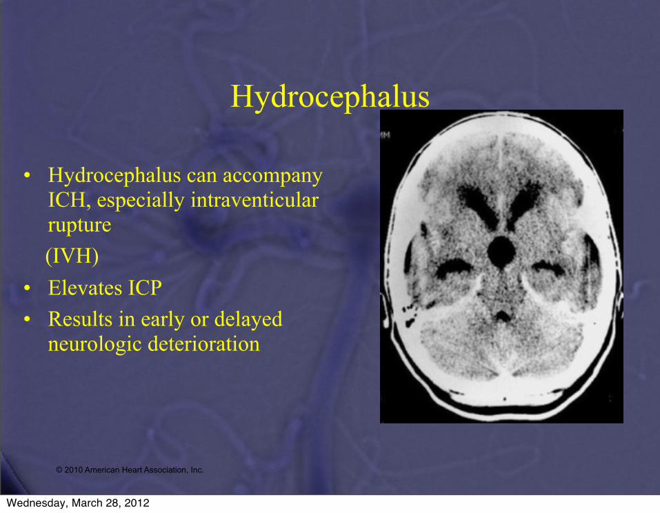
Hydrocephalus
• Hydrocephalus can accompany ICH, especially intraventicular rupture
(IVH)• Elevates ICP• Results in early or delayed
neurologic deterioration
© 2010 American Heart Association, Inc.
Wednesday, March 28, 2012
CLEAR-IVH Trial• 52 pts with IVH• Open-label intra-ventricular rt-PA to accelerate
blood clearance and lysis• Adverse events
– Symptomatic bleeding 4%– Bacterial ventriculitis 2%– 30 day mortality 17%
• Efficacy requires confirmation before use of intraventricular fibrinolysis can be recommended, Phase III trial in progress.© 2010 American Heart Association, Inc..
Wednesday, March 28, 2012
Management
• Surgical evacuation– Reduce mass effect, block the release of neuropathic
product from the hematoma– Surgery for supratentorial hemorrhage ?
• Hankey et al: 126 not undergo surgery 123 surgical evaculation through an open
craniotomy surgery higher rate of death (83% vs 70%) / 6m
Wednesday, March 28, 2012
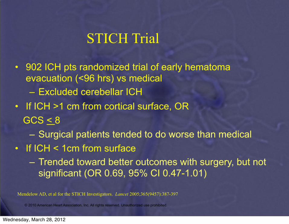
STICH Trial
• 902 ICH pts randomized trial of early hematoma evacuation (<96 hrs) vs medical – Excluded cerebellar ICH
• If ICH >1 cm from cortical surface, OR GCS < 8
– Surgical patients tended to do worse than medical• If ICH < 1cm from surface
– Trended toward better outcomes with surgery, but not significant (OR 0.69, 95% CI 0.47-1.01)
Mendelow AD, et al for the STICH Investigators. Lancet 2005;365(9457):387-397
© 2010 American Heart Association, Inc. All rights reserved. Unauthorized use prohibited.
Wednesday, March 28, 2012
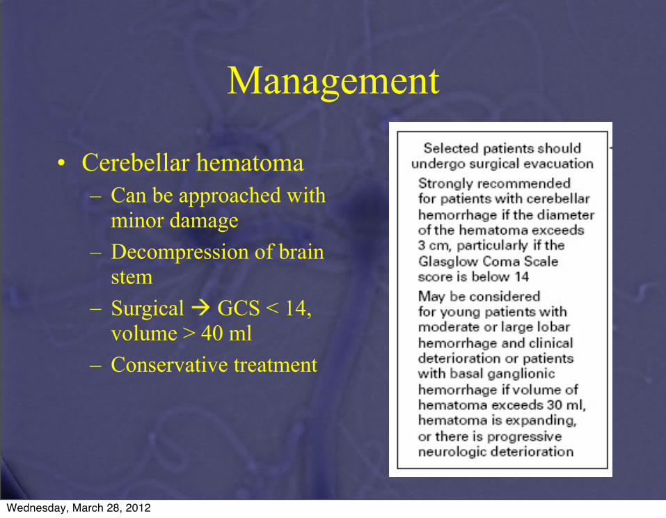
Management
• Cerebellar hematoma– Can be approached with
minor damage– Decompression of brain
stem– Surgical GCS < 14,
volume > 40 ml– Conservative treatment
Wednesday, March 28, 2012
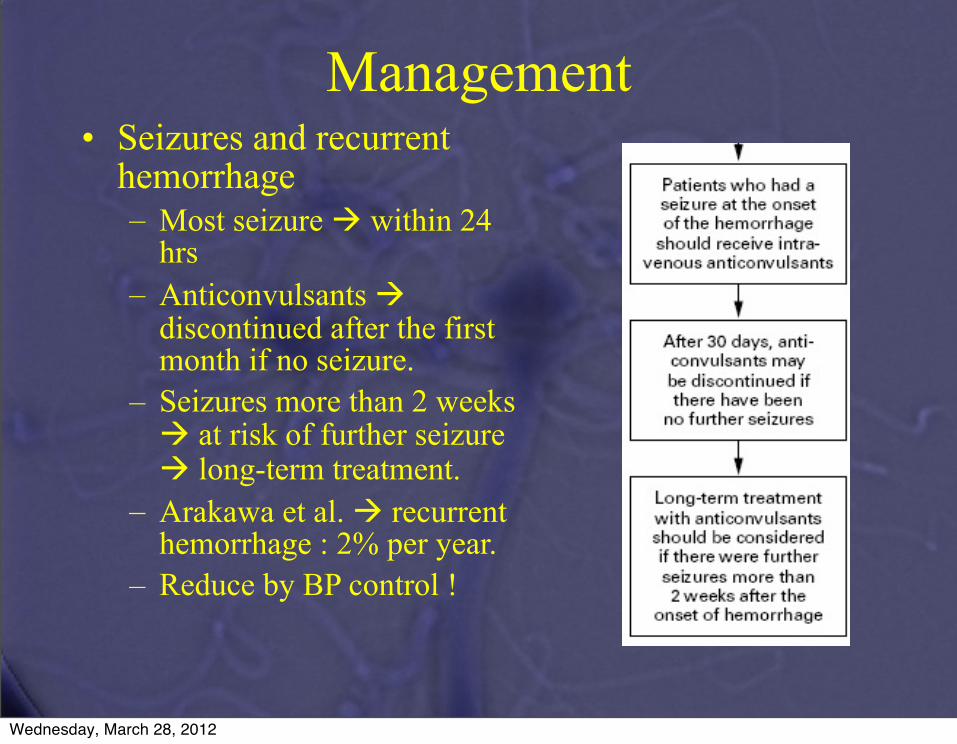
Management• Seizures and recurrent
hemorrhage– Most seizure within 24
hrs– Anticonvulsants
discontinued after the first month if no seizure.
– Seizures more than 2 weeks at risk of further seizure long-term treatment.
– Arakawa et al. recurrent hemorrhage : 2% per year.
– Reduce by BP control !
Wednesday, March 28, 2012
Conclusions:ICH
• Get a CTA!• Catheter angiography for CTA negative/ but
suspicious• Aggressive BP control/ICP management• Possible factor VIIa for pts with CTA spot
sign• Surgery only in select patients: <1cm from
cortex, cerebellar hematoma, large clot
Wednesday, March 28, 2012