Bleeding and coagulopathy
46
BLEEDING AND COAGULOPATHIES IN CRITICAL CARE Dr. Siba Prasad Dalai
-
Upload
buntyrocks -
Category
Health & Medicine
-
view
168 -
download
3
Transcript of Bleeding and coagulopathy

BLEEDING AND COAGULOPATHIES IN
CRITICAL CARE
Dr. Siba Prasad Dalai

WHAT IS COAGULOPATHY
• Coagulopathy (also called clotting and bleeding disorder) is a condition in which the blood's ability to clot (coagulate) is impaired.
• The term also covers thrombotic states, and because of the complexity of the hemostatic pathways these two conditions can exist simultaneously.
• Such states are common in patients in the intensive care unit (ICU) and require a clinico-pathological approach to ensure that the correct diagnosis is made and the appropriate treatment administered.

Mechanisms involved
• Vascular Integrity• Platelets• Clotting factors• Fibrinolysis
Derangement of any of these factors can cause abnormal bleeding and or coagulopathy
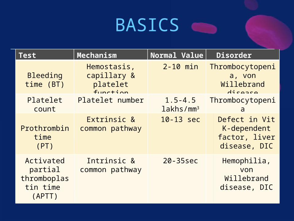
BASICS
Test Mechanism Tested
Normal Value Disorder
Bleeding time (BT)
Hemostasis, capillary &
platelet function
2-10 min Thrombocytopenia, von Willebrand
disease
Platelet count Platelet number 1.5-4.5 lakhs/mm3
Thrombocytopenia
Prothrombin time (PT)
Extrinsic & common pathway
10-13 sec Defect in Vit K-dependent factor, liver disease, DIC
Activated partial
thromboplastin time (APTT)
Intrinsic & common pathway
20-35sec Hemophilia, von Willebrand
disease, DIC

Key to diagnosis• A medical history taking and physical
examination are vital, since many different conditions can produce similar laboratory abnormalities.
• For example, end-stage liver failure and DIC produce thrombocytopenia and similar changes in standard tests of coagulation, and yet the management of and prognosis for these conditions are very different.
• A peripheral-blood smear is a vital investigation tool in most cases to confirm a low platelet count and the presence or absence of other diagnostic features, such as red-cell fragmentation, platelet morphologic abnormalities, or evidence of dysplasia or hematinic deficiency


Lab findings of common disorders

Management of coagulopathies• The first principle of the management of
coagulopathies in critical care is to avoid the correction of laboratory abnormalities with blood products unless there is a clinical bleeding problem, a surgical procedure is required, or both.

Major bleeding• In one study involving trauma patients, the
administration of more than 6 units of fresh-frozen plasma, as compared with no transfusion, was associated with an increase by a factor of 12 in the rate of ARDS and an increase by a factor of 6 in the multiple organ dysfunction syndrome *
* J Am Coll Surg 2010;210:957-65
• European practitioners have abandoned the use of fresh-frozen plasma, relying on the exclusive use of factor concentrates on the basis of rotational-elastometry (ROTEM) guided intervention with prothrombin complex concentrate, factor XIII, fibrinogen, tranexamic acid with red cells and IV fluids on an as-needed basis.

Major bleeding (contd..)
• Fibrinogen is the protein that ultimately forms fibrin, the ligand for platelet aggregation, and in patients with major bleeding, it is required to a larger extent than any other hemostatic protein.
• Guidelines for the management of traumatic bleeding now indicate that the trigger level for supplementing fibrinogen should be 1.5 to 2.0 g per liter rather than 1.0 g per liter. ( = EARLY USE )
• The use of recombinant factor VIIa, which has been shown to reduce the use of red cells in bleeding but not to reduce mortality, needs further evaluation.

Major bleeding (contd..)• Tranexamic acid should be administered to all
patients with major bleeding after trauma. This recommendation is supported by a large, RCT, the Clinical Randomization of an Antifibrinolytic in Significant Hemorrhage (CRASH-2) study. Patients who received tranexamic acid within 3 hours after injury had a one-third reduction in deaths from bleeding.
• Tranexamic acid should be administered as soon as possible after injury, since the drug ceased to confer benefit and appeared to be associated with increased mortality if it was administered more than 3 hours after injury.

Hemostatic support for invasive procedures in ICU
• There is no supportive evidence for the prophylactic use of fresh-frozen plasma to correct abnormal results on coagulation screening (for prothrombin time, activated partial-thromboplastin time, and fibrinogen) before an invasive procedure.
• Coagulation screening has no predictive value for later bleeding, and the use of fresh-frozen plasma may not correct abnormal results on coagulation tests.
• Prothrombin ratio of 1.5 or less is satisfactory for the insertion of a central venous or an arterial catheter in patients in whom direct compression can assist with hemostasis without the need for prophylactic supplementation with fresh-frozen plasma.

Hemostatic support for invasive procedures in ICU (contd..)
• As a general rule, dietary intake of vitamin K, which is necessary for the formation of coagulation factors II, VII, IX, and X, may be inadequate in critical care settings.
• Therefore supplementation of vitamin K1 (at a dose of at least 1 mg orally daily or 10 mg intravenously weekly) for critical care patients at risk is advisable.

DIC• defined by the International Society on
Thrombosis and Hemostasis (ISTH) as “an acquired syndrome characterized by the intravascular activation of coagulation with loss of localization arising from different causes.”
• typically originates in the microvasculature and can lead to MODS
• scoring system developed by the ISTH• usually presents as hemorrhage, with only 5 to
10% of cases presenting with microthrombi• Presentation depends on its cause and host
defenses.• Sepsis is the most common cause of
disseminated intravascular coagulation in critical care

DIC (contd..)

DIC (contd..)

DIC (contd..)

DIC (contd..)• cornerstone for managing this condition remains
the management of the underlying cause (e.g., sepsis)
• Replacement of coagulation proteins and platelets in patients who are bleeding.
• Platelet transfusion is indicated to maintain a platelet level of more than 50,000 per cubic millimeter
• administration ofa) fresh-frozen plasma to maintain a prothrombin
time and activated partial-thromboplastin time of less than 1.5 times the normal control
b) fibrinogen to maintain a fibrinogen level of more than 1.5 g per liter

DIC (contd..)
• The use of antifibrinolytic agents is contraindicated in the management of DIC, since the fibrinolytic system is required in recovery to ensure the dissolution of the widespread fibrin.
• Some guidelines recommend the administration of therapeutic doses of unfractionated heparin in patients with a thrombotic phenotype (e.g., gangrene), but such recommendations remain controversial because of the difficulties in monitoring treatment in a patient who already has a prolonged APTT.

Platelet Disorders
Thrombocytopenia Platelet dysfunction
PRIMARY
ITPTTPTAR syndromeWiskott-Aldrichsyndrome
SECONDARY
Malignancy
Aplastic anemia
DIC
Sepsis
Drug-induced
Hemolytic-uremic syndrome
Hypersplenism
Autoimmune(SLE)
HIV
Glanzmann’s thromboasthenia
Bernard-Soulier syndrome

Aetiology of thrombocytopenia
Decreased production of
platelets
Increased platelets
destruction
Sequestration
Dilutional
Nonimmunologic destruction (HUS, TTP,DIC)
Immunologic destruction (ITP, SLE )
Acquired (aplastic anaemia, marrow infiltration,drug induced )
Congenital (Fanconi anaemia, Wiskot-Aldrich Syndrome
Hypersplenism
Massive blood transfusion


Thrombocytopenia • Patients with thrombocytopenia tend to be more
sick, with higher illness-severity scores, than those who are admitted with normal platelet counts.
• A platelet threshold of 10,000 per mm3 for platelet transfusion in patients who are in stable condition is both hemostatically efficacious and cost-effective in reducing platelet-transfusion requirements.
• If the patient is actively bleeding, then a platelet count of 50,000 per mm3 should be maintained.
• Patients who have or are at risk for bleeding in the central nervous system or who are undergoing neurosurgery, a platelet count of more than 100,000 per cubic millimeter is often recommended

HITT• Heparin-induced thrombocytopenic thrombosis is
an uncommon, transient, drug-induced, autoimmune prothrombotic disorder caused by the formation of IgG antibodies that cause platelet activation by the formation of antibodies to complexes of platelet factor 4 and heparin.
• Not all people with a falling platelet count while receiving heparin turn out to have HIT.
• A commonly used score to predict the likelihood of HIT is the "4 Ts" score introduced in 2003.
• A score of 0–8 points is generated; 0-3, HIT is unlikely. 4–5 indicates intermediate probability 6–8 makes it highly likely

The 4T score for heparin-induced thrombocytopenia
Thrombocytopenia
2 points if the fall in platelet count is >50% of the previous value, or the lowest count (nadir) is 20–100 × 109/liter1 point if the fall is 30–50% or the nadir is 10–19 × 109/literNo points if the fall is less than 30% or the nadir is <10 × 109/liter.
Timing
2 points if the fall is between days 5–10 after commencement of treatment1 point if the fall is after day 10. If someone has been exposed to heparin within the last 30 days and then has a drop in platelet count within a day of reexposure, 2 points are given. If the previous exposure was 30–100 days ago, 1 pointIf the fall is early but there has been no previous heparin exposure, no points.
Thrombosis
2 points in new proven thrombosis, skin necrosis , or systemic reaction1 point if progressive or recurrent thrombosis, silent thrombosis or red skin lesionsNo points if there are no symptoms.
Alternative cause possible2 points if no other cause1 point if there is a possible alternative causeNo points if there is a definite alternative cause.

HITT (contd..)• Type 1 HIT presents within the first 2 days after
exposure to heparin, and the platelet count normalizes with continued heparin therapy. Type 1 HIT is a nonimmune disorder that results from the direct effect of heparin on platelet activation
• Type 2 HIT is an immune-mediated disorder that typically occurs 4-10 days after exposure to heparin and has life- and limb-threatening thrombotic complications (more common in icu setting)
• suspected when a patient who is receiving heparin has a decrease in the platelet count (>= 50% of the baseline) irrespective of the final platelet count.
• HIT is generally not marked by bleeding. Instead venous thromboembolism is the most common complication.

HITT (contd..)• Diagnosis of HIT is based on the combination of
clinical findings, thrombocytopenia characteristics, and laboratory studies of HIT antibodies.
• discontinue the heparin administration.• Alternate anticoagulation with either danaparoid,lepirudin, or argatroban.• Warfarin should not beused in HIT until theplatelet count is at least150 x 10^9/L becausethere is a very high riskof warfarin necrosis.

Post transfusion purpura• rare bleeding disorder caused by a platelet-specific
alloantibody (usually, anti–human platelet antigen 1a [HPA-1a]) in the recipient. HPA-1a reacts with donor platelets, destroying them and also the recipient's own platelets.
• majority of affected patients are multiparous women who have been sensitized during pregnancy.• Treatments include IVIg, glucocorticoids, and plasmapheresis

Thrombotic microangiopathies• Profound thrombocytopenia and microangiopathic
hemolytic anemia (red-cell fragmentation) characterize the thrombotic microangiopathies, which includes three major disorders: TTP, HUS and the HELLP syndrome .

Thrombotic microangiopathies• The absence of ADAMTS13 results in the
persistence of high-molecular-weight von Willebrand factor, which can cause spontaneous platelet aggregation when the protein is subjected to high shear stress.
• The rate of death in untreated cases is nearly 95% (usually from myocardial infarction due to platelet thrombi in the coronary arteries), but with early plasmapheresis, the survival rate is 80 to 90%.
• The use of rituximab, anti CD20 has been shown to reduce the rate of recurrence of the autoimmune form of this disorder from 57% to 10%.
• Thus, an active diagnosis of this disorder or failure to rule it out should lead to urgent plasmapheresis.

Liver disease• reduced hepatic synthetic function results in
prolongation of the screening tests of coagulation (particularly the PT) and reduced platelet counts, although levels of factor VIII and von Willebrand factor are increased.
• Acute alcohol intake inhibits platelet aggregation.• In CLD, there is also increased fibrinolytic potential
due to the failure of the liver to metabolize tissue plasminogen activator.
• In cholestatic liver disease, there is reduced absorption of lipid-soluble vit., so reduced amounts of the vitamin K–dependent coagulation factors II, VII, IX, and X
• In CLD, the failure of the normal enzymatic removal of sialic acid from fibrinogen results in dysfibrinogenemia

Liver disease (contd…)

Liver disease (contd…)• in parallel with the reduction in coagulation
factors, there is a similar reduction in the production of physiologic anticoagulants.
• Thus, patients with chronic liver disease and a prolonged PT are no longer considered to have a deficiency of coagulation factors, since their coagulation is rebalanced and thrombin generation is usually normal.
• In such cases, there is no need to treat prolonged coagulation times in the absence of bleeding.
• If bleeding does occur in liver disease, then consensus guidelines recommend blood-component management as determined by the results of testing of the platelet count, PT, APTT, TT, and fibrinogen.

Liver disease (contd…)• RCT revealed that in patients with acute UGI bleeding
patients who were treated with the restrictive strategy (Hb <7g/dl) had longer survival (6 weeks) and a lower rate of rebleeding than did those who were treated with the liberal red-cell transfusion strategy ( Hb <9g/dl) . [ Due to increased portal circulation pressures in the liberal-strategy group ]
• Adopt a restrictive approach to the use of FFP and platelets in patients with acute UGI bleeding.
• Vitamin K should be routinely administered to aid in the synthesis of coagulation factors
• The role of tranexamic acid in patients with gastrointestinal bleeding is under investigation in the ongoing randomized, controlled Hemorrhage Alleviation with Tranexamic Acid–Intestinal System (HALT-IT) trial.

Renal disease• Uremic bleeding typically presents with ecchymoses,
purpura, epistaxis, and bleeding from puncture sites due to impaired platelet function.
• The platelet dysfunction is a result of complex changes that include dysfunctional von Willebrand factor, decreased production of thromboxane, increased levels of cyclic AMP and cyclic GMP, uremic toxins, anemia, and altered platelet granules, all of which are necessary for adequate formation of a platelet plug.
• The anemia that commonly accompanies renal disease leads to the loss of laminar flow in arterioles so that red cells no longer push platelets and plasma to the endothelium, leading to prolongation of the bleeding time; treatment of the anemia partially corrects this problem.

Renal disease (contd…)

Renal disease (contd…)• Bleeding time is considered to be the most useful
clinical test of coagulation in patients with renal disease.
• Peritoneal dialysis, improves platelet function. • Erythropoietin, cryoprecipitate, conjugated estrogens,
desmopressin, and tranexamic acid have all independently been shown to reduce bleeding time.
• In the past decade, citrate has risen in popularity as a replacement anticoagulant in continuous renal-replacement therapy, with a reduction in bleeding, although data on its safety in patients with liver failure are lacking.

Fibrinolytic bleeding• Abnormal fibrinolytic activity may be difficult to
diagnose because of the absence of a specific routine assay.
• Clinical suspicion should be high in cases in which bleeding continues despite hemostatic replacement therapy, platelet levels are relatively conserved but fibrinogen levels are disproportionately low, and D-dimer levels are disproportionately high for DIC.
• Thromboelastography, which may help differentiate fibrinolytic activation from coagulation factor deficiency, is a crude tool, since it detects only the most marked changes.
• Fibrinolytic bleeding should be considered particularly in patients with liver disease and disseminated cancers.
• The use of tranexamic acid, either by infusion or orally (depending on the severity of the problem and the state of the patient), is beneficial in controlling bleeding.

VWD• A personal and family history of easy bruising and
bleeding should be sought as it may present with persistent oozing after an injury or surgery.
• Acquired von Willebrand's disease, which can be caused by several potential mechanisms due to autoantibodies, or due to breakdown of high-molecular-weight VWF multimers owing to high intravascular shear stress (eg. aortic-valve stenosis leading to gastrointestinal bleeding -Heyde's syndrome) or extracorporeal circuit shear stresses such as those caused by extracorporeal membrane oxygenation and left ventricular assist devices.

VWD (contd…)• Acquired von Willebrand's disease is treated with the
use of either desmopressin, which stimulates the release of residual stores of von Willebrand factor by endothelial cells, or von Willebrand factor concentrates, with the latter considered to be the more effective therapy.
• The use of antifibrinolytic agents may be considered to alleviate mucocutaneous bleeding.
• Acquired von Willebrand's disease due to high shear stresses requires the removal of the cause of the condition whenever possible.

Bleeding associated with antithrombotic therapies

Bleeding associated with antithrombotic therapies

Bleeding associated with antithrombotic therapies
• It is difficult to treat a bleeding patient who is receiving an oral anticoagulant such as dabigatran and rivaroxaban, since there is no specific antidote.
• Studies that have evaluated the reversal of the new oral anticoagulants have been limited to reversal of drug effect with the use of recombinant activated factor VII and prothrombin complex concentrate.
• Current evidence suggests that prothrombin complex concentrate may be the best option and that it reverses the effects of rivaroxaban better than the effects of dabigatran.

Bleeding associated with antithrombotic therapies
• General measures such as stopping the antithrombotic medication, documenting the time and amount of the last drug dose, and noting the presence of renal and hepatic impairment are suggested.
• Management may be aided by obtaining a full blood count and hemostatic screening, along with a specific laboratory test to measure the antithrombotic effect of the drug, if available.
• If the medication has been recently ingested and there is no specific antidote, oral activated charcoal may be given to absorb any residual drug in the stomach.

Conclusions • The management of bleeding in critically ill patients
remains a major clinical challenge. • The cause of a bleeding problem may be complex and
only partially understood, with limited diagnostic tools and management strategies currently available.
• The absence of robust evidence from clinical trials to guide the management of acquired bleeding disorders is very striking and points to the need for studies to address the many evidence gaps that currently exist.


![UvA-DARE (Digital Academic Repository) Coagulopathy and … · Major trauma is among the most common causes of death worldwide [1]. Whereas uncontrolled bleeding accounts for 50-80%](https://static.fdocuments.net/doc/165x107/5ebaf0e60f3be42737746a7e/uva-dare-digital-academic-repository-coagulopathy-and-major-trauma-is-among-the.jpg)