Historical and current status of cockle and mussel stocks ...
Journal of Ceramic Processing Research. Vol. 17, No. 7, pp. 699~706 (2016)
699
J O U R N A L O F
CeramicProcessing Research
Conversion of calcite from cockle shells to bioactive nanorod hydroxyapatite for
biomedical applications
Nur Ain Iftitah Mohamad Razalia, Sumit Pramanika, Noor Azuan Abu Osmana, Zamri Radzib and
Belinda Pingguan-Murphya,*
aDepartment of Biomedical Engineering, Faculty of Engineering, University of Malaya, Kuala Lumpur 50603, MalaysiabDepartment of Paediatric Dentistry & Orthodontics, Faculty of Dentistry, University of Malaya, Kuala Lumpur 50603, Malaysia
Bioactive ceramics such as hydroxyapatite (HA) can mimic the organic structure of human bone. HA was successfullysynthesized from animal bones, corals, and eggshells which have been studied for bone repairing treatment and as implantcoatings. This study aims to synthesize nanorod HA from cockle shells via two routes: calcination and the hydrothermalmethod. The raw cockle shells were converted to calcite by calcination method at 450 oC (CS450) and 800 oC (CS800) for 2 hrs.The calcite calcium carbonate samples were reacted with diammonium hydrogen phosphate and hydrothermally treated at110 oC. The pH of the solution was kept at 10.5 throughout the synthesis step by adding drops of ammonia. Product obtainedwas labelled as HA450 and HA800 containing HA powder. Presence of calcite phase in the raw cockle shells was characterizedby utilizing Thermogravimetric analysis (TGA), X-ray Diffraction (XRD) and Fourier transforms infrared spectrophotometer(FTIR) analyses and morphologically analyzed by Field emission scanning electron microscopy (FESEM). The best result wasobtained from the HA800 sample where nanoparticle with rod-like shape was observed (aspect ratio = 7) while needle-likeparticle was seen in HA450 sample (aspect ratio = 20). High purity HA was developed in HA800 sample while HA450 showedsmall presence of calcite phase. In vitro bioactivity test of HA powder samples incubated simulated body fluid (SBF) for 1, 3,8, 15 and 21 days showed high bioactivity in both samples by forming apatite agglomerate on the surfaces. Highermicrohardness strength was observed in HA800 compared to HA450, CS450 and CS800 sintered pellet samples.
Key words: Cockle shells, Hydroxyapatite, Nanorod, Hydrothermal.
Introduction
Human bone is composed of inorganic particles andorganic matrix materials. Inorganic material found inbone has a similar structure to hydroxyapatite (HA),with a chemical formula of Ca10(PO4)6(OH)2 [1-3].Synthetic HA has shown excellent biocompatibility andosteoconductivity in human body environment, such as inbone and teeth [4, 5]. In the case of bone fracture or boneloss, HA is the most appropriate biomaterial to be appliedas bone repair or replacement [6]. Osteogenesis improveswith the assistance of highly porous HA scaffold.Therefore, the time taken during the healing process ofdiseased or broken bone can be reduced by changing themorphology of synthetic HA. Other applications of HAinclude drug delivery, biosensors, adsorption ofbiomolecules, and metallic implant coatings [7-11].
Synthesis of HA includes the combination of calciumand phosphate precursors. Recently, numerous studiesin developing HA bioceramics were reported andfocused on producing nanoparticle structure [12, 13].Previous studies reported that the smaller the particle
size, the higher the porosity and surface area ofbioceramics desired for tissue engineering scaffolds [14].Various experimental methods have been used tosynthesise nano size HA, including solid state reactions[12], sol-gel techniques [15-18], vibro-milling method[19], hydrothermal methods [20-24], mechano-chemicaltechniques [25, 26], precipitation methods [27], and wetchemical methods [28]. Since 1990s, a lot of researchhas recognized the potential to synthesise HA fromnatural resources such as natural bones, corals, clams,and eggshells [2, 19, 25, 29-31]. Cockle shells (Anadara
granosa) are composed of ~ 98.70% Ca, along withother elements such as Mg (~ 0.05%), Na (~ 0.90%), K(~ 0.04%) and P (0.02%), similar to that in clams andcorals [32, 33]. Therefore, cockle shells are suited foruse as calcium source for HA synthesis. Cockle shellsare also abundant and cultured on a massive scale,unlike coral, which is under serious conservation.
Cockle shells were used in the development ofbioceramics, as reported by Mohammad et al [34].They conducted a comparative study of pure cockle shellsand synthetic HA-cockle shells. A mix of synthetic HA-cockle shell with 3 : 1 ratio was found to have a similarcompressive strength to human bone. In a separate studyby Zuki et al [35], a cockle shell based biocompositescaffold was confirmed to have osteoconductive andosteogenetic effects. In vitro studies of synthetic HA in
*Corresponding author: Tel : +6-03-79674580Fax: +6-03-79674579E-mail: [email protected]
700 Nur Ain Iftitah Mohamad Razali, Sumit Pramanik, Noor Azuan Abu Osman, Zamri Radzi and Belinda Pingguan-Murphy
simulated body fluid (SBF) gave a clear picture of itsinteraction with body fluid ions [12, 36].
The current study reports the conversion of cockleshells into hydroxyapatite through the formation ofcalcium carbonate precipitated from cockle shells.However, some traces of aluminium were detected inthe final product. An attempt to synthesize HA using acalcination process from cockle shells gives mixture ofHA, TCP, CaCO3 and Ca(OH)2 [37, 38]. In the presentstudy, calcite (calcium carbonate, CaCO3) is extractedfrom raw cockle shells through a simple calcinationprocess. HA is synthesized by using this cost effectivecalcite source as a calcium precursor following a time-effective hydrothermal method. The main aim of thisstudy is to produce pure HA with a nanorod shapefrom naturally abundant cockle shell waste. The mainadvantage of these nanorods is to increase the electricalsensitivity by increasing the electron mobility throughhigher aspect ratio (length to diameter ratio) particles[39, 40]. Subsequently, bioactivity and microhardnesstests of synthesized HA from cockle shells wereconducted to evaluate its viability as bone substitute.
Materials and Methods
Preparation and characterization of calcite fromcockle shells
Raw cockle shells were collected from the coast ofKuala Selangor, Malaysia. First, distilled water wasused to remove sand and dirt. Then, shells were soakedin 30% hydrogen peroxide (H2O2, supplied by Merck,Germany) to completely remove the outer slipperymucus layer. The cleaned cockle shells were then driedfor 3 days at room temperature. Non-calcined cockleshells (CS) were completely crushed using a pestle andmortar before being sent for thermogravimetricanalysis (TGA). The dried CS powder was calcined ateither 450 oC (hereafter, CS450) or 800 oC (hereafter,CS800), for 3 hrs. These temperatures were selected fortheir calcite phase, according to the TGA thermogramof the cleaned raw shell-powder.
Synthesis of hydroxyapatiteHydroxyapatite nanoparticles were prepared from
calcined cockle shells using a hydrothermal method. Inthis method, the calcined calcite (CaCO3) powder wasprepared from cockle shells, and di-ammonium hydrogenphosphate ((NH4)2HPO4) crystals (99% Sigma Aldrich,UK) were used as raw materials for both calcium andphosphorus sources. The reagent grade (NH4)2HPO4 wasused without further purification. Distilled water (H2O)was added to the mixture of CaCO3 and (NH4)2HPO4 inorder to dissolve the (NH4)2HPO4 crystals and to facilitatethe reaction. The 5 : 3 : 1 molar concentration ofCaCO3 : (NH4)2HPO4 : H2O was used to produce onemole HA according to a feasible chemical reaction (seereaction 1). Here, ammonia solution (32% Merck,
Germany) was added drop-wise until the pH of thesolution reached 10.5. The pH-adjusted solution washeated at 100 oC for 1 h in a closed vessel undercontinuous stirring in an active fume hood. An opaquewhite dilute solution appeared and gradually becamethicker over a period of 1 h. Several washing stepsusing distilled water removed all traces of ammonia inthe sample. All the procedures were performed in thefume hood since pungent ammonia gas was released tothe air during the reaction process. Finally, the samplewas dried in a drying oven (Memmert, USA) at 50 oCfor 3 days to obtain dry white HA powder. The sameprocess was used for both calcined (450 and 800 oC)powders separately to obtain differing morphologies ofHA. The HA synthesized from CS450 and CS800 isdenoted HA450 and HA800, respectively. Synthesizedsamples of HA450 and HA800 were a very fine whitepowder. The chemical reaction during the HA synthesisprocess can be expressed as reaction (1), below:
10 CaCO3 + 6 (NH4)2HPO4 + 2 H2O →
Ca10(PO4)6(OH)2 + 6 (NH4)2CO3 + 4 H2CO3 (1)
Ammonium carbonate, (NH4)2CO3 was further decomposedto ammonium bicarbonate and ammonia as per reaction (2):
(NH4)2CO3 → NH4HCO3 + NH3 (2)
Thermally-unstable ammonium bicarbonate, NH4HCO3
instantly degrades into carbon dioxide and ammoniagases. Therefore, repeated washing steps of the slurry arenecessary to remove ammonia by products from thepowder. The pH was maintained at 10.5 to control theparticle shape and size distribution [28].
In addition, another partial reaction (3) occurred dueto the formation of Ca(OH)2, which enhanced the extraHA in HA800 during the hydrothermal process. Thecalcium hydroxide (Ca(OH)2) might have been producedduring calcination at 800 oC (see reaction 4) followed bya reaction with adsorbed water (see reaction 5):
10 Ca(OH)2 + 6 (NH4)2HPO4 + 2 H2O →
Ca10(PO4)6(OH)2 + 12 NH4OH + 8 H2O (3)
CaCO3 + heat (800 oC) → CaO + CO2 ↑ (4)
CaO + H2O → Ca(OH)2 (5)
Characterization of hydroxyapatiteThermogravimetric analysis (TGA) was employed for the
cleaned raw powder, at temperatures ranging from roomtemperature to 1000 oC, at constant ramp of 10 oC/min in anitrogen gas atmosphere, using thermogravimetric analyzer(TGA/SDTA851eUltramicrobalance, Mettler Toledo, USA),in order to determine the proper sintering temperatures.Surface morphology and particle shape of the powderwas studied using a field emission scanning electron
Conversion of calcite from cockle shells to bioactive nanorod hydroxyapatite for biomedical applications 701
microscope (FESEM) (AURIGA, Carl Zeiss, Germany).Sample powder was dispersed thinly on the specimen platefor a better view of each particle. The particle size of as-synthesized nanorod HA powder was also measured usingthe FESEM image. Energy dispersive X-ray analyser(EDX) or energy dispersive analyser of X-ray(EDAX) attached to FESEM was employed for elementalmolar ratio analysis. An atomic force microscope(AFM) (Nanowizard BioScience AFM, JPK Instruments,Germany) was used to confirm the particle morphology ofthe HA samples derived from CS. The infrared absorptionand vibration spectra were collected by using a Fouriertransform infrared (FTIR) spectrophotometer (Spectrum400, Perkin Elmer, USA). As-synthesized dry HA powderwas mixed with dried spectroscopic-grade KBr powder(1 : 30 w/w) before being compressed into a pellet.Readings were taken in the range of 4000-500 cm−1 at aresolution of 4 cm−1. Furthermore, the crystal structure ofthe powder was determined using an X-ray diffractometer(XRD) (Empyrean, PANalytic, Netherlands) using a CuKα
radiation (λ = 0.1541 Å) with a tube voltage of 45 kV anda tube current of 40 mA. The XRD patterns were recordedin the 2θ range of 20 o to 80 o, with a step size of 0.026 o.The diffraction peaks were indexed from the standard HAcrystallite pattern obtained from the Joint Committee onPowder Diffraction Standards (JCPDS 09-0432). Amicro hardness tester (HMV-AD, Shimadzu, Japan) wasused to check the mechanical properties of the surface ofthe developed HA samples, and compared withcalcined CS samples, which were compacted with thesame unidirectional pressure (300 MPa) in a hydrauliccompaction machine, using a diamond indenter at a loadof 1.96 N, applied for 15 s. At least five indentationswere made to each sample to get the standard deviation(SD). The same pressure was used to compare themechanical properties of the surface of the differentpellet samples.
In vitro bioactivity testSimulated body fluid (SBF) was prepared by using
the method developed by Kokubo et al. [36]. EachHA450 and HA800 power were immersed in the SBF(Na+ 142.0, K+ 5.0, Mg2+ 1.5, Ca2+ 2.5, C− l47.8,HCO3
− 4.2, HPO42− 1.0, SO4
2− 0.5 m.mol/L) solutionfollowing the rule of 1 mg/ml buffered at pH 7.40. Thesamples were placed in a 37 oC incubator (Eppendorf,USA) and soaked for 1, 3, 8, 15, or 21 days. BothFESEM and whole frame EDAX were used to evaluatethe surface morphology and apatite formations sincethey have been widely been used by other studies asboth qualitative and quantitative characterization toolsto identify in vitro bioactivity [41].
Results and Discussion
Characterization of aragonite and calcite fromcockle shells
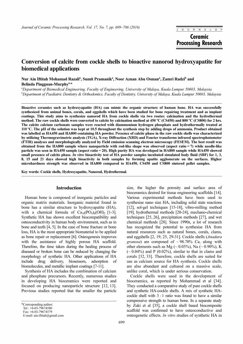
The thermogravimetric curve of cockle shells powderillustrated in Fig. 1 shows multi-step decomposition ofcockle shells. After losing water up to 200 oC, theorganic molecules degrade between 190 and 455 oC.Then, carbon dioxide was evolved rapidly between 650and 900 oC, indicating the maximum decomposition ofcalcium carbonate to calcium oxide. The derivative TGcurve shows the two major peaks at 290 oC and 890 oC,corresponding to the TGA curve. The derivative TGcurve shows a steady line from 400 to 500 oC, and thehighest rate of decomposition at 890 oC.
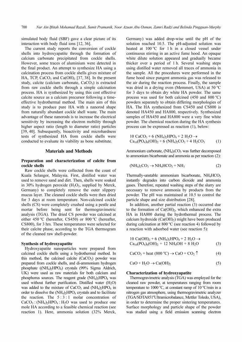
XRD patterns of CS (non-calcined), CS450 (sinteredat 450 oC), and CS800 (sintered at 800 oC) are depictedin Fig. 2 for structural phase comparison. Aragonitephase was identified from the CS sample as matched tothe standard aragonite reference card (JCPDS 98-015-7997) and as resembles other studies [42]. The greatestpeak of aragonite (111) was located at 2θ = 26.1 o, with100% intensity. A similar crystal phase was alsodetected by XRD, reported elsewhere [43] for cockleshells. An obvious shift of peaks occurred for CS450and CS800 samples. A highly intense and sharp peakof calcite (104) can be seen at 2θ = 29.3 o in both
Fig. 1. TG and DTG curves of cockle shells powder demonstratemulti-step decomposition process. Stable line implies completephase transformation of cockle shells along decompositionprocess.
Fig. 2. XRD patterns of aragonite in non-calcined cockle shellspowder (CS) and calcite for cockle shells powder after calcinationprocess at 450 oC (CS450) and 800 oC (CS800). Single Ca(OH)2peak is detected for CS800 spectrum.
702 Nur Ain Iftitah Mohamad Razali, Sumit Pramanik, Noor Azuan Abu Osman, Zamri Radzi and Belinda Pingguan-Murphy
CS450 and CS800 samples along with other weakpeaks, as seen in the standard calcite reference card(JCPDS 1-072-1937). No other extra peak for othermaterials, except for a small peak of calcium hydroxide(Ca(OH)2) formed at 2θ = 34.1 o in CS800 was found.The extra peak in CS800 for Ca(OH)2 indicates that thecalcium oxide (CaO) formed from calcite at hightemperature (800 oC) can easily be converted intoCa(OH)2 after reacting with adsorbed water (seereactions 4-5). Since the formation of CaO was notpossible at lower calcination temperatures (450 oC)according to TGA, the CS450 sample didn’t show anypeak for Ca(OH)2.
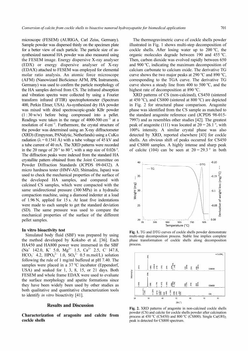
Fig. 3 illustrates FTIR spectra for the CS, CS450,and CS800 samples. The spectrum from the CS sampledepicts aragonite characteristic bands which areattributed to stretching at 1085 cm−1 and bending modesat 857 cm−1 of CO3
2−. A sharp band of CO32− can be
observed at 1455 cm−1, assigned to the fundamentalmode of vibration for CO3
2−. Weak double bands at712 cm−1 and 700 cm−1 are attributed to bending modesof CO3
2−. The FTIR spectrum for CS powder confirmsthe aragonite structure, as reported elsewhere [44].Meanwhile, the infrared spectrums of CS450 andCS800 samples are in agreement with the findings ofcalcite (CaCO3) formation by XRD analysis. Commonabsorption bands assigned to calcite were observed inCS450 and CS800 infrared spectrums. Prominentabsorption band of carbonate alkyl group was seen at1384 cm−1 instead of 1455 cm−1 in aragonite. Thecharacteristic absorption bands of calcite can beobserved at 875 cm−1 and a single peak at 714 cm−1,owing to vibrations of CO3
2− molecules. Other smallstretching bands of functional groups belong to amide at1792 cm−1 and carboxylic acids at 2507 cm−1. A weakOH− peak observed on CS800 spectrum located at3642 cm−1 depicts the presence of Ca(OH)2 at this stage.Both calcined powder samples, mostly containing calcite,
were used as a starting material for synthesis of HA andcompared. Unlike other reported methods, formation ofcalcite in the present study does not involve mixing withother unwanted compounds [37, 38].
Characterization of hydroxyapatite from cockle shellspowder
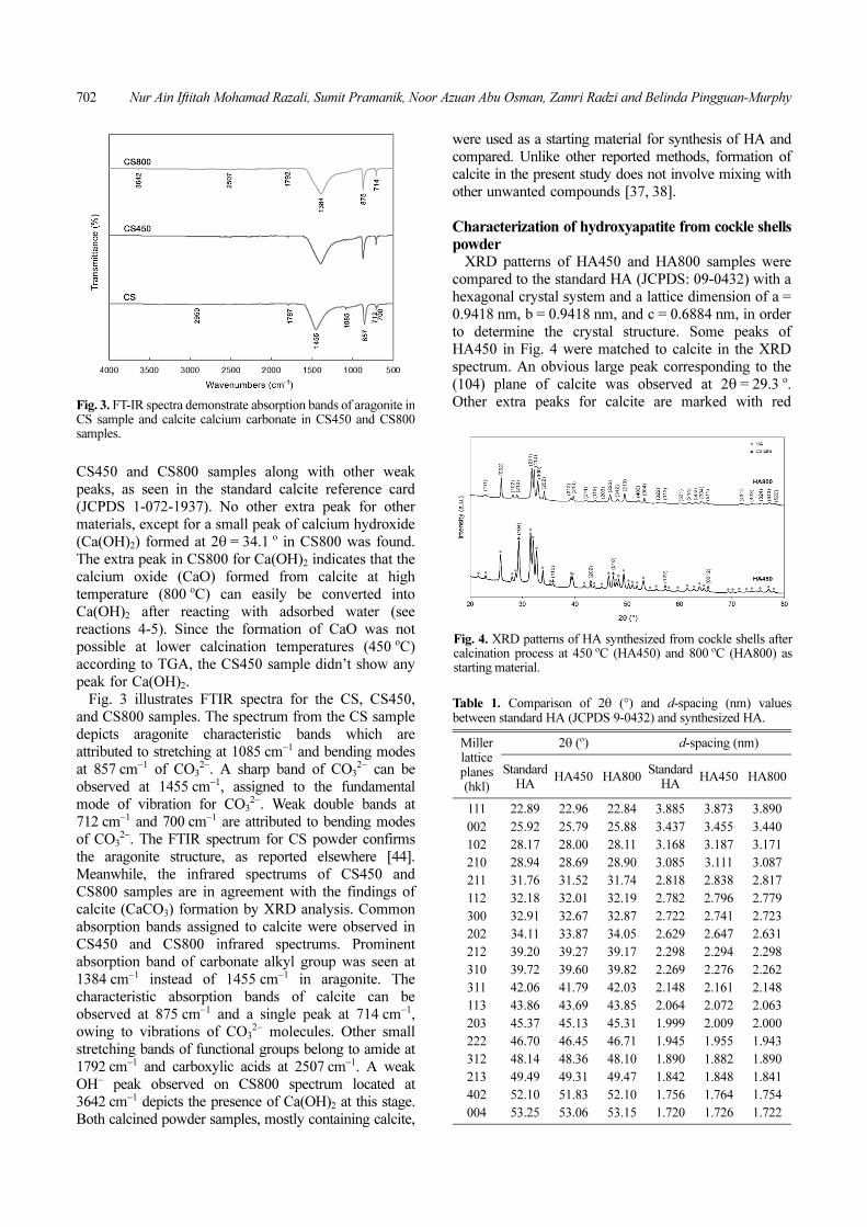
XRD patterns of HA450 and HA800 samples werecompared to the standard HA (JCPDS: 09-0432) with ahexagonal crystal system and a lattice dimension of a =0.9418 nm, b = 0.9418 nm, and c = 0.6884 nm, in orderto determine the crystal structure. Some peaks ofHA450 in Fig. 4 were matched to calcite in the XRDspectrum. An obvious large peak corresponding to the(104) plane of calcite was observed at 2θ = 29.3 o.Other extra peaks for calcite are marked with redFig. 3. FT-IR spectra demonstrate absorption bands of aragonite in
CS sample and calcite calcium carbonate in CS450 and CS800samples.
Fig. 4. XRD patterns of HA synthesized from cockle shells aftercalcination process at 450 oC (HA450) and 800 oC (HA800) asstarting material.
Table 1. Comparison of 2θ (°) and d-spacing (nm) valuesbetween standard HA (JCPDS 9-0432) and synthesized HA.
Miller lattice planes (hkl)
2θ (o) d-spacing (nm)
Standard HA
HA450 HA800Standard
HAHA450 HA800
111 22.89 22.96 22.84 3.885 3.873 3.890
002 25.92 25.79 25.88 3.437 3.455 3.440
102 28.17 28.00 28.11 3.168 3.187 3.171
210 28.94 28.69 28.90 3.085 3.111 3.087
211 31.76 31.52 31.74 2.818 2.838 2.817
112 32.18 32.01 32.19 2.782 2.796 2.779
300 32.91 32.67 32.87 2.722 2.741 2.723
202 34.11 33.87 34.05 2.629 2.647 2.631
212 39.20 39.27 39.17 2.298 2.294 2.298
310 39.72 39.60 39.82 2.269 2.276 2.262
311 42.06 41.79 42.03 2.148 2.161 2.148
113 43.86 43.69 43.85 2.064 2.072 2.063
203 45.37 45.13 45.31 1.999 2.009 2.000
222 46.70 46.45 46.71 1.945 1.955 1.943
312 48.14 48.36 48.10 1.890 1.882 1.890
213 49.49 49.31 49.47 1.842 1.848 1.841
402 52.10 51.83 52.10 1.756 1.764 1.754
004 53.25 53.06 53.15 1.720 1.726 1.722
Conversion of calcite from cockle shells to bioactive nanorod hydroxyapatite for biomedical applications 703
triangles, and the rest of the peaks which are in clearagreement with HA are marked with blue circle, asshown in HA450 of Fig. 4. Therefore, calcite is nottotally converted into HA in the HA450 sample. InHA800, the maximum diffraction peak was observed at2θ = 31.8 o, which corresponds to the (211) latticeplane. Other small peaks were also matched to thestandard for HA. No other element appeared, includingthe peak of Ca(OH)2 that was formed before thesynthesis process. Here, the HA800 sample consists ofsingle phase HA crystals which match the HAspectrum found elsewhere [15, 18, 45]. Comparing thediffraction angle (2θ) and d-spacing values betweenboth samples to the standard HA, it was found that theHA800 sample has the most similar profiles with thestandard HA. In contrary, a slight deviation of the 2θ andd-spacing values is depicted in the HA450 sample asillustrated in Table 1. This result proposed that particlesin HA450 are composed of distorted hexagonal crystals.The Debye Scherrer Equation (1) is applied to estimatethe crystallite size of the obtained HA powder[2].
(6)
Where D is the estimated crystallite size in nm, k is aconstant (k ≅ 0.9), λ is the X-ray diffraction wavelengthi.e., 0.154056 nm, β is the full width at half maximum(FWHM) in radian and θ is the Braggs’ angle in degree.The approximate crystallite size was determined byapplying FWHM and cosθ values at (002) reflection intoEquation (1). As a result, the crystallite size of thesample is estimated to be 39.9 nm (HA450) and 53.1 nm(HA800). These crystallite sizes of HA also resemble theHA developed by other studies [18]. The diameter ofthe HA nanorod will be measured from FESEMimages to visualize the actual particle dimensions,since the Debye Scherrer equation can only be used toknow the crystallite size.
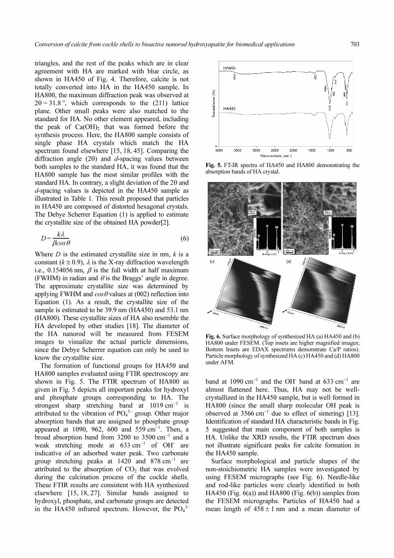
The formation of functional groups for HA450 andHA800 samples evaluated using FTIR spectroscopy areshown in Fig. 5. The FTIR spectrum of HA800 asgiven in Fig. 5 depicts all important peaks for hydroxyland phosphate groups corresponding to HA. Thestrongest sharp stretching band at 1019 cm−1 isattributed to the vibration of PO4
3− group. Other majorabsorption bands that are assigned to phosphate groupappeared at 1090, 962, 600 and 559 cm−1. Then, abroad absorption band from 3200 to 3500 cm−1 and aweak stretching mode at 633 cm−1 of OH- areindicative of an adsorbed water peak. Two carbonategroup stretching peaks at 1420 and 878 cm−1 areattributed to the absorption of CO2 that was evolvedduring the calcination process of the cockle shells.These FTIR results are consistent with HA synthesizedelsewhere [15, 18, 27]. Similar bands assigned tohydroxyl, phosphate, and carbonate groups are detectedin the HA450 infrared spectrum. However, the PO4
3-
band at 1090 cm−1 and the OH- band at 633 cm−1 arealmost flattened here. Thus, HA may not be well-crystallized in the HA450 sample, but is well formed inHA800 (since the small sharp molecular OH peak isobserved at 3566 cm−1 due to effect of sintering) [13].Identification of standard HA characteristic bands in Fig.5 suggested that main component of both samples isHA. Unlike the XRD results, the FTIR spectrum doesnot illustrate significant peaks for calcite formation inthe HA450 sample.
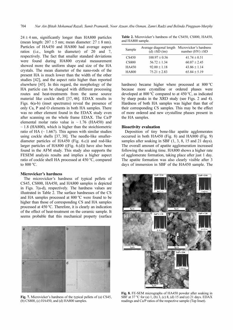
Surface morphological and particle shapes of thenon-stoichiometric HA samples were investigated byusing FESEM micrographs (see Fig. 6). Needle-likeand rod-like particles were clearly identified in bothHA450 (Fig. 6(a)) and HA800 (Fig. 6(b)) samples fromthe FESEM micrographs. Particles of HA450 had amean length of 458 ± 1 nm and a mean diameter of
Dkλ
β θcos--------------=
Fig. 5. FT-IR spectra of HA450 and HA800 demonstrating theabsorption bands of HA crystal.
Fig. 6. Surface morphology of synthesized HA (a) HA450 and (b)HA800 under FESEM. (Top insets are higher magnified images;Bottom Insets are EDAX spectrums demonstrate Ca/P ratios).Particle morphology of synthesized HA (c) HA450 and (d) HA800under AFM.
704 Nur Ain Iftitah Mohamad Razali, Sumit Pramanik, Noor Azuan Abu Osman, Zamri Radzi and Belinda Pingguan-Murphy
24 ± 4 nm, significantly longer than HA800 particles(mean length: 207 ± 5 nm; mean diameter: 27 ± 8 nm).Particles of HA450 and HA800 had average aspectratios (i.e., length to diameter) of 20 and 7,respectively. The fact that smaller standard deviationswere found during HA800 crystal measurementshowed more the uniform shape and size of the HAcrystals. The mean diameter of the nano-rods of thepresent HA is much lower than the width of the otherstudies [42], and the aspect ratio higher than reportedelsewhere [45]. In this regard, the morphology of theHA particle can be changed with different processingroutes and heat-treatments from the same sourcematerial like cockle shell [37, 46]. EDAX results inFigs. 6(a-b) (inset spectrums) reveal the presence ofonly Ca, P and O elements in both HA samples. Therewas no other element found in the EDAX study evenafter scanning on the whole frame EDAX. The Ca/Pelemental molar ratio value is ~ 1.76 (HA450) and~ 1.8 (HA800), which is higher than the stoichiometricratio of HA (~ 1.667). This agrees with similar studiesusing cockle shells [37, 38]. The needle-like smaller-diameter particles of HA450 (Fig. 6.c)) and rod-likelarger particles of HA800 ((Fig. 6.(d)) have also beenfound in the AFM study. This study also supports theFESEM analysis results and implies a higher aspectratio of cockle shell HA processed at 450 oC, comparedto 800 oC.
Microvicker’s hardnessThe microvicker’s hardness of typical pellets of
CS45, CS800, HA450, and HA800 samples is depictedin Figs. 7(a-d), respectively. The hardness values areillustrated in Table 2. The surface hardnesses of the CSand HA samples processed at 800 oC were found to behigher than those of corresponding CS and HA samplesprocessed at 450 oC. Therefore, it is clearly an indicationof the effect of heat-treatment on the ceramic sample. Itseems probable that this mechanical property (surface
hardness) became higher where processed at 800 oCbecause more crystalline or ordered phases weredeveloped at 800 oC compared to at 450 oC, as indicatedby sharp peaks in the XRD study (see Figs. 2 and 4).Hardness of both HA samples was higher than that oftheir corresponding CS samples. This may be the effectof more ordered and new crystalline phases present inthe HA samples.
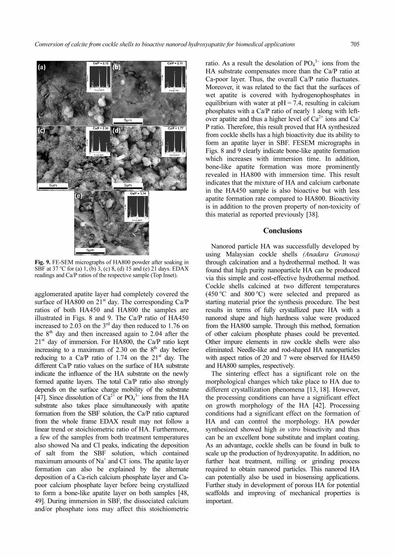
Bioactivity evaluationDeposition of tiny bone-like apatite agglomerates
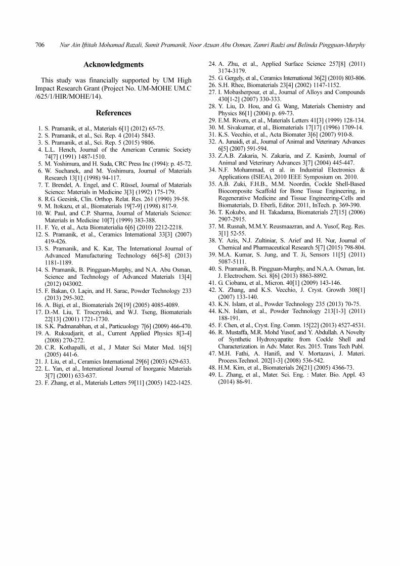
occurred in both HA450 (Fig. 8) and HA800 (Fig. 9)samples after soaking in SBF (1, 3, 8, 15 and 21 days).The overall amount of apatite agglomeration increasedfollowing the soaking time. HA800 shows a higher rateof agglomerate formation, taking place after just 1 day.The apatite formation was also clearly visible after 3days of immersion in SBF of the HA450 sample. The
Table 2. Microvicker’s hardness of the CS450, CS800, HA450,and HA800 sample.
SampleAverage diagonal length
(d) ±SD (ìm)Microvicker’s hardness
number (HV) ±SD
CS450 100.97 ± 0.56 41.74 ± 0.51
CS800 56.72 ± 1.34 60.87 ± 2.45
HA450 92.00 ± 1.18 43.86 ± 1.14
HA800 75.21 ± 2.83 65.84 ± 5.19
Fig. 7. Microvicker’s hardness of the typical pellets of (a) CS45,(b) CS800, (c) HA450, and (d) HA800 samples.
Fig. 8. FE-SEM micrographs of HA450 powder after soaking inSBF at 37 oC for (a) 1, (b) 3, (c) 8, (d) 15 and (e) 21 days. EDAXreadings and Ca/P ratios of the respective sample (Top Inset).
Conversion of calcite from cockle shells to bioactive nanorod hydroxyapatite for biomedical applications 705
agglomerated apatite layer had completely covered thesurface of HA800 on 21st day. The corresponding Ca/Pratios of both HA450 and HA800 the samples areillustrated in Figs. 8 and 9. The Ca/P ratio of HA450increased to 2.03 on the 3rd day then reduced to 1.76 onthe 8th day and then increased again to 2.04 after the21st day of immersion. For HA800, the Ca/P ratio keptincreasing to a maximum of 2.30 on the 8th day beforereducing to a Ca/P ratio of 1.74 on the 21st day. Thedifferent Ca/P ratio values on the surface of HA substrateindicate the influence of the HA substrate on the newlyformed apatite layers. The total Ca/P ratio also stronglydepends on the surface charge mobility of the substrate[47]. Since dissolution of Ca2+ or PO4
3− ions from the HAsubstrate also takes place simultaneously with apatiteformation from the SBF solution, the Ca/P ratio capturedfrom the whole frame EDAX result may not follow alinear trend or stoichiometric ratio of HA. Furthermore,a few of the samples from both treatment temperaturesalso showed Na and Cl peaks, indicating the depositionof salt from the SBF solution, which containedmaximum amounts of Na+ and Cl- ions. The apatite layerformation can also be explained by the alternatedeposition of a Ca-rich calcium phosphate layer and Ca-poor calcium phosphate layer before being crystallizedto form a bone-like apatite layer on both samples [48,49]. During immersion in SBF, the dissociated calciumand/or phosphate ions may affect this stoichiometric
ratio. As a result the desolation of PO43− ions from the
HA substrate compensates more than the Ca/P ratio atCa-poor layer. Thus, the overall Ca/P ratio fluctuates.Moreover, it was related to the fact that the surfaces ofwet apatite is covered with hydrogenophosphates inequilibrium with water at pH = 7.4, resulting in calciumphosphates with a Ca/P ratio of nearly 1 along with left-over apatite and thus a higher level of Ca2+ ions and Ca/P ratio. Therefore, this result proved that HA synthesizedfrom cockle shells has a high bioactivity due its ability toform an apatite layer in SBF. FESEM micrographs inFigs. 8 and 9 clearly indicate bone-like apatite formationwhich increases with immersion time. In addition,bone-like apatite formation was more prominentlyrevealed in HA800 with immersion time. This resultindicates that the mixture of HA and calcium carbonatein the HA450 sample is also bioactive but with lessapatite formation rate compared to HA800. Bioactivityis in addition to the proven property of non-toxicity ofthis material as reported previously [38].
Conclusions
Nanorod particle HA was successfully developed byusing Malaysian cockle shells (Anadara Granosa)
through calcination and a hydrothermal method. It wasfound that high purity nanoparticle HA can be producedvia this simple and cost-effective hydrothermal method.Cockle shells calcined at two different temperatures(450 oC and 800 oC) were selected and prepared asstarting material prior the synthesis procedure. The bestresults in terms of fully crystallized pure HA with ananorod shape and high hardness value were producedfrom the HA800 sample. Through this method, formationof other calcium phosphate phases could be prevented.Other impure elements in raw cockle shells were alsoeliminated. Needle-like and rod-shaped HA nanoparticleswith aspect ratios of 20 and 7 were observed for HA450and HA800 samples, respectively.
The sintering effect has a significant role on themorphological changes which take place to HA due todifferent crystallization phenomena [13, 18]. However,the processing conditions can have a significant effecton growth morphology of the HA [42]. Processingconditions had a significant effect on the formation ofHA and can control the morphology. HA powdersynthesized showed high in vitro bioactivity and thuscan be an excellent bone substitute and implant coating.As an advantage, cockle shells can be found in bulk toscale up the production of hydroxyapatite. In addition, nofurther heat treatment, milling or grinding processrequired to obtain nanorod particles. This nanorod HAcan potentially also be used in biosensing applications.Further study in development of porous HA for potentialscaffolds and improving of mechanical properties isimportant.
Fig. 9. FE-SEM micrographs of HA800 powder after soaking inSBF at 37 oC for (a) 1, (b) 3, (c) 8, (d) 15 and (e) 21 days. EDAXreadings and Ca/P ratios of the respective sample (Top Inset).
706 Nur Ain Iftitah Mohamad Razali, Sumit Pramanik, Noor Azuan Abu Osman, Zamri Radzi and Belinda Pingguan-Murphy
Acknowledgments
This study was financially supported by UM HighImpact Research Grant (Project No. UM-MOHE UM.C/625/1/HIR/MOHE/14).
References
1. S. Pramanik, et al., Materials 6[1] (2012) 65-75.2. S. Pramanik, et al., Sci. Rep. 4 (2014) 5843.3. S. Pramanik, et al., Sci. Rep. 5 (2015) 9806.4. L.L. Hench, Journal of the American Ceramic Society
74[7] (1991) 1487-1510.5. M. Yoshimura, and H. Suda, CRC Press Inc (1994): p. 45-72.6. W. Suchanek, and M. Yoshimura, Journal of Materials
Research 13[1] (1998) 94-117.7. T. Brendel, A. Engel, and C. Rüssel, Journal of Materials
Science: Materials in Medicine 3[3] (1992) 175-179.8. R.G. Geesink, Clin. Orthop. Relat. Res. 261 (1990) 39-58.9. M. Itokazu, et al., Biomaterials 19[7-9] (1998) 817-9.
10. W. Paul, and C.P. Sharma, Journal of Materials Science:Materials in Medicine 10[7] (1999) 383-388.
11. F. Ye, et al., Acta Biomaterialia 6[6] (2010) 2212-2218.12. S. Pramanik, et al., Ceramics International 33[3] (2007)
419-426.13. S. Pramanik, and K. Kar, The International Journal of
Advanced Manufacturing Technology 66[5-8] (2013)1181-1189.
14. S. Pramanik, B. Pingguan-Murphy, and N.A. Abu Osman,Science and Technology of Advanced Materials 13[4](2012) 043002.
15. F. Bakan, O. Laçin, and H. Sarac, Powder Technology 233(2013) 295-302.
16. A. Bigi, et al., Biomaterials 26[19] (2005) 4085-4089.17. D.-M. Liu, T. Troczynski, and W.J. Tseng, Biomaterials
22[13] (2001) 1721-1730.18. S.K. Padmanabhan, et al., Particuology 7[6] (2009) 466-470.19. A. Ruksudjarit, et al., Current Applied Physics 8[3-4]
(2008) 270-272.20. C.R. Kothapalli, et al., J Mater Sci Mater Med. 16[5]
(2005) 441-6.21. J. Liu, et al., Ceramics International 29[6] (2003) 629-633.22. L. Yan, et al., International Journal of Inorganic Materials
3[7] (2001) 633-637.23. F. Zhang, et al., Materials Letters 59[11] (2005) 1422-1425.
24. A. Zhu, et al., Applied Surface Science 257[8] (2011)3174-3179.
25. G. Gergely, et al., Ceramics International 36[2] (2010) 803-806.26. S.H. Rhee, Biomaterials 23[4] (2002) 1147-1152.27. I. Mobasherpour, et al., Journal of Alloys and Compounds
430[1-2] (2007) 330-333.28. Y. Liu, D. Hou, and G. Wang, Materials Chemistry and
Physics 86[1] (2004) p. 69-73.29. E.M. Rivera, et al., Materials Letters 41[3] (1999) 128-134.30. M. Sivakumar, et al., Biomaterials 17[17] (1996) 1709-14.31. K.S. Vecchio, et al., Acta Biomater 3[6] (2007) 910-8.32. A. Junaidi, et al., Journal of Animal and Veterinary Advances
6[5] (2007) 591-594.33. Z.A.B. Zakaria, N. Zakaria, and Z. Kasimb, Journal of
Animal and Veterinary Advances 3[7] (2004) 445-447.34. N.F. Mohammad, et al. in Industrial Electronics &
Applications (ISIEA), 2010 IEEE Symposium on. 2010.35. A.B. Zuki, F.H.B., M.M. Noordin, Cockle Shell-Based
Biocomposite Scaffold for Bone Tissue Engineering, inRegenerative Medicine and Tissue Engineering-Cells andBiomaterials, D. Eberli, Editor. 2011, InTech. p. 369-390.
36. T. Kokubo, and H. Takadama, Biomaterials 27[15] (2006)2907-2915.
37. M. Rusnah, M.M.Y. Reusmaazran, and A. Yusof, Reg. Res.3[1] 52-55.
38. Y. Azis, N.J. Zultiniar, S. Arief and H. Nur, Journal ofChemical and Pharmaceutical Research 5[7] (2015) 798-804.
39. M.A. Kumar, S. Jung, and T. Ji, Sensors 11[5] (2011)5087-5111.
40. S. Pramanik, B. Pingguan-Murphy, and N.A.A. Osman, Int.J. Electrochem. Sci. 8[6] (2013) 8863-8892.
41. G. Ciobanu, et al., Micron. 40[1] (2009) 143-146.42. X. Zhang, and K.S. Vecchio, J. Cryst. Growth 308[1]
(2007) 133-140.43. K.N. Islam, et al., Powder Technology 235 (2013) 70-75.44. K.N. Islam, et al., Powder Technology 213[1-3] (2011)
188-191.45. F. Chen, et al., Cryst. Eng. Comm. 15[22] (2013) 4527-4531.46. R. Mustaffa, M.R. Mohd Yusof, and Y. Abdullah. A Novelty
of Synthetic Hydroxyapatite from Cockle Shell andCharacterization. in Adv. Mater. Res. 2015. Trans Tech Publ.
47. M.H. Fathi, A. Hanifi, and V. Mortazavi, J. Materi.Process.Technol. 202[1-3] (2008) 536-542.
48. H.M. Kim, et al., Biomaterials 26[21] (2005) 4366-73.49. L. Zhang, et al., Mater. Sci. Eng. : Mater. Bio. Appl. 43
(2014) 86-91.