Connective tissue growth factor causes EMT-like cell fate...
12
Journal of Cell Science Connective tissue growth factor causes EMT-like cell fate changes in vivo and in vitro Sonali Sonnylal 1, *, Shiwen Xu 2 , Helen Jones 2 , Angela Tam 2 , Vivek R. Sreeram 2 , Markella Ponticos 2 , Jill Norman 2 , Pankaj Agrawal 1 , David Abraham 2 and Benoit de Crombrugghe 1 1 University of Texas M. D. Anderson Cancer Center, Department of Genetics, Houston, TX, USA 2 Division of Medicine, University College London, Royal Free Campus, London, UK *Author for correspondence ([email protected]) Accepted 19 February 2013 Journal of Cell Science 126, 2164–2175 ß 2013. Published by The Company of Biologists Ltd doi: 10.1242/jcs.111302 Summary Connective tissue growth factor (CTGF) plays an important role in the pathogenesis of chronic fibrotic diseases. However, the mechanism by which paracrine effects of CTGF control the cell fate of neighboring epithelial cells is not known. In this study, we investigated the paracrine effects of CTGF overexpressed in fibroblasts of Col1a2-CTGF transgenic mice on epithelial cells of skin and lung. The skin and lungs of Col1a2-CTGF transgenic mice were examined for phenotypic markers of epithelial activation and differentiation and stimulation of signal transduction pathways. In addition to an expansion of the dermal compartment in Col1a2-CTGF transgenic mice, the epidermis was characterized by focal hyperplasia, and basal cells stained positive for aSMA, Snail, S100A4 and Sox9, indicating that these cells had undergone a change in their genetic program. Activation of phosphorylated p38 and phosphorylated Erk1/2 was observed in the granular and cornified layers of the skin. Lung fibrosis was associated with a marked increase in cells co- expressing epithelial and mesenchymal markers in the lesional and unaffected lung tissue of Col1a2-CTGF mice. In epithelial cells treated with TGFb, CTGF-specific siRNA-mediated knockdown suppressed Snail, Sox9, S100A4 protein levels and restored E-cadherin levels. Both adenoviral expression of CTGF in epithelial cells and treatment with recombinant CTGF induced EMT-like morphological changes and expression of a-SMA. Our in vivo and in vitro data supports the notion that CTGF expression in mesenchymal cells in the skin and lungs can cause changes in the differentiation program of adjacent epithelial cells. We speculate that these changes might contribute to fibrogenesis. Key words: CTGF, Epithelial–mesenchymal transition, Dermal fibrosis, Pulmonary fibrosis Introduction Organ fibrosis is the outcome of a complex set of cellular interactions initially caused by a pathological injury followed by a process of dysregulated healing (Wynn, 2008). Tissue or organ fibrosis results in increased morbidity and mortality in humans. Hallmarks of fibrosis are the accumulation of fibroblasts and their contractile partners, the myofibroblasts. In addition to resident fibroblasts, perivascular fibroblasts or pericytes, mesenchymal stem cells, and circulating fibrocytes derived from the bone marrow compartment also contribute to the enhanced numbers of fibroblasts and to increased collagen production (Abraham et al., 2007; Krieg et al., 2007). A certain proportion of fibroblast-like cells in fibrotic lesions originate from epithelial cells that have acquired migratory and proliferative characteristics (Krieg et al., 2007). These cells are stimulated by a variety of cytokines, with TGFb being a key player in fibrosis (Pohlers et al., 2009). CTGF is a secreted matricellular protein that has been implicated as a regulator of cellular proliferation, angiogenesis and remodeling of the extracellular matrix (Friedrichsen et al., 2003; Ivkovic et al., 2003; Leask and Abraham, 2006). CTGF can act on the same cells that produce it in an autocrine manner elevating extracellular matrix (ECM) synthesis in normal fibroblasts to a level seen in cells of fibrotic lesions (Shi-wen et al., 2000). Our recent mouse model, in which CTGF is overexpressed in mesenchymal cells in vivo, clearly demonstrates that fibrosis of multiple organs can be mediated by CTGF alone independently of TGFb activation (Sonnylal et al., 2010). During normal development and during wound healing, epithelial mesenchymal transition (EMT) plays a very dynamic role (Kalluri and Neilson, 2003). During EMT, tightly adjoining epithelial cells resting on a basal lamina with apical–basolateral polarization convert to a non-polarized mesenchymal cell phenotype, detach from the epithelial layer, develop migratory properties and express proteins that are typical of mesenchymal cells. In certain pathological situations, such as in kidney and lung fibrosis, epithelial cells acquire mesenchymal characteristics and contribute to enhanced synthesis of collagen. During EMT, epithelial cells acquire the ability to interact with specific interstitial matrices subsequent to traversing the basement membrane. Generally such cell fate switches of cellular identity and function are important for disease progression. We have reported that transgenic mice expressing high levels of CTGF specifically in mesenchymal cells (Col1a2-CTGF) manifest hallmarks of sustained multi-organ fibrosis recapitulating key aspects of the fibrotic disease systemic sclerosis (SSc, scleroderma) (Sonnylal et al., 2010). In addition to overt changes in the connective tissue compartments, we observed focal hyperplasia in the epidermal layer of the fibrotic skin and in uninvolved areas of fibrotic lung. We hypothesize that 2164 Research Article
Transcript of Connective tissue growth factor causes EMT-like cell fate...

Journ
alof
Cell
Scie
nce
Connective tissue growth factor causes EMT-like cellfate changes in vivo and in vitro
Sonali Sonnylal1,*, Shiwen Xu2, Helen Jones2, Angela Tam2, Vivek R. Sreeram2, Markella Ponticos2,Jill Norman2, Pankaj Agrawal1, David Abraham2 and Benoit de Crombrugghe1
1University of Texas M. D. Anderson Cancer Center, Department of Genetics, Houston, TX, USA2Division of Medicine, University College London, Royal Free Campus, London, UK
*Author for correspondence ([email protected])
Accepted 19 February 2013Journal of Cell Science 126, 2164–2175� 2013. Published by The Company of Biologists Ltddoi: 10.1242/jcs.111302
SummaryConnective tissue growth factor (CTGF) plays an important role in the pathogenesis of chronic fibrotic diseases. However, themechanism by which paracrine effects of CTGF control the cell fate of neighboring epithelial cells is not known. In this study, weinvestigated the paracrine effects of CTGF overexpressed in fibroblasts of Col1a2-CTGF transgenic mice on epithelial cells of skin and
lung. The skin and lungs of Col1a2-CTGF transgenic mice were examined for phenotypic markers of epithelial activation anddifferentiation and stimulation of signal transduction pathways. In addition to an expansion of the dermal compartment in Col1a2-CTGFtransgenic mice, the epidermis was characterized by focal hyperplasia, and basal cells stained positive for aSMA, Snail, S100A4 andSox9, indicating that these cells had undergone a change in their genetic program. Activation of phosphorylated p38 and phosphorylated
Erk1/2 was observed in the granular and cornified layers of the skin. Lung fibrosis was associated with a marked increase in cells co-expressing epithelial and mesenchymal markers in the lesional and unaffected lung tissue of Col1a2-CTGF mice. In epithelial cellstreated with TGFb, CTGF-specific siRNA-mediated knockdown suppressed Snail, Sox9, S100A4 protein levels and restored E-cadherin
levels. Both adenoviral expression of CTGF in epithelial cells and treatment with recombinant CTGF induced EMT-like morphologicalchanges and expression of a-SMA. Our in vivo and in vitro data supports the notion that CTGF expression in mesenchymal cells in theskin and lungs can cause changes in the differentiation program of adjacent epithelial cells. We speculate that these changes might
contribute to fibrogenesis.
Key words: CTGF, Epithelial–mesenchymal transition, Dermal fibrosis, Pulmonary fibrosis
IntroductionOrgan fibrosis is the outcome of a complex set of cellular
interactions initially caused by a pathological injury followed by
a process of dysregulated healing (Wynn, 2008). Tissue or organ
fibrosis results in increased morbidity and mortality in humans.
Hallmarks of fibrosis are the accumulation of fibroblasts and
their contractile partners, the myofibroblasts. In addition to
resident fibroblasts, perivascular fibroblasts or pericytes,
mesenchymal stem cells, and circulating fibrocytes derived
from the bone marrow compartment also contribute to the
enhanced numbers of fibroblasts and to increased collagen
production (Abraham et al., 2007; Krieg et al., 2007). A certain
proportion of fibroblast-like cells in fibrotic lesions originate
from epithelial cells that have acquired migratory and
proliferative characteristics (Krieg et al., 2007). These cells are
stimulated by a variety of cytokines, with TGFb being a key
player in fibrosis (Pohlers et al., 2009).
CTGF is a secreted matricellular protein that has been
implicated as a regulator of cellular proliferation, angiogenesis
and remodeling of the extracellular matrix (Friedrichsen et al.,
2003; Ivkovic et al., 2003; Leask and Abraham, 2006). CTGF can
act on the same cells that produce it in an autocrine manner
elevating extracellular matrix (ECM) synthesis in normal
fibroblasts to a level seen in cells of fibrotic lesions (Shi-wen
et al., 2000). Our recent mouse model, in which CTGF is
overexpressed in mesenchymal cells in vivo, clearly demonstrates
that fibrosis of multiple organs can be mediated by CTGF alone
independently of TGFb activation (Sonnylal et al., 2010).
During normal development and during wound healing,
epithelial mesenchymal transition (EMT) plays a very dynamic
role (Kalluri and Neilson, 2003). During EMT, tightly adjoining
epithelial cells resting on a basal lamina with apical–basolateral
polarization convert to a non-polarized mesenchymal cell
phenotype, detach from the epithelial layer, develop migratory
properties and express proteins that are typical of mesenchymal
cells. In certain pathological situations, such as in kidney and
lung fibrosis, epithelial cells acquire mesenchymal characteristics
and contribute to enhanced synthesis of collagen. During EMT,
epithelial cells acquire the ability to interact with specific
interstitial matrices subsequent to traversing the basement
membrane. Generally such cell fate switches of cellular identity
and function are important for disease progression.
We have reported that transgenic mice expressing high levels
of CTGF specifically in mesenchymal cells (Col1a2-CTGF)
manifest hallmarks of sustained multi-organ fibrosis
recapitulating key aspects of the fibrotic disease systemic
sclerosis (SSc, scleroderma) (Sonnylal et al., 2010). In addition
to overt changes in the connective tissue compartments, we
observed focal hyperplasia in the epidermal layer of the fibrotic
skin and in uninvolved areas of fibrotic lung. We hypothesize that
2164 Research Article

Journ
alof
Cell
Scie
nce
the persistent overexpression of CTGF in mesenchymal cellsleads to epithelial cell proliferation and differentiation by a
paracrine mechanism.
In the present study, we explored the paracrine effects resultingfrom the production and secretion of CTGF by mesenchymal
cells on neighboring epithelial cells in fibrotic skin and lung ofCol1a2-CTGF transgenic mice. We found that some basalkeratinocytes in the skin and alveolar epithelial cells in fibrotic
foci and normal areas of the lung are morphologically andfunctionally different from epithelial cells in these tissues inwild-type (wt) mice. Cells that were committed to an epithelial
lineage are undergoing a change in their genetic program that isexemplified by expression of EMT marker genes. In the skinthese changes are associated with activated MAPK and ERKsignaling. In vitro studies in lung epithelial cells showed that the
onset of EMT marker gene expression due to exogenous TGFbcan be blocked by CTGF knockdown suggesting that CTGFmediates TGFb-induced EMT. Furthermore, expression of CTGF
in lung epithelial cells or treatment with exogenous CTGF alsoinduced EMT-like changes in vitro. Overall, our results providenew evidence that CTGF causes an EMT-like phenotype in skin
and lung in vivo, and that these changes are likely to occurthrough a paracrine mechanism.
ResultsAlteration in keratin and integrin b gene expression in theskin of Col1a2-CTGF transgenic mice
Mice overexpressing CTGF in mesenchymal cells developeddramatic and progressive thickening of the dermis. In addition,
we observed increased hyperplasia and hypertrophy ofkeratinocytes in focal areas of the dermal–epidermal junction.Evidence for hyper-proliferation of epidermal cells was shown bythe increase in the multilayer structure of the epidermis of
Col1a2-CTGF mice. Previous experiments had also shown athreefold increase in BrdU incorporation in the areas ofepidermal hyperplasia (data not shown). These results were
confirmed by increased expression of the K16 proliferationmarker in the interfollicular epidermis of transgenic micecompared to wt littermate controls (Fig. 1A,B, green), with an
increase in the number of positive cells to nearly 10% of the totalcell frequency.
Since the epidermis of Col1a2-CTGF mice showed increased
hyperplasia, we wanted to determine if the apical–basal polaritywas maintained by the basal cells in the epithelium. To addressthis, we analyzed the expression of integrins. Integrins are
transmembrane receptors that mediate cell–cell adhesion andterminal differentiation of keratinocytes. In wt skin thedistribution of integrin b4 was, as expected, mainly confined tothe basal side of the basal epithelial cells. In contrast, the
expression of integrin b4 in the epidermis of Col1a2-CTGFtransgenic mice was irregularly distributed all around the hyper-proliferative epithelial cells (Fig. 1A,B, red). Another epithelial
specific integrin, avb6, that is barely detectable in quiescentepithelial cells is rapidly induced upon epithelial injury andimparts increased migratory properties to these cells (Huang et al.,
1996). Interestingly, we observed a marked enhancement in theexpression of integrin b6 in the epidermis of Col1a2-CTGFtransgenic mice compared to littermate controls (Fig. 1C,D)
suggesting that these cells might have increased migratoryproperties. The redistribution of integrin b4 suggests that thebasal cells in the epidermis had lost their apical-basal polarity.
Based on the marked increase in integrin b6 expression, we
hypothesize that these cells had acquired increased migratory
properties. Both changes are likely the result of changes in the
genetic program of these cells.
Col1a2-CTGF mice exhibit focal hyperplasia of the
epidermis associated with cell fate changes in epithelial
cells
The increase in collagen in the dermis of Col1a2-CTGF
transgenic mice was associated with increased numbers of
myofibroblasts (Sonnylal et al., 2010). Immunostaining of skin
for aSMA revealed large numbers of cells positive for aSMA in
both the papillary and the reticular dermis of Col1a2-CTGF
transgenic mice (Sonnylal et al., 2010). Intriguingly, we have
observed that aSMA-positive cells are intermingled with basal
cells in the hypertrophic epidermis of Col1a2-CTGF transgenic
mice (Fig. 2A,C and corresponding DIC images B,D). We
explored this finding further and examined the origin of these
cells. Immunostaining for Snai1, a marker for EMT (Kalluri and
Neilson, 2003), revealed significant Snai1 levels in areas in
which there was focal hyperplasia of the epidermis in Col1a2-
CTGF transgenic mice. Intense cytoplasmic staining for Snai1
was seen in the basal cells adjacent to the dermis and clear Snai1
Fig. 1. Abnormal expression of hyperproliferative epidermal markers in
Col1a2-CTGF transgenic mice. The expression of keratin 16 (a
hyperproliferative epidermal marker), integrin b6, and integrin b4 in the skin
of wt and Col1a2-CTGF transgenic mice was examined by
immunofluorescence. Increased expression of keratin 16 (green) was
observed suprabasally in the epidermis in Col1a2-CTGF transgenic mice
(B) compared to wt controls (A). Increased expression of integrin b4 (red)
was observed in the hyperproliferative epidermis of Col1a2-CTGF transgenic
mice (B) whereas in wt skin integrin b4 expression was restricted to the basal
side of the basal layer and seemed reduced (A). Epidermal cells also showed a
marked increase in the level of integrin b6 in Col1a2-CTGF transgenic mice
(D), whereas no staining was observed in control skin (C). Scale bars: A,
B5100 mm (A,B), 50 mm (C,D). Quantification of the expression of these
proteins is shown below. Data are means 6 s.d.; n56 skin sections.
*P,0.0001, **P,0.001, ***P,0.0005, Student’s t-test.
Paracrine CTGF induces EMT-like changes 2165

Journ
alof
Cell
Scie
nce
staining primarily confined to the nucleus was seen in the upper layers
of the epidermis in Col1a2-CTGF transgenic mice. Snai1 staining was
absent in wt controls (Fig. 2E,G and corresponding DIC images F,H).
To determine if the epidermal cells of Col1a2-CTGF transgenic
mice displayed properties of newly formed mesenchymal cells,
immunofluorescence was performed with antibodies against S100A4
or FSP-1, a Ca2+ binding protein closely associated with EMT. We
observed that cells in both the basal and suprabasal layer of the
Col1a2-CTGF epidermis showed robust staining for S100A4, unlike
the skin epidermis of wt mice where staining for S100A4 was absent
(Fig. 2I,K and corresponding DIC images J,L). Marked staining for
aSMA, Snai1 and S100A4 in the epidermal cells within focal areas of
hyperplasia in the epidermis of Col1a2-CTGF transgenic mice
strongly suggest that these cells were undergoing an EMT-like
process.
We also observed staining for S100A4, a marker of newly formed
fibroblasts, in cells of the papillary dermis suggesting that these cells
either arose from resident fibroblasts or originated from epithelial
cells by EMT (Fig. 2I,K). Taken together these results strongly
suggest that overexpression of CTGF in the dermal compartment is
able to induce EMT-like changes in the adjacent epithelial cells of
the epidermis most likely by paracrine mechanisms.
Aberrant Sox9 expression in the epidermis of adult Col1a2-
CTGF transgenic mice
Sox9, which belongs to the HMG box super-family of DNA
binding proteins, is a key transcription factor for chondrocytes
and several other lineages (Pritchett et al., 2011; Bi et al., 1999).
In the skin, and particularly in the hair follicle, Sox9 has essential
roles in the development of the outer root sheath (ORS) and of
the stem cell compartment (the bulge) (Vidal et al., 2005). Recent
evidence in the literature has implicated Sox9 expression in
diseases that affect the extracellular matrix such as in skin keloids
(Naitoh et al., 2005), glomerular sclerosis of the kidney (Bennett
et al., 2007), and activated stellate cells in the liver (Hanley et al.,
2008). We wanted therefore to investigate whether the
distribution of Sox9 expression in the skin of Col1a2-CTGF
transgenic mice was altered. Using immunofluorescence, we
showed abundant expression of nuclear Sox9 in the basal cells of
the epidermis in Col1a2-CTGF transgenic mice but not in wt
mice (Fig. 3A,B and corresponding DIC images C,D). Increased
expression of Sox9 (over threefold) was also observed in the ORS
and in the bulge of hair follicles (Fig. 3E,F and corresponding
DIC images G,H).
Similar to the abnormal expression of aSMA, Snai1 and
S100A4, the anomalous Sox9 expression patterns strongly
suggest that overexpression of CTGF in mesenchymal cells
results in major cell fate changes in the basal layer of the
epidermis.
Multiple signaling pathways are activated in the epidermis
of Col1a2-CTGF transgenic mice
We have previously provided evidence that increased expression
of CTGF in mesenchymal cells causes constitutive activation of
multiple signaling molecules including phosphorylated p38 (p-
p38), Erk1/2 (pErk1/2), Akt (pAkt), and PI3K (Sonnylal et al.,
Fig. 2. Changes in the genetic program of the basal cells in the epidermis. Immunofluorescence was performed on skin sections of Col1a2-CTGF transgenic
mice and wt littermate controls with antibodies to aSMA, Snai1 and S100A4. De novo expression of aSMA was increased in the epidermis of Col1a2-CTGF
fibrotic skin (C; corresponding DIC overlay in D), indicated by white arrows, compared with wt littermate controls (A; corresponding DIC overlay in B). Cells
in the basal layer also stained positive for Snai1 (G; corresponding DIC overlay in H) indicated by white arrows, whereas no Snai1 staining was observed in
controls (E; corresponding DIC overlay in F). Similarly, S100A4 staining was observed in basal cells of the epidermis of Col1a2-CTGF transgenic mice
(K; corresponding DIC overlay in L), and absent in control sections (I; corresponding DIC overlay in J). Arrowheads indicate the cells expressing Snai1 and
S100A4 in the dermis. The horizontal dashed line marks the dermal–epidermal junction. Scale bar: 50 mm. Quantification of the expression of these proteins is
shown on the right. Data are means 6 s.d., n56 skin sections. *P,0.005, **P,0.001, ***P,0.001, Student’s t-test.
Journal of Cell Science 126 (10)2166
Journ
alof
Cell
Scie
nce
2010). Because integrin b6 causes the activation of Erk1/2 and
p38 signaling molecules in epithelial cells (Sullivan et al., 2011;
Ahmed et al., 2002), and because these molecules are known to
stimulate the proliferation of these cells, we examined the skin
using routine histology (Fig. 4A,B) and the phosphorylation
status of Erk1/2 and p38 in the skin of Col1a2-CTGF transgenic
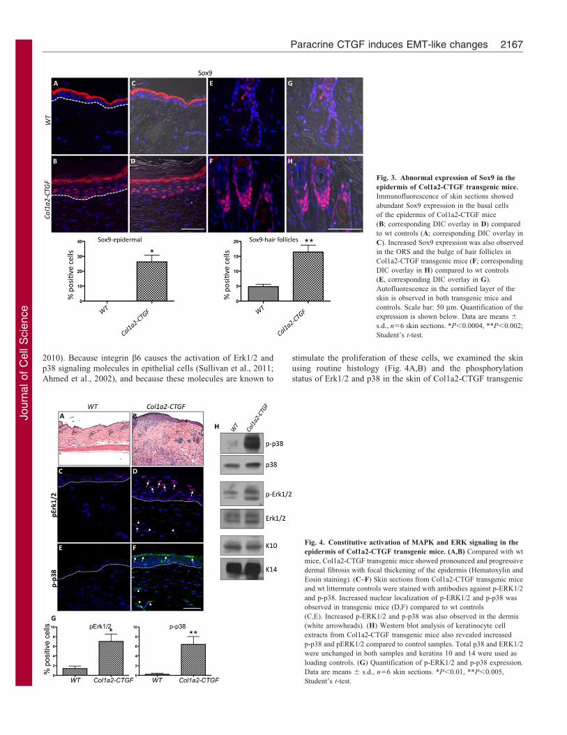
Fig. 3. Abnormal expression of Sox9 in the
epidermis of Col1a2-CTGF transgenic mice.
Immunofluorescence of skin sections showed
abundant Sox9 expression in the basal cells
of the epidermis of Col1a2-CTGF mice
(B; corresponding DIC overlay in D) compared
to wt controls (A; corresponding DIC overlay in
C). Increased Sox9 expression was also observed
in the ORS and the bulge of hair follicles in
Col1a2-CTGF transgenic mice (F; corresponding
DIC overlay in H) compared to wt controls
(E, corresponding DIC overlay in G).
Autofluorescence in the cornified layer of the
skin is observed in both transgenic mice and
controls. Scale bar: 50 mm. Quantification of the
expression is shown below. Data are means 6
s.d., n56 skin sections. *P,0.0004, **P,0.002;
Student’s t-test.
Fig. 4. Constitutive activation of MAPK and ERK signaling in the
epidermis of Col1a2-CTGF transgenic mice. (A,B) Compared with wt
mice, Col1a2-CTGF transgenic mice showed pronounced and progressive
dermal fibrosis with focal thickening of the epidermis (Hematoxylin and
Eosin staining). (C–F) Skin sections from Col1a2-CTGF transgenic mice
and wt littermate controls were stained with antibodies against p-ERK1/2
and p-p38. Increased nuclear localization of p-ERK1/2 and p-p38 was
observed in transgenic mice (D,F) compared to wt controls
(C,E). Increased p-ERK1/2 and p-p38 was also observed in the dermis
(white arrowheads). (H) Western blot analysis of keratinocyte cell
extracts from Col1a2-CTGF transgenic mice also revealed increased
p-p38 and pERK1/2 compared to control samples. Total p38 and ERK1/2
were unchanged in both samples and keratins 10 and 14 were used as
loading controls. (G) Quantification of p-ERK1/2 and p-p38 expression.
Data are means 6 s.d., n56 skin sections. *P,0.01, **P,0.005,
Student’s t-test.
Paracrine CTGF induces EMT-like changes 2167
Journ
alof
Cell
Scie
nce
mice and wt littermate controls. These experiments would help us
gain insights into the mechanism that could contribute to
increased proliferation and dedifferentiation of keratinocytes.
Immunofluorescence with antibodies against phosphorylated p38
(p-p38) and ERK1/2 (p-ERK1/2) showed three- to sevenfold
increases in the levels of both pErk1/2 (Fig. 4C,D,G) and p-p38
(Fig. 4E,F,G) in the granular and cornified layers of the
epidermis. We hypothesize that the increased expression of
integrin b6 and the increased activation of p38 and Erk1/2 could
account for the hyper-proliferation of keratinocytes in Col1a2-
CTGF transgenic mice.
Western blot analysis of protein extracts from primary
keratinocytes also showed constitutive activation of ERK1/2
and p38 in cells from Col1a2-CTGF transgenic mice (Fig. 4H).
Levels of total p38 and ERK1/2 were the same in cells derived
from both Col1a2-CTGF and wt mice (Fig. 4H).
Contribution of epithelial cells to fibrotic lung lesions in
Col1a2-CTGF transgenic mice
Col1a2-CTGF transgenic mice died early from extensive focal
fibrotic lesions within the lung parenchyma. The fibrosis was
characteristic with markedly increased Masson’s trichrome
staining and increased collagen type I, CTGF expression
compared to wt control mice (Sonnylal et al., 2010)
(supplementary material Fig. S1A–D). We also observed a
three to fourfold increase in BrdU-positive cells in the fibrotic
lesions suggesting increased numbers of cells undergoing cell
division (data not shown). To determine the proportion of cells
that are myofibroblasts and to distinguish them from alveolar
epithelial cells we performed co-immunostaining of lung sections
from Col1a2-CTGF transgenic mice and wt mice for aSMA and
TTF-1, markers for myofibroblasts and alveolar epithelial cells,
respectively. The immunofluorescence analysis revealed a
marked increase in aSMA-positive cells in the lesional areas of
the lungs of Col1a2-CTGF transgenic mice, whereas aSMA-
positive cells were lacking in the lungs of wt control mice
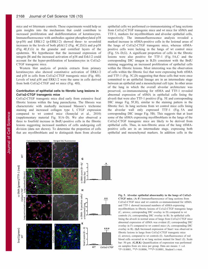
(Fig. 5A–D,G). A significant proportion of cells in the fibrotic
lesions were also positive for TTF-1 (Fig. 5A,C and the
corresponding DIC images in B,D) consistent with the BrdU
staining suggesting an increased proliferation of epithelial cells
within the fibrotic lesions. Most interesting was the observation
of cells within the fibrotic foci that were expressing both aSMA
and TTF-1 (Fig. 5C,D) suggesting that these cells that were once
committed to an epithelial lineage are in an intermediate stage
between an epithelial and a mesenchymal cell type. In other areas
of the lung in which the overall alveolar architecture was
preserved, co-immunostaining for aSMA and TTF-1 revealed
abnormal expression of aSMA in epithelial cells lining the
alveoli that were also TTF-1 positive (Fig. 5E and corresponding
DIC image Fig. 5F,H), similar to the staining pattern in the
fibrotic foci. In lung sections from wt control mice cells lining
the alveolar wall only expressed TTF-1 (Fig. 5A and
corresponding DIC image Fig. 5B). This suggests that at least
some of the aSMA expressing myofibroblasts in the lungs of the
Col1a2-CTGF transgenic mice are likely to be derived from
epithelial cells. Thus, in non-fibrotic areas of the lung aSMA-
positive cells are in an intermediate stage, expressing both
epithelial and mesenchymal markers. In addition cells in the
Fig. 5. Alveolar epithelial abnormality in the lungs of Col1a2-
CTGF mice. (A–F) Immunofluorescence of lung sections from
Col1a2-CTGF mice and wt controls co-immunostained for aSMA
and TTF-1 showed increased numbers of aSMA-expressing
myofibroblasts in fibrotic lesions of Co1a2-CTGF transgenic lungs
(C, arrows; corresponding DIC overlay in D) compared to wt
controls (A; corresponding DIC overlay in B). In epithelial cells
lining the alveoli in normal areas of lungs from Col1a2-CTGF mice
abnormal expression of aSMA was evident (E; corresponding DIC
overlay in F) compared to wt control mice (A; corresponding DIC
overlay in B). (I,J) Increased expression of Snai1 was observed in
fibrotic lesions in lungs from Col1a2-CTGF transgenic mice
(J, arrows), compared to wt controls (I). Autofluorescence of red
blood cells occurred in wt lung section stained for Snai1 (I). Scale
bar: 50 mm. (G,H,K) Quantification of expression was performed
on samples from six mice per group. Data are means 6 s.d.
*P,0.0001, **P,0.0006, ***P,0.0001, Student’s t-test.
Journal of Cell Science 126 (10)2168

Journ
alof
Cell
Scie
nce
fibrotic foci also stained for Snai1 suggesting that these cells have
undergone EMT (Fig. 5I,J,K). These results strongly suggest that
overexpression of CTGF in lung fibroblasts can induce EMT-like
changes in alveolar epithelial cells as assessed by co-expression of
epithelial and acquisition of mesenchymal cell markers.
EMT in lung fibrosis is associated with increased TGFb
signaling
To further understand the mechanisms of lung fibrosis in Col1a2-
CTGF transgenic mice, the expression levels of potential CTGF
target genes were measured by qRT-PCR. Since the fibrotic lung
had an abundance of macrophages, a known source of TGFb(Khalil et al., 1996), we also measured the mRNA levels of Tgfb1
and Tgfbr1. In addition to the expected increase in Ctgf mRNA,
we observed a marked enhancement of the levels of Tgfb1 and
Tgfbr1 mRNAs. Genes that are specific targets of the TGFbsignaling pathway such as Timp1, Serpine1 (PAI-I), Col1a1, Fn1
(fibronectin), Acta1 (alpha smooth muscle actin, aSMA) and
Sox9 were also overexpressed (Fig. 6). These alterations in
mRNA expression levels suggest that TGFb may be an important
participant in the fibrotic phenotype of the CTGF-induced lung
fibrosis and may also play a role in EMT of alveolar epithelial
cells described above.
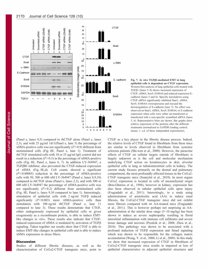
In vitro TGFb-mediated EMT in lung epithelial cells isinhibited by specific CTGF knockdown
There is evidence in the literature that TGFb can induce EMT in
lung epithelial cells in vitro (Xu et al., 2009). We wished to
determine whether this TGFb-induced EMT is mediated by
CTGF. Lung epithelial cells were treated in vitro with
recombinant TGFb1. We observed changes in cell morphology
from a rounded epithelial structure to a more spindle shaped
fibroblast-like structure (data not shown). In addition, western
blot analysis showed that the epithelial marker e-cadherin was
downregulated and other mesenchymal markers, including
aSMA, S100A4, SOX9, were upregulated in TGFb1-treated
cells (Fig. 7, lanes 5 and 6), indicating that EMT was occurring.
This evidence of EMT-like changes was accompanied by
increased expression of CTGF (Fig. 7, lanes 5 and 6). To
directly assess the role of increased CTGF in cellular changes in
epithelial cells, we used Ctgf siRNA to inhibit CTGF expression
after TGFb1 treatment. Specific CTGF knockdown significantly
decreased expression of SNAI1, aSMA and SOX9 expression
and increased expression of e-cadherin following TGFb1stimulation (Fig. 7, lanes 7 and 8). No effect was observed on
SNAI1, aSMA or SOX9 expression when cells were transfectedwith a non-specific scrambled siRNA (Fig. 7, lane 6). Hence,knockdown of CTGF blocks TGFb1-induced EMT in epithelialcells in vitro suggesting that CTGF mediates expression of EMT
markers in these TGFb1 treated cells.
CTGF treatment induces EMT-like changes in lungepithelial cells in vitro
As the TGFb1-induced EMT-like changes seen in lung epithelialcells are, at least in part, mediated by CTGF we investigated
whether treatment of cells with CTGF alone could induce EMT-like changes. Lung epithelial cells were transduced with anadenovirus expressing CTGF from the CMV promoter(Ad.CTGF). Phase contrast microscopy images of control cells
transduced with a GFP-expressing adenovirus (Ad.gfp) showed atypically epithelial cuboidal morphology (Fig. 8A,a), whereascells transduced with Ad.CTGF showed a change in morphology
to a spread, spindly morphology characteristic of mesenchymalcells (Fig. 8A,b–c). In addition, epithelial cells transduced withAd.CTGF show expression of the mesenchymal marker aSMA
(Fig. 8B,f), whereas this marker is absent in control cellstransduced with Ad.gfp (Fig. 8B,e). Deconvoluted z-stackimages (Fig. 8B,g) demonstrate that the expressed aSMA formsstress fibers. These experiments showed EMT-like changes occur
when CTGF is expressed by the epithelial cells themselves. Inorder to more closely mimic the in vivo situation in the Col1a2-CTGF mice, in which the CTGF is expressed by fibroblast cells
neighboring the epithelial cells, we treated epithelial cells in vitro
with recombinant human CTGF (rhCTGF). Treatment of cellswith rhCTGF in the absence of serum induced expression of
aSMA (Fig. 8B,b,c) with no expression seen in untreated cells(Fig. 8B,a), and deconvoluted z-stack images (Fig. 8B,d)demonstrate that the expressed aSMA forms stress fibers. The
induced expression seen by immunofluorescence was confirmedby western blot analysis (Fig. 8C) which showed a significant(P50.004) induction of a-SMA in treated cells. In order to assessany morphological changes, cells were stained with Rhodamine–
phalloidin and co-stained for aSMA (Fig. 8D). Untreated cells(Fig. 8D,a–c) show peripheral actin filaments (Fig. 8D,a) with noaSMA (Fig. 8D,b,c merge). Cells treated with 100 ng/ml
rhCTGF (Fig. 8D,d–f) show an increase in phalloidin staining(Fig. 8D,d) and an induction of aSMA (Fig. 8D,e,f merge).Furthermore, deconvoluted z-stack images (Fig. 8D,g–i) show
that the induced aSMA is incorporated into the actin filamentstress fibers.
As the fibrotic lungs of the Col1a2-CTGF mice show increasedTGFb signaling (Fig. 6), we investigated whether the EMT-like
changes induced by CTGF in vitro may be mediated by increasedTGFb signaling in a positive feedback loop. Epithelial cellsstimulated with rhCTGF were exposed to 1d11, a neutralizing
antibody to TGFb, or IgG control. Cells treated with rhCTGFalone induced expression of aSMA (Fig. 8E,b) compared tountreated cells (Fig. 8E,a). Cell counts showed a significantly
(P,0.00005) higher percentage of aSMA-positive cellsfollowing treatment with 50 or 100 ng/ml rhCTGF (Fig. 8E;Panel e, lanes 2,3). Antibody blockade of TGFb with 1d11
prevented the CTGF-induced expression of aSMA (Fig. 8E,c).Cell counts showed a significant (P,0.00005) reduction in thepercentage of aSMA-positive cells with 10 or 25 mg/ml 1d11
Fig. 6. Persistent activation of fibrotic and EMT marker genes in the
lungs of Col1a2-CTGF transgenic mice. qRT-PCR revealed a marked
increase in the expression of CTGF, TGFb, their target genes, and the EMT
markers Sox9 and aSMA in Col1a2-CTGF transgenic mice compared to wt
controls. Results are expressed as average fold change compared to wt control
mice; n54 mice per group.
Paracrine CTGF induces EMT-like changes 2169
Journ
alof
Cell
Scie
nce
(Panel e, lanes 4,5) compared to rhCTGF alone (Panel e, lanes
2,3), and with 25 mg/ml 1d11(Panel e, lane 5) the percentage of
aSMA-positive cells was not significantly (P.0.9) different from
unstimulated cells (Fig. 8E, Panel e, lane 1). Treatment of
rhCTGF stimulated cells with 10 or 25 mg/ml IgG control did not
result in a reduction (P.0.3) in the percentage of aSMA-positive
cells (Fig. 8E, Panel e, lanes 6, 7). In addition LY-364947, a
TGFbRI inhibitor, also prevented the CTGF-induced expression
of aSMA (Fig. 8E,d). Cell counts showed a significant
(P,0.00005) reduction in the percentage of aSMA-positive
cells with 50, 300 or 600 nM LY-364947 (Panel e, lanes 8,9,10)
compared to rhCTGF alone (Panel e, lanes 2,3), and with 300 or
600 nM LY-364947 the percentage of aSMA-positive cells was
not significantly (P.0.2) different from unstimulated cells
(Fig. 8E, Panel e, lanes 9,10 compared to lane 1). Interestingly,
stimulation of epithelial cells with 2 ng/ml TGFb induced
significantly (P,0.003) more aSMA-positive cells than
stimulation with 100 ng/ml rhCTGF (Panel e, lane 11
compared to lane 3). These results demonstrate that CTGF,
either endogenously expressed by epithelial cells or given
exogenously as a recombinant protein, is able to induce EMT-
like changes in vitro. These results also indicate that CTGF-
induced expression of aSMA is mediated by activation of TGFbsignaling. Taken together our results show that CTGF is able to
induce EMT-like changes in epithelial cells and is able to induce
aSMA expression in stress fibers.
DiscussionStudies of different fibrotic diseases, as well as the
characterization of Col1a2-CTGF transgenic mice, point to
CTGF as a key player in the fibrotic disease process. Indeed,
the relative levels of CTGF found in fibroblasts from these mice
are similar to levels observed in fibroblasts from systemic
sclerosis patients (Shi-wen et al., 2000). However, the paracrine
effects of CTGF on cellular targets outside of fibroblasts is
largely unknown as is the cell and molecular mechanism
underlying CTGF action on keratinocytes in skin, alveolar
epithelial cells in lung or tubulointerstitial cells in kidney. The
current study focuses primarily on the dermal and pulmonary
compartment, the most profoundly affected tissues in the Col1a2-
CTGF transgenic mice (Sonnylal et al., 2010). In most organs
Col1a2 expression is located in cells of mesenchymal origin
(Bou-Gharios et al., 1996), however in kidney, expression has
also been observed in tubular epithelial cells upon injury
(Fragiadaki et al., 2011). Interestingly, in a study using
administration of aristolochic acid (AA) to induce renal
fibrosis, the Col1a2-CTGF transgenic mice did not exhibit
more fibrosis compared with wt AA-treated mice (Fragiadaki
et al., 2011). This is however perhaps not surprising as AA-
administration at the similar dose range (5–10 mg/kg) has been
shown to induce as severe nephropathy resulting in florid
interstitial inflammation with immune cell infiltrates and severe
tissue damage and necrosis (Pozdzik et al., 2008; Zhou et al.,
2010). This pathology was shown to be associated with a
profound induction of TGFb expression and Smad signaling
which was shown to be responsible for the collagen matrix
accumulation (Wang, Y. et al., 2008; Li et al., 2009). In this study
we show that increased expression of CTGF in fibroblasts of
Col1a2-CTGF transgenic mice results in impaired or loss of
epithelial characteristics in adjacent epithelial structures and
Fig. 7. In vitro TGFb1-mediated EMT in lung
epithelial cells is dependent on CTGF expression.
Western blot analysis of lung epithelial cells treated with
TGFb1 (lanes 5–8) shows increased expression of
CTGF, aSMA, Sox9, S100A4 and reduced expression E-
cadherin (lanes 5 and 6). Specific knockdown using
CTGF siRNA significantly inhibited Snai1, aSMA,
Sox9, S100A4 overexpression and rescued the
downregulation of E-cadherin (lane 7). No effect was
observed on Snai1, aSMA, Sox9, S100A4, or E-cadherin
expression when cells were either un-transfected or
transfected with a non-specific scrambled siRNA (lanes
5, 6). Representative blots are shown. Bar graphs show
relative expression of the proteins after the different
treatments (normalized to GAPDH loading control;
means 6 s.d. of three independent experiments).
Journal of Cell Science 126 (10)2170

Journ
alof
Cell
Scie
nce
acquisition by these cells of mesenchymal properties in vivo in
the skin and lung presumably by paracrine mechanisms.
In addition to the fibrotic phenotype, Col1a2-CTGF transgenic
mice develop hair loss early on associated with hair follicle
abnormalities and focal hyperplasia of the epidermis. This hyper-
proliferation was associated with changes in cell morphology and
marked changes in gene expression including increased expression
of keratin K16. In the normal skin epidermis keratins K5 and K14
are expressed in the basal cells and keratins K1 and K10 are
expressed in the suprabasal layers (Koch and Roop, 2004). In some
diseased states such as in psoriasis where the epithelial cells
undergo hyperproliferation and/or aberrant differentiation,
expression of keratins K6 and its partner K16 are induced in the
inter-follicular epidermis (Bernot et al., 2002). Recent, proteomic
analysis of whole skin lesions from SSc patients shows abnormal
expression of K6 and K16 suggesting hyperproliferation of the
epidermal component in SSc (Aden et al., 2008).
In the skin of Col1a2-CTGF transgenic mice expression of the
epithelial-specific integrin b6 is also markedly increased in focal
areas of the epidermis. We speculate that the increase in b6
expression might eventually favor enhanced signaling by CTGF
since integrins have previously been shown to be involved in
such signaling. Mice in which integrin b6 overexpression is
targeted to the basal keratinocytes, showed loss of hair in the
lower back associated with fibrosis and chronic ulceration
characterized by hyperplasia of epithelial cells and thick
connective tissue (Hakkinen et al., 2004), a phenotype that is
analogous to that of Col1a2-CTGF transgenic mice.
Fig. 8. CTGF induces EMT-like changes in vitro, which are dependent on TGFb signaling. (A) CTGF expression induces a change in morphology in vitro.
Phase-contrast images of epithelial cells transduced with Ad.GFP (a) show a typically epithelial cobblestone morphology, whereas epithelial cells transduced
with Ad.CTGF (b,c) show a more spread, spindly morphology typical of mesenchymal cells. Scale bars: 20 mm. (B,C) CTGF induces expression of the
mesenchymal marker aSMA in epithelial cells in vitro. Epithelial cells were stained with a Cy3-conjugated anti-aSMA antibody (orange) and nuclei were counter-
stained with DAPI (blue). Cells were either exposed to exogenous rhCTGF (b–d) or transduced with adenovirus expressing CTGF (f,g) or gfp control
(e). Untreated (a) or Ad.gfp-transduced (e) epithelial cells show no aSMA expression, whereas cells treated with 50 ng/ml (b), 100 ng/ml (c), or 500 ng/ml
(d) rhCTGF, or transduced with Ad.CTGF (f,g) express aSMA. Deconvolution of z-stack images (d,g) shows that the expressed aSMA forms stress fibers. Scale
bars: 20 mm. In C, western blot analysis of epithelial cells treated with 10 or 50 ng/ml rhCTGF shows a significant (P50.004) induction of a-SMA compared to
controls. Representative blots for control and 50 ng/ml rhCTGF are shown. Graph shows densitometry of a-SMA (normalized to b-tubulin loading control). Data
are means 6 s.e.m. of three independent experiments. P-values were determined using Students t-test. (D) Exogenous CTGF induces a change in actin filaments in
vitro. Epithelial cells were co-stained with Rhodamine-conjugated phalloidin (red; a,d,g) and an anti-aSMA antibody (green; b,e,h), and images were merged
(c,f,i). Nuclei were counter-stained with DAPI (blue). Untreated cells (a–c) show mainly peripheral actin filaments that do not contain aSMA. Cells treated with
100 ng/ml rhCTGF (d–i) show an increase in actin filaments across the cells and these filaments include aSMA. Deconvoluted z-stack images (g–i) show the
formation of actin filaments and aSMA into stress fibers. Scale bars: 20 mm. (E) aSMA induction by CTGF is prevented by neutralization of TGFb or inhibition
of TGFbRI. Epithelial cells were stained with an anti-aSMA antibody (green), and nuclei were counter-stained with DAPI (blue). Cells were untreated
(a), exposed to rhCTGF (100 ng/ml; b), rhCTGF and a neutralizing anti-TGFB antibody, 1d11 (100 ng/ml and 25 mg/ml, respectively; c); or rhCTGF and the
small molecule TGFbRI inhibitor LY-364947 (100 ng/ml and 300 nM, respectively; d). Panel e, shows a graphical representation of the frequency of aSMA-
positive cells in treated and untreated cultures (as a percentage of total cell number) as means 6 s.e.m. of four fields of view at 206magnification. Lane 1, control
cultures; lanes 2 and 3, cells treated with rhCTGF at 50 and 100 ng/ml respectively; lanes 4 and 5, cells treated with rhCTGF at 100 ng/ml in the presence of
1d11 at 10 or 25 mg/ml, respectively; lanes 6 and 7, cells treated with rhCTGF at 100 ng/ml in the presence of a control IgG at 10 or 25 mg/ml, respectively;
lanes 8, 9 and 10, cells treated with rhCTGF at 100 ng/ml in the presence of the TGFbRI inhibitor LY-364947 at 50, 300 or 600 nM, respectively; lane 11,
cells treated with TGFb at 2 ng/ml. *P,0.00005 compared to untreated; #P,0.00005 compared to 100 ng/ml rhCTGF alone; ‘not significant (P.0.3) compared
to 100 ng/ml rhCTGF alone; Student’s t-test.
Paracrine CTGF induces EMT-like changes 2171
Journ
alof
Cell
Scie
nce
Furthermore the focal changes in the morphology of epidermalcells and the abnormal expression of aSMA, Snai1 and S100A4
by these cells in Col1a2-CTGF transgenic mice strongly suggeststhat the increased levels of CTGF that are secreted by the dermalfibroblasts resulting in an EMT-like process in neighboringepithelial cells presumably through a paracrine mechanism. EMT
is a process by which epithelial cells undergo a transition into amesenchymal cell-type giving rise to fibroblasts andmyofibroblasts. EMT is characterized by loss of epithelial
markers, loss of epithelial cell polarity, acquisition ofmesenchymal markers and an invasive phenotype (Kalluri andNeilson, 2003). This process is now being recognized to play an
important role in tissue repair and fibrosis. One recent reportprovides suggestive evidence consistent with EMT in post-menopausal frontal fibrosing alopecia (Nakamura and Tokura,2011). This disease is characterized by a recession of the hairline,
a reduction or loss of terminal hair follicles and fibrosis aroundthe affected hair follicles. Cells expressing Snai1 were present inthe fibrotic dermis raising the possibility that these cells might
have been derived from epithelial cells.
Emerging evidence from studies of liver fibrosis showed thatTGFb-induced expression of Sox9 in hepatic stellate cells (HSC)
in culture correlated with increased type I collagen production(Hanley et al., 2008). This increase can be inhibited byknockdown of Sox9 by a specific siRNA suggesting a role ofSox9 in ECM synthesis (Hanley et al., 2008).
In the Col1a2-CTGF transgenic mice the production of excessCTGF in dermal fibroblasts results in the abundant expression ofSox9 in the basal cells of the epidermis and in the outer root
sheath and bulge of hair follicles. We speculate that this occursthrough a paracrine mechanism. Interestingly embryonic ablationof Sox9 in K14 expressing epithelial cells of the skin results in
defects in the stem cell compartment and subsequent structuresthat arise from the stem cells. These mice also displayed impairedwound repair (Nowak et al., 2008). Our in vitro experiments in
lung A549 epithelial cells also show that knockdown of CTGF inthese cells is associated with markedly decreased levels of Sox9.Thus both in vivo and in vitro experiments establish a clearcorrelation between levels of CTGF and those of Sox9. Increased
expression of Sox9 is also observed in different cancers (Wanget al., 2007; Schaeffer et al., 2008). Inhibition of endogenousSox9 in CWR22Rv1, a human prostate cancer cell line, resulted
in decreased cellular proliferation (Wang et al., 2007).Transplantation of these cells in immuno-compromised miceresulted in decreased tumor growth correlating with its role in
cellular proliferation (Wang, H. et al., 2008). In in vitro
experiments, Sox9 activates the expression of the EMT markerSnail2 (Sakai et al., 2006) and co-expression of Sox9 and Snail2can induce EMT in neural epithelial cells (Cheung et al., 2005).
These lines of evidence would suggest the hypothesis that in ourmodel system increase in expression of Sox9 in the skin mightpromote proliferation of progenitor cells and changes in the
genetic program of existing epithelial cells or of newly formedepithelial cells undergoing EMT. In the epidermis of Col1a2-CTGF transgenic mice skin there was a marked increase in
phosphorylated p38 and ERK. We speculate that ERK and p38activation could mediate the paracrine effects of CTGF andtogether with the increased levels of b6 integrin have a role in
cell proliferation and migration. Previous in vivo studies of BLM-induced lung fibrosis have shown by fate mapping experimentsthat one third of cells expressing the fibroblast specific marker
S100A4 are derived from alveolar epithelial cells indicative of
EMT (Tanjore et al., 2009). Similar EMT-like event is also
observed in endoplasmic reticulum stress-induced lung fibrosis
(Tanjore et al., 2011). Other studies have suggested a link
between CTGF and lung fibrosis. Intratracheal bleomycin
treatment induces lung fibrosis with an increase in CTGF
expression in a bleomycin-sensitive mouse strain (C57BL/6) but
not in a bleomycin-resistant mouse strain (BALB/c) (Lasky et al.,
1998). Adenoviral-mediated CTGF expression in rat lungs
induces a transient fibrosis (Bonniaud et al., 2003).
Additionally, adenoviral expression of CTGF in the lungs of
bleomycin-resistant BALB/c mice leads to significant lung
fibrosis after treatment with bleomycin (Bonniaud et al., 2004).
The lung fibrosis seen in Col1a2-CTGF transgenic mice was
associated with increased proliferation of epithelial cells in the
fibrotic lesions exemplified by the increased number of cells that
expresses the epithelial specific transcription factor TTF-1. These
fibrotic lesions also contained a large number of myofibroblasts
expressing aSMA and a large percentage of these cells were co-
expressing both aSMA and TTF-1 suggesting that the
myofibroblast population arose from alveolar epithelial cells.
This hypothesis was also supported by evidence that in the
uninvolved areas of the transgenic mouse lung, which seemingly
displayed a normal architecture, epithelial cells lining the alveoli
co-expressed aSMA and TTF-1. Fibrotic lesions also displayed
cells positive for Snai1 suggesting that EMT is associated with
lung fibrosis. We propose that in the lung of Col1a2-CTGF
transgenic mice EMT is a consequence of the paracrine effects of
the abnormal high levels of secreted CTGF.
The results of the real-time PCR experiments on whole lung
RNA of Col1a2-CTGF transgenic mice showed markedly
increased RNA levels of TGFb and its downstream signature
targets. This is likely to be a consequence of the abundance of
macrophages in the fibrotic lung. In contrast, in the skin of
Col1a2-CTGF transgenic mice no macrophages were observed.
Microarrays of total skin RNA did not reveal any significant
increase in TGFb and TbR1 RNA levels (data not shown). We
propose that the combined increase in TGFb and CTGF in the
fibrotic lung not only causes increased myofibroblast
transformation and excess ECM deposition but could also
contribute to EMT in the lung epithelial cells. To understand
the role of CTGF in mediating EMT in the lung, we have used
lung epithelial cells to show that the activation of EMT marker
genes by TGFb can be inhibited by CTGF siRNA. We have also
demonstrated that CTGF expression by epithelial cells in vitro
causes EMT-like changes. Treatment of lung epithelial cells with
recombinant CTGF also induces EMT-like changes.
Additionally, we have demonstrated that the EMT-like changes
induced by recombinant CTGF in vitro can be prevented by
immunoinhibition of TGFb or by inhibition of TGFbRI
suggesting that TGFb signaling plays a central role in the EMT
process in lung epithelial cells. These in vitro findings strongly
suggest that paracrine CTGF is likely to have a direct effect on
inducing EMT in vivo. We speculate that these CTGF-induced
EMT-like changes may be a contributor to the development of
lung fibrosis. The results also suggest that the induction of TGFband TGFbRI by CTGF may establish a positive feedback loop
driving increased expression of endogenous CTGF and further
contributing to the EMT-like and fibrotic changes seen in the
lungs of Col1a2-CTGF mice.
Journal of Cell Science 126 (10)2172

Journ
alof
Cell
Scie
nce
In summary our in vivo observations strongly suggest thatparacrine effects of secreted CTGF produced in fibroblastic cellsof Col1a2-CTGF mice stimulate epithelial cells to modify their
genetic program in order to undergo changes that mimic featuresof EMT. The results of our in vitro experiments support theseconclusions. Thus CTGF plays a crucial role not only in
promoting a profibrotic program in fibroblastic cells but also inthe EMT-like transition of epithelial cells into mesenchymalcells. We speculate that the increase in Sox9 expression in skin
and lung epithelial cells may be an important factor in the EMT-like phenotype.
Materials and MethodsTransgenic mice
The Col1a2-CTGF transgenic mice used in this study have been describedpreviously (Sonnylal et al., 2010). Briefly, the murine homolog of CTGF (Fisp-12)was cloned into a vector containing the 6-kb enhancer and minimal promoter of themurine Col1a2 gene, an IRES-lacZ reporter, and the murine protamine polyAsignal. Transgenic mice were produced by the standard method of pronuclearinjection of linear DNA into a fertilized mouse egg. All experiments performedwith the mice were in compliance with the standards of care approved by the M. D.Anderson Cancer Center Institutional Animal Care and Use Committee.Genotyping was performed by PCR for the lacZ reporter on DNA extractedfrom ear biopsies.
Immunofluorescence of tissue sections and quantification
Skin and lung tissues from Col1a2-CTGF transgenic mice and wild-type (wt)littermate controls were fixed overnight in neutral buffered 4% paraformaldehydeat 4 C, processed through graded alcohols, and paraffin embedded.Immunofluorescence was performed on 5 mm sections with the followingprimary antibodies: Keratin 16 (Novus Biologicals, Littleton, CO, USA),integrin b4 (Southern Biotech, Birmingham, AL, USA), integrin b6 (R&DSystems, Minneapolis, MN, USA), aSMA (Sigma-Aldrich, St. Louis, MO, USA),Snai1 and CTGF (Santa Cruz Biotechnology Inc., Santa Cruz, CA, USA), S100A4(Abcam, Cambridge, MA, USA), Sox9 (Millipore, Billerica, MA, USA), TTF-1, p-p38, pERK (Cell Signaling, Lake Placid, NY, USA), and type I collagen (Novotec,Lyon, France). The secondary antibodies used were Alexa Fluor 488 goat anti-mouse IgG or Alexa Fluor 555 goat anti-rabbit IgG (Life Technologies, Carlsbad,CA, USA); nuclei were counterstained with Topro (Life Technologies, Carlsbad,CA, USA) and sections were mounted in ProLong Gold (Life Technologies,Carlsbad, CA, USA). Image stacks were collected using a Zeiss LSM 510 confocalmicroscope (Carl Zeiss, Oberkochen, USA). For type I collagen, sections werestained using the Vectastain ABC kit as previously described (Sonnylal et al.,2010). Quantitation for K16, integrin b4, integrin b6, a-SMA, Snai1, S100A4,Sox9, pErk1/2 and p-p38 staining of skin sections was performed usingMetaMorph software by counting the number of positive cells within a 100 mm2
box in at least six different tissue sections. The expression of a-SMA and Snai1 inlung sections was graded on a scale of 0 to 4 with 0 signifying no staining, 2signifying moderate staining and 3 or 4 signifying extensive staining. The numberof a-SMA-positive cells in uninvolved areas of lung was quantified as describedabove.
Cell culture
The lung epithelial cell lines A549 (ATCC: CCL-185) (Giard et al., 1973) andSV40-T2 (Clement et al., 1991) were maintained in DMEM medium (LifeTechnologies, Carlsbad, CA, USA) containing 10% FBS (BioSera, Ringmer, UK).For all treatments cells were serum starved overnight (cells were washed twice inPBS to remove serum then grown in DMEM without serum) and treatments wereperformed in DMEM without serum.
Keratinocytes were isolated from Col1a2-CTGF transgenic mice and wtlittermate controls as described previously (Flores et al., 2000). Briefly, the skinbiopsies were incubated in dispase II (Roche, Bassel, Switzerland) overnight at 4 Cto separate the epidermis from the dermis. The epidermis was carefully peeled offthe following day, chopped into small pieces, and incubated in 0.25% trypsin–EDTA (Life Technologies, Carlsbad, CA, USA) for 20 minutes at 37 C. Cellswere plated on J2-3T3 feeder cells and cultured in F medium (Sigma-Aldrich, St.Louis, MO, USA) supplemented with 0.4 mg/ml hydrocortisone, 24 ng/ml adenine,8.4 ng/ml cholera toxin, 5 mg/ml insulin, 13 ng/ml 3,3,5-triiodo-L-thyronine and10 ng/ml EGF.
Transfection with siRNA
Epithelial cells were transfected with ON-TARGETplus CTGF siRNA or non-targeting control siRNA (Dharmacon) using Fugene6 transfection reagent (Roche)in serum-free medium according to the manufacturers’ instructions. After 48 hours
transfected cells were treated with 4 ng/ml recombinant TGFb for a further24 hours before proteins were extracted for western blot analysis.
Adenoviral transduction
Epithelial cells were grown in T25 flasks (Corning Inc., Corning, NY, USA) to70% confluence then transduced at a multiplicity of infection of 100 with eitherAd.CTGF (Ad5 serotype adenovirus expressing human CTGF from the CMVpromoter) or Ad.gfp (Ad5 serotype adenovirus expressing GFP from the CMVpromoter, negative control). Briefly, the cell monolayer was washed in PBS toremove all serum, incubated for 2 hours with adenoviral particles in a minimalvolume of DMEM without serum, washed twice in PBS to remove any remainingvirus particles, then normal growth media was replaced. After viral transductionthe cells were allowed to recover for 24 hours then seeded into chamberslides (BDBioscience, Franklin Lakes, NJ, USA) at 70% confluence and allowed to attach for24 hours before fixing and staining.
Treatment with recombinant proteins, neutralizing antibodies andinhibitors
Recombinant human CTGF (rhCTGF) produced in HEK293 cells was bought fromEMP Genetech (Ingolstadt, Germany). Recombinant TGFb was from R&DSystems (Minneapolis, MN, USA). 1d11 (a mouse IgG1 neutralizing antibodyagainst TGFb) was from Abcam (Cambridge, MA, USA), purified mouse IgG(negative control) was from Vector Labs (Burlingame, CA, USA). LY-364947 (aselective TGFbRI inhibitor) was from Calbiochem (Merck KGaA, Darmstadt,Germany). For immunofluorescence, epithelial cells were seeded at 60%confluence in chamberslides, allowed to attach for 24 hours, serum-starvedovernight, then treated with rhCTGF, with and without 1d11, IgG or LY-364947,in DMEM without serum for 48 hours before fixing and staining. For western blotanalysis epithelial cells were seeded in six-well plates, allowed to attach for24 hours, serum-starved overnight, then treated with rhCTGF, with and without1d11, IgG or LY-364947, in DMEM without serum for 24 hours before proteinextraction.
Immunofluorescence of epithelial cell culture
Cells in chamber slides were fixed in ice-cold methanol (for staining with anit-aSMA antibody) or 2% PFA (for anti-aSMA and phalloidin co-staining). Cy3-conjugated anti- aSMA (clone 1A4, Sigma-Aldrich, St. Louis, MO, USA),unconjugated anti- aSMA (clone 1A4, Dako, Glostrup, Denmark), andRhodamine-conjugated phalloidin (Sigma-Aldrich, St. Louis, MO, USA) wereused at 1:100 dilution. Purified mouse IgG (Vector Labs, Burlingame, CA, USA)was used as negative control. Unconjugated anti-aSMA was detected with AlexaFluor 488 chicken anti-mouse IgG (Life Technologies, Carlsbad, CA, USA) at1:1000 dilution. Nuclei were counterstained with DAPI and mounted inVectashield (Vector Labs, Burlingame, CA, USA). Stained cells were imaged onan Axioskop Mot2 plus microscope with a monochrome camera and Axiovisionmicroscope control software (Carl Zeiss, Oberkochen, Germany). The IgG controlwas used to set the exposure time. Z-stack images were taken using automatedstage movement and inter-slice distances were optimised to fulfill the Nyquistcriterion. Z-stacks were deconvoluted using an iterative point spread function inAxiovision. Cell counts were performed using the cell counter plugin for ImageJ(US National Institutes of Health, Bethesda, MD, USA).
Western blot analysis
Cell extracts for western blot analysis has been described earlier (Sonnylal et al.,2007). Briefly, cells were cultured as monolayers, washed with ice-cold PBS, andproteins were extracted with RIPA buffer with protease inhibitors (50 mM Tris-HCl, pH 8.0, 1% NP-40, 0.5% sodium deoxycholate, 0.1% SDS, 150 mM NaCl,2 mM phenylmethylsulfonyl fluoride, 0.2 U/ml aprotinin). Cell extracts wereresolved on 10% SDS-PAGE and immunoblotted as described earlier (Sonnylalet al., 2007). In addition to the antibodies described above, the followingantibodies were also used in western blotting: CTGF (Santa Cruz BiotechnologyInc., Santa Cruz, CA, USA), p38, ERK (Cell Signaling, Lake Placid, NY, USA), e-cadherin (BD Biosciences, Franklin Lakes, NJ, USA), GAPDH, b-tubulin (Abcam,Cambridge, MA, USA). Blots were quantified with respect to the expression ofGAPDH by densitometric analysis using the UVP Biospectrum AC image systemwith VisionWorksLS software. The images present are representative of threeindependent experiments.
Real-time PCR
Standard RT-PCR was performed with the following primers: TGFb1 forward, 59-GAAGGGCCGGTTCATGTCATG-39; TGFb1 reverse, 59-TGTGACAGCAAAG-ATAACAAACTCCAC-39; TbR1 forward, 59-ATTGCTGGTCCAGTCTGCTT-39; TbR1-reverse, 59-CCTGATCCAGACCCTGATGT-39; Col1a1 forward, 59-CCCGCCGAT GTCGCTAT-39; Col1a1 reverse, 59-GCTACGCTG TTCTTGCA-GTGAT-39; Fibronectin forward, 59-GGTCTGCAGAGGTTGACAGTG-39,Fibronectin reverse, 59-GGAGAAGTTTGTGCATGGTGTCC-39; CTGFforward, 59-TTGTAATGGCAGGCACAGGTC-39; CTGF reverse, 59-CGCA-
Paracrine CTGF induces EMT-like changes 2173
Journ
alof
Cell
Scie
nce
CAAGAACCACCACTCTG-39 PAI 1 forward, 59-ATGCGGGCTGAGATGAC-AAAG-39; PAI 1 reverse, 59-ACTGCAAAAGGTCAGGATCGA-39; aSMAforward, 59-GCCAAGTCCAGACGCATGATG-39; aSMA reverse, 59-TATGC-CTCTGGACGTACAACT-39; TIMP 1 forward, 59-CACAAGCCTGGATTCCG-TGG-39; TIMP 1 reverse, 59-TCCCTTGCAAACTGGAGAGTGAC-39; Sox9forward, 59-GACGCTGGGCAAGCTCT-39; Sox9 reverse, 59-GTAATCCGGG-TGGTCCTTCT-39.
AcknowledgementsD.A. and B.de.C. are joint senior authors.
Author contributionsS.S., D.A., S.X., J.N. and B.d.C. developed the hypothesis, andconceived and designed the experiments. S.S., S.X., H.J., A.T.,V.R.S. P.A. and M.P. performed the experiments and contributed towriting the paper. S.S., H.J., D.A., J.N., M.P. and B.d.C. contributedto the supervision of the study, and helped in the analysis andinterpretation of data from experimental results. S.S., D.A. andB.d.C. wrote the paper. All authors provided critical comments ondrafts of the paper, and all read and approved the final manuscript.
FundingThis work was supported by National Institutes of Health/NationalInstitute of Arthritis and Musculoskeletal and Skin Diseases Centerof Research Translation in Scleroderma [grant numberP50AR054144 to B.d.C.]; Arthritis Research UK [grant numbers19427 and 18627 to D.A.]; the Medical Research Council [grantnumber G0801052 to D.A.]; and The Raynaud’s and SclerodermaAssociation [grant number RF27 to D.A.]. Deposited in PMC forrelease after 12 months.
Supplementary material available online at
http://jcs.biologists.org/lookup/suppl/doi:10.1242/jcs.111302/-/DC1
ReferencesAbraham, D. J., Eckes, B., Rajkumar, V. and Krieg, T. (2007). New developments in
fibroblast and myofibroblast biology: implications for fibrosis and scleroderma. Curr.
Rheumatol. Rep. 9, 136-143.
Aden, N., Shiwen, X., Aden, D., Black, C., Nuttall, A., Denton, C. P., Leask, A.,
Abraham, D. and Stratton, R. (2008). Proteomic analysis of scleroderma lesionalskin reveals activated wound healing phenotype of epidermal cell layer.Rheumatology (Oxford) 47, 1754-1760.
Ahmed, N., Niu, J., Dorahy, D. J., Gu, X., Andrews, S., Meldrum, C. J., Scott,R. J., Baker, M. S., Macreadie, I. G. and Agrez, M. V. (2002). Direct integrinalphavbeta6-ERK binding: implications for tumour growth. Oncogene 21, 1370-1380.
Bennett, M. R., Czech, K. A., Arend, L. J., Witte, D. P., Devarajan, P. and Potter,
S. S. (2007). Laser capture microdissection-microarray analysis of focal segmentalglomerulosclerosis glomeruli. Nephron Exp. Nephrol. 107, e30-e40.
Bernot, K. M., Coulombe, P. A. and McGowan, K. M. (2002). Keratin 16 expressiondefines a subset of epithelial cells during skin morphogenesis and the hair cycle.J. Invest. Dermatol. 119, 1137-1149.
Bi, W., Deng, J. M., Zhang, Z., Behringer, R. R. and de Crombrugghe, B. (1999).Sox9 is required for cartilage formation. Nat. Genet. 22, 85-89.
Bonniaud, P., Margetts, P. J., Kolb, M., Haberberger, T., Kelly, M., Robertson,J. and Gauldie, J. (2003). Adenoviral gene transfer of connective tissue growthfactor in the lung induces transient fibrosis. Am. J. Respir. Crit. Care Med. 168, 770-778.
Bonniaud, P., Martin, G., Margetts, P. J., Ask, K., Robertson, J., Gauldie, J. andKolb, M. (2004). Connective tissue growth factor is crucial to inducing a profibroticenvironment in ‘‘fibrosis-resistant’’ BALB/c mouse lungs. Am. J. Respir. Cell Mol.
Biol. 31, 510-516.
Bou-Gharios, G., Garret, L. A., Rossert, J., Niederreither, K., Eberspaecher, H.,
Smith, C. et al. (1996). A potent far-upstream enhancer in the mouse proa2(I)collagen gene regulates expression of reporter genes in transgenic mice. J. Cell Biol.
132, 1333-1344.
Cheung, M., Chaboissier, M. C., Mynett, A., Hirst, E., Schedl, A. and Briscoe,J. (2005). The transcriptional control of trunk neural crest induction, survival, anddelamination. Dev. Cell 8, 179-192.
Clement, A., Steele, M. P., Brody, J. S. and Riedel, N. (1991). SV40T-immortalizedlung alveolar epithelial cells display post-transcriptional regulation of proliferation-related genes. Exp. Cell Res. 196, 198-205.
Flores, E. R., Allen-Hoffmann, B. L., Lee, D. and Lambert, P. F. (2000). The humanpapillomavirus type 16 E7 oncogene is required for the productive stage of the virallife cycle. J. Virol. 74, 6622-6631.
Fragiadaki, M., Witherden, A. S., Kaneko, T., Sonnylal, S., Pusey, C. D., Bou-
Gharios, G. and Mason, R. M. (2011). Interstitial fibrosis is associated withincreased COL1A2 transcription in AA-injured renal tubular epithelial cells in vivo.
Matrix Biol. 30, 396-403.
Friedrichsen, S., Heuer, H., Christ, S., Winckler, M., Brauer, D., Bauer, K. and
Raivich, G. (2003). CTGF expression during mouse embryonic development. Cell
Tissue Res. 312, 175-188.
Giard, D. J., Aaronson, S. A., Todaro, G. J., Arnstein, P., Kersey, J. H., Dosik,
H. and Parks, W. P. (1973). In vitro cultivation of human tumors: establishment
of cell lines derived from a series of solid tumors. J. Natl. Cancer Inst. 51, 1417-1423.
Hakkinen, L., Koivisto, L., Gardner, H., Saarialho-Kere, U., Carroll, J. M., Lakso,
M., Rauvala, H., Laato, M., Heino, J. and Larjava, H. (2004). Increased expression
of beta6-integrin in skin leads to spontaneous development of chronic wounds. Am. J.
Pathol. 164, 229-242.
Hanley, K. P., Oakley, F., Sugden, S., Wilson, D. I., Mann, D. A. and Hanley, N. A.
(2008). Ectopic SOX9 mediates extracellular matrix deposition characteristic of organfibrosis. J. Biol. Chem. 283, 14063-14071.
Huang, X. Z., Wu, J. F., Cass, D., Erle, D. J., Corry, D., Young, S. G., Farese, R. V.,
Jr and Sheppard, D. (1996). Inactivation of the integrin beta 6 subunit gene reveals a
role of epithelial integrins in regulating inflammation in the lung and skin. J. Cell
Biol. 133, 921-928.
Ivkovic, S., Yoon, B. S., Popoff, S. N., Safadi, F. F., Libuda, D. E., Stephenson, R. C.,
Daluiski, A. and Lyons, K. M. (2003). Connective tissue growth factor coordinateschondrogenesis and angiogenesis during skeletal development. Development 130,
2779-2791.
Kalluri, R. and Neilson, E. G. (2003). Epithelial-mesenchymal transition and itsimplications for fibrosis. J. Clin. Invest. 112, 1776-1784.
Khalil, N., Corne, S., Whitman, C. and Yacyshyn, H. (1996). Plasmin regulates theactivation of cell-associated latent TGF-beta 1 secreted by rat alveolar
macrophages after in vivo bleomycin injury. Am. J. Respir. Cell Mol. Biol. 15,252-259.
Koch, P. J. and Roop, D. R. (2004). The role of keratins in epidermal development and
homeostasis—going beyond the obvious. J. Invest. Dermatol. 123, x-xi.
Krieg, T., Abraham, D. and Lafyatis, R. (2007). Fibrosis in connective tissue disease:
the role of the myofibroblast and fibroblast-epithelial cell interactions. Arthritis Res.
Ther. 9 Suppl 2, S4.
Lasky, J. A., Ortiz, L. A., Tonthat, B., Hoyle, G. W., Corti, M., Athas, G.,
Lungarella, G., Brody, A. and Friedman, M. (1998). Connective tissue growthfactor mRNA expression is upregulated in bleomycin-induced lung fibrosis. Am. J.
Physiol. 275, L365-L371.
Leask, A. and Abraham, D. J. (2006). All in the CCN family: essential matricellular
signaling modulators emerge from the bunker. J. Cell Sci. 119, 4803-4810.
Li, J., Zhang, Z., Wang, D., Wang, Y., Li, Y. and Wu, G. (2009). TGF-beta 1/Smadssignaling stimulates renal interstitial fibrosis in experimental AAN. J. Recept. Signal
Transduct. Res. 29, 280-285.
Naitoh, M., Kubota, H., Ikeda, M., Tanaka, T., Shirane, H., Suzuki, S. and Nagata,
K. (2005). Gene expression in human keloids is altered from dermal to chondrocytic
and osteogenic lineage. Genes Cells 10, 1081-1091.
Nakamura, M. and Tokura, Y. (2011). Epithelial-mesenchymal transition in the skin.
J. Dermatol. Sci. 61, 7-13.
Nowak, J. A., Polak, L., Pasolli, H. A. and Fuchs, E. (2008). Hair follicle stem
cells are specified and function in early skin morphogenesis. Cell Stem Cell 3,
33-43.
Pohlers, D., Brenmoehl, J., Loffler, I., Muller, C. K., Leipner, C., Schultze-Mosgau,
S., Stallmach, A., Kinne, R. W. and Wolf, G. (2009). TGF-beta and fibrosis indifferent organs – molecular pathway imprints. Biochim. Biophys. Acta 1792, 746-
756.
Pozdzik, A. A., Salmon, I. J., Husson, C. P., Decaestecker, C., Rogier, E.,
Bourgeade, M. F., Deschodt-Lanckman, M. M., Vanherweghem, J. L. and
Nortier, J. L. (2008). Patterns of interstitial inflammation during the evolution ofrenal injury in experimental aristolochic acid nephropathy. Nephrol. Dial. Transplant.
23, 2480-2491.
Pritchett, J., Athwal, V., Roberts, N., Hanley, N. A. and Hanley, K. P. (2011).
Understanding the role of SOX9 in acquired diseases: lessons from development.
Trends Mol. Med. 17, 166-174.
Sakai, D., Suzuki, T., Osumi, N. and Wakamatsu, Y. (2006). Cooperative action of
Sox9, Snail2 and PKA signaling in early neural crest development. Development 133,1323-1333.
Schaeffer, E. M., Marchionni, L., Huang, Z., Simons, B., Blackman, A., Yu, W.,
Parmigiani, G. and Berman, D. M. (2008). Androgen-induced programs for prostateepithelial growth and invasion arise in embryogenesis and are reactivated in cancer.
Oncogene 27, 7180-7191.
Shi-wen, X., Pennington, D., Holmes, A., Leask, A., Bradham, D., Beauchamp, J. R.,
Fonseca, C., du Bois, R. M., Martin, G. R., Black, C. M. et al. (2000). Autocrineoverexpression of CTGF maintains fibrosis: RDA analysis of fibrosis genes in
systemic sclerosis. Exp. Cell Res. 259, 213-224.
Sonnylal, S., Denton, C. P., Zheng, B., Keene, D. R., He, R., Adams, H. P., Vanpelt,
C. S., Geng, Y. J., Deng, J. M., Behringer, R. R. et al. (2007). Postnatal induction
of transforming growth factor beta signaling in fibroblasts of mice recapitulatesclinical, histologic, and biochemical features of scleroderma. Arthritis Rheum. 56,
334-344.
Journal of Cell Science 126 (10)2174

Journ
alof
Cell
Scie
nce
Sonnylal, S., Shi-Wen, X., Leoni, P., Naff, K., Van Pelt, C. S., Nakamura, H., Leask,A., Abraham, D., Bou-Gharios, G. and de Crombrugghe, B. (2010). Selectiveexpression of connective tissue growth factor in fibroblasts in vivo promotes systemictissue fibrosis. Arthritis Rheum. 62, 1523-1532.
Sullivan, B. P., Kassel, K. M., Manley, S., Baker, A. K. and Luyendyk, J. P. (2011).Regulation of transforming growth factor-b1-dependent integrin b6 expression by p38mitogen-activated protein kinase in bile duct epithelial cells. J. Pharmacol. Exp. Ther.
337, 471-478.Tanjore, H., Xu, X. C., Polosukhin, V. V., Degryse, A. L., Li, B., Han, W., Sherrill,
T. P., Plieth, D., Neilson, E. G., Blackwell, T. S. et al. (2009). Contribution ofepithelial-derived fibroblasts to bleomycin-induced lung fibrosis. Am. J. Respir. Crit.
Care Med. 180, 657-665.Tanjore, H., Cheng, D. S., Degryse, A. L., Zoz, D. F., Abdolrasulnia, R., Lawson,
W. E. and Blackwell, T. S. (2011). Alveolar epithelial cells undergo epithelial-to-mesenchymal transition in response to endoplasmic reticulum stress. J. Biol. Chem.
286, 30972-30980.Vidal, V. P., Chaboissier, M. C., Lutzkendorf, S., Cotsarelis, G., Mill, P., Hui, C. C.,
Ortonne, N., Ortonne, J. P. and Schedl, A. (2005). Sox9 is essential for outer root
sheath differentiation and the formation of the hair stem cell compartment. Curr. Biol.
15, 1340-1351.
Wang, H., McKnight, N. C., Zhang, T., Lu, M. L., Balk, S. P. and Yuan, X. (2007).
SOX9 is expressed in normal prostate basal cells and regulates androgen receptor
expression in prostate cancer cells. Cancer Res. 67, 528-536.
Wang, H., Leav, I., Ibaragi, S., Wegner, M., Hu, G. F., Lu, M. L., Balk, S. P. and
Yuan, X. (2008). SOX9 is expressed in human fetal prostate epithelium and enhances
prostate cancer invasion. Cancer Res. 68, 1625-1630.
Wang, Y., Zhang, Z., Shen, H., Lu, Y., Li, H., Ren, X. and Wu, G. (2008). TGF-
beta1/Smad7 signaling stimulates renal tubulointerstitial fibrosis induced by AAI.
J. Recept. Signal Transduct. Res. 28, 413-428.
Wynn, T. A. (2008). Cellular and molecular mechanisms of fibrosis. J. Pathol. 214, 199-210.
Xu, J., Lamouille, S. and Derynck, R. (2009). TGF-b-induced epithelial to
mesenchymal transition. Cell Res. 19, 156-172.
Zhou, L., Fu, P., Huang, X. R., Liu, F., Chung, A. C., Lai, K. N. and Lan, H. Y.
(2010). Mechanism of chronic aristolochic acid nephropathy: role of Smad3. Am. J.
Physiol. Renal Physiol. 298, F1006-F1017.
Paracrine CTGF induces EMT-like changes 2175