Comparing open and minimally invasive surgical procedures...
101
Metcalfe, C., Avery, K., Noble, S., Moure Fernandez, A., Donovan, J., & Blazeby, J. (2016). Comparing open and minimally invasive surgical procedures for oesophagectomy in the treatment of cancer: the ROMIO (Randomised Oesophagectomy: Minimally Invasive or Open) feasibility study and pilot trial. Health Technology Assessment, 20(48). DOI: 10.3310/hta20480 Publisher's PDF, also known as Version of record License (if available): Other Link to published version (if available): 10.3310/hta20480 Link to publication record in Explore Bristol Research PDF-document This is the final published version of the article (version of record). It first appeared online via NIHR Health Technology Assessment Programme at http://dx.doi.org/10.3310/hta20480. Please refer to any applicable terms of use of the publisher. University of Bristol - Explore Bristol Research General rights This document is made available in accordance with publisher policies. Please cite only the published version using the reference above. Full terms of use are available: http://www.bristol.ac.uk/pure/about/ebr-terms
Transcript of Comparing open and minimally invasive surgical procedures...
Metcalfe, C., Avery, K., Noble, S., Moure Fernandez, A., Donovan, J., &Blazeby, J. (2016). Comparing open and minimally invasive surgicalprocedures for oesophagectomy in the treatment of cancer: the ROMIO(Randomised Oesophagectomy: Minimally Invasive or Open) feasibilitystudy and pilot trial. Health Technology Assessment, 20(48). DOI:10.3310/hta20480
Publisher's PDF, also known as Version of record
License (if available):Other
Link to published version (if available):10.3310/hta20480
Link to publication record in Explore Bristol ResearchPDF-document
This is the final published version of the article (version of record). It first appeared online via NIHR HealthTechnology Assessment Programme at http://dx.doi.org/10.3310/hta20480. Please refer to any applicable termsof use of the publisher.
University of Bristol - Explore Bristol ResearchGeneral rights
This document is made available in accordance with publisher policies. Please cite only the publishedversion using the reference above. Full terms of use are available:http://www.bristol.ac.uk/pure/about/ebr-terms
HEALTH TECHNOLOGY ASSESSMENTVOLUME 20 ISSUE 48 JUNE 2016
ISSN 1366-5278
DOI 10.3310/hta20480
Comparing open and minimally invasive surgical procedures for oesophagectomy in the treatment of cancer: the ROMIO (Randomised Oesophagectomy: Minimally Invasive or Open) feasibility study and pilot trial
Chris Metcalfe, Kerry Avery, Richard Berrisford, Paul Barham, Sian M Noble, Aida Moure Fernandez, George Hanna, Robert Goldin, Jackie Elliott, Timothy Wheatley, Grant Sanders, Andrew Hollowood, Stephen Falk, Dan Titcomb, Christopher Streets, Jenny L Donovan and Jane M Blazeby


Comparing open and minimally invasivesurgical procedures for oesophagectomyin the treatment of cancer: the ROMIO(Randomised Oesophagectomy: MinimallyInvasive or Open) feasibility study andpilot trial
Chris Metcalfe,1,2* Kerry Avery,2 Richard Berrisford,3
Paul Barham,4 Sian M Noble,2 Aida Moure Fernandez,2
George Hanna,5 Robert Goldin,6 Jackie Elliott,7
Timothy Wheatley,3 Grant Sanders,3
Andrew Hollowood,4 Stephen Falk,8 Dan Titcomb,4
Christopher Streets,4 Jenny L Donovan2
and Jane M Blazeby2,4
1Bristol Randomised Trials Collaboration, University of Bristol, Bristol, UK2School of Social and Community Medicine, University of Bristol, Bristol, UK3Department of Upper Gastrointestinal Surgery, Plymouth Hospitals NHS Trust,Plymouth, UK
4Division of Surgery, Head and Neck, University Hospitals Bristol NHS FoundationTrust, Bristol, UK
5Department of Surgery and Cancer, Imperial College London, London, UK6Department of Cellular Pathology, Imperial College London, London, UK7Gastro-Oesophageal Support and Help Group, Kingswood, Bristol, UK8Bristol Oncology Centre, University Hospitals Bristol NHS Foundation Trust,Bristol, UK
*Corresponding author
Declared competing interests of authors: none
Published June 2016DOI: 10.3310/hta20480


This report should be referenced as follows:
Metcalfe C, Avery K, Berrisford R, Barham P, Noble SM, Fernandez AM, et al. Comparing open and
minimally invasive surgical procedures for oesophagectomy in the treatment of cancer: the ROMIO
(Randomised Oesophagectomy: Minimally Invasive or Open) feasibility study and pilot trial. HealthTechnol Assess 2016;20(48).
Health Technology Assessment is indexed and abstracted in Index Medicus/MEDLINE, ExcerptaMedica/EMBASE, Science Citation Index Expanded (SciSearch®) and Current Contents®/Clinical Medicine.

Health Technology Assessment HTA/HTA TAR
ISSN 1366-5278 (Print)
ISSN 2046-4924 (Online)
Impact factor: 4.058
Health Technology Assessment is indexed in MEDLINE, CINAHL, EMBASE, The Cochrane Library and the ISI Science Citation Index.
This journal is a member of and subscribes to the principles of the Committee on Publication Ethics (COPE) (www.publicationethics.org/).
Editorial contact: [email protected]
The full HTA archive is freely available to view online at www.journalslibrary.nihr.ac.uk/hta. Print-on-demand copies can be purchased from thereport pages of the NIHR Journals Library website: www.journalslibrary.nihr.ac.uk
Criteria for inclusion in the Health Technology Assessment journalReports are published in Health Technology Assessment (HTA) if (1) they have resulted from work for the HTA programme, and (2) theyare of a sufficiently high scientific quality as assessed by the reviewers and editors.
Reviews in Health Technology Assessment are termed ‘systematic’ when the account of the search appraisal and synthesis methods (tominimise biases and random errors) would, in theory, permit the replication of the review by others.
HTA programmeThe HTA programme, part of the National Institute for Health Research (NIHR), was set up in 1993. It produces high-quality researchinformation on the effectiveness, costs and broader impact of health technologies for those who use, manage and provide care in the NHS.‘Health technologies’ are broadly defined as all interventions used to promote health, prevent and treat disease, and improve rehabilitationand long-term care.
The journal is indexed in NHS Evidence via its abstracts included in MEDLINE and its Technology Assessment Reports inform National Institutefor Health and Care Excellence (NICE) guidance. HTA research is also an important source of evidence for National Screening Committee (NSC)policy decisions.
For more information about the HTA programme please visit the website: http://www.nets.nihr.ac.uk/programmes/hta
This reportThe research reported in this issue of the journal was funded by the HTA programme as project number 10/50/65. The contractual start datewas in January 2013. The draft report began editorial review in February 2015 and was accepted for publication in December 2015. Theauthors have been wholly responsible for all data collection, analysis and interpretation, and for writing up their work. The HTA editors andpublisher have tried to ensure the accuracy of the authors’ report and would like to thank the reviewers for their constructive comments onthe draft document. However, they do not accept liability for damages or losses arising from material published in this report.
This report presents independent research funded by the National Institute for Health Research (NIHR). The views and opinions expressed byauthors in this publication are those of the authors and do not necessarily reflect those of the NHS, the NIHR, NETSCC, the HTA programmeor the Department of Health. If there are verbatim quotations included in this publication the views and opinions expressed by theinterviewees are those of the interviewees and do not necessarily reflect those of the authors, those of the NHS, the NIHR, NETSCC, the HTAprogramme or the Department of Health.
© Queen’s Printer and Controller of HMSO 2016. This work was produced by Metcalfe et al. under the terms of a commissioningcontract issued by the Secretary of State for Health. This issue may be freely reproduced for the purposes of private research andstudy and extracts (or indeed, the full report) may be included in professional journals provided that suitable acknowledgementis made and the reproduction is not associated with any form of advertising. Applications for commercial reproduction should beaddressed to: NIHR Journals Library, National Institute for Health Research, Evaluation, Trials and Studies Coordinating Centre,Alpha House, University of Southampton Science Park, Southampton SO16 7NS, UK.
Published by the NIHR Journals Library (www.journalslibrary.nihr.ac.uk), produced by Prepress Projects Ltd, Perth, Scotland(www.prepress-projects.co.uk).
Editor-in-Chief
Health Technology Assessment
NIHR Journals Library
Professor Tom Walley Director, NIHR Evaluation, Trials and Studies and Director of the EME Programme, UK
NIHR Journals Library Editors
Professor Ken Stein Chair of HTA Editorial Board and Professor of Public Health, University of Exeter Medical School, UK
Professor Andree Le May Chair of NIHR Journals Library Editorial Group (EME, HS&DR, PGfAR, PHR journals)
Dr Martin Ashton-Key Consultant in Public Health Medicine/Consultant Advisor, NETSCC, UK
Professor Matthias Beck Chair in Public Sector Management and Subject Leader (Management Group), Queen’s University Management School, Queen’s University Belfast, UK
Professor Aileen Clarke Professor of Public Health and Health Services Research, Warwick Medical School, University of Warwick, UK
Dr Tessa Crilly Director, Crystal Blue Consulting Ltd, UK
Dr Peter Davidson Director of NETSCC, HTA, UK
Ms Tara Lamont Scientific Advisor, NETSCC, UK
Professor Elaine McColl Director, Newcastle Clinical Trials Unit, Institute of Health and Society, Newcastle University, UK
Professor William McGuire Professor of Child Health, Hull York Medical School, University of York, UK
Professor John Norrie Health Services Research Unit, University of Aberdeen, UK
Professor John Powell Consultant Clinical Adviser, National Institute for Health and Care Excellence (NICE), UK
Professor James Raftery Professor of Health Technology Assessment, Wessex Institute, Faculty of Medicine, University of Southampton, UK
Dr Rob Riemsma Reviews Manager, Kleijnen Systematic Reviews Ltd, UK
Professor Helen Roberts Professor of Child Health Research, UCL Institute of Child Health, UK
Professor Helen Snooks Professor of Health Services Research, Institute of Life Science, College of Medicine, Swansea University, UK
Professor Jim Thornton Professor of Obstetrics and Gynaecology, Faculty of Medicine and Health Sciences, University of Nottingham, UK
Please visit the website for a list of members of the NIHR Journals Library Board: www.journalslibrary.nihr.ac.uk/about/editors
Editorial contact: [email protected]
Professor Geoffrey Meads Professor of Health Sciences Research, Health and Wellbeing Research andDevelopment Group, University of Winchester, UK
Editor-in-Chief
Professor Hywel Williams Director, HTA Programme, UK and Foundation Professor and Co-Director of theCentre of Evidence-Based Dermatology, University of Nottingham, UK
Professor Jonathan Ross Professor of Sexual Health and HIV, University Hospital Birmingham, UK
Dr Eugenia Cronin Senior Scientific Advisor, Wessex Institute, UK
Professor Martin Underwood Director, Warwick Clinical Trials Unit, Warwick Medical School,University of Warwick, UK
NIHR Journals Library www.journalslibrary.nihr.ac.uk
Abstract
Comparing open and minimally invasive surgical proceduresfor oesophagectomy in the treatment of cancer: the ROMIO(Randomised Oesophagectomy: Minimally Invasive or Open)feasibility study and pilot trial
Chris Metcalfe,1,2* Kerry Avery,2 Richard Berrisford,3 Paul Barham,4
Sian M Noble,2 Aida Moure Fernandez,2 George Hanna,5
Robert Goldin,6 Jackie Elliott,7 Timothy Wheatley,3 Grant Sanders,3
Andrew Hollowood,4 Stephen Falk,8 Dan Titcomb,4
Christopher Streets,4 Jenny L Donovan2 and Jane M Blazeby2,4
1Bristol Randomised Trials Collaboration, University of Bristol, Bristol, UK2School of Social and Community Medicine, University of Bristol, Bristol, UK3Department of Upper Gastrointestinal Surgery, Plymouth Hospitals NHS Trust, Plymouth, UK4Division of Surgery, Head and Neck, University Hospitals Bristol NHS Foundation Trust, Bristol, UK5Department of Surgery and Cancer, Imperial College London, London, UK6Department of Cellular Pathology, Imperial College London, London, UK7Gastro-Oesophageal Support and Help Group, Kingswood, Bristol, UK8Bristol Oncology Centre, University Hospitals Bristol NHS Foundation Trust, Bristol, UK
*Corresponding author [email protected]
Background: Localised oesophageal cancer can be curatively treated with surgery (oesophagectomy) butthe procedure is complex with a risk of complications, negative effects on quality of life and a recoveryperiod of 6–9 months. Minimal-access surgery may accelerate recovery.
Objectives: The ROMIO (Randomised Oesophagectomy: Minimally Invasive or Open) study aimed toestablish the feasibility of, and methodology for, a definitive trial comparing minimally invasive and opensurgery for oesophagectomy. Objectives were to quantify the number of eligible patients in a pilot trial;develop surgical manuals as the basis for quality assurance; standardise pathological processing; establish amethod to blind patients to their allocation in the first week post surgery; identify measures of postsurgicaloutcome of importance to patients and clinicians; and establish the main cost differences between thesurgical approaches.
Design: Pilot parallel three-arm randomised controlled trial nested within feasibility work.
Setting: Two UK NHS departments of upper gastrointestinal surgery.
Participants: Patients aged ≥ 18 years with histopathological evidence of oesophageal oroesophagogastric junctional adenocarcinoma, squamous cell cancer or high-grade dysplasia, referred foroesophagectomy or oesophagectomy following neoadjuvant chemo(radio)therapy.
Interventions: Oesophagectomy, with patients randomised to open surgery, a hybrid open chest andminimally invasive abdomen or totally minimally invasive access.
DOI: 10.3310/hta20480 HEALTH TECHNOLOGY ASSESSMENT 2016 VOL. 20 NO. 48
© Queen’s Printer and Controller of HMSO 2016. This work was produced by Metcalfe et al. under the terms of a commissioning contract issued by the Secretary of State forHealth. This issue may be freely reproduced for the purposes of private research and study and extracts (or indeed, the full report) may be included in professional journalsprovided that suitable acknowledgement is made and the reproduction is not associated with any form of advertising. Applications for commercial reproduction should beaddressed to: NIHR Journals Library, National Institute for Health Research, Evaluation, Trials and Studies Coordinating Centre, Alpha House, University of Southampton SciencePark, Southampton SO16 7NS, UK.
vii
Main outcome measure: The primary outcome measure for the pilot trial was the number of patientsrecruited per month, with the main trial considered feasible if at least 2.5 patients per monthwere recruited.
Results: During 21 months of recruitment, 263 patients were assessed for eligibility; of these, 135 (51%)were found to be eligible and 104 (77%) agreed to participate, an average of five patients per month. Intotal, 41 patients were allocated to open surgery, 43 to the hybrid procedure and 20 to totally minimallyinvasive surgery. Recruitment is continuing, allowing a seamless transition into the definitive trial.Consequently, the database is unlocked at the time of writing and data presented here are for patientsrecruited by 31 August 2014. Random allocation achieved a good balance between the arms of the study,which, as a high proportion of patients underwent their allocated surgery (69/79, 87%), ensured a faircomparison between the interventions. Dressing patients with large bandages, covering all possibleincisions, was successful in keeping patients blind while pain was assessed during the first week postsurgery. Postsurgical length of stay and risk of adverse events were within the typical range for this groupof patients, with one death occurring within 30 days among 76 patients. There were good completionrates for the assessment of pain at 6 days post surgery (88%) and of the patient-reported outcomes at6 weeks post randomisation (74%).
Conclusions: Rapid recruitment to the pilot trial and the successful refinement of methodology indicatedthe feasibility of a definitive trial comparing different approaches to oesophagectomy. Although we haveshown a full trial of open compared with minimally invasive oesophagectomy to be feasible, this isnecessarily based on our findings from the two clinical centres that we could include in this smallpreliminary study.
Trial registration: Current Controlled Trials ISRCTN59036820.
Funding: This project was funded by the NIHR Health Technology Assessment programme and will bepublished in full in Health Technology Assessment; Vol. 20, No. 48. See the NIHR Journals Library websitefor further project information.
ABSTRACT
NIHR Journals Library www.journalslibrary.nihr.ac.uk
viii
Contents
List of tables xiii
List of figures xv
List of boxes xvii
List of abbreviations xix
Plain English summary xxi
Scientific summary xxiii
Chapter 1 Introduction 1Funding history 1Structure of this report 1Background 1Literature review 2Other trials evaluating minimal-access surgery for oesophageal cancer 3
The French MIRO trial 3The Dutch TIME trial 3
Aim and rationale 4Rationale for a UK trial 4Rationale for a feasibility study and pilot trial 4
Feasibility study objectives 4
Chapter 2 Methods 7Patients 7Open and minimally invasive approaches to oesophagectomy 7
Open surgery 7Laparoscopically assisted oesophagectomy 8Totally minimally invasive oesophagectomy 8
Design of the pilot randomised trial 8Setting 8Allocation to treatment arm 8Sample size 8Statistical methods 9Planned follow-up 9Primary outcome measure 9Secondary outcome measures 10Further measures 10
Integrated qualitative research to optimise recruitment 11Understanding recruitment issues 11Development of a recruitment strategy 11
Blinding to treatment allocation 12Quality assurance and monitoring fidelity to operative protocols 12
Semistructured interviews and structured observations 12Hierarchical task analysis for oesophagectomy 13Delphi consensus process for the oesophagectomy hierarchical task analysis 13
DOI: 10.3310/hta20480 HEALTH TECHNOLOGY ASSESSMENT 2016 VOL. 20 NO. 48
© Queen’s Printer and Controller of HMSO 2016. This work was produced by Metcalfe et al. under the terms of a commissioning contract issued by the Secretary of State forHealth. This issue may be freely reproduced for the purposes of private research and study and extracts (or indeed, the full report) may be included in professional journalsprovided that suitable acknowledgement is made and the reproduction is not associated with any form of advertising. Applications for commercial reproduction should beaddressed to: NIHR Journals Library, National Institute for Health Research, Evaluation, Trials and Studies Coordinating Centre, Alpha House, University of Southampton SciencePark, Southampton SO16 7NS, UK.
ix

Development of an operation manual for oesophagectomy 14Development of an operation note for oesophagectomy 14Development and examination of the reliability of a video assessment toolfor oesophagectomy 14Development of a photographic assessment tool for oesophagectomy 15
Processing of the pathological specimens 15Development of a core outcome set to assess outcome for oesophageal cancer surgery 15Economic evaluation 16
Chapter 3 Results 17Results of the pilot randomised controlled trial 17
Recruitment 17Balance across treatment arms 18Adherence to allocated treatment 19Postsurgical measures 20Completeness of follow-up 20
Quality assurance and monitoring fidelity to operative protocols 22Semistructured interviews and structured observations 22Hierarchical task analysis 22Operation manual and essential tasks 22Operation note 23Video and photographic assessment tools 23Implementation in the feasibility study 23
Processing of the pathological specimens 23Blinding to treatment allocation 23Between-centre variability and learning effects 24Integrated qualitative research to optimise recruitment 24Development of a core outcome set to assess outcome for oesophageal cancer surgery 25Economic evaluation 26
Initial inpatient stay incorporating the oesophagectomy 26Follow-up inpatient stays and outpatient visits 26Resource use logs (diaries) 26Initial findings on this process 27Establishing cost differences between the arms 27
Chapter 4 Discussion 29Summary of findings 29Approaches to oesophagectomy 29Recruitment and sample size 30
Qualitative recruitment intervention 30Sample size for the planned definitive trial 30
Outcome measures and follow-up 31Quality assurance and monitoring fidelity to operative protocols 32Learning effects 32Economic evaluation 32
Chapter 5 Conclusions 35
Acknowledgements 37
References 39
CONTENTS
NIHR Journals Library www.journalslibrary.nihr.ac.uk
x
Appendix 1 Oesophagectomy hierarchical task analysis 43
Appendix 2 Oesophagectomy manual (all tasks) 47
Appendix 3 Oesophagectomy manual (essential tasks) 51
Appendix 4 Operation note 53
Appendix 5 Video assessment tool 65
Appendix 6 Photograph assessment tool 67
DOI: 10.3310/hta20480 HEALTH TECHNOLOGY ASSESSMENT 2016 VOL. 20 NO. 48
© Queen’s Printer and Controller of HMSO 2016. This work was produced by Metcalfe et al. under the terms of a commissioning contract issued by the Secretary of State forHealth. This issue may be freely reproduced for the purposes of private research and study and extracts (or indeed, the full report) may be included in professional journalsprovided that suitable acknowledgement is made and the reproduction is not associated with any form of advertising. Applications for commercial reproduction should beaddressed to: NIHR Journals Library, National Institute for Health Research, Evaluation, Trials and Studies Coordinating Centre, Alpha House, University of Southampton SciencePark, Southampton SO16 7NS, UK.
xi

List of tables
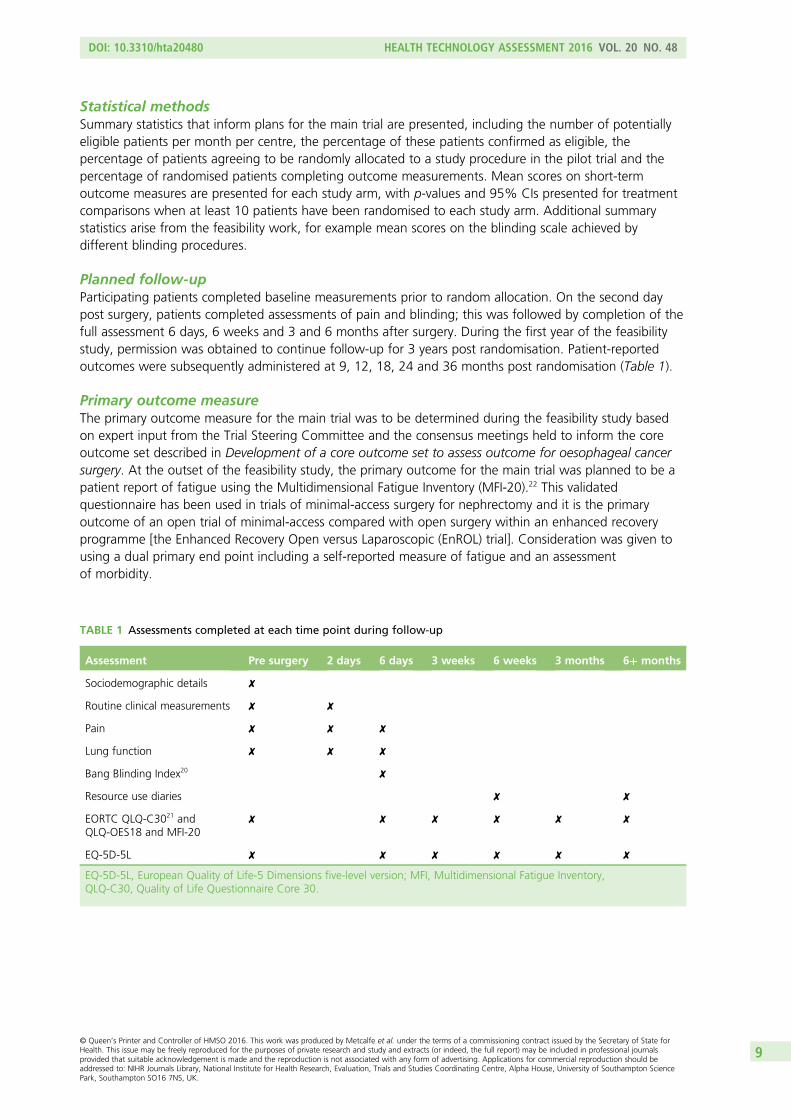
TABLE 1 Assessments completed at each time point during follow-up 9
TABLE 2 The TNM system applied to oesophageal tumours 10
TABLE 3 Definitions of technical terms used in the quality assurance section 13
TABLE 4 Baseline variable summary statistics presented by allocated arm withineach centre 18
TABLE 5 Baseline pathological staging by allocated arm within each centre 19
TABLE 6 Surgical approach undergone by allocated approach for the Bristol centre 19
TABLE 7 Surgical approach undergone by allocated approach for thePlymouth centre 20
TABLE 8 Postsurgical measures 20
TABLE 9 Completion of pain scales at days 2 and 6 post surgery 21
TABLE 10 Completion of the QLQ-C30 at baseline, 6 days post surgery and6 weeks and 6 months post randomisation 21
TABLE 11 Surgeon demographics from the semistructured interviews 22
TABLE 12 Synopsis of the thematic analysis of the semistructured interviews 22
TABLE 13 Patient beliefs about the surgical approach undergone by actualallocation at day 2 and day 6 at the Bristol centre 24
TABLE 14 Patient beliefs about the surgical approach undergone by actualallocation at day 2 and day 6 at the Plymouth centre 24
DOI: 10.3310/hta20480 HEALTH TECHNOLOGY ASSESSMENT 2016 VOL. 20 NO. 48
© Queen’s Printer and Controller of HMSO 2016. This work was produced by Metcalfe et al. under the terms of a commissioning contract issued by the Secretary of State forHealth. This issue may be freely reproduced for the purposes of private research and study and extracts (or indeed, the full report) may be included in professional journalsprovided that suitable acknowledgement is made and the reproduction is not associated with any form of advertising. Applications for commercial reproduction should beaddressed to: NIHR Journals Library, National Institute for Health Research, Evaluation, Trials and Studies Coordinating Centre, Alpha House, University of Southampton SciencePark, Southampton SO16 7NS, UK.
xiii


List of figures
FIGURE 1 Actual and target recruitment up to December 2014 17
DOI: 10.3310/hta20480 HEALTH TECHNOLOGY ASSESSMENT 2016 VOL. 20 NO. 48
© Queen’s Printer and Controller of HMSO 2016. This work was produced by Metcalfe et al. under the terms of a commissioning contract issued by the Secretary of State forHealth. This issue may be freely reproduced for the purposes of private research and study and extracts (or indeed, the full report) may be included in professional journalsprovided that suitable acknowledgement is made and the reproduction is not associated with any form of advertising. Applications for commercial reproduction should beaddressed to: NIHR Journals Library, National Institute for Health Research, Evaluation, Trials and Studies Coordinating Centre, Alpha House, University of Southampton SciencePark, Southampton SO16 7NS, UK.
xv


List of boxes
BOX 1 Core outcome set for oesophageal cancer surgery 25
DOI: 10.3310/hta20480 HEALTH TECHNOLOGY ASSESSMENT 2016 VOL. 20 NO. 48
© Queen’s Printer and Controller of HMSO 2016. This work was produced by Metcalfe et al. under the terms of a commissioning contract issued by the Secretary of State forHealth. This issue may be freely reproduced for the purposes of private research and study and extracts (or indeed, the full report) may be included in professional journalsprovided that suitable acknowledgement is made and the reproduction is not associated with any form of advertising. Applications for commercial reproduction should beaddressed to: NIHR Journals Library, National Institute for Health Research, Evaluation, Trials and Studies Coordinating Centre, Alpha House, University of Southampton SciencePark, Southampton SO16 7NS, UK.
xvii

List of abbreviations
CI confidence interval
CRF Clinical Report Form
EORTC European Organization forResearch and Treatment of Cancer
EQ-5D-5L European Quality of Life-5Dimensions five-level version
HRG Healthcare Resource Group
HRQoL health-related quality of life
HTA Health Technology Assessment
IDEAL Innovation, Development,Exploration, Assessment,Long-term study
LAO laparoscopically assistedoesophagectomy
MFI-20 Multidimensional Fatigue Inventory
MIO minimally invasive oesophagectomy
MIRO oesphagectoMIe pour cancer paRvoie conventionnelle oucoeliO-assistée
NIHR National Institute for HealthResearch
ProtecT Prostate testing for cancer andTreatment
PSS Personal Social Services
QALY quality-adjusted life-year
QLQ-C30 Quality of Life QuestionnaireCore 30
QLQ-OES18 Quality of Life QuestionnaireOesophageal Cancer Module
RCT randomised controlled trial
ROMIO Randomised Oesophagectomy:Minimally Invasive or Open
TIME Traditional Invasive versusMinimally invasive Esophagectomy
TNM tumour, node, metastasis
DOI: 10.3310/hta20480 HEALTH TECHNOLOGY ASSESSMENT 2016 VOL. 20 NO. 48
© Queen’s Printer and Controller of HMSO 2016. This work was produced by Metcalfe et al. under the terms of a commissioning contract issued by the Secretary of State forHealth. This issue may be freely reproduced for the purposes of private research and study and extracts (or indeed, the full report) may be included in professional journalsprovided that suitable acknowledgement is made and the reproduction is not associated with any form of advertising. Applications for commercial reproduction should beaddressed to: NIHR Journals Library, National Institute for Health Research, Evaluation, Trials and Studies Coordinating Centre, Alpha House, University of Southampton SciencePark, Southampton SO16 7NS, UK.
xix

Plain English summary
The survival of some patients with oesophageal (gullet) cancer can be improved by surgery(oesophagectomy). Surgery traditionally requires large incisions to be made in the abdomen, the
chest and sometimes the neck (open surgery). Complications are common and recovery takes ≥ 6 months.Minimally invasive ‘keyhole’ surgery may achieve the same survival benefit, with quicker recovery.However, to confirm this, a randomised controlled trial (RCT) needs to be carried out to make a faircomparison between the surgical approaches. The present study conducted preparatory work in a smallRCT in two departments of surgery.
This trial indicated the feasibility of a full-scale evaluation, with 104 patients agreeing to take part over21 months. The random allocation of a surgical approach to each patient resulted in similar groups ofpatients undergoing the different approaches, which, with most patients undergoing their allocated surgery(87%), ensured a fair comparison between the approaches. By bandaging all possible incision points forthe first week post surgery, it proved possible to keep patients from knowing which surgical approach theyhad undergone, improving the assessment of postsurgical pain. Participants are reporting, with highcompletion rates, on outcomes such as physical function and fatigue over a 3-year period. Patients andclinicians are being consulted on the most important measures of outcome following oesophagectomy.The steps in performing an oesophagectomy have been documented, including the important differencesbetween the approaches, allowing quality control of surgery. Finally, the important costs and methods ofmeasurement have been determined, allowing a cost-effectiveness analysis in the full-scale evaluation.
DOI: 10.3310/hta20480 HEALTH TECHNOLOGY ASSESSMENT 2016 VOL. 20 NO. 48
© Queen’s Printer and Controller of HMSO 2016. This work was produced by Metcalfe et al. under the terms of a commissioning contract issued by the Secretary of State forHealth. This issue may be freely reproduced for the purposes of private research and study and extracts (or indeed, the full report) may be included in professional journalsprovided that suitable acknowledgement is made and the reproduction is not associated with any form of advertising. Applications for commercial reproduction should beaddressed to: NIHR Journals Library, National Institute for Health Research, Evaluation, Trials and Studies Coordinating Centre, Alpha House, University of Southampton SciencePark, Southampton SO16 7NS, UK.
xxi

Scientific summary
Background
Localised oesophageal cancer can be curatively treated with surgery (oesophagectomy). The national audit ofpatients in England and Wales (2011–20) undergoing oesophagectomy collected details of 1220 operations.Oesophagectomy had a 3% risk of in-hospital mortality, a 9% risk of reoperation and a 16% risk ofrespiratory complications. Health-related quality of life is significantly worsened after oesophagectomy, withpatients reporting major impacts in terms of physical and social function, fatigue, breathlessness and pain forat least 3 months. Recovery takes about 6–9 months after open surgery.
There is therefore a need to improve outcomes of patients undergoing oesophagectomy. Minimal-accesssurgical techniques may cause less tissue damage and allow a more rapid recovery. Whether or not this isa cost-effective approach, however, is unknown, as high-quality comparative evidence is limited.
Two previous trials had methodological flaws that preclude firm conclusions being drawn from their results.In particular, the sample sizes were small and, therefore, the studies made only a modest contribution tothe evidence of equivalent survival benefits with the different approaches to oesophagectomy. The primaryend points reflected surgical interest and did not incorporate meaningful benefits for minimal-accesssurgery from the patients’ perspective. Both trials were at risk of biased outcome assessment as assessorswere not blinded and one trial used sealed envelopes for randomisation, bringing the concealment ofallocation into question.
A UK trial is needed to provide definitive evidence on the relative cost-effectiveness of minimally invasiveand open surgery for oesophagectomy. However, there are many challenges to conducting high-qualityrandomised trials of surgery. The particular hurdles that may affect a full trial of open oesophagectomyand minimally invasive oesophagectomy (MIO) are (1) strong preferences held by surgeons about the twoprocedures, which may mean that it is difficult to recruit centres to the main trial, and (2) preferences ofpatients that do not permit randomisation. The feasibility of the main trial would also be in doubt if(3) the number of eligible patients was lower than a mean of 2.5 patients per month per centre. Theseand further methodological issues were addressed in the feasibility study reported here.
Objectives
The ROMIO (Randomised Oesophagectomy: Minimally Invasive or Open) feasibility study aimed to establishthe methodology and infrastructure for a definitive trial comparing the cost-effectiveness of minimallyinvasive and open surgical techniques for oesophagectomy in the treatment of cancer. The core ofthis preliminary work was an assessment of the feasibility of comparing surgical procedures foroesophagectomy in a pilot two-centre randomised trial. Specific objectives were:
l To pilot the randomisation process and investigate reasons for any difficulties that affect recruitment sothat these can be tackled before the main trial.
l To establish the proportion of potentially eligible patients who can be approached about the trial, whoare confirmed as eligible, who are successfully recruited and randomised and who are able and willingto undergo research assessments. This addresses the feasibility of the main trial by indicating theachievable sample size and the number of centres required.
DOI: 10.3310/hta20480 HEALTH TECHNOLOGY ASSESSMENT 2016 VOL. 20 NO. 48
© Queen’s Printer and Controller of HMSO 2016. This work was produced by Metcalfe et al. under the terms of a commissioning contract issued by the Secretary of State forHealth. This issue may be freely reproduced for the purposes of private research and study and extracts (or indeed, the full report) may be included in professional journalsprovided that suitable acknowledgement is made and the reproduction is not associated with any form of advertising. Applications for commercial reproduction should beaddressed to: NIHR Journals Library, National Institute for Health Research, Evaluation, Trials and Studies Coordinating Centre, Alpha House, University of Southampton SciencePark, Southampton SO16 7NS, UK.
xxiii
l To document in detail, using IDEAL (Innovation, Development, Exploration, Assessment, Long-termstudy) recommendations, the technical developments of the totally minimally invasive approach foroesophagectomy, to inform the design and choice of interventions in the main trial. This workdeveloped manuals for the different surgical procedures, and methods of monitoring adherence tothem, which will then be available for the main trial. It also informed the development of a competencyassessment tool for objective evaluation of technical performance to be used to evaluate surgeons’skills before participating in the main trial.
l To develop a manual for the cutting up of specimens, specimen fixing and pathology to optimise thequality of lymph node counts and ascertainment of positive resection margins.
l To consider the appropriate statistical model for estimating treatment effectiveness while allowing for‘clustering’ in the data because of between-surgeon variation.
l To develop and evaluate feasible, acceptable and effective methods of keeping patients blind to theirtreatment for the first week after surgery.
l To establish outcome measures for the main trial that are recognised as a comprehensive, valid andreliable assessment of oesophagectomy outcome by patients and the clinical community.
Methods
The ROMIO feasibility study was based around a pilot parallel three-arm randomised controlled trial (RCT)nested within feasibility work. This was conducted in two centres, University Hospitals Bristol NHSFoundation Trust and Plymouth Hospitals NHS Trust, which each have a team of upper gastrointestinalcancer surgeons.
Patients were eligible for the pilot trial if they were aged ≥ 18 years with confirmed histopathological evidenceof oesophageal or oesophagogastric junctional adenocarcinoma, squamous cell cancer or high-grade dysplasiathat was referred for oesophagectomy or oesophagectomy following neoadjuvant chemo(radio)therapy.The technology was oesophagectomy, with patients randomised to open surgery, a hybrid open chest andminimally invasive abdomen [laparoscopically assisted oesophagectomy (LAO)] or totally MIO.
The primary outcome measure for the pilot trial was the number of patients recruited per month, with themain trial being considered feasible if at least 2.5 patients per month were recruited. The primary outcomemeasure for the main trial was to be confirmed during the feasibility study based on expert input fromthe Trial Steering Committee and the consensus meetings held to inform the core outcome set. At theoutset of the feasibility study the initial candidate for the primary outcome of the main trial was a patientreport of fatigue.
Participants completed baseline measurements prior to random allocation. On the second day post surgery,patients completed assessments of pain and blinding, followed by completion of the full assessment6 days, 6 weeks and 3 and 6 months after surgery. During the first year of the feasibility study, permissionwas obtained to continue follow-up for 3 years post randomisation. Patient-reported outcomes weresubsequently administered at 9, 12, 18, 24 and 36 months.
Allocation of patients to surgical procedure was at random, the allocation being conducted separately forthe two centres and further stratified by whether or not patients had undergone neoadjuvant treatment.Patients were randomly allocated to one of the three procedures at Bristol and to the open procedure orthe two-phase laparoscopically assisted procedure at Plymouth. Allocation was concealed throughcentralised randomisation.
Recruitment to the pilot RCT was planned for a 12-month period, with 72 potentially eligible patientsbeing expected during that time. This would allow a true 50% recruitment rate to be estimated with a95% confidence interval of approximately 38% to 62%.
SCIENTIFIC SUMMARY
NIHR Journals Library www.journalslibrary.nihr.ac.uk
xxiv
Summary statistics that inform plans for the main trial are presented including the number of potentiallyeligible patients per month per centre, the percentage of these patients confirmed as eligible, thepercentage of patients agreeing to be randomly allocated to a study procedure in the pilot trial and thepercentage of randomised patients completing outcome measurements.
Key aspects of the associated methodological work included the qualitative recruitment intervention,the documentation of the different surgical procedures and the production of materials to allow qualityassurance of surgery and pathology, determination of outcome measures that are important to patientsundergoing oesophagectomy and their clinicians, identification of the key cost differences betweenthe surgical approaches and methods to capture these and establishing a method to keep patients blindto their treatment allocation while pain levels were assessed in the first week post surgery.
Results
In the first 21 months of recruitment (until the end of December 2014) 263 patients have been assessedfor eligibility, of whom 135 (51%) were found to be eligible and 104 (77%) were randomised acrossthe two centres. Of the recruited patients, 46 were from Plymouth (n= 22 open surgery, n= 24 LAO) and58 were from Bristol (n= 19 open surgery, n= 19 LAO, n= 20 MIO). To allow continuation into thedefinitive trial, recruitment was continued beyond the planned 12-month period and was ongoing at thetime of writing. In this report we focus on those patients randomised on or before 31 August 2014 forwhom 6-week follow-up data were expected to be available for analysis.
The majority of participants were male and the mean age at randomisation was approximately 66 years foreach treatment group (standard deviation approximately 7 years). A lower proportion of patients hadundergone neoadjuvant treatment before surgery in Plymouth than in Bristol, although the proportion ineach treatment arm was well balanced within each centre. Considering disease stage at diagnosis, mostparticipants had a tumour that was growing through the wall of the oesophagus but involving one to twolymph nodes at most (stages 2a and 2b) or a cancer that had just started to spread to the tissuessurrounding the oesophagus (stage 3a). There was no convincing evidence of imbalance between therandomised intervention arms in terms of disease staging.
The large majority of patients underwent their allocated surgical approach (69/79, 87%). The threepatients for whom the allocation to open surgery could not be followed were found to be inoperable.Of the patients allocated to LAO, one converted to open surgery to stop a bleed, one requested MIO havingspoken to a patient who had undergone this procedure previously and one underwent a gastrectomy formore extensive disease. Four patients allocated to MIO underwent LAO, two because of the position of thetumour and two because a surgeon was not available to undertake the MIO procedure.
The median length of postsurgical stay across the two centres was 10 days, with stays typically lastingbetween 8 and 16 days (interquartile range). Among the first 76 patients recruited there was one deathoccurring within 30 days of surgery, with a further death occurring on the 31st postsurgical day. Fouranastomotic leaks occurred, all of which resulted in a return to theatre. A further nine patients returned totheatre one or more times. Eight patients returned to the intensive care unit, six because of respiratoryproblems and two because of renal failure. Eight patients were readmitted within 30 days of surgery.
Completion of the pain scale at day 2 post surgery was low (39/73, 53%), the reasons for non-completionbeing illness or insufficient recovery (9/73, 12%), lack of cover to administer the scale at the weekend(16/73, 22%) and administrative error (9/73, 12%). Completion of the pain scale was considerably betterat day 6 (64/73, 88%). The primary outcome measure for the planned definitive trial is likely to be apatient-reported outcome at 6 weeks post randomisation; the European Organization for Researchand Treatment of Cancer (EORTC) Quality of Life Questionnaire Core 30 (QLQ-C30) was available for74% (54/73) of participants at that time point.
DOI: 10.3310/hta20480 HEALTH TECHNOLOGY ASSESSMENT 2016 VOL. 20 NO. 48
© Queen’s Printer and Controller of HMSO 2016. This work was produced by Metcalfe et al. under the terms of a commissioning contract issued by the Secretary of State forHealth. This issue may be freely reproduced for the purposes of private research and study and extracts (or indeed, the full report) may be included in professional journalsprovided that suitable acknowledgement is made and the reproduction is not associated with any form of advertising. Applications for commercial reproduction should beaddressed to: NIHR Journals Library, National Institute for Health Research, Evaluation, Trials and Studies Coordinating Centre, Alpha House, University of Southampton SciencePark, Southampton SO16 7NS, UK.
xxv
Dressing patients with large bandages, covering all possible incisions, was successful in keeping patientsblind while pain was assessed during the first week post surgery. On both day 2 and day 6, the majority ofresponders believed that they had undergone the LAO procedure, irrespective of their allocation.
Hierarchical task analysis has allowed the documentation of the steps of oesophagectomy and thedifferences between the open approach and the minimally invasive approach. Assessment tools havebeen produced for quality assurance of surgery, recorded on video or with photographs. This will besupplemented with an ‘op note’ allowing the recording of each step of the operation. The studypathologists at the two centres and the study’s independent advisor have reached consensus on astandard approach to pathological processing and recording and have planned quality assuranceprocedures for the proposed main trial.
In conducting the qualitative recruitment intervention, the patient eligibility and recruitment pathwayat each centre has been mapped. In-depth interviews with nine clinical investigators and staff undertakingrecruitment have been conducted and analysed to explore views about the evidence on which the trial isbased, perceptions of levels of equipoise in relation to the trial arms, how the arms are or can be deliveredin their clinical centre and methods for identifying eligible patients. Audio recordings of > 100 consultationshave also been conducted and analysed to scrutinise recruiters’ ability to summarise the details of the trialdesign and protocol and provide information about the trial and to identify examples of actual recruitmentsuccesses and challenges. This has informed feedback and training meetings with recruiters. Furthermore,the patient information provided at recruitment has been updated and a ‘recruitment tips’ guidancedocument has been developed.
In developing a core outcome set to evaluate the clinical effectiveness of oesophageal cancer surgery,a literature review identified 901 measures, which were synthesised into 67 outcome domains andoperationalised into 68 questions. Delphi methodology was used to reduce the initial list of outcomedomains to a final core set according to prespecified criteria, by surveying key stakeholders (consultantsurgeons, clinical nurse specialists and patients). Analyses of responses led to 41 and 19 outcomes beingretained as important after the first and second surveys, respectively. The retained outcomes from thesecond survey were presented at a face-to-face patient consensus meeting at which patients were askedto anonymously rate their importance. The final core outcome set consists of 10 outcomes.
One area in which cost differences will exist between the approaches to oesophagectomy is in relation tothe actual operation. Piloting of a Clinical Report Form was ongoing at the time of writing, which aimed tocapture use of equipment and consumables in terms of brand and quantity and use of operation staff interms of role and time. This level of detail of the operation itself is unlikely to be available from patients’medical records. Readmissions to the treating hospital will be captured in a review of medical records andit seems reasonable to rely on Healthcare Resource Group codes to indicate the cost of these. Secondarycare visits and inpatient stays at other hospitals will not be captured in this way and neither willcommunity-based NHS resource use. These aspects require the patients to provide information and apatient diary with a nurse-led telephone interview has been piloted. Early experience suggests that patientswould prefer a narrower NHS and Personal Social Services perspective to reduce the burden of this aspectof the study and that patients vary in whether they prefer to provide their resource use diary to the studyteam or take part in the telephone interview while referring to the diary.
SCIENTIFIC SUMMARY
NIHR Journals Library www.journalslibrary.nihr.ac.uk
xxvi
Conclusions
This feasibility study has demonstrated that different approaches to oesophagectomy can be compared inan unbiased fashion in a RCT and we are now planning the definitive trial.
At the time of writing we are proposing to focus on a comparison between LAO and openoesophagectomy as MIO continues to be subject to refinement. We plan to recruit 406 patients, allowinga clinically important benefit for postsurgical recovery of physical function to be detected with 90% power,and making a major contribution to an individual patient meta-analysis of survival. A seven-centre study isplanned; the new centres have varying experience of recruiting to surgical trials and so the qualitativerecruitment intervention will be continued. Currently, the plan is for two centres to randomly allocatepatients to three interventions, the third being MIO, allowing further unbiased data to be collected onthat approach.
Trial registration
This trial is registered as ISRCTN59036820.
Funding
Funding for this study was provided by the Health Technology Assessment programme of the NationalInstitute for Health Research.
DOI: 10.3310/hta20480 HEALTH TECHNOLOGY ASSESSMENT 2016 VOL. 20 NO. 48
© Queen’s Printer and Controller of HMSO 2016. This work was produced by Metcalfe et al. under the terms of a commissioning contract issued by the Secretary of State forHealth. This issue may be freely reproduced for the purposes of private research and study and extracts (or indeed, the full report) may be included in professional journalsprovided that suitable acknowledgement is made and the reproduction is not associated with any form of advertising. Applications for commercial reproduction should beaddressed to: NIHR Journals Library, National Institute for Health Research, Evaluation, Trials and Studies Coordinating Centre, Alpha House, University of Southampton SciencePark, Southampton SO16 7NS, UK.
xxvii

Chapter 1 Introduction
Funding history
The ROMIO (Randomised Oesophagectomy: Minimally Invasive or Open) feasibility study and pilot trialwas funded by the National Institute for Health Research (NIHR) Health Technology Assessment (HTA)programme (reference number 10/50/65). The ROMIO pilot study was a researcher-led proposal. Thefunding contract was agreed in October 2012 and the study opened in January 2013. The study was dueto close in December 2014 but a contract variation was agreed in November 2014 for a close in December2015, allowing continuation into a main trial should an application for funding of that subsequent studybe successful.
Structure of this report
In this chapter the importance of a rigorous comparison between open oesophagectomy and minimallyinvasive oesophagectomy (MIO) is made and the need for a preceding feasibility study and pilot trialjustified. In Chapter 2 the pilot randomised controlled trial (RCT) procedure and associated methodologicalwork are described, with the results of this work being presented in Chapter 3. The implications of ourfindings are discussed in Chapter 4 and the conclusions drawn with regard to the planned full trial arepresented in Chapter 5.
Background
Oesophageal cancer was the 13th most common cancer in the UK in 2011, with 8332 people diagnosedthat year.1 Two-thirds of these cancers are adenocarcinoma and one-third are squamous cell cancer andabout one-quarter of cases are diagnosed while the disease is localised to the oesophagus. Localisedoesophageal cancer and high-grade dysplasia (a pre-malignant condition) can be curatively treated withsurgery (oesophagectomy). Surgery is frequently combined with neoadjuvant treatment.
The most recent (2011–12) national audit of patients in England and Wales undergoing oesophagectomycollected details of 1220 operations.2 Oesophagectomy had a 3% risk of in-hospital mortality, a 9% risk ofreoperation and a 16% risk of respiratory complications. Complications of any type occurred in 30% ofpatients. Health-related quality of life (HRQoL) is significantly worsened after oesophagectomy, with patientsreporting major reductions in physical and social function and more problems with fatigue, breathlessnessand pain for at least 3 months. Recovery takes about 6–9 months after open surgery although patients dyingwithin a year of surgery do not recover preoperative HRQoL. Persistent long-term deficits in HRQoL occur.
There is therefore a need to improve outcomes for patients undergoing oesophagectomy. The pastdecade has seen improvements in patient selection, neoadjuvant treatment and perioperative care.Minimal-access surgical techniques have been introduced into UK clinical practice, with the theoreticaladvantages of causing less tissue damage and increasing the speed of recovery. Whether or not this is aneffective and cost-effective approach, however, is unknown as high-quality comparative evidence is limited.
There is a growing use of minimal-access surgical techniques for all types of cancer. Whether or not theseprovide patient benefit in the short term and maintain long-term survival is important to establish so that ahigh standard of surgical care can be provided. In some cancer sites there is good evidence that minimal-access techniques are beneficial. For example, minimal-access surgery for colorectal cancer was evaluatedin several large-scale trials3 in the 1990s, providing evidence of better recovery and equivalent survival.These trials led to changes in practice and surgical training.
DOI: 10.3310/hta20480 HEALTH TECHNOLOGY ASSESSMENT 2016 VOL. 20 NO. 48
© Queen’s Printer and Controller of HMSO 2016. This work was produced by Metcalfe et al. under the terms of a commissioning contract issued by the Secretary of State forHealth. This issue may be freely reproduced for the purposes of private research and study and extracts (or indeed, the full report) may be included in professional journalsprovided that suitable acknowledgement is made and the reproduction is not associated with any form of advertising. Applications for commercial reproduction should beaddressed to: NIHR Journals Library, National Institute for Health Research, Evaluation, Trials and Studies Coordinating Centre, Alpha House, University of Southampton SciencePark, Southampton SO16 7NS, UK.
1
Surgery for upper gastrointestinal cancer, however, is much more complex than colorectal cancer surgeryand is associated with high rates of mortality and morbidity. Minimal-access surgery may make theprocedure even more technically demanding, potentially resulting in greater surgical risks. Although therehas been an increase in the UK and worldwide in the uptake of minimal-access techniques for thetreatment of oesophageal cancer, there are also centres and surgeons who continue with standard opensurgery. It is necessary to ensure that these approaches are effective and cost-effective. If high-qualityevidence can be created then a standard of surgery for patients can be provided and health-care policyformulated to support this approach.
National audit data2 show that, of the 1220 oesophagectomies performed between 2011 and 2012 thatinclude details of the procedure, 40% (n= 492) used minimal-access surgical techniques. The majority(n= 314) were laparoscopically assisted (minimal-access approach for the abdomen and standard openright chest incision) and 140 were oesophagectomies performed by totally minimally invasive techniques.
The 2011/12 national audit did not suggest any marked differences in complications or length of hospitalstay following open surgery, laparoscopically assisted surgery or minimally invasive surgery.2 However, it islikely that patients were specifically selected for these approaches and that the patients had small tumoursand little comorbidity. Hence, there is a high chance of bias in observational data such as these, with thepatients’ prognosis being a factor in determining the method of surgery undertaken. Only a RCT canreliably control for such ‘confounding by indication’.
Literature review
At the time of planning this feasibility study we undertook a systematic literature review in the MEDLINEdatabase and Cochrane Central Register of Controlled Trials.4 Papers were eligible for inclusion in thereview if they reported the short-term clinical outcomes of surgery (with or without neoadjuvant treatment)as observed in both randomised and non-randomised studies of patients with oesophageal cancer(squamous cell and adenocarcinoma). Papers reporting outcomes of surgery for high-grade dysplasia only,outcomes of oesophagectomy combined with other procedures such as gastrectomy, based on < 50oesophagectomies, and retrospective study designs were excluded. MEDLINE and the Cochrane ControlledTrials Register were searched for abstracts with keywords for cancer (neoplasms or cancer or neoplasm$),oesophagus (esophag$or oesophagi$) and surgery (surg$or operation or operable or resect$). The searchwas limited to human studies published in English between January 2004 and March 2009. Reportsavailable only in abstract form were excluded. Titles and abstracts were screened for eligibility by tworeviewers and the full texts of studies meeting the inclusion criteria were analysed by three reviewers. Thereference lists of retrieved articles were used to identify additional potentially relevant studies. Dataextracted from the included studies were subject to a narrative synthesis.
We identified 23 non-randomised studies describing outcomes of minimally invasive procedures foroesophageal cancer. Sixteen papers described outcomes of totally minimally invasive surgery and sevenreported outcomes of laparoscopically assisted two-phase surgery using minimal-access techniques for theabdomen or chest.4 Three other systematic reviews were identified but none included a randomised trial.5–7
In a series of 222 patients undergoing totally minimally invasive surgery,8 the short-term clinical outcomes(morbidity and technical data) were similar to those published in series of open surgery. Few of the abovestudies reported short-term oncological end points (e.g. lymph node count), although UK national auditdata show similar lymph node counts with minimally invasive surgery to those achieved by openprocedures, with 68% of open and 78% of minimally invasive procedures yielding > 15 nodes.2 Onecohort study compared the outcomes of open oesophagectomy (n= 114), a combined approach (n= 309)and totally minimally invasive surgery (n= 23) and found no differences in 3- or 5-year survival.9 There wasa lack of published data on cost-effectiveness and only two studies measured HRQoL.6,10 One usedvalidated generic and disease-specific tools for a year after minimal-access surgery and showed an earlyrecovery of most aspects of health, but the study was small and without a comparison group.11
INTRODUCTION
NIHR Journals Library www.journalslibrary.nihr.ac.uk
2
All of these studies have methodological weaknesses because of their small sample sizes and observationaldesigns, with limited details regarding patient selection and outcome assessment. It is not possible to drawmeaningful conclusions from the available non-randomised studies and the evidence base for minimallyinvasive surgery for oesophageal resection is weak. A well-designed and conducted RCT comparing theeffectiveness and cost-effectiveness of minimal-access and open surgery is needed to inform current NHSpractice, health policy and individual surgeon and patient clinical decision-making. Open oesophagectomycosts about £6000 but the inclusion of reoperations, readmission to intensive care and prolonged staysmay significantly increase this cost. Minimally invasive surgery requires additional operative equipment butmay reduce hospital stay. An economic analysis, embedded within a pragmatic RCT, is required toestablish the relative cost-effectiveness of the different procedures when adopted into routineclinical practice.
Other trials evaluating minimal-access surgery foroesophageal cancer
The French MIRO trialThe oesphagectoMIe pour cancer paR voie conventionnelle ou coeliO-assistée (MIRO) trial of patientswith oesophageal cancer, excluding patients with type II and III tumours involving the gastro-oesophagealjunction, compared open two-phase surgery (abdomen and right chest) with two-phase laparoscopicallyassisted oesophagectomy (LAO; minimal access for the abdomen and open right chest incision)[see http://clinicaltrials.gov/show/NCT00937456 (accessed 21 January 2016)]. The primary end point was30-day morbidity (a composite of all complications from grade II to grade IV on the Clavien-Dindosystem12) and the trial was powered to test the hypothesis that minimal-access surgery leads to a reducedrate of complications (45% vs. 25%) at 30 days. The MIRO trial recruited 207 patients from 12 centres.The results, to date reported only at the American Society of Clinical Oncology Gastrointestinal CancersSymposium 2015,13 found that 67 (64.4%) patients in the open surgery group had major morbiditycompared with 37 (35.9%) in the minimal-access group [odds ratio 0.31, 95% confidence interval (CI)0.18 to 0.55; p= 0.0001]. There are, however, a number of methodological weaknesses. Although therandomisation sequence was computer generated, allocation concealment was unclear, using sealedenvelopes. The outcome assessors were not blinded to the intervention type and methods to quality assuresurgical procedures are not described.14
The Dutch TIME trialThe Traditional Invasive versus Minimally invasive Esophagectomy (TIME) trial included patients withoesophageal cancer, excluding patients with type II and III tumours involving the gastro-oesophagealjunction.15 It compared open two- or three-phase oesophagectomy with totally MIO (both abdomen andchest access performed with minimal-access approaches in the prone position). Pulmonary complications,strictly defined and graded, were the primary measure of outcome. The criteria for surgeon involvement inthis trial were evidence of prior completion of 10 minimally invasive procedures and production of onevideo showing surgical competence. This trial recruited 120 patients from seven surgical centres in fourcountries (the Netherlands, Spain, India and Italy). In total, 115 patients were recruited, with 16 out of 56(29%) patients undergoing open surgery having a pulmonary infection in the first 2 weeks compared withfive out of 59 (8%) in the minimally invasive surgery group (relative risk 0.30, 95% confidence interval0.12 to 0.76; p= 0.005). The trial includes a comprehensive assessment of HRQoL with the Short Formquestionnaire-36 items (SF-36)16 and the European Organization for Research and Treatment of Cancer(EORTC) Quality of Life Questionnaire Oesophageal Cancer Module (QLQ-OES18),17 but there are no costanalyses and no monitoring of surgical procedures.
DOI: 10.3310/hta20480 HEALTH TECHNOLOGY ASSESSMENT 2016 VOL. 20 NO. 48
© Queen’s Printer and Controller of HMSO 2016. This work was produced by Metcalfe et al. under the terms of a commissioning contract issued by the Secretary of State forHealth. This issue may be freely reproduced for the purposes of private research and study and extracts (or indeed, the full report) may be included in professional journalsprovided that suitable acknowledgement is made and the reproduction is not associated with any form of advertising. Applications for commercial reproduction should beaddressed to: NIHR Journals Library, National Institute for Health Research, Evaluation, Trials and Studies Coordinating Centre, Alpha House, University of Southampton SciencePark, Southampton SO16 7NS, UK.
3
Aim and rationale
The overall aim of the ROMIO trial was to compare, in patients with cancer of the oesophagus, the clinicaleffectiveness and cost-effectiveness of minimally invasive and open surgical procedures in terms ofrecovery, HRQoL, cost and survival.
Rationale for a UK trialAlthough the two previously conducted trials (MIRO13 and TIME15) provide some evidence to informpractice, both have methodological flaws that preclude firm conclusions being drawn from their resultsand neither will be applicable to the NHS and UK surgeons. In particular, the sample sizes are small andonly a modest contribution is made to the evidence of equivalent survival benefits with the differentsurgical approaches. The primary end points reflect surgical interest and do not incorporate meaningfulbenefit for minimal-access surgery from the patients’ perspective. Both trials are at risk of biased outcomeassessment without blinding of assessors and the French trial used sealed envelopes for randomisationwith the concealment of allocation consequently being questionable.14 In addition, the interventions in theDutch trial15 (totally minimally invasive surgery) are still being developed in the UK and, as this is anevolving procedure, few UK surgeons and anaesthetists are comfortable with oesophagectomy in theprone position.
Rationale for a feasibility study and pilot trialThere are many challenges to conducting high-quality randomised trials of non-pharmaceuticalinterventions; the particular hurdles that may affect a full trial of open oesophagectomy and MIO are(1) strong preferences held by surgeons about the two procedures, which may mean that it is difficult torecruit centres to the main trial, and (2) preferences of patients that will not permit randomisation.Addressing the first issue, we planned to secure provisional agreement from clinical centres to take part inthe main trial. The recruitment of patients to the study was addressed by the qualitative researchintegrated within this feasibility study. The feasibility of the main trial would also be in doubt if (3) thenumber of eligible patients was lower than a mean of 2.5 patients per month per centre. Further potentialchallenges to the quality of the main trial are (4) an inability to specify the surgical procedures beingevaluated and to demonstrate that the specified procedures are being adhered to; (5) bias in outcomemeasures because of patients and assessors not being blinded to treatment allocation; (6) the lack of awidely accepted battery of outcome measures on which to compare different surgical procedures; and(7) poor standards of pathology with consequently a weak measure of oncological success. These were alladdressed by the feasibility study and an unexpected failure to find a practical solution to any of themwould compromise the main trial.
Feasibility study objectives
The ROMIO feasibility study established the methodology and infrastructure for the main trial. The core ofthis preliminary work was an assessment of the feasibility of comparing surgical procedures foroesophagectomy in a pilot two-centre RCT. Specific objectives were:
l To pilot the randomisation process and investigate reasons for any difficulties that affect recruitment sothat these can be tackled before the main trial.
l To establish the proportion of potentially eligible patients who can be approached about the trial, whoare confirmed as eligible, who are successfully recruited and randomised and who are able and willingto undergo research assessments. This addressed the feasibility of the main trial by indicating theachievable sample size and the number of centres required.
l To document in detail, using IDEAL (Innovation, Development, Exploration, Assessment, Long-termstudy) recommendations,18 the technical developments of the totally minimally invasive approach foroesophagectomy, to inform the design and choice of interventions in the main trial. This workdeveloped manuals for the different surgical procedures, and methods of monitoring adherence to
INTRODUCTION
NIHR Journals Library www.journalslibrary.nihr.ac.uk
4
them, which were then available for the main trial. It also informed the development of a competencyassessment tool for objective evaluation of technical performance to be used to evaluate surgeons’skills before participating in the main trial.
l To develop a manual for the cutting up of specimens, specimen fixing and pathology, thereforeoptimising the quality of lymph node counts and ascertainment of positive resection margins, both ofwhich are likely to be important short-term outcome measures for the main trial.
l To consider the appropriate statistical model for estimating treatment effectiveness while allowing for‘clustering’ in the data because of between-surgeon variation. This will allow the statistical analysis planto be written during the early stages of the main trial.
l To develop and evaluate feasible, acceptable and effective methods of keeping patients blind to theirtreatment for the first week after surgery, so reducing bias in self-reported outcomes during themain trial.
l To establish outcome measures for the main trial that are recognised as a comprehensive, valid andreliable assessment of oesophagectomy outcome by patients and the clinical community and whichinclude a set of core outcome measures considered to be essential in studies of oesophageal cancer.
DOI: 10.3310/hta20480 HEALTH TECHNOLOGY ASSESSMENT 2016 VOL. 20 NO. 48
© Queen’s Printer and Controller of HMSO 2016. This work was produced by Metcalfe et al. under the terms of a commissioning contract issued by the Secretary of State forHealth. This issue may be freely reproduced for the purposes of private research and study and extracts (or indeed, the full report) may be included in professional journalsprovided that suitable acknowledgement is made and the reproduction is not associated with any form of advertising. Applications for commercial reproduction should beaddressed to: NIHR Journals Library, National Institute for Health Research, Evaluation, Trials and Studies Coordinating Centre, Alpha House, University of Southampton SciencePark, Southampton SO16 7NS, UK.
5

Chapter 2 Methods
This feasibility study was built around a pilot two-centre pragmatic RCT comparing minimally invasiveand open surgical procedures in the treatment of oesophageal cancer. Both the pilot RCT and the
integrated feasibility work are described in this chapter.
Patients
The initial plan was to have a 12-month period during which consecutive referrals of patients withoesophageal cancer for oesophagectomy at the Bristol and Plymouth centres were invited to take part inthe pilot RCT. Inclusion criteria were oesophageal adenocarcinoma, lower-third squamous cell cancer orhigh-grade dysplasia; selected for surgery by an upper gastrointestinal multidisciplinary cancer team; andage ≥ 18 years. Patients with stage-4 disease, or evidence of a previous complex thoracotomy orlaparotomy, were excluded from participation. Previous neoadjuvant chemotherapy did not excludeparticipants from the trial. All trial participants were asked to provide written informed consent. Weanticipated six potentially eligible patients being identified each month, giving a total of 72 patients invitedto participate in the pilot trial.
Open and minimally invasive approaches to oesophagectomy
Open surgeryThe oesophagectomy was carried out in two or three phases according to the surgeons’ judgement withregard to the patient and the tumour.
l Abdominal phase. After initial inspection of the abdomen to exclude inoperability, complete gastricmobilisation was performed with the surgeons’ usual practice based on the right gastroepiploic andright gastric arteries. Pyloroplasty, pyloromyotomy or no drainage were at the surgeons’ discretion.Lymphadenectomies along the common hepatic artery and left gastric and splenic arteries either enbloc or separately were performed and removal of sufficient crural fibres and a cuff of diaphragm wasperformed if required for tumour clearance. The pericardial fat pad and strips of pleura were removed.Transection of the lesser curve was undertaken during the abdominal phase or left to the thoracicphase of the operation. Placement of a feeding jejunostomy or nasojejunal tube was optional at thesurgeons’ discretion as was placement of intra-abdominal and intra-thoracic drains. Methods to closethe abdomen were at the surgeons’ discretion.
l Thoracic phase. After initial inspection of the chest to exclude inoperability, the mediastinal pleuraoverlying the oesophagus was excised in continuity with the oesophagus. The posterior limit of thedissection was normally the anterolateral wall of the aorta, so that the thoracic duct was mobiliseden bloc or separately to the oesophagus and peri-oesophageal tissues. The thoracic duct was ligatedand divided at the level of the diaphragm. The oesophagus was mobilised to the level of at least theaortic arch. Para-oesophageal and diaphragmatic nodes were removed in continuity or separately withthe oesophagus (at the surgeons’ discretion). Lymph nodes at the tracheal bifurcation and along theright and left main bronchi to the pulmonary hilus were removed en bloc or separately at the surgeons’discretion. The anastomotic technique and use of chest drainage was at the surgeons’ discretion.Methods to close the chest were at the surgeons’ discretion.
l Cervical phase. A left neck incision was made if a three-phase operation was undertaken.The oesophagus was mobilised preserving the recurrent laryngeal nerve and the anastomosiswas performed using the surgeons’ preferred methods. Use of a drain was optional.
DOI: 10.3310/hta20480 HEALTH TECHNOLOGY ASSESSMENT 2016 VOL. 20 NO. 48
© Queen’s Printer and Controller of HMSO 2016. This work was produced by Metcalfe et al. under the terms of a commissioning contract issued by the Secretary of State forHealth. This issue may be freely reproduced for the purposes of private research and study and extracts (or indeed, the full report) may be included in professional journalsprovided that suitable acknowledgement is made and the reproduction is not associated with any form of advertising. Applications for commercial reproduction should beaddressed to: NIHR Journals Library, National Institute for Health Research, Evaluation, Trials and Studies Coordinating Centre, Alpha House, University of Southampton SciencePark, Southampton SO16 7NS, UK.
7
Laparoscopically assisted oesophagectomyThe oesophagectomy was performed as described in the previous section and access to the abdominalcavity was achieved using between four and six small incisions, which were placed any way in theabdominal wall at the surgeons’ discretion. Placement of a feeding jejunostomy was at the surgeons’discretion and was performed laparoscopically or by extending a port site to an 8-cm abdominal incision.The thoracic part of the operation was performed as described in the previous section with a standardopen incision. A cervical incision and a neck anastomosis was normally undertaken if required fortumour clearance.
Totally minimally invasive oesophagectomyThis consisted of performing the steps of the abdominal and chest phases of the operation as described inOpen surgery but using laparoscopic and thoracoscopic techniques for access to each body cavity for eachphase, respectively. A two- or three-phase minimally invasive operation was permissable. In the two-phaseprocedure the anastomosis was performed in the chest if necessary creating a 15-cm incision in addition tothe minimal-access ports. In the three-phase operation the anastomosis was performed with a leftcervical incision.
Design of the pilot randomised trial
SettingTwo centres, University Hospitals Bristol NHS Foundation Trust and Plymouth Hospitals NHS Trust, recruitedpatients and carried out procedures within the pilot trial. Both centres have teams of upper gastrointestinalcancer surgeons (six in Bristol and five in Plymouth). These two centres also undertook the associatedfeasibility work. Methodological support for the pilot trial was predominantly based in Bristol, with thedevelopment of quality assurance protocols for surgical procedures and pathology being based at ImperialCollege London.
Allocation to treatment armAllocation of patients to surgical procedure was at random, the allocation being conducted separately forthe two centres and further stratified by whether or not patients had undergone neoadjuvant treatment.Patients were randomly allocated to one of the three procedures at Bristol and to one of the open ortwo-phase laparoscopically assisted procedures at Plymouth. Randomisation within blocks of varying sizeprevented large imbalances in the number of patients in each treatment arm. Allocation was concealedthrough centralised randomisation, with patients being logged into the study before their allocation wasrevealed to the surgical team.
Sample sizeAt the two lead centres, recruitment to the pilot RCT was planned for a 12-month period, with 72potentially eligible patients being expected during that time. This would allow a true 50% recruitmentrate to be estimated with a 95% CI of approximately 38% to 62% (CI calculation based on the binomialdistribution – the Clopper–Pearson approach19). If 11 patients were randomly allocated to each surgicalprocedure, this would allow a true difference of 1.25 standard deviations between two procedures on acontinuous measure of early outcome to be detected with 80% power at the 5% significance level. Hence,the pilot RCT was planned to provide an acceptably precise estimate of the recruitment rate to inform plansfor the main trial and perhaps to provide evidence suggestive of an intervention having promise for abeneficial impact on short-term outcomes.
METHODS
NIHR Journals Library www.journalslibrary.nihr.ac.uk
8
Statistical methodsSummary statistics that inform plans for the main trial are presented, including the number of potentiallyeligible patients per month per centre, the percentage of these patients confirmed as eligible, thepercentage of patients agreeing to be randomly allocated to a study procedure in the pilot trial and thepercentage of randomised patients completing outcome measurements. Mean scores on short-termoutcome measures are presented for each study arm, with p-values and 95% CIs presented for treatmentcomparisons when at least 10 patients have been randomised to each study arm. Additional summarystatistics arise from the feasibility work, for example mean scores on the blinding scale achieved bydifferent blinding procedures.
Planned follow-upParticipating patients completed baseline measurements prior to random allocation. On the second daypost surgery, patients completed assessments of pain and blinding; this was followed by completion of thefull assessment 6 days, 6 weeks and 3 and 6 months after surgery. During the first year of the feasibilitystudy, permission was obtained to continue follow-up for 3 years post randomisation. Patient-reportedoutcomes were subsequently administered at 9, 12, 18, 24 and 36 months post randomisation (Table 1).
Primary outcome measureThe primary outcome measure for the main trial was to be determined during the feasibility study basedon expert input from the Trial Steering Committee and the consensus meetings held to inform the coreoutcome set described in Development of a core outcome set to assess outcome for oesophageal cancersurgery. At the outset of the feasibility study, the primary outcome for the main trial was planned to be apatient report of fatigue using the Multidimensional Fatigue Inventory (MFI-20).22 This validatedquestionnaire has been used in trials of minimal-access surgery for nephrectomy and it is the primaryoutcome of an open trial of minimal-access compared with open surgery within an enhanced recoveryprogramme [the Enhanced Recovery Open versus Laparoscopic (EnROL) trial]. Consideration was given tousing a dual primary end point including a self-reported measure of fatigue and an assessmentof morbidity.
TABLE 1 Assessments completed at each time point during follow-up
Assessment Pre surgery 2 days 6 days 3 weeks 6 weeks 3 months 6+ months
Sociodemographic details ✗
Routine clinical measurements ✗ ✗
Pain ✗ ✗ ✗
Lung function ✗ ✗ ✗
Bang Blinding Index20 ✗
Resource use diaries ✗ ✗
EORTC QLQ-C3021 andQLQ-OES18 and MFI-20
✗ ✗ ✗ ✗ ✗ ✗
EQ-5D-5L ✗ ✗ ✗ ✗ ✗ ✗
EQ-5D-5L, European Quality of Life-5 Dimensions five-level version; MFI, Multidimensional Fatigue Inventory,QLQ-C30, Quality of Life Questionnaire Core 30.
DOI: 10.3310/hta20480 HEALTH TECHNOLOGY ASSESSMENT 2016 VOL. 20 NO. 48
© Queen’s Printer and Controller of HMSO 2016. This work was produced by Metcalfe et al. under the terms of a commissioning contract issued by the Secretary of State forHealth. This issue may be freely reproduced for the purposes of private research and study and extracts (or indeed, the full report) may be included in professional journalsprovided that suitable acknowledgement is made and the reproduction is not associated with any form of advertising. Applications for commercial reproduction should beaddressed to: NIHR Journals Library, National Institute for Health Research, Evaluation, Trials and Studies Coordinating Centre, Alpha House, University of Southampton SciencePark, Southampton SO16 7NS, UK.
9
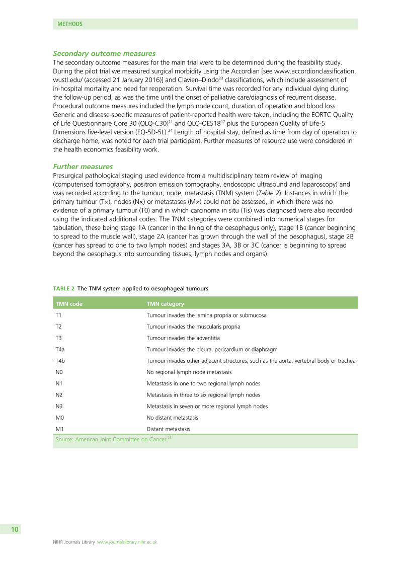
Secondary outcome measuresThe secondary outcome measures for the main trial were to be determined during the feasibility study.During the pilot trial we measured surgical morbidity using the Accordian [see www.accordionclassification.wustl.edu/ (accessed 21 January 2016)] and Clavien–Dindo23 classifications, which include assessment ofin-hospital mortality and need for reoperation. Survival time was recorded for any individual dying duringthe follow-up period, as was the time until the onset of palliative care/diagnosis of recurrent disease.Procedural outcome measures included the lymph node count, duration of operation and blood loss.Generic and disease-specific measures of patient-reported health were taken, including the EORTC Qualityof Life Questionnaire Core 30 (QLQ-C30)21 and QLQ-OES1817 plus the European Quality of Life-5Dimensions five-level version (EQ-5D-5L).24 Length of hospital stay, defined as time from day of operation todischarge home, was noted for each trial participant. Further measures of resource use were considered inthe health economics feasibility work.
Further measuresPresurgical pathological staging used evidence from a multidisciplinary team review of imaging(computerised tomography, positron emission tomography, endoscopic ultrasound and laparoscopy) andwas recorded according to the tumour, node, metastasis (TNM) system (Table 2). Instances in which theprimary tumour (T×), nodes (N×) or metastases (M×) could not be assessed, in which there was noevidence of a primary tumour (T0) and in which carcinoma in situ (Tis) was diagnosed were also recordedusing the indicated additional codes. The TNM categories were combined into numerical stages fortabulation, these being stage 1A (cancer in the lining of the oesophagus only), stage 1B (cancer beginningto spread to the muscle wall), stage 2A (cancer has grown through the wall of the oesophagus), stage 2B(cancer has spread to one to two lymph nodes) and stages 3A, 3B or 3C (cancer is beginning to spreadbeyond the oesophagus into surrounding tissues, lymph nodes and organs).
TABLE 2 The TNM system applied to oesophageal tumours
TMN code TMN category
T1 Tumour invades the lamina propria or submucosa
T2 Tumour invades the muscularis propria
T3 Tumour invades the adventitia
T4a Tumour invades the pleura, pericardium or diaphragm
T4b Tumour invades other adjacent structures, such as the aorta, vertebral body or trachea
N0 No regional lymph node metastasis
N1 Metastasis in one to two regional lymph nodes
N2 Metastasis in three to six regional lymph nodes
N3 Metastasis in seven or more regional lymph nodes
M0 No distant metastasis
M1 Distant metastasis
Source: American Joint Committee on Cancer.25
METHODS
NIHR Journals Library www.journalslibrary.nihr.ac.uk
10
Integrated qualitative research to optimise recruitment
A major focus of the feasibility study was to evaluate the acceptability of the ROMIO trial to patients andclinicians and to investigate the reasons underlying recruitment difficulties to ensure high levels ofrecruitment and informed consent.26,27 To this end, an integrated qualitative study, based on previous workby Donovan and colleagues28–31 in the NIHR HTA programme-funded ProtecT (Prostate testing for cancerand Treatment) study, was undertaken. There were two aspects to this work: understanding recruitmentissues and development of a recruitment strategy.
Understanding recruitment issuesThe first aspect was addressed through in-depth interviews, undertaken by an experienced qualitativeresearcher, with a maximum variation sample of between 10 and 15 patients eligible for the trial(selected to include the range of age, disease severity, socioeconomic status and centre). These interviewsexplored patient perspectives of oesophageal cancer and its treatment, views about surgery and studyinformation and the acceptability of randomisation between procedures, including potential barriers torandomisation. In-depth interviews with clinical investigators and staff undertaking recruitment to the trialexplored their ability to summarise the details of the trial design and protocol, their views about theevidence on which it is based, their perceptions of levels of uncertainty/equipoise in relation to the trialarms, their views about how the arms are or can be delivered in their clinical centre, methods foridentifying eligible patients and examples of actual recruitment successes and difficulties. Numbers ofinterviews were guided by the concept of data saturation: the need to continue sampling until findingsbecome repetitious. Interview data were analysed thematically and using case study approaches, lookingfor shared or disparate views among investigators, clinicians, recruiters and potential participants andwithin or between centres.32,33
Digital audio recorders were provided to the recruitment staff so that they could audio record appointmentsat which they provided information for patients and attempted to recruit them to the ROMIO trial. Theserecordings were analysed thematically and also using a version of conversation analysis pioneered inprevious studies33–35 to identify aspects of information provision that were unclear, disrupted and hinderrecruitment and that could thus be targeted for improvement.
The qualitative researcher worked with the two study nurses to map the patient eligibility and recruitmentpathways that are established, including when patients receive information about the trial and whichmembers of the clinical team they meet and when. Logs of eligible and recruited patients were assembledusing simple flow charts and counts to display numbers and percentages of patients at each stage of theeligibility and recruitment process and allow comparison with the original protocol. Previous research29–31
has shown that logistical and other local issues sometimes prevent full implementation of the protocol andcan consequently lead to less efficient recruitment pathways.
Development of a recruitment strategyThe second aspect was the development of a recruitment strategy. The findings from these qualitativeinvestigations and their potential impact on recruitment were summarised and presented to the ROMIOinvestigators. When necessary the recruitment process was modified in light of the specific qualitativeresearch findings, the changes including a mix of bespoke and generic measures. Actions specific to theROMIO trial were grounded in the findings of the integrated qualitative research and could include, forexample, changes to the descriptions of surgical procedures in study information. Training of recruitmentstaff is an example of a generic approach that has been found to be useful in previous trials,29,32 with topicsincluding how to elicit and address patient preferences and how to manage personal (clinical) preferences.
Once the recruitment plan was agreed it was implemented during the latter part of the feasibility studyrecruitment period. Recruiters were strongly encouraged to continue recording their appointments withpotential participants so that these could be analysed iteratively to check that the new process was beingimplemented and to identify any emergent difficulties. Individual feedback to recruiters was provided
DOI: 10.3310/hta20480 HEALTH TECHNOLOGY ASSESSMENT 2016 VOL. 20 NO. 48
© Queen’s Printer and Controller of HMSO 2016. This work was produced by Metcalfe et al. under the terms of a commissioning contract issued by the Secretary of State forHealth. This issue may be freely reproduced for the purposes of private research and study and extracts (or indeed, the full report) may be included in professional journalsprovided that suitable acknowledgement is made and the reproduction is not associated with any form of advertising. Applications for commercial reproduction should beaddressed to: NIHR Journals Library, National Institute for Health Research, Evaluation, Trials and Studies Coordinating Centre, Alpha House, University of Southampton SciencePark, Southampton SO16 7NS, UK.
11
confidentially by the qualitative researcher and anonymised findings from the recordings were used in arecruitment training programme for the main trial. Logs of eligible patients and randomisation ratescontinued to be measured.
Blinding to treatment allocation
Methods to achieve blinding of patients and outcome assessors were piloted during the feasibility work,to inform whether or not this could be achieved by methods acceptable for the main trial. Attempts weremade to blind patients during the first week post surgery when they were completing pain assessments onday 2 and day 6. On day 6 participants also complete questionnaires assessing generic and disease-specificaspects of HRQoL before being unblinded. Blinding was piloted using large adhesive dressings that wereprovided to participating sites by the trial office for the dressing of patients’ surgical wounds. The dressingwas positioned similarly on all trial patients regardless of the type of surgery (covering the positions ofabdominal, thoracic and cervical incisions). The first dressing was applied by the surgical team in theoperating theatre. The dressing was not changed unless required (because of soiling or lack of adherence)until day 3. It was then changed by the research nurses, who were not routinely involved with the patients’care. During the dressing changes patients were asked to turn their head away to prevent them observingthe wounds. The nurses washed and cleaned the sites of all actual and potential incisions on the abdomen.Dressings were supplied as part of the set-up process, with additional dressings supplied as required. Ondays 2 and 6 the success of blinding was assessed. Patients were asked to complete the Bang BlindingIndex,20 which assesses the success of blinding by asking patients to guess which arm of the trial they wereallocated to. Dressings were removed after 1 week (after the second questionnaire assessment).
Quality assurance and monitoring fidelity tooperative protocols
With regard to quality assurance and monitoring fidelity to operative protocols, the aims of the feasibilitywork were to develop (1) a manual for mandatory and optional operative steps of oesophagectomy to befollowed in the definitive trial; (2) an operation note to record the execution of the oesophagectomy bythe surgeon; (3) a reliable video assessment tool to evaluate the technical performance of oesophagectomyprior to a surgeon’s entry into the trial; and (4) a photographic evaluation tool for oesophagectomy toassess adherence to the trial protocol. Full ethical approval of this aspect of the research was obtained(Research Ethics Committee reference 11/NW/0895) and confirmed locally as appropriate. Table 3 definesthe technical terms used in the following sections to maintain the clarity and conciseness of themethods described.
Semistructured interviews and structured observationsSemistructured interviews and structured observations,36 performed with peer-identified expert surgeons atspecialist centres for oesophagectomy in the UK, the USA and Japan, provided experience of variations inclinical practice worldwide. Interviews were digitally audio recorded (with the participants’ verbal consent)before being transcribed, checked for accuracy and qualitatively analysed using thematic analysis.37
By means of contingency, shorthand written records were also kept at the time of interview in case ofdigital data loss. Structured observations were written in a research diary kept by the researcher. A secondresearcher was present for some of the procedures in the UK and all of those in Japan. A debrief was heldat the end of each observed operation, which permitted comparison of notes between researchers. Asecond researcher was not present in the USA, although digital video recordings were obtained, with theappropriate approvals in place, for analysis in the UK.
METHODS
NIHR Journals Library www.journalslibrary.nihr.ac.uk
12
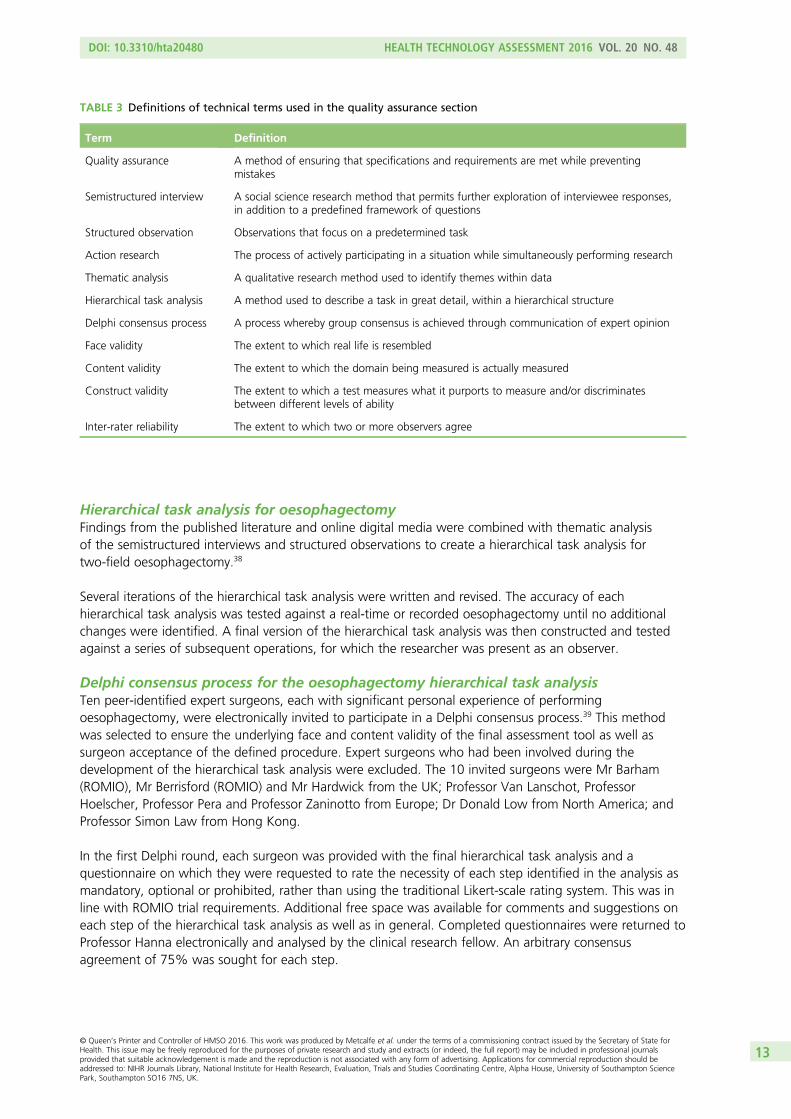
Hierarchical task analysis for oesophagectomyFindings from the published literature and online digital media were combined with thematic analysisof the semistructured interviews and structured observations to create a hierarchical task analysis fortwo-field oesophagectomy.38
Several iterations of the hierarchical task analysis were written and revised. The accuracy of eachhierarchical task analysis was tested against a real-time or recorded oesophagectomy until no additionalchanges were identified. A final version of the hierarchical task analysis was then constructed and testedagainst a series of subsequent operations, for which the researcher was present as an observer.
Delphi consensus process for the oesophagectomy hierarchical task analysisTen peer-identified expert surgeons, each with significant personal experience of performingoesophagectomy, were electronically invited to participate in a Delphi consensus process.39 This methodwas selected to ensure the underlying face and content validity of the final assessment tool as well assurgeon acceptance of the defined procedure. Expert surgeons who had been involved during thedevelopment of the hierarchical task analysis were excluded. The 10 invited surgeons were Mr Barham(ROMIO), Mr Berrisford (ROMIO) and Mr Hardwick from the UK; Professor Van Lanschot, ProfessorHoelscher, Professor Pera and Professor Zaninotto from Europe; Dr Donald Low from North America; andProfessor Simon Law from Hong Kong.
In the first Delphi round, each surgeon was provided with the final hierarchical task analysis and aquestionnaire on which they were requested to rate the necessity of each step identified in the analysis asmandatory, optional or prohibited, rather than using the traditional Likert-scale rating system. This was inline with ROMIO trial requirements. Additional free space was available for comments and suggestions oneach step of the hierarchical task analysis as well as in general. Completed questionnaires were returned toProfessor Hanna electronically and analysed by the clinical research fellow. An arbitrary consensusagreement of 75% was sought for each step.
TABLE 3 Definitions of technical terms used in the quality assurance section
Term Definition
Quality assurance A method of ensuring that specifications and requirements are met while preventingmistakes
Semistructured interview A social science research method that permits further exploration of interviewee responses,in addition to a predefined framework of questions
Structured observation Observations that focus on a predetermined task
Action research The process of actively participating in a situation while simultaneously performing research
Thematic analysis A qualitative research method used to identify themes within data
Hierarchical task analysis A method used to describe a task in great detail, within a hierarchical structure
Delphi consensus process A process whereby group consensus is achieved through communication of expert opinion
Face validity The extent to which real life is resembled
Content validity The extent to which the domain being measured is actually measured
Construct validity The extent to which a test measures what it purports to measure and/or discriminatesbetween different levels of ability
Inter-rater reliability The extent to which two or more observers agree
DOI: 10.3310/hta20480 HEALTH TECHNOLOGY ASSESSMENT 2016 VOL. 20 NO. 48
© Queen’s Printer and Controller of HMSO 2016. This work was produced by Metcalfe et al. under the terms of a commissioning contract issued by the Secretary of State forHealth. This issue may be freely reproduced for the purposes of private research and study and extracts (or indeed, the full report) may be included in professional journalsprovided that suitable acknowledgement is made and the reproduction is not associated with any form of advertising. Applications for commercial reproduction should beaddressed to: NIHR Journals Library, National Institute for Health Research, Evaluation, Trials and Studies Coordinating Centre, Alpha House, University of Southampton SciencePark, Southampton SO16 7NS, UK.
13
This process was repeated in the second Delphi round, during which the nine respondents from the firstDelphi round were electronically sent the percentage agreement and anonymised comments for each stepof the hierarchical task analysis (rather than the traditional mean and standard deviation). The originalanalysis was resent, along with a fresh Delphi questionnaire. If respondents’ answers remained outside ofthe majority agreement, they were asked to provide reasons for this in the comments section. For stepsin which consensus agreement could not be reached by the end of the second Delphi round, theresearcher consulted Professor Hanna at Imperial College London. It was decided that a majority opinioncould be upheld if it reflected the findings of the evidence-based hierarchical task analysis.
Development of an operation manual for oesophagectomyA full operation manual was constructed for circulation to surgeons entering the ROMIO trial, based onthe Delphi consensus approved hierarchical task analysis and its original evidence base (i.e. the publishedliterature, semistructured interviews and structured observations). The operative steps for both openoesophagectomy and MIO were identical. As such, each step of the operation, both mandatory andoptional, was described in detail and diagrams illustrated the required en-bloc lymphadenectomy.
Development of an operation note for oesophagectomyAn operation note was constructed by way of an iterative process. It was designed to satisfy both theclinical requirement of providing a legal record of the operation performed and the research requirementof quality assurance. Its structure was based on the standard operation note currently in use at St Mary’sHospital, London, and combined its generic content (namely patient identifiers, operating surgeon andassistants, procedure performed, operation date, incision, findings, procedure, closure, and postoperativeinstructions) with the evidence-based operation manual specific to the ROMIO trial.
The body of the operation note included a tick-box version of the operation manual, permitting surgeonsto rapidly provide a detailed outline of the procedure performed. White-space boxes were incorporated forpatient identifiers; operative findings; specific details of a task, for example the anastomosis; deviationsfrom the trial protocol; errors and their recovery; and postoperative instructions. Two upper gastrointestinalcancer surgeons at St Mary’s Hospital, London, piloted the operation note over the course of 3 monthsand provided informal verbal constructive feedback to the primary researcher. Surgeons at the two centresinvolved in the ROMIO feasibility study also piloted the operation note.
Development and examination of the reliability of a video assessment toolfor oesophagectomyResults obtained from the semistructured interviews and structured observations performed previouslyconfirmed the importance of the technical performance (i.e. process) and oncological quality (i.e. immediateoutcome) of the operation, in accordance with the Systems Engineering Initiative for Patient Safety (SEIPS)model using a structure–process–outcome approach.40 Various techniques were considered that wouldpermit independent remote blind evaluation of (1) the process (safety and efficiency) and (2) the quality ofthe end product. It was decided that only a video assessment tool could address all of these aspects.
Elements relevant to the safety and efficiency of the operative process, as well as the oncological quality ofthe end product, were identified from thematic analysis of the semistructured interviews and structuredobservations such that clear definitions for each of the terms used were composed. An existing validatedconsultant-level surgical assessment tool41 was deconstructed and its underlying principles adapted duringthe structural development of this video assessment tool.
METHODS
NIHR Journals Library www.journalslibrary.nihr.ac.uk
14
Several different video assessment tools were written and piloted at St Mary’s Hospital, London, over thecourse of 3 months. Each iteration placed a different emphasis on rating the element being assessed, withthe intention of balancing the technical safety with which each task was performed with the surgeons’efficiency and the oncological quality of their dissection. Eight videos, randomly selected from the twocentres involved in the study, were rated by two independent blind assessors (senior consultant surgeonwith 20 years’ experience in upper gastrointestinal surgery and a post Certificate of Completion ofSpecialist Training clinical fellow at consultant grade) to determine inter-rater reliability, which wascalculated using Cohen’s kappa.
Development of a photographic assessment tool for oesophagectomyWhile in Japan, researchers from Imperial College London had witnessed how photographs takenintraoperatively could be used for quality assurance purposes. Therefore, the outcome component of thefinal video assessment tool was isolated for use as a photographic assessment tool, focusing on thecompleteness of the lymphadenectomy and exposure of the relevant anatomical landmarks.
Processing of the pathological specimens
This aspect of the feasibility work aimed to reach agreement on a uniform approach to the processing ofpathological specimens, through collaboration between histopathologists at the two centres and theindependent study pathology lead. This included the approach to the dissection of lymph nodes fromthe main specimen and lymphadenectomy specimens, standardised techniques for the sampling of lymphnodes to achieve the maximum yield from all cases and microscopic and macroscopic assessment of thesurgical resection margin. Quality control was based on regular exchange of material for cross-evaluation.
Development of a core outcome set to assess outcome foroesophageal cancer surgery
The ROMIO feasibility study incorporated work to develop a core outcome set, defining adverse surgicalevents and benefits, using similar methodology as used by the Outcome Measures in Rheumatology(OMERACT) group.42 The basic aim of core outcome set work is to ensure that trials in an area report atleast a common set of core outcomes, allowing for evidence synthesis and cross-study comparisons as wellas reducing reporting bias.
Systematic literature reviews identified all of the current reported clinical outcomes of oesophageal cancersurgery (and their definitions). Qualitative interviews with clinical nurse specialists, dieticians, patients andprimary care representatives identified additional potential outcomes of importance that were not identifiedfrom literature searches. Delphi methodology with surgeons and patients reduced the potential list to ashorter list of outcomes to be discussed at the consensus meetings. In the Delphi survey, stakeholders wereasked to rate the importance of each potential outcome in the core outcome set and two rounds wereundertaken to reduce the list according to prespecified criteria. Each Delphi round was analysed to identifykey or redundant items in the list. Consensus meetings were convened to allow further anonymised ratingof the importance of retained items. This work linked with the Medical Research Council methodologyhub research network-funded project Core Outcome Measures in Effectiveness Trials (COMET)43 led byProfessors Paula Williamson, Jane Blazeby, Doug Altman and Mike Clarke.
DOI: 10.3310/hta20480 HEALTH TECHNOLOGY ASSESSMENT 2016 VOL. 20 NO. 48
© Queen’s Printer and Controller of HMSO 2016. This work was produced by Metcalfe et al. under the terms of a commissioning contract issued by the Secretary of State forHealth. This issue may be freely reproduced for the purposes of private research and study and extracts (or indeed, the full report) may be included in professional journalsprovided that suitable acknowledgement is made and the reproduction is not associated with any form of advertising. Applications for commercial reproduction should beaddressed to: NIHR Journals Library, National Institute for Health Research, Evaluation, Trials and Studies Coordinating Centre, Alpha House, University of Southampton SciencePark, Southampton SO16 7NS, UK.
15
Economic evaluation
The objective of the economic evaluation work was to explore the best way of capturing resource use andcosts in relation to the interventions and follow-up in secondary care. Questionnaires and resource use logswere also developed to collect use of community-based NHS services and social services and direct andindirect costs incurred by patient and carers. Unit costs from hospital finance and routine sources werethen to be applied to the resource use data.
The original intention had been to establish the face validity of the resource use questionnaires for patientsthrough qualitative interviewing using ‘thinking aloud’ techniques. However, following development of the3-month follow-up questionnaire, clinician opinion indicated that patients would frequently be too ill tocomplete the questionnaire. As decision was made to redevelop the questionnaires into a telephoneinterview format, which could be administered by research nurses at 3 and 6 months post surgery.
The resource use data were separated into different categories (e.g. theatre, outpatient visits, generalpractitioner visits) and the average costs for all of the different categories were compared by arm. Thisenabled the main cost drivers of the interventions to be established and the identification of potentialareas where cost differences exist between the arms in addition to identifying areas where obtainingaccurate estimates of cost is problematic. This would enable a more focused collection of resource usedata in the main trial, which will result in a more accurate estimate of cost-effectiveness.
There are two different types of costing methodology that can be used within an economic evaluation:the use of Healthcare Resource Groups (HRGs) and microcosting. HRGs are groups of different events(based on procedures and/or diagnoses) that are deemed to use similar amounts of resources. Costs canthen be applied to the HRGs using national reference costs. Microcosting is a methodology in whichresource use at a more granular level is captured (e.g. number of consultant contacts, medication use,time in theatre). Given that the different types of oesophagectomy are incorporated within the same HRG,a microcosting approach is needed in terms of the initial inpatient stay for the oesophagectomy, whereas itis possible to use HRGs for subsequent inpatient stays. Within the feasibility trial it was explored how bestto microcost the initial inpatient stay and how best to obtain the information needed to use HRGs for thefollow-up period.
METHODS
NIHR Journals Library www.journalslibrary.nihr.ac.uk
16
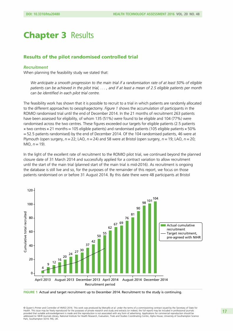
Chapter 3 Results
Results of the pilot randomised controlled trial
RecruitmentWhen planning the feasibility study we stated that:
We anticipate a smooth progression to the main trial if a randomisation rate of at least 50% of eligiblepatients can be achieved in the pilot trial, . . . , and if at least a mean of 2.5 eligible patients per monthcan be identified in each pilot trial centre.
The feasibility work has shown that it is possible to recruit to a trial in which patients are randomly allocatedto the different approaches to oesophagectomy. Figure 1 shows the accumulation of participants in theROMIO randomised trial until the end of December 2014. In the 21 months of recruitment 263 patientshave been assessed for eligibility, of whom 135 (51%) were found to be eligible and 104 (77%) wererandomised across the two centres. These figures exceeded our targets for eligible patients (2.5 patients× two centres × 21 months= 105 eligible patients) and randomised patients (105 eligible patients × 50%= 52.5 patients randomised) by the end of December 2014. Of the 104 randomised patients, 46 were atPlymouth (open surgery, n= 22; LAO, n= 24) and 58 were at Bristol (open surgery, n= 19; LAO, n= 20;MIO, n= 19).
In the light of the excellent rate of recruitment to the ROMIO pilot trial, we continued beyond the plannedclosure date of 31 March 2014 and successfully applied for a contract variation to allow recruitmentuntil the start of the main trial (planned start of the main trial is mid-2016). As recruitment is ongoingthe database is still live and so, for the purposes of the remainder of this report, we focus on thosepatients randomised on or before 31 August 2014. By this date there were 48 participants at Bristol
120
100
80
60
Cu
mu
lati
ve t
ota
l rec
ruit
ed
40
20
0
April 2013
49
1216
2024
27 30
3742
5055
6267 69
7681
90
98101
104
August 2013 December 2013Recruitment period
April 2014 August 2014 December 2014
Actual cumulativerecruitmentTarget recruitment, pre-agreed with NIHR
FIGURE 1 Actual and target recruitment up to December 2014. Recruitment to the study is continuing.
DOI: 10.3310/hta20480 HEALTH TECHNOLOGY ASSESSMENT 2016 VOL. 20 NO. 48
© Queen’s Printer and Controller of HMSO 2016. This work was produced by Metcalfe et al. under the terms of a commissioning contract issued by the Secretary of State forHealth. This issue may be freely reproduced for the purposes of private research and study and extracts (or indeed, the full report) may be included in professional journalsprovided that suitable acknowledgement is made and the reproduction is not associated with any form of advertising. Applications for commercial reproduction should beaddressed to: NIHR Journals Library, National Institute for Health Research, Evaluation, Trials and Studies Coordinating Centre, Alpha House, University of Southampton SciencePark, Southampton SO16 7NS, UK.
17
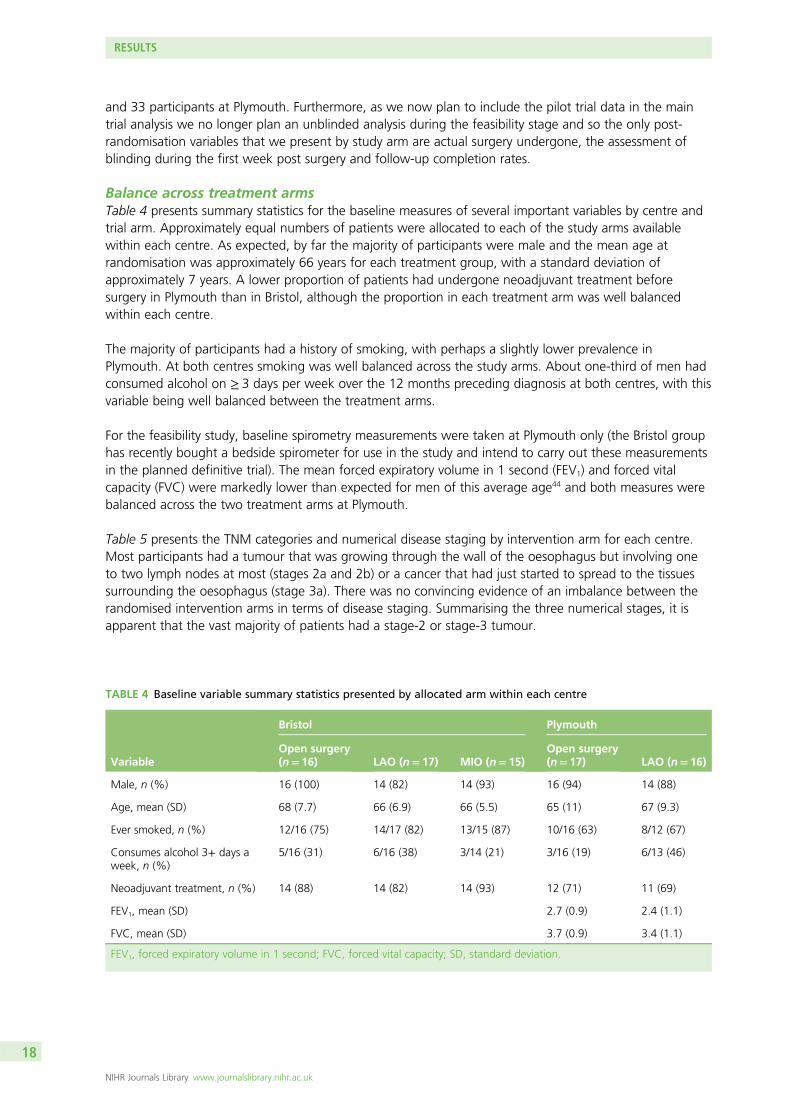
and 33 participants at Plymouth. Furthermore, as we now plan to include the pilot trial data in the maintrial analysis we no longer plan an unblinded analysis during the feasibility stage and so the only post-randomisation variables that we present by study arm are actual surgery undergone, the assessment ofblinding during the first week post surgery and follow-up completion rates.
Balance across treatment armsTable 4 presents summary statistics for the baseline measures of several important variables by centre andtrial arm. Approximately equal numbers of patients were allocated to each of the study arms availablewithin each centre. As expected, by far the majority of participants were male and the mean age atrandomisation was approximately 66 years for each treatment group, with a standard deviation ofapproximately 7 years. A lower proportion of patients had undergone neoadjuvant treatment beforesurgery in Plymouth than in Bristol, although the proportion in each treatment arm was well balancedwithin each centre.
The majority of participants had a history of smoking, with perhaps a slightly lower prevalence inPlymouth. At both centres smoking was well balanced across the study arms. About one-third of men hadconsumed alcohol on ≥ 3 days per week over the 12 months preceding diagnosis at both centres, with thisvariable being well balanced between the treatment arms.
For the feasibility study, baseline spirometry measurements were taken at Plymouth only (the Bristol grouphas recently bought a bedside spirometer for use in the study and intend to carry out these measurementsin the planned definitive trial). The mean forced expiratory volume in 1 second (FEV1) and forced vitalcapacity (FVC) were markedly lower than expected for men of this average age44 and both measures werebalanced across the two treatment arms at Plymouth.
Table 5 presents the TNM categories and numerical disease staging by intervention arm for each centre.Most participants had a tumour that was growing through the wall of the oesophagus but involving oneto two lymph nodes at most (stages 2a and 2b) or a cancer that had just started to spread to the tissuessurrounding the oesophagus (stage 3a). There was no convincing evidence of an imbalance between therandomised intervention arms in terms of disease staging. Summarising the three numerical stages, it isapparent that the vast majority of patients had a stage-2 or stage-3 tumour.
TABLE 4 Baseline variable summary statistics presented by allocated arm within each centre
Variable
Bristol Plymouth
Open surgery(n= 16) LAO (n= 17) MIO (n= 15)
Open surgery(n= 17) LAO (n= 16)
Male, n (%) 16 (100) 14 (82) 14 (93) 16 (94) 14 (88)
Age, mean (SD) 68 (7.7) 66 (6.9) 66 (5.5) 65 (11) 67 (9.3)
Ever smoked, n (%) 12/16 (75) 14/17 (82) 13/15 (87) 10/16 (63) 8/12 (67)
Consumes alcohol 3+ days aweek, n (%)
5/16 (31) 6/16 (38) 3/14 (21) 3/16 (19) 6/13 (46)
Neoadjuvant treatment, n (%) 14 (88) 14 (82) 14 (93) 12 (71) 11 (69)
FEV1, mean (SD) 2.7 (0.9) 2.4 (1.1)
FVC, mean (SD) 3.7 (0.9) 3.4 (1.1)
FEV1, forced expiratory volume in 1 second; FVC, forced vital capacity; SD, standard deviation.
RESULTS
NIHR Journals Library www.journalslibrary.nihr.ac.uk
18
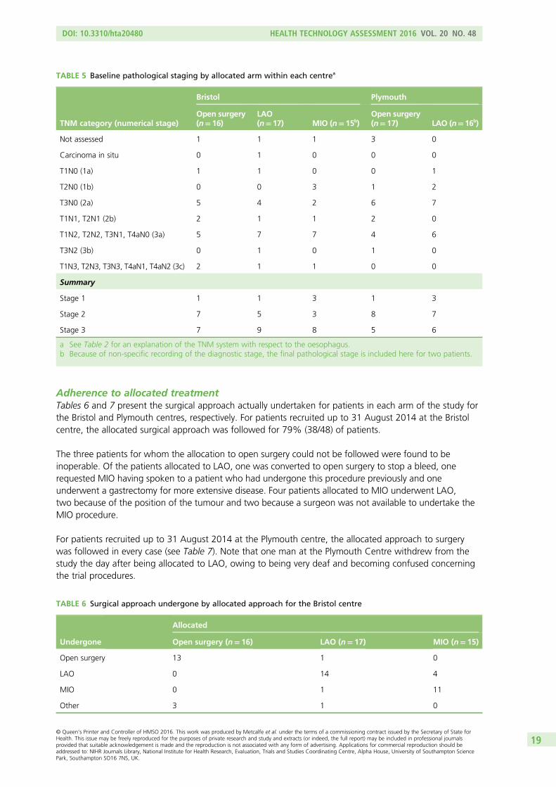
Adherence to allocated treatmentTables 6 and 7 present the surgical approach actually undertaken for patients in each arm of the study forthe Bristol and Plymouth centres, respectively. For patients recruited up to 31 August 2014 at the Bristolcentre, the allocated surgical approach was followed for 79% (38/48) of patients.
The three patients for whom the allocation to open surgery could not be followed were found to beinoperable. Of the patients allocated to LAO, one was converted to open surgery to stop a bleed, onerequested MIO having spoken to a patient who had undergone this procedure previously and oneunderwent a gastrectomy for more extensive disease. Four patients allocated to MIO underwent LAO,two because of the position of the tumour and two because a surgeon was not available to undertake theMIO procedure.
For patients recruited up to 31 August 2014 at the Plymouth centre, the allocated approach to surgerywas followed in every case (see Table 7). Note that one man at the Plymouth Centre withdrew from thestudy the day after being allocated to LAO, owing to being very deaf and becoming confused concerningthe trial procedures.
TABLE 5 Baseline pathological staging by allocated arm within each centrea
TNM category (numerical stage)
Bristol Plymouth
Open surgery(n=16)
LAO(n=17) MIO (n=15b)
Open surgery(n=17) LAO (n=16b)
Not assessed 1 1 1 3 0
Carcinoma in situ 0 1 0 0 0
T1N0 (1a) 1 1 0 0 1
T2N0 (1b) 0 0 3 1 2
T3N0 (2a) 5 4 2 6 7
T1N1, T2N1 (2b) 2 1 1 2 0
T1N2, T2N2, T3N1, T4aN0 (3a) 5 7 7 4 6
T3N2 (3b) 0 1 0 1 0
T1N3, T2N3, T3N3, T4aN1, T4aN2 (3c) 2 1 1 0 0
Summary
Stage 1 1 1 3 1 3
Stage 2 7 5 3 8 7
Stage 3 7 9 8 5 6
a See Table 2 for an explanation of the TNM system with respect to the oesophagus.b Because of non-specific recording of the diagnostic stage, the final pathological stage is included here for two patients.
TABLE 6 Surgical approach undergone by allocated approach for the Bristol centre
Undergone
Allocated
Open surgery (n= 16) LAO (n= 17) MIO (n= 15)
Open surgery 13 1 0
LAO 0 14 4
MIO 0 1 11
Other 3 1 0
DOI: 10.3310/hta20480 HEALTH TECHNOLOGY ASSESSMENT 2016 VOL. 20 NO. 48
© Queen’s Printer and Controller of HMSO 2016. This work was produced by Metcalfe et al. under the terms of a commissioning contract issued by the Secretary of State forHealth. This issue may be freely reproduced for the purposes of private research and study and extracts (or indeed, the full report) may be included in professional journalsprovided that suitable acknowledgement is made and the reproduction is not associated with any form of advertising. Applications for commercial reproduction should beaddressed to: NIHR Journals Library, National Institute for Health Research, Evaluation, Trials and Studies Coordinating Centre, Alpha House, University of Southampton SciencePark, Southampton SO16 7NS, UK.
19
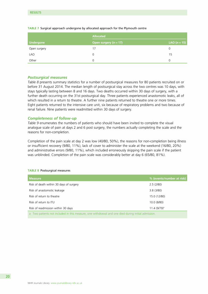
Postsurgical measuresTable 8 presents summary statistics for a number of postsurgical measures for 80 patients recruited on orbefore 31 August 2014. The median length of postsurgical stay across the two centres was 10 days, withstays typically lasting between 8 and 16 days. Two deaths occurred within 30 days of surgery, with afurther death occurring on the 31st postsurgical day. Three patients experienced anastomotic leaks, all ofwhich resulted in a return to theatre. A further nine patients returned to theatre one or more times.Eight patients returned to the intensive care unit, six because of respiratory problems and two because ofrenal failure. Nine patients were readmitted within 30 days of surgery.
Completeness of follow-upTable 9 enumerates the numbers of patients who should have been invited to complete the visualanalogue scale of pain at days 2 and 6 post surgery, the numbers actually completing the scale and thereasons for non-completion.
Completion of the pain scale at day 2 was low (40/80, 50%), the reasons for non-completion being illnessor insufficient recovery (9/80, 11%), lack of cover to administer the scale at the weekend (16/80, 20%)and administrative errors (9/80, 11%), which included erroneously skipping the pain scale if the patientwas unblinded. Completion of the pain scale was considerably better at day 6 (65/80, 81%).
TABLE 7 Surgical approach undergone by allocated approach for the Plymouth centre
Undergone
Allocated
Open surgery (n= 17) LAO (n= 15)
Open surgery 17 0
LAO 0 15
Other 0 0
TABLE 8 Postsurgical measures
Measure % (events/number at risk)
Risk of death within 30 days of surgery 2.5 (2/80)
Risk of anastomotic leakage 3.8 (3/80)
Risk of return to theatre 15.0 (12/80)
Risk of return to ITU 10.0 (8/80)
Risk of readmission within 30 days 11.4 (9/79)a
a Two patients not included in this measure, one withdrawal and one died during initial admission.
RESULTS
NIHR Journals Library www.journalslibrary.nihr.ac.uk
20
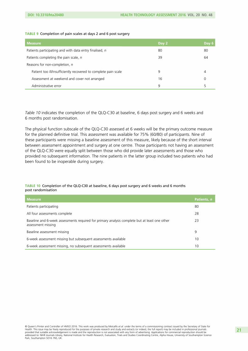
Table 10 indicates the completion of the QLQ-C30 at baseline, 6 days post surgery and 6 weeks and6 months post randomisation.
The physical function subscale of the QLQ-C30 assessed at 6 weeks will be the primary outcome measurefor the planned definitive trial. This assessment was available for 75% (60/80) of participants. Nine ofthese participants were missing a baseline assessment of this measure, likely because of the short intervalbetween assessment appointment and surgery at one centre. Those participants not having an assessmentof the QLQ-C30 were equally split between those who did provide later assessments and those whoprovided no subsequent information. The nine patients in the latter group included two patients who hadbeen found to be inoperable during surgery.
TABLE 9 Completion of pain scales at days 2 and 6 post surgery
Measure Day 2 Day 6
Patients participating and with data entry finalised, n 80 80
Patients completing the pain scale, n 39 64
Reasons for non-completion, n
Patient too ill/insufficiently recovered to complete pain scale 9 4
Assessment at weekend and cover not arranged 16 0
Administrative error 9 5
TABLE 10 Completion of the QLQ-C30 at baseline, 6 days post surgery and 6 weeks and 6 monthspost randomisation
Measure Patients, n
Patients participating 80
All four assessments complete 28
Baseline and 6-week assessments required for primary analysis complete but at least one otherassessment missing
23
Baseline assessment missing 9
6-week assessment missing but subsequent assessments available 10
6-week assessment missing, no subsequent assessments available 10
DOI: 10.3310/hta20480 HEALTH TECHNOLOGY ASSESSMENT 2016 VOL. 20 NO. 48
© Queen’s Printer and Controller of HMSO 2016. This work was produced by Metcalfe et al. under the terms of a commissioning contract issued by the Secretary of State forHealth. This issue may be freely reproduced for the purposes of private research and study and extracts (or indeed, the full report) may be included in professional journalsprovided that suitable acknowledgement is made and the reproduction is not associated with any form of advertising. Applications for commercial reproduction should beaddressed to: NIHR Journals Library, National Institute for Health Research, Evaluation, Trials and Studies Coordinating Centre, Alpha House, University of Southampton SciencePark, Southampton SO16 7NS, UK.
21
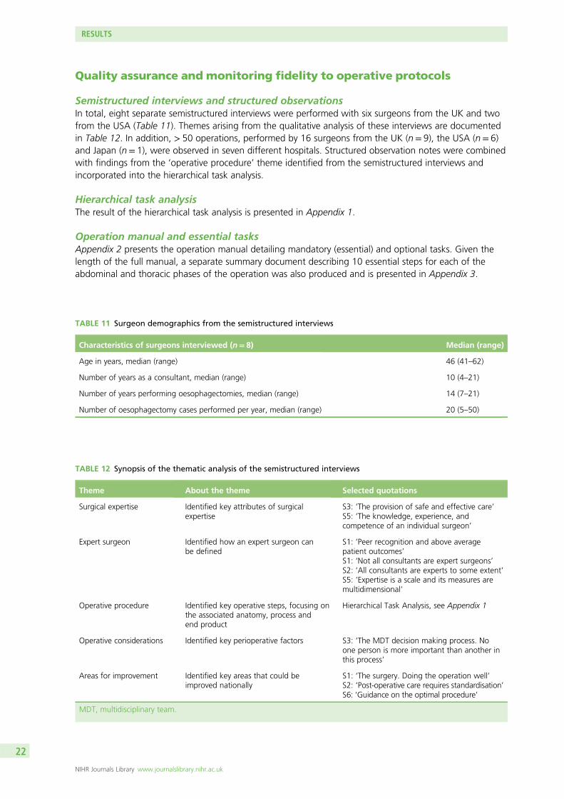
Quality assurance and monitoring fidelity to operative protocols
Semistructured interviews and structured observationsIn total, eight separate semistructured interviews were performed with six surgeons from the UK and twofrom the USA (Table 11). Themes arising from the qualitative analysis of these interviews are documentedin Table 12. In addition, > 50 operations, performed by 16 surgeons from the UK (n= 9), the USA (n= 6)and Japan (n= 1), were observed in seven different hospitals. Structured observation notes were combinedwith findings from the ‘operative procedure’ theme identified from the semistructured interviews andincorporated into the hierarchical task analysis.
Hierarchical task analysisThe result of the hierarchical task analysis is presented in Appendix 1.
Operation manual and essential tasksAppendix 2 presents the operation manual detailing mandatory (essential) and optional tasks. Given thelength of the full manual, a separate summary document describing 10 essential steps for each of theabdominal and thoracic phases of the operation was also produced and is presented in Appendix 3.
TABLE 12 Synopsis of the thematic analysis of the semistructured interviews
Theme About the theme Selected quotations
Surgical expertise Identified key attributes of surgicalexpertise
S3: ‘The provision of safe and effective care’S5: ‘The knowledge, experience, andcompetence of an individual surgeon’
Expert surgeon Identified how an expert surgeon canbe defined
S1: ‘Peer recognition and above averagepatient outcomes’S1: ‘Not all consultants are expert surgeons’S2: ‘All consultants are experts to some extent’S5: ‘Expertise is a scale and its measures aremultidimensional’
Operative procedure Identified key operative steps, focusing onthe associated anatomy, process andend product
Hierarchical Task Analysis, see Appendix 1
Operative considerations Identified key perioperative factors S3: ‘The MDT decision making process. Noone person is more important than another inthis process’
Areas for improvement Identified key areas that could beimproved nationally
S1: ‘The surgery. Doing the operation well’S2: ‘Post-operative care requires standardisation’S6: ‘Guidance on the optimal procedure’
MDT, multidisciplinary team.
TABLE 11 Surgeon demographics from the semistructured interviews
Characteristics of surgeons interviewed (n= 8) Median (range)
Age in years, median (range) 46 (41–62)
Number of years as a consultant, median (range) 10 (4–21)
Number of years performing oesophagectomies, median (range) 14 (7–21)
Number of oesophagectomy cases performed per year, median (range) 20 (5–50)
RESULTS
NIHR Journals Library www.journalslibrary.nihr.ac.uk
22

Operation noteThe operation note is presented in Appendix 4. This note allows recording of performance of or variationfrom steps in the manual, errors incurred and recovery mechanisms. This will allow root cause analysis ofadverse events.
Video and photographic assessment toolsTo allow a standardised assessment of video and photographic recording of surgical procedures,assessment tools were produced and are presented in Appendices 5 and 6, respectively.
Implementation in the feasibility studyThe manuals, notes and assessment tools for quality assurance were approved by the ROMIO Trial SteeringCommittee on 15 November 2013. The approved documents were piloted during the ROMIO feasibility phase.An online encrypted file transfer process was set up by the team at Imperial College London. The ImperialCollege London Clinical Governance Officer arranged local access to the server and a nominee from each site,for example a nurse specialist, was identified to upload the files. The system was tested in both centres and allsurgeons submitted video records from their surgery. Twenty-five operative videos were transferred using theonline system. Blind independent assessors evaluated the operative performance using the video assessmenttool, indicating that an objective video-based assessment system is feasible and can be applied in the main trial.Sets of intraoperative photographs of the operative field were obtained by the surgeons and transferred usingthe same online system. Surgeons used the operation notes to record the steps of the procedures.
Processing of the pathological specimens
In two face-to-face meetings between the lead pathologists at each of the two feasibility study centres andProfessor Goldin, the study’s independent histopathologist, consensus was reached on the followingapproach to the processing and reporting of pathological specimens. All trial pathology specimens will beprepared and macroscopically and microscopically assessed in a uniform manner as per the current RoyalCollege of Pathologists Dataset for Oesophageal Carcinomas.45 The pathology data for the trial will becollected using a standardised form and will represent data points included within the Royal College ofPathologists data set. Data points that will serve as surgical quality assurance indicators include the length ofthe oesophagus and the number of harvested lymph nodes. Data points that will serve as surrogate markersfor patient survival include pT (pathologically determined tumour) stage, pN (pathologically determinednode) stage and pM (pathologically determined metastasis) stage. For pathology quality assurance purposes,the slides of 10% of all cases from each centre will be reviewed by the lead pathologist.
Blinding to treatment allocation
During the pilot trial, patients were asked at 2 and 6 days post surgery to state which surgical technique theyhad undergone. Table 13 gives the responses by allocation for the two assessments for patients randomised upuntil 31 August 2014 at the Bristol centre (total n= 47). Those patients who did not undergo their allocatedprocedure are excluded from this table (n= 9), whereas those patients who became unblinded are included asknowing their allocation. For the day-2 assessment, 12 measurements are missing because of administrativeerrors (largely problems organising weekend cover in the first few weeks of the study, n= 9) and because ofpatients not being sufficiently recovered from surgery to complete the inventory (n= 3). The day 6 assessmentis a little more complete. These data indicate that blinding was successful during the first week post surgery,with the majority of patients who stated a surgical approach believing that they had undergone LAO at day 2and LAO or MIO at day 6, irrespective of the approach that they had actually undergone.
Table 14 provides the patient beliefs about treatment received by allocated treatment arm for thePlymouth centre (total randomised by 31 August 2014 n= 28). As for the Bristol centre, the majority ofpatients believed that they had undergone the LAO procedure at both day 2 and day 6, irrespective of thearm to which they had been allocated.
DOI: 10.3310/hta20480 HEALTH TECHNOLOGY ASSESSMENT 2016 VOL. 20 NO. 48
© Queen’s Printer and Controller of HMSO 2016. This work was produced by Metcalfe et al. under the terms of a commissioning contract issued by the Secretary of State forHealth. This issue may be freely reproduced for the purposes of private research and study and extracts (or indeed, the full report) may be included in professional journalsprovided that suitable acknowledgement is made and the reproduction is not associated with any form of advertising. Applications for commercial reproduction should beaddressed to: NIHR Journals Library, National Institute for Health Research, Evaluation, Trials and Studies Coordinating Centre, Alpha House, University of Southampton SciencePark, Southampton SO16 7NS, UK.
23
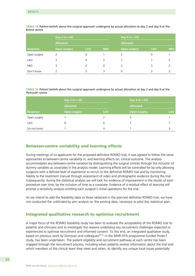
Between-centre variability and learning effects
During meetings of co-applicants for the proposed definitive ROMIO trial, it was agreed to follow the sameapproaches to between-centre variability in, and learning effects on, clinical outcome. The analysisaccommodates any between-centre variation by distinguishing the surgical centres through the inclusion ofdummy variables as covariates in the analysis model. Learning effects will be controlled for by only allowingsurgeons with a defined level of experience to recruit to the definitive ROMIO trial and by monitoringfidelity to the treatment manual through assessment of video and photographic evidence during the trial.Subsequently, during the statistical analysis we will look for evidence of improvement in the results of eachprocedure over time, by the inclusion of time as a covariate. Evidence of a residual effect of learning willprompt a sensitivity analysis omitting each surgeon’s initial operations for the trial.
As we intend to add the feasibility data to those obtained in the planned definitive ROMIO trial, we havenot conducted the unblinded by arm analysis on the existing data, necessary to pilot this statistical plan.
Integrated qualitative research to optimise recruitment
A major focus of the ROMIO feasibility study has been to evaluate the acceptability of the ROMIO trial topatients and clinicians and to investigate the reasons underlying any recruitment challenges expected orexperienced to optimise recruitment and informed consent. To this end, an integrated qualitative study,based on previous work by Donovan and colleagues28–31 in the NIHR HTA programme-funded ProtecTstudy, has been undertaken. The patient eligibility and recruitment pathway at each centre has beenmapped through the recruitment process, including when patients receive information about the trial andwhich members of the clinical team they meet and when, to identify any unique local issues potentially
TABLE 13 Patient beliefs about the surgical approach undergone by actual allocation at day 2 and day 6 at theBristol centre
Response
Day 2 (n= 26) Day 6 (n= 35)
Allocated Allocated
Open surgery LAO MIO Open surgery LAO MIO
Open surgery 2 0 1 2 0 1
LAO 3 4 5 7 7 5
MIO 3 0 0 2 3 3
Don’t know 2 3 3 0 3 2
TABLE 14 Patient beliefs about the surgical approach undergone by actual allocation at day 2 and day 6 at thePlymouth centre
Response
Day 2 (n= 23) Day 6 (n= 24)
Allocated Allocated
Open surgery LAO Open surgery LAO
Open surgery 1 2 5 1
LAO 9 6 7 7
Do not know 1 4 1 3
RESULTS
NIHR Journals Library www.journalslibrary.nihr.ac.uk
24
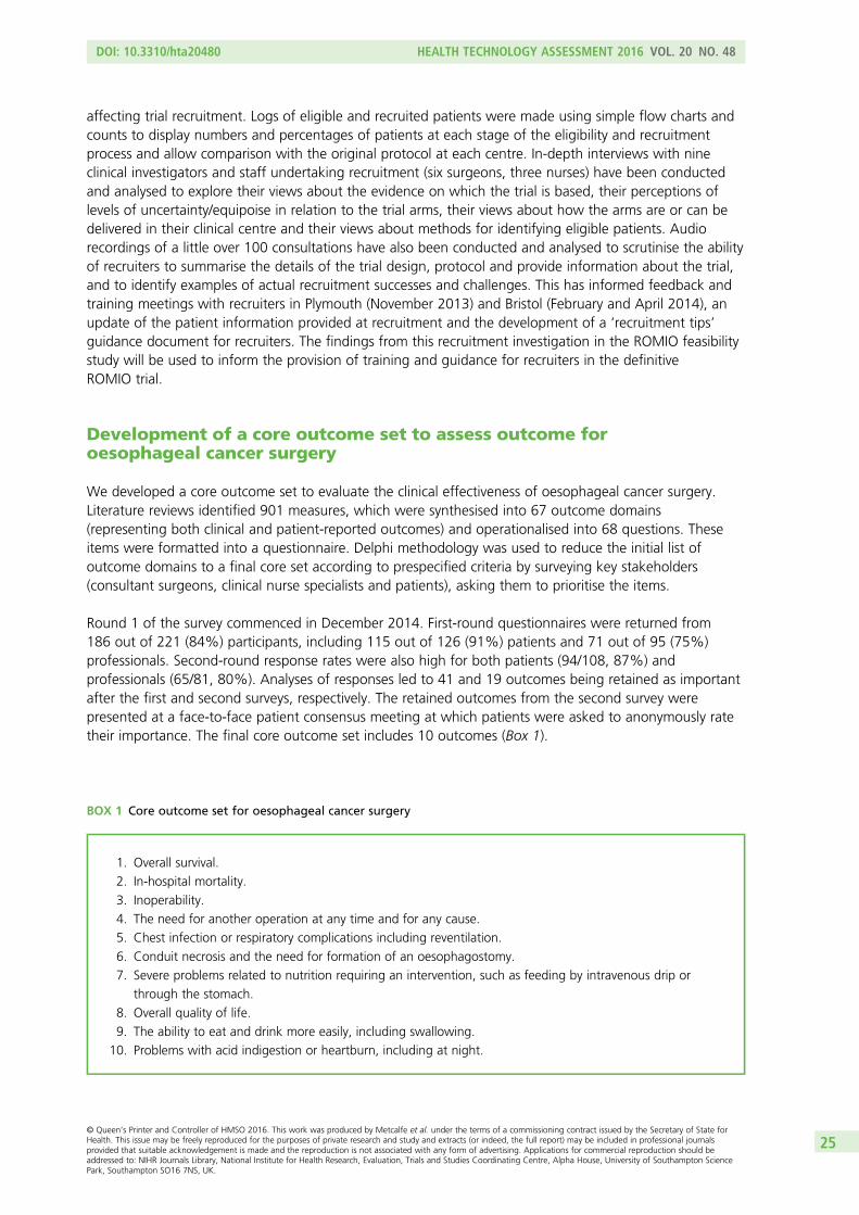
affecting trial recruitment. Logs of eligible and recruited patients were made using simple flow charts andcounts to display numbers and percentages of patients at each stage of the eligibility and recruitmentprocess and allow comparison with the original protocol at each centre. In-depth interviews with nineclinical investigators and staff undertaking recruitment (six surgeons, three nurses) have been conductedand analysed to explore their views about the evidence on which the trial is based, their perceptions oflevels of uncertainty/equipoise in relation to the trial arms, their views about how the arms are or can bedelivered in their clinical centre and their views about methods for identifying eligible patients. Audiorecordings of a little over 100 consultations have also been conducted and analysed to scrutinise the abilityof recruiters to summarise the details of the trial design, protocol and provide information about the trial,and to identify examples of actual recruitment successes and challenges. This has informed feedback andtraining meetings with recruiters in Plymouth (November 2013) and Bristol (February and April 2014), anupdate of the patient information provided at recruitment and the development of a ‘recruitment tips’guidance document for recruiters. The findings from this recruitment investigation in the ROMIO feasibilitystudy will be used to inform the provision of training and guidance for recruiters in the definitiveROMIO trial.
Development of a core outcome set to assess outcome foroesophageal cancer surgery
We developed a core outcome set to evaluate the clinical effectiveness of oesophageal cancer surgery.Literature reviews identified 901 measures, which were synthesised into 67 outcome domains(representing both clinical and patient-reported outcomes) and operationalised into 68 questions. Theseitems were formatted into a questionnaire. Delphi methodology was used to reduce the initial list ofoutcome domains to a final core set according to prespecified criteria by surveying key stakeholders(consultant surgeons, clinical nurse specialists and patients), asking them to prioritise the items.
Round 1 of the survey commenced in December 2014. First-round questionnaires were returned from186 out of 221 (84%) participants, including 115 out of 126 (91%) patients and 71 out of 95 (75%)professionals. Second-round response rates were also high for both patients (94/108, 87%) andprofessionals (65/81, 80%). Analyses of responses led to 41 and 19 outcomes being retained as importantafter the first and second surveys, respectively. The retained outcomes from the second survey werepresented at a face-to-face patient consensus meeting at which patients were asked to anonymously ratetheir importance. The final core outcome set includes 10 outcomes (Box 1).
BOX 1 Core outcome set for oesophageal cancer surgery
1. Overall survival.
2. In-hospital mortality.
3. Inoperability.
4. The need for another operation at any time and for any cause.
5. Chest infection or respiratory complications including reventilation.
6. Conduit necrosis and the need for formation of an oesophagostomy.
7. Severe problems related to nutrition requiring an intervention, such as feeding by intravenous drip or
through the stomach.
8. Overall quality of life.
9. The ability to eat and drink more easily, including swallowing.
10. Problems with acid indigestion or heartburn, including at night.
DOI: 10.3310/hta20480 HEALTH TECHNOLOGY ASSESSMENT 2016 VOL. 20 NO. 48
© Queen’s Printer and Controller of HMSO 2016. This work was produced by Metcalfe et al. under the terms of a commissioning contract issued by the Secretary of State forHealth. This issue may be freely reproduced for the purposes of private research and study and extracts (or indeed, the full report) may be included in professional journalsprovided that suitable acknowledgement is made and the reproduction is not associated with any form of advertising. Applications for commercial reproduction should beaddressed to: NIHR Journals Library, National Institute for Health Research, Evaluation, Trials and Studies Coordinating Centre, Alpha House, University of Southampton SciencePark, Southampton SO16 7NS, UK.
25
Economic evaluation
Initial inpatient stay incorporating the oesophagectomyInitial meetings with finance personnel at one of the sites and subsequent approaches to financedepartments within all of the sites established how resource use was costed within the different hospitals.This will enable resource use information to be collected in the correct way in terms of measurement(e.g. different types of medical staff contacts can be measured using the number of visits to the patient orthe actual time spent visiting the patient), which will then ensure that costs from the finance departmentscan be applied.
Follow-up inpatient stays and outpatient visitsThe follow-up Clinical Report Form (CRF) was designed to enable HRGs to be created. The level of detailneeded for this to be carried out correctly can be time-consuming. Different approaches to obtaining theHRGs for any subsequent inpatient stays have been explored.
Coders within the hospitals were initially approached as they allocate codes to diagnoses, procedures,complications and comorbidities. These codes are then submitted into a HRG ‘grouper’, a softwarepackage available for each financial year from the Health and Social Care Information Centre(www.hscic.gov.uk), which produces the HRG code for an individual’s inpatient stay. Time constraintsmay, however, limit their ability to obtain HRG codes for each individual within the trial.
Clinical commissioning groups were approached as they use individual patient HRGs to reimbursehospitals. However, the groups obtain patient HRGs only in a pseudo-anonymised format.
Finally, the use of individual patient records as the source to obtain data on resource use was explored.Finance and costing departments for Bristol, Plymouth and Bath were contacted. Bristol and Plymouth haveso far confirmed that individual patient records are available for ROMIO patients. If individual patientrecords are available for all other sites in the main ROMIO trial then this would be the preferred option.However, although information on operating times would be available through the individual patientrecords, it is unlikely that differences in equipment use for the main surgical intervention would beaccurately captured as needed. This emphasises the need to use microcosting techniques to cost the initialoperation, even if individual patient records are used for the remaining aspects of the initial inpatient stay.
Resource use logs (diaries)These were designed to be used by the patient as an ‘aide memoire’ to prospectively record health andsocial service contacts and other expenses incurred, with the format of the logs mirroring that of thequestionnaires. Resource use logs have the potential to reduce missing data46 and therefore lead to moreaccurate estimates of costs. To allow piloting of the resource use diaries and the telephone-administeredquestionnaires, a substantial amendment to the ethical approval was sought and this was granted in April2014. Patients were given the 0–3 months resource use diary by a research nurse at hospital dischargefollowing their oesophagectomy. If they were too unwell to record events a relative or friend could recordthis information for them. At 3 months following their discharge, a nurse telephoned those patients giventhe resource use diary to ask them questions about their resource use during that period of time. To notoverburden the patients, nurses were encouraged to call back or speak with a relative who could answerthe questions on a patient’s behalf if the patient did not feel able to answer the questions at that time.
To assess the feasibility of the 6-month telephone administration of the resource use questionnaire thefollowing process was established. Participants who had been discharged prior to ethical approval beinggranted but who had not yet reached 3 months post discharge were posted the 4–6 months resource usediary at 3 months post discharge and asked to complete it for the following 3 months. At 6 monthsfollowing their discharge from hospital a research nurse telephoned those patients who had received a4–6 months resource use diary and asked questions about their resource use.
RESULTS
NIHR Journals Library www.journalslibrary.nihr.ac.uk
26
Initial findings on this processThe organisation of the study, especially for patients who undergo surgery in one site and are followed upin another site (as in the case of Bath patients), means that, if resource use diaries are to be used in themain trial, site-specific resource use diaries and resource use questionnaires will have to be created. Somepatients did not want to complete the questionnaire over the telephone; one patient wanted only to readout what he had written in the resource use diary rather than be asked questions. Another patient wantedto return the resource use diary rather than complete the telephone questionnaire. These findings haveimplications for how resource use is obtained for the main trial, as these two actions resulted inmissing data.
To reduce the burden on patients, one suggestion is to use a much narrower NHS perspective or NHS andPersonal Social Services (PSS) perspective for the main trial and include an exploration of other resourceuse within the qualitative component of the trial.
A shortened resource use instrument will still be required as not all secondary care visits and inpatient staystake place within the main treating hospitals where the initial surgery took place, and therefore medicalrecords cannot provide this information. In some instances, when a substantial group of patients isfollowed up at another site, for example as in Bath in the feasibility study, it makes sense for the correctapprovals to be gained so that access to medical records can be granted. In addition, community-basedNHS resource use still needs to be obtained.
One solution would be to use the patient diary as the main patient source of information, using telephoneinterviews for those patients who do not return the diaries. Alternatively, given that patients did seem tobe able to complete the resource use diaries, a shortened resource use questionnaire may be acceptable.
Establishing cost differences between the armsOne potential area in which cost differences will exist between the arms of the trial is in relation to theactual operation. A CRF was created that outlined potential equipment and consumables that couldbe used in all of the operations in terms of brand and quantity and the operation staff potentially involvedin terms of their role and time present during the operation. Currently, this CRF is being piloted inPlymouth on the two different types of oesophagectomy. Once the main differences in theatre resourcehave been identified in this pilot, theatre records will be obtained from Bristol to ascertain whether or notthe main differences in resource use can be captured easily within the Bristol centre.
DOI: 10.3310/hta20480 HEALTH TECHNOLOGY ASSESSMENT 2016 VOL. 20 NO. 48
© Queen’s Printer and Controller of HMSO 2016. This work was produced by Metcalfe et al. under the terms of a commissioning contract issued by the Secretary of State forHealth. This issue may be freely reproduced for the purposes of private research and study and extracts (or indeed, the full report) may be included in professional journalsprovided that suitable acknowledgement is made and the reproduction is not associated with any form of advertising. Applications for commercial reproduction should beaddressed to: NIHR Journals Library, National Institute for Health Research, Evaluation, Trials and Studies Coordinating Centre, Alpha House, University of Southampton SciencePark, Southampton SO16 7NS, UK.
27

Chapter 4 Discussion
Summary of findings
This feasibility study has demonstrated that different approaches to oesophagectomy can be compared inan unbiased fashion in a RCT. Recruitment to the pilot trial was soon ahead of target at the two centres,Bristol and Plymouth, with the qualitative recruitment intervention overcoming the widely recogniseddifficulties of presenting to patients the idea of equipoise between surgical techniques and the need forrandom allocation. The high proportion of eligible patients consenting to join the trial (76.5%; all figuresfor patients recruited until 31 August 2014) was likely to be one factor behind the profile of participantsbeing typical of the profile of patients seen in clinical practice. The large majority of patients underwentoesophagectomy according to their allocated approach (79% at Bristol, 100% at Plymouth; patientsrecruited until 31 August 2014). Four out of 10 patients not following their allocated approach at Bristolwere found to be inoperable during surgery. Blinding participants to their allocation during the firstweek post surgery using a large bandage that covered all possible incision sites was found to be successfulon the whole, with a similar majority of patients believing that they had undergone the LAO or MIOprocedures across all intervention arms at a centre.
Follow-up completion rates were good considering that this is a severely ill population of patients with apoor medium-term prognosis even when suitable for surgery. Surgical complications were common,as they are in clinical practice, and progress was made in agreeing a format for effectively presentingthese data. The resource use data collection method was piloted and the appropriate focus of thecost-effectiveness analysis was discussed within the study team. Consensus was reached that the main costand benefit differences will occur at surgery and in terms of recovery during the subsequent 6 weeks.Differences in costs are likely to arise from the different resources required for the different surgicalapproaches and from the subsequent time spent in hospital, including the duration of intensive care,and the care required for any complications. The resources used during each surgical approach need to bedetermined for each centre using microcosting methods applied to several example operations of each type.HRG codes are not sensitive to the differences between the different approaches to oesophagectomy,but they are sufficiently accurate to provide the costs of any subsequent care. The choice of a telephoneinterview or a patient-completed diary appears to encourage patients to provide data on the NHS and socialservices care that they receive. Clinical benefits will be measured using quality-adjusted life-year (QALYs),based on the EQ-5D-5L.24
The remainder of this chapter considers in more detail the findings of the feasibility study that haveparticularly influenced the planned approach to the definitive ROMIO trial.
Approaches to oesophagectomy
Minimally invasive oesophagectomy continued to be the subject of ongoing refinements during thefeasibility study and the procedure is not yet sufficiently stable for evaluation in a pragmatic Phase III trial.Also, fewer centres are currently using this approach to oesophagectomy. In view of this, in the definitiveROMIO trial we plan to continue to collect robust prospective data on the safety and outcomes of MIO byincluding it as a third randomised arm in those main trial centres where it is in routine use. This third groupof the trial forms a nested IDEAL Phase IIb study,18 which may inform a future pragmatic trial beyond thedefinitive ROMIO study with the aim of a full evaluation of MIO. The definitive ROMIO trial at all centreswill focus on a comparison between LAO and open oesophagectomy.
DOI: 10.3310/hta20480 HEALTH TECHNOLOGY ASSESSMENT 2016 VOL. 20 NO. 48
© Queen’s Printer and Controller of HMSO 2016. This work was produced by Metcalfe et al. under the terms of a commissioning contract issued by the Secretary of State forHealth. This issue may be freely reproduced for the purposes of private research and study and extracts (or indeed, the full report) may be included in professional journalsprovided that suitable acknowledgement is made and the reproduction is not associated with any form of advertising. Applications for commercial reproduction should beaddressed to: NIHR Journals Library, National Institute for Health Research, Evaluation, Trials and Studies Coordinating Centre, Alpha House, University of Southampton SciencePark, Southampton SO16 7NS, UK.
29
Recruitment and sample size
Qualitative recruitment interventionRecruitment was successful in no small part because, as our qualitative investigation has demonstrated,surgeons were able to present the equipoise that exists between the different approaches in consultations.The qualitative recruitment intervention has identified the key ways to balance the presentation ofinterventions and to explain to patients the need for a trial.47,48 We will continue the qualitative recruitmentintervention in the proposed definitive trial to ensure that the five centres joining the main trial will meetwith the same success so that the definitive ROMIO trial as a whole recruits on time and to target.
Sample size for the planned definitive trialIn this section we illustrate the thinking behind the sample size calculation, as included in the currentproposal for a definitive ROMIO trial. For simplicity, and to indicate the minimum statistical power that willbe achieved for the comparison of recovery, we consider just the 6-week assessment of physical function.The planned analysis, with the baseline assessment of physical function as a covariate, is likely to havegreater power than indicated here. We are assuming that having adjusted analyses for centre, there will nofurther need to accommodate clustering of outcomes by surgeon. In fact, as a team of surgeons isinvolved in each case (in decision-making and hospital care and often in theatre), it would be difficult todo this in practice.
A recent review of patient-reported outcomes has indicated that the minimum clinically importantdifference on the QLQ-C30 physical function subscale is 0.4 standard deviations.49 Allowing for 5% ofpatients allocated to LAO actually undergoing open surgery and 10% of patients in each arm being foundduring surgery to have more extensive disease can be achieved by reducing the effect size to be detectedto 0.34 standard deviations. In this situation 182 patients in each arm (364 patients in total) will allow atrue treatment effect (LAO vs. open oesophagectomy) of 0.4 standard deviations to be detected with 90%power at the 5% significance level when up to 15% of patients are not able to follow their allocatedprocedure. Allowing for up to 10% of missing primary outcome data, for example because of patientsbeing unwell, increases the target sample size to 364/0.9= 406 patients in total. Hence, our sample sizetarget for the definitive ROMIO trial is 203 patients allocated to LAO and 203 patients allocated toopen oesophagectomy.
This sample size will also give adequate statistical power to detect a clinically important reduction inpostsurgical length of stay. The mean length of stay in the pilot trial (all arms combined) was 13 days(standard deviation 8 days). Allowing for the skewed distribution,50 182 patients per group will allow aratio of means of 0.84 to be detected with 80% power at the 5% significance level. This ratio of meanscorresponds to a 2.25-day reduction, from 14 to 11.75 days. Meta-analysis with data from the feasibilitystudy will allow us to detect smaller differences in the average length of stay.
We are discussing carrying out an individual patient data meta-analysis of survival data from the pilot(n≈ 150) and main (n= 406) ROMIO trials, the TIME trial15 (n= 115) and the MIRO trial14 (n= 200), giving acombined data set of > 800 patients. A data set of this size will rule out an absolute difference in mortalityexceeding 6% (e.g. 7.5% with open surgery and 13.5% with minimally invasive surgery at 1 year) with85% power and a 95% one-sided CI if true survival is equivalent.
Our experience with the pilot trial indicates that it is reasonable to expect an average of 22 patients to berecruited per centre per year. Seven centres recruiting for 2.5 years will enrol 2.5 × 7 × 22= 385 patients.The two established centres will continue recruiting up to the definitive trial period and hence will alsorecruit during the 6 months preceding the main recruitment phase (0.5 × 2 × 22= 22 patients), hencemeeting the sample size target of 406 patients.
DISCUSSION
NIHR Journals Library www.journalslibrary.nihr.ac.uk
30
Outcome measures and follow-up
During the conduct of the ROMIO feasibility study the trial management group, which includesoesophageal cancer surgeons, methodologists and patient representatives, discussed outcome measuresand follow-up in detail and reviewed the end points of the two previous trials in oesophagectomy.14,15
We discussed the end points with members of the Gastro-Oesophageal Support and Help (GOSH) Groupin Bristol, and all said that the most important end point of oesophageal cancer surgery is long-termsurvival. Patients did not want to undergo an operation that may risk long-term survival even if there was ashort-term benefit with the minimally invasive approach. However, the patients stated that if minimallyinvasive surgery led to a similar survival rate as that achieved by standard open surgery then a measure ofrecovery after hospital discharge was most important to them. The patients defined recovery as apost-discharge experience of being able to perform household activities and get out and about.
The primary end point of the Dutch TIME trial (n= 115) was pulmonary infection within the first 2 weeksafter surgery and during the whole stay in hospital.15 The primary end point in the French MIRO trial14
(n= 200) was 30-day major postoperative complications. These two trials are too small to establishequivalent survival between the two surgical approaches. This, and the theoretical benefits of less tissuetrauma with minimally invasive surgery, are likely to be behind the choice of a measure of surgicalcomplications as the primary end point in both trials. The two end points of importance to patients,survival and recovery of physical function, remain unaddressed in a randomised comparison of openoesophagectomy and MIO.
Although observational data must be interpreted with caution, because of the high risk of bias in suchstudies, there is no suggestion in the available evidence of a survival advantage of one technique over theother. We have also reviewed the evidence for minimally invasive versus open surgery for colorectal cancer,which has accumulated in several randomised trials reported during the past 15 years. These trials showthat minimally invasive surgery has similar overall mortality, total recurrence and overall 5-year survival ratesto those for standard open surgery.51 It is important to patients that the same equivalence in survival isdemonstrated for minimally invasive compared with open surgery for oesophageal cancer, but this wouldrequire a very large trial by surgical standards. We propose instead to collaborate with the TIME and MIROteams in an individual patient meta-analysis, with the resulting sample size of about 800 allowing largedifferences to be ruled out if indeed survival is equivalent.
As long as the accumulating evidence indicates equivalent survival for minimally invasive and openoesophagectomy, then the patient view, and experience with colorectal cancer surgery suggests thatevidence of faster recovery with minimally invasive techniques would be practice changing. A meta-analysisof the short-term outcomes of laparoscopic resection for colorectal cancer showed a reduction in woundinfection and less pain but other morbidities were similar in both groups52 and, in a Cochrane review basedon evidence mainly from non-randomised studies, laparoscopic excision appears to have clinicallymeasurable short-term advantages in patients with primary resectable rectal cancer.53 Minimally invasivetechniques are now the surgical standard for colorectal cancer.53
We have therefore selected the primary end point to be the assessment of a validated patient report ofphysical function 6 weeks after randomisation. This primary end point is therefore patient centred and issensitive to recovery from surgery and from complications of surgery. We are assessing the primary outcomeat 6 weeks post randomisation as this time point (typically 2–3 weeks after discharge) is likely to be themost sensitive to differences in recovery between LAO and open surgery, although of course informationfrom later assessments will be important in interpreting the clinical importance of any difference in theprimary outcome. Secondary outcomes will include a broader range of patient-reported outcomes,including blinded assessments of pain during the first week post surgery and validated generic anddisease-specific measures. Measures of prognosis, such as positive lymph node count, and of surgicalquality, such as lymph node yield, will be determined using measures developed in the feasibility study.This will provide surrogate information and we will follow up patients for at least 2 years to assess survival.
DOI: 10.3310/hta20480 HEALTH TECHNOLOGY ASSESSMENT 2016 VOL. 20 NO. 48
© Queen’s Printer and Controller of HMSO 2016. This work was produced by Metcalfe et al. under the terms of a commissioning contract issued by the Secretary of State forHealth. This issue may be freely reproduced for the purposes of private research and study and extracts (or indeed, the full report) may be included in professional journalsprovided that suitable acknowledgement is made and the reproduction is not associated with any form of advertising. Applications for commercial reproduction should beaddressed to: NIHR Journals Library, National Institute for Health Research, Evaluation, Trials and Studies Coordinating Centre, Alpha House, University of Southampton SciencePark, Southampton SO16 7NS, UK.
31
Quality assurance and monitoring fidelity to operative protocols
Tools and processes developed in the feasibility study will ensure surgical quality in the proposed definitiveROMIO trial. The operation manual that was developed during the feasibility study details the essential andoptional steps of the operations. The manual will guide surgical performance and the evaluation ofsurgical quality.
i. Surgeons’ entry into the trial. Surgeons with a minimum procedural volume of 30 cases will be invited.Before participation in the trial, independent assessors who are blinded to the identity of the operatingsurgeons will evaluate two unedited video records using an objective video-based assessment tool.
ii. Monitoring adherence to the trial surgical manual. This will be ensured by the assessment of thephotographs of the operative field, which reflect the quality of the lymphadenectomy as an endproduct of the procedure, using the photograph assessment tool. Two video recordings will also beassessed annually for every participating surgeon.
iii. Analysis of adverse events and trends in surgical performance and outcomes. A modified HealthcareFailure Mode and Effects Analysis and frameworks for assessing adverse events54 that have beenestablished at the Safety Unit at Imperial College London will be used. Operation notes have beendesigned to report on deviation from protocols, intraoperative errors/adverse events andrecovery mechanisms.
Video and photograph assessment tools have been developed in the feasibility study. If a surgeon’sperformance does not meet safety and efficiency criteria, he or she will need to carry out more operationsbefore recruiting patients into the trial, whereas those surgeons who do not meet the lymphadenectomycriteria will be required to standardise their technique to that specified in the operation manual. Thisapproach will avoid the limitations of using procedural volume as the only criterion for entry into the trial.
Learning effects
Although there are statistical approaches to capturing learning effects, there is currently no satisfactoryway to adjust an estimated difference in outcome between interventions to reflect what would have beenachieved had all operators been fully competent throughout. Hence, for the definitive ROMIO trial wepropose to ensure that only skilled operators (see previous section) recruit patients to the trial. Althougheven skilled operators are likely to refine their technique as they acquire further experience, the effect ofthis on the outcome is unlikely to be large. In any case, in terms of implications for routine clinical practice,the effect of a surgical intervention performed by competent operators is arguably more relevant than thatachieved by a handful of the most highly skilled surgeons.
Economic evaluation
The in-theatre costs for LAO are likely to be higher than those for open surgery for the treatment ofoesophageal cancer but, if quicker recovery results, postsurgical costs may be reduced and a better clinicaloutcome secured. Therefore, the balance of costs and benefits between surgical approaches will beestablished using techniques piloted in the feasibility study. Previous research55 and the findings of thefeasibility study indicate that NHS and PSS costs are likely to be the main cost drivers and principal sourceof difference between the approaches to surgery. The economic evaluation will therefore adopt thisperspective. We will conduct the primary evaluation at 6 weeks post randomisation, at which point it isanticipated that the major differences in costs between the groups will be detected. We will also carry outan evaluation at 24 months to identify the long-term cost-effectiveness of LAO compared with opensurgery in the treatment of oesophageal cancer.
DISCUSSION
NIHR Journals Library www.journalslibrary.nihr.ac.uk
32
NHS costs include those associated with (1) the operation, (2) the postoperative inpatient stay and(3) the period after discharge until follow-up. PSS costs will relate to social care during the follow-upperiod. In-theatre costs will be estimated using a microcosting approach. We will collect data from eachcentre on staff requirements and equipment used for five patients undergoing each type of operation toestimate a typical unit cost per centre per type of operation. In-hospital resource use during thepostoperative stay will be driven by length of stay and all-cause complications such as bleeding and returnto theatre. We will collect data on these events using the piloted trial CRF developed in the feasibility studyand events will be costed using information from hospital finance departments. Resource use during theperiod between discharge and 6 weeks will include that related to readmissions, outpatient care, primaryand community care, medications and use of social services. Information on the use of these services willbe collected using a procedure being refined in the feasibility study: patients will be asked to keep adiary of contacts with health and social services and will then take part in a telephone interview with ahospital research nurse. Piloting has identified that some patients preferred to submit the diary rather thantake part in the interview and allowing this option is currently being explored. These data will be costedusing nationally published sources.56–58
Collection of detailed NHS resource by telephone will continue until 6 months and, thereafter, for thelong-term follow-up at 24 months, will focus on secondary care, principally hospital admissions andpalliative care. The main outcome measure for the economic evaluation will be QALYs, estimated using theEQ-5D-5L,24 which will be administered at baseline and 6 days post surgery, with subsequent assessmentsbeing carried out by post or online. We will estimate the mean costs and QALYs per patient in each groupand the differences between the trial arms will be used to estimate an incremental cost per QALY gain.Uncertainty will be addressed in sensitivity analyses and by using bootstrapping to estimate the netmonetary benefit and by plotting cost-effectiveness acceptability curves. Longer-term costs and benefitswill be discounted in line with recommendations prevailing at the time.59
On the more practical side, the importance of close communication between the health economists andthe researchers responsible for collecting resource use data was evident during the course of the feasibilitystudy. Resource use data are less familiar to clinical research staff and guidance in their collection improvestheir quality and completeness. For the proposed definitive trial, this will begin with the health economistsplaying a part in site initiation visits.
DOI: 10.3310/hta20480 HEALTH TECHNOLOGY ASSESSMENT 2016 VOL. 20 NO. 48
© Queen’s Printer and Controller of HMSO 2016. This work was produced by Metcalfe et al. under the terms of a commissioning contract issued by the Secretary of State forHealth. This issue may be freely reproduced for the purposes of private research and study and extracts (or indeed, the full report) may be included in professional journalsprovided that suitable acknowledgement is made and the reproduction is not associated with any form of advertising. Applications for commercial reproduction should beaddressed to: NIHR Journals Library, National Institute for Health Research, Evaluation, Trials and Studies Coordinating Centre, Alpha House, University of Southampton SciencePark, Southampton SO16 7NS, UK.
33

Chapter 5 Conclusions
In this section the conclusions arising from the feasibility study and pertaining to the design and conductof the main trial are summarised. The planned definitive trial will focus on a comparison between LAO
and open oesophagectomy, as MIO continues to be subject to refinement. At present the plan is to recruita total of 406 patients to the definitive ROMIO trial, randomising 50% to each of the two approaches tooesophagectomy. This will allow a clinically important benefit for postsurgical recovery of physical functionto be detected with 90% power and will also make a major contribution to an individual patientmeta-analysis, which will also include data from the TIME15 and MIRO14 studies, to allow evaluation of anyeffect on survival. This is a large trial by surgical standards and will require a multicentre approach.Provisional agreement has been reached with an additional five centres to participate in the definitiveROMIO trial. These centres have varying degrees of experience with regard to recruiting to surgical trialsand consequently we plan to continue with the qualitative recruitment intervention at the new sitesand also with any new recruiters at the established pilot trial sites. Currently the plan is for two of theseven sites to randomly allocate patients to one of three interventions, the third being MIO, so allowingfurther unbiased effectiveness data to be collected in an embedded early-phase RCT of this approachto oesophagectomy.
As presented in the appendices to this report, we have produced a surgical manual and note for use in thedefinitive ROMIO trial, which will contribute to the standardisation of the interventions across trial centresand provides a standard against which to assess the quality of surgery. We have built up experience ofrecording procedures using video and photography and have a better understanding of the resourcesneeded to achieve this for the main study. In the definitive ROMIO trial the assessment tools will be appliedto submitted videos and photographs, both to ensure that surgeons have completed their initial learning ofthe procedures being evaluated before recruiting to the trial and to increase acceptance of the trial resultsby the surgical community through showing that a high standard of surgery has been maintained.
Trial conduct will be closely monitored at each centre to ensure that the high uptake of the allocatedinterventions continues and that the success of blinding to allocation during the first postsurgical weekcontinues. The primary outcome measure is now planned to be the physical function scale of the QLQ-C30,which members of our group, based on experience of other surgical trials, believe will be more sensitive torecovery than our original proposal of fatigue measured by the MFI-20. We have found participants to bewilling to complete several measures of HRQoL at each assessment during a 3-year follow-up period and wewill continue to do this in the definitive trial. Clinical data will be collected around the time of surgery usingrefinements of our CRFs, notably the operation note and the CRF for histopathology.
Members of the study group are building up experience of collecting and presenting data on treatmentcomplications from the several surgical studies that they are involved with, with these methods not yetbeing well adapted from pharmacological to surgical trials. This also applies to the methods of collection ofresource use data to allow a cost-effectiveness analysis for a surgical trial. The cost-effectiveness analysiswill now focus on NHS and PSS costs during the first 6 weeks, when both the major cost differencesbetween the two surgical approaches and the benefits of LAO for postsurgical recovery are likely to berealised. In addition, there will be an analysis of cost-effectiveness over the full 2-year follow-up. Themethodology for these two aspects may develop during the conduct of the definitive ROMIO trial and wewill look to adopt improved practice where possible.
Further to these refinements to study methodology for the definitive trial, the ROMIO feasibility study hasalso equipped the study team with practical experience of resourcing and running a complex surgical RCTto a consistently high standard.
DOI: 10.3310/hta20480 HEALTH TECHNOLOGY ASSESSMENT 2016 VOL. 20 NO. 48
© Queen’s Printer and Controller of HMSO 2016. This work was produced by Metcalfe et al. under the terms of a commissioning contract issued by the Secretary of State forHealth. This issue may be freely reproduced for the purposes of private research and study and extracts (or indeed, the full report) may be included in professional journalsprovided that suitable acknowledgement is made and the reproduction is not associated with any form of advertising. Applications for commercial reproduction should beaddressed to: NIHR Journals Library, National Institute for Health Research, Evaluation, Trials and Studies Coordinating Centre, Alpha House, University of Southampton SciencePark, Southampton SO16 7NS, UK.
35

Acknowledgements
The ROMIO study team acknowledges the support of the NIHR through the West of England ClinicalResearch Network and of the Medical Research Council ConDuCT-II Hub for Trials Methodology
Research. We are particularly grateful to everyone who gave their time freely to participate in the trial andto the surgical research teams who contributed so much to the study, including Karen Bobruk, LouiseFlintoff, Rebecca Houlihan, Katrina Hurley, Amelia Lowe, Jo Nicklin and Catherine Philpott at the BristolRoyal Infirmary and Catherine Harden and Abigail Patrick at Plymouth’s Derriford Hospital.
The ROMIO Trial Steering Committee membership consists of Professor Craig Ramsay (Chair, Universityof Aberdeen), Mr William Allum (Royal Marsden NHS Foundation Trust, London), Dr Heike Grabsch(University of Leeds), Mr Richard Hardwick (Addenbrooke’s Hospital NHS Foundation Trust, Cambridge),Dr Anthony Ingold (Oesophageal Patients Association representative) and Professor Sally Stenning(Medical Research Council Clinical Trials Unit, London).
Contributions of authors
Dr Chris Metcalfe (Reader, Medical Statistics) was the chief investigator and a study management groupmember, developed the protocol and carried out pilot trial monitoring, statistical analysis andreport writing.
Dr Kerry Avery (Research Fellow, Health Services Research) was the study manager and a studymanagement group member, developed the protocol, developed the qualitative recruitment interventionand carried out pilot trial monitoring and report writing.
Mr Richard Berrisford (Consultant, Surgery) was a study management group member and the principalinvestigator for the Plymouth centre, provided surgical expertise and developed the protocol.
Mr Paul Barham (Consultant, Surgery) was a study management group member, provided surgicalexpertise and developed the protocol.
Dr Sian M Noble (Senior Lecturer, Health Economics) was the study lead for the health economicsanalysis, identified key resource use and developed methods of measurement.
Miss Aida Moure Fernandez (Research Assistant, Health Economics) carried out the piloting of theresource use measures.
Professor George Hanna (Professor, Surgical Sciences) was the study lead for quality assurance, carriedout the hierarchical task analysis of oesophagectomy, documented the key steps in oesophagectomy anddeveloped the operation note and the video and photograph assessments.
Professor Robert Goldin (Professor, Liver and Gastrointestinal Pathology) provided pathology expertiseand worked with the pathologists at Bristol and Plymouth to agree methods of processing and reporting.
Mrs Jackie Elliott (Patient and Public Involvement Representative) was a study management groupmember and provided a link with the Bristol patient group, advising on patient information andoutcome measurement.
Mr Timothy Wheatley (Consultant, Surgery) provided surgical expertise and developed the protocol.
Mr Grant Sanders (Consultant, Surgery) provided surgical expertise and developed the protocol.
DOI: 10.3310/hta20480 HEALTH TECHNOLOGY ASSESSMENT 2016 VOL. 20 NO. 48
© Queen’s Printer and Controller of HMSO 2016. This work was produced by Metcalfe et al. under the terms of a commissioning contract issued by the Secretary of State forHealth. This issue may be freely reproduced for the purposes of private research and study and extracts (or indeed, the full report) may be included in professional journalsprovided that suitable acknowledgement is made and the reproduction is not associated with any form of advertising. Applications for commercial reproduction should beaddressed to: NIHR Journals Library, National Institute for Health Research, Evaluation, Trials and Studies Coordinating Centre, Alpha House, University of Southampton SciencePark, Southampton SO16 7NS, UK.
37
Mr Andrew Hollowood (Consultant, Surgery) provided surgical expertise and developed the protocol.
Dr Stephen Falk (Consultant, Clinical Oncology) provided oncology expertise and developed the protocol.
Mr Dan Titcomb (Consultant, Surgery) provided surgical expertise and developed the protocol.
Mr Christopher Streets (Consultant, Surgery) provided surgical expertise and developed the protocol.
Professor Jenny L Donovan (Professor, Social Medicine) was the study lead for the qualitativerecruitment intervention.
Professor Jane M Blazeby (Professor, Surgery) was the clinical lead for the trial and the principalinvestigator for the Bristol centre and a study management group member and developed the protocol.
Publications
Avery KN, Barham CP, Berrisford R, Blazeby JM, Blencowe NS, Donovan J, et al. Understanding surgicalinterventions in RCTs: the need for better methodology. Lancet 2013;381:27–8.
Avery KNL, Metcalfe C, Berrisford RG, Barham CP, Donovan JL, Elliott J, et al. The feasibility of arandomized controlled trial of esophagectomy for esophageal cancer – the ROMIO (RandomizedOesophagectomy: Minimally Invasive or Open) study: protocol for a randomized controlled trial.Trials 2014;15:200–9.
Data sharing statement
We shall make data available to the scientific community with as few restrictions as feasible, while retainingexclusive use until the publication of major outputs. The data will be made available via application toDr Chris Metcalfe, the corresponding author. Data on the feasibility of the trial (i.e. measures taken duringthe feasibility study, prior to random allocation of surgical technique) will be available 6 months afterpublication of the main feasibility study publication in a peer-reviewed journal. Data on the comparativeeffectiveness of the oesophagectomy techniques will be available 6 months after publication of the primaryresults paper from the main trial.
ACKNOWLEDGEMENTS
NIHR Journals Library www.journalslibrary.nihr.ac.uk
38
References
1. Cancer Research UK. Oesophageal Cancer Incidence Statistics. URL: www.cancerresearchuk.org/cancer-info/cancerstats/types/oesophagus/incidence/ (accessed 23 October 2015).
2. Groene O, Cromwell DA, Hardwick RH, Riley SA, Crosby T, Greenaway K. NationalOesophago-Gastric Cancer Audit 2012. London, England: Royal College of Surgeons; 2012.
3. Morneau M, Boulanger J, Charlebois P, Latulippe J-F, Lougnarath R, Thibault C, et al. Laparoscopicversus open surgery for the treatment of colorectal cancer: a literature review and recommendationsfrom the Comité de l’évolution des pratiques en oncologie. Can J Surg 2013;56:297–310.
4. Blencowe NS, Strong S, McNair AG, Brookes ST, Crosby T, Griffin SM, et al. Reporting of short-termclinical outcomes after oesophagectomy: a systematic review. Ann Surg 2012;255:658–66.http://dx.doi.org/10.1097/SLA.0b013e3182480a6a
5. Gemmill EH, McCulloch P. Systematic review of minimally invasive resection for gastro-oesophagealcancer. Br J Surg 2007;94:1461–7. http://dx.doi.org/10.1002/bjs.6015
6. Decker G, Coosemans W, De Leyn P, Decaluwé H, Nafteux P, Van Raemdonck D, et al. Minimallyinvasive esophagectomy for cancer. Eur J Cardiothorac Surg 2009;35:13–20. http://dx.doi.org/10.1016/j.ejcts.2008.09.024
7. Verhage RJ, Hazebroek EJ, Boone J, Van HR. Minimally invasive surgery compared to openprocedures in esophagectomy for cancer: a systematic review of the literature. Minerva Chir2009;64:135–46.
8. Luketich JD, Alvelo-Rivera M, Buenaventura PO, Christie NA, McCaughan JS, Litle VR, et al.Minimally invasive esophagectomy: outcomes in 222 patients. Ann Surg 2003;238:486–95.http://dx.doi.org/10.1097/01.sla.0000089858.40725.68
9. Smithers BM, Gotley DC, Martin I, Thomas JM. Comparison of the outcomes between open andminimally invasive esophagectomy. Ann Surg 2007;245:232–40. http://dx.doi.org/10.1097/01.sla.0000225093.58071.c6
10. Boshier P, Anderson O, Hanna GB. Transthoracic versus transhiatal esophagectomy for thetreatment of esophagogastric cancer: a meta-analysis. Ann Surg 2011;254:894–906.http://dx.doi.org/10.1097/SLA.0b013e3182263781
11. Parameswaran R, Blazeby JM, Hughes R, Mitchell K, Berrisford RG, Wajed SA. Health-relatedquality of life after minimally invasive oesophagectomy. Br J Surg 2010;97:525–31.http://dx.doi.org/10.1002/bjs.6908
12. Clavien PA, Barkun J, de Oliveira ML, Vauthey JN, Dindo D, Schulick RD, et al. The Clavien-DindoClassification of Surgical Complications. Five-year experience. Ann Surg 2009;250:187–96.
13. Mariette C, Meunier B, Pezet D, Dalban C, Collet D, Thomas PA, et al. Hybrid minimally invasiveversus open oesophagectomy for patients with oesophageal cancer: a multicenter, open-label,randomized Phase III controlled trial, the MIRO trial. J Clin Oncol 2015;33(Suppl. 3):abstract 5.
14. Briez N, Piessen G, Bonnetain F, Brigand C, Carrere N, Collet D, et al. Open versus laparoscopically-assisted oesophagectomy for cancer: a multicentre randomised controlled phase III trial – the MIROtrial. BMC Cancer 2011;11:310.
15. Biere SS, van Berge Henegouwen MI, Maas KW, Bonavina L, Rosman C, Garcia JR, et al. Minimallyinvasive versus open oesophagectomy for patients with oesophageal cancer: a multi-centre,open-label, randomised controlled trial. Lancet 2012;379:1887–92. http://dx.doi.org/10.1016/S0140-6736(12)60516-9
DOI: 10.3310/hta20480 HEALTH TECHNOLOGY ASSESSMENT 2016 VOL. 20 NO. 48
© Queen’s Printer and Controller of HMSO 2016. This work was produced by Metcalfe et al. under the terms of a commissioning contract issued by the Secretary of State forHealth. This issue may be freely reproduced for the purposes of private research and study and extracts (or indeed, the full report) may be included in professional journalsprovided that suitable acknowledgement is made and the reproduction is not associated with any form of advertising. Applications for commercial reproduction should beaddressed to: NIHR Journals Library, National Institute for Health Research, Evaluation, Trials and Studies Coordinating Centre, Alpha House, University of Southampton SciencePark, Southampton SO16 7NS, UK.
39
16. Ware JE, Sherbourne CD. The MOS 36-item short-form health survey (SF-36). I. Conceptualframework and item selection. Med Care 1992;30:473–83.
17. Blazeby JM, Conroy T, Hammerlid E, Fayers P, Sezer O, Koller M, et al. Clinical and psychometricvalidation of an EORTC questionnaire module, the EORTC QLQ-OES18, to assess quality of life inpatients with oesophageal cancer. Eur J Cancer 2003;39:1384–94. http://dx.doi.org/10.1016/S0959-8049(03)00270-3
18. McCulloch P, Altman DG, Campbell WB, Flum DR, Glasziou P, Marshall JC, et al. No surgicalinnovation without evaluation: the IDEAL recommendations. Lancet 2009;374:1105–12.http://dx.doi.org/10.1016/S0140-6736(09)61116-8
19. Clopper CJ, Pearson SE. The use of confidence of fiducial limits illustrated in the case of thebinomial. Biometrika 1934;26:404–13.
20. Bang H, Ni L, Davis CE. Assessment of blinding in clinical trials. Control Clin Trials 2004;25:143–56.http://dx.doi.org/10.1016/j.cct.2003.10.016
21. Aaronson N, Ahmedzai S, Bergman B, Bullinger M, Cull A, Duez N, et al. The EuropeanOrganization for Research and Treatment of Cancer QLQ-C30: a quality-of-life instrument for usein international clinical trials in oncology. J Natl Cancer Inst 1993;85:365–76. http://dx.doi.org/10.1093/jnci/85.5.365
22. Smets EM, Garssen B, Cull A, de Haes JC. Application of the Multidimensional Fatigue Inventory(MFI-20) in cancer patients receiving radiotherapy. Br J Cancer 1996;73:241–5. http://dx.doi.org/10.1038/bjc.1996.42
23. Dindo D, Demartines N, Clavien PA. Classification of surgical complications: a new proposal withevaluation in a cohort of 6336 patients and results of a survey. Ann Surg 2004;240:205–13.http://dx.doi.org/10.1097/01.sla.0000133083.54934.ae
24. The EuroQol Group. EQ-5D-5L URL: www.euroqol.org (accessed 23 October 2015).
25. American Joint Committee on Cancer. Esophagus and Esophagogastric Junction. In Edge SB,Byrd D, Compton C, Fritz AG, Greene FL, Trotti A, editors. AJCC Cancer Staging Manual. 7th edn.New York, NY: Springer; 2010. pp. 103–11.
26. Cook JA. The challenges faced in the design, conduct and analysis of surgical randomisedcontrolled trials. Trials 2009;10:1–30. http://dx.doi.org/10.1186/1745-6215-10-9
27. Avery KNL, Metcalfe C, Berrisford RG, Barham CP, Donovan JL, Elliott J, et al. The feasibility of arandomized controlled trial of esophagectomy for esophageal cancer – the ROMIO (RandomisedOesophagectomy: Minimally Invasive or Open) study: protocol for a randomized controlled trial.Trials 2014;15:200–9. http://dx.doi.org/10.1186/1745-6215-15-200
28. de Salis I, Tomlin Z, Toerien M, Donovan J. Qualitative research to improve RCT recruitment:issues arising in establishing research collaborations. Contemp Clin Trials 2008;29:663–70.http://dx.doi.org/10.1016/j.cct.2008.03.003
29. Paramasivan S, Huddart R, Hall E, Lewis R, Birtle A, Donovan JL. Key issues in recruitment torandomised controlled trials with very different interventions: a qualitative investigation ofrecruitment to the SPARE trial (CRUK/07/011). Trials 2011;12:78. http://dx.doi.org/10.1186/1745-6215-12-78
30. Hamilton DW, de Salis I, Donovan JL, Birchall M. The recruitment of patients to trials in head andneck cancer: a qualitative study of the EaStER trial of treatments for early laryngeal cancer.Eur Arch Otorhinolaryngol 2013;270:2333–7. http://dx.doi.org/10.1007/s00405-013-2349-8
REFERENCES
NIHR Journals Library www.journalslibrary.nihr.ac.uk
40
31. Howard L, de Salis I, Tomlin Z, Thornicroft G, Donovan J. Why is recruitment to trials difficult?An investigation into recruitment difficulties in an RCT of supported employment in patientswith severe mental illness. Contemp Clin Trials 2009;30:40–6. http://dx.doi.org/10.1016/j.cct.2008.07.007
32. Donovan JL, Lane JA, Peters TJ, Brindle L, Salter E, Gillatt D, et al. Development of a complexintervention improved randomisation and informed consent in a randomized controlled trial.J Clin Epidemiol 2009;62:29–36. http://dx.doi.org/10.1016/j.jclinepi.2008.02.010
33. Donovan JL, Sanders C. Key Issues in the Analysis of Qualitative Data in Health Services Research.In Bowling A, Ebrahim S, editors. Handbook of Health Research Methods. Maidenhead: OpenUniversity Press; 2005. pp. 515–32.
34. Donovan JL, Mills N, Smith M, Brindle L, Jacoby A, Peters TJ, et al. Improving the design andconduct of randomised trials: the role of qualitative research methods in the ProtecT study.BMJ 2002;325:766–70. http://dx.doi.org/10.1136/bmj.325.7367.766
35. Miles MB, Huberman AM. Qualitative Data Analysis. 2nd edn. London: Sage Publications; 1994.
36. Marshall CL, Rossman G. Designing Qualitative Research. London: Sage Publications; 1995.
37. Flick U. An Introduction to Qualitative Research. London: Sage Publications; 2009.
38. Sarker S, Chang A, Albrani T, Vincent C. Contructing hierarchical task analysis in surgery.Surg Endosc 2008;22:107–11. http://dx.doi.org/10.1007/s00464-007-9380-z
39. Palter V, Macrae HM, Grantcharov T. Development of an objective evaluation tool to assesstechnical skills in laparoscopic colorectal surgery: a Delphi methodology. Am J Surg2011;201:251–9. http://dx.doi.org/10.1016/j.amjsurg.2010.01.031
40. Carayon P, Schoofs Hundt A, Karsh B-T, Gurses A, Alvarado C, Smith M, et al. Work system designfor patient safety: the SEIPS model. Qual Saf Health Care 2006;15(Suppl. 1):i50–8. http://dx.doi.org/10.1136/qshc.2005.015842
41. Miskovic D, Ni M, Wyles S, Kennedy R, Francis N, Parvaiz A, et al. Is competency assessment at thespecialist level achievable? A study for the national training programme in laparoscopic colorectalsurgery in England. Ann Surg 2013;257:476–82. http://dx.doi.org/10.1097/SLA.0b013e318275b72a
42. Tugwell P, Boers M, Brooks P, Simon L, Strand V, Idzerda L. OMERACT: an international initiativeto improve outcome measurement in rheumatology. Trials 2007;8:38. http://dx.doi.org/10.1186/1745-6215-8-38
43. North West Hub for Trials Methodology Research. COMET. URL: www.liv.ac.uk/translational-medicine/departmentsandgroups/nwhtmr/theme-2/ (accessed 23 October 2015).
44. Entwistle IR. Exacta Medica. London: Churchill Livingstone; 2003.
45. Mapstone NP. Dataset for the Histopathological Reporting of Oesophageal Carcinoma (2nd edition).London, England: Royal College of Pathologists; 2007.
46. Marques E, Johnson EC, Gooberman-Hill R, Blom AW, Noble S. Using resource use logs to reduce theamount of missing data in economic evaluations alongside trials. Value Health 2013;16:195–201.http://dx.doi.org/10.1016/j.jval.2012.09.008
47. Donovan JL, de Salis I, Toerien M, Paramasivan S, Hamdy FC, Blazeby JM. The intellectualchallenges and emotional consequences of equipoise contributed to the fragility of recruitment insix randomized controlled trials. J Clin Epidemiol 2014;67:912–20. http://dx.doi.org/10.1016/j.jclinepi.2014.03.010
DOI: 10.3310/hta20480 HEALTH TECHNOLOGY ASSESSMENT 2016 VOL. 20 NO. 48
© Queen’s Printer and Controller of HMSO 2016. This work was produced by Metcalfe et al. under the terms of a commissioning contract issued by the Secretary of State forHealth. This issue may be freely reproduced for the purposes of private research and study and extracts (or indeed, the full report) may be included in professional journalsprovided that suitable acknowledgement is made and the reproduction is not associated with any form of advertising. Applications for commercial reproduction should beaddressed to: NIHR Journals Library, National Institute for Health Research, Evaluation, Trials and Studies Coordinating Centre, Alpha House, University of Southampton SciencePark, Southampton SO16 7NS, UK.
41
48. Donovan JL, Paramasivan S, De Salis I, Toerien M. Clear obstacles and hidden challenges:understanding recruiter perspectives in six pragmatic randomised controlled trials. Trials2014;15:1–12. http://dx.doi.org/10.1186/1745-6215-15-5
49. Cocks K, King MT, Velikova G, Martyn St-James M, Fayers PM, Brown JM. Evidence-basedguidelines for determination of sample size and interpretation of the European Organisation forthe Research and Treatment of Cancer Quality of Life Questionnaire Core 30. J Clin Oncol2010;48:1713–21. http://dx.doi.org/10.1200/JCO.2010.28.0107
50. Wolfe R, Carlin JB. Sample-size calculation for a log-transformed outcome measure. Control ClinTrials 1999;20:547–54. http://dx.doi.org/10.1016/S0197-2456(99)00032-X
51. Kuhry E, Schwenk WF, Gaupset R, Romild U, Bonjer HJ. Long-term results of laparoscopiccolorectal cancer resection. Cochrane Database Syst Rev 2008;16:CD003432. http://dx.doi.org/10.1002/14651858.cd003432.pub2
52. Abraham NS, Young JM, Solomon MJ. Meta-analysis of short-term outcomes after laparoscopicresection for colorectal cancer. Br J Surg 2004;91:1111–24. http://dx.doi.org/10.1002/bjs.4640
53. Vennix S, Pelzers L, Bouvy N, Beets GL, Pierie JP, Wiggers T, et al. Laparoscipic versus open totalmesorectal excision for rectal cancer. Cochrane Database Syst Rev 2014;15:CD005200.http://dx.doi.org/10.1002/14651858.CD005200.pub3
54. Shebl N, Franklin B, Barber N, Burnett S, Parand A. Failure mode and effects analysis: views ofhospital staff in the UK. J Health Serv Res Policy 2012;17:37–43.
55. Kennedy K, Francis E, Wharton R, Blazeby JM, Quirke P, West NP, et al. A multicentre randomisedcontrolled trial of conventional versus laparoscopic surgery for colorectal cancer within anenhanced recovery programme (EnROL). J Clin Oncol 2014;32:1804–11. http://dx.doi.org/10.1200/JCO.2013.54.3694
56. Department of Health. NHS Reference Costs. 23 January 2014. URL: www.gov.uk/government/collections/nhs-reference-costs (accessed 23 October 2015).
57. Curtis L. Unit Costs of Health and Social Care. Canterbury: PSSRU, University of Kent; 2014.URL: www.pssru.ac.uk/project-pages/unit-costs (accessed 23 October 2015).
58. Joint Formulary Committee. British National Formulary. 68 ed. London: BMJ Group andPharmaceutical Press; 2014.
59. National Institute for Health and Care Excellence (NICE). Guide to the Methods of TechnologyAppraisal. London: NICE; 2004.
REFERENCES
NIHR Journals Library www.journalslibrary.nihr.ac.uk
42
Appendix 1 Oesophagectomy hierarchicaltask analysis
Abdominal phase
Task 1: Abdominal access1.1 Opening the abdomen
1.1.1 Obtain safe access to the abdominal cavity.
1.1.2 Confirm the absence of metastatic disease and the appropriateness of the planned procedure.
Task 2: Diaphragmatic hiatus2.1 Diaphragmatic hiatus
2.1.1 Mobilise the oesophagus from the diaphragmatic hiatus to the gastro-oesophageal junction,resecting a cuff of diaphragm and right and left paracardial lymph (LN) tissue (LN stations 1 and 2).
2.1.2 Dissect along the pericardial adventitia to remove the pericardial fat.
2.1.3 Resect the right pleura to expose the right lung.
2.1.4 Resect the left pleura to expose the left lung.
2.1.5 Dissect along the pre-aortic fascia.
Task 3: Gastric mobilisation3.1 Gastric mobilisation
3.1.1 Identify the right gastroepiploic artery, which will provide the blood supply to the gastric tube.
3.1.2 Divide the greater omentum, ensuring that the gastroepiploic artery is preserved.
3.1.3 Enter the lesser sac and continue the dissection along the greater curvature of the stomachtowards the spleen, dividing the short gastric and left gastroepiploic vessels and resecting the associatedLN tissue (LN stations 4sa and 4sb).
Task 4: Coeliac axis4.1 Portal vein and coeliac axis
4.1.1 Retract the stomach and dissect LN tissue along the superior border of the pancreas, to expose theportal vein.
4.1.2 Sling the common hepatic artery.
4.1.3 Dissect LN tissue along the proper hepatic artery, common hepatic artery, coeliac artery, left gastricartery and proximal splenic artery (LN stations 7, 8a, 9, 11p and 12a).
4.1.4 Ligate and divide the left gastric vein at the portal vein.
DOI: 10.3310/hta20480 HEALTH TECHNOLOGY ASSESSMENT 2016 VOL. 20 NO. 48
© Queen’s Printer and Controller of HMSO 2016. This work was produced by Metcalfe et al. under the terms of a commissioning contract issued by the Secretary of State forHealth. This issue may be freely reproduced for the purposes of private research and study and extracts (or indeed, the full report) may be included in professional journalsprovided that suitable acknowledgement is made and the reproduction is not associated with any form of advertising. Applications for commercial reproduction should beaddressed to: NIHR Journals Library, National Institute for Health Research, Evaluation, Trials and Studies Coordinating Centre, Alpha House, University of Southampton SciencePark, Southampton SO16 7NS, UK.
43

4.1.5 Ligate and divide the left gastric artery at its origin from the coeliac artery.
4.1.6 Dissect LN tissue from the left side of the coeliac artery to the left crus at the oesophageal hiatusand left side of Gerota’s fascia.
4.2 Splenic artery
4.2.1 Continue the dissection along the anterior surface of the proximal splenic artery towards thesplenic hilum.
4.2.2 Ligate the posterior gastric vessels at their origin from the splenic artery.
4.2.3 Dissect the remaining LN tissue along the distal splenic artery, clearing to the splenic vein inferiorlyand the abdominal wall posteriorly, until the splenic hilum is reached (LN station 11d).
4.2.4 Clear the splenic hilum of LN tissue (LN station 10).
Note: This dissection should marry up with that performed in Task 3.
Task 5: Gastric tube5.1 Gastric tube
5.1.1 Perform a lymphadenectomy along the lesser curvature of the stomach until the expected lowerresection margin is reached (LN stations 3a and 3b).
5.1.2 Create the gastric tube.
5.1.3 Pyloroplasty, pyloromyotomy, other (e.g. botulinum toxin, Botox®, Allergan) or no action maybe performed.
Task 6: Insertion of surgical adjuncts6.1 Feeding jejunostomy
6.1.1 A feeding jejunostomy may be placed.
6.1.2 If the abdominal phase of the operation has been performed minimally invasively, a port site maybe extended to an 8-cm incision to facilitate the placement of a feeding jejunostomy.
6.2 Abdominal drains
6.2.1 Abdominal drain(s) may be placed.
Task 7: Abdominal closure7.1 Abdominal closure
7.1.1 Perform abdominal lavage.
7.1.2 Confirm haemostasis.
7.1.3 Close the abdomen.
7.1.4 Dress the wound.
APPENDIX 1
NIHR Journals Library www.journalslibrary.nihr.ac.uk
44
Abdominal lymph node stationsJapanese Gastric Cancer Association. Japanese classification of gastric carcinoma: 3rd English Edition.Gastric Cancer 2011;14:101–12.
See table 5 in the above paper.
Thoracic phase
Task 1: Thoracic access1.1 Thoracic access
1.1.1 Obtain safe access to the patient’s right chest.
Task 2: Thoracic lymphadenectomy2.1 Thoracic lymphadenectomy
2.1.1 Mobilise the lower lobe of the right lung.
2.1.2 Ligate and divide the azygos vein at the azygos arch.
2.1.3 Dissect along the pericardium until the left lung is reached including the pleura of the left lung inthe radial excision margin.
2.1.4 Perform a subcarinal lymphadenectomy (LN station 107).
2.1.5 Clear both bronchi of LN tissue until the hilum of each lung is reached (LN station 109).
2.1.6 Dissect along the right pulmonary veins, continuing posteriorly until the left pulmonary veinsare reached.
2.1.7 Dissect the mediastinal pleura at the anterolateral border of the thoracic aorta.
2.1.8 Dissect along the pre-aortic fascia from the proximal resection margin towards the diaphragm(LN station 112).
2.1.9 Dissect LN tissue along the aorto-pulmonary window, clearing the arch of the aorta, pulmonaryartery and recurrent laryngeal nerve as it hooks around the arch of the aorta.
2.1.10 Identify and ligate the thoracic duct at the proximal resection margin and at the level of thediaphragm such that it is resected with the specimen.
Task 3: Specimen excision3.1 Specimen excision
3.1.1 Ensure that the thoracic part of the specimen is circumferentially free, from the previouslycompleted diaphragmatic mobilisation (performed during the abdominal phase) to at least the level of theaortic arch (LN stations 108, 110 and 111).
3.1.2 Deliver the stomach into the right chest cavity, ensuring that the gastric tube can reach the site ofanastomosis without tension or torsion.
3.1.3 Excise the specimen with a suitable distal resection margin.
3.1.4 Send the specimen to pathology as per protocol.
DOI: 10.3310/hta20480 HEALTH TECHNOLOGY ASSESSMENT 2016 VOL. 20 NO. 48
© Queen’s Printer and Controller of HMSO 2016. This work was produced by Metcalfe et al. under the terms of a commissioning contract issued by the Secretary of State forHealth. This issue may be freely reproduced for the purposes of private research and study and extracts (or indeed, the full report) may be included in professional journalsprovided that suitable acknowledgement is made and the reproduction is not associated with any form of advertising. Applications for commercial reproduction should beaddressed to: NIHR Journals Library, National Institute for Health Research, Evaluation, Trials and Studies Coordinating Centre, Alpha House, University of Southampton SciencePark, Southampton SO16 7NS, UK.
45
Task 4: Anastomosis4.1 Oesophago-gastrostomy
4.1.1 Perform an oesophago-gastrostomy.
4.1.2 If performing a two-phase minimally invasive procedure, an incision of up to 5 cm may be made inaddition to the existing ports.
4.1.3 If performing a three-phase minimally invasive procedure, a left cervical incision is permitted for theanastomosis to be made.
Task 5: Insertion of surgical adjuncts5.1 Nasogastric/nasojejunal tube
5.1.1 A nasogastric or nasojejunal tube may be placed.
5.2 Thoracic drains
5.2.1 Thoracic drains should be placed prior to the closure of the thoracic incision.
Task 6: Thoracic closure6.1 Thoracic closure
6.1.1 Perform lavage.
6.1.2 Confirm haemostasis.
6.1.3 Close the chest.
6.1.4 Dress the wound.
Thoracic lymph node stationsJapan Esophageal Society. Japanese classification of esophageal cancer, tenth edition: part 1.Esophagus 2009;6:1–25.
See page 12 in the above paper.
APPENDIX 1
NIHR Journals Library www.journalslibrary.nihr.ac.uk
46
Appendix 2 Oesophagectomy manual (all tasks)
Operative steps are mandatory unless otherwise stated (O= optional) and may be performed inany order.
Abdominal phase
Task 1: Abdominal access1.1 Abdominal access
1.1.1 Obtain safe access to the abdominal cavity.
1.1.2 Confirm the absence of metastatic disease and the appropriateness of the planned procedure.
Task 2: Diaphragmatic hiatus2.1 Diaphragmatic hiatus
2.1.1 Mobilise the oesophagus from the diaphragmatic hiatus to the gastro-oesophageal junction,resecting right and left paracardial lymph node (LN) tissue (LN stations 1 and 2).
2.1.2 A cuff of diaphragm should be resected in advanced disease to achieve a clear circumferentialmargin if advanced disease.
2.1.3 Dissect along the pericardial adventitia to remove the pericardial fat.
2.1.4 Resect the right pleura to expose the right lung in advanced disease.
2.1.5 Resect the left pleura to expose the left lung in advanced disease.
2.1.6 Dissect along the pre-aortic fascia.
Task 3: Gastric mobilisation3.1 Gastric mobilisation
3.1.1 Identify the right gastroepiploic artery, which will provide the blood supply to the gastric tube.
3.1.2 Divide the greater omentum to enter the lesser sac, ensuring that the gastroepiploic vesselsare preserved.
3.1.3 Continue the dissection along the greater curvature of the stomach towards the spleen, dividingthe short gastric and left gastroepiploic vessels.
3.1.4 Resect the associated LN tissue (O) (LN stations 4sa and 4sb).
Task 4: Coeliac axis4.1 Portal vein and coeliac axis
4.1.1 Retract the stomach and dissect along the superior border of the pancreas to expose the portalvein (O).
4.1.2 Sling the common hepatic artery (O).
DOI: 10.3310/hta20480 HEALTH TECHNOLOGY ASSESSMENT 2016 VOL. 20 NO. 48
© Queen’s Printer and Controller of HMSO 2016. This work was produced by Metcalfe et al. under the terms of a commissioning contract issued by the Secretary of State forHealth. This issue may be freely reproduced for the purposes of private research and study and extracts (or indeed, the full report) may be included in professional journalsprovided that suitable acknowledgement is made and the reproduction is not associated with any form of advertising. Applications for commercial reproduction should beaddressed to: NIHR Journals Library, National Institute for Health Research, Evaluation, Trials and Studies Coordinating Centre, Alpha House, University of Southampton SciencePark, Southampton SO16 7NS, UK.
47
4.1.3 Dissect LN tissue along the proper hepatic artery (O) (LN station 12a).
4.1.4 Dissect LN tissue along the common hepatic artery, coeliac artery, left gastric artery and proximalsplenic artery (LN stations 7, 8a, 9 and 11p).
4.1.5 Ligate and divide the left gastric vein close to the portal vein.
4.1.6 Ligate and divide the left gastric artery at its origin from the coeliac artery.
4.1.7 Dissect LN tissue from the left side of the coeliac artery to the left crus at the oesophageal hiatusand left side of Gerota’s fascia.
4.2 Splenic artery
4.2.1 Continue the dissection along the anterior surface of the proximal splenic artery towards thesplenic hilum.
4.2.2 Ligate the posterior gastric vessels at their origin from the splenic artery.
4.2.3 Dissect the remaining LN tissue along the distal splenic artery, clearing to the splenic vein inferiorlyand the abdominal wall posteriorly, until the splenic hilum is reached (O) (LN station 11d).
4.2.4 Clear the splenic hilum of LN tissue (O) (LN station 10).
Task 5: Gastric tube5.1 Gastric tube
5.1.1 The lesser curvature of the stomach is cleared of LN tissue at the appropriate level until theexpected distal resection margin is reached (LN stations 3a and 3b).
5.1.2 Create the gastric tube. This may be performed in the chest.
5.1.3 Oversew the staple line (O).
5.1.4 Pyloroplasty, pyloromyotomy or other may be performed (O).
Task 6: Insertion of surgical adjuncts6.1 Feeding jejunostomy
6.1.1 A feeding jejunostomy may be placed (O).
6.1.2 If the abdominal phase of the operation has been performed minimally invasively, a port site maybe extended to facilitate the placement of a feeding jejunostomy (O).
6.2 Abdominal drains
6.2.1 Abdominal drain(s) may be placed (O).
Task 7: Abdominal closure7.1 Abdominal closure
7.1.1 Perform abdominal lavage (O).
APPENDIX 2
NIHR Journals Library www.journalslibrary.nihr.ac.uk
48
7.1.2 Confirm haemostasis.
7.1.3 Close the abdomen.
7.1.4 Nursing staff dress the wound as per ROMIO trial guidelines.
Abdominal lymph node stationsJapanese Gastric Cancer Association. Japanese classification of gastric carcinoma: 3rd English Edition.Gastric Cancer 2011;14:101–12.
See table 5 in the above paper.
Note: The abdominal lymphadenectomy is identical to that for a D2 gastrectomy except LN stations 4d(right gastroepiploic artery), 5 (suprapyloric LN) and 6 (infrapyloric LN) are not included.
Thoracic phase
Task 1: Thoracic access1.1 Thoracic access
1.1.1 Obtain safe access to the patient’s right chest.
1.1.2 Confirm the absence of metastatic disease in the chest.
Task 2: Thoracic lymphadenectomy2.1 Thoracic lymphadenectomy
2.1.1 Divide the inferior pulmonary ligament.
2.1.2 Ligate and divide the azygos arch.
2.1.3 Dissect along the pericardium until the left lung is reached, including resection of the left pleura inadvanced disease to achieve a clear circumferential margin.
2.1.4 Perform a subcarinal lymphadenectomy (LN station 107).
2.1.5 Clear both bronchi of LN tissue until the hilum of each lung is reached (LN station 109).
2.1.6 Dissect along the right pulmonary veins, continuing posteriorly until the left pulmonary veins arereached (O).
2.1.7 Dissect the mediastinal pleura at the anterolateral border of the thoracic aorta.
2.1.8 Dissect along the pre-aortic fascia, from the proximal resection margin towards the diaphragm(LN station 112).
2.1.9 Dissect LN tissue along the aorto-pulmonary window, clearing the arch of the aorta, pulmonaryartery and recurrent laryngeal nerve as it hooks around the arch of the aorta (O).
2.1.10 Identify and ligate the thoracic duct at the proximal resection margin and above the level of thediaphragm such that it is resected with the specimen.
DOI: 10.3310/hta20480 HEALTH TECHNOLOGY ASSESSMENT 2016 VOL. 20 NO. 48
© Queen’s Printer and Controller of HMSO 2016. This work was produced by Metcalfe et al. under the terms of a commissioning contract issued by the Secretary of State forHealth. This issue may be freely reproduced for the purposes of private research and study and extracts (or indeed, the full report) may be included in professional journalsprovided that suitable acknowledgement is made and the reproduction is not associated with any form of advertising. Applications for commercial reproduction should beaddressed to: NIHR Journals Library, National Institute for Health Research, Evaluation, Trials and Studies Coordinating Centre, Alpha House, University of Southampton SciencePark, Southampton SO16 7NS, UK.
49
Task 3: Specimen excision3.1 Specimen excision
3.1.1 Ensure that the thoracic part of the specimen is circumferentially free, from the previouslycompleted diaphragmatic mobilisation (performed during the abdominal phase) to at least the level of theaortic arch (LN stations 108, 110 and 111).
3.1.2 Deliver the stomach into the right chest cavity ensuring that the gastric tube can reach the site ofanastomosis without tension or torsion.
3.1.3 Excise the specimen with suitable proximal and distal resection margins.
3.1.4 Send the specimen to pathology as per the ROMIO trial protocol.
Task 4: Anastomosis4.1 Oesophago-gastrostomy
4.1.1 Perform an oesophago-gastrostomy using your preferred method.
4.1.2 If performing a two-phase minimally invasive procedure, a further incision may be made in additionto the existing port sites (O).
4.1.3 If performing a three-phase procedure, a left cervical incision is permitted for the anastomosis to bemade (O).
Task 5: Insertion of surgical adjuncts5.1 Nasogastric/nasojejunal tube
5.1.1 A nasogastric or nasojejunal tube may be placed (O).
5.2 Thoracic drains
5.2.1 Thoracic drain(s) should be placed prior to the closure of the thoracic incision.
Task 6: Thoracic closure6.1 Thoracic closure
6.1.1 Perform lavage (O).
6.1.2 Confirm haemostasis.
6.1.3 Reinflate the lung under direct vision.
6.1.4 Close the chest.
6.1.5 Nursing staff dress the wound as per ROMIO trial guidelines.
Thoracic lymph node stationsJapan Esophageal Society. Japanese classification of esophageal cancer, tenth edition: part 1.Esophagus 2009;6:1–25.
See page 12 in the above paper.
APPENDIX 2
NIHR Journals Library www.journalslibrary.nihr.ac.uk
50
Appendix 3 Oesophagectomy manual(essential tasks)
Abdominal phase
Abdominal access
1. Confirm the absence of metastatic disease.
Diaphragmatic hiatus
2. Mobilise the gastro-oesophageal junction, resecting right and left paracardial lymphatic (LN) tissue(LN stations 1 and 2).
3. Resect a cuff of diaphragm and pleura to achieve a clear circumferential margin in advanced disease.4. Dissect along the pre-aortic fascia.
Gastric mobilisation
5. Mobilise the stomach based on the right gastroepiploic vessels.
Coeliac axis
6. Dissect LN tissue along the common hepatic artery, coeliac artery, left gastric artery and proximal splenicartery (LN stations 7, 8a, 9 and 11p).
7. Ligate and divide the left gastric vein close to the portal vein and the left gastric artery at thecoeliac artery.
8. Dissect LN tissue from the left side of the coeliac artery to the left crus at the oesophageal hiatus andthe left side of Gerota’s fascia.
9. Continue the dissection along the anterior surface of the proximal splenic artery towards the splenichilum and ligate the posterior gastric vessels at their origin from the splenic artery.
Gastric tube
10. Create the gastric tube, removing tissue along the lesser curvature of the stomach (LN stations 3a and3b). This step may be carried out in the chest.
DOI: 10.3310/hta20480 HEALTH TECHNOLOGY ASSESSMENT 2016 VOL. 20 NO. 48
© Queen’s Printer and Controller of HMSO 2016. This work was produced by Metcalfe et al. under the terms of a commissioning contract issued by the Secretary of State forHealth. This issue may be freely reproduced for the purposes of private research and study and extracts (or indeed, the full report) may be included in professional journalsprovided that suitable acknowledgement is made and the reproduction is not associated with any form of advertising. Applications for commercial reproduction should beaddressed to: NIHR Journals Library, National Institute for Health Research, Evaluation, Trials and Studies Coordinating Centre, Alpha House, University of Southampton SciencePark, Southampton SO16 7NS, UK.
51
Thoracic phase
Thoracic access
1. Exclude metastatic disease in the chest.
Thoracic lymphadenectomy
2. Divide the inferior pulmonary ligament and ligate and divide the azygos arch.3. Dissect along the pericardium until the left pulmonary vein is reached, including the left pleura in
advanced disease.4. Perform a subcarinal lymphadenectomy (LN station 107) and clear both bronchi of LN tissue
(LN station 109).5. Dissect the mediastinal pleura at the anterolateral border of the thoracic aorta and dissect along the
pre-aortic fascia from the proximal resection margin towards the diaphragm (LN station 112).6. Identify and ligate the thoracic duct at the proximal resection margin and above the diaphragm.
Specimen excision
7. Ensure that the thoracic part of the specimen is circumferentially free, from the previously completeddiaphragmatic mobilisation (performed during the abdominal phase) to at least the level of the aorticarch (LN stations 108, 110 and 111).
8. Deliver the stomach into the right chest cavity ensuring that the gastric tube can reach the site ofanastomosis without tension or torsion.
9. Excise the specimen with suitable proximal and distal resection margins and send it to pathology as perthe ROMIO trial protocol.
Anastomosis
10. Perform an oesophago-gastrostomy using the preferred technique.
APPENDIX 3
NIHR Journals Library www.journalslibrary.nihr.ac.uk
52
Appendix 4 Operation note
This operative case record has specifically been designed for recording the performance of:
Transthoracic oesophagectomy with infra-carinal lymphadenectomy
The primary operating surgeon should complete all sections. Any deviation from the agreed approach to perform the procedure should be explained in the relevant section.
ROMIO ID
Patient name: ………………………….…………………………………......
ABDOMINAL PHASE
Instructions for use:
Please tick the appropriate box to indicate if each task was performed (Column Y), or not
(Column N).
Please explain any deviations from this protocol in the allocated box at the end of each
section.
However, this is not necessary for optional tasks (o), which can be omitted if not applicable.
Please note that the order in which these tasks may be performed is flexible.
DOI: 10.3310/hta20480 HEALTH TECHNOLOGY ASSESSMENT 2016 VOL. 20 NO. 48
© Queen’s Printer and Controller of HMSO 2016. This work was produced by Metcalfe et al. under the terms of a commissioning contract issued by the Secretary of State forHealth. This issue may be freely reproduced for the purposes of private research and study and extracts (or indeed, the full report) may be included in professional journalsprovided that suitable acknowledgement is made and the reproduction is not associated with any form of advertising. Applications for commercial reproduction should beaddressed to: NIHR Journals Library, National Institute for Health Research, Evaluation, Trials and Studies Coordinating Centre, Alpha House, University of Southampton SciencePark, Southampton SO16 7NS, UK.
53

OPERATIVE FINDINGS
Please state operative findings
APPENDIX 4
NIHR Journals Library www.journalslibrary.nihr.ac.uk
54
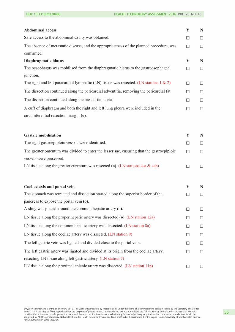
Abdominal access Y N
Safe access to the abdominal cavity was obtained.
The absence of metastatic disease, and the appropriateness of the planned procedure, was
confirmed.
Diaphragmatic hiatus Y N
The oesophagus was mobilised from the diaphragmatic hiatus to the gastroesophageal
junction.
The right and left paracardial lymphatic (LN) tissue was resected. (LN stations 1 & 2)
The dissection continued along the pericardial adventitia, removing the pericardial fat.
The dissection continued along the pre-aortic fascia.
A cuff of diaphragm and both the right and left lung pleura were included in the
circumferential resection margin (o).
Gastric mobilisation Y N
The right gastroepiploic vessels were identified.
The greater omentum was divided to enter the lesser sac, ensuring that the gastroepiploic
vessels were preserved.
LN tissue along the greater curvature was resected (o). (LN stations 4sa & 4sb)
Coeliac axis and portal vein
Y
N
The stomach was retracted and dissection started along the superior border of the
pancreas to expose the portal vein (o).
A sling was placed around the common hepatic artery (o).
LN tissue along the proper hepatic artery was dissected (o). (LN station 12a)
LN tissue along the common hepatic artery was dissected. (LN station 8a)
LN tissue along the coeliac artery was dissected. (LN station 9)
The left gastric vein was ligated and divided close to the portal vein.
The left gastric artery was ligated and divided at its origin from the coeliac artery,
resecting LN tissue along left gastric artery. (LN station 7)
LN tissue along the proximal splenic artery was dissected. (LN station 11p)
DOI: 10.3310/hta20480 HEALTH TECHNOLOGY ASSESSMENT 2016 VOL. 20 NO. 48
© Queen’s Printer and Controller of HMSO 2016. This work was produced by Metcalfe et al. under the terms of a commissioning contract issued by the Secretary of State forHealth. This issue may be freely reproduced for the purposes of private research and study and extracts (or indeed, the full report) may be included in professional journalsprovided that suitable acknowledgement is made and the reproduction is not associated with any form of advertising. Applications for commercial reproduction should beaddressed to: NIHR Journals Library, National Institute for Health Research, Evaluation, Trials and Studies Coordinating Centre, Alpha House, University of Southampton SciencePark, Southampton SO16 7NS, UK.
55
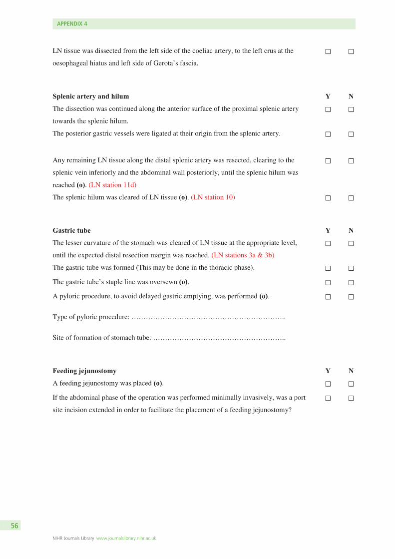
LN tissue was dissected from the left side of the coeliac artery, to the left crus at the
oesophageal hiatus and left side of Gerota’s fascia.
Splenic artery and hilum Y N
The dissection was continued along the anterior surface of the proximal splenic artery
towards the splenic hilum.
The posterior gastric vessels were ligated at their origin from the splenic artery.
Any remaining LN tissue along the distal splenic artery was resected, clearing to the
splenic vein inferiorly and the abdominal wall posteriorly, until the splenic hilum was
reached (o). (LN station 11d)
The splenic hilum was cleared of LN tissue (o). (LN station 10)
Gastric tube Y N
The lesser curvature of the stomach was cleared of LN tissue at the appropriate level,
until the expected distal resection margin was reached. (LN stations 3a & 3b)
The gastric tube was formed (This may be done in the thoracic phase).
The gastric tube’s staple line was oversewn (o).
A pyloric procedure, to avoid delayed gastric emptying, was performed (o).
Type of pyloric procedure: ………………………………………………………..
Site of formation of stomach tube: ………………………………………………..
Feeding jejunostomy Y N
A feeding jejunostomy was placed (o).
If the abdominal phase of the operation was performed minimally invasively, was a port
site incision extended in order to facilitate the placement of a feeding jejunostomy?
APPENDIX 4
NIHR Journals Library www.journalslibrary.nihr.ac.uk
56
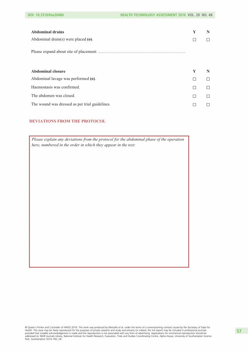
Abdominal drains Y N
Abdominal drain(s) were placed (o).
Please expand about site of placement: ..……………………………………………………
Abdominal closure Y N
Abdominal lavage was performed (o).
Haemostasis was confirmed.
The abdomen was closed.
The wound was dressed as per trial guidelines.
DEVIATIONS FROM THE PROTOCOL
Please explain any deviations from the protocol for the abdominal phase of the operation here, numbered in the order in which they appear in the text:
DOI: 10.3310/hta20480 HEALTH TECHNOLOGY ASSESSMENT 2016 VOL. 20 NO. 48
© Queen’s Printer and Controller of HMSO 2016. This work was produced by Metcalfe et al. under the terms of a commissioning contract issued by the Secretary of State forHealth. This issue may be freely reproduced for the purposes of private research and study and extracts (or indeed, the full report) may be included in professional journalsprovided that suitable acknowledgement is made and the reproduction is not associated with any form of advertising. Applications for commercial reproduction should beaddressed to: NIHR Journals Library, National Institute for Health Research, Evaluation, Trials and Studies Coordinating Centre, Alpha House, University of Southampton SciencePark, Southampton SO16 7NS, UK.
57
ERRORS AND THEIR RECOVERY
Please describe any errors that occurred, and how they were recovered.
Example errors include damage to surrounding structures and bleeding from major vessels.
APPENDIX 4
NIHR Journals Library www.journalslibrary.nihr.ac.uk
58
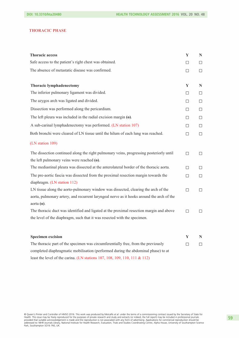
THORACIC PHASE
Thoracic access Y N
Safe access to the patient’s right chest was obtained.
The absence of metastatic disease was confirmed.
Thoracic lymphadenectomy Y N
The inferior pulmonary ligament was divided.
The azygos arch was ligated and divided.
Dissection was performed along the pericardium.
The left pleura was included in the radial excision margin (o).
A sub-carinal lymphadenectomy was performed. (LN station 107)
Both bronchi were cleared of LN tissue until the hilum of each lung was reached.
(LN station 109)
The dissection continued along the right pulmonary veins, progressing posteriorly until
the left pulmonary veins were reached (o).
The mediastinal pleura was dissected at the anterolateral border of the thoracic aorta.
The pre-aortic fascia was dissected from the proximal resection margin towards the
diaphragm. (LN station 112)
LN tissue along the aorto-pulmonary window was dissected, clearing the arch of the
aorta, pulmonary artery, and recurrent laryngeal nerve as it hooks around the arch of the
aorta (o).
The thoracic duct was identified and ligated at the proximal resection margin and above
the level of the diaphragm, such that it was resected with the specimen.
Specimen excision Y N
The thoracic part of the specimen was circumferentially free, from the previously
completed diaphragmatic mobilisation (performed during the abdominal phase) to at
least the level of the carina. (LN stations 107, 108, 109, 110, 111 & 112)
DOI: 10.3310/hta20480 HEALTH TECHNOLOGY ASSESSMENT 2016 VOL. 20 NO. 48
© Queen’s Printer and Controller of HMSO 2016. This work was produced by Metcalfe et al. under the terms of a commissioning contract issued by the Secretary of State forHealth. This issue may be freely reproduced for the purposes of private research and study and extracts (or indeed, the full report) may be included in professional journalsprovided that suitable acknowledgement is made and the reproduction is not associated with any form of advertising. Applications for commercial reproduction should beaddressed to: NIHR Journals Library, National Institute for Health Research, Evaluation, Trials and Studies Coordinating Centre, Alpha House, University of Southampton SciencePark, Southampton SO16 7NS, UK.
59
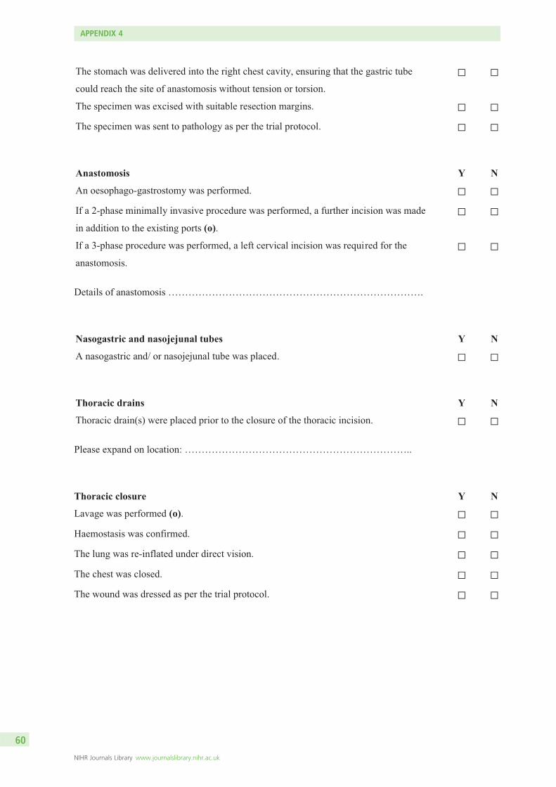
The stomach was delivered into the right chest cavity, ensuring that the gastric tube
could reach the site of anastomosis without tension or torsion.
The specimen was excised with suitable resection margins.
The specimen was sent to pathology as per the trial protocol.
Anastomosis Y N
An oesophago-gastrostomy was performed.
If a 2-phase minimally invasive procedure was performed, a further incision was made
in addition to the existing ports (o).
If a 3-phase procedure was performed, a left cervical incision was required for the
anastomosis.
Details of anastomosis ………………………………………………………………….
Nasogastric and nasojejunal tubes Y N
A nasogastric and/ or nasojejunal tube was placed.
Thoracic drains Y N
Thoracic drain(s) were placed prior to the closure of the thoracic incision.
Please expand on location: …………………………………………………………..
Thoracic closure Y N
Lavage was performed (o).
Haemostasis was confirmed.
The lung was re-inflated under direct vision.
The chest was closed.
The wound was dressed as per the trial protocol.
APPENDIX 4
NIHR Journals Library www.journalslibrary.nihr.ac.uk
60
DEVIATIONS FROM THE PROTOCOL
Please explain any deviations from the protocol for the thoracic phase of the operation here, numbered in the order in which they appear in the text:
DOI: 10.3310/hta20480 HEALTH TECHNOLOGY ASSESSMENT 2016 VOL. 20 NO. 48
© Queen’s Printer and Controller of HMSO 2016. This work was produced by Metcalfe et al. under the terms of a commissioning contract issued by the Secretary of State forHealth. This issue may be freely reproduced for the purposes of private research and study and extracts (or indeed, the full report) may be included in professional journalsprovided that suitable acknowledgement is made and the reproduction is not associated with any form of advertising. Applications for commercial reproduction should beaddressed to: NIHR Journals Library, National Institute for Health Research, Evaluation, Trials and Studies Coordinating Centre, Alpha House, University of Southampton SciencePark, Southampton SO16 7NS, UK.
61
ERRORS AND THEIR RECOVERY
Please describe any errors that occurred, and how they were recovered.
Example errors include damage to surrounding structures and bleeding from major vessels.
APPENDIX 4
NIHR Journals Library www.journalslibrary.nihr.ac.uk
62
POSTOPERATIVE INSTRUCTIONS
Please describe instructions for postoperative care
Surgeon name
Surgeon signature
DOI: 10.3310/hta20480 HEALTH TECHNOLOGY ASSESSMENT 2016 VOL. 20 NO. 48
© Queen’s Printer and Controller of HMSO 2016. This work was produced by Metcalfe et al. under the terms of a commissioning contract issued by the Secretary of State forHealth. This issue may be freely reproduced for the purposes of private research and study and extracts (or indeed, the full report) may be included in professional journalsprovided that suitable acknowledgement is made and the reproduction is not associated with any form of advertising. Applications for commercial reproduction should beaddressed to: NIHR Journals Library, National Institute for Health Research, Evaluation, Trials and Studies Coordinating Centre, Alpha House, University of Southampton SciencePark, Southampton SO16 7NS, UK.
63

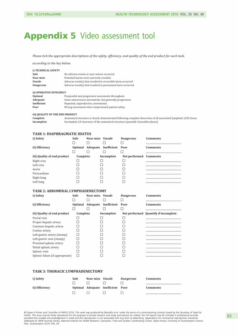
Appendix 5 Video assessment tool
Please tick the appropriate descriptions of the safety, ef�iciency, and quality of the end product for each task,
according to the key below.
i) TECHNICAL SAFETY Safe No adverse events or near misses occurred.
Near miss Potential harms were narrowly avoided.
Unsafe Adverse event(s) that resulted in reversible harm occurred.
Dangerous Adverse event(s) that resulted in permanent harm occurred.
ii) OPERATIVE EFFICIENCY Optimal Purposeful and progressive movements throughout.
Adequate Some unnecessary movements, but generally progressive.
Inef�icient Repeated, unproductive, movements.
Poor Wrong movements that compromised patient safety.
iii) QUALITY OF THE END PRODUCT Complete Anatomical structure is clearly demonstrated following complete dissection of all associated lymphatic (LN) tissue. Incomplete Incomplete LN clearance of the anatomical structure (quantify if possible please)
TASK 1: DIAPHRAGMATIC HIATUS i) Safety Safe Near miss Unsafe Dangerous Comments ................................................
ii) Ef�iciency Optimal Adequate Inef�icient Poor Comments ................................................
iii) Quality of end product Complete Incomplete Not performed Comments Right crus ................................................
Left crus ................................................
Aorta ................................................
Pericardium ................................................
Right lung ................................................
Left lung ................................................
TASK 2: ABDOMINAL LYMPHADENECTOMY i) Safety Safe Near miss Unsafe Dangerous Comments ................................................
ii) Ef�iciency Optimal Adequate Inef�icient Poor Comments ................................................
iii) Quality of end product Complete Incomplete Not performed Quantify if incomplete Portal vein ................................................
Proper hepatic artery ................................................
Common hepatic artery ................................................
Coeliac artery ................................................
Left gastric artery (stump) ................................................
Left gastric vein (stump) ................................................
Proximal splenic artery ................................................
Distal splenic artery ................................................
Splenic vein ................................................
Splenic hilum (if appropriate) ................................................
TASK 3: THORACIC LYMPHADENECTOMY
i) Safety Safe Near miss Unsafe Dangerous Comments ................................................
ii) Ef�iciency Optimal Adequate Inef�icient Poor Comments
................................................
DOI: 10.3310/hta20480 HEALTH TECHNOLOGY ASSESSMENT 2016 VOL. 20 NO. 48
© Queen’s Printer and Controller of HMSO 2016. This work was produced by Metcalfe et al. under the terms of a commissioning contract issued by the Secretary of State forHealth. This issue may be freely reproduced for the purposes of private research and study and extracts (or indeed, the full report) may be included in professional journalsprovided that suitable acknowledgement is made and the reproduction is not associated with any form of advertising. Applications for commercial reproduction should beaddressed to: NIHR Journals Library, National Institute for Health Research, Evaluation, Trials and Studies Coordinating Centre, Alpha House, University of Southampton SciencePark, Southampton SO16 7NS, UK.
65
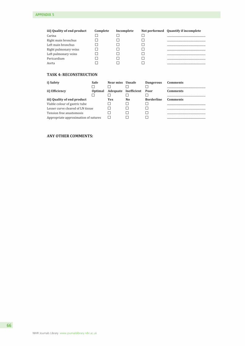
iii) Quality of end product Complete Incomplete Not performed Quantify if incomplete Carina ................................................
Right main bronchus ................................................
Left main bronchus ................................................
Right pulmonary veins ................................................
Left pulmonary veins ................................................
Pericardium ................................................
Aorta ................................................
TASK 4: RECONSTRUCTION
i) Safety Safe Near miss Unsafe Dangerous Comments ................................................ ii) Ef�iciency Optimal Adequate Inef�icient Poor Comments ................................................ iii) Quality of end product Yes No Comments Viable colour of gastric tube ................................................
Lesser curve cleared of LN tissue ................................................
Tension free anastomosis ................................................ Appropriate approximation of sutures
Borderline
................................................
ANY OTHER COMMENTS:
APPENDIX 5
NIHR Journals Library www.journalslibrary.nihr.ac.uk
66
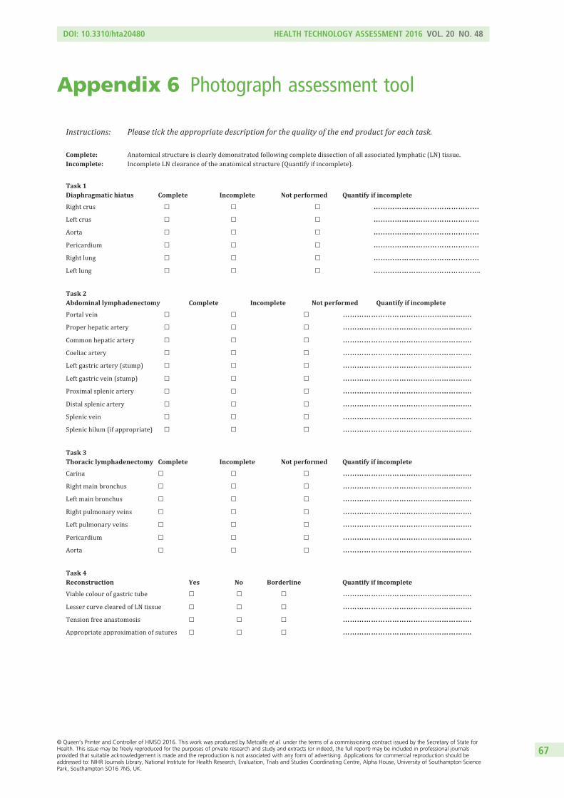
Appendix 6 Photograph assessment tool
DOI: 10.3310/hta20480 HEALTH TECHNOLOGY ASSESSMENT 2016 VOL. 20 NO. 48
© Queen’s Printer and Controller of HMSO 2016. This work was produced by Metcalfe et al. under the terms of a commissioning contract issued by the Secretary of State forHealth. This issue may be freely reproduced for the purposes of private research and study and extracts (or indeed, the full report) may be included in professional journalsprovided that suitable acknowledgement is made and the reproduction is not associated with any form of advertising. Applications for commercial reproduction should beaddressed to: NIHR Journals Library, National Institute for Health Research, Evaluation, Trials and Studies Coordinating Centre, Alpha House, University of Southampton SciencePark, Southampton SO16 7NS, UK.
67


Part of the NIHR Journals Library www.journalslibrary.nihr.ac.uk
Published by the NIHR Journals Library
This report presents independent research funded by the National Institute for Health Research (NIHR). The views expressed are those of the author(s) and not necessarily those of the NHS, the NIHR or the Department of Health
EMEHS&DRHTAPGfARPHR