Minimally invasive treatment for esthetic enhancement … · Minimally invasive treatment for...
5
The Journal of Advanced Prosthodontics 359 Minimally invasive treatment for esthetic enhancement of white spot lesion in adjacent tooth Ji-Hye Lee 1 , DDS, MSD, Dae-Gon Kim 2 , DDS, MSD, Chan-Jin Park 2 , DDS, MSD, PhD, Lee-Ra Cho 2 *, DDS, MSD, PhD 1 Department of Dentistry, Uijeongbu St. Mary’s Hospital, Catholic University of Korea, Uijeongbu, Republic of Korea 2 Department of Prosthodontics and Research Institute of Oral Science, Gangneung-Wonju National University, Gangneung, Republic of Korea This article describes the treatment provided to a patient with the maxillary anterior teeth exhibiting severe secondary caries beneath the previous restoration and a white spot lesion on the adjacent incisor. Two implants were placed after extraction of hopeless teeth with the guided bone regeneration technique. A white spot lesion of the adjacent incisor was treated with minimally invasive treatment. This clinical report describes the multidisciplinary treatment for the white spot lesion and esthetic restoration of missing anterior teeth. [ J Adv Prosthodont 2013;5:359-63] KEYWORDS: Bleaching; Clinical report; Resin infiltration; White spot http://dx.doi.org/10.4047/jap.2013.5.3.359 http://jap.or.kr J Adv Prosthodont 2013;5:359-63 INTRODUCTION The white spot lesion is the first visible evidence of a caries in the enamel, characterized by demineralized lesion under- neath an intact surface. 1 The lesion is caused by the accu- mulation of plaque and bacteria, developing among young adolescents with insufficient oral hygiene. 2 The increased pore volume inside the demineralized lesion body leads to a different refraction index from the sound enamel. 3 An inac- tive white spot lesion might act as arrested dental caries and impair only the esthetic appearance by displaying a milky white color from its interior opacity. 4,5 Attempts have been made to conserve tooth structures with highly concentrated fluoride solutions to hyper-mineralize the surface of the lesion. 6 Since these solutions do not thoroughly penetrate into the lesion, the opaque whitish aspect remains, compro- mising the appearance. 6,7 For esthetic improvement of non-cavitated white spot lesions with remineralized surface, treatment may consist of tooth bleaching, , macroabrasion, composite resin bond- ing, prosthetic restoration or some combination depending on the severity of the lesion and its etiology. 8 It remains unclear which treatment is most effective for white spot lesions. Tooth whitening is the most conservative approach. As a part of minimally invasive treatment, tooth whitening to mask the white spot lesion or achieve more uniform appearance has been reported. 9-11 However, tooth whitening treatment alone to camouflage the white spot lesion seemed to be relatively unpredictable. 12 Moreover, if tooth whitening is used in decalcified areas, the microhardness of the enamel surface may be reduced. 13 The conventional treatment approach is based on resto- ration, which, in most instances, is quite invasive. 14,15 Efforts should be made to achieve an ideal function and esthetics along with preserving the sound tooth structures as much as possible. 16,17 Since most patients requiring treatment for white spot lesions are adolescents or young adults, minimal- Corresponding author: Lee-Ra Cho Department of Prosthodontics, Department of Prosthodontics and Research Institute of Oral Science, Gangneung-Wonju National University, Gangneung, Republic of Korea Tel. 82336403153: e-mail, [email protected] Received April 18, 2013 / Last Revision July 11, 2013 / Accepted July 15, 2013 © 2013 The Korean Academy of Prosthodontics This is an Open Access article distributed under the terms of the Creative Commons Attribution Non-Commercial License (http://creativecommons. org/licenses/by-nc/3.0) which permits unrestricted non-commercial use, distribution, and reproduction in any medium, provided the original work is properly cited.
Transcript of Minimally invasive treatment for esthetic enhancement … · Minimally invasive treatment for...
The Journal of Advanced Prosthodontics 359
Minimally invasive treatment for esthetic enhancement of white spot lesion in adjacent tooth
Ji-Hye Lee1, DDS, MSD, Dae-Gon Kim2, DDS, MSD, Chan-Jin Park2, DDS, MSD, PhD, Lee-Ra Cho2*, DDS, MSD, PhD 1Department of Dentistry, Uijeongbu St. Mary’s Hospital, Catholic University of Korea, Uijeongbu, Republic of Korea2Department of Prosthodontics and Research Institute of Oral Science, Gangneung-Wonju National University, Gangneung, Republic of Korea
This article describes the treatment provided to a patient with the maxillary anterior teeth exhibiting severe secondary caries beneath the previous restoration and a white spot lesion on the adjacent incisor. Two implants were placed after extraction of hopeless teeth with the guided bone regeneration technique. A white spot lesion of the adjacent incisor was treated with minimally invasive treatment. This clinical report describes the multidisciplinary treatment for the white spot lesion and esthetic restoration of missing anterior teeth. [ J Adv Prosthodont 2013;5:359-63]
KEYWORDS: Bleaching; Clinical report; Resin infiltration; White spot
http://dx.doi.org/10.4047/jap.2013.5.3.359http://jap.or.kr J Adv Prosthodont 2013;5:359-63
INTRODUCTION
The white spot lesion is the first visible evidence of a caries in the enamel, characterized by demineralized lesion under-neath an intact surface.1 The lesion is caused by the accu-mulation of plaque and bacteria, developing among young adolescents with insufficient oral hygiene.2 The increased pore volume inside the demineralized lesion body leads to a different refraction index from the sound enamel.3 An inac-tive white spot lesion might act as arrested dental caries and impair only the esthetic appearance by displaying a milky white color from its interior opacity.4,5 Attempts have been
made to conserve tooth structures with highly concentrated fluoride solutions to hyper-mineralize the surface of the lesion.6 Since these solutions do not thoroughly penetrate into the lesion, the opaque whitish aspect remains, compro-mising the appearance.6,7
For esthetic improvement of non-cavitated white spot lesions with remineralized surface, treatment may consist of tooth bleaching, , macroabrasion, composite resin bond-ing, prosthetic restoration or some combination depending on the severity of the lesion and its etiology.8 It remains unclear which treatment is most effective for white spot lesions. Tooth whitening is the most conservative approach. As a part of minimally invasive treatment, tooth whitening to mask the white spot lesion or achieve more uniform appearance has been reported.9-11 However, tooth whitening treatment alone to camouflage the white spot lesion seemed to be relatively unpredictable.12 Moreover, if tooth whitening is used in decalcified areas, the microhardness of the enamel surface may be reduced.13
The conventional treatment approach is based on resto-ration, which, in most instances, is quite invasive.14,15 Efforts should be made to achieve an ideal function and esthetics along with preserving the sound tooth structures as much as possible.16,17 Since most patients requiring treatment for white spot lesions are adolescents or young adults, minimal-
Corresponding author: Lee-Ra ChoDepartment of Prosthodontics, Department of Prosthodontics and Research Institute of Oral Science, Gangneung-Wonju National University, Gangneung, Republic of KoreaTel. 82336403153: e-mail, [email protected] April 18, 2013 / Last Revision July 11, 2013 / Accepted July 15, 2013
© 2013 The Korean Academy of ProsthodonticsThis is an Open Access article distributed under the terms of the Creative Commons Attribution Non-Commercial License (http://creativecommons.org/licenses/by-nc/3.0) which permits unrestricted non-commercial use, distribution, and reproduction in any medium, provided the original work is properly cited.
360
J Adv Prosthodont 2013;5:359-63
ly invasive treatments are needed to prevent excessive sacri-fice of tooth material at an early age.
Recently, the resin infiltration technique was introduced as a type of minimally invasive treatment. The resin infiltra-tion technique prevents further progression of the lesion using a low-viscosity resin with a high penetration coeffi-cient, filling the enamel intercrystalline spaces.18-20 This technique has been reported to remove the whitish opaque color thereby changing the color and translucency of the white lesion.1,7 The purpose of this clinical report was to describe and illustrate a minimally invasive technique that improves the esthetic aspect of the white spot lesion adja-cent to the restoring implant-supported prostheses via the multidisciplinary approach.
CASE REPORT
A 22-year-old woman who lost her maxillary right central and lateral incisor prosthesis presented to the Gangneung-Wonju National University Dental Hospital for treatment (Fig. 1A). The prosthesis was lost with severe secondary caries around the metal post. Each root canal was incom-
pletely filled with a radiopaque resinous hard material, and the right lateral incisor root had apical rarefying osteitis (Fig. 1B). Extraction was necessary due to the large extent of secondary caries and the nonremovable canal filling material. A white spot was found in the maxillary left cen-tral incisor with poor oral hygienic control.
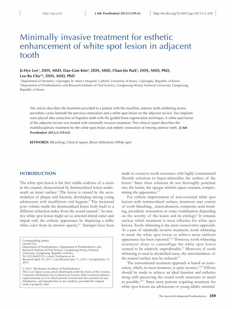
Because the patient refused tooth preparation, restoring the teeth with an implant prosthesis was planned. A diag-nostic waxing of the teeth determined the installing site of implants to meet the esthetic needs and to fabricate a radio-graphic stent and a surgical stent. Six weeks after extrac-tion, severe bone loss of the buccal plate was confirmed (Fig. 2A). In view of the insufficient implant primary stabil-ity, guided bone regeneration was performed as defects were filled with a xenograft material (Bioss; Geistlich Pharma AG, Wolhusen, Switzerland) and covered with a resorbable bilayer collagen membrane (Bio-gide; Geistlich Pharma AG) (Fig. 2B). Four months after guided bone regeneration, sufficient hard and soft tissue volume was acquired. A tapered internal hex titanium implants (Imp-lantium; Dentium, Seoul, Korea) were installed, oriented by a surgical guide. An acrylic resin RPD was fabricated to
Fig. 1. (A) Pre-treatment clinical view, (B) Pre-treatment radiograph. Note the cervical white spot lesion with poor oral hygiene.
A B
Fig. 2. (A) Extraction socket after 6-week-healing, (B) Defects covered with resorbable bilayer collagen membrane.
A B
The Journal of Advanced Prosthodontics 361
replace the missing tooth during the healing period.Six month after implant placement, a tissue-punch tech-
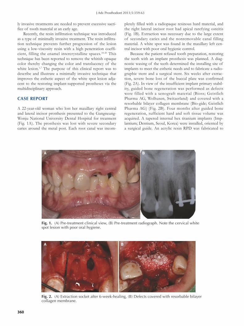
nique was used to place a screw-retained provisional resto-ration with a provisional plastic abutment (Plastic tempo-rary abutment, Dentium). The treatment for the white spot of the left maxillary central incisor was also needed. Dia-gnosis for the white spot was performed with visual-tactile inspection evaluating change of the surface gloss and color after dessication. The lesion surface was hard and shiny, it was diagnosed as an inactive ICDAS (International Caries Detection and Assessment System) code 2 which requires esthetic improvement only. Regenerating the white spot on the prosthesis was not esthetic and removal of the white spot lesion was thus required. Gingival recession after a surgical procedure highlights the white spot in the cervical area (Fig. 3). Furthermore, bandlike whitish discoloration along the perikymata in the middle third coronal area also needed esthetic enhancement. The proposed minimally invasive technique is based on a combined approach of bleaching, resin infiltration and composite resin bonding. In-office bleaching treatment was applied according to the manufacturer’s (Lumawhite; Luma Lite, Spring Valley, CA,
Fig. 3. Placement of provisional restoration.
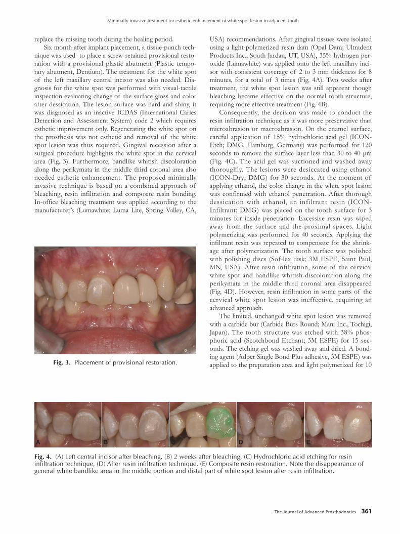
USA) recommendations. After gingival tissues were isolated using a light-polymerized resin dam (Opal Dam; Ultradent Products Inc., South Jardan, UT, USA), 35% hydrogen per-oxide (Lumawhite) was applied onto the left maxillary inci-sor with consistent coverage of 2 to 3 mm thickness for 8 minutes, for a total of 3 times (Fig. 4A). Two weeks after treatment, the white spot lesion was still apparent though bleaching became effective on the normal tooth structure, requiring more effective treatment (Fig. 4B).
Consequently, the decision was made to conduct the resin infiltration technique as it was more preservative than microabrasion or macroabrasion. On the enamel surface, careful application of 15% hydrochloric acid gel (ICON-Etch; DMG, Hamburg, Germany) was performed for 120 secondstoremovethesurface layer lessthan30to40μm(Fig. 4C). The acid gel was suctioned and washed away thoroughly. The lesions were desiccated using ethanol (ICON-Dry; DMG) for 30 seconds. At the moment of applying ethanol, the color change in the white spot lesion was confirmed with ethanol penetration. After thorough dessication with ethanol, an infiltrant resin (ICON-Infiltrant; DMG) was placed on the tooth surface for 3 minutes for inside penetration. Excessive resin was wiped away from the surface and the proximal spaces. Light polymerizing was performed for 40 seconds. Applying the infiltrant resin was repeated to compensate for the shrink-age after polymerization. The tooth surface was polished with polishing discs (Sof-lex disk; 3M ESPE, Saint Paul, MN, USA). After resin infiltration, some of the cervical white spot and bandlike whitish discoloration along the perikymata in the middle third coronal area disappeared (Fig. 4D). However, resin infiltration in some parts of the cervical white spot lesion was ineffective, requiring an advanced approach.
The limited, unchanged white spot lesion was removed with a carbide bur (Carbide Burs Round; Mani Inc., Tochigi, Japan). The tooth structure was etched with 38% phos-phoric acid (Scotchbond Etchant; 3M ESPE) for 15 sec-onds. The etching gel was washed away and dried. A bond-ing agent (Adper Single Bond Plus adhesive, 3M ESPE) was applied to the preparation area and light polymerized for 10
Fig. 4. (A) Left central incisor after bleaching, (B) 2 weeks after bleaching, (C) Hydrochloric acid etching for resin infiltration technique, (D) After resin infiltration technique, (E) Composite resin restoration. Note the disappearance of general white bandlike area in the middle portion and distal part of white spot lesion after resin infiltration.
A B C D E
Minimally invasive treatment for esthetic enhancement of white spot lesion in adjacent tooth
362
J Adv Prosthodont 2013;5:359-63
seconds. Composite resin (Filtek Supreme XT; 3M ESPE) was placed and polymerized for 40 seconds. Finally, polish-ing the restoration surface was performed with polishing discs (Sof-lex disk; 3M ESPE) (Fig. 4E).
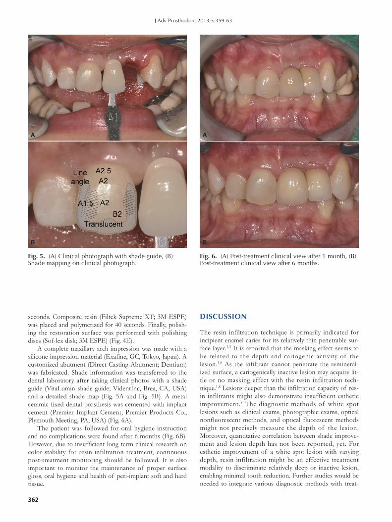
A complete maxillary arch impression was made with a silicone impression material (Exafine, GC, Tokyo, Japan). A customized abutment (Direct Casting Abutment; Dentium) was fabricated. Shade information was transferred to the dental laboratory after taking clinical photos with a shade guide (VitaLumin shade guide; VidentInc, Brea, CA, USA) and a detailed shade map (Fig. 5A and Fig. 5B). A metal ceramic fixed dental prosthesis was cemented with implant cement (Premier Implant Cement; Premier Products Co., Plymouth Meeting, PA, USA) (Fig. 6A).
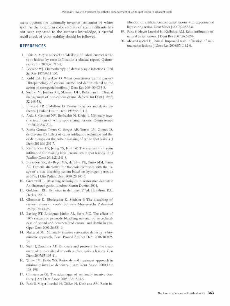
The patient was followed for oral hygiene instruction and no complications were found after 6 months (Fig. 6B). However, due to insufficient long term clinical research on color stability for resin infiltration treatment, continuous post-treatment monitoring should be followed. It is also important to monitor the maintenance of proper surface gloss, oral hygiene and health of peri-implant soft and hard tissue.
DISCUSSION
The resin infiltration technique is primarily indicated for incipient enamel caries for its relatively thin penetrable sur-face layer.1,3 It is reported that the masking effect seems to be related to the depth and cariogenic activity of the lesion.1,8 As the infiltrant cannot penetrate the remineral-ized surface, a cariogenically inactive lesion may acquire lit-tle or no masking effect with the resin infiltration tech-nique.1,8 Lesions deeper than the infiltration capacity of res-in infiltrants might also demonstrate insufficient esthetic improvement.8 The diagnostic methods of white spot lesions such as clinical exams, photographic exams, optical nonfluorescent methods, and optical fluorescent methods might not precisely measure the depth of the lesion. Moreover, quantitative correlation between shade improve-ment and lesion depth has not been reported, yet. For esthetic improvement of a white spot lesion with varying depth, resin infiltration might be an effective treatment modality to discriminate relatively deep or inactive lesion, enabling minimal tooth reduction. Further studies would be needed to integrate various diagnostic methods with treat-
Fig. 5. (A) Clinical photograph with shade guide, (B) Shade mapping on clinical photograph.
A
B
Fig. 6. (A) Post-treatment clinical view after 1 month, (B) Post-treatment clinical view after 6 months.
A
B
The Journal of Advanced Prosthodontics 363
ment options for minimally invasive treatment of white spot. As the long term color stability of resin infiltrants has not been reported to the author’s knowledge, a careful recall check of color stability should be followed.
REFERENCES
1. Paris S, Meyer-Lueckel H. Masking of labial enamel white spot lesions by resin infiltration-a clinical report. Quinte-ssence Int 2009;40:713-8.
2. Loesche WJ. Chemotherapy of dental plaque infections. Oral Sci Rev 1976;9:65-107.
3. Kidd EA, Fejerskov O. What constitutes dental caries? Histopathology of carious enamel and dentin related to the action of cariogenic biofilms. J Dent Res 2004;83:C35-8.
4. Suzuki M, Jordan RE, Skinner DH, Boksman L. Clinical management of non-carious enamel defects. Int Dent J 1982; 32:148-58.
5. Ellwood RP, O’Mullane D. Enamel opacities and dental es-thetics. J Public Health Dent 1995;55:171-6.
6. Ardu S, Castioni NV, Benbachir N, Krejci I. Minimally inva-sive treatment of white spot enamel lesions. Quintessence Int 2007;38:633-6.
7. Rocha Gomes Torres C, Borges AB, Torres LM, Gomes IS, de Oliveira RS. Effect of caries infiltration technique and flu-oride therapy on the colour masking of white spot lesions. J Dent 2011;39:202-7.
8. Kim S, Kim EY, Jeong TS, Kim JW. The evaluation of resin infiltration for masking labial enamel white spot lesions. Int J Paediatr Dent 2011;21:241-8.
9. Bussadori SK, do Rego MA, da Silva PE, Pinto MM, Pinto AC. Esthetic alternative for fluorosis blemishes with the us-age of a dual bleaching system based on hydrogen peroxide at 35%. J Clin Pediatr Dent 2004;28:143-6.
10. Greenwall L. Bleaching techniques in restorative dentistry: An illustrated guide. London: Martin Dunitz; 2001.
11. Goldstein RE. Esthetics in dentistry. 2nded. Hamilton: B.C. Decker; 2001.
12. Glockner K, Ebeleseder K, Städtler P. The bleaching of stained anterior teeth. Schweiz Monatsschr Zahnmed 1997;107:413-25.
13. Basting RT, Rodrigues Júnior AL, Serra MC. The effect of 10% carbamide peroxide bleaching material on microhard-ness of sound and demineralized enamel and dentin in situ. Oper Dent 2001;26:531-9.
14. Malterud MI. Minimally invasive restorative dentistry: a bio-mimetic approach. Pract Proced Aesthet Dent 2006;18:409-14.
15. Stahl J, Zandona AF. Rationale and protocol for the treat-ment of non-cavitated smooth surface carious lesions. Gen Dent 2007;55:105-11.
16. White JM, Eakle WS. Rationale and treatment approach in minimally invasive dentistry. J Am Dent Assoc 2000;131: 13S-19S.
17. Christensen GJ. The advantages of minimally invasive den-tistry. J Am Dent Assoc 2005;136:1563-5.
18. Paris S, Meyer-Lueckel H, Cölfen H, Kielbassa AM. Resin in-
filtration of artificial enamel caries lesions with experimental light curing resins. Dent Mater J 2007;26:582-8.
19. Paris S, Meyer-Lueckel H, Kielbassa AM. Resin infiltration of natural caries lesions. J Dent Res 2007;86:662-6.
20. Meyer-Lueckel H, Paris S. Improved resin infiltration of nat-ural caries lesions. J Dent Res 2008;87:1112-6.
Minimally invasive treatment for esthetic enhancement of white spot lesion in adjacent tooth