
BookabsISCP12 Final
95
B B O O O O K K O O F F A A B B S S T T R R A A C C T T S S International Student Conference on Photonics 2012
-
Upload
boni-mihai -
Category
Documents
-
view
106 -
download
2
Transcript of BookabsISCP12 Final
BBOOOOKK
OOFF
AABBSSTTRRAACCTTSS
International Student
Conference on Photonics
2012

II
I N T E R N A T I O N A L S T U D E N T
C O N F E R E N C E O N P H O T O N I C S
S i n a i a , R o m a n i a 8 - 1 1 M a y 2 0 1 2
EDITORS
Cristina Achim
Mihai Boni
Viorel Nastasa National Institute for Laser, Plasma and Radiation Physics,
Bucharest, Romania
ORGANISED BY:
National Institute for Laser, Plasma and Radiation Physics,
Bucharest, Romania
Insitute for Atomic Physics SPIE Student Chapter

III
ORGANIZED AND SPONSORED BY

IV
SCOPE
The aim of the ISCP 2012, the 3rd edition of International Student Conference
on Photonics, is to reassemble young researchers from different countries with the
aim of exchanging information in photonics and related fields.
ISCP is the annual conference organized by Romanian SPIE Student Chapter
and follows the previous ISWLA conferences. This edition celebrates the 50th
anniversary since the first Romanian laser was developed by Prof. Ion I. Agarbiceanu
and coworkers.
ISCP can represent a good opportunity for underlining the importance to
belong to a professional association and the prospects offered by SPIE and OSA to
permanently up-date and increase the professional level of their members.
The involvement of young researchers (physicists, mathematicians, engineers,
biologists, medical doctors and educators from all over the world) in organizing
scientific events adds new values to their professional profile, such as the
management skills and the innovative thinking. Such skills are benefic for young
scientist to develop a current and future successful scientific career.
Research papers are expected from all domains of the scientific and
technological fields related to ISCP.
MODE OF PRESENTATION
Round table (60 minutes);
Invited presentations (30 minutes);
Oral presentations (15 minutes);
Poster session;
Young scientist’s awards.
KEY TOPICS
High power lasers and applications
Optics and Optoelectronics
Micro and nanotechnologies
Advanced Materials (meta & nanomaterials)
Lasers in life sciences
Laser metrology and industry
Physics of plasma sources and applications

V
ORGANIZING CHAPTER
Institute for Atomic Physics SPIE Student Chapter
COORDINATORS
Mihai Boni
Viorel Nastasa
ADVISER
Dr. Angela Staicu
CO-ORGANIZING CHAPTERS
Munich SPIE Student Chapter Germany
Nicolaus Copernicus University SPIE Chapter Poland
Taurida National V.I. Vernadsky University SPIE
Chapter
Ukraine
Vladivostok SPIE Student Chapter Russian Federation
Yerevan State University SPIE Chapter Armenia
National Research University of Information
Technologies, Mechanics and Optics SPIE Chapter
Russian Federation
Sofia University OSA Student Chapter Bulgaria
CIMAP/University de Caen SPIE Student Chapter France
INTERNATIONAL SCIENTIFIC COMMITTEE
Prof. Dr. Frank Burnet Professor of Science Communication/SPIE
trainer
Dr. Eng. Ion Morjan NILPRP, General Manager
Prof. Dr Nicolae Zamfir IFIN-HH, General Manager
Prof. Dr. Alexandru Jipa Dean, Faculty of Physics, University of
Bucharest
Prof. Dr. Stefan Antohe Dean, Faculty of Physics, University of
Bucharest
Dr. Viorica Stancalie NILPRP, Head of Laser Department
Prof. Dr. Mihail Lucian
Pascu
NILPRP, Laser Spectroscopy Group
Prof. Dr. Dan Dumitras NILPRP, Optics and Lasers in Life Sciences,
Environment and Manufacturing Group
Dr. Cristian Ruset NILPRP, Head of Plasma Surface Engineering
Laboratory
Dr. Maria Dinescu NILPRP, Photonic Processing of Advanced

VI
Materials Group
Dr. Constantin Grigoriu NILPRP, Quantum Dots, Nanopowders And
Thin Films Group
Dr. Angela Staicu SPIE Student Chapter Adviser
LOCAL ORGANISING COMMITTEE
Dr. Alexandru Achim Iulian Pana
Cristina Achim Laurentiu Rusen
Alina Ionescu Gabriela Salamu
Florin Jipa
YOUNG SCIENTIST’S AWARDS
The best presentation as well as the best poster displayed by young scientists
will be awarded during the ISCP conference. The competition is conceived to
encourage young scientists to properly disseminate the results of the scientific
activity. General principles of competition:
Every young scientist can take part in competition, including PhD
students;
The young scientist must be first author or co-author of a paper
presented in this conference;
The participant in competition must present his own research.
PUBLISHED MANUSCRIPTS
The accepted manuscripts of the International Student Conference on
Photonics will be published by Optoelectronics and Advanced Materials – Rapid
Communications (OAM-RC) and Journal of Optoelectronics and Advanced Materials
(JOAM).
The proceedings papers must comply with the ethical standards as described
at: http://oam-rc.inoe.ro and http://joam.inoe.ro.
It will be necessary a copyright releases signed by at least one author for each
paper. The signed copyright releases should be sent to the Editors. Then, the Editors
will forward the manuscripts to reviewers.

VII
PROGRAM
Monday May 07, 2012
16.00 Opening of the Registration Desk
19.30 Welcome Party/Dinner
Tuesday May 08, 2012
09.30 Opening Ceremony
Gen. Dir. INFLPR Dr. Ion Morjan, Gen. Dir.
IFIN-HH Prof. Dr. Nicolae Zamfir
Micro and nano technologies
INVITED LECTURE 10.00 “Challenges in nanoparticles synthesis by laser
pyrolysis”, Dr. Catalin Luculescu, National
Institute for Laser, Plasma and Radiation Physics,
Laser Department, Bucharest, Romania
Oral communication 10.30 “Laser near-field processing using photopolymer
microstructures”, Jipa Florin, National Institute
for Laser, Plasma and Radiation Physics, Laser
Department, Bucharest, Romania
Oral communication 10.45 “Laser-based polymerization of ormosils for bio-
applications”, Andreea Matei, Petru Poni
Institute of Macromolecular Chemistry, Iasi,
Romania
Oral communication 11.00 “Properties of BaxSr1-xTiO3 thin films obtained by
pulsed laser deposition”, Valentin Ion , National
Institute for Laser, Plasma and Radiation Physics,
Laser Department, Bucharest, Romania
11.15 Coffee Break
Oral communication 11.30 “Periodical structures induced by femtosecond
laser on Tungsten in air and liquid environments”,
Albu Catalina, National Institute for Laser,
Plasma and Radiation Physics, Laser Department,
Bucharest, Romania
INVITED LECTURE 11.45 “Investigation of the mechanism of quasi-
periodical surface nanostructures formation by
“pump and probe” experiments”, Dr. Marian
Zamfirescu, National Institute for Laser, Plasma
and Radiation Physics, Laser Department,
Bucharest, Romania
Oral communication 12.15 “Optical and electrical properties of YBa2Cu3O7-
thin films deposited by radio -frequency assisted
pulsed laser deposition”, George Stanciu,
University Politehnica of Bucharest, Faculty of
Applied Chemistry and Material Science,
Bucharest, Romania
12.30 Lunch
INVITED LECTURE 14.15 “The Centre for Advanced Laser Tehnologies
(CETAL) opportunity for cutting-edge research in
Photonics ”, Dr. Constantin Grigoriu, National
Institute for Laser, Plasma and Radiation Physics,
VIII
Laser Department, Bucharest, Romania
Photonics and Optics
Oral communication 14.45 “Simple modeling of optical feshbach
resonances”, Dariusz Swierad, Copernicus
University, Torun, Poland
Oral communication 15.00 “Transversal strain induced birefringence effects
on Fiber-Bragg-Gratings”, Andre Heßke,
Institute for Measurement Systems and Sensor
Technology, Technical University Munich,
Theresienstr, Germany
Oral communication 15.15 “Classification of Gear-Oil Raman Spectra by
Support Vector Machine”, Daniel Dorigo,
Institute for Measurement Systems and Sensor
Technology, Technical University Munich,
Theresienstr, Germany
Oral communication 15.30 “In-band Pumped Nd:LuVO4 Laser Mode Locked
by ÷(2)
-Lens Formation in an LBO Nonlinear
Crystal”, Veselin Aleksandrov, Department of
Physics, University of Sofia, Sofia, Bulgaria
Oral communication 15.45 “Study of combustion process for a methane-air
mixture using a microlaser system”, Gabriela
Salamu, Laboratory of Solid-State Quantum
Electronics, National Institute for Lasers, Plasma
and Radiation Physics, Magurele, Bucharest,
Romania
Oral communication 16.00 “Analysis of polarization state losses in optical
fibers ”, Avram Ioana , The Technical University
of Cluj Napoca, Cluj Napoca, Romania
16.15 Coffee Break
Oral communication 16.30 “Analysis of the inscription process of FBGs in a
Panda-type-high birefingent fiber”, Bianca
Berrang, University of Applied Sciences Munich,
Laboratory of Photonics, Munich, Germany
Oral communication 16.45 “Optical spectrum of a coupled chaotic system”,
Ionut Relu Andrei, National Institute for Laser,
Plasma and Radiation Physics, Laser Department,
Bucharest, Romania
Oral communication 17.00 “Laser Induced Damage Threshold Test Station:
Development and measurements - preliminary
results”, Alexandru Zorila, National Institute for
Laser, Plasma and Radiation Physics, Laser
Department, Bucharest, Romania
17.15 End of session
19.00 Dinner
Wednesday May 9 , 2012
Advanced Materials
INVITED LECTURE 09.00 “Phase relation, dielectric and ferroelectric
properties of lead-free ferroelectric thin films
obtained by PLD and RF-PLD”, Dr. Nicu
Scarisoreanu, National Institute for Laser, Plasma
and Radiation Physics, Laser Department,
Bucharest, Romania
Oral communication 09.30 “Numerical analysis of non-linear cavity modes in
IX
a silicon hole-array photonic crystal “, Camelia
Daniela Sold, Faculty of Physics, West University
of Timisoara, Timisoara, Romania
INVITED LECTURE 09.45 “Advanced nonlinear optic crystals for high
efficiency visible and UV laser sources based on
frequency conversion processes”, Dr. Gheorghe
Lucian, National Institute for Laser, Plasma and
Radiation Physics, Solid-State Quantum
Electronics Laboratory, Romania
Oral communication 10.15 “Characterization of a low pressure expanding RF
plasma jet generated in Ar/H2/C2H2 admixture
used for carbon nanowalls synthesis”, Daniel
Stoica, National Institute for Laser, Plasma and
Radiation Physics, Plasma and Radiation Physics,
Solid-State Quantum Electronics Laboratory,
Romania
INVITED LECTURE 10.30 “Laser processing of soft materials”, Prof. Dr.
Maria Dinescu, National Institute for Laser,
Plasma and Radiation Physics, Laser Department,
Bucharest, Romania
11.00 Coffee Break
Laser History
INVITED LECTURE 11.15 “50 years of laser research in Romania
(Department of LASERS)”, Prof. Dr. Dan
Dumitras, National Institute for Laser, Plasma and
Radiation Physics, Laser Department, Bucharest,
Romania
Spectroscopy. Lasers in Life Science
Oral communication 11.45 “Miniaturized Mid-IR-Spectrometer for On-line
and On-Site Condition Monitoring of Stationary
Biogas Engines.”, Benjamin Wiesent, Institute for
Measurement Systems and Sensor Technology,
Technical University Munich, Theresienstr,
Germany
Oral communication 12.00 “Laser beams interaction with pharmaceutical
foams: Aethoxysklerol case”, Adriana
Smarandache, National Institute for Laser,
Plasma and Radiation Physics, Laser Department,
Bucharest, Romania
Oral communication 12.15 “Quantitative analysis of surgical smoke by laser
photoacoustic spectroscopy”, Ana Bratu, National
Institute for Laser, Plasma and Radiation Physics,
Laser Department, Bucharest, Romania
Oral communication 12.30 “Temperature distribution analysis in laser
irradiated tissue by numerical analysis and
experimental data“, Mioara Petrus, National
Institute for Laser, Plasma and Radiation Physics,
Laser Department, Bucharest, Romania
Oral communication 12.45 “Calibration and artefact minimization in a CW
Diffuse Optical Tomography system”, Mihai
Patachia, National Institute for Laser, Plasma and
Radiation Physics, Laser Department, Bucharest,
Romania

X
13.00 Lunch
INVITED LECTURE 14.15 “Progress in 3rd
and 4th
generation of photovoltaic
cells at Research and Development Centre for
Materials and Electronic & Optoelectronic
Devices (MDEO)”, Prof. Dr. Stefan Antohe,
Dean of Faculty of Physics, University of
Bucharest, Romania
INVITED LECTURE 14.45 “Studies about the laser radiation interaction with
beads of microliter volumes”, Prof. Dr. Mihai
Lucian Pascu, National Institute for Laser, Plasma
and Radiation Physics, Laser Department,
Bucharest, Romania
Oral communication 15.15 “Study of the properties of micro- and nano-
droplets by using the interaction with laser
radiation”, Viorel Nastasa, National Institute for
Laser, Plasma and Radiation Physics, Laser
Department, Bucharest, Romania
Oral communication 15.30 “Measurements of Raman Spectra induced by
laser beams on droplets”, Mihai Boni, National
Institute for Laser, Plasma and Radiation Physics,
Laser Department, Bucharest, Romania
Oral communication 15.45 “Exposure of Chlorpromazine to 266 nm laser
beam generates new species with antibacterial
properties”, Tatiana Alexandru, National
Institute for Laser, Plasma and Radiation Physics,
Laser Department, Bucharest, Romania
INVITED LECTURE 16.00 “Laser induced breakdown spectroscopy:
stratigraphy on painted mock-ups”, Dr. Angela
Staicu, National Institute for Laser, Plasma and
Radiation Physics, Laser Department, Bucharest,
Romania
16.30 Coffee Break
Poster Session 16.45
19.00 Dinner
Thursday May 10, 2012
INVITED LECTURE 09.00 “Why and how to communicate your research“,
Prof. Dr. Frank Burnet, United Kingdom (Part I)
11.00 Coffee Break
11.15 “Why and how to communicate your research “,
Prof. Dr. Frank Burnet, United Kingdom (Part II)
13.00 Lunch
Presentations by the sponsoring companies
14.00 "High repetition rate PetaWatt level Titanium
Sapphire laser system for laser wakefield
acceleration", Dr. Olivier Chalus, Laser Solution
Department,Thales Optronique, France
14.30 “High performance spectrometry”, Dan Bulik,
BERD Trading, Bucharest, Romania
14.45 “Photonics solutions: Lasers and Spectroscopy”
Chiricuta Bogdan, APEL Laser, Bucharest,
Romania

XI
High power lasers
INVITED LECTURE 15.00 “High-peak power passively Q-switched
Nd:YAG/Cr4+
:YAG lasers”, Dr. Nicolaie Pavel,
Laboratory of Solid-State Quantum Electronics
National Institute for Laser, Plasma and Radiation
Physics, Bucharest, Romania
Oral communication 15.30 “28-mJ, single frequency, sub-nanosecond Nd-
MOPA system, at kHz repetition rate”, Bozhidar
Oreshkov, Department of Physics, Sofia
University, Sofia, Bulgaria
Oral communication 15.45 “Simple method for synchronization of pulses in
GRIP x-ray laser scheme”, Romeo Banici,
National Institute for Laser, Plasma and Radiation
Physics, Laser Department, Bucharest, Romania
16.00 Coffee break
Oral communication 16.15 “Progress at the multi-PW ELI-NP laser facility”,
Liviu Neagu, National Institute for Laser, Plasma
and Radiation Physics, Laser Department,
Bucharest, Romania
Oral communication 16.30 “Study of high harmonics generation at the
interaction of an ultrashort and intense laser pulse
with an overdense plasma layer”, Andreea
Mihailescu, National Institute for Laser, Plasma
and Radiation Physics, Laser Department,
Bucharest, Romania
Oral communication 16.45 “Operation of Mo XRL in ultra-low pumping
regime”, Gabriel Cojocaru, National Institute for
Laser, Plasma and Radiation Physics, Laser
Department, Bucharest, Romania
Oral communication 17.00 “Simulation of spatio-temporal distortions in
ultra-short laser pulses”, Razvan Ungureanu,
National Institute for Laser, Plasma and Radiation
Physics, Laser Department, Bucharest, Romania
17.15 End of session
19.00 Dinner
Friday May 11, 2012
INVITED LECTURE 09.00 “Project LASERLAB EUROPE: The Integrated
Initiative of European Laser Research
Infrastructure”, Prof. Dr. Traian Dascalu,
National Institute for Laser, Plasma and Radiation
Physics, Laser Department, Bucharest, Romania
Physics of Plasma
INVITED LECTURE 09.30 “Dusty Plasmas: a review of experiments and
possible applications”, Dr. Catalin Ticos,
National Institute for Laser, Plasma and Radiation
Physics, Laser Department, Bucharest, Romania
Oral communication 10.00 “Helmholtz equations in rectangular-shaped
curved optical fiber”, Daniel Gustaw, Nicolaus
Copernicus University, Faculty of Physics,
Astronomy and Informatics, Torun , Poland
Oral communication 10.15 “Dose distributions in water-equivalent materials
irradiated with hadron beams”, Chirvase

XII
Cezarina-Isabela, Faculty of Physics, “Al. I.
Cuza” University, Iasi, Romania
Oral communication 10.30 “Preparation and characterization of double
perovskite targets for thin films deposition by PLD
method”, Robert Lowndes, National Institute of
Materials Physics, Bucharest, Romania
10.45 Coffee break
ROUND TABLE 11.00 “How to develop a successful scientific research
carrier”
12.00 Lunch
13.30 Visit to Peles Castle
19.00 Banquet
Saturday May 12 , 2012
09.00 Poster and Oral Presentation Awards
09.30 Conference Closing

XIII
C O N T E N T
Session 1: Micro and nano technologies ……………………......1
Session 2: Photonics and Optics …………………………….…10
Session 3: Advanced Materials ………………………………...20
Session 4: Lasers History …………………………………........24
Session 5: Spectroscopy. Lasers in Life Sciences…………..….26
Poster Session……………………………………………………37
Session 6: High Power Lasers……………………………….…67
Session 7: Physics of Plasma……….…………………………...76
Participants List………………………………………………...81

1
Session 1
Micro and
nano technologies
2
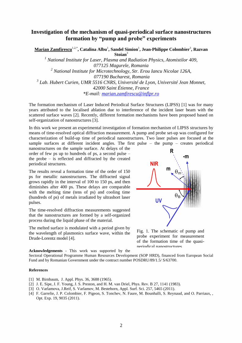
Investigation of the mechanism of quasi-periodical surface nanostructures
formation by “pump and probe” experiments
Marian Zamfirescu1,2,*
, Catalina Albu1, Sandel Simion
1, Jean-Philippe Colombier
3, Razvan
Stoian3
1 National Institute for Laser, Plasma and Radiation Physics, Atomistilor 409,
077125 Magurele, Romania
2 National Institute for Microtechnology, Str. Erou Iancu Nicolae 126A,
077190 Bucharest, Romania 3 Lab. Hubert Curien, UMR 5516 CNRS, Université de Lyon, Université Jean Monnet,
42000 Saint Étienne, France
*E-mail: [email protected]
The formation mechanism of Laser Induced Periodical Surface Structures (LIPSS) [1] was for many
years attributed to the localised ablation due to interference of the incident laser beam with the
scattered surface waves [2]. Recently, different formation mechanisms have been proposed based on
self-organization of nanostructures [3].
In this work we present an experimental investigation of formation mechanism of LIPSS structures by
means of time-resolved optical diffraction measurement. A pump and probe set-up was configured for
characterization of build-up time of periodical nanostructures. Two laser pulses are focused at the
sample surfaces at different incident angles. The first pulse – the pump – creates periodical
nanostructures on the sample surface. At delays of the
order of few ps up to hundreds of ps, a second pulse –
the probe – is reflected and diffracted by the created
periodical structures.
The results reveal a formation time of the order of 150
ps for metallic nanostructures. The diffracted signal
grows rapidly in the interval of 100 to 150 ps, and then
diminishes after 400 ps. These delays are comparable
with the melting time (tens of ps) and cooling time
(hundreds of ps) of metals irradiated by ultrashort laser
pulses.
The time-resolved diffraction measurements suggested
that the nanostructures are formed by a self-organized
process during the liquid phase of the material.
The melted surface is modulated with a period given by
the wavelength of plasmonics surface wave, within the
Drude-Lorentz model [4].
Acknowledgements - This work was supported by the
Sectoral Operational Programme Human Resources Development (SOP HRD), financed from European Social
Fund and by Romanian Government under the contract number POSDRU/89/1.5/ S/63700.
References
[1] M. Birnbaum, J. Appl. Phys. 36, 3688 (1965).
[2] J. E. Sipe, J. F. Young, J. S. Preston, and H. M. van Driel, Phys. Rev. B 27, 1141 (1983).
[3] O. Varlamova, J.Reif, S. Varlamov, M. Bestehorn, Appl. Surf. Sci. 257, 5465 (2011).
[4] F. Garrelie, J. P. Colombier, F. Pigeon, S. Tonchev, N. Faure, M. Bounhalli, S. Reynaud, and O. Parriaux, ,
Opt. Exp. 19, 9035 (2011).
NIR
UV B
m
R -m
m
Fig. 1. The schematic of pump and
probe experiment for measurement
of the formation time of the quasi-
periodical nanostructures.

3
Laser Near-field processing using photopolymer microstructures
F. Jipa, I. Anghel, C. Luculescu, M. Zamfirescu, R. Dabu
National Institute for Laser Plasma and Radiation Physics, Atomistilor
409, 077125 Magurele, Bucharest, Romania
Optical near-field enhancement of the electromagnetic field using micro- and nano-optical
components represents an versatile method to induce local modifications on the material surface [1].
Because of its potential to focus the light under diffraction limit, this method it is used to create
nanopatterns on large surface area. This method presents few advantages like lower energy densities
and short processing time on large surface. However, when self-assembled monolayers of dielectric
microspheres are used as focusing optics, the nanopattern imprinted on the material surface is limited
to hexagonal arrangement [2].
In this work we presented an alternative method to overcome the hexagonal geometrical limitation, by
creating transparent photopolymer masks for near-field lithography, with arbitrary designed geometry.
An inorganic-organic hybrid photoresist- Ormocer, with good optical properties as well as mechanical
and thermal stability was used to create transparent mask by Two Photon Polymerization (TPP)
method [3]. The propagation of the electromagnetic field through the transparent mask and the
intensification factor was computed by finite-difference time domain (FDTD) method (Fig.1),
demonstrating the feasibility of the proposed method to process large material surfaces.
Experimentally, the transparent
mask created in Ormocer-
photoresist was used to process
the Si material surface by
intensifying the electromagnetic
field of a femtosecond laser
(Clark-CPA).
Figure1. Electromagnetic field
intensification. a) photopolymer
microstructures; b) intensification
value
Acknowledgements: This work is
supported by National Authority for
Scientific Research, Project
LAPLAS3, No. PN-0939/2012.
References:
[1]Theppakuttai and S. Chen, Applied Physics Letters, vol. 83, 758 ( 2003).
[2]M. Ulmeanu, M. Zamfirescu, L. Rusen, C. Luculescu, A. Moldovan, A. Stratan, and
R. Dabu, Journal of Applied Physics, vol.106, 114908 (2009).
[3]Maruo S, Nakamura O, Kawata S, Optics Letters, Vol. 22, pp.132 (1997).
a)
b)
4
Laser-based polymerization of ormosils for bio-applications
A. Matei1,2
, M. Zamfirescu2, C. Albu
2, E.C. Buruiana
1, T. Buruiana
1, C. Mustaciosu
3, M.
Dinescu2
1Petru Poni Institute of Macromolecular Chemistry,
Grigore Ghica Voda 41A, 6600 Iasi, Romania 2National Institute for Lasers, Plasma and Radiation Physics, Atomistilor 409,
77125 Bucharest-Magurele, Romania 3Department of Environmental and Life Physics, National Institute for Physics and Nuclear
Engineering ‘Horia Hulubei’, Bucharest, Romania
Micro and nano-structured polymeric thin films and multilayers are used in a wide range of
applications in electronics, optoelectronics, sensors, medicine, tissue engineering etc. There is an
increasing interest in obtaining controlled two- and three-dimensional soft materials structures on the
sub-micron scale [1].
This work presents results on photo-polymerization by direct laser writing (DLW) of new organic
modified silicates (ormosils), with application in tissue engineering. DLW is a technique that uses a
femtosecond laser to create 2D and 3D structures with micron or nanometric size [2]. The polymeric
structures with controlled architecture find applications in medical implants and medicine, as well as
in optical components. Hybrid methacrylates based on silane derivatives were synthesized and
polymerized in 2D and 3D scaffolds and then tested in fibroblast cells culture for morphology,
proliferation, and attachment [3, 4].
Acknowledgements: Two authors (AM and TB) acknowledge the financial support of European Social Fund –
„Cristofor I. Simionescu” Postdoctoral Fellowship Programme (ID POSDRU/89/1.5/S/55216), Sectoral
Operational Programme Human Resources Development 2007 – 2013.
References [1] Matei A.; Zamfirescu M.; Jipa F.; et al., INTERNATIONAL SYMPOSIUM ON HIGH POWER LASER
ABLATION 2010 Book Series: AIP Conference Proceedings Volume: 1278 Pages: 843-851,
(2010)
[2] Matei A.; Dinescu M.; Buruiana E. C.; et al., DIGEST JOURNAL OF NANOMATERIALS AND
BIOSTRUCTURES Volume: 6 Issue: 1, Pages: 29-35 (2011)
[3] Matei A.; Zamfirescu M.; Radu C.; et al., APPLIED PHYSICS A-MATERIALS SCIENCE &
PROCESSING Volume: 104 Issue: 3 Pages: 821-827, (2011)
[4] L. E. Sima, E. C. Buruiana, T. Buruiana, A. Matei, G. Epurescu, M. Zamfirescu, A. Moldovan, S. M.
Petrescu, M. Dinescu, JOURNAL OF TISSUE ENGINEERING AND REGENERATIVE MEDICINE, DOI:
10.1002/term.507

5
Properties of BaxSr1-xTiO3 thin films obtained by pulsed laser deposition
V. Ion1, 2
, N. D. Scarisoreanu,1, A. Andrei
1, A. Nedelcea
1 and M. Dinescu
1 1National Institute for Laser, Plasma and Radiation Physics, Magurele, Bucharest, Romania
2 University of Bucharest, Faculty of Physics, Bucharest, Romania
valentin.ion@inflpr,ro
Barium strontium titanate (BaxSr1-xTiO3) thin films were obtained by Pulsed Laser Deposition (PLD)
and radio-frequency assisted PLD (RF-PLD) techniques on Pt/Si, MgO and Si substrates.
Control of the BST (BaxSr1-xTiO3) thin film epitaxial growth and microstructure quality are important
factors for the electronic devices applications.
A parametric study on the influence of substrate temperature, composition and gas pressure on the
properties of the thin BST thin layer was carried out.
Crystalline properties and topography of surface of BST thin films were studied using X-ray
diffraction and atomic force microscopy (AFM).
Optical properties were investigated by spectroscopic ellipsometry (SE). The dispersion of the
refractive index and optical absorption in the 300 − 1700 nm range was calculated. Thicknesses of the
films and of their rough layer are extracted from the simulations using Cauchy-Urbach model. The
thicknesses of rough layer were found to be in agreement with atomic force microscopy results. The
final values of refractive indices and extinction coefficients were calculated using a Lorentz oscillator
model which is fully Kramers-Kronig consistent.
Keywords: SE, spectroscopic ellipsometry, PLD, AFM, XRD, BST, barium strontium titanate
Acknowledgements: This work was supported by the project POSDRU/88/1.5/S/56668.

6
Challenges in nanoparticles synthesis by laser pyrolysis
Catalin Luculescu1
1Laser Department, National Institute for Laser, Plasma and Radiation Physics, Bucharest-
Magurele, Romania
e-mail: [email protected]
The laser pyrolysis technique for nanoparticles synthesis will be presented in close relation with its
present challenges. Its advantages and disadvantages will be related with the latest advances in the
field of laser engineering and applications in the field of nanotechnology.
Among the broad spectrum of nanoscale materials being investigated, magnetic nanoparticles (MNPs)
have gained significant attention due to their intrinsic magnetic properties, which enable the controlled
release under exposure to external magnetic fields [1-2]. The advances in iron-based nanoparticles
synthesis by laser pyrolysis will be presented in regards to their magnetic properties and diameters.
From TEM and XRD analysis of the iron-based nanoparticles obtained by laser pyrolysis some
interesting core-shell structures were obtained with a few nanometers iron bcc core as depicted bellow.
(a) (b) A TEM image of an usual Fe-based nanoparticle (a) and its schematically representation (b)
Acknowledgements: Part of financial support was obtained from EU program, POSDRU/89/1.5/S/60746.
References [1] R. Alexandrescu, I. Morjan, F. Dumitrache, R. Birjega, C. Fleaca, I. Soare, L. Gavrila, C. Luculescu, G.
Prodan, V. Kuncser, G. Filoti, Recent developments in the formation and structure of tin-iron oxides by laser
pyrolysis, Applied Surface Science 257 5460-5464 (2011)
[2] I. Morjan, F. Dumitrache, R. Alexandrescu, C. Fleaca, R. Birjega, C.R. Luculescu, I. Soare, E. Dutu, G.
Filoti, V. Kuncser, G. Prodan, N.C. Popa, L. Vékás, Laser synthesis of magnetic iron–carbon nanocomposites
with size dependent properties, Advanced Powder Technology 23 88-96 (2012)

7
Periodical structures induced by femtosecond laser on Tungsten in air and
liquid environments
Catalina Albu*, Magdalena Ulmeanu, C. Luculescu, M. Zamfirescu
National Institute for Laser, Plasma and Radiation Physics, Atomistilor 409,
077125 Magurele, Romania
*E-mail of corresponding author: [email protected]
Laser Induced Periodical Surface Structures – LIPSS, commonly known as ripples, obtained in metals
under femtosecond laser irradiation have been extensively investigated by many research groups. Such
ripples were observed after laser irradiation in air of materials such as stainless steel, Ti, Al, Cu, Si,
etc [1,2]. Similarly, ripples or nanostructures formation on solid materials covered with different
liquids has been recently reported under ablation with femtosecond laser pulses [3-5].
In our experiments, periodic ripples were obtained on
tungsten sample by irradiation with femtosecond laser
pulses in air and liquid environments. Metallic samples
were processed by linearly polarized Ti:Sapphire laser with
wavelength 775 nm, repetition rate 2 kHz and pulse
duration 200 fs. A laser scanning head with galvanometric-
mirrors and focusing lens of 100 mm focal length was used
for fast processing of the samples.
To determine the influence of the irradiation conditions
such as laser fluence, scanning speed, or the influence of the
processing environments (air or liquid) on the surface
morphology, parallel series of lines were produced on the
sample. In our experiments the structures formed in liquid
are generally several times smaller than those formed in air.
The surface morphology is determined from SEM images
(Fig. 1). The formation mechanisms of ripples structures are
discussed.
Acknowledgments
This work is supported by National Authority for Scientific Research, Project LAPLAS3, No. PN-
0939/2012.
References
[1] B.K. Nayak, M.C.Gupta, Optics and Lasers in Engineering 48, 940–949 (2010).
[2] R. Le Harzic, H. Schuck, D. Sauer, T. Anhut, I. Riemann, K. König, Optics Express 13, 6651-6656 (2005).
[3] C. Wang, H. Huo, M. Johnson, M. Shen, E. Mazur, Nanotechnology 21, 075304 (2010).
[4] C. Radu, S. Simion, M. Zamfirescu, M. Ulmeanu, M. Enculescu, M. Radoiu, Journal of Applied Physics 110,
034901 (2011).
[5] E.V. Barmina, E. Stratakis, M. Barberoglou, V.N. Stolyarov, I.N. Stolyarov, C. Fotakis, G.A. Shafeev,
Applied Surface Science 258, 5898-5902 (2012).
E
Fig. 1. SEM images of ripples obtained
on tungsten in air. Direction of the laser
polarization is indicated with double
arrows. Scale bar is 1 m.

8
Optical and Electrical properties of YBa2Cu3O7- thin films deposited by
radio -frequency assisted pulsed laser deposition
George Stanciu1*
, Nicu Doinel Scarisoreanu2,
Valentin Ion2,3
, Antoniu Moldovan2, Maria Dinescu
2 and Ecaterina Andronescu
1
1University Politehnica of Bucharest, Faculty of Applied Chemistry and
Material Science, Bucharest, Romania 2 National Institute for Lasers, Plasma and Radiation Physics, Magurele, Romania
3 University of Bucharest, Faculty of Physics, Bucharest, Romania
*Corresponding author: [email protected]
YBa2Cu3O7- (YBCO) superconducting films were grown on (001) oriented SrTiO3 (STO) single
substrates using a pulsed laser deposition (PLD) and radio - frequency assisted pulsed laser deposition
(RF-PLD) [1,2].
Control of the YBCO thin film epitaxial growth and microstructure quality are important factors for
superconducting electronic device applications. In this study, the influence of the substrate
temperature and post annealing treatment on the optical and electrical properties of YBCO thin films
have been investigated using spectroscopic ellipsometry (SE) [3,4] and electrical conductivity
measurements.
Crystallinity and morphologycal properties of YBCO thin films were also studied, using X-ray
diffraction and atomic force microscopy (AFM) tehniques.
References:
1. E. Morintale, C. Constantinescu, M. Dinescu, Thin films development by pulsed laser-assisted
deposition, Physics AUC, vol. 20 (part 1), 43-56, 2010;
2. V. Leca, D. Neagu, E. Stefan, E. Andronescu, Growth mechanism and properties of YBa2Cu3O7- thin
films deposited by laser ablation on (001) SrTiO3, Revista Română de Materiale / Romanian Journal of
Materials, 40 (4), 365-369, 2010;
3. M. Branescu, A. Vailionis, M. Gartner, and M. Anastasescu, Spectroscopic and X-ray diffraction study
of high Tc epitaxial YBCO thin films obtained by pulsed laser deposition, Applied Surface Science 253, 400,
2006;
4. H. Fujiwara, Spectroscopic Ellipsometry Principles and Applications, Maruzen Co. Ltd., Tokyo, Japan,
2007.

9
The centre for Advanced Laser Tehnologies (CETAL)
Opportunity for Cutting-Edge research in photonics
Constantin Grigoriu, Constantin Fenic, Dan Sporea
National Institute for Laser, Plasma and Radiation Physics
Atomistilor 409, Bucharest-Magurele, 077125, Jud. Ilfov, Romania
The CETAL facility is being developed at the National Institute for Laser, Plasma and Radiation
Physics, Bucharest-Magurele. It is the first centre for research in the field of photonics in Romania and
in South-Eastern Europe.
CETAL will enable new basic/applied exploratory research activities in physics, chemistry, biology
/medicine, energy, material science, manufacturing, etc., providing a direct benefit to the Romanian
economy and to society.
One of the main research fields will be in the frontier scientific domain of laser beam-matter
interaction at levels of the electromagnetic radiation density over 1021
W/cm2. The main equipment is
a high power femtosecond laser of 1 PW/25 fs. Specific experiments: physics of extreme states of
matter in hyper-intense optical fields, accelerated particle beams, higher harmonic generation, X-ray
beams, etc.).
A suite of equipment (pulsed and CW lasers) will be dedicated for diverse exploratory research
activities with applications in material processing or material synthesis, from macro to micro and
down to the nanoscale level (drilling, welding, cutting, thermal treatments, cladding, prototyping,
PLD, etc.). New advanced technologies will be especially developed for Small-Medium-Enterprises
(SMEs). The synthesis of new materials (metamaterials, photonic crystals, nanomaterials, etc.) will
also be promoted.
Another area of investigations in the field of photonics will deal with the evaluation and application of
optical radiation over the entire spectral domain between 180 nm (UV) and 1 mm (THz)
(measurements, testing, metrology and education). The laboratory will facilitate studies such as:
optical frequency reference based on frequency comb laser, optical clocks, chemical
identification/imaging, THz technologies, coherent and non-coherent optical spectroscopy, metrology,
etc.
CETAL will be an opportunity for the scientific photonics community to accede to the forefront of
advanced research and to strengthen the innovative and technological capabilities of SMEs. The
implementation of CETAL will foster mutually beneficial research collaboration at a national and
European level.
10
Session 2
Photonics and Optics

11
Simple modeling of optical feshbach resonances
Dariusz Świerad1
1Nicolaus Copernicus University, Grudziadzka 5,
87-100 Torun,SPIE Chapter Torun
The word ‘laser’ means different things to different physicists, but, no matter which branch of Physics
you are interested in, you cannot deny their great significance in modern science . Among countless
examples of their application, it is worth mentioning that they enable us to trap atoms in the Magneto-
Optical Trap, which can be very helpful in building the apparatus for Bose-Einstein Condensate.
As far as ultracold atoms are concerned, the scattering length seems to be the term of crucial
relevance. It appears in many aspects, for example it determines the possibility of making a
condensate from the atoms. It also helps to decide if an interaction between two atoms is attractive. It
should be pointed out that a very slight change in the interaction potential can implicate enormous
change in the scattering length. Furthermore, if one of the pair of colliding atoms is excited by using a
laser beam, a new molecule is produced - this phenomenon is called photoassociation process. The
wave functions in both ground and excited channel can be computed by solving two-channel
Schrödinger equation in the matrix form. Then it is possible to observe the relation between laser
detuning from the resonance and the scattering length. What is more, adding next channel and laser
coupling can make this problem look a little bit more complicated.
In my talk I would like to present the approach to solve this problem analytically on the example of
square-shaped potential. I shall compare my simple model with experimental data and present
arguments for its support.
References
[1] R. Ciuryło, E. Tiensiga, P.S Julienne, Physical Review A, 74, 022710, 1(2006).
[2] John L. Bohn and P. S. Julienne, Phisical Review A, 60, 414 (1999).
[3] E. Enomoto, K. Kasa, M. Kitagawa, Y. Takahashi, Physical Review Letters 101, 203201, 1(2008).
[4] S. Blatt, T. L. Nicholson, B. J. Bloom, J. R. Williams, J. W. Thomsen, P. S. Julienne, J. Ye, Physical Review
Letters 107, 073202, 1(2011).
[5] M. Theis, G. Thalhammer, K. Winkler, M. Hellwig, G. Ruff, R. Grimm, J. Hecker Denschlag, Physical
Review Letters 93, 12, 123001-1(2004).
[6] R. Ciuryło, E. Tiesinga, S. Kotochigova, P. S. Julienne, Physical Review A 70, 062710, 1(2004).
[8] M. Borkowski, R. Ciuryło, P. S. Julienne, S. Tojo, K. Enomoto, Y. Takahashi, Physical Review A 80,
012715, 1(2009)
[9] Kevin M. Jones, Eite Tiesinga, Paul D. Lett, Paul S. Julienne, Reviews Of Modern Physics, 78, 483(2006).

12
Transversal Strain induced Birefringence Effects on Fiber-Bragg-Gratings
A. Heßke1, M. R. Rößner
1, A. W. Koch
1
1Technische Universität München,
Institute for Measurement Systems and Sensor Technologies,
Theresienstr. 90 / N5, 80333 Munich
The usage of fiber Bragg gratings (FBG) sensors is already becoming a common sensor technology to
measure linear strains or temperatures. Newest applications are the detection of torque and strain in
blades of wind turbines. Here the main reason is the electromagnetic insensibility and their corrosion
resistance of these sensors.
The correct application of the FBG can be a decisive factor of the measuring accuracy. An influencing
temperature field or a longitudinal strain in the fiber’s direction results in a shifting of the individual
Bragg wavelength λB. In this case, the spectrum has one main peak, whose center wavelength is λB.
Transversal strain causes a bifurcation of the characteristic spectrum of the FBG. We developed an
experimental setup to induce transversal strain to an FBG. With a load cell, detecting strains in three
dimensions, we were able to measure all parasite strains to the FBG. Due to the induced internal stress,
we got a double peak spectrum. The Bragg wavelength shift ΔλB is derived by the principal strains pij,
the effective refractive index neff, and the strain-optic coefficients εk,- the so called components of the
Pockels strain-optic tensor. It is obvious to select a coordinate system fitting to the elongation of the
fiber and the induced force direction. So we were able to differentiate a ΔλB,x and a ΔλB,y [1]. Thus, the
pressing strain raises the x-direction Bragg wavelength, whereas the y-direction λB,y does not change
its value. We got a birefringence effect in the stressed area of the FBG. Figure 1 shows a typical shift
of the induced fast-axis (parallel to the perturbation) and the slow-axis (perpendicular) λB-shift.
In previous works [2, 3] we derived the transversal strain effects on FBGs in high birefringence fibers.
The developed setup is also able to detect the therein predicted effects, like wavelength shift or mode
coupling between the fiber’s fast- and slow-axis Bragg spectra.
1548
1548.2
1548.4
1548.6
1548.8
0 50 100 150
wavele
ngth
in n
m
load in N/mm
unperturbated
induced fast-axis
induced slow-axis
Figure 1: Splitting and peak-wavelength shift of a transversal strain perturbed FBG.
Acknowledgements We would like to thank fos4X company for providing the tested FBGs, as well as the TUM Graduate School and
DFG for funding this work.
References [1] C. Lawrence, D. Nelson, E. Udd,, Measurement of transverse strains with fiber bragg gratings, SPIE 3042,
pp. 218 (1997).
[2] M. S. Müller, T. C. Buck, H. J. El-Khozondar, A. W. Koch, Shear-Strain Influence on Fiber Bragg Grating
Measurement Systems, Journal of Lightwave Technology, 27 (23), pp. 5223 (2009).
[3] A. Heßke, M. S. Müller, T. C. Buck, F. Jülich, J. Roths, A. W. Koch, Preliminary results of an experimental
verification of shear strain influence on fibre Bragg grating reflection spectra, Proc. SPIE 8173, (2011).
13
Classification of Gear-Oil Raman Spectra by Support Vector Machine
D. G. Dorigo1, B. R. Wiesent
1, T. N. Le
1, A. Pérez Grassi
1, A. W. Koch
1
1 Institute for Measurement Systems and Sensor Technology, Technische Universität
München, Theresienstr. 90 / N5, D-80333, Munich
The increasing energy demand and the goal set by some European governments to increase the
contingent of renewable energy make wind power one of the most promising green energy sources.
The efficiency of plants built in offshore regions, however, is accompanied by considerable
maintenance costs for corrective operations. One preventive action for reducing such operations is the
periodic offline control of gear-box oil samples. However, a disadvantage of such analysis is the time
delay (up to 5 days) between sample submission and result availability. A solution of this problem is
given by in-situ condition monitoring, which allows a better scheduling of preventive actions and a
reduction of downtime periods.
Important oil parameters affect the Raman [1] spectra and can therefore be deduced and classified by
spectral analysis. One of the most important of these parameters is the total acid number (TAN). This
is because the TAN, a measure of sample acidity [2], is considered to be a proxy variable for oil age.
In this paper, gear-oil classification by means of Support Vector Machine (SVM) is presented. SVM is
a supervised learning machine.
Gear-oil analysis was performed on Raman spectra gained by excitation of the sample with an
800 mW laser at 1064 nm. The scattered Raman signal was collected by a Fourier Transform Infrared
(FTIR) Spectrometer.
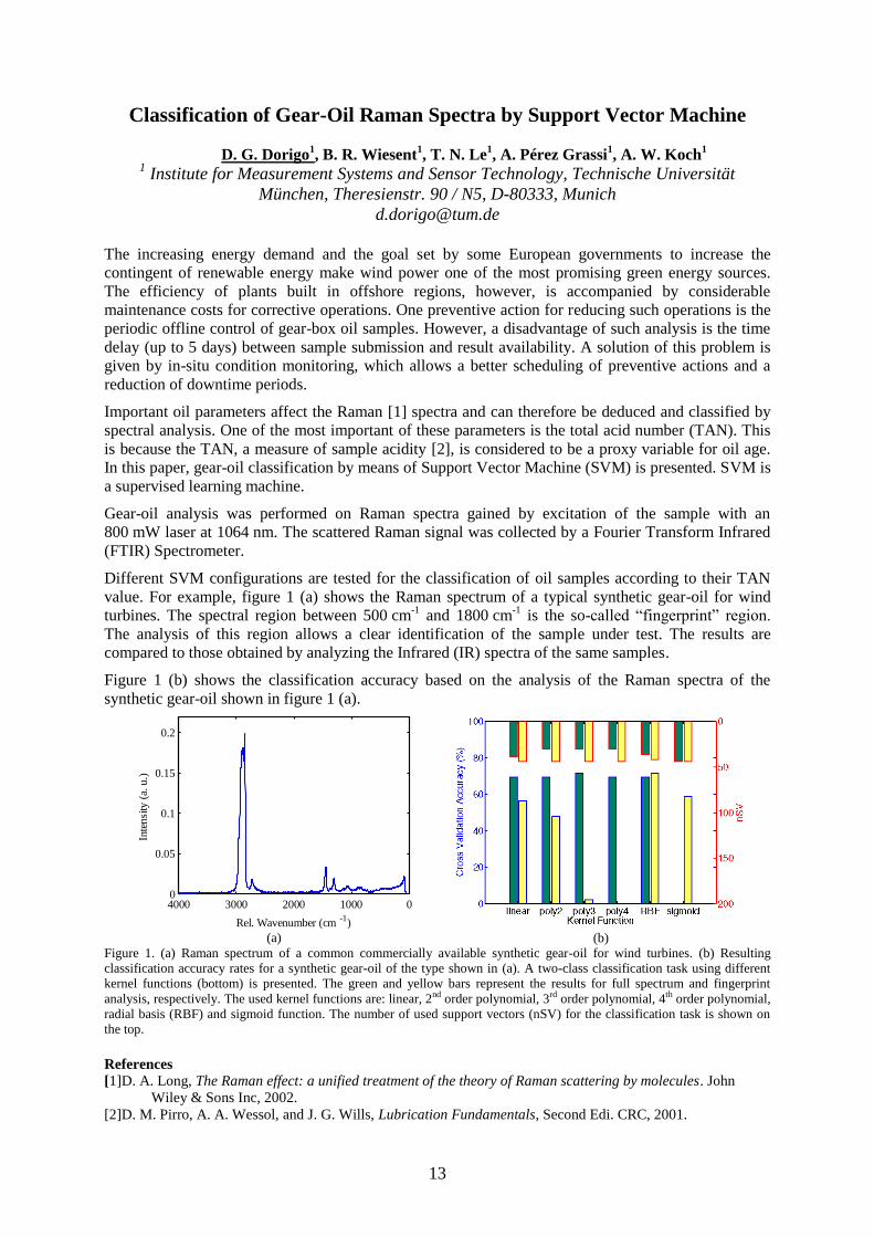
Different SVM configurations are tested for the classification of oil samples according to their TAN
value. For example, figure 1 (a) shows the Raman spectrum of a typical synthetic gear-oil for wind
turbines. The spectral region between 500 cm-1
and 1800 cm-1
is the so-called “fingerprint” region.
The analysis of this region allows a clear identification of the sample under test. The results are
compared to those obtained by analyzing the Infrared (IR) spectra of the same samples.
Figure 1 (b) shows the classification accuracy based on the analysis of the Raman spectra of the
synthetic gear-oil shown in figure 1 (a).
010002000300040000
0.05
0.1
0.15
0.2
Inte
nsi
ty (
a. u
.)
Rel. Wavenumber (cm-1
) (a)
(b)
Figure 1. (a) Raman spectrum of a common commercially available synthetic gear-oil for wind turbines. (b) Resulting
classification accuracy rates for a synthetic gear-oil of the type shown in (a). A two-class classification task using different
kernel functions (bottom) is presented. The green and yellow bars represent the results for full spectrum and fingerprint
analysis, respectively. The used kernel functions are: linear, 2nd order polynomial, 3rd order polynomial, 4th order polynomial,
radial basis (RBF) and sigmoid function. The number of used support vectors (nSV) for the classification task is shown on
the top. References [1]D. A. Long, The Raman effect: a unified treatment of the theory of Raman scattering by molecules. John
Wiley & Sons Inc, 2002.
[2]D. M. Pirro, A. A. Wessol, and J. G. Wills, Lubrication Fundamentals, Second Edi. CRC, 2001.

14
In-band Pumped Nd:LuVO4 Laser Mode Locked by χ(2)
-Lens Formation in
an LBO Nonlinear Crystal
V. Aleksandrov, H. Iliev, I. Buchvarov
Department of Physics, University of Sofia, 5 James Bourchier Boulevard,
BG-1164 Sofia, Bulgaria
e-mail: [email protected]
Multi-Watt operation of picosecond diode-pumped Nd
+3 doped laser oscillators has been demonstrated
mainly by two passive mode-locking techniques, one based on semiconductor saturable absorber
mirrors (SESAMs) and the other on intracavity frequency doubling. Although passive mode-locking
by SESAMs is well established approach for ps-pulse generation around 1 µm, their residual
absorption, leading to heating, is an intrinsic drawback that limits their power-scaling capabilities.
Besides, their production requires complicate equipment. However, the potential of χ(2)
-lens formation
in nonlinear crystal for second harmonic generation (SHG) related to its transparency at the
fundamental wave and shorter pulse generation seems not to have been exploited effectively, yet.
Indeed, in diode-pumped mode-locked Nd-lasers using intracavity SHG for instance, mostly crystals
with higher nonlinearly have been used up to now and the shortest pulses of 2.8 ps were obtained in
the case of Nd:GdVO4 lasers emitting at 1.06 μm while the output power is modest [1]. Although the
Nd: LuVO4 has broader bandwidth among vanadate family, the shortest pulses obtained so far at
multi-watt operation are longer than 10 ps exploiting SESAM mode-locking technique only [2]. On
the other hand, χ(2)
-lens mode locking technique shows capability to keep relatively shorter pulse
duration at muti-watt level of laser output power [3]. This indicates that this technique has strong
potential for high-power mode-locking of broader bandwidth Nd-materials.
In this work we present the results on passive χ(2)
-lens mode-locking of a Nd: LuVO4 laser pumped by
808 nm laser diode as well as in-band pumped at 880 nm. An LBO nonlinear crystal has been used for
intracavity SHG. With 808 nm pump source and 30 % output coupling, we achieve maximum output
power of 2.7 W and 36 % slope efficiency while the measured pulse duration is 7.5 ps. By replacing
the pump source with the one having central wavelength around 880 nm, (using the advantages of in-
band pumping,) we were able to increase the output power with ~ 89 % up to 5.1 W while the pulse
duration was 5.6 ps. The shortest pulse duration obtained for Nd:LuVO4 is 1.6 ps while the output
power is 0.7 W. The repetition rate in both cases was ~ 111 MHz, determined by the cavity roundtrip
time.
Acknowledgements: We acknowledge financial support under bilateral scientific project between Romania and
Bulgaria (grant number DNTS 02/24/2010) and grant number DDVU 02/105/2010 of the Bulgarian national
science fund.
References [1] S. Holmgren, V. Pasiskevicius, F. Laurell, Optics Express, vol. 13, pp. 5270 (2005).
[3] G. Xie, D. Tang, H. Luo, H. Yu, H. Zhang, L. Qian, Laser Physics Letters, vol. 5, pp. 647 (2008).
[3] H. Iliev, I. Buchvarov, S. Kurimura, V. Petrov, Optics Letters, vol. 35, pp. 1016 (2010).

15
Study of combustion process for a methane-air mixture using
a microlaser system
Salamu Gabriela1, Sandu Oana
1, Dejanu Marcel
2, Voicu Flavius
1, Ticos Catalin
3, Popa Dinel
2,
Parlac Sebastian2, Pavel Nicolaie
1, Dascalu Traian
1
1Laboratory of Solid-State Quantum Electronics, National Institute for Lasers, Plasma and
Radiation Physics, Magurele, PO Box MG-36, 077125, Bucharest, Romania 2Faculty of Mechanics and Technology, University of Pitesti, Targu din Vale Street,
110040, Pitesti, Romania 3Low Temperature Plasma Laboratory, National Institute for Lasers, Plasma and Radiation
Physics, Magurele, PO Box MG-36, 077125, Bucharest, Romania
Lean combustion is currently under investigation due to its potential advantages in limiting NOx
emissions and in reducing fuel consumption. It has been used in gas turbines and direct injection spark
ignition (DISI) engines. In this type of engine (DISI), the fuel is directly injected into the combustion
chamber resulting in a distribution of lean fuel/air mixtures.
In this presentation we report a compact, diode-pumped, passively Q-switched Nd:YAG/ Cr4+:YAG
micro-laser used to replace the spark plug as the source for internal combustion engines. A
comparative study of laser spark plug and laser induced ignition in methane-air mixture using this
laser system is discussed.
Air breakdown
Spark Plug
Laser
Fig. 1. Sketch of the experimental
set-up.
Fig. 2. Laser and conventional spark
plug.
The experimental set-up is shown in Figure 1. The ignition process of CH4/ air mixture was studied
experimentally in a constant- volume vessel at filling pressures between 0.1 and 0.5 MPa. Also, the
pressure developed during ignition was measured with a piezoelectric pressure transducer (PCB
112B10 type) mounted on a spark plug-like adaptor (PCB 65 A). For a better understanding of the
combustion process different concentrations of the CH4/ air mixture investigated. We have observed
that the cross-section area of the flame kernel generated by the laser is larger than the one generated by
the spark plug for the same time range. In Figure 2 are depicted the laser and the conventional
electrical spark plug.
In conclusion, a laser system was developed for ignition of gas mixtures, which has the overall
dimensions smaller than that of an electrical spark plug.
Keywords: Methane-air mixture, spark-plug, laser ignition, solid-state lasers, internal combustion.
This work was supported through the project 72150/01.10.2008 that is financed by the Romanian Ministry of
Education, Research, Youth and Sports.
References:
[1] H. Kofler, J. Tauer, G. Tartar, K. Iskra, J. Klausner, G. Herdin, E. Wintner, “An innovative solid-state laser
for engine ignition,“ Laser Phys. Lett. 4, 322-327 (2007).
[2] M. Tsunekane, T. Inohara, A. Ando, N. Kido, K. Kanehara, T. Taira, “High Peak Power, Passively Q-
switched Microlaser for Ignition of Engines,” IEEE J. Quantum Elecron. 46 (2), 277-284 (2010).

16
Analysis of polarization state losses in optical fibers
Ioana Moldovean (Avram)1, Ioan G. Tarnovan
2, Septimiu Crisan
3
The Technical University of Cluj Napoca, 28 Memorandumului str. 400114,
Cluj Napoca, Romania
The optical fiber sensor and the communication through the optical fiber are influenced by the state of
polarization. The optical fiber core imperfections influence the current state of polarization along the
fiber optic cable. The state of polarization is influenced by twisting and coiling the fiber. The paper
presents an analysis of the losses of the polarization state in fiber optic cables. Measurement and
simulation using Comsol software were made with multimode silica and plastic fiber optic and
monomod fiber optic.
Propagation of electromagnetic wave through a plastic optical fiber for 3 different dimensions
of optical fiber.
The initial dimension of fiber Increase the initial size of fiber Increase the initial size of
fiber with 20% with 40%
Acknowledgements
This paper was supported by the project "Improvement of the doctoral studies quality in engineering science for
development of the knowledge based society-QDOC” contract no. POSDRU/107/1.5/S/78534, project co-funded
by the European Social Fund through the Sectorial Operational Program Human Resources 2007-2013
References
1. Arun Kumar, Ajoy Ghatak – Polarization of Light with Application in Optical Fibers – Spie Press
2. Giancarlo C. Righini, Anna Grazia Miagnani, Ilaria Cacciari and Massimo Brenci – Fiber and
integrated optics sensors: Fundamentals and applications - An introduction to optoelectronic sensors,
World Scientific Publishing Co.Pte.Lte
3. Handbook of optics –Third Edition Volume I – Geometrical And Physical Optics, Polarized light,
Components and Intrumentation
4. Hassan Abid Yasser – Polarization Losses in optical Fibers
5. N.Gisin, B.Huttner, N.Cyr Influence of polarization dependent loss on birefringent optical networks –
Baltimore, USA, 2000
6. Petr Drexler and Pavel Fiala – Optical Fiber Birefringence Effects - Sources, Utilization and Methods
of Suppression
7. R.L.Sharma, Dr. Ranjit Singh, Vinod Kumar – Polarization Mode Dispersion (PMD), its limits,
compensation and effect on optical fiber networks – Journal of computing, Volume 3, Issue 8, August
2011
8. R.Ulrich and A.Simon – Polarization optics of twisted single-mode fibers- Optical Society of America
1979
9. Ray Williamson - Polarization Optics Tutorial: Polarizers, Waveplates, Rotators and Lyot Filters
17
Analysis of the inscription process of FBGs in a Panda-type-high
birefingent fiber
B. Berrang, F. Jülich, R. Kuttler, J.Roths
University of Applied Sciences Munich, Laboratory of Photonics, Lothstr. 34, 80335 Munich,
Germany
We investigated the inscription process of fiber Bragg gratings (FBGs) in high birefringent (HiBi),
polarization maintaining (PM) fibers of the PANDA type from Nufern (see Figure 1). FBGs in HiBi-
PM fibers have the potential for multi parameter sensing [1] and improved accuracy for temperature
and strain sensing [2], compared with FBGs in standard single-mode fibers.
Since PANDA fibres are not rotationaly symmetric, the dependence of the
FBG writing process with respect to the azimuthal orientation was evaluated
in this study. FBGs were inscribed using an Eximer laser operating at 248 nm
and the fibers were placed directly infront of a phase mask [3]. Figure 1
shows the alignment of the fiber and the stress applying parts relative to the
UV-laser beam and the alignment microscope. The azimuthal alignment
angle α is defined as the angle between the center of the UV inscription beam
and the slow axis of the fiber.
To monitor the inscription process a multichannel FBG interrogator (SM125-
500, Micron Optics) in combination with a polarization beam splitter (PBS)
was used (see Figure 2a). The reflection spectra (Figure 2b) of the FBG in the
fast and slow axis were observed simultaneously. A delay of 10 m was used
between the channels. The coherence length of the SM125-500 light source is in the order of 1 m.
Figure 3a shows the growth process of the FBGs created in the fast and slow axis as a function of applied
exposure dose and this growth process was largely insensitive to the orientation of the azimuthal angle.
However there was a small dependency of the ratio of the reflectivities of the FBGs in the slow and fast axis
with azimuthal angle as shown in Figure 3b. The reflectivities of FBGs in the fast axis Rf were in every case
greater than the reflectivities of the slow axis FBGs Rs (Figure 3c).
Figure 2: a) schematic of interrogation setup while inscription b) reflection spectra of one HiBi-FBG
Figure 3: a) graph of inscription process, b) ratio Rs / Rf c) transmission spectra of one HiBi-FBG measured
with a broad band source, linear polariser and an optical spectrum analyzer
Acknowledgements: This work was supported by the Bavarian State Minister of Science, Research and the Arts.
References
[1] D. Wada, H. Murayama, H. Igawa, K. Kageyama, K. Uzawa, Smart Materials and Structures, 20, pp. 8(2011)
[2] F. Jülich, A. Koch, J. Roths, Technisches Messen, 1, pp. 52 (2012)
[3] E. Mayer, D. Gillett, S. Govorkov, Fiber and Integrated Optics, 18:3, 189-198 (2010)
Figure 1: Definition of
azimuthal alignment
angle (here: α = 45°)

18
Optical spectrum of a coupled chaotic system
I. R. Andrei, G.V. Popescu, C.M. Ticos and M.L. Pascu
National Institute for Laser, Plasma and Radiation Physics, Department of Lasers, str.
Atomistilor 409, 077125, Magurele, Romania
e-mail of corresponding author: [email protected]
Chaotic synchronization effects [1] on the optical spectrum of coupled system were investigated
experimentally using two bidirectionaly coupled identical SLs as master and slave. The master is an
external-cavity semiconductor laser (LSCE) system with chaotic low-frequncy fluctuations (LFF)
dynamics [2] and the slave can be an identical LSCE system or a solitary diode laser with free
emission (Fig. 1). The semiconductor laser operating parameters of two lasers were chosen so that the
emission spectra to be similar in the absence of external feedback, and for injection currents near the
threshold current (the condition to obtaining LFF fluctuations). In the presence of the external optical
feedback, in these particular experimental conditions, both laser output emisions shown multimod
structures, but in close spectral range.
In the present work we experimentally evaluated the effects that the synchronization regimes (lag, zero
lag or anticipated synchronization) [3,4] have on optical spectrum of the coupled system. These results
are important to understand the mechanisms that contribute to the optical spectrum formation of a
mutually coupled systems. It was observed that in the emission spectrum of the coupled system there
is the trend to manifest the master or slave modes depending on the synchronization regime. In the
optical spectrum there are activated dominant modes that coincide with the laser modes of the two
systems operating in LFF regime without coupling. Under synchronization conditions in the optical
spectrum of the coupled system there are either the master in the lag synchronization or the slave in
anticipated synchronization.The experimental setup used is schematically shown in Figure 1.
Fig.1. Experimental setup of mutually coupling of the coupled
system. SL, semiconductor laser; TEC, SL mount with thermo-
electric cooler; L, collimation system; BS, beamsplitter; NDF,
neutral density filter; ER, external optical reflector; PD, photo-
detector.
Acknowledgements This work was supported by the National Centre for the Management of Programs (CNMP)
under contract no. 72-219.2008 within the PNCDI2 program and by the National Authority for Scientific
Research (ANCS) under contract Nucleu-LAPLAS 2011
References [1] H. Fujino and J. Ohtsubo, Experimental synchronization of chaotic oscillations in external-cavity
semiconductor lasers, Opt. Lett., 25, 625–627 (2000).
[2] J. Mork, B. Tromborg, P. L. Christiansen, Bistabily and Low-Frequency-Fluctuations with Optical
Feedback: a Theoretical Analysis, IEEE J. QE-24, 2, 123 (1988).
[3] S. Sivaprakasam, P.S. Spencer, P. Rees, and K.A. Shore, Regimes of chaotic synchronization in external-
cavity laser diodes, IEEE J. Quantum Electron., 38, 9, 1155 (2002).
[4] H.U. Voss, Anticipating chaotic synchronization, Phys. Rev. E., 61, 5115–5118 (2000).
19
Laser Induced Damage Threshold Test Station:
Development and Measurements - preliminary results
Alexandru Zorilă1, 2
, Laurenţiu Rusen1, Simion Sandel
1, Aurel Stratan
1,
Constantin Blanaru1, Constantin Fenic
1, George Nemeş
1, 3
1Solid State Laser Laboratory, Laser Department, National Institute for Laser, Plasma and
Radiation Physics, 077125 Măgurele, Romania,
http://ssll.inflpr.ro; [email protected] 2"Politehnica" University, 313 Splaiul Independentei, 060042 Bucharest, Romania
3ASTiGMAT
TM, Sacramento, CA 95827, USA,
http://astigmat-us.com; [email protected]
Keywords: laser-induced damage-threshold, ISO standard, automated test procedure.
The presentation reports the development of an automated test station for Laser Induced Damage
Thresholds (LIDT) measurements on various optical surfaces, by irradiating a certain site with
multiple-pulses, referred to as the S-on-1 test in the ISO 21254 standard [1]. This test is based on a
protocol that applies a series of up to S laser pulses with constant energy density at each unexposed
test site, and stops the delivery of the remaining pulses immediately after a permanent damage occurs
at that site, generally after N pulses (N ≤ S).
Therefore, a real-time damage detection system is necessary. Its function is to determine in real-time
the appearance of a permanent damage on the irradiated site. This information is further used to
determine the exact number of pulses, N, at which that particular site was permanently damaged and to
stop the subsequent laser pulses to hit the site after the damage occurred. The damage threshold of a
site is defined as the minimum fluence or irradiance level at which a permanent laser radiation-
induced change of the surface characteristics of the specimen can be observed by an incident-light
microscope having Nomarski-type differential-interference contrast with a total magnification of at least 100x - 150x [2].
The presentation describes the automated S-on-1 procedure and the main characteristics of the
measurement setup. Some LIDT measurement results obtained on optical components (laser mirrors and anti-reflecting coatings) for high-power lasers, are presented, too.
The automated test-station is developed within the framework of the Project ISOTEST – "Facility for
laser beam diagnosis and ISO characterization/certification of behavior of optical
components/materials subjected to high power laser beams".
Acknowledgments: This work is done within the framework of the Project No. 172/2010 - ISOTEST-
sponsored by the National Authority for Scientific Research (ANCS-POSCCE), Romania.
References:
[1] ISO 21254 - 2:2011, "Lasers and laser-related equipment - Test methods for laser-radiation-induced damage
threshold - Part 2: Threshold determination".
[2] ISO 21254 – 1:2011, "Lasers and laser-related equipment - Test methods for laser-radiation-induced damage
threshold - Part 1: Definitions and general principles".

20
Session 3
Advanced Materials

21
Phase relation, electro-optic and dielectric properties of lead-free
ferroelectric thin films.
N.D. Scarisoreanu
National Institute for Lasers, Plasma and Radiation Physics, Bucharest, Romania
Multiple nowadays applications are using ferroelectric ceramic materials. There are many types of
ferroelectric materials: some of them containing potentially dangerous elements like lead zirconate
titanate (Pb(Zr1-x,Tix)O3), others having different crystallographic structures (tungsten-bronse,
perovskite, etc), and the list can go on. Tungsten-bronze and perovskite types are considered among
the best lead-free candidate materials for tomorrow’s devices which are predicted by today’s life
standards and environmental laws. Taking into account the toxicity of lead-based systems like Pb(Zr1-
x,Tix)O3 (PZT), there are a lot of lead-free piezoelectric materials under investigation in worldwide
spread laboratories for replacing PZT in future devices. Constant efforts are made to find viable
replacements for all these materials which contain harmful elements. Solid-solution systems based on
lead-free perovskites such as Na0.5K0.5NbO3 (NKN), BaTiO3 (BT), Na0.5Bi0.5TiO3 (NBT) or bismuth
layered-structured SrBi2Ta2O9 (SBT), SrBi2Nb2O9, together with tungsten-bronze niobates like
SrxBa1-xNb2O6 are considered as viable alternatives for replacing lead-based materials.
Lead-free (Na0.5Bi0.5)1-xBaxTiO3 ferroelectric thin films obtained by pulsed laser deposition (PLD),
have been optical, structural and electrical investigated for compositions at and around morphotropic
phase boundary (MPB). Unusual characteristics have been found in the meaning of phase transitions
temperatures, enhancement of ferroelectric phases or variations of tetragonality ratio. The appearance
of these effects has been attributed to the existing intrinsic surface stress in film’s elongated
nanograins. Using phase transitions measurements, the dielectric anomalies present in NBT-BT thin
films are identified and discussed.
The electric and electro-optic properties of heteroepitaxial strontium barium niobate (SrxBa1-xNb2O6)
thin films deposited by pulsed laser deposition and radiofrequency assisted pulsed laser deposition on
MgO and Nb:STO substrates. As a function of an electric applied field, the birefringence behavior and
electro-optic coefficient of the thin films have been measured by reflection-type spectroscopic
ellipsometry method using coplanar AZO electrodes.
References :
[1] N.D. Scarisoreanu et al, Thin Solid Films, http://dx.doi.org/10.1016/j.tsf.2011.11.066. (2011).
[2] N.D. Scarisoreanu et al, Applied Physics A 93, 795–800 (2008).
[3] N.D. Scarisoreanu et al, Applied Surface Science 2541292–1297, (2007).

22
Numerical analysis of non-linear cavity modes in a silicon hole-array
photonic crystal
C. D. Sold1, O. M. Bunoiu
1, C. G. Biris
1,2
1Faculty of Physics, West University of Timisoara, Timisoara, Romania
2High Performance Computing Center, West University of Timisoara, Timisoara, Romania
corresponding author: [email protected]
Photonic crystals (PhCs) are well-known to have important applications in the study and manipulation
of light at the micro and nano scales. In this type of structures, one often encounters cavity defect
modes, which occur within the photonic bandgaps. Linear defect modes have been shown to exhibit
very large quality factors1, an important property in applications to active optical devices. However,
there is little information of the behaviour of such modes in the non-linear regime. We show in our
work that the properties of cavity defect modes in a PhC can be greatly enhanced by the use of surface
second harmonic generation. Our geometry consists of a Si slab with cylindrical air holes in a
hexagonal lattice. The central hole has been filled in, in order to form the defect [see Fig. 1a].
Fig. 1 Photonic crystal geometry a); Band structure showing location of defect modes and band-gaps b); Electric
field profile for the lower frequency (FF) and higher frequency (SH) defect modes, c) and d) respectively.
The band structure of the crystal was computed using a commercial band solver and clearly shows the
formation of two band-gaps and two corresponding defect modes with the second at close to double
the frequency of the first [see Fig. 1b]. Figs. 1c-d show the field profiles of these two modes at the
fundamental frequency (FF) and the second harmonic (SH), respectively. The profiles were obtained
using an advanced implementation of the MSM Algorithm2. It is easy to see that the formation of the
defect mode at the FF leads directly to a strong non-linear enhancement at the SH due to surface SHG,
which implies that these devices could potentially have novel applications in the field of active optical
non-linear devices as we will show during the conference.
Acknowledgements: This research is partially supported by the grant FP7-REGPOT-2011-1-284595 (HOST).
The authors would also like to extend their gratitude to N. C. Panoiu for his many useful suggestions.
References:
[1] H.Y. Ryu, M. Natomi, G.H. Kim and Y.H. Lee, Opt. Express 12, 1708 (2004)
[2] C.G. Biris and N.C. Panoiu, Phys. Rev. B 81, 195102 (2010)
23
Advanced nonlinear optic crystals for high efficiency visible and UV laser
sources based on frequency conversion processes
L. Gheorghe
National Institute for Laser, Plasma and Radiation Physics, Solid-State Quantum Electronics
Laboratory, P.O. Box MG 36, 077125 Magurele, Bucharest, Romania
Currently, depending on the emission wavelength, solid-state lasers are used in a wide variety of
applications such as scientific research, telemetry, information technology (processing, transmission,
storage or information display), surgery and medicine, materials processing, remote detection of utile
materials or pollutants, photolithography, laser printing, laser display technology, entertainment, etc.
To satisfy all these needs, the laser emission wavelengths must cover a wide spectral range from
infrared (IR) to ultraviolet (UV) passing through the visible (VIS) domain. At present, there are very
few solid-state lasers with efficient emission in the VIS and UV spectral ranges, and the accessible
wavelengths are very limited. The most efficient method to achieve such laser sources is the frequency
conversion of near-IR solid-state laser emission by nonlinear optical (NLO) processes in suitable NLO
crystals [1]. Thus, NLO crystals play a key role in the development of coherent radiation sources as
they permit the frequency conversion of mature solid-state lasers into spectral ranges where lasers do
not exist or perform poorly.
Up to now, many efforts have been carried out in order to develop new NLO materials and/or to
improve their specific properties or workability. The experimental results demonstrated that borate-
type compounds constitute a veritable source of NLO crystals with good nonlinear properties [2, 3].
Thus, all the recent NLO crystals discovered in the last 10 years are in majority borate-based
compounds such as: CsLiB6O10 (CLBO), BiB3O6 (BiBO), K2Al2B2O7 (KABO), KBe2BO3F2 (KBBF),
RECa4O(BO3)3 (RECOB, RE = Gd, Y, La), LnMe3(BO3)4 (Ln = lanthanide, Me = Al, Ga, Sc) [2, 10]
etc. Nowadays, two families of borate crystals are known that melt congruently or can include
congruently melting compounds which can be grown with high quality and large size by Czochralski
method: (i) rare-earth calcium oxyborates RECOB and (ii) binary borates LnMe3(BO3)4 with trigonal
huntite-type structure. Two types of new NLO borate crystals with congruent melting behavior
belonging to these two families are presented in this work:
- Gd1-xRxCa4O(BO3)3 (R3+
= Sc3+
, Lu3+
) crystals in which function of the substitution degree with R3+
ions it is possible to achieve second harmonic generation (SHG) in non-critical phase matching
(NCPM) conditions of some important laser emissions at specific wavelengths in the near-IR range,
conditions which ensure obtaining of maximum conversion efficiency into VIS spectral range;
- LaxGdyScz(BO3)4 (x + y + z = 4) crystals with wide transparency range from the UV to the IR
domain and excellent NLO properties characteristics to huntite-type borate crystals.
Acknowledgements: This work was supported by the Joint Research Project Romania - France, Project 3 RO-
FR / 03.01.2012 (project code: BORATESYB).
References
[1] D. N. Nikogosyan, “Nonlinear Optical Crystals: A Complete Survey”, Springer, N.Y. (2005).
[2] T. Sasaki, et al., Mat. Sci. Eng., 30, pp. 1 (2000).
[3] P. Becker, et al., J. Cryst. Growth, 203, pp. 149 (1999).
[4] C. Zhang, et al., J. Cryst. Growth, 235, pp. 1 (2002).
[5] J. Lu, et al., Opt. Comm., 200, pp. 415 (2001).
[6] M. Iwai, et al., Jpn. J. Appl. Phys., 36, pp. L276 (1997).
[7] G. Aka, et al., J. Opt. Soc. Am. B, 14, pp. 2238 (1997).
[8] H. J. Zhang, et al., Appl. Phys. A, 78, pp. 889 (2004)
[9] G. A. Peterson, et al., Intern. J. Inorg. Mat., 2, pp. 101 (2000).
[10] S. T. Durmanov, et al., Opt. Mater., 18, pp. 243 (2001).

24
Session 4
Lasers History

25
50 years of laser research in Romania
(Department of LASERS)
Dan C. Dumitraş
Department of Lasers, National Institute for Laser, Plasma and Radiation Physics,
Bucharest, Romania (e-mail: [email protected])
Last year we celebrated 50 years of laser history. If fifty years ago people thought that the laser is “a
solution looking for a problem”, today lasers have gone on to be one of the outstanding success stories
in physics.
The development of lasers was possible owing to the general progress in physics and particularly in
optics and quantum electronics, with the contributions of Fabry-Pérot (1899), Einstein (1916),
Ladenburg (1928), Fabrikant (1939), Lamb (1947), Kastler (1950), Purcell (1951), Weber (1953), and
many others. But, the first device based on the principles developed by Einstein was built in December
1953 by Townes – the ammonia MASER. Other maser types were described theoretically by Basov
and Prokhorov (1954) and Bloembergen (1956), and subsequently operated in different laboratories
(1957 - 1960). Scientists were looking for a maser at optical frequencies since 1957, and the race to
build a LASER was won by Maiman on May 16th, 1960.
Since then, many laser pioneers have contributed to the discovery of new lasers and laser operating
regimes. These steps will be presented chronologically, emphasizing the role of many brilliant
scientists (1960 - 2011). Detailed information will be given on first commercial lasers and on Nobel
Prizes for laser related discoveries.
The second part of the talk will focus on Romanian contributions in the field (1960 - 1975). Romania
is believed to be the forth country in the world that succeeded to operate a laser (October 20th, 1962).
Many details will be presented regarding the scientific and technologic contributions of Romanian
scientists in the first two decades of laser history (published papers, communications at conferences,
books, PhD Thesis, patents). An important role for our community have played the international
conferences organized by our institute (1982 - 2011), that offered the possibility to present our results
at international level and to meet researchers from many other countries.
2010 was a yearlong celebration of the 50th anniversary of the laser. Many conferences had special
sessions dedicated to this event. The author have participated in several of them (CLEO/QELS, San
Jose, May; Laser in the City of Light, Paris, June; LPHYS, Foz do Iguacu, July; ALT, Egmont aan
Zee, September) and the presentation will be accompanied by numerous pictures from those
celebrations.
In 2009 Romania was chosen as a site of the ambitious European project Extreme Light Infrastructure
(ELI) as a result of our efforts and contributions to ELI – Preparatory Phase (FP 7 Program, 2007-
2010). The mission of ELI – NP (Nuclear Physics) pillar, the laser architecture, the experiments
envisaged, and the structure of the future infrastructure will be mentioned.
The last question approached is: do masers and lasers exist in nature? The answer is
affirmative.
26
Session 5
Spectroscopy. Lasers in
Life Sciences
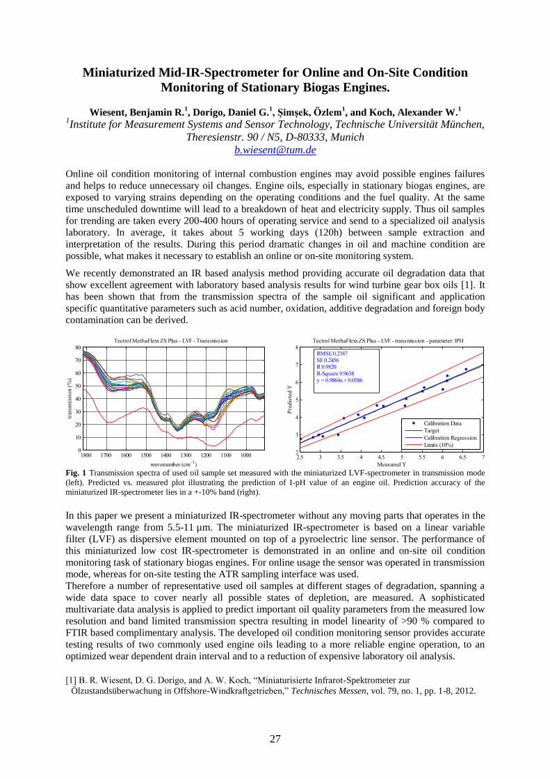
27
Miniaturized Mid-IR-Spectrometer for Online and On-Site Condition
Monitoring of Stationary Biogas Engines.
Wiesent, Benjamin R.1, Dorigo, Daniel G.
1, Şimşek, Özlem
1, and Koch, Alexander W.
1
1Institute for Measurement Systems and Sensor Technology, Technische Universität München,
Theresienstr. 90 / N5, D-80333, Munich
Online oil condition monitoring of internal combustion engines may avoid possible engines failures
and helps to reduce unnecessary oil changes. Engine oils, especially in stationary biogas engines, are
exposed to varying strains depending on the operating conditions and the fuel quality. At the same
time unscheduled downtime will lead to a breakdown of heat and electricity supply. Thus oil samples
for trending are taken every 200-400 hours of operating service and send to a specialized oil analysis
laboratory. In average, it takes about 5 working days (120h) between sample extraction and
interpretation of the results. During this period dramatic changes in oil and machine condition are
possible, what makes it necessary to establish an online or on-site monitoring system.
We recently demonstrated an IR based analysis method providing accurate oil degradation data that
show excellent agreement with laboratory based analysis results for wind turbine gear box oils [1]. It has been shown that from the transmission spectra of the sample oil significant and application
specific quantitative parameters such as acid number, oxidation, additive degradation and foreign body
contamination can be derived.
1000110012001300140015001600170018000
10
20
30
40
50
60
70
80
wavenumber (cm-1
)
Tectrol MethaFlexx ZS Plus - LVF - Transmission
tran
smis
sio
n (
%)
2.5 3 3.5 4 4.5 5 5.5 6 6.5 72
3
4
5
6
7
8
Measured Y
Pre
dic
ted
Y
Tectrol MethaFlexx ZS Plus - LVF - transmission - parameter: IPH
Calibration Data
Target
Calibration Regression
Limits (10%)
RMSE 0.2387SE 0.2456R 0.9820R-Square 0.9638y = 0.9864x + 0.0586
Fig. 1 Transmission spectra of used oil sample set measured with the miniaturized LVF-spectrometer in transmission mode
(left). Predicted vs. measured plot illustrating the prediction of I-pH value of an engine oil. Prediction accuracy of the
miniaturized IR-spectrometer lies in a +-10% band (right).
In this paper we present a miniaturized IR-spectrometer without any moving parts that operates in the
wavelength range from 5.5-11 µm. The miniaturized IR-spectrometer is based on a linear variable
filter (LVF) as dispersive element mounted on top of a pyroelectric line sensor. The performance of
this miniaturized low cost IR-spectrometer is demonstrated in an online and on-site oil condition
monitoring task of stationary biogas engines. For online usage the sensor was operated in transmission
mode, whereas for on-site testing the ATR sampling interface was used.
Therefore a number of representative used oil samples at different stages of degradation, spanning a
wide data space to cover nearly all possible states of depletion, are measured. A sophisticated
multivariate data analysis is applied to predict important oil quality parameters from the measured low
resolution and band limited transmission spectra resulting in model linearity of >90 % compared to
FTIR based complimentary analysis. The developed oil condition monitoring sensor provides accurate
testing results of two commonly used engine oils leading to a more reliable engine operation, to an
optimized wear dependent drain interval and to a reduction of expensive laboratory oil analysis.
[1] B. R. Wiesent, D. G. Dorigo, and A. W. Koch, “Miniaturisierte Infrarot-Spektrometer zur
Ölzustandsüberwachung in Offshore-Windkraftgetrieben,” Technisches Messen, vol. 79, no. 1, pp. 1-8, 2012.

28
Laser beams interaction with pharmaceutical foams:
Aethoxysklerol case
Adriana Smarandache1, M. Trelles
2, J. Moreno-Moraga
3, Angela Staicu
1, M.L. Pascu
1
1 National Institute for Lasers, Plasma and Radiation Physics, Laser Department,
P.O.Box, MG-36, 077125, Bucharest, Romania 2 Instituto Médico Vilafortuny, Cambrils (Tarragona), Spain
3 Instituto Médico Láser, 28010 Madrid, Spain
E-mail of the corresponding author: [email protected]
The goal of this study is to investigate the laser light scattering involved in the laser irradiation process
of Aethoxysklerol (Polidocanol) foam samples. It is the commercially available medicine used in the
sclerotherapy of small veins (less than 4 mm in diameter).
Clinical experimental results prove that the exposure of tissues impregnated with Polidocanol in foam
form to laser radiation emitted at 1064 nm improves the efficiency of the treatment [1].
Previous absorption studies on Aethoxysklerol 2% solution before and after exposure to Nd:YAG
1064 nm laser radiation have not shown important spectral modifications of it [2]. The effect of the
laser light may be enhanced if the Polidocanol is introduced as foam. It is important to investigate the
laser light scattering involved in the laser irradiation process of this drug foam samples. To do that, we
produced foam by mixing Aethoxysklerol 2% solution and atmospheric air (1:4) using the Tessari
method. The batch is passed between the two syringes about 40 times and the resulting foam is stable
during 5-6 min.
A 10 mm optical cell containing foam sample was introduced into a home-made Raman spectroscopy
system. The laser radiation used to excite the Raman emission is the second harmonic of a pulsed
Nd:YAG laser (10 Hz frequency, 5 ns pulse duration, 250 mJ energy at 532 nm). The detection is
made by a high resolution spectrograph and ICCD camera.
The obtained Raman spectra were more intense in foam form than in simple liquid solution (see
figure). This shows the fact that the laser light scattering produces a larger optical path of the laser
beam in the sample and consequently a larger absorption of it by the foam components.
The compared Raman spectra of the Polidocanol in solution and foam presentation
Acknowledgements: This research work was supported by the ANCS (RO), project LAPLAS 3-PN 09 33.
References:
[1] M. Trelles, J. Moreno-Moraga, J. Alcolea, A. Smarandache, M.L. Pascu In: Synopsis of Aesthetic
Dermatology & Cosmetic Surgery, (Nova Science Publishers Inc, NY, USA), Chapter 3, 2012. [2] A. Smarandache, M. Trelles, M.L. Pascu, J. Optoelectronics Advanced Materials, vol. 12, pp. 1942, 2010.

29
Quantitative analysis of surgical smoke by laser photoacoustic spectroscopy
Ana Bratu, Mioara Petrus, Cristina Achim, Consuela Matei, Mihai Patachia,
Stefan Banita and D.C. Dumitras
Department of Lasers, National Institute for Laser, Plasma and Radiation Physics,
409 Atomistilor St., PO Box MG-36, 077125, Bucharest, Romania
[email protected] Personnel in operating rooms have been exposed to surgical smoke for many years, unaware that it
may create certain health risks. Exposure has increased as surgical procedures have developed and the
use of laser surgery has increased. With the development of laser surgery technology the elements of
surgical smoke have come over investigation.
Surgical smoke is referred to as the smoke created when energy is transported to tissue cells during
surgical procedures [1]. When laser is transferred to a cell, heat is created. This heat vaporizes cellular
fluid, which increases the pressure inside the cell and eventually causes the cell membrane rupture [2-
3]. The intense heat created burns the protein and other organic matter in the cell, and causes thermal
necrosis in neighboring cells. This charring of cells, which creates smoke, not only hinders the vision
of the surgical stuff, but it also release harmful contaminants, both biological and chemical, into the air
[4].
Exposure to the noxious odor of surgical smoke is accepted as part of working in an operating theatre,
but this odor is an indication of the chemicals which it contains and has been reported to cause
headache, irritation and soreness in the eyes, nose and throat [5].
CO2 laser photoacoustic spectroscopy offers a sensitive technique for detection and monitoring of
trace gases at low concentrations. The CO2 laser is of special interest, as it ensures high output power
in a wavelength region (9-11 m) where more than 250 molecular gases/vapors exhibit strong
absorption bands. It is an ideal source to push the sensitivity of photoacustic (PA) gas detection into
the concentration range of parts per billion (ppbV) or even lower [6].
The chemical composition of smoke produced with a CO2 laser on different animal in vitro tissues was
investigated. Our study employs laser photoacoustic spectroscopy to determine quantitative chemical
composition of surgical smoke. Measurements were carried out at ambient pressure and room
temperature. A gas of choice (N2 or synthetic air) was pumped into the cell through the gas inlet, while
the produced smoke was evacuated through the gas outlet. A fully analysis of the surgery smoke
obtained by the surgery CO2 laser is presented; dependences of the smoke composition with respect of
the surgery CO2 laser parameters is analyzed.
The concentrations of the components in the smoke sample depend on how much animal tissue was
cauterized. The qualitative composition of all the samples that we studied was very similar: water,
methane, carbon dioxide and ethane were always found, plus additional broad absorptions that could
not be identified.
References [1] Biggins, Jill, Renfree, Steve, The Hazards of Surgical Smoke are not to be Sniffed at! Journal of Peri-
Operative Nursing, (April 2002).
[2] Ott, D. E. Smoke Production and Smoke Reduction in Endoscopic Surgery: Preliminary Report. Endosc Surg
Allied Tech (1993).
[3] Smith, Jerome, Hsu-Chi Yeh, Bruce Muggenburg, Raymond Guilmetter, Linda S. Martin, and Phillip W.
Strine, Study Design for the Characterization of Aerosols During Surgical Procedures, Scandinavian Journal of
work Environmental Health 2 (1992; suppl 2).
[4] Winston, Charlene, The Effects of Smoke Plume Generated During Laser and Electrosurgical Procedures,
Minimally Invasive Surgical Nursing, (1994).
[5] Ferenczy A, Bergeron C and Richard RM, Human papillomavirus DNA in CO2 laser-generated Plume of
Smoke and Consequences to the Surgeon. Obstet Gynecol, (1990).
[6] D. C. Dumitras, D. C. Dutu, C. Matei, A. M. Magureanu, M. Petrus, C. Popa “Laser photoacoustic
spectroscopy: principles, instrumentation, and characterization” Journal of Optoelectronics and Advanced
Materials, Vol 9, No. 12, (2007).

30
Temperature distribution analysis in laser irradiated tissue by
numerical analysis and experimental data
M. Petrus, C. Matei and D.C. Dumitras
Department of Lasers, National Institute for Laser, Plasma and Radiation Physics,
409 Atomistilor St., PO Box MG-36, 077125, Bucharest, Romania
Laser is an effective tool in clinical applications [1]. In treatments, that use laser radiation it is
important to have a control of temperature, high temperature could lead to thermal damage in the
surrounding tissue [1]. Computer models have been used to determine tissue temperatures
during tissue ablation laser irradiation. For numerical modeling, the temperature evolution in
tissue is described by the bioheat equation [2]. The numerical solutions are obtained by the
Finite Element Method (FEM) [3-5]. To generate the finite element model we used the
software Comsol Multiphysics.
The wavelength of 10.6 m is strongly absorbed by the water and the CO2 laser is suitable for
vaporization and the precise thermal cutting of tissue and can be considered as a device whose
irradiation is absorbed near the surface [1]. Experimentally, porcine vocal cords were irradiated in
vitro by a CO2 laser at different laser power and different exposure time.
We applied CO2 laser light at different power density and exposure time and the
computational model was simulated for a single tissue layer. For the numerical model validation
we compare the crater ablation depth from the experimental images with the temperature distribution
from the numerical analysis. This numerical method of temperature distribution modeling can also be
used for extensive parametric studies in order to characterize the stability of various treatment
parameters and would allow obtaining a faster and better simulation of laser treatment of biological
tissues. The results obtained from the study reveal the important parameters, which significantly affect
the thermal response of soft tissue, as exposure duration, power, wavelength of the beam, as well as
the area and type of the tissue.
a) b)
Fig. 1: a) 3D numerical simulation on soft tissue irradiated with a CO2 laser at 24 W and an exposure time of 100
ms, b) OCT image of crater ablation in simulation on soft tissue irradiated with a CO2 laser at 24 W and an
exposure time of 100 ms.
References [1] RH Ossoff, Reinisch L, Chapter 12: Laser Surgery: Basic Principles and Safety Considerations, (C.V.
Mosby Company, St. Louis (1986).
[2] HH Pennes, Analysis of tissue and arterial blood temperature in the resting human fore-arm, Journal of
Applied Physiology, 1: 93 – 122 (1948).
[3] Maruyama S, Okajima J, Komiya A, Takeda H, Estimation of Temperature Distribution in Biological Tissue
by Using Solutions of Bioheat Transfer Equation, Heat Transfer—Asian Research 2008; 37 (6): 375-386.
[4] Gupta PK, Singh J, Rai KN, Numerical simulation for heat transfer in tissue during thermal therapy, Journal
of Thermal Biology; 35:295-301 (2010).
[5]Fanjul-Velez F, Romanov OG, Arce-Dego JL, Efficient 3D numerical approach foe temperature prediction in
laser irradiated biological tissues, Computers in Biology and medicine; 39: 810-817 (2009).

31
Calibration and artefact minimization in a CW Diffuse Optical
Tomography system
M. Patachia1,2
, D. C. Dutu1, S. Banita
1, C. Achim
1, M. Petrus
1, A. Bratu
1,
C. Matei1 and D.C. Dumitras
1
1Department of Lasers, National Institute for Laser, Plasma and Radiation Physics,
409 Atomistilor St., PO Box MG-36, 077125 Bucharest, Romania 2Faculty of Physics, University of Bucharest, Romania
e-mail : [email protected]
Keywords: Laser diode, diffuse optical tomography, fiber optics, tissue phantom, image
reconstruction.
Abstract: In most of the tissues, light propagation is dominated by scattering. As results, after
travelling the length of a few millimeters, light propagation in tissues can be described as a diffusive
process. Different tissue types have distinct scattering properties, and therefore this distinction can be
imaged. Using optical measurements at multiple source-detector positions on the tissue surface, one
can reconstruct the internal distribution of the absorption coefficient (µa) and the reduced scattering
coefficient (µ´s) based on the diffusion and the light transport model in a diffusive media.
Measurements on phantoms are used to evaluate the performance of systems and to validate imaging
algorithms. The calibration is the pivotal part of the data acquisition due to the variation in
characteristics of each laser source, optical fibers, detectors and optic elements. An accurate
calibration is achieved in homogeneous phantoms. The work describes how the homogeneous
phantoms are used to evaluate the performance of the system and to minimize the artifacts.
Acknowledgements Thank you to Prof. Dr. H. Jiang and Mrs. Xiaoping Liang from the Department of Biomedical Engineering,
University of Florida, Gainesville, U.S.A. and Dr. N.V. Iftimia, Physical Sciences, Inc., 20 New England
Business Center Drive, Andover, U.S.A.
References
Schmitz, C. H., Graber, H. L., Luo, H.,, 2000. Instrumentation and calibration protocol for imaging dynamic features in
dense-scattering media by optical tomography, Applied Optics, 39, 6466-6485.
Bishop, Y. M. M., Fienberg, S. E., Holland, P. W., 1991. Maximum likelihood estimates for complete tables, Discrete
Multivariate Analysis: Theory and Practice, Chap. 3, MIT, Cambridge, Mass.
Jiang, H. B., Xu, Y., Iftimia, N.V., 2000. Experimental three-dimensional optical image reconstruction of heterogeneous
turbid media from continuous-wave data, Optics Express 7, 204-209.

32
Studies about the laser radiation interaction with beads of microliter
volumes
Mihail - Lucian Pascu
National Institute for Laser, Plasma and Radiation Physics, Laser Department, Magurele,
077125, Romania. e-mail: [email protected]
A survey is made about the optofluidics at micrometre scale, treating on one hand the generation of
single droplets and on the other the production of large number of beads in suspensions that contain
immiscible materials. The interaction of distilled water microdroplets (volumes of some microliters)
suspended individually on a capillary, with pulsed laser beams emitted at 532nm is described. The
532nm wavelength is used because here the distilled water absorption is very low and the interaction
of a water bead with the laser radiation is dominated by unresonant phenomena. Following the
collision of such a laser beam with a water microdroplet in air, deformations and mechanical
vibrations of it are produced. The conditions in which the droplets lose material as a consequence of
the impact with laser beams are explored and reported, as well. The effects produced on the droplet
were studied pulse by pulse. They depend on: droplet’s content (in this case water), laser beam
wavelength, power and focusing conditions, bead exposure geometry, possibly bead’s volume and its
adhesion to the capillary on which it is suspended. The laser pulses energies were varied between
0.25mJ and 1mJ. The pulse full time width was 10ns and the typical beam focus diameter on the
droplet was 90µm. The microdroplets and the behaviour of their shapes after exposure to the laser
beam were measured by recordings performed at 10kfr/s. Following the microdroplet interaction with
the laser beam one may also produce nanodroplets propagating at high (probably supersonic or even
hypersonic) speeds and microdroplets propagating at slower speeds. One may produce pendant
droplets of smaller dimensions than the initial one as well as micro/nano gas bubbles in the pendant
droplet’s material/volume [1]. Another set of experiments recorded at high speed the evolution of
beads of Rhodamine 6G in distilled water at resonant interaction with similar laser pulses (emitted at
532nm), at the same energies. It is shown that function of the laser beam energy, concentration of the
absorbent and excitation and optical signal collection geometry, fluorescence emission by droplets
may be measured as well as mechanical effects on them.
REFERENCES
[1] M. L. Pascu, G. V. Popescu, C. M. Ticos, I. R. Andrei. Unresonant interaction of laser beams with
microdroplets, Journal of the European Optical Society - Rapid Publications 7, 120 (2012).

33
Study of the properties of micro- and nano- droplets by using the
interaction with laser radiation
V. Nastasa1,2
, M. Boni 1,2
, I.R.Andrei1, M.L. Pascu
1,2
1National Institute for Laser Plasma and Radiation Physics, Bucharest, Romania
2Faculty of Physics, University of Bucharest, Romania
e-mail of corresponding author: [email protected]
There is currently significant interest in the multiple resistance to treatment using drugs
(MDR), developed by bacteria and malignant tumors. One of the alternatives to the existing medicines
and treatment procedures in fighting MDR is strengthening the effects of medicines by improving their
delivery methods. Such a method is represented by the generation, transport and use of micro/nano-
droplets which contain drugs. This approach can reduce the medicines consumption by generating
micro-droplets which contain drugs incorporated in solvents substances; the micro-/nano-droplets can
favour a faster delivery to the targets and a higher drug concentration in them. Another studied method
used to improve the effect of medicines is represented by the laser irradiation of these solutions. It was
noticed that laser irradiation can induce molecular modifications in the studied drugs.
We have studied the induced modifications on irradiated medicines through different methods: surface
tension measurements (ST), laser induced fluorescence (LIF), absorbtion spectra, thin layer
chromatography (TLC) and the effect on bacteria strains.
The surface tension measurements were made with a Drop Profile Analysis Tensiometer (PAT 1 from
SINTERFACE) which allows surface or interfacial tension and oscillation measurements over a period
of several hours.
The methods to generate layered micro- and nano-droplets presented in this paper are; the use of a
coaxial capillary or the generation of multiple emulsions with the help of surfactants. For this we have
studied the generation of emulsions of vitamin A diluted in sunflower oil and a solution of Tween 80
surfactant in distilled water. The concentration of surfactant in water was, typically, 4*10-5
M. We have
studied the dependence of the droplets dimensions in emulsion on the mixing rotation speed, agitation
time and components ratio. The droplets diameters were measured using a light scattering method. It is
found that at appreciably high energy input (high rotation speed, large pressure drop) and relatively
small oil/water ratio, droplets diameters smaller than 100 nm were obtained.
References:
[1] V. Nastasa, K. Samaras, I.R. Andrei, M.L. Pascu, T. Karapantsios “Study of the formation of micro and
nano-droplets containing immiscible solutions”Colloids and Surfaces A: Physicochem. Eng. Aspects 382 (2011)
246–250
Acknowledgements: The research was funded by the PALIRT, 41-018/2007 project of the Romanian CNMP, the ANCS Nucleu
Project PN 09 39 01/2009 – 2011, the COST P21 Action - Physics of droplets, the POSDRU/88/1.5/S/56668 and
POSDRU 107/1.5/S/80765 Project.
34
Measurements of Raman Spectra induced by laser beams on droplets
M. Boni1,2
, V. Nastasa1,2
, A. Pascu1, Angela Staicu
1, M. L. Pascu
1,2
1 National Institute for Laser, Plasma & Radiation Physics (INFLPR), Bucharest, Romania
2Faculty of Physics, University of Bucharest
The paper reports results regarding Raman spectra measurements we performed on (micro-) droplets
of ultra-pure water and dimethyl sulfoxide (DMSO). The ultra-pure water and the (DMSO) are some
of the most utilised solvents of medicines applied in basic and clinical studies. To understand the
interaction of the solutions of drugs with different targets of theirs it is necessary to understand the
behaviour of solvents in the conditions in which the drugs are used.
The droplets (3 mm diameter and 12.5 μl volume), were generated using a computer controlled
capillary system (Hamilton - Microlab 500).
The water and, respectively, DMSO droplets were exposed to a pulsed, Nd:YAG laser beams (pulse
time width at half peak 6ns, λ=355 nm λ=532 nm, laser pulses repetition rate 10 pps). The beam was
focused into the droplet’s volume so that the diameter at the focus spot was 2mm and the peak power
energy density in the focus was (around) 3.18W/cm2.
The Raman spectra were recorded using a computer controlled system (SpectraPro - 2750
monochromator coupled with a Princeton Instruments PI-MAX Intensified CDD camera) arranged in a
homemade system.
The Raman spectra of DMSO and water were measured in two cases: bulk and droplets. In this paper
the Raman spectra obtained from droplets were measured and a comparison was made with the spectra
measured on bulk samples. The Raman spectra of the DMSO and water in droplets shapes, when
excited at 355nm and 532nm are the Raman spectra of DMSO and water obtained from bulk samples
and are in agreement with literature reports [1], [2].
The temperature of DMSO and water droplets, changes insignificantly in particular conditions during
the exposure to laser radiation. This is investigated and reported for actually unresonant interaction of
the laser beams with micro-droplets.
Acknowledgements The research was funded by the PALIRT, 41-018/2007 project of the Romanian CNMP, the
ANCS Nucleu Project PN 09 39 01/2009 – 2011, the COST P21 Action - Physics of droplets, and the
POSDRU/88/1.5/S/56668 Project.
References [1] M.L. Pascu, , G.V Popescu, C.M Ticos, I.R Andrei, Unresonant interaction of laser beams with
microdroplets, Journal of the European Optical Society - Rapid Publications 7, 120 (2012)
[2] R. G. Pinnick, A. Biswas, R. L. Armstrong, H. Latifi, E. Creegan, V. Srivastava, and G. Fernandez,
"Stimulated Raman scattering in micrometer-sized droplets: measurements of angular scattering characteristics,"
Opt. Lett. 13, 1099-1101 (1988)

35
Exposure of Chlorpromazine to 266 nm laser beam generates new species
with antibacterial properties
T. Alexandru1*
, M. L. Pascu1,5,7
, B. Danko2,5
, V. Nastasa1,5,7
, M. Boni1,7
, A. Militaru1,5,7
, I. R.
Andrei1, A. Staicu
1, A. Hunyadi
2,6, S. Fanning
3,5, L. Amaral
4,5
1 National Institute for Laser, Plasma and Radiation Physics, Laser Department,
Atomistilor str., Nr. 409, 077125, Magurele, Romania 2
Institute of Pharmacognosy, Faculty of Pharmacy, University of Szeged, Eötvös u. 6.,
H-6720 Szeged, Hungary 3 School of Agriculture, Food Science & Veterinary Medicine University College Dublin,
Centres for Food Safety & Food-borne Zoonomics, 4 Dublin, Ireland 4 Group of Mycobacteria, Unit of Microbiology, Institute of Hygiene and Tropical Medicine,
Universidade Nova de Lisboa, Lisbon, Portugal. 5 COST Action BM0701 (ATENS) of the European Commission, Brussels, Belgium
6 COST Action CM0804 (Chemical Biology with Natural Products) of the European
Commission, Brussels, Belgium 7Faculty of Physics, University of Bucharest, Magurele, Romania
*Author for correspondence. E-mail: [email protected]
Phenothiazines exposed to white light or UV radiation undergo a variety of reactions that result in the
degradation of the parental compound and the formation of new species. This process is slow and may
be sped up with exposure to high energy light such as that produced by a laser. Varying concentrations
of Chlorpromazine (CPZ) (2-20 mg/L in distilled water) were exposed to 266 nm laser beam (time
intervals: 1-24 hrs). At distinct intervals the irradiation products were evaluated by spectrophotometry
between 200-1500 nm, thin layer chromatography, and for activity against the Chlorpromazine
sensitive test organism ATCC 25923 Staphylococcus aureus.
Chlorpromazine exposed to the 266 nm laser beam of given energy levels yielded species derived
from it, whose number increased with duration of exposure. Although the major species produced
were Promazine (PZ), Hydroxypromazine or PZ sulfoxide, and CPZ sulfoxide, over 200 compounds
were generated with exposure of 20 mg/L of CPZ for 24 hrs. Evaluation of the irradiation products
indicated that the bioactivity against the test organism increased despite the total disappearance of
CPZ, that is due, most probably, to one or more new species that yet remain unidentified.
The amounts of products identified via TLC increased, at any given concentration of CPZ, with
prolonged exposure to the laser beam, all of the products formed were more polar than the control un-
irradiated CPZ. Irradiation of the smallest concentration of CPZ (2mg/mL) resulted in the complete
conversion of CPZ into products that fluoresced under the 366 nm UV lamp whereas CPZ did not emit
fluorescence. Increasing the concentration of CPZ exposed to the laser afforded the identification of
TLC products that could not be detected with the smallest concentration of CPZ (2 mg/mL).
Prolongation of exposure increased the presence of these products such that by the end of 16 hrs, the
major product was one that migrated behind CPZ in the TLC system. This product could be seen to
decrease with further prolongation of exposure to 24 hrs and was identified as PZ.
Acknoledgements: The research was funded by the PALIRT, 41-018/2007 project of the Romanian CNMP, the
ANCS Nucleu Project PN 09 39 01/2009 – 2011, the POSDRU/88/1.5/S/56668 and POSDRU 107/1.5/S/80765
Project.

36
Laser induced break down spectroscopy: stratigraphy on painted mock-ups
A. Staicu, I. Apostol, A. Pascu, I. Iordache, V. Damian, M. L. Pascu
Laser Department, National Institute for Lasers, Plasma and Radiation Physics,
Atomistilor 409, 077125 Magurele, Bucharest, Romania
Laser spectroscopy techniques are modern and competitive methods for elemental analysis. Laser
induced breakdown spectroscopy (LIBS), due to its advantages as minimally invasive method that
provides real time monitoring and selectivity, is a suitable tool to analyze sample composition.
Based on the known emission spectra of heavy metals such as Pb, Zn, Au, Ca, a stratigraphic study
regarding the identification of the painting layers content of different mock-up samples was
performed.
LIBS was used to monitor the laser induced stepwise selective removal of the painting layers and to
analyze their composition. The obtained LIBS spectra were correlated with profilometric
measurements.
Figure 1: LIBS spectra recorded for 4.3 J/cm2 laser fluence and for 10 laser shots on a mock-up
sample with three layers: gold acrylic spray - lead white - ground.
Acknowledgements This work was supported by ANCS through the Programmes Capacities (ctr. 240 no.
22/2007) and Nucleu (LAPLAS3 Project).
References [1] A. Mizioley, V. Palleschi, I. Schchter (Eds.), Laser-Induced Breakdown Spectroscopy (LIBS):
Fundamentals and Applications, Cambridge University Press, 2006.
[2] D. Anglos, Laser-induced breakdown spectroscopy in art and archaeology, Appl. Spectrosc. 55 (2001)
186A.

37
Poster
Session

38
Femtosecond laser ablation of multilayer coatings
Catalina Albu1*
, Biljana Gaković2, C. Luculescu
1, Iuliana Iordache
1, M. Zamfirescu
1
1. National Institute for Laser, Plasma and Radiation Physics, Atomistilor 409, 077125 Magurele, Romania.
2. Institute of Nuclear Sciences Vinča, University of Belgrade, POB 522, 11001 Belgrade, Serbia.
*E-mail of corresponding author: [email protected]
Alumina ceramic (Al2O3), due to its physical properties like high hardness, dielectric strength,
chemical and radiation resistance, is used in a wide range of applications such as protective coatings
and substrates for electronic circuits [1,2]. Titanium-aluminum based nitride (TiAlN) obtained by
incorporation of aluminum in TiN is a material with excellent hardness and a significantly enhanced
oxidation resistance [3]. A bi-layered coating composed of dielectric alumina and metallic nitride
ceramic (Al2O3/TiAlN) deposited on steel has excellent physical and chemical characteristics, which combine good properties of both coating materials [4].
In this work we investigated the surface modification of double
coatings Al2O3/ TiAlN irradiated with single and multiple
femtosecond laser pulses in air. The sample structure consists of
Al2O3 layer with 1.7 μm thickness deposited on 1.9 μm TiAlN, with
steel substrate. The sample was processed by linearly polarized
femtosecond Ti:Sapphire laser with central wavelength at 775 nm,
repetition rate 2 kHz and pulse duration of 200 fs. The focusing
optics is a 75 mm lens, with 28 m minimum diameter of the
focused spot.
The sample irradiation was done in air by single and multiple laser
pulses, with energy ranging from 0.3 J to 50 J. After single laser
pulse irradiation with highest fluence used, the alumina layer was
completely removed without affecting the TiAlN layer underneath.
After 10 laser pulses irradiation and the same laser fluence, the
second layer (TiAlN) begins to be modified but not completely
removed from the steel substrate (Fig.1). In our experiments, single
and multipulse laser irradiation produced a range of modifications, from deep craters in Al2O3, to well
defined parallel surface structures on TiAlN. The surface morphology after irradiation was measured
by profilometry and SEM images. EDX analyzer was used for determining the sample surface
composition.
Acknowledgments
This work is supported by National Authority for Scientific Research, Project LAPLAS3, No. PN-
0939/2012.
References
[1] M. Astrand, T.I. Selinder, F. Fietzke, H. Klostermann, Surface & Coatings Technology 188–189, 186–192
(2004).
[2] X.C. Wang, H.Y. Zheng, P.L. Chu, J.L. Tan, K.M. The, T. Liu, B.C.Y. Ang, G.H. Tay, Applied Physics A
101 271–278 (2010).
[3] R.M. Souto, H. Alanyali, Corrosion Science 42, 2201-2211 (2000).
[4] B. Gakovic, C. Radu, M. Zamfirescu, B. Radak, M. Trtica, S. Petrovic, P. Panjan, F. Zupanic, C. Ristoscu,
I.N. Mihailescu, Surface & Coatings Technology 206, 411–416 (2011).
1 pulse 10 pulses
Fig. 1. SEM images of single and
multiple laser ablated spot at fluence
level 8.27 J/cm2.

39
Waveguides in two-dimensional photonic crystals obtained by femtosecond
laser direct writing
I. Anghel
1*, F. Jipa
1, A. Rizea
2, M. Zamfirescu
1
1 National Institute for Laser, Plasma and Radiation Physics,
Atomistilor 409, 077125 Magurele, Romania 2S.C. PROOPTICA S.A, Gh. Petrascu 67, 745081, Bucharest, Romania
*E-mail of corresponding author: [email protected]
The high refractive index of TiO2 (about 2.3) and the ability of the direct laser writing method to
pattern the surfaces at sub-micrometer resolution, make such material ideal for the realization of
photonic crystal for Near Infrared (NIR) spectral range. Periodical structures were processed by
femtosecond laser with 200 fs pulse duration, 775 nm wavelength, energy of hundreds of nJ per pulse,
and repetition rate of 2 kHz. A triangular lattice with period of 1.5 m was created by femtosecond
laser ablation on a surface of 400 x 400 m2. The energy dispersion diagram, calculated by the plane
wave expansion technique, shows that the structure of two-dimensional photonic crystal (2D-PhC) has
a photonic band gap (PBG) for the TE mode in the telecomunication wavelength range. The 2D
pattern includes a waveguide which is surrounded by the PhC lattice. This waveguide can be
considered a defect in the photonic structure allowing the control of the light on a wavelength scale.
This line defect consists of missing air holes from the periodical lattice.
Before the fabrication of the 2D-PhC waveguide, the photonic crystal structure was designed and
calculated with professional soft-ware (RSoft). Figure 1 represents the flow of light in the photonic
crystal waveguide.
Figure 1. Simulated flow of light in the photonic crystal
waveguide
Acknowledgements This work was financially supported by National Authority for Scientific Research,
Project LAPLAS3, No. PN-0939/2012.
References
[1] M. De Vittorio, M.T. Todaro, T. Stomeo, R. Cingolani, D. Cojoc, E. Di Fabrizio, Microelectronic
Engineering 73–74, 388–391, (2004)
[2] J.D. Joannopoulos, P.R. Villeneuve, S. Fan, Nature 386, 143, (1997)
[3] M. Khatibi Moghadam, Mir Mojtaba Mirsalehi, Amir Reza Attari, Photonics and Nanostructures –
Fundamentals and Applications 6, 142–147 (2008)
[4] J. Joannopoulos et al, Photonic Crystals - Molding the Flow of Light (2nd Ed - Prince Ton), (2008).
40
The influence of material with different densities from the environment on
the dose distribution in homogeneous media
Cezarina-Isabela Chirvase1, Cătălin Borcia
1
1Faculty of Physics, “Al. I. Cuza” University
[email protected] [email protected]
The aim of this study was to highlight the significantly induced changes by materials with high or low
density in dose calculation process in order to be administrated to patients undergoing external beam
radiotherapy with electrons.
When passing through matter ionizing radiation loses some energy, producing excitation and
ionization. The physical quantity expressed as the energy deposited in a medium by ionizing radiation
per unit mass. After radiation interaction, biological effects occur and the exposed person can be
clinically affected. The nature and the severity of these effects depend on the amount of absorbed
radiation and its debit. Quantifying these effects is the basis for radiation protection standards.
Monte Carlo simulation methods are useful in understanding the physical interactions of radiation with
matter and the calculation of absorbed doses in different situations of exposure to ionizing radiation. In
this paper we presented the results of simulation study on induced changes in dose distribution in a
homogeneous material by different material densities significantly higher or lower. These situations
can be encountered when a medical physicist is preparing a treatment plan for patients who have
implants. The study we have established in the areas of homogeneous material (simulating tissue
irradiated) effects occur overdosing or underdosing and should be taken as measures to avoid the
undesirable effects.

41
Petawatt Laser Facilities in Romania
N. Banu, L.M. Munteanu, A. Scurtu, D. Ursescu, R. Dabu, D. Martin, M. Toma,
C. M. Ticos, C. Grigoriu
National institute for Laser, Plasma, and Radiation Physics (INFLPR),
Str. Atomistilor Nr. 409, 077125 Bucharest, Romania
The Center for Applied Technologies (CETAL) and the Extreme Light Infrastructure-Nuclear Pillar
(ELI-NP) are major facilities which will be commissioned during the next 5 years in Romania. While
CETAL is more of a national project hosted by INFLPR, ELI-NP is a European infrastructure based
on an international scientific collaborative effort. Both facilities will host pulsed lasers with peak
powers in the Petawatt regime: a single laser delivering 1 PW for CETAL and two lasers each with 10
PW. The CETAL building is currently under construction, and the state-of-the art laser is due to be
installed in 2013. ELI-NP is under evaluation by the EU commission and it is expected to be funded
starting with 2012. These two unique facilities will perform cutting edge research by bringing together
several fields of physics such as high-power lasers and optics, relativistic plasma physics, particle
physics, nuclear physics, ultrahigh pressure and nonlinear physics. Major scientific breakthroughs in
these fields are expected with the potential of applications in particle accelerators, nuclear
pharmacology, oncology, X-ray and gamma-ray imaging and radioactive waste management. Among
the 4 branches of ELI (Ultra-High-Field Science, Attosecond Laser Science, High-Energy Beam
Facility, Nuclear Physics Facility) ELI-NP will be particularly devoted to studying the interaction of
ultra-intense laser pulses with accelerated electron bunches with energies up to 600 MeV which will
generate brilliant gamma beams (up to 19 MeV) for nuclear and particle physics research.
Acknowledgements: The authors acknowledge support from ANCS from contract ELI-NP37

42
Laser beams interaction on droplets
M. Boni1,2
, V. Nastasa1,2
,I.R Andrei, Angela Staicu, M. L. Pascu1
1 National Institute for Laser, Plasma & Radiation Physics (INFLPR)
2Faculty of Physics, University of Bucharest
In this paper we present lasing effect in pendant droplets having different volumes. The droplets were
seeded with an organic dye (Rhodamine 6G – R6G) in ultrapure water at different concentrations and
irradiated by pulsed laser beam emitted at 532 nm by a SHG Nd:YAG laser beam, (pulse time width at
half maximum 6ns, laser pulses repetition rate 10 pps , peak power per pulse 330 kW/pulse) [1]. The
droplets were generated using a computer controlled system Hamilton Microlab 500. The liquid
volumes pumped were typically 12.5 μl and the produced droplets had diameters around 3mm. In
order to observe the lasing emission, we measured the laser induced fluorescence (LIF) emitted by the
pendant droplets when excited at 532 nm. The fluorescence signal is collected by an optical fiber
(1mm core), and analyzed with HR4000 Ocean Optics spectrometer (0.65mm resolution, 200-
1100nm).
We observed that the lasing effect depends on several parameters, such as: the concentration of the Rh
6G in water; the droplet’s volume; the interaction angle/geometry of the pumping laser beam with the
droplet’s surface. By varying the concentration of the R6G in water we obtained the typical
fluorescence broad band and a narrow peak assigned to the lasing effect. The best results obtained on
the measured samples were at the R6G 10-3M concentration in ultra pure water. In the Figure 1 it is
shown the typical spectrum of the LIF signal emitted by the droplet when pumped at 532 nm with a
pulsed beam having the energy 0.330 mJ.
Figure 1. The emission spectrum of a droplet (R=1.5mm) containing R6G at 10
-3M in ultrapure water.
Acknowledgements The authors from NILPRP acknowledge the financing of the research by Rom. ANCS
PALIRT, 41-018/2007 project of the Romanian CNMP, the ANCS Nucleu Project PN 09 39 01/2009 – 2011, the
COST P21 Action - Physics of droplets, the POSDRU/88/1.5/S/56668 and POSDRU 107/1.5/S/80765 Project.
References [1] J.Opt. Am. B/Vol. 16, No.8/August 1999, Chemical lasing in pendant droplets; lasing-spectra, emission-
pattern and cavity-lifetime measurements, Seongsik Chang, Nathan B. Rex, and Richard K. Chang
[2] M. Sc. Thesis, Hatim Azzouz, Liquid Droplet Dye Laser.

43
Layered Double Hydroxides (LDHs) thin films deposited by pulsed laser
deposition and matrix assisted pulsed laser evaporation
A. Matei1, R. Birjega
1, M. Filipescu
1, F. Stokker-Cheregi
1, C. Luculescu
1, M. Dinescu
1, R.
Zavoianu2, O.D. Pavel
2
1National Institute for Lasers, Plasma and Radiation Physics, 409 Atomistilor Str., 76900
Bucharest-Magurele, Romania 2University of Bucharest, Faculty of Chemistry, Department of Chemical Technology and
Catalysis, 4-12 Regina Elisabeta Bd., Bucharest, Romania
LDH are a class of lamellar materials with layers positively charged, and anions trapped in the region
between layers. The general chemical formula is: [M(II)1-xM(III)x(OH)2]x+(An-x/n) • mH2O, where
M(II) is a divalent cation (Mg, Ni, Zn, Cu or Co) and M(III) is a trivalent cation (Al, Cr, Fe, or Ga) [1].
There is an increasing demand for the production of thin crystalline films of LDHs (also known as
hydrotalcite-like materials) for possible application as chemical sensors, corrosion resistant coatings,
components in optical and magnetic devices etc.
In this work, laser based techniques are employed as innovative techniques for the deposition of Mg-
Al based LDH thin films. The successful deposition of crystalline films grown by pulsed laser
deposition (PLD) and matrix assisted pulsed laser evaporation (MAPLE) is emphasized. X-Ray
Diffraction, Atomic Force Microscopy, Scanning Electron Microscopy and Secondary Ions Mass
Spectrometry were performed in order to investigate the morphological and structural properties of the
thin films.
Acknowledgements: The financial support offered by UEFISCDI through 98/2010 contract is gratefully
acknowledged.
[1] E. M. Seftel, E. Dvininov, D. Lutic, E. Popovici, C. Ciocoiua, J. Optoel. Adv. Mater, Vol. 7, No. 6, (2005),
2869-2874

44
Second harmonic generation in new nonlinear optical crystals of
Y1-xRxCa4O(BO3)3 (R = Lu, Sc) through non-critical phase matching
A. Achim, L. Gheorghe , F. Voicu
National Institute R&D for Laser, Plasma and Radiation Physics, Laboratory of Solid-State
Quantum Electronics, P.O. Box MG 36, 077125, Bucharest-Magurele, Romania
At this time, the availability of laser frequencies in the visible and UV is limited by laser materials and
pump sources. Frequency conversion of solid-state lasers operating in the near infrared range by
nonlinear optical (NLO) crystals has become the most available method to obtain shorter wavelength
lasers with high beam stability, low cost and compactness. Thus, the reliance on nonlinear methods of
frequency generation demonstrates the need for new nonlinear harmonic crystals with the ability to
frequency convert a wide variety of laser wavelengths.
YCa4O(BO3)3 (YCOB) has attracted great attention as a new NLO crystal for frequency generation
since its earliest development [1]. YCOB is a congruent melting non linear material allowing the
growth of large dimensions and high optical quality crystals to be used as frequency converters in
solid-state laser systems [1-3]. Our previous researches [4] showed that in YCOB crystal, the Y3+
ions
can be partially substituted by smaller radius ions Sc3+
or Lu3+
(rLu = 0.861 Å, rSc = 0.745 Å, rY = 0.9
Å) in order to tune the chemical composition of the crystal. By changing the compositional parameter
x of Y1-xRxCa4O(BO3)3 (R = Lu, Sc) crystals, their optical birefringence can be controlled in order to
perform non-critical phase matching (NCPM) second harmonic generation (SHG) of specific near
infrared laser emission wavelengths shorter than phase matching cutoff wavelengths of YCOB crystal
(724 nm along Y axis and 832 nm along Z axis at room temperature [2]). For biaxial crystals like
YCOB family compounds, NCPM is the phase matching along one principal axis of the crystal. For
frequency conversion applications, NCPM is advantageous because of its large angular acceptance and
because it eliminates walk-off between fundamental and harmonic radiations which leads to the
highest efficiency. Five new nonlinear crystals of Y1-xLuxCa4O(BO3)3 and Y1-xScxCa4O(BO3)3, with x
= 0.19, 0.29, 0.39 and x = 0.07, 0.11, respectively, of good quality with no cracks and bubbles have
been grown by Czochralski method, and their NCPM properties were investigated. We have
demonstrated that efficient room temperature type-I NCMP SHG of any wavelength from 692.6 - 724
nm and 791.4 - 832 nm spectral ranges, can be achieved in Y1-xRxCa4O(BO3)3 crystals by tuning the
composition. This result has very important implications for many of today’s tunable solid state lasers
(Ti: Sapphire, Cr: LiSAF, Cr: LiCAF, Alexandrite) and laser diodes (AlGaAs, AlGaInP) with
emission in these spectral ranges, in order to obtain specific blue and/or near UV laser emissions.
Acknowledgements : This work was supported by the Joint Research Project Romania - France, Project 3 RO-
FR / 03.01.2012 (project code: BORATESYB).
References
[1] M. Iwai et al., Jpn. J. Appl. Phys., 36, pp. L276 (1997).
[2] D. Vivien et al., J. Cryst. Growth, 237-239, pp. 621 (2002).
[3] Y. Fei et al., J. Cryst. Growth, 290, pp. 301 (2006).
[4]. L. Gheorghe et al., Optoelectron. Adv. Mater. - Rapid Commun., 4, pp. 318 (2010).

45
Multi-laser System for Surface Processing
Vasile Sava a,c)
, Nicolae Dan Becherescua,c)
, Bogdan Chiricutab,c)
,
Mihai Selageab,c)
, Mircea Udreac)
a) University of Bucharest, Faculty of Physics
b) University “Politehnica” of Bucharest
c) SC Apel Laser SRL, Bucharest
Laser surface processing is a modern area of study that is covered by a lot of recent research works. In
this paper we study the processing of the Si (100) and of the titanium alloy Ti6Al4V. These materials
are extensively studied in the literature having a large area of applications and also very wide used in
research, medicine and industry. An multi-laser system enables us to use three different laser
wavelengths (10,6µm from a RF excited CO2 laser, 193nm,248 nm and 308nm from an excimer laser,
and 1,06 µm from a pulsed Nd: YAG laser). The lasers are coupled to a 5-axis CNC system. The
results obtained with this laser system are presented and compared. For investigation we used SEM
and EDX.
a) b) c) d)
Fig.1 a) SEM image for texturized Si (100) b) microchannel in Si (100) c) SEM image for
texturized Ti6Al4V d) EDX graphic for texturized Ti6Al4V
Acknowledgements: This work was supported by project: POSDRU/88/1.5/S/56668
References:
[1] M. Halbwax, T. Sarnet Ph. Delaporte, M. Sentis, H. Etienne, F. Torregrosa, V. Vervisch ,I. Perichaud, S.
Martinuzzi, Thin Solid Films 516 (2008) 6791–6795
[2] Multi-Scale Microstructural Characterization of Micro-Textured Ti-6Al-4V Surfaces W.O. Soboyejo, C.
Mercer, S. Allameh, B. Nemetski, N. Marcantoniox and J. Ricci
[3] Vilhena, L.M.; Sedlacek, M.; Podgornik, B.; Vizintin, J.; Babnik, A.; Mozina, Tribology
InternationalVolume 42, Issue 10, October 2009, Pages 1496–1504

46
LIGA and Laser tehnology for developing bio-compatible micro-pumps
Vasile Sava a)
, Cristinel Ilie b)
,Marius Popa b)
a) University of Bucharest, Faculty of Physics
b) National Institute for R&D in Electrical Engineering ICPE-CA
In this paper we describe the results obtained by combining two procesing methods to produce a
micro-pump. The two methods used are LIGA (Lithography, Electroplating, and Molding) and
surface micro-machining laser system containing an excimer laser, homogenizer optical system with a
5-axis CNC system. This systems can be found in ICPE-CA laboratories. The main purpose was to
create a bio-compatible micro-pump by using bio-compatible materials . One of their main
characteristic is the capability of controlling the flow rate in ml/min or µl /min, with very good
accuracy. We describe the steps done in the production process and present the results of some
elementary analysis of the LIGA processed materials . The measured characteristics are also
presented.
Acknowledgements: This research was funded by core research program of ICPE-CA/ 2009 no. 09350101 .
Vasile Sava is grateful for financial support from the POSDRU/88/1.5/S/56668 contract .
References: [1] Brian D. Iverson, Suresh V. Garimella, Microfluidics and Nanofluidics 145–174, (2008)
[2] D.E. Lee, S. Soper, W. Wang, Proceedings of SPIE, vol. 6465, San Jose, p. 64650B, 2007.
[3] Cristinel Ilie, Marius Popa, Paula Prioteasa, Ionel Chirita, Nicolae Tanase, U.P.B. Sci. Bull., Series D, Vol.
73, Iss. 2, 2011

47
Implementation of Python Programming
for the Analysis and Interpretation of LIDAR Signals
Mihail Nicolae Dănilă
1, Florin Unga
1, Marius Mihai Cazacu
1, Silviu Gurlui
1
1 “Alexandru Ioan Cuza” University of Iasi, Faculty of Physics, 11 Carol I Blvd.,
700506 Iasi, Romania [email protected]
In our days, a complex research of the atmosphere requires cutting-edge remote sensing techniques.
The LIDAR (LIght Detection And Ranging) and RADAR systems represent the main methods of
profiling the atmosphere. The high spatial and temporal resolutions (3.5 meters and 1 minute
respectively) and the possibility to monitor Earth's atmosphere to heights up to 100 kilometers, make
the LIDAR a very attractive and necessary system. The variety of interactions between the
atmospheric constituents and the radiation emitted by a LIDAR system allows the determination of the
atmospheric variables of state (i.e. temperature, pressure, humidity) and the determination of the
aerosol concentration [1, 2]. The implementation of Python programming [3] for the analysis and
interpretation of LIDAR signals was initiated in order to obtain the RCS Time Series (Range
Corrected Signal) (see figure 1), the atmosphere's molecular parameters and the atmospheric variables
of state. Measurements were made with the mini-LIDAR system from the Atmosphere Optics,
Spectroscopy and Lasers Laboratory, "Alexandru Ioan Cuza" University of Iasi, partner of Romanian
Atmospheric Research 3D Observatory (RADO).
Figure 1: RCS Time Series example, made using Python programming
Acknowledgements: This work was supported by Romanian Atmospheric Research 3D Observatory (RADO).
The financial support from the Grant POSDRU/89/1.5/S/63663 is highly acknowledged.
References: [1] M. M. Cazacu, Contributions to the implementation of the first national LIDAR network for atmospheric
aerosols optical characterization, Ph. D. Thesis (2011);
[2] A. Timofte, M. M. Cazacu, R. Radulescu., C. Talianu, D. Dimitriu, S. Gurlui, Environmental Engineering
and Management Journal, vol. 10, No.1, 91 – 97 (2011);
[3] The Python Tutorial - http://docs.python.org/tutorial.

48
Computer generated holograms for chirped pulse amplification beam
correction
L. Ionel
National Institute for Laser, Plasma and Radiation Physics, Laser Department, Atomistilor
Str. 409, P. O. Box MG-36, 077125 Magurele-Bucharest, Romania
Abstract. An alternative method to correct the shape of the Gaussian beam profile propagated through
a chirped pulse amplification (CPA) system employing computer generated holograms (CGHs) is
presented. Considering the optical beam path from the experimental setup, the CPA Gaussian beam
intensity profile was simulated using the numerical ray-tracing model from Rayica module of
Mathematica. An iterative code based on Gerchberg-Saxton algorithm (GSA) was used to design
computer generated holograms in order to make the correction of the aberrated Gaussian beam. The
input images for GSA were generated in different cases after the stretcher and after the compressor
using dispersive and absorbent elements for pulse shaping and varying the values of the angle of
incidence on the diffraction grating for angular dispersion study and correction.
In this work, two spatial light modulator (SLMs) devices are combined in order to create a dynamic
and compact adaptive optical system to correct the beam profile resulted after propagation through a
CPA laser system. All the intensity patterns were generated in Mathematica in accordance with the
optical path from the experimental CPA laser system, and were addressed on the first SLM. Starting
with these images, we calculate CGHs using a MATLAB code based on Gerchberg-Saxton algorithm
to correct the altered profiles. These CGHs are addressed on the second SLM which creates in far field
the corrected Gaussian beam profile. Thus, it was performed the correction of the beam shape resulted
after the CPA laser system by addressing on the SLMs the associate phase-only map in all cases: after
the stretcher and compressor using dispersive and absorbent elements and varying the angle of
incidence on the diffraction grating. It was demonstrated that this combination for shape of the
intensity profile correction is possible using CGHs addressed on the SLM computed with a given
incident illumination. This method will be easily implemented in our CPA laser system output in order
to compensate the distortions of the pulse during the propagation through the laser system and to
improve the quality of the laser beam profile for different applications [1-3].
Fig.1. Correction of the beam shape resulted after the optical compressor when we use an absorbent element
positioned in the right side of the laser beam spectrum (red clipping). a) output intensity distribution from the
first SLM corresponding to the aberrated beam obtained after the compressor; b) Ideal Gaussian beam profile -
desired pattern in the image plan; c) associated phase-only map to be addressed on the second SLM; d) corrected
beam profile intensity employing CGHs
References [1] L. Ionel, C. P. Cristescu, , Optoelectron. Adv. Mater. - Rapid Commun, 5(9), 906-910 (2011)
[2] L. Ionel, C. P. Cristescu, F. Jipa, M. Enculescu, M. Radoiu, R. Dabu, M. Zamfirescu, M. Ulmeanu,
Optoelectron. Adv. Mater. - Rapid Commun. 4(11), 1920 (2010).
[3] D. Ursescu, L. Ionel, J. Optoelectron. Adv. Mater. 4(1), 662 (2009).
b a c d
49
ISO Procedure for Laser Beam Diagnosis
Laurenţiu Rusen1, 4
, Alexandru Zorilă1, 2
, Liviu Neagu1, Aurel Stratan
1, George Nemeş
1, 3
1Solid State Laser Laboratory, Laser Department, National Institute for Laser, Plasma and Radiation
Physics, 077125 Măgurele, Romania, http://ssll.inflpr.ro; [email protected] 2”
Politehnica” University, 313 Splaiul Independentei, 060042 Bucharest, Romania 3ASTiGMAT
TM, Sacramento, CA 95827, USA, http://astigmat-us.com; [email protected]
4University of Bucharest, 36-46 M. Kogălniceanu Blvd., 050107 Bucharest, Romania
Keywords: beam width, beam propagation, beam propagation ratio M2, CCD beam profiler,
ISO procedure
In the last years an increasing demand for laser manufacturing requires new methods and instruments
to characterize the laser beam. Accordingly, this paper presents the development of a method for
measuring the spatial characteristics of the laser beams according to ISO 11146:2005 [1]. The method
can be applied for different types of laser beams with wavelengths from near ultraviolet to near infrared and pulse durations between about 200 fs and the continuous wave (CW) regime.
We used the method according to ISO 11146 recommendations and performed a series of
measurements on different laser sources: a home-made diode-pumped picosecond laser system, a
Thorlabs He-Ne laser, and a femtosecond Clark MRX 2101 laser. The measured beam characteristics
and an evaluation of the measurement errors are presented. Some of these results are presented. These
preliminary measurements aim at reducing the uncertainty of the measurement results by adjusting the experimental setup parameters.
This procedure will be implemented in a facility for laser beam diagnosis under development within
the framework of the Project ISOTEST – "Facility for laser beam diagnosis and ISO
characterization/certification of behavior of optical components/materials subjected to high power
laser beams"[2].
Acknowledgments: This work is done within the framework of the Project No. 172/2010 -
ISOTEST- sponsored by the National Authority for Scientific Research (ANCS-POSCCE), Romania.
References: [1] ISO 11146-1,2,3, "Lasers and laser-related equipment - Test methods for laser beam widths,
divergence angles and beam propagation ratios".
[2] http://ssll.inflpr.ro/isotest/index.htm .

50
Changes on the Surface of Polymer Films Induced by
Femtosecond Laser Irradiation
Laurentiu Rusen, Marian Zamfirescu, Catalin Luculescu and Valentina Dinca*
National Institute for Lasers, Plasma and Radiation Physics, 077125 Măgurele, Romania,
Keywords: femtosecond laser irradiation, natural polymers, bio-applications.
The surface morphological characteristics of chitosan and chitosan/collagen films after
Ti:Sapphire laser irradiation (λ = 775 nm) were investigated by scanning electron microscopy, atomic force microscopy, and optical microscopy.
The laser irradiation of naturally derived polymers films caused different modifications of
their morphological characteristics, from polymer material expansion/swelling onto irradiated surface,
to its melting. Due to the local heat and pressure generated by the laser radiation, two and three-
dimensional polymer “bubble”, “fingertips”, and ”sponge-like” structures on the surface were
obtained.
In the case of “bubble” or “fingertips” types of structures, the heights of the irradiated area in
the case of chitosan and chitosan/collagen film were between several hundred nanometers up to few
micrometers (for fluences between 400 mJ/cm2 - 1500 mJ/cm
2). The threshold of morphological
changes on the irradiated film surfaces with a single pulse is around 350 mJ/cm2. Along with
increasing fluences (beginning from the fluence of 1500 mJ/cm2), the characteristic “sponge-like” and
even folded filaments at the irradiated area edges are observed. We briefly discuss the observed morphological changes.
These results can be of interest for cellular platforms bio-applications based on micro- and
nano-structuring of polymeric surfaces.
Acknowledgments: This work is done within the framework of the National Program TE 43: “Antimicrobial
and degradable biohybrid substrates with controlled surface architecture combining localized bio activation with
antifouling properties”. The financial support is gratefully acknowledged.

51
Constructing specified coherent superposition states of atoms in
macroscopic volume
G.Grigoryan1, V. Chaltykyan
1, E.Gazazyan
1, O.Tichova
2, A.Hovannisyan
2
1Institute for Physical Research, 0203 Ashtarak-2, Armenia
2Russian-Armenian (Slavonic) University, Yerevan, Armenia
Creation of needed coherent superposition states of atoms is a key problem of quantum information
processing, nonlinear optics, control of chemical reaction (see, for instance, [1]). The adiabatic process
is very popular techniques for atomic population transfer [2]. In spite of huge number of theoretical
studies on efficient transfer and control of atomic level populations, all of them are mainly
concentrated on microscopic processes in isolated atomic systems. Meanwhile, at propagation of laser
pulses in resonant media energy transfer occurs unavoidably between these pulses, which may result
in essential changes in time envelopes, in parametric broadening of spectra, in breaking of the
interaction adiabaticity, and, hence, loss of control of populations [3,4]. In the present work we use
relatively simple quasienergy technique to study the possibilities of producing controllable
superposition states preserving their stability at propagation of pulses in media. We consider a five-
level atomic system driven by four near-resonant laser pulses and study the possibilities of population
transfer via different combination of Stimulated Raman Adiabatic Passage (STIRAP) [2] and bright-
STIRAP [4] processes. We analyze this processes in connection with their realization in macroscopic
volumes und use for designing logic gates.
References
[1] P.Kral, I.Thanopulos, M.Shapiro, Rev.Mod.Phys.79,53 (2007).
[2] K.Bergman, H.Theuer, B.Shore, Rev.Mod.Phys.70,1003 (2004).
[3] Shore B.W., , Acta Phys. 58, 243-486 (2008).
[4] G.G. Grigoryan, G. Nikoghosyan, T. Halfmann, Y.T. Pashayan-Leroy, C. Leroy, S.Guerin,
Phys.Rev. A 80, 033402 (2009).
52
Biocompatible nanosized structures based on mixed iron-cobalt
with native cyclodextrin
D. Vrincianu1, C. Nadejde
1, C. Astefanoaei
1, V. Nica
1, D. Creanga
1
1University “Al.I. Cuza”, Faculty of Physics, Carol I Bd, No. 11A, 700506, Iasi, Romania
e-mail:[email protected]
The ability of cyclodextrins to interact with cobalt ions suggested the current application in producing
complex nanosystems type iron-cobalt oxide cores/beta-cyclodextrin shell for further biological uses
[1-2].
The iron-cobalt oxide cores were prepared by modified Massart’s method from precursor metal salts
solutions in stoichiometric ratio 2:1 through careful mixing at high temperature. The precipitated that
resulted following drop wise addition of 25% sodium hydroxide was several times washed with
deionized water and dried within electric oven. X-ray diffraction technique was applied to characterize
the crystallite properties of the nanosized powder. Further the metal oxide powder was mixed with
aqueous solution of native beta-cyclodextrin (0.016 g/ml) under continuous stirring at high
temperature to form coated nanoparticles.
The resulted suspension was analyzed from rheological viewpoint by measuring dynamic viscosity
coefficient and superficial tension as well as the volume fraction of the suspended colloidal
nanoparticles. UV-VIS and infrared spectroscopy techniques were applied to record electronic and
vibration transitions of the diluted nanoparticle suspension for confirming the interaction of coating
molecular shell with iron-cobalt oxides. The magnetic properties were investigated by means of the
magnetization curve and its interpretation with Langevin’s theory.
The main intended application of the magnetic colloidal suspension is focused on possible magnetic
carriers of various guest molecules that could be trapped in the host cyclodextrins attached to the
nanosized magnetic vectors.
Acknowledgements: This research was partially supported by the projects “The transnational network of
integrated management for postdoctoral research in the field of Science Communication. Institutional
construction (post-doctoral school) and fellowship programme (CommScie)”, Code POSDRU/89/1.5/S/63663
and Code POSDRU/89/1.5/S/49944.
References [1] E. Deunf, O. Buriez, E. Labbé, J.-N. Verpeaux, Ch. Amatore, Electrochemistry Communications, 11(1), pp.
114 (2009).
[2] E. Norkus, Journal of Inclusion Phenomena and Macrocyclic Chemistry, 65(3-4), pp. 237 (2009).
53
Radiofrequency assisted pulsed laser deposition (RF-PLD) of
In2O3 thin films
A. Nedelcea, M. Dumitru, A. Moldovan, I. Valentin, R. Birjega, M. Dinescu
National Institute for Lasers,Plasma and Radiation Physics,
Bucharest Magurele, PO BOX MG36 Romania
Keywords: In2O3, thin films, RF-PLD
In2O3 is a transparent conductive oxide with applications as thin film for infrared reflectors transparent
for visible light (hot mirrors), antistatic coatings, diffusion barriers ("barrier metals")
in semiconductors, e.g. to inhibit diffusion between aluminium and silicon.
In2O3 have been deposited by Radiofrequency Assisted Pulsed Laser Deposition (PLD) starting from
an In target and working in oxygen atmosphere. The samples have been investigated with Atomic
Force Microscopy techniques, Scanning Electron Microscopy, Spectroellipsometry and X Ray
Diffraction.

54
Optical parameters characterization of a volcanic ash intrusion over
Northern Romania following the Grímsvötn volcano eruption in May 2011
Florin Unga1, Mihail Nicolae Dănilă
1, Silviu Gurlui
1, Dan Dimitriu
1,
Nicolae Ajtai2, Adrian Timofte
1, 3, Marius Mihai Cazacu
1
1”Alexandru Ioan Cuza” University of Iasi, Faculty of Physics, 11 Carol I Blvd.,
700506 Iasi, Romania 2
“Babes-Bolyai” University, Faculty of Environmental Science and Engineering, 30
Fantanele St., 400294, Cluj-Napoca, Romania 3 National Meteorological Administration, Regional Forecast Center Bacau, 1 Cuza Voda
Str., 600274 Bacau, Romania
The eruption of the Grímsvötn volcano (from South-East Iceland: Lat: 64.42 Lon: -17.33) and its long
range influence on the environment have been studied by means of specialized instruments (optical
remote sensing technology), thus confirming the transportation over great distances of sulphates and
ash aerosols produced by the volcano in question. Using the LIDAR methodology [1], the moment of
intrusion (fine ash particle intrusion), height of the observed local ash cloud and the atmospheric
layers has been observed at IASI_UAIC station (Lat: 47.2 Lon: 27.6). The LIDAR data was processed
in LabView, the RCS time series (Range Corrected Signal) being calculated and graphically
represented considering the signal correction parameters such as: overlap function, noise correction,
background correction, etc. [2]. Optical parameters of the atmospheric layers were also derived from
AERONET data collected at CLUJ_UBB station (Lat: 46.46 Lon: 23.33) with a CIMEL 318 A sun
and sky tracking radiometer. Several Direct Sun parameters were analyzed (AOD – aerosol optical
depth, Angstrom exponent -α) along with Inversion data regarding Size Distribution, Single Scattering
Albedo and Refractive Index. AERONET data [3] clearly shows a coarse particle intrusion on 26th and
27th of May characterized by increased AOD, low α and the increase of the coarse fraction in the size
distribution showed in Figure1 below:
Figure 1: Sun photometer data (left): Size Distribution for CLUJ_UBB station on May 26th
2011 and LIDAR
data (right): RCS Time Series for IASI_UAIC station
Acknowledgements: This work was supported by Romanian Atmospheric Research 3D Observatory (RADO).
The financial support from the Grant POSDRU/89/1.5/S/63663 is highly acknowledged.
References [1] A. Timofte, M.M. Cazacu, R. Radulescu., C. Talianu, D. Dimitriu, S. Gurlui, Environmental Engineering and
Management Journal, vol. 10, No.1, 91 – 97 (2011);
[2] C. Talianu, D. Nicolae, C. P.Cristescu, J. Ciuciu, A. Nemuc, E. Carstea, L. Belegante M. Ciobanu, Scientific,
Computing in Electrical Engineering, Vol. 11, 55- 61 (2007);
[3] O. Dubovik, B. Holben, T.F. Eck, A. Smirnov, Y. J Kaufman., M. D. King, D. Tanre, I. Slutsker, Journal of
the Atmospheric Sciences 59:590-608 (2002).

55
First LIDAR observations using the YR-INC airborne laboratory
capabilities
Octavian Carp1,2
, Andreea Boscornea1,2
, Mihai Floriean1
1 National Institute for Aerospace Research “Elie Carafoli”,
220 Iuliu Maniu Bvd. 061126 Bucharest, Romania 2
Universitatea București, Faculty of Physics,
405 Atomistilor Str, Magurele, Jud. Ilfov, Romania
The Airborne Laboratory for Enviromental Atmospherical Research (ATMOSLAB) is based on the
Beechcraft King Air C90GTx (registered YR-INC) with its pressurised cabin and twin turboprop
engines, capable of ataining an altitude of 9100m and a range of 2200Km, while providing 5 operator
seats for the on board instrumentation.
The RIEGL LMS-Q680i lidar, at the heart of the ground observations pod placed on the left wing, is
based on a 1550nm fiber laser Er-doped[1]
, with a maximum repetition rate of 400KHz.[2]
The
equipment is capable of scanning a wide field of view of up to 60 degrees[2]
with a maximum scan
speed of 200 lines per second[2]
, meanwhile recording full waveform data on the returned signal to
allow for full waveform postprocessing[3]
thus enabling observations of vegetated areas.[3]
The system
features a rotating mirror for parallel scan lines[2]
and is capable of multiple-time-around processing
meaning that it has more than one laser pulse in the air at any given moment.[4]
Complementing the
laser scanner is a DigiCAM optical camera with a 60 Mpixel CCD[5]
to allow for the coloration of the
obtained point cloud[6]
as well as to permit the operator optical verification of the scanned region.[5]
For georeferencing purposes the ground observations pod is also equipped with a dual band GPS and
inertial navigation device capable of recording flight path information down to decimeter accuracy and
which is also used to aid the pilot in following the planned flight paths.[5]
In this paper we intend to present the first observations for a calibration flight performed with
ATMOSLAB. It will consist of several scan lines at right angle to each other to allow for boresight
calibration calculations performed with the help of the postprocessing software. Classifications of the
observed points will differentiate between local vegetation and man-made structures. Furthermore
aerial images will be integrated for the coloration of the point cloud generating the basic appearance of
a 3D model of the area. Further analysis will generate a digital terrain model and then we will attempt
to automatically detect buildings and generate 3D building models from the lidar point cloud.
From the above mentioned analysis boresight corrections values will be calculated for use in further
research projects and conclusions on the actual capabilities of the ground observations pod can be
drawn.
Acknowledgements: Beechcraft Berlin for providing test flight data.
References
[1] Martin Pfenningbauer, Andreas Ulrich, “Multi-Wavelength Airborne Laser Scanning”, International Lidar
Mapping Forum 2011, New Orleans, February 7 – 9, 2011
[2] Riegl laser measurement systems, (01/04/2012) http://www.riegl.com/nc/products/airborne-
scanning/, Data Sheets RIEGL LMS-Q680i.
[3] Mallet, C., Bretar, F., “Full waveform topographic lidar: State-of-the-art”, ISPRS Journal of Photogrammetry
and Remote Sensing 64 (2009) pp.1-16
[4] Peter Rieger, Andreas Ullrich, "Resolving range ambiguities in high repetition rate airborne LIDAR
applications", Proc. SPIE 8186A, 8186A-9, (2011).
[5] Ingenieur-Gesellschaft fur Interfaces mbH, (01/04/2012), http://www.igi.eu/brochures.html, DigiCam,
AEROcontrol
[6] Cihan Altuntas, Ferruh Yildiz, (2012) "Range and image sensor combination for three-dimensional
reconstruction of objects or scenes", Sensor Review, Vol. 32 Iss: 3

56
THz Time Domain Spectroscopy in studying supported lipid membranes
A. Ionescu1, C.A. Brandus
1, O.V. Sandu
1, M.E. Barbanta-Patrascu
2, L. Tugulea
2, T.
Dascalu1
1 National Institute for Laser, Plasma and Radiation Physics, Laboratory of Solid-State
Quantum Electronics,Magurele , P.O.Box MG-36,077125, Bucharest, Romania 2 Faculty of Physics- University of Bucharest, Department of Electricity, Solid state and
Biophysics, P.O.Box MG-11, Magurele, 077125, Bucharest Romania
e-mail: [email protected]
Terahertz Time-Domain Spectroscopy (THz-TDS) is a new technique of material investigation and
study of chemicals and biological structures as it covers the vibrational, rotational and torsional modes
of molecular components [1]. In this study model membranes of bilayer lipids deposited on two
different supports were investigated by THz-TDS in the 0.2-3 THz (6 - 100 cm-1
) region. The
multilamellar vesicles (MLV) and small unilamellar vesicles (SUV) were prepared according to the
thin-film hydration method from lipids of dipalmitoyl phosphatidylcholine (DPPC) and lecithin. Thus
SUV and MLV were used to obtain SPBs (supported phospholipid bilayers) by the vesicle spreading
(adsorption) method: a suspension of SUVs (50-100 l) in PBS (phosphate buffer solution) is spread
on a support of silicon wafers and kept in the dark, at room temperature, for minimum 6 hours [2].
Broadband THz pulses are generated and detected using photoconductive antennas optically excited
by a femtosecond laser pulse emitted from a self mode locked fiber laser (TOPTICA GmbH) SHG at a
wavelength of 760nm with a pulsewidths of 100fs [3]. The THz transmission measurements at room
temperature provides the transient electric field which allow the determination of both the amplitude
and the phase of the spectral components that make up the pulse and thus the absorption coefficient
and refractive index of the sample are calculated. In the THz absorption spectrum of both SUV and
MLV, the absorption mode at 1.097 THz (36.56cm-1
) was attributed to the presence of water
molecules in the sample. There was observed a broadband absorption increasing with frequency for
both lipids in the THz frequency range which suggest that the collective modes for these systems are
IR active.
Acknowledgements This work was supported by the Romanian Ministry of Education, Research, Youth and Sport through the
“IDEAS” project 37 / 06.10.2011 (Ultraintense THz wave generated in air-plasma by short-pulse high-intensity
laser beam) and by University of Bucharest Faculty of Physics, Department of Electricity, Solid state and
Biophysics.
References [1] B. Yu, F. Zeng, Y. Yang, Q. Xing, A. Chechin, X. Xin, I. Zeylikovich, R.R. Alfano, Biophysical Journal, 86,
1649 (2004) [2] T. Stefanescu, C. Manolea, C. Parvu, M. E. Barbinta Patrascu, L. Tugulea, Optoelectronics and Advanced
Materials - Rapid Communications, 4, 33 (2010)
[3] M. P. Dinca, A. Leca, D. APostol, M. Mernea, O. Calaborean, D. Mihailescu, T. Dascalu, Journal of
Optoelectronics and Advanced Materials, 12, 110 (2010)

57
Laser measurement system for fast flexural analysis of marble slabs
G. Monti1, D. G. Dorigo
1, A. W. Koch
1
1Institute for Measurement Systems and Sensor Technology, Technische Universität München,
Theresienstr. 90 / N5, D-80333, Munich
An evaluation of structural damage cases with significant economical and security relevance [1]
caused by mechanical failure of marble slabs clarifies that a comprehensive quality control of each
slab used for construction purposes is indispensable prior to their application, ideally immediately
after their fabrication. Considering marbles’ anisotropic fabric, mechanical failure occurs on marble
slabs mainly by spreading of internal microcracks caused by mechanical or thermal induced flexural
load cycles. Therefore, a non-destructive determination of flexural moduli of elasticity by
consideration of this anisotropic behaviour is necessary. Measurement methods of the state of
technology determine marble slab flexural moduli of elasticity by analysis of ultrasonic wave
propagation properties through marbles fabric. Because such methods can be only applied on
specimens with different geometries than slab shapes, merely statistic statements from a strongly
limited number of specimens of a charge are possible.
This paper introduces a measurement system with a Laser Doppler Vibrometer (LDV) and a related
eigenfrequency method. The system is designed for automatic quality control environments and allows
a fast and non-destructive determination of a reliable value range of flexural moduli over a multitude
of marble varieties. In a first step the measurement system calculates the first order eigenfrequency
from a transient oscillation measured by the LDV on the surface of a single-edge clamped marble slab,
which has been previously excited by a force impulse. Within a further step a reliable range for the
flexural modulus of elasticity Ex/y is calculated for an orthogonal direction x or y of the slab’s larger
surfaces under knowledge of an anisotropic parameter ζE and a reliability value ΔE. Both have been
calculated through measurements within a charge composed by common aesthetic criteria, applying a
comparative aging method, also introduced in this paper which can be additionally optimized through
the operating experience of the manufacturer.
Measurement results (e.g. Fig. 1) and advantages of the laser measurement system are compared and
discussed respectively to a common standardized ultrasonic method [3], based on the evaluation of
quasi-longitudinal waves, for a multitude of marble varieties. A fast execution of measurement
sequences has been achieved by use of a Graphics Processing Unit (GPU) based signal processing
taking consideration of industrial environment conditions.
Fig.1: a) Comparison of measurement results over different aging conditions or rather different quasi-longitudinal velocities
VD, between the ultrasonic method and the presented eigenfrequency method applied in y-direction of a Thassos Extra
marble (ζE=3,9) slab main surface. b) Reliable range of eigenfrequency method results marked by broken lines.
References [1] S. Siegesmund, T. Weiss, “Formbeständigkeit von Fassadenplatten”, Naturstein 9, 2000, pp. 80-84.
[2] S. Siegesmund, K. Ullmeyer, T.Weiss, E.K. Tschegg 2000, “Physical weathering of marbles caused by
anisotropic thermal expansion”. International Journal of Earth Sciences, Vol. 89, pp. 170-182.
[3] EN14146, “Prüfverfahren für Naturstein”, Deutsche Fassung EN14146 2004-06, Beuth-Verlag.
58
Non-destructive measurement method for evaluation of marble slab shear
properties
G. Monti1, D. G. Dorigo
1, A. W. Koch
1
1Institute for Measurement Systems and Sensor Technology, Technische Universität München,
Theresienstr. 90 / N5, D-80333, Munich
A comprehensive quality control of each marble slab used for construction purposes becomes
indispensable if its application accounts for binding safety regulations. Moreover, financial loss in
consequence of possible damage cases [1] should be avoided. The mechanical failure of marble slabs
will be accelerated if they exhibit insufficient mechanical resistance against shear stress, which can be
effectively determined by measurement of torsional moduli of elasticity. In consideration of marbles
anisotropic behavior, measurements have to be executed into different suitable slab directions.
Adequate measurement methods of the state of technology apply ultrasonic waves exclusively on rod-
shaped specimens, therefore such methods allow the examination of slab-shaped objects only in a
destructive manner. The eigenfrequency method introduced in this paper explains how to obtain
reliable ranges of torsional moduli over a multitude of marble varieties. To this aim a first order
eigenfrequency is derived from an oscillation transient measured by a Laser Doppler Vibrometer
(LDV) on the surface of a single-edge clamped marble slab, by fast means of a GPU-FFT (Graphics
Processing Unit-Fast Fourier Transform). The measurement achieved by the LDV is contactless and
non-destructive. A reliable torsional modulus range Gx/y is subsequently calculated for an orthogonal
direction x or y of the slab’s larger surfaces by knowledge of an anisotropic parameter ζG, a mean
poisson’s ratio D and of a reliability value, which considers all relevant measurement uncertainties
under worst case conditions. Aiming for a preferable small reliable value range, ζG is calculated from
measurement values delimited within a marble charge composed by common aesthetic criteria.
Therefore a related cyclic aging process is effected simultaneously on different marble slabs and
specimens, which have been cut out along the edges of the same slabs. After each aging progress
torsional moduli are detected on those slabs using the eigenfrequency method and on the belonging
specimens using a common ultrasonic method. ζG is finally calculated through a ΔG-minimizing
comparison of result trends, obtained from a number of aging cycles necessary to reach slab’s
mechanical failure. Considering the normalized variance of marble variety charge related poisson
ratios, it has been found, that Gx/y can be calculated with acceptable influence on the reliable range,
including mean values of poisson’s ratio. Measurement results (see Fig. 1) and advantages achieved by
application of the introduced method are compared and discussed respectively to results of a common
ultrasonic method, based on the evaluation of shear waves, for a multitude of marble varieties.
Fig.1: a) ΔG–minimizing comparison of result trends over ultrasonic shear velocities Vs, applied in x-direction of a Thassos Extra
marble slab main surface ; b) Probability density of poisson’s ratio ν within the considered Thassos Extra marble charge obtained
from 225 measurements.
References
[1] A. Koch, “Deformation von Fassadenplatten aus Marmor: Schadenskartierung und gesteinstech-
nische Untersuchungen zur Verwitterungsdynamik von Marmorfassaden”, Niedersächsische Staats-
und Universitätsbibliothek, 09/2006, ISBN 3938616474

59
A proposed algorithm for tree detection and forest parameters estimation
using LIDAR data processing
Mursa Paula Florina1
1Military Technical Academy, George-Cosbuc Street, No. 39-49, Bucharest
An airborne optical remote-sensing technology, similar to RADAR, which can also be defined by
another two words: laser and receiver system, LIDAR offers several advantages over the conventional
methods of topographic data collection. Such high resolution gives higher accuracy for the
measurement of the height of feature on the ground and above the ground. For this reason, LIDAR
quickly sparked interest among the foresters and forest management received important answers
regarding the single tree detection, parameters estimation, species type.Therefore it is desirable to
automatically determinate the forest parameters. To make this aim realistic an algorithm has been
developed to detect trees top and estimate their shape, an algorithm based on point cloud analysis.
Detecting and separating trees provides useful information -parameters-, which can be used in many
applications.
The algorithm consists out of three main parts. The first part studies the whole points cloud in the
segment even if there are more trees, detecting the highest point and defining the levels, the second
part treats individual each tree detected and in the last part there are constructed ellipses in order to
separate the trees and define their shape.
The principle is quiet simple based on math relations, ellipses construction but its importance is clearly
highlighted by results. The purpose is to detect trees which had not been detected after watershed
segmentation, maxima extraction and selection, to define the shape of the tree in order to estimate
parameters and also to separate them.
Figure 1. Algorithm steps

60
Biophysical studies on carbon nanotubes/biomimetic membranes
biohybrids
V. S. Raducanu 1, P. I. Varasteanu
1, A. Matei
1, G. Baranga
1, S. M. Iordache
2,
L. Tugulea1, M. E. Barbinta Patrascu
1*
1 University of Bucharest, Faculty of Physics, Department of Electricity, Solid state and
Biophysics, 405, Atomistilor Street, Magurele, judet Ilfov, Romania, Postal Code RO- 077125 2 University of Bucharest, Faculty of Physics, Nanosciences&Alternative Energy Sources
(3Nano-SAE) Res Centre, 405, Atomistilor Street, Magurele,
judet Ilfov, Romania, Postal Code RO- 077125
e-mail of corresponding author: [email protected]
This paper reports the design of new bionanohybrid systems via the self-assembly of carbon nanotubes
(CNTs) and biomimetic membranes (liposomes). Carbon nanotubes (CNTs) have attracted a lot of
attention in the last decade, due to their fascinating properties [1]. Liposomes are spherical lipid
vesicles formed by one or more curved lipid bilayers enclosing one or more aqueous volumes between
concentric lipid layers [2]. Liposomes were obtained by the hydration of a thin lipid film [3] and the
nanohybrids were prepared as described previously [4]. The bioconstructs were investigated by
spectral methods (absorption spectroscopy, DLS) and the resistance to oxidative stress was checked by
luminol chemiluminescence (CL) assay. The CNTs/liposomes hybrids represent promising carriers for
biomedical applications.
Acknowledgements:
This work was supported by the strategic grants POSDRU/89/1.5/S/58852, Project „Postdoctoral programme for
training scientific researchers” (M.E.B-P.) and POSDRU/107/1.5/S/80765 „Excelency and Interdisciplinarity in
doctoral studies for an informational society” (G. B.), cofinanced from the ESF within the Sectorial Operational
Program Human Resources Development 2007 – 2013.
References
[1] P. Singh, R. M. Tripathi, A. Saxena, Journal of Optoelectronics and Biomedical Materials, Vol. 2, Issue 2,
pp. 91 (2010).
[2] R. R. C. New (ed.), Liposomes: A practical approach, IRL press, Oxford University (1990).
[3] M. E. Barbinta-Patrascu, L. Tugulea, A. Meghea, Revista de Chimie, 60(4), pp. 337 (2009).
[4] M. E. Barbinta Patrascu, A. Cojocariu, L Tugulea, N. M. Badea, I. Lacatusu, A. Meghea, Journal of
Optoelectronics and Advanced Materials, 13(9), pp. 1165 (2011).

61
Automation development for the laser induced damage threshold
ISOTEST facility
S. Simion1, C. Blanaru
1, C. Baicu
2
1Solid State Laser Laboratory, Laser Department, National Institute for Laser, Plasma, and
Radiation Physics, 077125 Măgurele, Romania, http://ssll.inflpr.ro; 2S. C. DELISOFT S. R. L.
The major task of the ISOTEST Project is to develop a facility to test and measure the damage
threshold of optical surfaces, induced by laser pulses. The method used to perform these
measurements is based on the S-on-1 algorithm, as it is described in ISO 21254-1,2,3,4 standards [1].
According to the S-on-1 procedure, each site is irradiated to S pulses, and the irradiation is stopped
after the first N pulses induce a permanent damage (N ≤ S). For statistical reasons, multiple sites are
irradiated with the same pulse energy, and this energy is varied throughout the test. The total number
of pulses inducing a permanent damage on a specific site, N, and the energy density (fluence, in J/cm2)
applied on that site are the main quantities to be measured and recorded for each interrogated site.
These results are used as input data for the computing unit to perform the calculations stated by the S-
on-1 procedure. Finally, a report containing information about the tested optical surface is generated,
with emphasis on the curve of damage threshold as a function of number of pulses which induced a
permanent damage on a single site, N. The facility uses two lasers: a 5.5 ns pulsed laser, with 10 Hz
repetition frequency, and maximum pulse energy of 500 mJ @ 1064 nm, 200 mJ @ 532 nm, 100 mJ
@ 355 nm; the second one, a 200 fs pulsed laser, with 2 kHz repetition frequency and maximum pulse
energy of 500 μJ @ 775 nm.
The maximum number of applied pulses on a single site is S = 500 for the nanosecond pulsed laser
and S = 100 000 for the femtosecond pulsed laser. To complete one test, about 300 sites are explored
on a 25 mm diameter sample, which might take about three working days if the operation is performed
manually. Considering the amount of necessary time and the fact that most of the operations are
performed repetitively, it is necessary and appropriate to implement the automation technology for the
test operation.
In this paper the automation development of the facility is presented. The architecture uses one
Personal Computer (PC) with associated operating system and programs which directly and indirectly
controls the peripheral devices located on the optical table (i.e., laser beam attenuator, shutter, probe
positioning motorized stage, etc). The communication between the PC and the peripherals control unit
(PCU) is realized with a specific protocol, using the MASTER/SLAVE concept and technology. The
MASTER is the PC and the SLAVE is the peripherals control unit. Briefly said, the PC transmits to
the PCU instruction and command messages and the PCU responds back the results of the executed
tasks or status messages, upon request. The poster depicts, in a functional block diagram manner, the
concept and the architecture of the automation hardware and software of the test station.
Acknowledgments: This work is done within the framework of the Project No. 172/2010 "Facility for laser
beam diagnosis and ISO characterization/certification of behavior of optical components/materials subjected to
high power laser beams" -ISOTEST- sponsored by the National Authority for Scientific Research (ANCS-
POSCCE), Romania.
References:
[1] ISO 21254 – 1, 2, 3, 4: 2011, "Lasers and laser-related equipment - Test methods for laser-radiation-induced
damage threshold - Part 1: Definitions and general principles; Part 2: Threshold determination; Part 3: Assurance
of laser power (energy) handling capabilities; Part 4: Inspection, detection, and measurement".

62
Preparation of Nd3+
: YAG and Sm3+
: Sc2O3 transparent ceramics
1Flavius Voicu,
1Cristina Gheorghe and
2Alina Crisan
1National Institute for Laser, Plasma and Radiation Physics, Magurele, Ilfov, Romania
2National Institute of Material Phisics, Magurele, Ilfov, Romania
Cubic YAG and Sc2O3 crystals has been investigated for a long time as a laser-host materials due to
their high thermal conductivity, broad spectral region, chemical stability, strong Stark-splitting and
relatively low phonon energies. However, it is very difficult to grow large size single crystal with high
quality because of their high melting point (~24850C for Sc2O3 and ~1950
0C for YAG). Transparent
polycrystalline materials are very good alternative to single crystals because of their sintering
temperature, which is much lower than melting point, possibility of having much more active ions,
larger size in comparing with single crystal[1]. In the present work we present preliminary search in
producing transparent polycrystalline oxides using ceramics technology[2]. Samarium doped
Scandium Oxide (0.1%, 1%, 3% and 5%) and Neodymium (1%) doped YAG translucent ceramics
were fabricated by solid-state reaction. Commercial nanopowders was use as the starting materials:
Sm2O3 (6 µm) and Sc2O2 (20-50 nm) for Sm3+
:Sc2O3 and α-Al2O3, Y2O3 and Nd2O3 (20- 50 nm) for
Nd3+
: YAG. For preparation of Nd3+
: YAG was use as sintering additive 0.5 wt% tetraethyl
orthosilicate (TEOS) and PEG (polyethylene glycol-400) as dispersant (1 at %). Powders were mixed
in stoichiometric ratio with ball mill in anhydrous ethylic alcohol using alumina balls and jar for 48h
for Nd3+
:YAG. For Sm3+
:Sc2O3, the magnetic stirrer was used for mixing the powders, also in absolut
ethilic acohol. Then, the alcohol solvent was removed by spray drying the milled slurrys. The spray-
dried powders were pressed with low pressure (10MPa) into pellets with half inch diameter in a metal
mold and then cold isostatically pressed at 200 MPa. Translucent ceramics were obtained by sintering
16h at 1730°C for YAG: Nd3+
and 10h at 15500C for Sc2O3: Sm
3+ in air atmosphere. SEM
investigation revealed the average of grains sizes of prepared ceramics were between 5-15 μm.
Acknowledgements:
This work was supported by CNCSIS –UEFISCSU, project number PNII – Human Resources PD-160/2010.
References:
[1] A. Ikesue, T. Kinoshita, K. Kamata, and K. Yoshida, J. Am. Ceram. Soc., 78 [4] 1033 (1995);
[2] A.C. Bravo, L. Longuet, D. Autissier, J.F. Baumard, P. Vissie, J.L. Longuet, Optical Materials 31 (2009)
734–739.

63
Rayleigh scattering of photons by 2p electrons
M. L. Munteanu1, 2
, A. Costescu2, S. Spanulescu
2, 3
1National Institute for Lasers, Plasma and Radiation Physics,
Bucharest-Magurele 077125, Romania 2Department of Physics, University of Bucharest, MG11,
Bucharest-Magurele 077125,Romania 3Department of Physics, Hyperion University of Bucharest, Bucharest 030629, Romania
The scattering of photons by atoms is a standard means to investigate the structure of a target as well
as the dynamics of its interaction with an electromagnetic field. Theoretical and experimental studies
show the importance of electron correlation effects near the photoionization threshold of K- and L-
shells. Many theoretical calculations (analytical and numerical) have shown the importance of the
dynamics effects due to the multipoles and retardation both in inelastic (Compton scattering[1] and
photoionization) and elastic (Rayleigh) scattering of photons by K-shell electrons[2]. However they
were performed for the K-shell electrons in the case of Coulomb field.
We want to obtain the analytical expression of the nonrelativistic amplitudes and cross-section in the
case of the elastic scattering of photons by 2p subshell electrons. This completes our previous result in
the case of elastic scattering of photons by 1s and 2s subshell electrons [3,4].
Using the Coulomb–Green function method and considering the nonrelativistic limit for the two-
photon S-matrix element, the right nonrelativistic 2p Rayleigh scattering amplitudes are obtained. Our
result takes into account all multipoles, retardation and relativistic kinematics contributions, and the
old dipole approximation result of Costescu [5] is retrieved as a limit case. The total photoeffect cross-
section which is related to the imaginary part of the Rayleigh forward scattering amplitude trough the
optical theorem is also obtained. Our Coulombian formulae are used in the more realistic case of
elastic scattering of photons by bound 2p electrons in ions and neutral atoms. Screening effects are
considered in the independent particle approximation through the Hartree–Fock method. The effective
charge Zeff is obtained by fitting the Hartree–Fock charge distribution by a Coulombian one [3]. For
the K-shell, these screening effects are small (less than one unit), but for other shells they are more
important. That is why we want to extend the calculations for complete atoms, both for the K-shell and
the L-shell electrons.
Acknowledgements: The work was supported by the Romanian Executive Unit for Financing Higher
Education, Research and Innovation (UEFISCDI), under grant PN II 22-139/2008.
References
[1] A. Costescu , S. Spanulescu 2006, Phys. Rev. A 73 , 022702
[2] A. Costescu, S. Spanulescu and C. Stoica J. Phys. B : At., Mol. , Opt. Phys. 40,2995 (2007);
[3] A. Costescu, K. Karim, M. Moldovan, S. Spânulescu, C. Stoica, J. Phys. B: At. Mol. Opt. Phys. 44, 045204
(2011);
[4] K. Karim, M.L. Munteanu, S. Spânulescu, C. Stoica “Elastic scattering of X-rays and gamma rays by 2S
electrons in ions and neutral atoms” Paper to be published by Romanian Reports in Physics journal.
[5] A. Costescu Rev. Roum. Phys. 21 3 (1976);
64
Application of graphene in Polymer electrolyte membrane (PEM) fuel cells
N. Banu1,2
, A. Ducu2 E.C. Serban
2, A. Trefilov
2, S-M. Iordache
2, L. Popovici
2
1 National Institute for Laser, Plasma and Radiation Physics, Magurele, Romania
2 University of Bucharest, 3 Nano-SAE Research Centre, Bucharest, Romania,
Recent research has proved that synthesis of Platinum (Pt) nanoparticles with controlled size and
shape is one of the most important goals in developing highly active Pt catalysts for many industrial
applications. The purpose of this paper is to study the properties of a new anode electrocatalyst based
on graphene oxide (GOx) coated with Pt “star-shape” nanoparticles. By controlling the molar ratio of
polyvinylpyrrolidone (PVP) and Pt in a polyol process, Pt nanoparticles have been successfully
synthesized. Pt catalyst with different size and morphologies was deposited by thermal evaporation
process on GOx substrate. The synthetic Pt/GOx composites were characterized by scanning electron
microscopy, and cyclic voltammetry.
Acknowledgements: N. Banu acknowledges with thanks for the financial support from national projects National
Program PN-II, PN-II-ID-PCE-2011-3-0815.
References:
[1] S. Sun, F. Jaouen, J-P. Dodelet, Advanced Materials, vol.20,3900-3904 (2008).
[2] J. Fang, S. Du, S. Lebedkin, Z. Li, R. Kruk, M. Kappes, and H. Hahn, Nanoletters, 10, 5006-5013 (2010).
[3] Y. Bing, H. Liu, L. Zhang, D. Ghosh and J.Zhang, The Royal Society of Chemistry, 39, 2184-2202 (2010).
[4]N. Banu, E.C. Serban, A. Marinescu, A. Trefilov, A. Andronie, A. Cucu, S.N. Stamatin, M. Ceaus, S.
Iordache, C. Ceaus, I. Stamatin, OAM-RC,5, 1186 – 1189 (2011)
[5] S. Stamatin A. Andronie, C. Cristesccu, S. M. Iordache, A. Cucu, C. Luculescu, C. Ceaus, I. Stamatin,
OAM-RC,12, 2010, 937 – 940 (2010)

65
Electrical conduction mechanisms in a MFM structure for the Pb(Zr,Ti)O3
epitaxial thin film with different top electrodes
G.Ibanescu1, C. Dragoi
1, I. Pintilie
1, L. Pintilie
1
1National Institute of Materials Physics, Atomistilor 105bis, Magurele, Ilfov, 77125, Romania
Here we report on the charge transport mechanisms and ferroelectric properties of a metal-feroelectric-
metal (MFM) structures based on 300 nm epitaxial grown Pb(Zr,Ti)O3 (PZT) film and several types of
top electrodes (SRO, Pt, Cu, Al).
The MFM structures were grown by PLD using a KrF excimer laser with a wavelength of 248 nm.
The PZT thin film was deposited on a STO(100) substrate after a SRO epitaxal buffer layer has been
deposited on this substrate. This is acting as a bottom electrode and also as an excellent template for
the heteroepitaxial growth of a high-quality ferroelectric perovskite.
The electrical and ferroelectric properties of these structures were studied via hysteresis, capacitance-
voltage (C-V), current-voltage (I-V) measurements performed at different temperatures between
150K-400K. These investigations revealed that the properties of MFM structures based on epitaxial
thin films are strongly dependent on the interface with the metal electrodes.

66
Common ways in the description of the “classical” plasma and quark-gluon
plasma
A. Scurtu1,2
, A. Jipa2, V. Covlea
2, C.Besliu
2
1 National Institute for Laser, Plasma and Radiation Physics, Magurele, Romania
2 University of Bucharest, Romania
Recent experimental results on the possible formation of the quark-gluon plasma in Au-Au collisions
at the maximum energy of the Relativistic Heavy Ion Collider (RHIC) from the Brookhaven National
Laboratory (BNL), USA, and Large Hadron Collider (LHC) from CERN opened the discussions on
the possibility to use notions, phenomena and specific parameters from the Plasma Physics in the
description of the quark-gluon plasma, trying to exceed the differences between the different nature of
the basic interactions in the two types of states of the matter. The present work is such attempt. We
discuss the possibility to describe the observed quark-gluon plasma at the RHIC maximum energy,
supposed in liquid phase, using the parameters for dusty plasmas, strongly coupled plasmas, mainly
Coulomb parameter and different wave lengths. The analogies lead at the idea that there are the
common behaviours of the parameters sustaining the formation of the quark-gluon plasma in liquid
state.
Acknowledgements : A.Scurtu acknowledges with thanks for the financial support from University of Bucharest.
References [1] A. Jipa,V. Covlea, C. Besliu, A. Scurtu, et al., Indian Journal of Physics, vol.85, nr.1, 167-175, 2009
[2] C. Besliu, Al. Jipa, V. Covlea, M. Calin, T. Esanu, I.V. Grossu, B. Iliescu, C. Bordeianu, A. Scurtu, A.
Jinaru, Nuclear Physics A, 820, 235c–238c, 2009
[3] V. Covlea, A. Jipa, C. Besliu, M. Calin, T. Esanu, A.Scurtu, JOURNAL OF OPTOELECTRONICS AND
ADVANCED MATERIALS Vol. 10, No. 8, p. 1958 – 1963, 2008

67
Session 6
High power lasers

68
High-peak power passively Q-switched Nd:YAG/Cr4+
:YAG lasers
Nicolaie PAVEL and Traian DASCALU
Laboratory of Solid-State Quantum Electronics
National Institute for Laser, Plasma and Radiation Physics, Bucharest R-077125, Romania
Email: [email protected]
Passive Q-switching technique is attractive particularly for scientific, medical, or industrial
applications that do not require temporal accuracy better than microseconds range. Although this
method yields lower output compared to electro-optic or acousto-optic Q-switched lasers, it has the
advantage of a simple design, with good efficiency, reliability and compactness. In this talk we will
discuss the performances of various passively Q-switched Nd:YAG-Cr4+
:YAG lasers realized in our
laboratory. The Nd:YAG laser medium and the Cr4+
:YAG saturable absorber were single-crystal or
poly-crystalline media, and the optical pumping was performed in continuous-wave (cw) or quasi-cw
regime by diode lasers.
A cw-pumped Nd:YAG-Cr4+
:YAG laser that was build of single-crystal discrete elements will be
described. The laser pulse energy was Ep= 270
power reached 16 kW. This device was used to demonstrate the first passively Q-switched Nd:YAG-
Cr4+
YAG laser with generation into green visible spectrum at 532 nm by intracavity frequency
doubling with LiB3O5 nonlinear crystal.
Composite, all-polycrystalline ceramics, passively Q-switched Nd:YAG-Cr4+
:YAG lasers with high
(MW-level) peak power were realized using quasi-cw pumping. The discussion will focus on three
applications of such a laser: (i) laser ignition of an automobile engine. Lasers with up to three output
beams, each beam inducing air breakdown, were realized; (ii) generation of high-peak power laser
pulses in the green visible spectrum at 532 nm by extra-cavity, single-pass frequency doubling of the
1.064-4+
: YAG laser with the
pulse energy Ep= 0.54 mJ was used as a master oscillator in a master-oscillator-power-amplifier
system, yielding pulses with energy up to 11 mJ (peak power of ~12.8 MW) at a high (250 Hz)
repetition rate.
A method to control the output performances of a passively Q-switched Nd:YAG-Cr4+
:YAG laser by
using a volume Bragg grating (VBG) as output coupler is discussed. Compared with operation at room
temperature, the Q-switch laser pulse energy was increased, by a factor of two or more, by elevating
Nd:YAG temperature and locking the wavelength of emission with the VBG. Furthermore, the
emission wavelength was tuned over 0.7-nm bandwidth by changing the VBG temperature, while
maintaining laser pulses of mJ-level energy and short less than 4-ns width duration.

69
28-mJ, single frequency, sub-nanosecond Nd-MOPA system, at kHz
repetition rate
B. Oreshkov1, D. Chuchumishev
1, A. Gaydardzhiev
1, I. Buchvarov
1
1 Department of Physics, Sofia University, 5 James Bourchier Blvd., BG-1164 Sofia, Bulgaria
A number of scientific and industrial applications can benefit from reliable laser system providing
both high-energy (in the tens of mJ range) and high-peak power (>10-MW) at repetition rate around
one kilohertz. These include but are not limited to: high efficient nonlinear optical conversion [1],
optical parametric processes [2], LIDAR and remote sensing [3]. However, in the vast variety of the
existing kHz ns-laser systems the output pulse energy is not much than few10-ths of mJ while, on the
other hand, the repetition rate of the presently existing high pulse-energy systems does not exceed
100-Hz. Besides in the most of the cases the high energy lasers are not single TEM00 mode and single
frequency lasers. In this work, we report on the amplification of pulses from a near diffraction limited,
single frequency, passively Q-switched Nd:YAG laser (240-µJ, 830-ps at 0.5-kHz) up to 28-mJ in a
one Nd:YVO4 preamplifier and a diode pumped boost YAG-amplifier, whilst preserving pulse
duration, beam quality and linear polarization.
The signal from the master oscillator is pre-amplified in one pass through an end-pumped, 9-mm long
Nd:YVO4 crystal, with 0.25 at. % doping. The pre-amplifier is longitudinally pumped by a fiber-
coupled quasi-cw diode laser (Jenoptik Laser GmbH, JOLD70-QPXF-1L) driven with 120-μs 80-A
current pulses (60W peak power) at 0.5-kHz repetition rate. Further amplification is done by utilizing
a boost amplification stage operated in a double-pass with transversely diode-pumped Nd:YAG
module. The second stage employs a 0.6 at. % doped Nd:YAG crystal (dia. 3-mm and 90-mm long),
pumped by three linear stacks of laser diode bars in a three-fold geometry; each stack composed of
five 100-W quasi-cw laser diode bars. Two double-pass amplification configurations are realized and
studied. The duration of pulses from the oscillator and from the output of the MOPA system are
measured by a 1.5-GHz oscilloscope and an InGaAs photodiode. Beam quality is characterized with a
CCD beam-analyzer.
The efficient energy extraction from an amplifier stage increases by choosing the energy density of the
input pulse close to the saturation density of the laser material used. Nd:YAG has a saturation density
of 0.66-J/cm2 and the diameter of the used crystal is 3-mm, therefore it is needed to reach an energy of
~1-mJ for the ~0.8-ns input pulses in order to achieve efficient amplification. By taking advantage of
the low saturation density (0.12-J/cm2) of Nd:YVO4, we were able to achieve high amplification in a
single pass through the preamplifier. The preamplifier is operated in a saturated regime, providing 1-
mJ energy pulses at 0.5-kHz repetition rate, with 10% extraction efficiency and near-diffraction-
limited beam quality. The achieved output pulse energy from the boost amplification stage is 28-mJ, at
0.5-kHz repetition rate, which corresponds to 18 % extraction efficiency. The beam quality after the
master oscillator was measured to be Mx2 x My
2=1.2 x 1.4 and no significant deterioration was
observed after the pre-amplifier and at the output of first stage.
The presented work shows an intermediate stage in a development process aimed at the construction
of a high-energy (~100mJ) and high-average power (~100W) master-oscillator power-amplifier
system. We plan to equip this system with a down-conversion stage for mid-IR generation in order to
explore its potential for advanced medical applications and laser ablation.
Acknowledgements: We acknowledge financial support under bilateral scientific project between Romania and
Bulgaria (grant DNTS 02/24/2010) and grant DDVU 02/105/2010 of the Bulgarian national science fund.
References: [1] P. Cerny, H. Jelinkova, P. G. Zverev and T. T. Basiev, Quantum Electronics 28, 113 (2004).
[2] T. Debuisschert, Quantum and Semiclassical Optics 9, 209 (1997).
[3] S. T. Du, J. Zhou, F. B. Zhang, Y. T. Feng, Q. H. Lou and W. B. Chen, Microwave and Optical Technology
Letters 50, 2546 (2008).

70
Simple method for synchronization of pulses in GRIP x-ray laser scheme
R. Banici1, G. Cojocaru
1, R. Ungureanu
1, R. Dabu
1, D. Ursescu
1,*
1National Institute for Lasers, Plasma and Radiation Physics,
409 Atomistilor St., PO Box MG-36, 077125 Bucharest, Romania
Laser controlled pump-probe experiments with sub-picosecond accuracy are nowadays ubiquitous.
Here it is proposed a new approach in pump-probe investigations, using the chirp of one broadband
laser pulse. We provide a proof of principle experiment whit applicability in the set-up of plasma x-ray
lasers where the synchronization of pump laser pulses with picosecond resolution is essential.
A very simple method using basic optical elements is proposed for the synchronization between the
long and the short pulse in the Grazing-Incidence Pumped(GRIP) X-Ray Lasers [1,2].
The method is based on the plasma mirror generation using the short pumping pulse on a transparent
substrate (TS). The TS is placed at the position of the target. The long pulse is transmitted through the
TS, as long as the short pumping pulse didn't reached the TS. The transmitted part of the long pulse is
monitored with a spectrometer. Due to the Group Delay Dispersion of the long pulse there is a direct
correspondence between the registered spectrum and the temporal domain. With the arrival of the
short pulse and subsequent plasma mirror generation, the spectrometer registers a modified long pulse
spectrum differing from the full long pulse spectrum. This difference consists in a suppression of a
part from the spectrum.
This spectral cut-off corresponds to a specific delay between the long and the short pulses.
The main advantages of this synchronization method are: it does not need expensive non-linear
crystals or broadband oscilloscopes, and the delay is measured directly at the target place. Other
advantages are: it can be used in vacuum; the synchronization can easily be rechecked during the
experiment; sub-picosecond resolution can be achieved. The method can also be used for a better
characterization of the long pulse.
Acknowledgments: The studies were carried out during my work in solid state laser laboratory using financial
support from project PN 09 39-LAPLAS 3/2099 References [1] V. N. Shlyaptsev, J. Dunn, S. Moon, R. Smith, R. Keenan, J. Nilsen, K. B. Fournier, J. Kuba, A. L.
Osterheld, J. J. G. Rocca, B. M. Luther, Y. Wang, and M. C. Marconi, in Soft X-Ray Lasers and Applications V,
Vol. 5197, edited by E. E. F. S. Suckewer (SPIE, 2003) pp. 221-228.
[2] R. Keenan, J. Dunn, P. K. Patel, D. F. Price, R. F. Smith, and V. N. Shlyaptsev, Physical Review Letters 94,
103901 (2005).
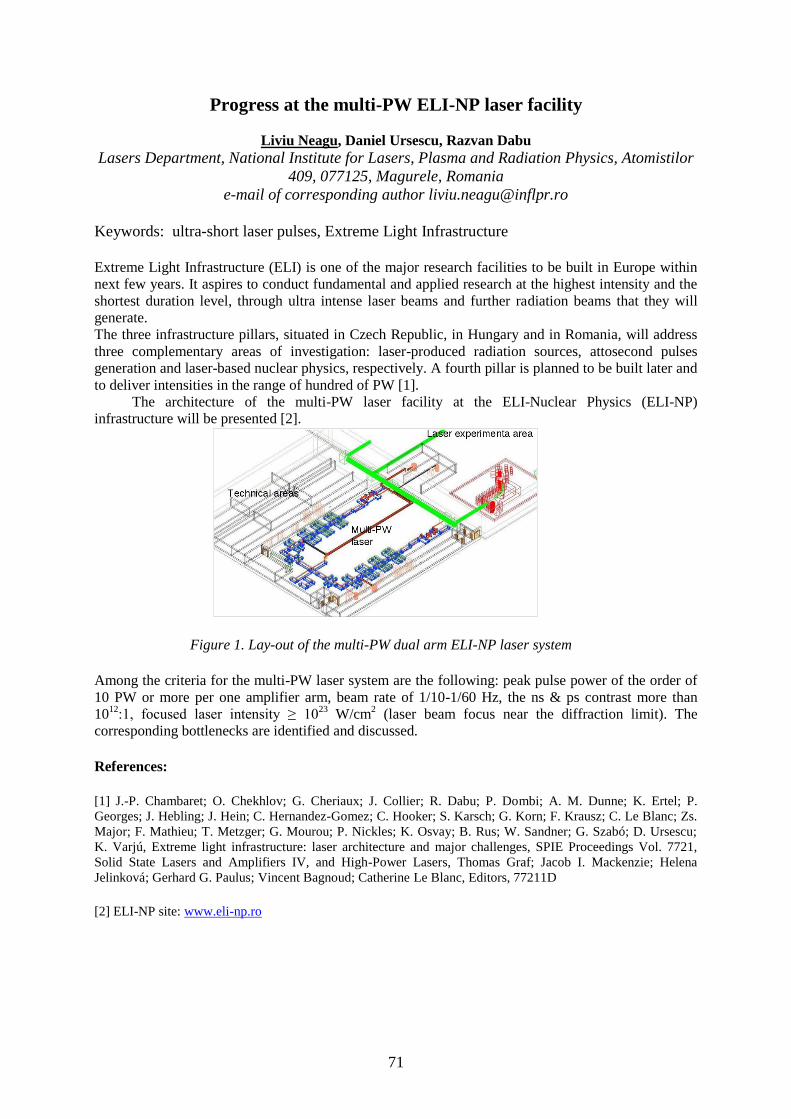
71
Progress at the multi-PW ELI-NP laser facility
Liviu Neagu, Daniel Ursescu, Razvan Dabu
Lasers Department, National Institute for Lasers, Plasma and Radiation Physics, Atomistilor
409, 077125, Magurele, Romania
e-mail of corresponding author [email protected]
Keywords: ultra-short laser pulses, Extreme Light Infrastructure
Extreme Light Infrastructure (ELI) is one of the major research facilities to be built in Europe within
next few years. It aspires to conduct fundamental and applied research at the highest intensity and the
shortest duration level, through ultra intense laser beams and further radiation beams that they will
generate.
The three infrastructure pillars, situated in Czech Republic, in Hungary and in Romania, will address
three complementary areas of investigation: laser-produced radiation sources, attosecond pulses
generation and laser-based nuclear physics, respectively. A fourth pillar is planned to be built later and
to deliver intensities in the range of hundred of PW [1].
The architecture of the multi-PW laser facility at the ELI-Nuclear Physics (ELI-NP)
infrastructure will be presented [2].
Figure 1. Lay-out of the multi-PW dual arm ELI-NP laser system
Among the criteria for the multi-PW laser system are the following: peak pulse power of the order of
10 PW or more per one amplifier arm, beam rate of 1/10-1/60 Hz, the ns & ps contrast more than
1012
:1, focused laser intensity ≥ 1023
W/cm2 (laser beam focus near the diffraction limit). The
corresponding bottlenecks are identified and discussed.
References:
[1] J.-P. Chambaret; O. Chekhlov; G. Cheriaux; J. Collier; R. Dabu; P. Dombi; A. M. Dunne; K. Ertel; P.
Georges; J. Hebling; J. Hein; C. Hernandez-Gomez; C. Hooker; S. Karsch; G. Korn; F. Krausz; C. Le Blanc; Zs.
Major; F. Mathieu; T. Metzger; G. Mourou; P. Nickles; K. Osvay; B. Rus; W. Sandner; G. Szabó; D. Ursescu;
K. Varjú, Extreme light infrastructure: laser architecture and major challenges, SPIE Proceedings Vol. 7721,
Solid State Lasers and Amplifiers IV, and High-Power Lasers, Thomas Graf; Jacob I. Mackenzie; Helena
Jelinková; Gerhard G. Paulus; Vincent Bagnoud; Catherine Le Blanc, Editors, 77211D
[2] ELI-NP site: www.eli-np.ro

72
Study of high harmonics generation at the interaction of an ultrashort and
intense laser pulse with an overdense plasma layer
A. Mihailescu, V. Stancalie, V.F. Pais
National Institute for Laser, Plasma and Radiation Physics, P.O.Box MG-36, Magurele,
077125 ROMANIA, Association EURATOM MEdC
Ultrashort and intense laser pulses with durations close to an optical cycle in the infrared and visible
domains have proven to be well suited in experiments looking for non-linear effects such as high
harmonics generation. Focused intensities of about 217 /10 cmW from a Ti-Sapphire terawatt laser
( m 8.00 and pulse durations around 100-150 fs) allowed the experimental observation of
harmonics up to the 15th order [1, 2]. Using a Nd: glass laser system ( m 05.10 , 2.5 ps pulses)
reaching an intensity of 219 /10 cmW in the relativistic regime, Norreys et al.[3] have detected
harmonics as far as the 68th order.
In this work the interaction of a short and intense laser beam (2219172 1010 mWcmI ) with an
overdense plasma layer is simulated with the purpose of studying the high harmonics generation
mechanism and its efficiency. A 1D3V relativistic particle in cell (PIC) code is employed. Charge
densities, currents and the electromagnetic field are therefore defined on a 1D grid and are updated
after each particle push. Various pulse shapes with different durations– normal and oblique incidence-
have been chosen while the intensity and the polarization of the laser beam were varied. Influences of
the plasma density and of the initial electron temperature have also been taken under consideration.
Numerical errors have been quantified by modifying the size of the simulation box, the time steps per
laser cycle and the number of macroparticles per cell.
Harmonics emission has been found to increase with the intensity of the laser and also when lowering
the plasma density. Electron density profiles as well as harmonic spectra are shown and the harmonics
emission efficiency is discussed.
References
[1] S. Kohlweyer, G.D. Tsakiris, C.G. Wahlstrom, C. Tillman and I. Mercer, Opt. Commun. 117, 431 (1995).
[2] D. von der Linde, T.Engers, G. Jenke, P. Agostini, G. Grillon, E. NIbbering, A. Mysyrowitz and A.
Antonetti, Phys. Rev. A 52, R 25 (1995).
[3] P. A. Norreys, M. Zepf, S. Moustaizis, A.P. Fews, J. Zhang, P.Lee, M. Bakarezos, C. N. Danson, A. Dyson,
P. Gibbon, P. Loukakos, D. Neely, F. N. Walsh, J.S. Wark and A. E. Dangor, Phys Rev. Lett. 76, 1832 (1996).

73
Operation of Mo XRL in ultra-low pumping regime
G. Cojocaru1, R. Banici
1, R. Ungureanu
1, R. Dabu
1, H. Stiel
2, D. Ursescu
1,*
1National Institut for Lasers, Plasma and Radiation Physics,
409 Atomistilor St., PO Box MG-36, 077125 Bucharest, Romania 2Max Born Institut, Berlin, Germany
With the advancement of surface and material sciences, and for the development of unique imaging
and metrology tools, there is a major interest in the development of coherent, shorter wavelength
sources capable of producing high average power irradiation. Plasma-based x-ray lasers are such
sources, that aim to complement large scale facilities such as synchrotrons.
First plasma X-ray laser emissions were obtained with high pumped energy in the domain of kJoules
[1]. Later, after 1995, femtosecond laser pulses have brought tremendous improvements in obtaining
of soft x-ray laser emission [2,3]. Pumping of the 18.9 nm line of Ni-like Mo at 14 degrees grazing
incidence with 150 mJ-300mJ of total pumping energy from a 10 Hz laser was reported [4].
TEWALAS laser facility from National Institute of Lasers Plasma and Radiation Physics provides
ultrashort laser pulses with central wavelength at 800 nm and 10 Hz repetition rate with an energy up
to 460mJ per pulse. It offered us the possibility to implement the grazing incidence pumping scheme
for XRL which demands a normal incidence long pulse (hundreds of ps duration) and a grazing
incidence short pulse (few ps duration).
After careful optimization, a Mo XRL at 18.9 nm was demonstrated at 10 Hz repetition rate. The
minimum pump energy during for obtaining lasing during our investigation was 85 mJ, almost a factor
of two less than previously reported [4]. Our results, together with the advances in high repetition rate
pump lasers with pulse energies in the 0.1J-1J range, can bring XRL to at least one order of magnitude
higher repetition rate (100Hz-1 kHz).
Acknowledgements: The studies were carried out during my work in solid state laser laboratory using financial
support from project PN 09 39-LAPLAS 3/2099
References
[1] H. Daido, Rep. Prog. Phys. 65, 1513¨C1576
, (2002)
[2] P. V. Nickles et al., Phys. Rev. Lett. 78, 2748¨C2751 (1997)
[3] V. N. Shlyaptsev, J. Dunn, S. Moon, R. Smith, R. Keenan, J. Nilsen, K. B. Fournier, J. Kuba, A. L.
Osterheld, J. J. G. Rocca, B. M. Luther, Y. Wang, and M. C. Marconi, in Soft X-Ray Lasers and Applications V,
Vol. 5197, edited by E. E. F. S. Suckewer (SPIE, 2003) pp. 221-228.
[4] R. Keenan, J. Dunn, P. K. Patel, D. F. Price, R. F. Smith, and V. N. Shlyaptsev, Physical Review Letters 94,
103901 (2005).

74
Simulation of spatio-temporal distortions in ultra-short laser pulses
R. Ungureanu1, D. Ursescu
1,*
1National Institut for Lasers, Plasma and Radiation Physics,
409 Atomistilor St., PO Box MG-36, 077125 Bucharest, Ronmania
The space and time dependence of an ultrashort pulse's electric field are often assumed to be separable
into independent functions. This assumption fail when coupling occurs between the pulse electric
field's space and time dependence, and this is referred to as a spatio-temporal distortion. They are
common in ultrafast optics because the generation, amplification, and manipulation of ultrashort
pulses all involve the deliberate introduction of such spatio-temporal distortions.[1]
Evaluation of the spatio-temporal distortion of ultra short pulses is an active field of research. First-
order beam propagation for ultra short pulses is usually described in a matrix formalism, known as
Kostenbauder matrix formalism (KMF), which is an extension of the ABCD ray matrix formalism.
To study laser pulse distortions, today it is very common to use KMF which take in consideration four
parameters in the description of a ray of light: space, angle time and frequency. The formalism
assumes that optical systems are represented by 4x4 matrices which fulfill the symplecticity property
and rays are represented by ray vectors with four elements. [2]
We present a friendly and also powerful tool for studying spatio-temporal couplings in ultrashort laser
pulses based on (KMF). A direct benefit is the symbolic power of computation offered by
Mathematica. This instrument, which is a package of functions in Wolfram Mathematica language
aims to provide support for characterizing distortions
introduced by optical systems to ultrashort laser pulses.
The package contains object functions which describe
the basic optical systems such as lens, prism, grating,
free space propagation and a general system. Each object
contains the 4x4 the ray transfer matrix, the
symplecticity conditions and the type of the
representation space. The symplecticity property of the
4x4 matrix comes from the energy conservation law and
is specific to the representation space assumed.
In order to analyze distortions, the program has built-in
functions for processing and analyzing, as shown in the
figure.
One can obtain the transfer matrix for optical systems by
composing the matrices of each basic element. For
instance, the matrix for a prisms based optical
compressor can be generated and then analysed, in order to obtain the ray transfer matrix and to study
further distortions of the system.
Acknowledgements : The studies were carried out during my work in solid state laser laboratory using financial
support from project PN 09 39-LAPLAS 3/2099 and project 37N
References
[1] S.Akturk, X.Gu, E.Zeek, R.Trebino, Pulse-front tilt caused by spatial and temporal chirp, Opt. Express 2004
[2] ELI White book, pp. 238 - 240 2011.
[3] Miguel Augusto Vieira Fernandes, Analysis of spatial-temporal distortions in chirped pulse Msc Thesis
Portugal, 2009

75
Project LASERLAB EUROPE
The Integrated Initiative of European Laser Research Infrastructure
Traian DASCALU
1
1National Institute for lasers, Plasma and Radiation Physics, Atomistilor Street,
No. 409, Magurele, Romania
LASERLAB-EUROPE is in the third phase of its successful cooperation in new shape: the
Consortium has become much larger, involving 26 Laser Research Infrastructures from 16 European
member states. Over 20 facilities offer access to their labs for European research teams. Given the
importance of lasers and their applications in all areas of sciences, technologies and life sciences, the
Consortium set up the main objectives:
To form a competitive, inter-disciplinary network of European national laser laboratories;
To strengthen the European leading role in laser research through Joint Research Activities
(JRA), pushing the laser concept into new directions and opening up new applications of key
importance;
To engage in the Transnational Access Programme in a co-ordinated fashion for the benefit of
the European research community.
To increase the European basis in laser research and applications by reaching out to
neighboring scientific communities and by assisting the development of Laser Research
Infrastructures on both the national and the European level.
We will present the role of LaserLAB III in Romania and Europe, the scientific objectives,
Joint Research Activities, possibilities for Networking.
76
Session 7
Physics of Plasma
77
Dusty Plasmas: a Review of Experiments and Possible Applications
C. M. Ticoş1,2
, O. Sandu2, N. Banu
2, , M.L. Munteanu
2, A. Scurtu
2
1National Institute for Research and Development in Microtechnologies,
077190 Bucharest, Romania 2National Institute for Laser, Plasma and Radiation Physics,
077125 Bucharest,Romania
Dusty plasmas are often called complex plasmas due do their intricate structure: a collection of
charged ions or molecules, electrons, fields and electrically charged particles of matter. This last
component differentiates dusty plasmas from all other types of plasmas and confers them some unique
properties. The recorded trajectories of dust particles moving in plasma can provide direct information
about the physical processes involving dust-plasma and dust-dust interactions. A review of some
experiments and their imaging techniques employed in tracking dust present in low ionized gases and
in high density plasma jets is presented. Digital recording cameras are used to track the motion of slow
or hypervelocity dust particles dragged by plasma jets, or to evidence single or collective dust particles
oscillations and vibrations in the plasma sheath. When the charged dust particles are strongly coupled
electrostatically, self-organization of the particles into 3-D periodic structures called ‘plasma crystals’
takes place. A possible application based on scattering of THz waves by a plasma crystal is discussed.
The wavelength of the electromagnetic waves is in the range of frequencies between 0.5 to a few THz
and corresponds roughly to the interparticle separation distance. It has been suggested recently that a
plasma crystal made of micron size dust particles can potentially work as a tunable filter in the
terahertz (THz) domain.
78
The Template for the ISCP 2012 Summary
D. Gustaw1
1Student at the Nicolaus Copernicus University in Torun. Faculty of Physics, Astronomy and
Informatics. Poland, 87-100 Torun Grudziadzka 5
e-mail [email protected]
The optical fiber is applied in optoelectronics systems, as well as in integrated optics systems and
communication. It can also be used to investigate gentle quantum efects such as quantum force or
Bohm potential. Equations describing classical light in optical fiber (Helmholtz equations) are very
similar to Schrödinger time-independent equation. Some analogical effect can be observed for massive
particle in constrained cavity and for light in the optical fiber. Interesting result appears when the
trajectory is curved.
The speach presents solutions of the Helmholtz equations in cylindrical coordinate system on the
example of a rectangular-shaped curved optical fiber. To simplify the problem, an assumption is made
that the solutions of the equations approach to zero on the bounds of the fiber.
The most important point of the presentation seems to be apperance of the non-integer order of Bessel
function. This result is significantly different than the one in cylindrical fiber. The difference will be
considered in the context of angular momentum, Bohm potential and quantum force. There will be
presented deep analogy not only between massive particle and classic light, but also between
Shrodinger equation and Helmholtz equation.
In my consideration the first step is taken to find general solutions of the Helmholtz equtions in
cylindrical coordinate system. Next part is dedicated to bound conditions. In the end, there are
traversed properties of the solutions and interpretations of the parameters. As it will be shown,
rectangular-shaped curved optical fiber is an interesting example, relatively simple in calculations.
References [1] Jan Petykiewicz - Optyka falowa,(wave optics), PWN, Warszawa 1986
[2] C.C. Gerry, P.L. Knight - Wstęp do optyki kwantowej,(introduction to quantum optics), PWN, Warszawa
2007
[3] Ramamurti Shankar - Mechanika kwantowa,(quantum mechanics), PWN, Warszawa 2007

79
Dose distributions in water-equivalent materials irradiated with hadron
beams
C.I. Chirvase1
1Faculty of Physics, “Al. I. Cuza” University
The purpose of this paper is to analyze the dosimetric properties of some common tissue-equivalent
materials, by simulating the interactions of charged particles beams with phantoms of different
materials.
Application of proton therapy in medical practice requires a special equipment to simulate a human
body, organs and tissues as well as the reference medium, water. The proton interaction cross sections
of the phantom materials are to be close as possible to those of respective biological tissues. This
problem is well known in conventional radiation therapy by photon and electron beams. Numerous
tissue and water equivalent materials were developed for radiation therapy in the last decades and high
accuracy in the interaction cross sections simulation was achieved for modern substitutes. In
particular, water and tissue equivalent plastics provided by ICRU are close to liquid water and
respective tissues within 0.5-1.0% for photon and electron beams. Nevertheless, the applicability of
these materials in proton beams is to be specially verified.
Using beams of hadrons - protons or light ions - radiotherapists can overcome the limitations of
photon beams. As well as depositing most of their energy at the end of their range, hadrons penetrate
the patient with practically no diffusion. This means that they can bring about severe damage to the
DNA in cancer cells while sparing both traversed and deeper healthy tissue. This characteristic also
enables them to be used to accurately irradiate any part of the tumor. Hadron beams allow highly
conformal treatment (in which the beam conforms to the shape of the tumor) of deep-seated tumors to
the nearest millimeter, while delivering minimal doses to surrounding tissues.
In this work, we have performed a theoretical evaluation of tissue and water equivalence of seven
phantom materials originally developed for conventional radiation therapy. The simulations were
made using Fluka software. Three types of charged particles beam were used: electron beams (at 10,
20 and 40 MeV), proton beams (at 70, 100 and 130 MeV) and carbon ion beams (at 200, 250 and 300
MeV). The materials used for building the phantoms were: PMMA, Polystyrene, Solid water RMI-457,
Plastic water (all of these being water-equivalent materials), as well as A-150 (soft tissue-equivalent
plastic) and B100 (bone-equivalent plastic).The resulting data was then plotted as depth dose
distributions, comparing the depth dose distributions of these materials with the depth dose
distributions of water, soft tissue and bone, respectively.

80
Preparation and characterization of double perovskite targets for thin films
deposition by PLD method
*Robert Lowndes
National Institute of Materials Physics, P.O. Box MG-7, Bucharest-Magurele,
077125, Romania
*Corresponding author: [email protected]
A multiferroic material possesses two or more ferroic orders from ferroelectricity, ferromagnetism or
ferroelasticity within a single phase. Such materials find application in data storage devices and
resonant circuits for mobile telephone communication systems. There are challenges that are yet to be
overcome; single phase multiferroic materials are rare and the magnetoelecctric coupling effect is
often weak. Pulsed laser deposition is a frequently used technique to fabricate multiferroics double
perovskites in thin film form with epitaxy of the films a key requirement. Multiferroic double
perovskites based on Bi2CoMnO6, Sr2FeMoO6 and Bi2CrFeO6 were prepared by sol-gel and solid-state
reaction methods. The aim of this study is the optimization of the processing parameters to obtain high
quality targets for the preparation of epitaxial thin films. Pellets were heat treated by conventional
methods and spark plasma sintering. The phase development and microstructure of the powders and
sintered pellets were investigated by SEM, EDAX, XRD and TEM. The Bi-based compositions were
found to have multiple phases present in contrast to Sr2FeMoO6 which was found to be a single
perovskite phase. SEM analysis revealed the presence of equiaxed grains with sizes typically less than
5µm.

81
Participants list
Antohe Stefan [email protected] Romania
Chalus Olivier France
Dinescu Maria [email protected] Romania
Dumitras Dan [email protected] Romania
Burnet Frank [email protected] United Kingdom
Gheorhe Lucian [email protected] Romania
Grigoriu Constantin [email protected] Romania
Luculescu Catalin [email protected] Romania
Pascu Mihai Lucian [email protected] Romania
Pavel Nicolaie [email protected] Romania
Scarisoreanu Nicu [email protected] Romania
Staicu Angela [email protected] Romania
Traian Dascalu [email protected] Romania
Zamfirescu Marian [email protected] Romania
1. Achim Alexandru [email protected] Romania 2. Achim Cristina [email protected] Romania 3. Albu Catalina [email protected] Romania 4. Aleksandrov Veselin [email protected] Bulgaria 5. Alexandru Tatiana [email protected] Romania 6. Ana Magureanu [email protected] Romania 7. Anghel Iulia [email protected] Romania 8. Avram Ioana [email protected] Romania 9. Banici Romeo [email protected] Romania 10. Banita Stefan [email protected] Romania 11. Banu Nicoleta [email protected] Romania 12. Becherescu Nicu Romania 13. Berrang Bianca [email protected] Germany 14. Boni Mihai [email protected] Romania 15. Bratu Ana [email protected] Romania 16. Carp Octavian [email protected] Romania 17. Chiricuta Bogdan Romania 18. Chirvase Cezarina-Isabela [email protected]
m
Romania
19. Cojocaru Gabriel [email protected] Romania 20. Matei Consuela [email protected] Romania 21. Dorigo Daniel [email protected] Germany 22. Gazazyan Emil [email protected] Armenia 23. Gustaw Daniel [email protected] Poland 24. Heßke Andre [email protected] Germany 25. Ibanescu Andra Georgia [email protected] Romania 26. Ion Valentin [email protected] Romania 27. Ionel Laura [email protected] Romania 28. Ionescu Alina [email protected] Romania 29. Jipa Florin [email protected] Romania 30. Lowndes Robert [email protected] United Kingdom 31. Matei Andreea
[email protected] Romania

82
32. Mihailescu Andreea [email protected] Romania 33. Monti Gianni [email protected] Germany 34. Munteanu Maria-Luiza [email protected] Romania 35. Mursa Paula Florina [email protected] Romania 36. Nadejde Claudia [email protected] Romania 37. Nastasa Viorel [email protected] Romania 38. Neagu Liviu [email protected] Romania 39. Nedelcea Anca [email protected] Romania 40. Nicolae Danila Mihail [email protected] Romania 41. Oreshkov Bozhidar [email protected] Romania 42. Patachia Mihai [email protected] Romania 43. Petrus Mioara [email protected] Romania 44. Raducanu Vlad Stefan [email protected] Romania 45. Relu Ionut Andrei [email protected] Romania 46. Rusen Laurentiu [email protected] Romania 47. Salamu Gabriela [email protected] Romania 48. Sava Vasile [email protected] Romania 49. Simion Sandel [email protected] Romania 50. Smarandache Adriana adriana.smarandache@inflpr.
ro
Romania
51. Sold Camelia Daniela [email protected] Romania 52. Stanciu George [email protected] Romania 53. Swierad Dariusz [email protected] Poland 54. Unga Florin [email protected] Romania 55. Ungureanu Razvan
Georgian
[email protected] Romania
56. Varasteanu Pericle Romania 57. Voicu Flavius [email protected] Romania 58. Wiesent Benjamin [email protected] Germany 59. Zorila Alexandru [email protected] Romania