BI-RADS By Nina Zahedi MDBy Nina Zahedi MD. Why BI-RADS?
61
BI-RADS By Nina Zahedi MD
-
Upload
bridget-hart -
Category
Documents
-
view
235 -
download
3
Transcript of BI-RADS By Nina Zahedi MDBy Nina Zahedi MD. Why BI-RADS?

BI-RADS
By Nina Zahedi MD

Why BI-RADS?

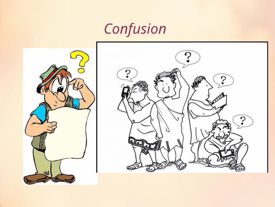
Confusion

If I report she is really sick what happens if she is not?
If I report normal what happens if she is really sick?

Consultation

And now
Baby of 1997 !

Breast Imaging Reporting and Data System (BI-RADS).
• Having a standard way of reporting mammogram results , meaning:
• Lets doctors use the same words and terms for describing the findings ,
• Reduce confusion in breast imaging interpretations,
• and Facilitate outcome monitoring.
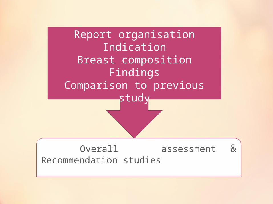
Overall assessment & Recommendation studies
Report organisationIndication
Breast compositionFindings
Comparison to previous study
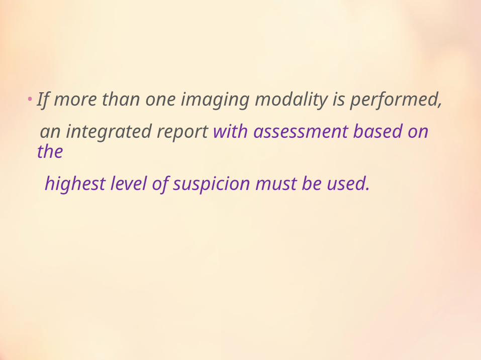
• If more than one imaging modality is performed,
an integrated report with assessment based on the
highest level of suspicion must be used.

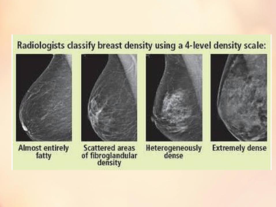
Mammographic Breast Composition
The breast is almost entirely fat(<25% FGT)
Scattered fibroglandular densities (25-50%)
Heterogeneously dense breast tissue(51-75%)
Extremely dense (> 75% glandular)


BIRADS Lexicon
• Mass
• Architectural distortion
• Asymmetry
• Calcification

BI-RADS 1
Negative:
There is nothing to comment on.
( either abnormal or normal)

BI-RADS 2 :Benign Lesion

Benign Masses can be Ignored• a-Raised skin lesions
Seborrheic keratosis
• b-Intramammary lymph nodes
• c-Fat containing lesions(Encapsulated lucent lesions) Lipomas Fat necrosis forming oil cysts Galactoceles
• c-Mixed-density lesions Hamartomas, Hematomas
• d-Multiple rounded densities
• e-Benign calcified masses Calcifying involuting fibroadenomas
• f-Benign masses with peripheral calcifications Calcifying involuting fibroadenomas Cysts with calcified walls Fat necrosis
• g-Calcifying large duct papillomas
• h-Cysts with precipitated calcium
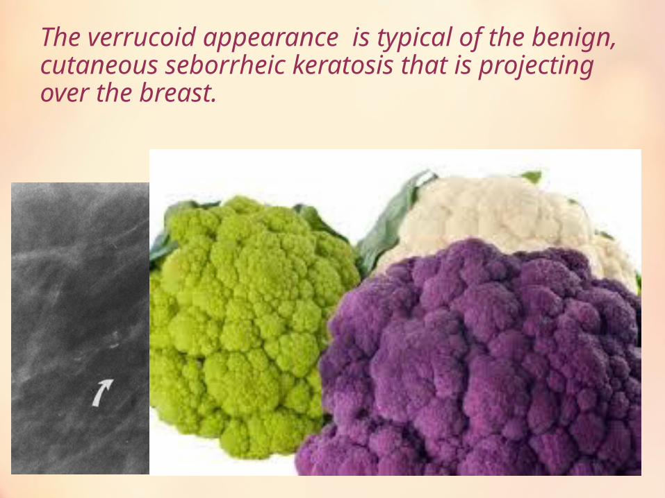
The verrucoid appearance is typical of the benign, cutaneous seborrheic keratosis that is projecting over the breast.
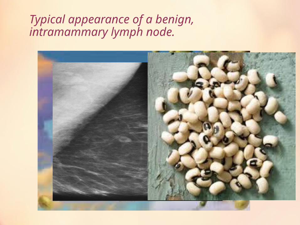
Typical appearance of a benign, intramammary lymph node.
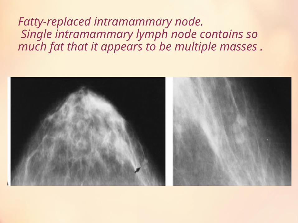
Fatty-replaced intramammary node. Single intramammary lymph node contains so much fat that it appears to be multiple masses .
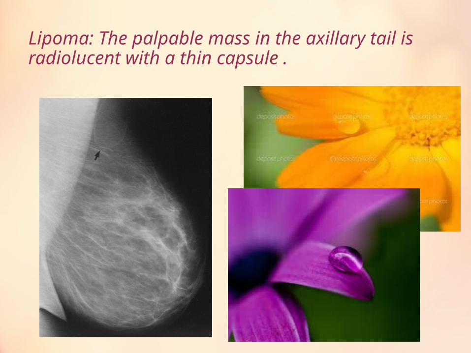
Lipoma: The palpable mass in the axillary tail is radiolucent with a thin capsule .
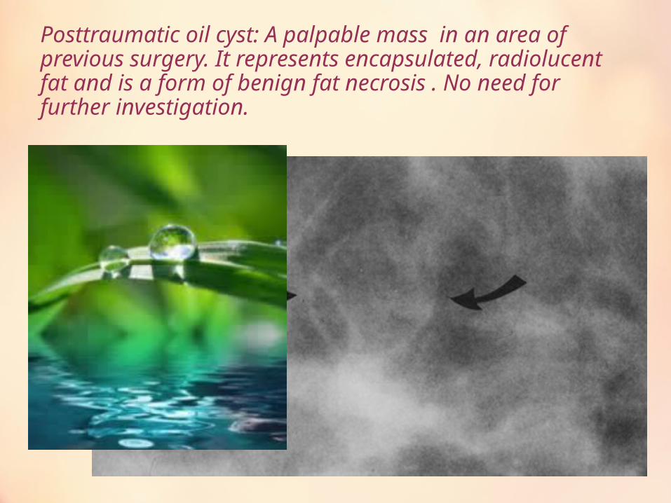
Posttraumatic oil cyst: A palpable mass in an area of previous surgery. It represents encapsulated, radiolucent fat and is a form of benign fat necrosis . No need for further investigation.
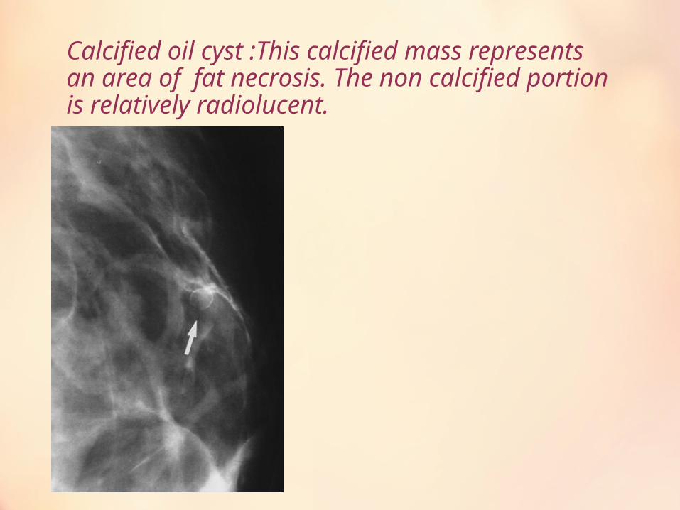
Calcified oil cyst :This calcified mass represents an area of fat necrosis. The non calcified portion is relatively radiolucent.
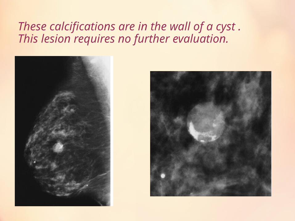
These calcifications are in the wall of a cyst .This lesion requires no further evaluation.
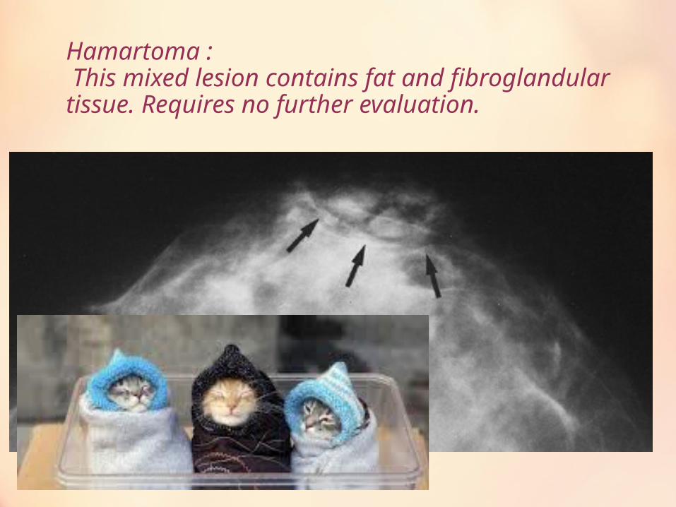
Hamartoma : This mixed lesion contains fat and fibroglandular tissue. Requires no further evaluation.
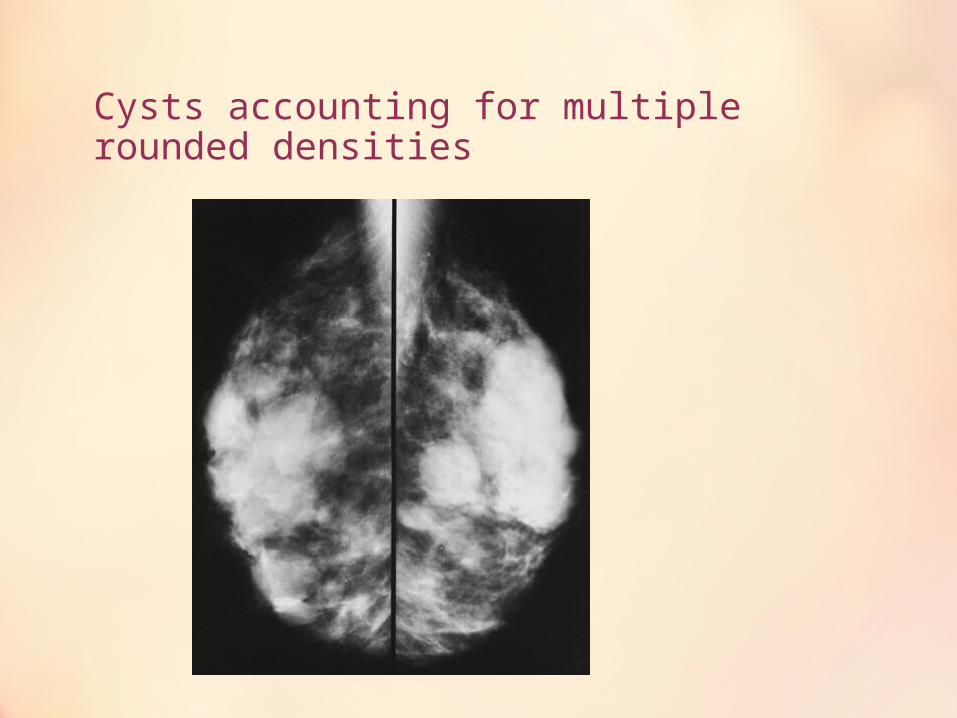
Cysts accounting for multiple rounded densities
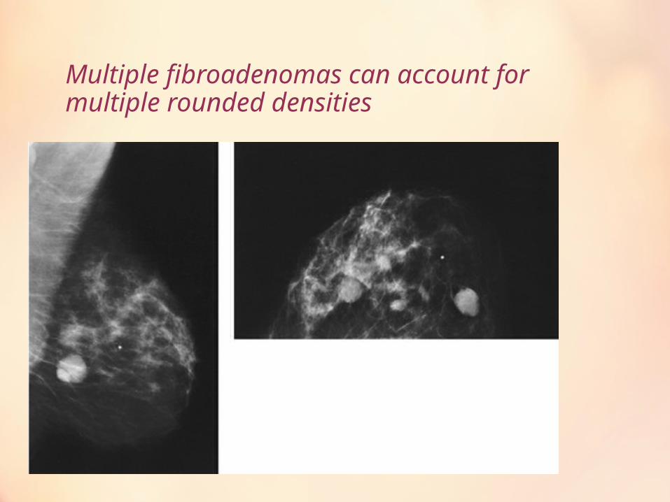
Multiple fibroadenomas can account for multiple rounded densities
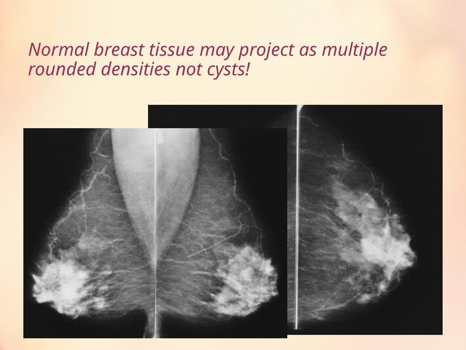
Normal breast tissue may project as multiple rounded densities not cysts!
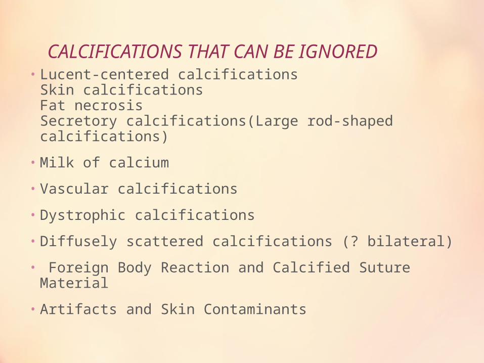
CALCIFICATIONS THAT CAN BE IGNORED• Lucent-centered calcifications
Skin calcificationsFat necrosisSecretory calcifications(Large rod-shaped calcifications)
• Milk of calcium
• Vascular calcifications
• Dystrophic calcifications
• Diffusely scattered calcifications (? bilateral)
• Foreign Body Reaction and Calcified Suture Material
• Artifacts and Skin Contaminants
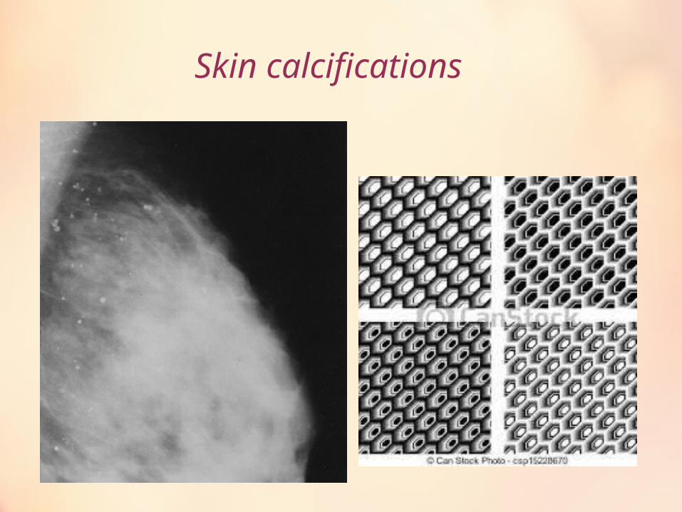
Skin calcifications
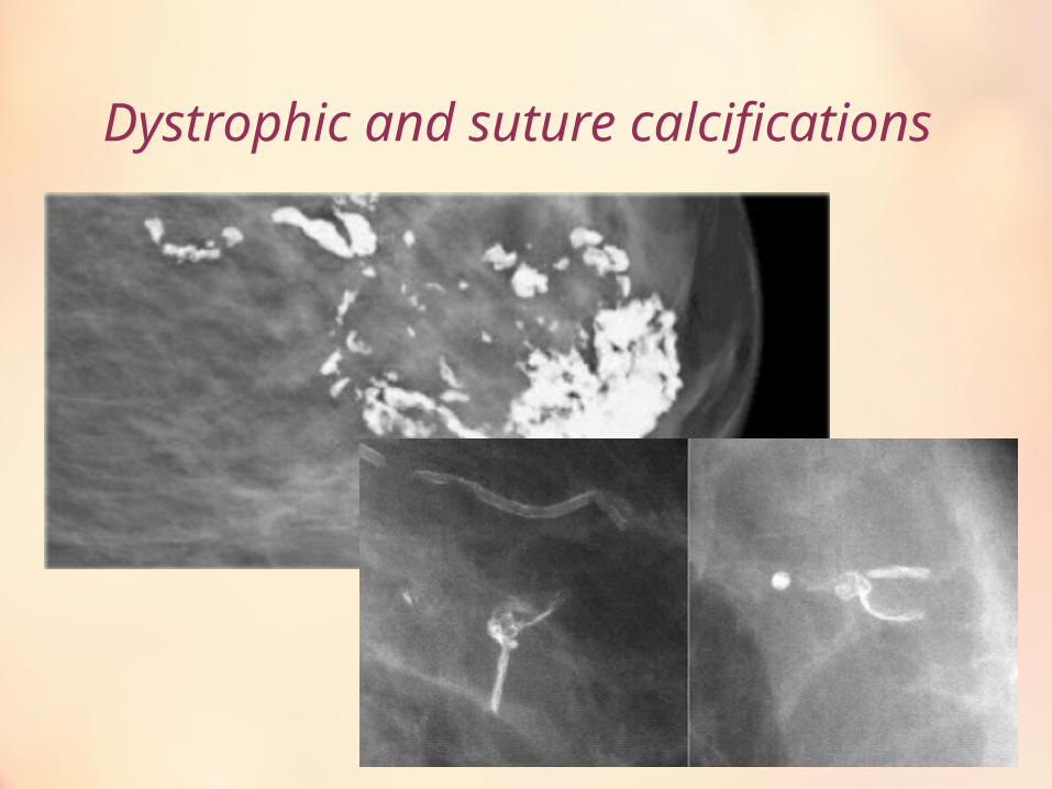
Dystrophic and suture calcifications
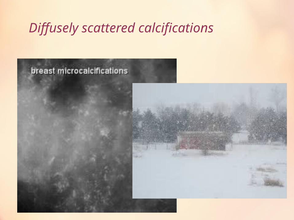
Diffusely scattered calcifications
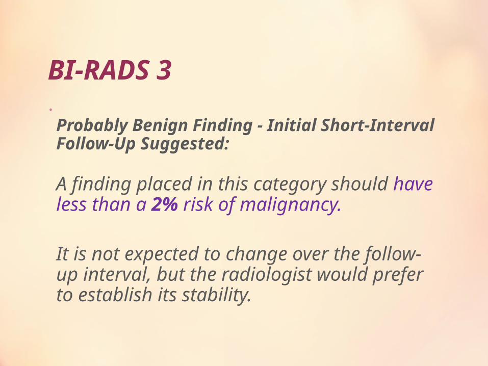
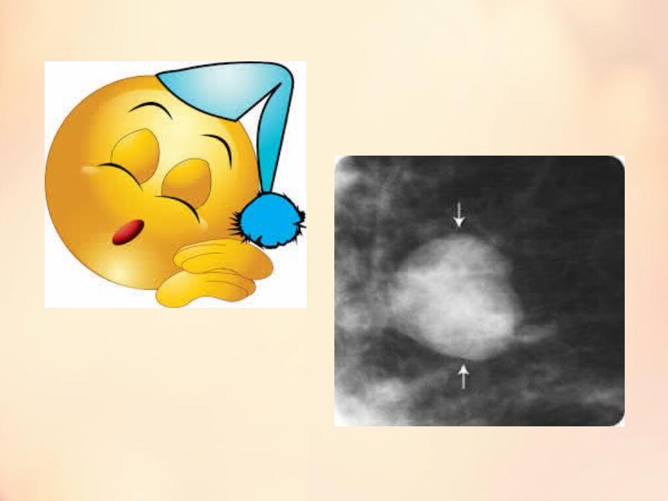
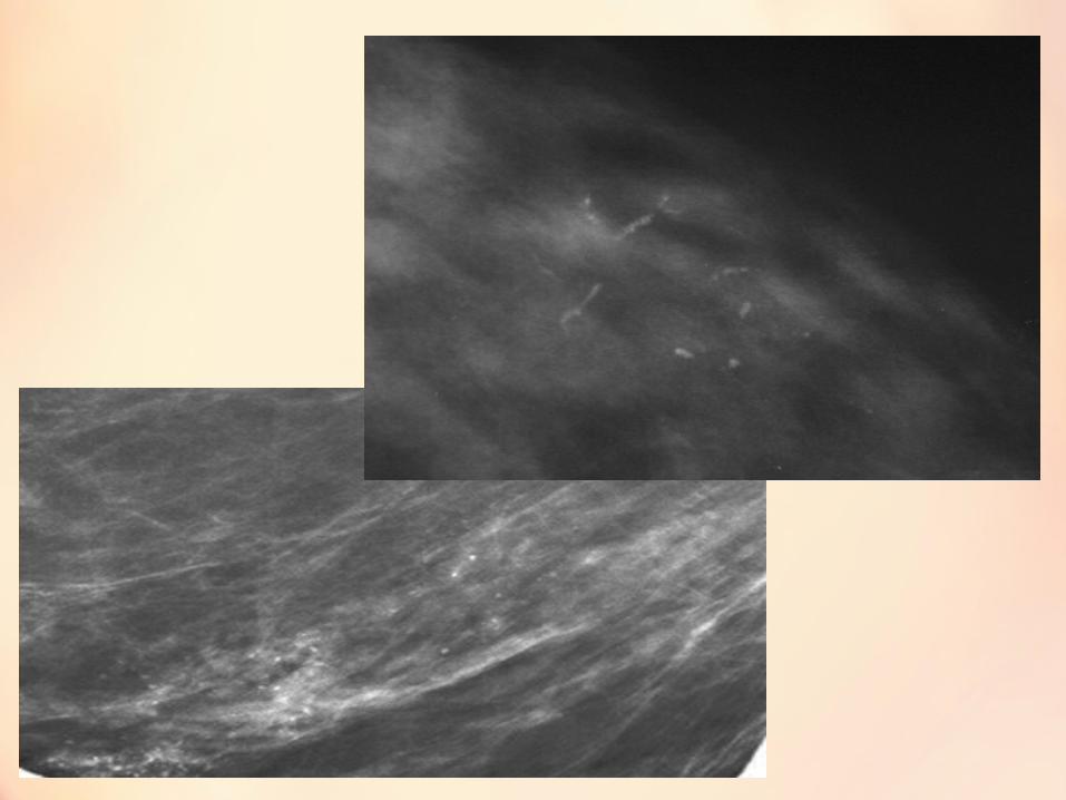
BI-RADS 3•
Probably Benign Finding - Initial Short-Interval Follow-Up Suggested:
A finding placed in this category should have less than a 2% risk of malignancy.
It is not expected to change over the follow-up interval, but the radiologist would prefer to establish its stability.
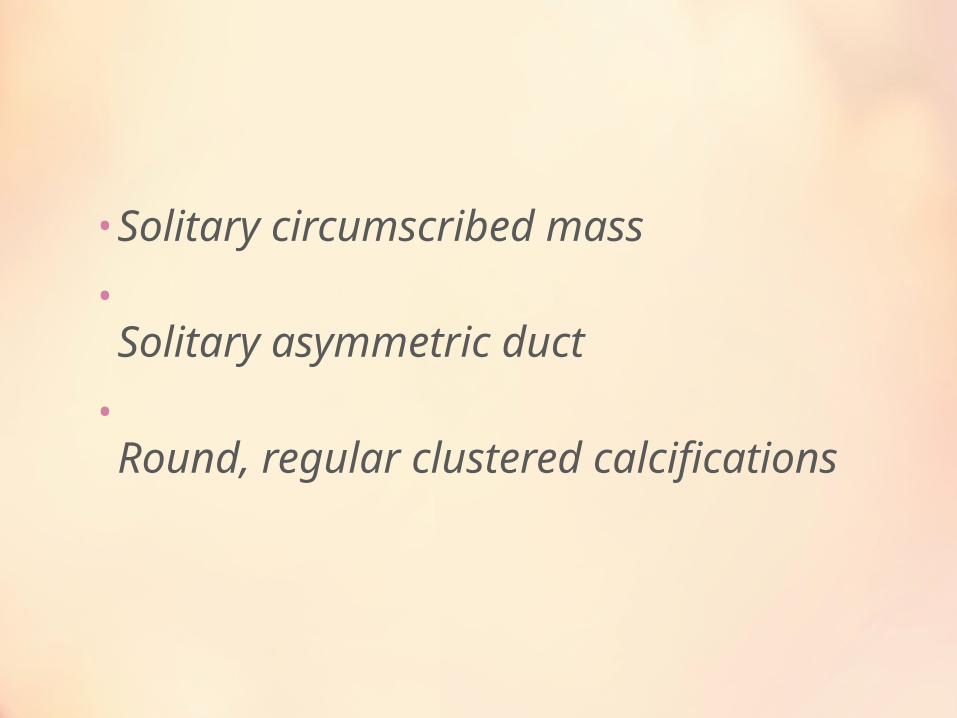
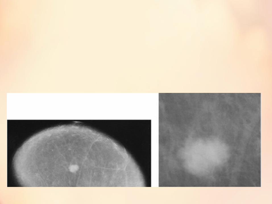
• Solitary circumscribed mass
•Solitary asymmetric duct
•Round, regular clustered calcifications
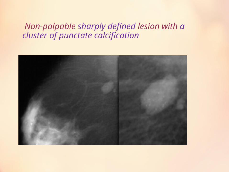
Non-palpable sharply defined lesion with a cluster of punctate calcification
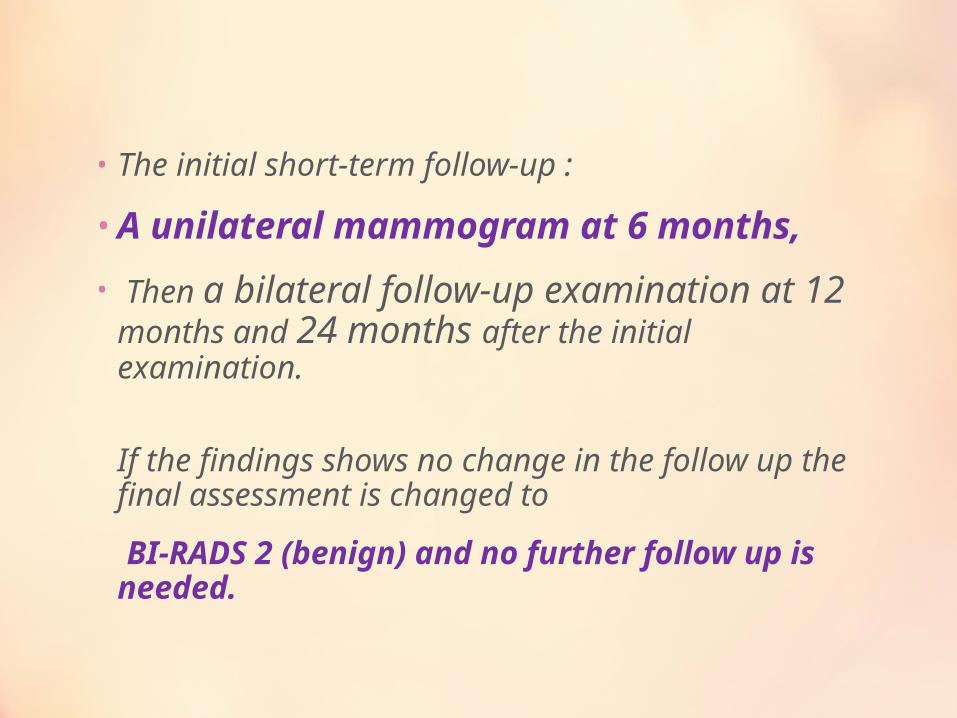
• The initial short-term follow-up :
• A unilateral mammogram at 6 months,
• Then a bilateral follow-up examination at 12 months and 24 months after the initial examination.
If the findings shows no change in the follow up the final assessment is changed to
BI-RADS 2 (benign) and no further follow up is needed.
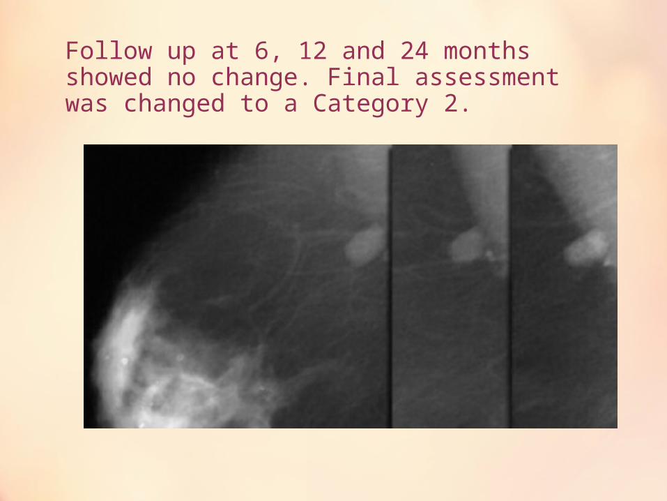
Follow up at 6, 12 and 24 months showed no change. Final assessment was changed to a Category 2.
• If a BI-RADS 3 lesion shows any change during follow up,
• It will change into a BI-RADS 4 or 5 and appropriate action should be taken.
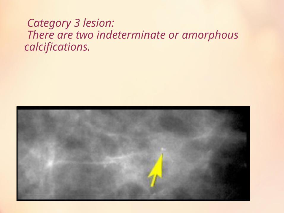
Category 3 lesion: There are two indeterminate or amorphous calcifications.
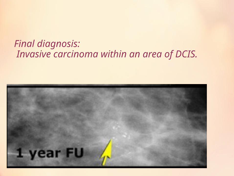
Final diagnosis: Invasive carcinoma within an area of DCIS.
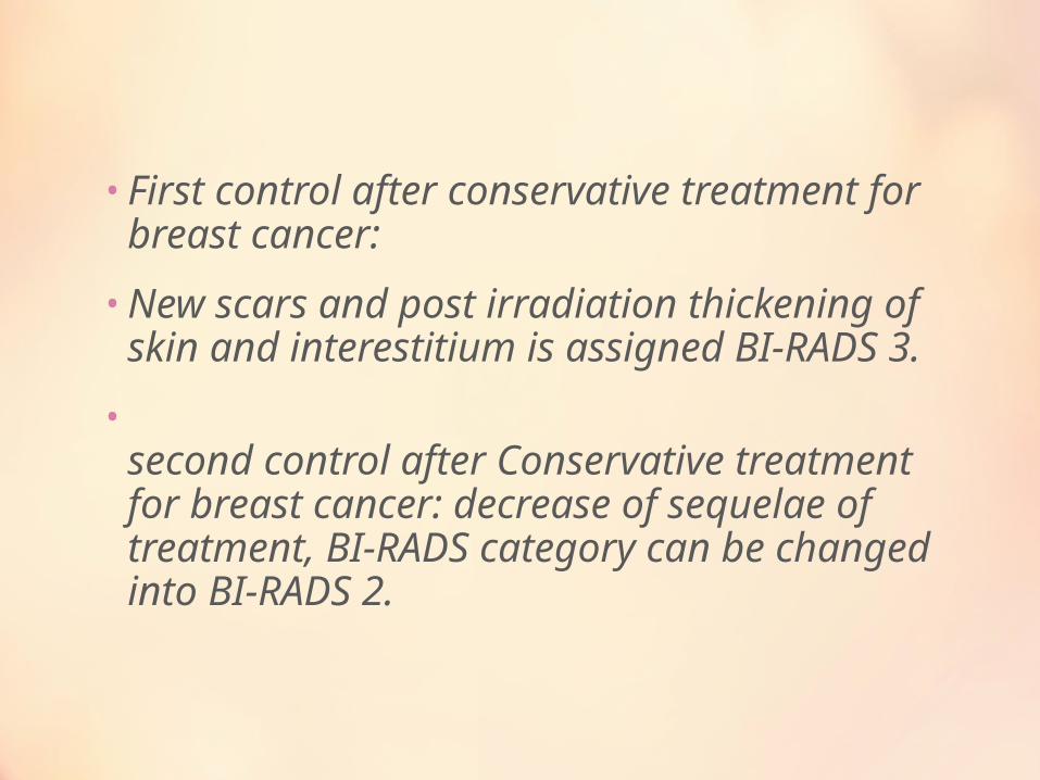
• First control after conservative treatment for breast cancer:
• New scars and post irradiation thickening of skin and interestitium is assigned BI-RADS 3.
•second control after Conservative treatment for breast cancer: decrease of sequelae of treatment, BI-RADS category can be changed into BI-RADS 2.
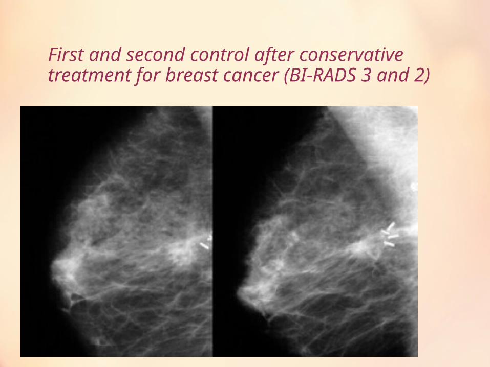
First and second control after conservative treatment for breast cancer (BI-RADS 3 and 2)
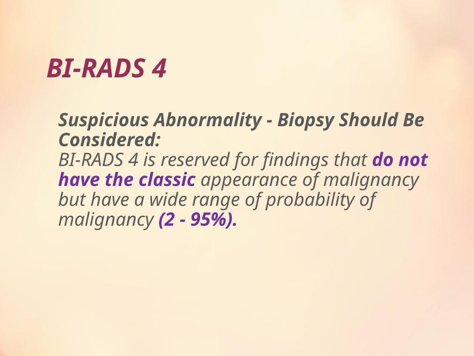
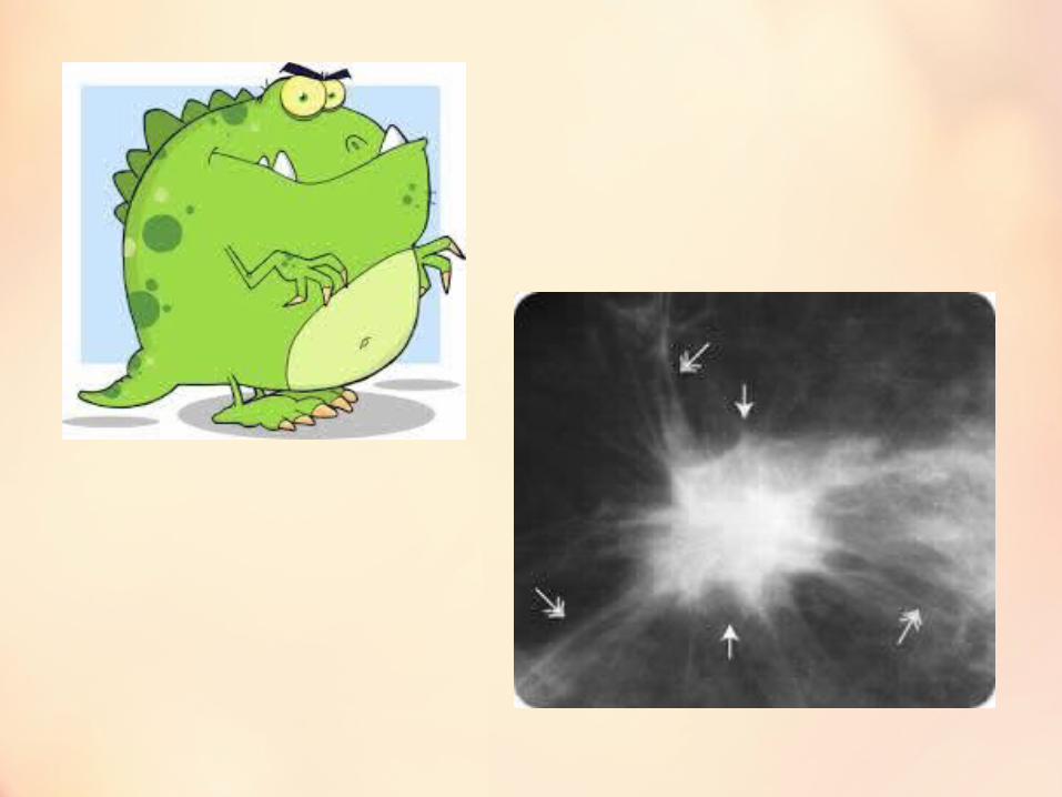
BI-RADS 4
Suspicious Abnormality - Biopsy Should Be Considered:BI-RADS 4 is reserved for findings that do not have the classic appearance of malignancy but have a wide range of probability of malignancy (2 - 95%).
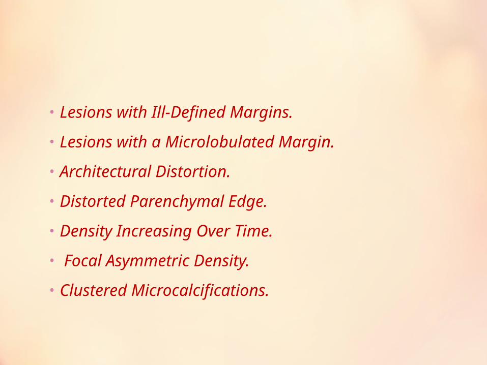
• Lesions with Ill-Defined Margins.
• Lesions with a Microlobulated Margin.
• Architectural Distortion.
• Distorted Parenchymal Edge.
• Density Increasing Over Time.
• Focal Asymmetric Density.
• Clustered Microcalcifications.
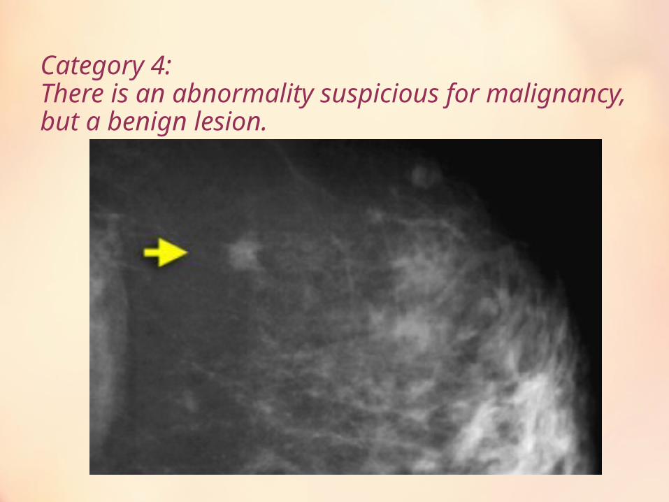
Category 4: There is an abnormality suspicious for malignancy, but a benign lesion.
BI-RADS 5
Highly Suggestive of Malignancy. Appropriate Action Should Be Taken:
classic breast cancers, with a >95% likelihood of malignancy. A spiculated, irregular high-density mass,
a segmental or linear arrangement of fine linear calcifications
or an irregular spiculated mass with associated pleomorphic calcification.

•
New oncologic management requires percutaneous tissue sampling is included in surgical treatment or when neoadjuvant chemotherapy is administered.

BI-RADS 6•
Known Biopsy Proven Malignancy. Appropriate Action Should Be Taken lesions identified on the imaging study with biopsy proof of malignancy prior to definitive therapy.
•These patients are treated with neo-adjuvant chemotherapy. During the course of the treatment the tumor may be less visible, while still you know you are dealing with cancer.
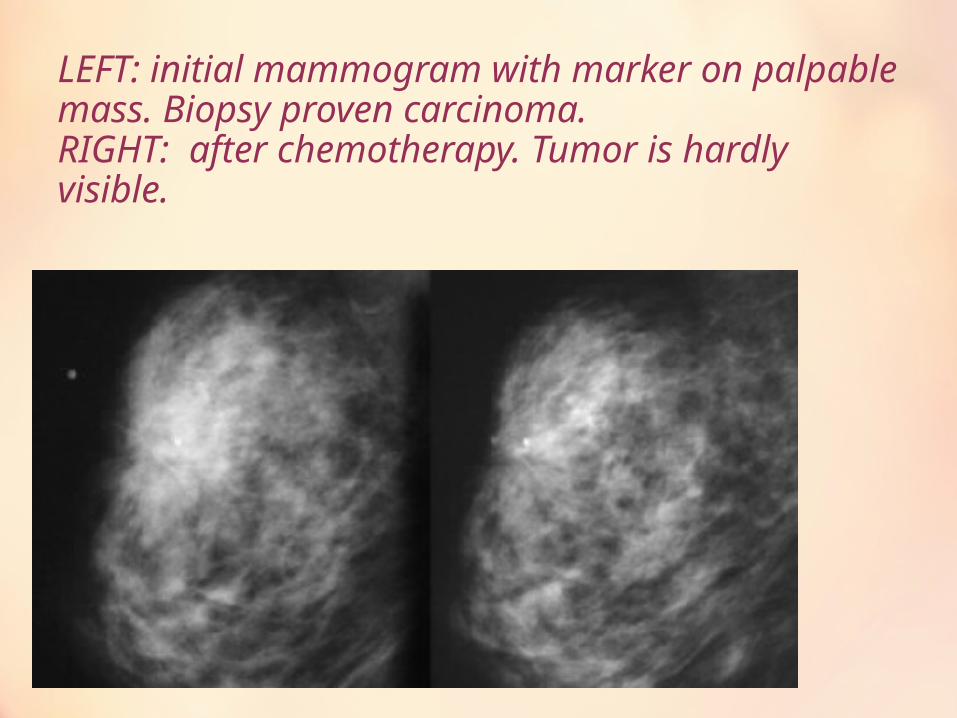
LEFT: initial mammogram with marker on palpable mass. Biopsy proven carcinoma.RIGHT: after chemotherapy. Tumor is hardly visible.
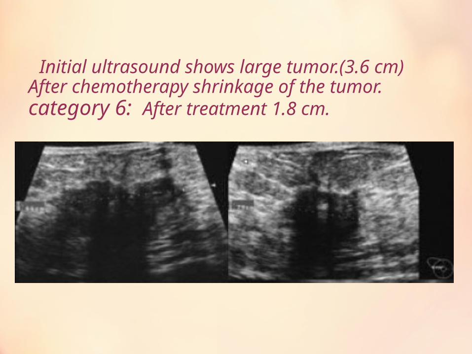
Initial ultrasound shows large tumor.(3.6 cm) After chemotherapy shrinkage of the tumor. category 6: After treatment 1.8 cm.
BI-RADS 0• Need Additional Imaging Evaluation and/or Prior
Mammograms For Comparison:
When additional imaging studies are completed, a final assessment is made.
Always try to avoid this category by immediately doing additional imaging or retrieving old films before reporting.
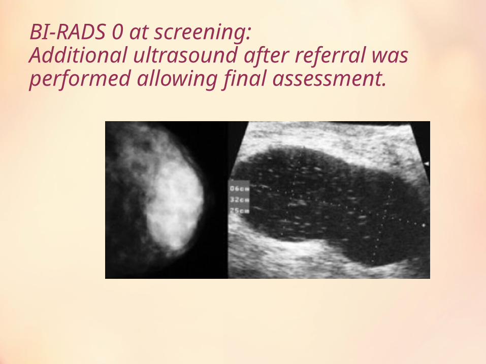
BI-RADS 0 at screening:Additional ultrasound after referral was performed allowing final assessment.