Toward a Standardized Breast Ultrasound Lexicon, BI-RADS ...
Understanding BI-RADS
Pailin Kongmebhol, M.D. Department of Radiology
Faculty of Medicine
Chiang Mai University

References
• ACR BI-RADS ATLAS
• BI-RADS frequently asked questions section from www.acr.org
• Breast Imaging by Bassett et al.

• ACR BI-RADS was initiated in 1988
• Clinicians complain about mammography reports that are lack of uniformity & vague management recommendation
• ACR developed a lexicon of terminology to standardize reporting & management recommendation

• The report should include:
– Significant findings using standard terminology
– Overall assessment (BI-RADS category)
– Management recommendation
The findings, BI-RADS category and management recommendation should be concordant

• The assessment (BI-RADS) category should reflect the imaging findings of the case
• A concordant management recommendation should be provide
• An additional sentence should recommend the additional (discordant) management appropriate for the scenario

Assessment categories • BIRADS 0 – need additional image evaluation/ prior mammogram for
comparison
• BIRADS 1 - Negative (0% likelihood of malignancy)
• BIRADS 2 - Benign (0% likelihood of malignancy)
• BIRADS 3 - Probably benign (> 0 to 2% likelihood of
malignancy)
• BIRADS 4 - Suspicious abnormality (>2 to < 95%
malignancy; average 30%)
• BIRADS 5 - Highly suggestive of malignancy (>/=
95% malignancy)
• BIRADS 6 – Known biopsy-proven malignancy

BI-RADS 0
• Most used in a screening – Wait for additional mammographic view
– Wait for additional US
– Wait for old films
• Should not be used for diagnostic breast imaging findings that warrant further MRI

BI-RADS 1
• There is nothing to comment on. This is a normal examination.
• May have some benign findings that the interpretor choose not to describe such findings eg intramammary LN, benign calcifications
• Management recommendation is routine mammography screening

BI-RADS 1 – Negative (not describe the benign calcifications)

BI-RADS 2 • Considered as normal assessment
• No mammographic evidence of malignancy
• Management recommendation is routine mammography screening

BI-RADS 2
• Benign findings were described in the report eg.
– Intramammary LN
– Benign calcifications
– Degenerating fibroadenoma
– Surgical clips
– Fat-containing lesions
– Implants
– Architectural distortion from surgical scar

BI-RADS 2 :calcified degenerating fibroadenoma

BI-RADS 2 : intramammary LN

BI-RADS 2: post surgical scar

BI-RADS 2: surgical clips

BI-RADS 3
• The lesion is not expected to change over the follow up periods
• Most lesions will have short interval follow up (6 mo.) x 2 times then 1 year; if stable after 2-3 years then change to BI-RADS 2
• Shorter time follow up may be used eg. 1 mo in cases suspected trauma or infection

• Should not use BI-RADS 3 on screening mammogram
• It should be used after diagnostic work up
• The lesion may appears definitely benign on work up such as cyst, intramammary LN, skin calcifications no need to be anxiety for 6 mo.
• Some lesions may appear suspicious on diagnostic work up and biopsy should not be delayed

On mammogram
• Suggested BI-RADS 3:
– Noncalcified circumscribed solid mass
– Focal asymmetry
– Solitary group of punctate calcifications
*No robust scientific data for using BI-RADS 3 on palpable lesion

BI-RADS 3: a noncalcified circumscribed solid mass

yr 2010
BI-RADS 3: asymmetry (rt.)
yr 2012 yr 2015

BI-RADS 3: grouped punctate calcifications

On ultrasound

BI-RADS 3 : an oval, circumscribed, hypoechoic solid mass, parallel orientation, no posterior feature or minimal posterior enhancement
*If the mass is palpable, the documents strong only in women < 40 years old

BI-RADS 3: an isolated complicated cyst with uniform low level echo

Less strong evidence* for clustered microcysts (Size < 2-3 mm each; thin septum (<0.5 mm thick)
* <500 cases on references, 0.5% malignancy

• BI-RADS 3 lesions based on expert’s opinion
– A hyperechoic mass with central hypoechoic components and surrounding edema, consistent with but not diagnostic of fat necrosis
– Architectural distortion thought to be due to postsurgical scar

BI-RADS 3 : probably fat necrosis (history of trauma)

• If the lesion increase in size (> 20% diameter) during follow up change to BI-RADS 4 and suggest biopsy
• If the lesion disappear or become apparently benign before 2 years BI-RADS 2
• FNA or biopsy may be performed on BI-RADS 3 lesion when patient anxiety, patient cannot come for follow up

BI-RADS 4
• Findings that do not have classic appearance of malignancy and not typically benign; but findings suspicious enough to biopsy
• Subdivision into BI-RADS 4 A, 4 B, 4 C is an option
• Management recommendation is biopsy

BI-RADS 4 A
• Chance >2 to 10 % malignancy
• A malignant finding is not expected
• Recommendation if biopsy/ FNA result is benign:
– 6 mo. follow up
or
– Routine follow up
*Some patient may choose to decline biopsy if they accept the risk of 10% chance of malignancy

BI-RADS 4 A
• Examples :
– A partially (<75%) circumscribed solid mass with US features suggestive of a fibroadenoma
– Palpable solitary complicated cyst
– Probable abscess (if typical abscess on US then it is BI-RADS 2)
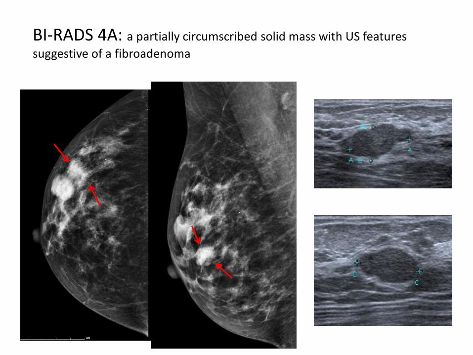
BI-RADS 4A: a partially circumscribed solid mass with US features
suggestive of a fibroadenoma

BI-RADS 4A: palpable complicated cyst. (must be homogeneously low level echoes or fluid-fluid level, no thick wall, no thick septa, no internal solid component)

BI-RADS 4 A: probable abscess, aspiration showed pus.

BI-RADS 4 B
• Chance malignancy >10 to </= 50% malignancy
• Recommended follow up with benign biopsy result will depend on concordance

BI-RADS 4 B
• Examples of findings:
– Coarse heterogeneous calcifications (13% malignancy)
– A group of amorphous calcifications
– A group of fine, pleomorphic calcifications
– An otherwise nondescript solid mass with indistinct margin

CC MLO
Bx = degenerating fibroadenoma
BI-RADS 4B: coarse heterogeneous calcifications
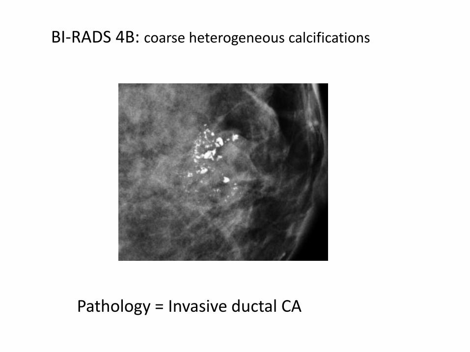
Pathology = Invasive ductal CA
BI-RADS 4B: coarse heterogeneous calcifications

Biopsy = DCIS
BI-RADS 4B: grouped amorphous calcifications

Bx = fibrocystic change
BI-RADS 4B: grouped amorphous calcifications

Fibrocystic change Benign calcifications in ducts
BI-RADS 4B:Grouped pleomorphic calcifications

Bx = DCIS in all cases
BI-RADS 4B:Grouped pleomorphic calcifications

BI-RADS 4B: a solid mass with (partly) indistinct margin
Core biopsy showed fibroadenoma

BI-RADS 4 C
• Findings high suspicion for malignancy but not highly suggestive of malignancy
• Chance malignancy > 50% to <95%

BI-RADS 4 C
• Examples
– A new indistinct, irregular solid mass
– A new group of fine linear calcifications
• Pathologist may initiate further histological evaluation of benign results of category 4C lesions

BI-RADS 4C: an indistinct, irregular solid mass

BIRADS 4C: new grouped fine linear branching calcifications
baseline 1 year later
BI-RADS 5 • Chance of malignancy >/= 95%
• Used in classic examples of malignancy
• If biopsy result is benign suggest repeat (usually surgical) biopsy
• Recommendation is “biopsy should be performed in the absence of clinical contraindication” rather than “appropriate action should be taken”

BI-RADS 5 • Examples
– An irregular, spiculated, high density mass with associated microcalcifications
– New fine linear and branching calcifications + segmental distribution
• There is no single mammographic feature that is associated with a likelihood of malignancy of >= 95%, it takes a combination of suspicious imaging findings to justify a category 5 assessment

BI-RADS 5: An irregular, high density mass with associated
microcalcifications, axillary lymphadenopathy

BI-RADS 5: segmental, fine linear branching calcifications

BI-RADS 5: segmental, fine linear branching calcifications

BI-RADS 5: a spiculated, irregular shaped, high density mass with skin retraction

BI-RADS 6 • Biopsy was already performed and the result was
malignancy
• Examples:
• Used for assessment before complete surgical excision
• Follow up response to neoadjuvant chemotherapy
• After attempted complete removal of target lesion by percutaneous core biopsy
BI-RADS 6 • If mammogram is done for evaluation after
attempted complete surgical excision with positive resection margin on pathology:
• If there are residual positive imaging findings of malignancy BI-RADS 6
• If imaging shows only post surgical scarring (although pathology result was margin positive); then BI-RADS 2 should be given with extra-sentence stating that pathology report suggest residual tumor

BI-RADS 6
• If suspicious findings other than the known cancer is found, then BI-RADS 4 or 5 should be given
• If the other findings are not suspicious (benign or probably benign), then BI-RADS 6 should be given and add a sentence for proper management of the additional findings

Palpable left breast mass: FNA showed ductal CA

Palpable left breast mass: FNA showed ductal CA

Palpable left breast mass: FNA showed ductal CA A spiculated, irregular mass on the right side.

Left: Known ductal CA Right: angular margin, irregular mass with posterior shadow
Final BI-RADS 5 (right breast mass)

BI-RADS 6
• Recommendation should be :
“ surgical excision when clinically appropriate”
instead of
“ appropriate action should be taken” (previous BI-RADS ed.)
• Some specific clinical scenarios may have management discordant with the assessment category or may confused radiologists

Scenario 1
• Patient with palpable breast abnormality
• Mammogram negative, ultrasound negative
• The radiologist may feels that the palpable lesion is worrisome and should be biopsy
• What BI-RADS category & recommendation should be?

• BI-RADS 1 – Negative (due to negative mammogram, US)
• Management recommendation: Routine mammography screening
• Adding sentence:
-surgical consultation or tissue diagnosis if clinically indicated
-since the palpable lesion is imaging negative; management should be based on clinical concern

• There is no test that ensure that a woman does not have breast cancer
• In case of palpable mass with imaging negative, management decision must be made based on clinical findings
• The likelihood of malignancy for palpable mass with negative mammogram and negative US is 0.1-4%

Scenario 2
• A simple cyst on US with pain or tenderness that need therapeutic aspiration to relieve symptom
• What BI-RADS category & recommendation should be?

• BI-RADS 2 – Benign finding
• Recommendation: routine mammography screening
• Adding sentence: aspiration to relieve the discomfort produced by the cyst

Scenario 3
• A woman with a ruptured implant but no imaging finding of malignancy
• Should have surgical consultation for implant removal and possible replacement
• What BI-RADS category & recommendation should be?

• BI-RADS 2 – Benign finding
• Recommendation: routine mammography screening
• Adding sentence: Surgical consultation that addresses proper treatment for the rupture implant

Scenario 4
• A woman with isolated complicated cyst on US. Mammogram negative
• The radiologist wants to follow up US in 6 mo. • The woman cannot come for follow up
• What BI-RADS category & recommendation
should be?

• BI-RADS 3 : probably benign finding
• Recommendation: short interval follow up in 6 mo. by US
• Adding sentence: The patient cannot come for follow up so FNA was performed

Scenario 5
• Screening mammogram shows suspicious microcalcifications
• The radiologist suggests biopsy but the patient ask for follow up instead
• What BI-RADS category & recommendation should be?

• BI-RADS 4 – suspicious abnormality
• Recommendation: stereotactic biopsy grouped microcalcifications
• Adding sentence: the patient does not want to biopsy the microcalcifications and ask for short interval follow up. Follow up left mammogram next 6 mo. is planned
• On 6 mo. follow up, the microcalcifications are stable. What BI-RADS assessment should be?
• BI-RADS 4 – suspicious abnormality

Scenario 6
• Screening mammogram found an asymmetry.
• Diagnostic mammogram with ultrasound found no more abnormal finding (only asymmetry found)
• The radiologist wants to further investigate with MRI
• What BI-RADS assessment should be?

• Should not use BI-RADS 0 on diagnostic mammogram that warrant further evaluation with MRI
• BI-RADS (1-6) should be given (BI-RADS 3 for this case) and add a sentence to suggest MRI on the recommendation
• If MRI is not performed or negative/ benign, the management can still be done based on diagnostic study

Scenario 7
• A post-lumpectomy patient was sent for mammogram 6 mo. after surgery, what BI-RADS should be?
• After lumpectomy, the usual mammographic appearance is architectural distortion caused by surgery

• If no other suspicious sign BI-RADS 2 is appropriate
• BI-RADS 3 should not be used when radiologist is “not sure” whether a finding is benign or suspicious
• During the post lumpectomy period, if the subsequent mammogram shows increased architectural distortion or other suspicious findings BI-RADS 4 is appropriate

Scenario 8
• Do mammography examination performed on men require a BI-RADS assessment?
• Yes.
• All mammography examination are require to have final assessment category but the management recommendations may differ because annual screening mammography is not usually appropriate

Conclusion
• Using proper BI-RADS assessment category with concordant management recommendation can guide clinician for appropriate treatment or follow up, reduce confusion in management
• Radiologists should have well knowledge and understand how to use the proper BI-RADS category

THANK YOU


![Bi Rads Patologias 2 [Salvo Automaticamente]](https://static.fdocuments.net/doc/165x107/577c7a571a28abe05494cc50/bi-rads-patologias-2-salvo-automaticamente.jpg)