Assessment of Genetic Stability of Micropropagated Curcuma ...
9
Assessment of Genetic Stability of Micropropagated Curcuma caesia through Cytophotometric and Molecular Analysis S. Mohanty 1 , R. K. Joshi 1 , E. Subudhi 1 , S. Sahoo 2 and S. Nayak 1, * 1 Centre of Biotechnology, Siksha O Anusandhan University, Khandagiri, Bhubaneswar-30, Orissa, India 2 P.G. Department of Botany, Utkal University, Vanivihar, Bhubaneswar, Orissa, India Received January 21, 2010; accepted February 1, 2010 Summary Protocol has been standardized for micropropagation of medicinally important Curcuma caesia using sprouted bud explants from rhizome. Tissue culture raised plantlets of C. caesia could be conserved in vitro through subculturing at an interval of 4 months. The genetic stability of micropropagated plants was studied with an interval of 6 months up to 30 months in culture using cytophotometric, random amplified polymorphic DNA (RAPD) and inter simple sequence repeats (ISSR) analysis. Cytophotometric analysis of 151 plants revealed a unimodal distribution of the DNA content with a peak corresponding to 4C nuclear DNA (3.5 pg). RAPD and ISSR analysis revealed mopnomorphic bands showing the absence of polymorphism in all 73 regenerants analyzed, thus confirming the genetic uniformity among in vitro grown somaclones of Curcuma caesia. Key words Curcuma caesia, Cytophotometric analysis, Genetic stability, Micropropagation, Molecular marker. Curcuma caesia Roxb., commonly known as black turmeric is a wild species of the family Zingiberaceae. C. caesia is known especially in North East India and Bangladesh. It is rarely found in other parts of India, such as Madhya Pradesh, Jharkhand, Chattisgarh, and Orissa. Rhizomes of the plant are used for sprains and bruises, and are also employed as cosmetics (CSIR 1962). It is also used as a carminative, and for the treatment of headaches and rheumatic pains. The rhizomes are aromatic and yield an essential oil. The inner part of the rhizome is bluish-black in colour and emits a characteristic sweet smell due to the presence of essential oil. The oils possess antibacterial and antifungal properties (Rahman and Yusuf 1996) and are claimed to be useful in treating piles, leprosy, bronchitis, asthma, cancer, epilepsy, fever, wounds, impotency and migraines etc. A paste made from the rhizome is used to cure blood dysentery and as poultice in rheumatic pain. The leaves are used as a wrapping material and dry leaves are used as fuel by Bengalis in Bangladesh (Yusuf et al. 2002). North Indian tribes use black turmeric as a talisman to keep the evil spirits away. This plant is in great demand in Central India and due to indiscriminate exploitation without ensuring its regeneration and conservation, the plant has recently been categorized as an endangered species (Kumar 1998). This necessitates the application of the tissue culture technique for micropropagation and in vitro conservation of C. caesia and to ensure genetic stability for production of true-to-type plantlets. The tissue culture technique is a powerful tool which can be employed as an alternative to the conventional method of vegetative propagation with the objective of rapid clonal multiplication of desired genotypes (Hussey 1986, Murashige and Skoog 1990). Periodic monitoring of the degree of * Corresponding author, e-mail: [email protected] © 2010 The Japan Mendel Society Cytologia 75(1): 73–81, 2010
Transcript of Assessment of Genetic Stability of Micropropagated Curcuma ...
Assessment of Genetic Stability of MicropropagatedCurcuma caesia through Cytophotometric and
Molecular Analysis
S. Mohanty1, R. K. Joshi1, E. Subudhi1, S. Sahoo2 and S. Nayak1,*
1 Centre of Biotechnology, Siksha O Anusandhan University, Khandagiri, Bhubaneswar-30, Orissa, India
2 P.G. Department of Botany, Utkal University, Vanivihar, Bhubaneswar, Orissa, India
Received January 21, 2010; accepted February 1, 2010
Summary Protocol has been standardized for micropropagation of medicinally importantCurcuma caesia using sprouted bud explants from rhizome. Tissue culture raised plantlets of C.caesia could be conserved in vitro through subculturing at an interval of 4 months. The geneticstability of micropropagated plants was studied with an interval of 6 months up to 30 months inculture using cytophotometric, random amplified polymorphic DNA (RAPD) and inter simplesequence repeats (ISSR) analysis. Cytophotometric analysis of 151 plants revealed a unimodaldistribution of the DNA content with a peak corresponding to 4C nuclear DNA (3.5 pg). RAPD andISSR analysis revealed mopnomorphic bands showing the absence of polymorphism in all 73regenerants analyzed, thus confirming the genetic uniformity among in vitro grown somaclones ofCurcuma caesia.
Key words Curcuma caesia, Cytophotometric analysis, Genetic stability, Micropropagation,Molecular marker.
Curcuma caesia Roxb., commonly known as black turmeric is a wild species of the familyZingiberaceae. C. caesia is known especially in North East India and Bangladesh. It is rarely foundin other parts of India, such as Madhya Pradesh, Jharkhand, Chattisgarh, and Orissa. Rhizomes ofthe plant are used for sprains and bruises, and are also employed as cosmetics (CSIR 1962). It isalso used as a carminative, and for the treatment of headaches and rheumatic pains. The rhizomesare aromatic and yield an essential oil. The inner part of the rhizome is bluish-black in colour andemits a characteristic sweet smell due to the presence of essential oil. The oils possess antibacterialand antifungal properties (Rahman and Yusuf 1996) and are claimed to be useful in treating piles,leprosy, bronchitis, asthma, cancer, epilepsy, fever, wounds, impotency and migraines etc. A pastemade from the rhizome is used to cure blood dysentery and as poultice in rheumatic pain. Theleaves are used as a wrapping material and dry leaves are used as fuel by Bengalis in Bangladesh(Yusuf et al. 2002). North Indian tribes use black turmeric as a talisman to keep the evil spiritsaway. This plant is in great demand in Central India and due to indiscriminate exploitation withoutensuring its regeneration and conservation, the plant has recently been categorized as anendangered species (Kumar 1998). This necessitates the application of the tissue culture techniquefor micropropagation and in vitro conservation of C. caesia and to ensure genetic stability forproduction of true-to-type plantlets.
The tissue culture technique is a powerful tool which can be employed as an alternative to theconventional method of vegetative propagation with the objective of rapid clonal multiplication ofdesired genotypes (Hussey 1986, Murashige and Skoog 1990). Periodic monitoring of the degree of
* Corresponding author, e-mail: [email protected]
© 2010 The Japan Mendel Society Cytologia 75(1): 73–81, 2010
73_81.pdf 1 10.5.26 9:10:17 AM

genetic stability of in vitro conserved plants is of utmost importance for commercial utilization ofthe technique for large scale production of true-to-type plants of the desired genotype. Theassessment of the genetic integrity of in vitro grown regenerants at regular intervals cansignificantly reduce or eliminate the chance of occurence of somaclonal variations (Larkins andScowcroft 1981) at the early or late phase of culture. Cytophotometric analysis is very useful forquick determination of the extent of the ploidy level of regenerated plants with a large number ofcells (Nayak and Sen 1995). Of several molecular markers used for the assessment of geneticstability, RAPD and ISSR analysis are the simplest, cheapest and quickest method for determiningthe genetic fidelity of in vitro grown plants as reported in many species (Williams et al. 1990, Routet al. 1998, Martins et al. 2004, Venkatachalam et al. 2007, Joshi and Dhawan 2007). Although mi-cropropagation and in vitro conservstion of C. caesia has been reported (Balachandran et al. 1990,Raju et al. 2005, Tyagi et al. 2004), there is no report regarding the extent of stability of themicropropagated clones.
We report rapid in vitro multiplication of C..caesia and in vitro monitoring of the geneticstability of micropropagated plants through cytophotometric estimation of the 4C nuclear DNAcontent RAPD and ISSR analysis at regular intervals. This will definitely meet the increasingdemand of this endangered species with its conserved genetic integrity.
Materials and methods
Plant materialRhizome of C. caesia was collected from a high altitude research station in Pottangi, Koraput,
Orissa, and was grown in the medicinal plant garden of the Centre of Biotechnology, Bhubaneswar,Orissa (India). Rhizomes containing sprouted apical buds were used as explants and werethoroughly washed with water. The explants were kept under running water for 10–15 min and thendipped in liquid detergent (Extran, Merck) for 3–5 min. They were thoroughly washed with distilledwater to remove the last drop of detergent. Surface sterilization was done by 0.1% mercuricchloride solution for 8–12 min depending upon the nature and size of the explants. Aftersterilization the explants containing buds were washed several times with sterile distilled waterunder aseptic conditions prior to inoculation.
In vitro multiplicationExplants were inoculated to the basal medium of Murashige and Skoog (MS) (1962) with
varying combinations of benzyl adenine (1–5 mg/l), indole-3-acetic acid (0.5–2 mg/l), naphthaleneacetic acid (0.5–2 mg/l) and kinetine (0.5–2 mg/l). fifteen replicas were used for each treatment. TheMS media was gelled with 0.8% (m/v) agar (Himedia Laboratories Pvt. Ltd., Mumbai, India) andsterilized at 121°C and 1.06 kg/cm2 pressure for 15 min. The pH of the media was adjusted to5.73–5.74. After sterilization of the media, the explants were inoculated into the medium. Theexplants were inoculated under aseptic conditions and then kept in a culture room under whitefluorescent light of 50 mmol m�2 s�1 for 16 h photoperiod. The temperature of the culture room wasmaintained at 25�2°C. Explants with well developed shoots and roots were subcultured at aninterval of 3–4 months in culture. Each individual shoot was separated and inoculated to a freshmedia for further growth. In vitro grown plantlets with well developed shoots and roots weretransferred to pots containing sterilized soil. These were then kept in a greenhouse foracclimatization. After 1 month they were transferred to normal field conditions and grown tomaturity.
Cytophotometric analysisFor cytophotometric analysis, explants’ tissue was collected from a field-grown source plant
74 Cytologia 75(1)S. Mohanty et al.
73_81.pdf 2 10.5.26 9:10:18 AM

and the root tips of in vitro grown regenerants aseptically from culture tubes and fixed over night inpropionic acid/ethanol (1 : 3). This treatment was followed by hydrolysis in 1 M HCl (v/v) at 60°Cfor 12 min. The root tips were washed in distilled water, stained in Schiff’s reagent for 2 h at 14°Cand squashed with 45% acetic acid. The DNA content of nuclei was measured with a Leitz Wetzlarmicro-spectrophotometer using the single wavelength (550 nm) method (Sharma and Sharma 1980).In situ DNA values were obtained on the basis of optical densities which were converted to pico-grams (pg) by using 4C nuclear DNA value of Allium cepa (67.1 pg) as standard (Van’t Hof 1965).
Isolation of DNAFresh young leaves were collected and washed thoroughly with cold autoclaved distilled water,
then blotted to dry. About 2 g of leaves were taken for extraction. The genomic DNA was isolatedfollowing the protocol of Doyle and Doyle (1990) with a little modification; insolublepolyvinylpolypyrolidone was added to the leaf sample prior to grinding. The crude DNA waspurified with RNase A (60 mg/ml of crude DNA solution) followed by washing thrice withphenol/chloroform/isoamyl alcohol (25 : 24 : 1 v/v/v) and subsequently with chloroform/isoamyl-alcohol (24 : 1 v/v). Quantification of DNA was done by 0.8% agarose gel electrophoresis of thesamples along with a known amount of uncut lambda DNA (Bangalore Genei Pvt. Ltd, Bangalore,India) as standard. DNA samples were diluted to 25 ng/m l for RAPD-PCR analysis.
RAPD and ISSR analysesFor RAPD analysis a total of 30 random primers were used out of which 19 random decamer
primers (Operon Tech, Almeda, USA) were selected. In the case of ISSR, out of 10 primers, 8 wereselected. The RAPD analysis was performed as per the method of Williams et al. (1990) and forISSR analysis the method of Zeitkiewicz et al. (1994) was followed. RAPD and ISSRamplifications were performed routinely using a PCR mixture (25 m l) containing 25 ng of genomicDNA as a template, 10�PCR buffer (Bangalore Genei, India), 200 mM dNTPs (Bangalore Genei,India ), 0.5 unit (U) of Taq polymerase (Bangalore Genei, India), and 15 ng of RAPD primer or40 ng of ISSR primer. The amplification was carried out in a thermal cycler (Gene Amp PCRsystem 9700, Applied Biosystems, CA, USA). In RAPD, PCR was performed at an initialtemperature of 94°C for 5 min for complete denaturation. The second step consisted of 42 cycleshaving 3 ranges of temperature, i.e. at 92°C for 1 min for denaturation of template DNA, at 37°Cfor 1 min for primer annealing, at 72°C for 2 min for primer extension, followed by running thesamples at 72°C for 7 min for complete polymerization. For ISSR the same temperature profile wasfollowed, but the primer annealing temperature was set at 5°C lower than the melting temperature.The PCR products obtained from RAPD were analyzed in 1.5% agarose gel whereas the ISSRproducts were analyzed in 2% agarose gel stained with ethidium bromide (0.5 mg m l�1). The size ofthe amplicons was estimated using 100 bp DNA ladder plus or DNA ladder mix (MBI Fermentas,Lithuania) and documented in the Gel Doc (Bio-Rad, USA).
Results
In vitro multiplicationSprouted rhizome buds of C. caesia were cultured as explants (Fig. 1A) on MS media with
various combinations of BA (1–5 mg/l), IAA (0.5–2 mg/l), NAA (0.5–2 mg/l) and Kinetin(0.5–2 mg/l). The percentage of explants forming shoots was maximum in media with BA (3 mg/l)and IAA (0.5 mg/l) i.e., 92.8�0.3 (Table 1). A higher concentration of BA showed less responsetowards in vitro shoot initiation. Media with BA (1 mg/l) and IAA (0.5 mg/l) was optimum for invitro shoot multiplication with 3.5�0.2 number of shoots and 1.1�0.2 number of roots (Fig. 1B,C). Media with BA and NAA were also effective for shoot initiation and multiplication. Media with
2010 75Cytophotometric and Molecular Analysis of Curcuma caesia
73_81.pdf 3 10.5.26 9:10:18 AM
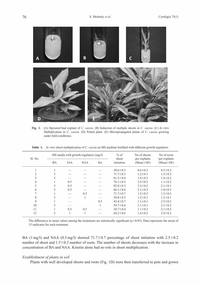
BA (1 mg/l) and NAA (0.5 mg/l) showed 71.7�0.7 percentage of shoot initiation with 2.5�0.2number of shoot and 1.5�0.2 number of roots. The number of shoots decreases with the increase inconcentration of BA and NAA. Kinetin alone had no role in shoot multiplication.
Establishment of plants in soilPlants with well developed shoots and roots (Fig. 1D) were then transferred to pots and grown
76 Cytologia 75(1)S. Mohanty et al.
Fig. 1. (A) Sprouted bud explant of C. caesia. (B) Induction of multiple shoots in C. caesia. (C) In vitroMultiplication in C. caesia. (D) Potted plant. (E) Micropropagated plants of C. caesia growingunder field conditions.
Table 1. In vitro shoot multiplication of C. caesia on MS medium fortified with different growth regulators
MS media with growth regulators (mg/l) % of No of shoots No of roots Sl. No. shoot per explants per explants
BA IAA NAA Kn initiation (Mean�SE) (Mean�SE)
1 1 — — — 30.6�0.5 0.6�0.2 0.5�0.22 3 — — — 71.7�0.3 1.2�0.1 1.5�0.23 5 — — — 61.5�0.4 1.8�0.3 1.4�0.24 1 0.5 — — 76.1�0.5 3.5�0.2 1.1�0.25 3 0.5 — — 92.8�0.3 3.2�0.2 2.1�0.16 5 0.5 — — 66.1�0.6 2.1�0.3 1.0�0.37 1 — 0.5 — 71.7�0.7 1.5�0.2 1.5�0.38 3 — 1 — 54.8�0.5 1.2�0.1 1.2�0.19 1 — 0.5 43.4�0.7 1.1�0.1 2.5�0.2
10 3 — 1 54.7�0.6 1.1�0.1 2.1�0.211 1 0.5 0.5 — 38.7�0.4 1.1�0.3 2.1�0.312 3 1 1 — 64.2�0.6 1.4�0.2 2.4�0.2
The differences in mean values among the treatments are statistically significant (p�0.05). Data represents the mean of15 replicates for each treatment.
73_81.pdf 4 10.5.26 9:10:18 AM
in green house for 1 month for acclimatization. After 1 month the acclimatized plants weretransferred to the field and were established showing a survival rate of 90% (Fig. 1E).
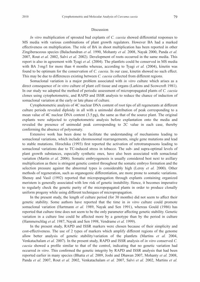
Cytophotometric analysisCytophotometric analysis of the original explant was carried out before explantation which
revealed a peak showing 2C value (1.7 pg). The cytophotometric analysis of the 4C nuclear DNAcontent of root tips from a total of 151 in vitro grown regenerants, carried out at 6 month, intervalsup to 24 months in culture, revealed diploidy in all with a unimodal distribution with 1 distinct peakcorresponding to a 4C value (3.5 pg). This is the same same as obtained in the source plant. Therange of mean DNA content in root tips of in vitro conserved plantlets of different ages was almostsimilar to the range obtained in root tips of 10 control plants from the field (Table 2). The plantletsof C. caesia used for the cytophotometric study were labeled properly to be used for the molecularanalysis with RAPD and ISSR markers.
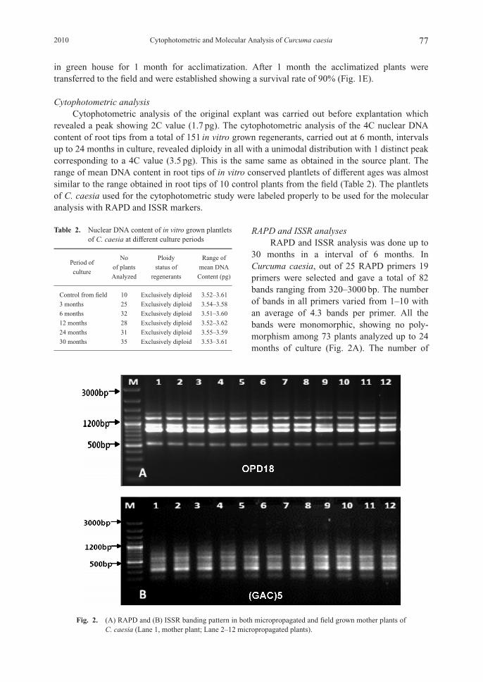
RAPD and ISSR analysesRAPD and ISSR analysis was done up to
30 months in a interval of 6 months. InCurcuma caesia, out of 25 RAPD primers 19primers were selected and gave a total of 82bands ranging from 320–3000 bp. The numberof bands in all primers varied from 1–10 withan average of 4.3 bands per primer. All thebands were monomorphic, showing no poly-morphism among 73 plants analyzed up to 24months of culture (Fig. 2A). The number of
2010 77Cytophotometric and Molecular Analysis of Curcuma caesia
Table 2. Nuclear DNA content of in vitro grown plantletsof C. caesia at different culture periods
Period of No Ploidy Range of
of plants status of mean DNA culture
Analyzed regenerants Content (pg)
Control from field 10 Exclusively diploid 3.52–3.61
3 months 25 Exclusively diploid 3.54–3.58
6 months 32 Exclusively diploid 3.51–3.60
12 months 28 Exclusively diploid 3.52–3.62
24 months 31 Exclusively diploid 3.55–3.59
30 months 35 Exclusively diploid 3.53–3.61
Fig. 2. (A) RAPD and (B) ISSR banding pattern in both micropropagated and field grown mother plants ofC. caesia (Lane 1, mother plant; Lane 2–12 micropropagated plants).
73_81.pdf 5 10.5.26 9:10:20 AM

monomorphic bands was highest, 10 in primer OPC11 (ranging from 450–1900 bp) and lowest, 1 inprimer OPD12 (850 bp). A total of 5986 bands [(number of plantlets analyzed)�(number of bandswith all primers)] were generated by the RAPD techniques (Table 3). Out of 10 ISSR primers, 8primers formed a total of 63 bands(ranging from 200–1900 bp) which are all monomorphic innature. The number of bands varied from 5–12 in all primers with an average of 7.8 bands perprimer. A total of 4599 bands [(number of plantlets analyzed)�(number of bands with all primers)]were generated by the ISSR techniques (Table 3). The highest number of bands i.e. 12 in primer(GAC)5 (ranging from 200–1200 bp), and lowest number of bands, i.e. 5 in primer (GA)9T(350–975 bp), were amplified by ISSR analysis.Thus no variation was detected among in vitroconserved plantlets (Fig. 2B).
In the present study, 2 PCR based molecular markers, RAPD and ISSR, were used to show thegenetic integrity in micropropagated C. caesia because of their cost effectiveness and simplicity. Asthere was no variation in the banding pattern both in RAPD and ISSR in all the regenerantsanalyzed in comparison with the mother plants, we conclude that the protocol developed could beused as a method for large scale propagation of true-to-type plantlets and conservation of C. caesiain vitro.
78 Cytologia 75(1)S. Mohanty et al.
Table 3. RAPD and ISSR banding pattern of micropropagated and field-grown mother plants of C. caesia
Primer Sequence Total bandsRange of
amplicons [bp]
RAPD OPA4 AATCGGGCTG 6 320–1500OPA7 GAAACGGGTG 2 600–2200OPA9 GGGTAACGCC 5 850–1750OPA18 AGGTGACCGT 3 450–900OPC2 GTGAGGCGTC 6 650–1300OPC5 GATGACCGCC 3 1031–1700OPC11 AAAGCTGCGG 10 450–1900OPD3 GTCGCCGTCA 2 800–1250OPD7 TTGGCACGGG 8 450–2000OPD8 GTGTGCCCCA 4 850–1700OPD12 CACCGTATCC 1 850OPD18 GAGAGCCAAC 5 450–1031OPD20 ACCCGGTCAC 6 600–3000OPN4 GACCGACCCA 3 500–1200OPN16 AAGCGACCTG 2 350–1031OPN18 GGTGAGGTCA 2 400–1031OPAF5 CCCGATCAGA 6 650–2200OPAF14 GGTGCGCACT 3 900–3000OPAF15 CACGAACCTC 5 550–2000
Total 82
ISSR SPS 1 (GAC)5 12 200–1200SPS 2 (GTGC)4 6 300–1050SPS 3 (GACA)4 8 475–1900SPS 4 (AGG)6 8 400–1031SPS 5 (GA)9T 5 350–975SPS 6 T(GA)9 7 500–1100SPS 7 (GTG)5 8 250–850SPS 8 (GGA)4 9 300–1500
Total 63
73_81.pdf 6 10.5.26 9:10:21 AM
Discussion
In vitro multiplication of sprouted bud explants of C. caesia showed differential responses toMS media with various combinations of plant growth regulators. However BA had a markedeffectiveness on multiplication. The role of BA in shoot multiplication has been reported in otherZingiberaceous species (Balachandran et al. 1990, Mohanty et al. 2008, Nayak 2000, Panda et al.2007, Rout et al. 2002, Salvi et al. 2002). Development of roots occurred in the same media. Thisreport is also in agreement with Tyagi et al. (2004). The plantlets could be conserved in MS mediawith BA 3 mg/l for more than 4 months whereas, according to Tyagi et al. (2004), kinetin wasfound to be optimum for the conservation of C. caesia. In our case, kinetin showed no such effect.This may be due to differences existing between C. caesia collected from different regions.
Somaclonal variation is a major problem associated with in vitro culture which arises as adirect consequence of in vitro culture of plant cell tissue and organs (Larkins and Scowcroft 1981).In our study we adopted the method of periodic assessment of micropropagated plants of C. caesiaclones using cytophotometric, and RAPD and ISSR analysis to reduce the chance of induction ofsomaclonal variation at the early or late phase of culture.
Cytophotometric analysis of 4C nuclear DNA content of root tips of all regenerants at differentculture periods revealed diploidy in all with a unimodal distribution of peak corresponding to amean value of 4C nuclear DNA content (3.5 pg), the same as that of the source plant. The originalexplants were subjected to cytophotometric analysis before explantation onto the media andrevealed the presence of unimodal peak corresponding to 2C value in each case, therebyconferming the absence of polysomaty.
Extensive work has been done to facilitate the understanding of mechanisms leading tosomaclonal variations, which include chromosomal rearrangements, single gene mutations and leadto stable mutations. Hirochika (1993) first reported the activation of retrotransposons leading tosomaclonal variations due to TC-induced stress in tobacco. The sub- and supra-optimal levels ofplant growth substances, especially synthetic ones, have also been associated with somaclonalvariation (Martin et al. 2006). Somatic embryogenesis is usually considered best next to axillarymultiplication as there is stringent genetic control throughout the somatic embryo formation and theselection pressure against the abnormal types is considerably high (Leroy et al. 2000). Othermethods of regeneration, such as organogenic differentiation, are more prone to somatic variations.Shenoy and Vasil (1992) reported that micropopagation through explants containing organizedmeristem is generally associated with low risk of genetic instability. Hence, it becomes imperativeto regularly check the genetic purity of the micropopagated plants in order to produce clonallyuniform progeny while using different techniques of micropropagation.
In the present study, the length of culture period (for 30 months) did not seem to affect theirgenetic stability. Some authors have reported that the time in in vitro culture could promotesomaclonal variation (Hartmann et al. 1989, Nayak and Sen 1991), whereas Gould (1986) hasreported that culture time does not seem to be the only parameter affecting genetic stability. Geneticvariation in a culture line could be affected more by a genotype than by the period in culture(Hammerschlag et al. 1987, Nayak and Sen 1998, Vendrame et al. 1999).
In the present study, RAPD and ISSR markers were chosen because of their simplicity andcost-effectiveness. The use of 2 types of markers which amplify different regions of the genomeallow better analysis of genetic stability/variation of the plantlets (Martins et al. 2004,Venkatachalam et al. 2007). In the present study, RAPD and ISSR analysis of in vitro conserved C.caesia showed a profile similar to that of the control, indicating that no genetic variation hadoccurred in vitro. This confirmed the genetic integrity by RAPD and ISSR analysis that had beenreported earlier in many species (Bhatia et al. 2009, Joshi and Dhawan 2007, Mohanty et al. 2008,Panda et al. 2007, Rout et al. 2002, Venkatachalam et al. 2007, Salvi et al. 2002, Martins et al.
2010 79Cytophotometric and Molecular Analysis of Curcuma caesia
73_81.pdf 7 10.5.26 9:10:21 AM

2004). Our study, in close agreement with Zuchi et al. (2002), reveals that the absence ofpolymorphism in micropropagated C. caesia could be due to the absence of polymorphism insource plants.
Acknowledgements
The authors are grateful to Dr. S. C. Si, Dean, Centre of Biotechnology and Dr. M. R. Nayak,President, Siksha O Anusandhan University for providing facilities and encouragement throughout.Financial assistance from Department of Biotechnology, New Delhi, India, is also dulyacknowledged.
References
Balachandran, S. M., Bhat, S. R. and Chandel, K. P. S. 1990. In vitro clonal multiplication of turmeric (Curcuma longa) andginger (Zingiber officinale Rosc.). Plant Cell Rep. 3: 521–524.
Bhatia, R., Singh, K. P., Jhang, T. and Sharma, T. R. 2009. Assesment of genetic fidelity of micropropagated gerbera plantsby ISSR markers. Sci. Hortic 11: 208–211.
CSIR (ed.). 1962. The Wealth of India, Raw Materials. CSIR, New Delhi. p. 402.Doyle, J. J. and Doyle, J. L. 1990. A rapid DNA isolation procedure for small quantities of fresh leaf tissue. Phytochem.
Bull. 19: 11–15.Gould, A. R. 1986. Factors controlling generation of variability in vitro. In: Vasil, I. K. (ed.). Cell culture and somatic cell
genetics in plants. 3. Plant regeneration and genetic variability. Academic, Orlando. pp. 549–567.Hammerschlag, F. A., Bauchan, G. R. and Scorza, R. 1987. Factors influencing in vitro multiplication and rooting of peach
cultivars. Plant Cell Tissue Organ Cult. 8: 235–242.Hartmann, C., Henry, Y., De Buyser, J., Aubry, C. and Rode A. 1989. Identification of new mitochondrial genome
organizations in wheat plants regenerated from somatic tissue cultures. Theor. Appl. Genet. 77: 169–175.Hirochika, H. 1993. Activation of tobacco transposons during tissue culture. EMBO J. 12: 2521–2528.Hussey, G. 1986. Problems and prospects in the in vitro propagation of herbaceous plants. In: Withers L. A. and Aldeson P.
G. (eds.). Plant Tissue Culture and its Agricultural Application. Butterworths, London. pp. 113–123.Joshi, P. and Dhawan, V. 2007. Assesment of genetic fidelity of micropropagated Swertia chirayita plantlets by ISSR marker
assay. Biol Plant. 51: 22–26.Kumar, S., Singh, J., Shah, N. C. and Ranjan, V. 1998. Indian Medicinal Plants Facing Genetic Erosion. CIMAP, Lucknow.
p. 219.Larkins, P. and Scowcroft, W. R. 1981. Somaclonal variation, a novel source of variability from cell cultures for plant
improvement. Theor. Appl. Genet. 60: 197–214.Leroy, X. J., Leon, K., Charles, G. and Branchard, M. 2000. Cauliflower somatic embryogenesis and analysis of regenerant
stability by ISSRs. Plant. Cell. Rep. 19: 1102–1107.Martin, K. P., Pachathundkandi, S. K., Zhang, C. L. and Slater, A. 2006. RAPD analysis of a variant of Banana (Musa sp.)
cv. Grande Naine and its propagation via shoot culture. In Vitro Cell Dev. Biol. 42: 188–192.Martins M., Sarmento D. and Oliveira M. M. 2004. Genetic stability of micropropagated almond plantlets, as assessed by
RAPD and ISSR markers. Plant Cell Rep. 23: 492–496.Mohanty, S., Panda, M. K., Subudhi, E., Acharya, L. and Nayak, S. 2008. Genetic Stability of Micropropagated Ginger
Derived from Axillary Bud through Cytophotometric and RAPD Analysis. Z Naturforsch. 63c: 747–754.Murashige, T. and Skoog, N. S. 1990. Plant propagation by tissue culture. A practice with unrealized potential. In:
Anmirato, P. V., Even, D. R., Sharp, W. R. and Bajaj. Y. P. S. (eds.). Handbook of Plant Cell Culture, McGraw HillPublishing Company, New York. pp. 3–9.
— and Skoog, F. 1962. A revised medium for rapid growth and bioassays with tobacco tissue culture. Physiol Plant. 15:473–479.
Nayak, S. and Sen, S. 1991. Cytological and cytophotometric analysis of direct explant and callus derived plants ofOrnithogalum thyrsoides Jacq. Cytologia. 56: 297–302.
— and — 1995. Rapid and stable propagation of Ornithogalum umbelatum L. in long term culture. Plant Cell Rep. 15:150–153.
— and — 1998. Differential resistance of three species of Ornithogalum to polyploidization in vitro. Nucleus. 41: 48–52.— 2000. In vitro multiplication and microrhizome induction in Curcuma aromatica Salisb. Plant Growth Regul. 32: 41–47.Panda, M. K., Mohanty, S., Subudhi, E., Acharya, L. and Nayak, S. 2007. Assessment of genetic stability of
micropropagated plants of Curcuma longa L. by cytophotometry and RAPD analysis. Int. J. Integ. Biol. 1:
80 Cytologia 75(1)S. Mohanty et al.
73_81.pdf 8 10.5.26 9:10:21 AM

189–195.Rahman, M. A. and Yusuf, M. 1996. Diversity, Ecology and Ethnobotany of the Zingiberaceae of Bangladesh. J. Econ.
Taxon. Bot. Addl. Series. 12: 13–19.Raju, B., Anita, D. and Kalita, M. C. 2005. In vitro clonal propagation of Curcuma caesia Roxb, and Curcuma zedoaria
Rosc. from rhizome bud explants. J. Plant Biochem Biotechnol. 14: 61–63. (eds.).Rout, G. R. and Das, P. 2002. In vitro studies of ginger: A review of recent progress In: Goril J. N., Anand Kumar P. and
Singh V. K. (eds.). recent progress is Medicinal Plants, Vol. 4–Biotechnology and Genetic Engineering, ScienceTechnology Publication, city. pp. 307–326.
—, Das, P., Goel, G. and Raina, S. N. 1998. Determination of genetic stability of micropropagated plants of ginger usingRandom Amplified Polymorphic DNA (RAPD) markers. Bot. Bull. Acad. Sinica (Taiwan) 39: 23–27.
Salvi, N. D., George, L. and Eapen, S. 2002. Micropropagation and field evaluation of micropropagated plants of turmeric.Plant Cell Tissue Organ Cult. 68: 143–151.
Sharma and Sharma 1980Shenoy, V. B. and Vasil, I. K. 1992. Biochemical and molecular analysis of plants derived from embryogenic tissue cultures
of napiergrass (Penisetum purpureum K. Schum.). Theor Appl Genet. 83: 947–955.Shirin, F., Kumar, S. and Mishra, Y. 2000. In vitro plantlet production system for Kaempferia galanga, a rare Indian
medicinal herb. Plant Cell Tissue Organ Cult. 63: 193–197.Tyagi, R. K., Yusuf, A., Dua, P. and Agrawal, A. 2004. In vitro plant regeneration and genotype conservation of eight wild
species of Curcuma. Biol. Plant. 48: 129–132.Van’t Hof, J. 1965. Relationships between mitotic cycle duration, S period duration and the average rate of DNA synthesis
in the root meristem cells of several plants. Exp. Cell Res. 39: 48–58.Vendrame, W. A., Kochert, G. and Wetzstein, H. Y. 1999. AFLP analysis of variation in pecan somatic embryos. Plant Cell
Rep. 18: 853–857.Venkatachalam et al. 2007Williams, J. G. K., Kubelik, A. R., Liva, K. J., Rafalski, J. A. and Tingey, S. V. 1990. DNA polymorphisms amplified by
arbitrary primers are useful as genetic markers. Nucl. Acid Res. 18: 6531–6535.Yusuf, M., Rahman, M. A., Chowdhury, J. U. and Begum, J. 2002. Indigenous knowledge about the use of Zingibers in
Bangladesh. J. Econ. Taxon. Bot. 26: 566–570.Zeitkiewicz, E., Rafalski, A. and Labuda, D. 1994. Genome finger printing by simple sequence repeat (SSR)-anchored PCR
amplification. Genomics 20: 176–183.Zucchi, M. I., Arizono, H., Morais, V. I., Fungaro, M. H. P., Vieira, and M. L. C. 2002. Genetic instability of sugarcane
plants derived from meristem cultures. Genet. Mol. Biol. 25: 91–96.
2010 81Cytophotometric and Molecular Analysis of Curcuma caesia
73_81.pdf 9 10.5.26 9:10:21 AM