Cardiovascular System. I. System Anatomy Heart Pumps Blood Blood Vessels Deliver blood…
Upload
jaycee-engelbyCategory
view
215download
0
1
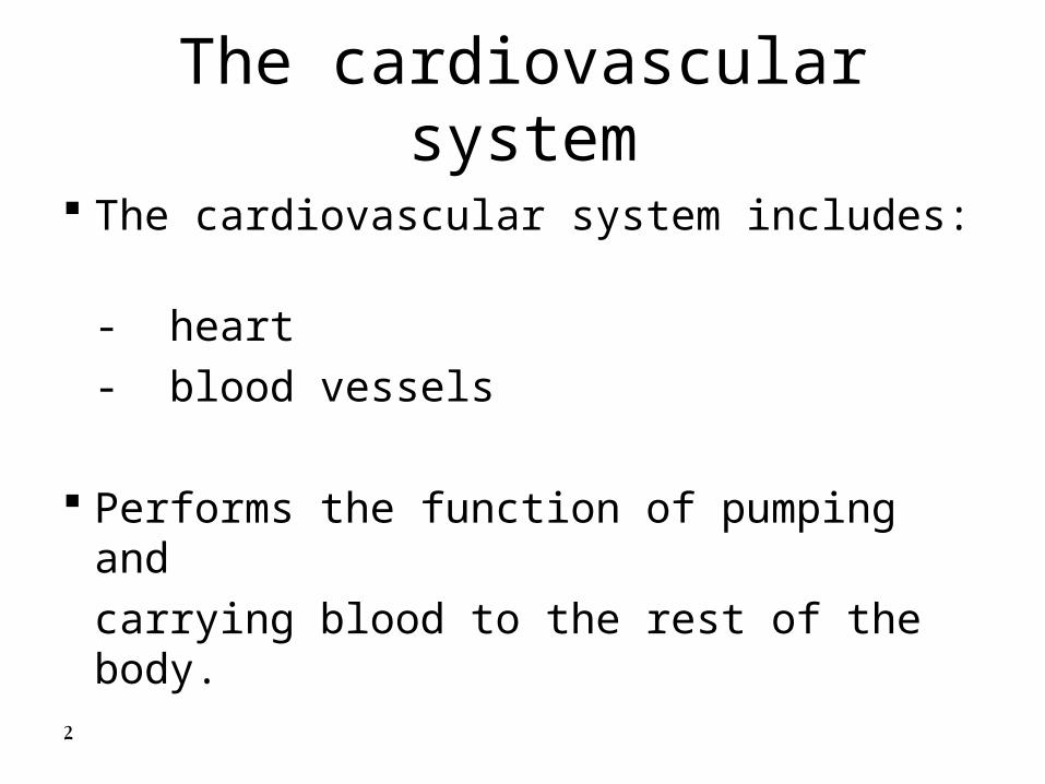
The Cardiovascular system
2
The cardiovascular system includes:
- heart
- blood vessels
Performs the function of pumping and
carrying blood to the rest of the body.
The cardiovascular system

3
The Heart
-The heart is the central organ of the thorax
- In the dog, the heart lies caudoventrally between the third and seventh ribs and slightly to the left.
- In the horse, the heart lies dorsalventraly between the third and sixth ribs and slightly to the left.
4
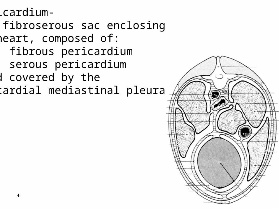
Pericardium- the fibroserous sac enclosingthe heart, composed of: A. fibrous pericardium B. serous pericardium - and covered by the pericardial mediastinal pleura

5
A. Fibrous pericardium- tough, fibrous sacsurrounding the serous pericardium, the heartand pericardial cavity

6
B. Serous pericardium- a serous membrane forminga closed cavity, composed of :
1. Parietal layer of Serous pericardium -
lines the inner surface of the fibrous pericardium

7
B. Serous pericardium- cont.2. Visceral layer of Serous pericardium -
covers the myocardium of heartalso called: epicardium

8
C. pericardial cavity: potential space betweenthe visceral and parietal layers of serous pericardium
contains ~ 1 ml of serous fluid that acts as a lubricant

9 the pericardial mediastinal pleura covers the fibrous pericardium


11
Structure
The heart is roughly conical with the base being dorsal and the apex close to the sternum opposite the sixth costal cartilage.

12
Structure - The base is formed by the thin walled atria which are clearly separated from the ventricles by an encircling coronary groove
- coronary groove contains the main coronary vessels.

13
General anatomy of the heart
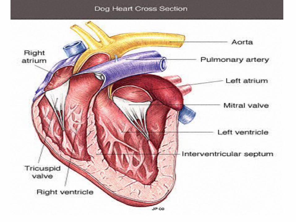
In the adult it consists of four chambers:
1. Right atrium
2. Left atrium
3. Right ventricle
4. Left ventricle

14
- The ventricles constitute a much larger part of - The ventricles constitute a much larger part of the heart which is much firmer than the atria the heart which is much firmer than the atria due to muscular wallsdue to muscular walls
- The left being much thicker than the right.- The left being much thicker than the right.
- The right ventricle lies as much cranially as to - The right ventricle lies as much cranially as to the right of the left ventricle. the right of the left ventricle.
StructureStructure

15
Right atrium The caudal vena cava enters the
chamber above the opening of the coronary sinus.
The cranial vena cava opens at the terminal crest.
The intervenous tubercle separates the two caval openings preventing confrontation between the two streams.
- The right azygous vein enters dorsally .

16

17
Caudal to the tubercle lies the fossa ovalis Caudal to the tubercle lies the fossa ovalis which corresponds to the foramen ovale of which corresponds to the foramen ovale of fetal life. fetal life.
The right auricle is a blind-ended sac extending to The right auricle is a blind-ended sac extending to the cranial face of the pulmonary trunk.the cranial face of the pulmonary trunk.
Pectinate muscles within the auricle branch from Pectinate muscles within the auricle branch from the terminal crest which marks the boundary the terminal crest which marks the boundary between the auricle and the main atrium.between the auricle and the main atrium.
Right atriumRight atrium

18

19
Left atrium
Similar form to the right atrium.
Receives pulmonary veins which enter at two or three sites.
Auricle similar to right.
20
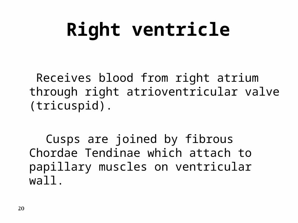
Right ventricle
Receives blood from right atrium through right atrioventricular valve (tricuspid).
Cusps are joined by fibrous Chordae Tendinae which attach to papillary muscles on ventricular wall.

21

22
Lumen of ventricle crossed by septomarginalis Lumen of ventricle crossed by septomarginalis trabecula trabecula (muscle band extend from septum to (muscle band extend from septum to lateral wall).lateral wall).
Blood flows through semilunar pulmonary valve Blood flows through semilunar pulmonary valve to pulmonary trunk. to pulmonary trunk.
Right ventricleRight ventricle
23

24
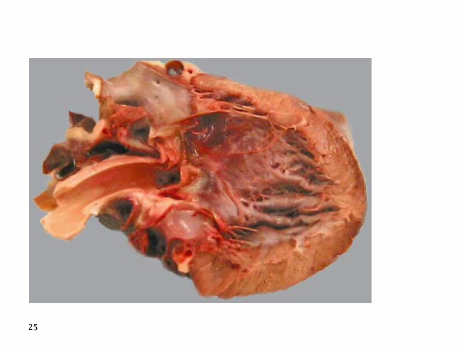
Left ventricle Left atrioventricular valve is bicuspid and
much larger and stronger than the right. The ventricular wall is much thicker than
on the right due to high resistance of the systemic circulation.
Blood leaves the ventricle via the
semilunar aortic valve into the aorta.
25

26

27
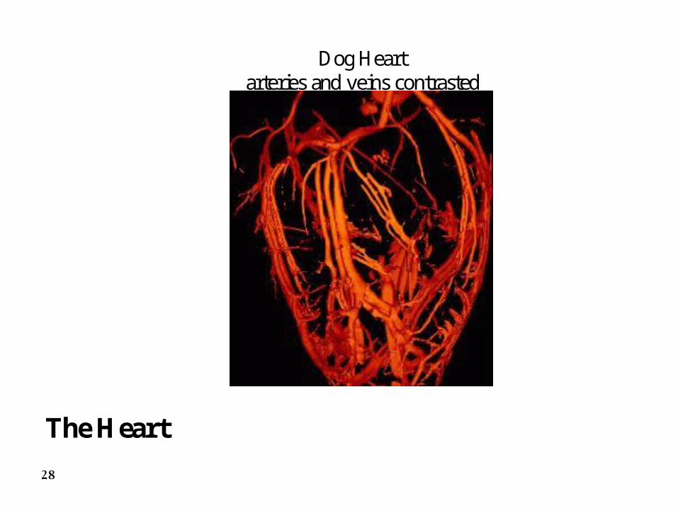
Cardiac vessels
• • The heart tissue receives 15% of the cardiac output from the left ventricle.
• This travels in two arteries from sinuses just above the aortic valve.
28
Dog Heart arteries and veins contrasted
The Heart

29
The coronary veins
– Blood returns to the heart via the cardiac vein.
– This opens into the the right atrium via the
coronary sinus just below the entrance of the caudal vena cava.

30
Systemic circulation
• The systemic arteries:
• The aortic arch
• The origin of the aorta is similar to that of the pulmonary trunk but is from the left ventricle.
31

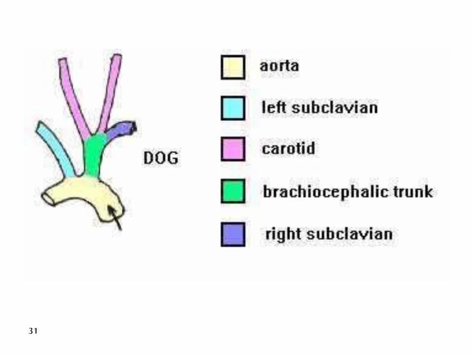
Arteries and Veins Cranial to the Heart
1. Brachiocephalic trunk- 1st branch from the aortic arch, gives rise to: (2) left common carotid artery and terminates as the (3)right common carotid artery and (4) right subclavian artery
5. left subclavian artery- originates from aortic arch beyond the brachiocephalic trunk
dog-dorsal view
1
2 3
4
5 11Cow Horse
2 23 3
6 65
5
4
4
77
88
I. Arteries- Branches of the Aortic Arch
B. Horse and Ruminant: 1. Brachiocephalic trunk- only branch from the aortic arch. (2) left and (3) right common carotid arteries do not arise separately as in dog, but from a (6)common bicarotid trunk
A. Dog
heart
heartheart
Dog-ventral view
1
23
4
5

33
The common carotid arteries supply structures of the head.
The subclavian artery supplies blood to the forelimb and to structures of the neck and cerviocothoracic junction .
It winds around the cranial border of the first rib to enter the limb through the axilla ,it changes its name to axillary at this point.

34
The subclavin detaches four branches in its intrathoracic course:
1. The vertebral artery
1. The costocervical trunk
1. The internal thoracic artery
1. The superficial cervical artery

35
The thoracic aorta
• The thoracic aorta runs caudally below the roof of the thorax to enter the abdomen by the aortic hiatus of the diaphragm .
• It continues as the abdominal aorta in company with the azygous vein and thoracic duct.

36
The systemic veins
The systemic veins return blood to the heart through the cranial vena cava , caudal vena cava and coronary sinus.
The cranial vena cava :
- is formed close to the entrance to the chest by the union of the external jugular and subclavian veins.

37
The caudal vena cava:The caudal vena cava:
- is formed on the abdomen near the pelvic - is formed on the abdomen near the pelvic inlet , by the union of right and left common inlet , by the union of right and left common iliac veins.iliac veins.
The systemic veinsThe systemic veins

Diaphragm
Diaphragm consists of contractile skeletal muscle, innervated by phrenic nerve
Openings in Diaphragm: 1. Aortic hiatus-(hiatus- term for gap, cleft, or opening)-dorsal part of diaphragm, passage of aorta, azygos, and thoracic duct 2. Esophageal hiatus- centrally located and ventral to aortic hiatus,passage of esophagus
3. Caval foramen- to right\ventral of esophageal hiatus, passage of caudal vena cava
dog