The Cardiovascular system Components: –Blood –Heart –Blood vessels.

Your Blood & Cardiovascular System

Cardiovascular system:
• Heart
• Blood vessels
• Blood
Major function:
Carry gases, nutrients, wastes
1 place another place in body
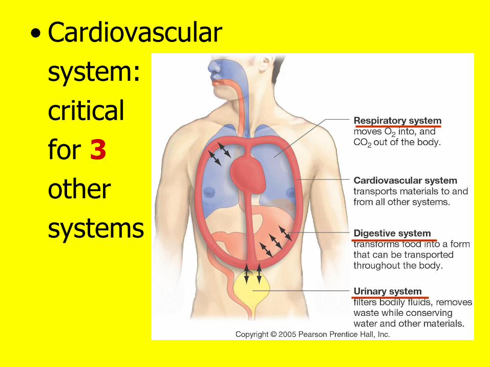
• Cardiovascular
system:
critical
for 3
other
systems

What’s carried in your blood?
1. Cells: red, white, platelets
2. Nutrients: glucose, amino acids, fats
3. Vitamins: A, B, C, D, E, K
4. Wastes: urea, CO2
5. Gases: O2, CO2
6. Hormones: insulin
7. Proteins: hemoglobin

Blood centrifuge spin
Red, Plasma
white cells, (liquid)
platelets

Red Blood Cells (RBC’s)
• Contain hemoglobin
• Carry O2: Lungs Tissues
• CO2 (cell respiration)
carried in plasma + hemoglobin
Exhaled lungs

RBC: cell membrane + hemoglobin • No nucleus • Biconcave shape • Live 120 days • Wear out: Trapped in liver, spleen, bone marrow: destroyed- phagocytes: parts recycled

RBC’s, white blood cells, platelets: produced from stem cells in red blood marrow

White blood cells (WBC’s)
• Important: your immune system
• Travel in blood
Tissue- site
of injury/invasion
bacteria/viruses/
foreign organism

Platelets: not cells- parts of cells: contain enzymes
Important: blood clotting: temporary plug in injured blood vessel
Followed by clotting
process-
permanent
plug

Plasma: 92% water
Plasma proteins
1. Albumin (liver): carry hormones & fatty acids
2. Fibrinogen (liver): important- clotting process
3. Immunoglobulins= antibodies
Attack foreign proteins/disease causing organisms
4. Lipoproteins: carry fat: LDL & HDL

Blood vessels: different size tubes- like plumbing pipes

Draw the circulatory system Arteries Veins Capillaries

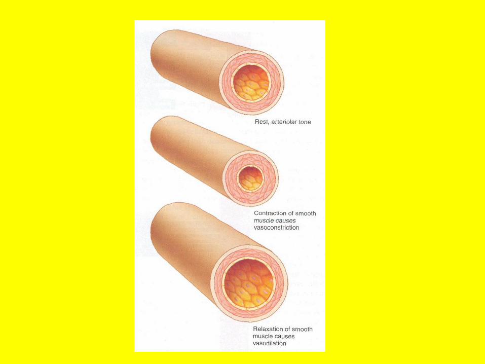
Blood vessels: can change diameter: nerve impulses
smooth muscle cells in walls
1. Vasoconstriction: smaller
2. Vasodilation: larger

1. Arteries: carry blood away from heart A=AWAY
2. Connect to capillaries: walls- single layer of cells: O2 & CO2
move in & out of tissues
3. Veins: return blood back to heart


Pulse: Throbbing- expansion/contraction of artery in time with heartbeat
Resting Heart Rate: rate needed to supply tissues at rest
• Measure- morning before getting up
• Aerobic exercise: resting rate

Pulse rate (beats/minute)
78-82
70-72

Your heart 2 upper chambers: right & left atria 2 lower chambers right & left ventricles

Pretend you are a single red blood cell
Trace your movement though the circulatory system

• Right atrium to right ventricle • Right atrium
• 2 vena cava (large veins) • Blood in veins- returning from tissues- darker red (deoxygenated)

Valves close- no
backflow
Right ventricle:
blood to lungs
Give up CO2
Pick up O2

Blood (redder- oxygenated)
returns to left
atrium
left atrium to
left ventricle
Values: backflow

Left ventricle to
aorta (large artery)
valves: backflow
Blood to head & entire body
Supply tissues O2

End in capillary
bed where you
started

Artery
Arteriole
Capillary (bed)
Venule
Vein

Capillary bed: where the action is in all tissues

Capillaries:
small,
very thin-
walls: single
cell layer
RBC’s move
through
single file

O2, glucose
diffuse from
inside capillary
to fluid
surrounding
cells cells
for metabolism:
Energy & ATP’s

O2, glucose diffuse from high concentration (blood) to lower concentration (cells): concentration gradient

CO2 (waste from cell respiration) diffuses from cells to blood in capillaries lungs (high low)
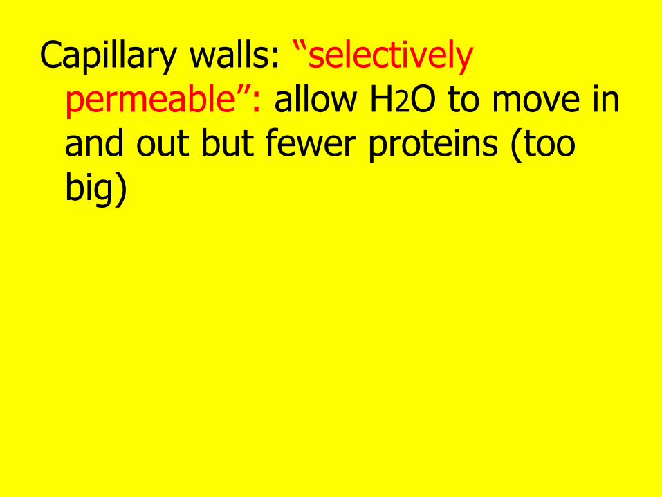
Capillary walls: “selectively permeable”: allow H2O to move in and out but fewer proteins (too big)

At this end (high pressure)- water moves out of capillaries to cells
At this end (lower pressure)- water moves back into capillary blood by osmosis

Blood pressure in veins: low
How does blood get back to heart?
Answer: your
skeletal
muscles- when
they contract:
squeeze veins-
helps blood move
back to heart

Valves in veins prevent backflow

Varicose veins:
defective veins-
backflow of blood
Pregnancy, jobs
people standing
all day, obesity

William Harvey:
discovered
circulatory
system: 1628
Demonstrated
valves in veins

How your heart beats
Pumping
(contraction)
and filling
(relaxation)
= cardiac
cycle

Heart Rate:
~ 72
beats/min
1. Entire heart
relaxed:
Diastole
“filling phase”
0.4 sec

Blood flows
into all
4 chambers
“AV” valves
between atria &
ventricles: open
Ventricles: fill
with blood

Systole:
“contraction
Phase”
First: 0.1 sec
contraction-
of atria:
ventricles fill
completely

2d part:
Ventricles
contract
(0.3 sec)
AV
valves
close

“AV” Valves Opened and Closed

“Semilunar” (half-moons) valves
open: Blood pumped
Right Ventricle Left Ventricle
Lungs Aorta & rest
of body

Semilunar Valves opened and closed

Each ventricle
Pumps
~ 70 ml blood/beat:
Cardiac output
Well- trained athlete:
stronger/enlarged
heart
Cardiac Output

Heart murmurs:
valves don’t close
properly- blood
turbulents
Heart sounds:
lub & dup
valves closing
Lub: AV
Dup: Semilunar

Blood Pressure: depends on
A) Volume blood pumped by heart
B) Resistance to blood flow: blood vessels
Systolic pressure: ventricles contract: blood flow from big aorta to small
arterioles: creates pressure
Arterioles: elastic stretch

Diastolic
Pressure:
arterioles
“snap back”
pressure

Blood
Velocity
(speed)
• High in
aorta
• Slow-
Capillaries
• Speeds up-
veins

Blood Pressure Measurement

• Wrap cuff- upper arm & inflate
• Cuff pressure closes artery: cuts off blood flow
• Listen with Stethoscope

• Deflate cuff
• Hear 1st sound: blood spurts through constricted artery= systolic pressure

• Continue to deflate, hear blood flow
• Sound stops: even blood flow
• Artery pressure > cuff pressure
• Diastolic pressure

Cardiac muscle cells:
• “inherent” ability
to beat (contract &
relax) without
nervous system
• Heart can beat in
lab dish

Heart also has
2 sets of nerves:
Speed up or Slow
Down
Hormones
(epinephrine) also
affect heart rate

What sets pace of Heart?
Pacemaker:
Sinoatrial
node
Upper
right wall-
Right atrium

SA node
1st Signals
Right atrium
&
Left atrium
Contract together

2d signals
Relay
point
Atrioventricular
(AV)
Node

AV Node
Heart apex:
Spread up
Walls
Right Left
Ventricles
Strong Contractions

Electrical Signals
in heart:
electrical
changes
in skin
record:
Electrocardiogram
(ECG, EKG)

See electrical events in heart
Detect abnormal electrical activity
arrhythmias- abnormal heart rhythms

Heart attack:
Abnormal rhythm
of ventricles
“Ventricular
Fibrillation”
(bag of worms)
Defibrillators
Electric shocks to chest
Re-set heart electrical system

Artificial Pacemakers
If heart doesn’t
keep normal
rhythm
Surgically
implanted
near heart
battery, signals:
normal heart beat

Defibrillators also implanted
1. Detect abnormal rhythms
2. Send jolt to heart
3. Restore rhythm

Cardiovascular Disease
• Heart takes care
of itself first
• 2 arteries: blood
from aorta to
heart muscle
(myocardium)
Supply O2 +
glucose

Coronary arteries:
branches form
coronary
circulation
“coronary”-
Latin for
“crown” –
encircles
heart

Beginning in children (5-12 years)
see thickenings & fatty streaks in coronary arteries

Disease process: atherosclerosis-
Accumulation of lipids (cholesterol), protein, calcium, scar tissue in arteries
Going on
quietly
in you now
Also in arteries of
brain, arms, legs


Can cause heart attack: coronary arteries in heart

Heart attack: Warning signs
• Heavy pressure, fullness, squeezing pain in center of chest
• Pain may spread: arms, back, neck, jaw, or stomach
• Cold sweat
• Nausea and vomiting
• Lightheadedness

Heart Attack Pain: may spread

Heart attacks: more common morning, on birthdays

Peripheral Artery Disease

Blockage of neck carotid arteries
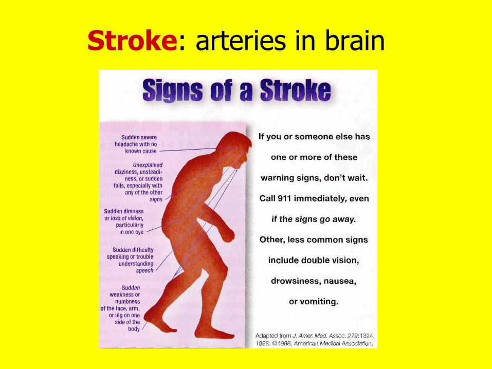
Stroke: arteries in brain

Heart Disease Risk Factors
1. Men > Women
2. Family history: early heart attacks
3. Age
4. Genetics: African Americans (high blood pressure), Mexican and native Americans (obesity & diabetes)

Heart Disease Risk Factors
5. Smoking
6. Blood lipids LDL HDL
7. High Blood pressure
8. Diabetes
9. Obesity
10.Sedentary Lifestyle
11.Job stress

Heart Disease Risk Factors
12. Diet & Exercise
Saturated (animal) fat, trans fat, cholesterol, salt
Fiber, fruits & veggies, exercise

High Blood Pressure

Know these numbers
Systolic/Diastolic
Normal 120/80
Pre-hypertension 121-139/81-89
Hypertension 140/90 or >
Systolic: heart pumping
Diastolic: heart relaxing

DIETARY GUIDELINES SODIUM & POTASSIUM TOO MUCH & TOO LITTLE

TOO MUCH SALT (SODIUM)
Blood pressure
Heart attack (#1 killer)
Stroke (#3 killer)
Heart failure
Kidney Disease

Where does sodium come from?
12%: naturally- foods
11%: you- salt shaker
77%: processed foods- added by companies


Kidney stones US children (2008)
Oxalates (food)
binds to
calcium stone
2 risk factors:
1) not enough
drinking of fluids Tessa Cesario
2) too much salt 11 years old

Potassium & Blood Pressure
Potassium: Anti-salt
Blood pressure
Risk- stroke
Kidney stones
Bone loss
Recommendation: 4,700 mg/day
Average American: ½ this amount

Simple way: better balance
Added salt/processed foods
Fruits & veggies
(low sodium, high potassium)

Cold Therapy: Therapeutic
Hypothermia
“Quasi-hibernating” state

1999: 29 year old woman doctor falls into river in Norway while skiing
• Carried by currents- stuck in ice flow
• 1 hour later: rescued- no heart beat
• Temperature 57 F
• CPR 9 hours treatment slow warming
• 60 days- intensive care
• Back to work: 5 months, skiing year later

What happened to her body?
• Body cooled
• Cells need less O2
• Metabolism 10% of baseline value
• She was in “suspended animation”
• Between life and death

Cooling Treatment: Medical Applications
• Today- induced mild hypothermia for delicate heart, brain, spinal cord surgery
• Cooling techniques: cooling blankets,
ice packets, circulating ice cold saline, cooled blood through heart-lung bypass machine
• Body cooled to 60 F: heart stops beating (cardiac standstill)

Hypothermia: reduces clotting, slows metabolism, O2 demand
After surgery: heat exchanger on heart-lung machine:
slowly raises
body temperature

Emergency Applications
• Some hospitals put comatose cardiac arrest patients “on ice” after heart
re-started
• Reduces
brain damage,
reduces
inflammation
after resuscitation

January 2008
Directive:
NY City
ambulances:
Take cardiac
arrest patients to
hospitals with Cooling Therapy
available: to protect the brain
even if not nearest hospital

Cocaine mimics heart attack

Hospital emergency rooms
admitting young people:
Chest pain, shortness
breath, anxiety, palpitations,
dizziness, nausea, heavy sweating
• All heart attack symptoms
• But without heart disease risk factors
• Cause: cocaine use ( B.P., heart rate, vasoconstriction)
• Real heart attack vs. cocaine use: important differences in treatment