VISUAL LOSS AND EYE CONDITIONS - CECity...2 OBJECTIVES Know and understand: • The leading causes...
43
1 VISUAL LOSS AND EYE CONDITIONS
Transcript of VISUAL LOSS AND EYE CONDITIONS - CECity...2 OBJECTIVES Know and understand: • The leading causes...
1
VISUAL LOSS AND EYE CONDITIONS

2
OBJECTIVES
Know and understand:
• The leading causes and pathophysiology of visual loss
• Techniques for preventing and treating visual loss
• The signs of and treatments for common eye disorders in older people
• Techniques for low-vision rehabilitation

3
TOPICS COVERED
• Common Eye Conditions • Causes of Visual Loss
Ø Refractive Error Ø Cataract Ø Age-related Macular Degeneration (ARMD) Ø Diabetic Retinopathy Ø Glaucoma Ø Ischemic Optic Neuropathy
• Low-Vision Rehabilitation Strategies
4
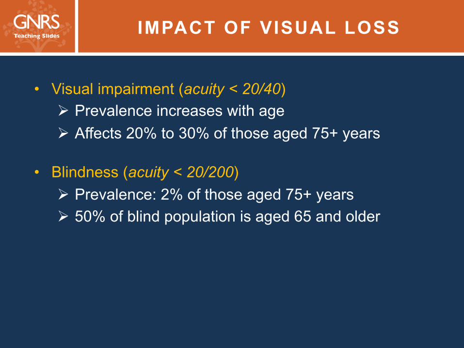
IMPACT OF VISUAL LOSS
• Visual impairment (acuity < 20/40) Ø Prevalence increases with age Ø Affects 20% to 30% of those aged 75+ years
• Blindness (acuity < 20/200) Ø Prevalence: 2% of those aged 75+ years Ø 50% of blind population is aged 65 and older
5 SCREENING TO PREVENT VISUAL LOSS
The American Academy of Ophthalmology recommends comprehensive eye examinations every 1 to 2 years for people ages 65 years and older

6 COMMON EYE CONDITIONS IN OLDER ADULTS
• Red eye, ocular swelling or discomfort, diplopia, sudden loss of vision, and floaters are common eye complaints
• Ask, “Has your vision changed?”
• Decreased vision can indicate a serious condition
Ø Check visual acuity Ø Check for afferent pupillary defect

7 S I G N S A N D S Y M P TO M S O F E Y E C O N D I T I O N S R E Q U I R I N G I M M E D I AT E R E F E R R A L TO
O P H T H A L M O L O G I S T ( 1 o f 2 )
Condition Symptoms and signs Retinal detachment Flashes, floaters, decreased vision
Acute angle-closure glaucoma
Eye pain or headache, ocular hyperemia, hazy cornea, dilated pupil, decreased vision, nausea, vomiting
Ischemic optic neuropathy
Sudden loss of vision (complete or partial) in one eye, swollen optic nerve
Central artery occlusion or giant cell arteritis
Sudden painless loss of vision in one eye; if from giant cell arteritis, then review of symptoms may reveal jaw claudication, headache, transient diplopia, etc.
8 S I G N S A N D S Y M P TO M S O F E Y E C O N D I T I O N S R E Q U I R I N G I M M E D I AT E R E F E R R A L TO
O P H T H A L M O L O G I S T ( 2 o f 2 )
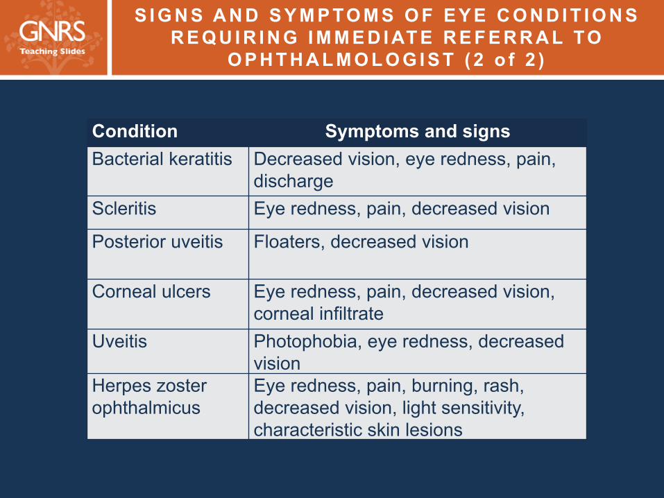
Condition Symptoms and signs Bacterial keratitis Decreased vision, eye redness, pain,
discharge Scleritis Eye redness, pain, decreased vision
Posterior uveitis Floaters, decreased vision
Corneal ulcers Eye redness, pain, decreased vision, corneal infiltrate
Uveitis Photophobia, eye redness, decreased vision
Herpes zoster ophthalmicus
Eye redness, pain, burning, rash, decreased vision, light sensitivity, characteristic skin lesions

9
T R E AT M E N T O F E Y E C O N D I T I O N S C O M M O N LY S E E N B Y P R I M A RY C A R E P R O V I D E R S ( 1 o f 3 )
Condition Treatment and/or cause Red eye
Subconjunctival hemorrhage
Supportive treatment with artificial tears
Dry eye Artificial tears, cyclosporin 0.2% eye drops Blepharitis Lid scrubs, ophthalmic antibiotic ointment qhs
to eyelids, oral doxycycline Lid malposition or lid exposure
Ocular lubricant, refer for surgical repair
Allergic conjunctivitis Cold compresses, allergen avoidance, topical/systemic antihistamines

10
T R E AT M E N T O F E Y E C O N D I T I O N S C O M M O N LY S E E N B Y P R I M A RY C A R E P R O V I D E R S ( 2 o f 3 )
Condition Treatment and/or cause Red eye (continued)
Viral conjunctivitis Supportive treatment with artificial tears; refer to ophthalmologist if vision significantly affected
Chalazion Warm compresses, may refer for excision Herpes simplex keratitis
Trifluridine eye drops, refer to ophthalmologist
Herpes zoster ophthalmicus
Tear drops, refer to ophthalmologist immediately if there are lesions on tip of nose (Hutchinson’s sign)
Angle-closure glaucoma
Acetazolamide oral or i.v., refer to ophthalmologist immediately
11
T R E AT M E N T O F E Y E C O N D I T I O N S C O M M O N LY S E E N B Y P R I M A RY C A R E P R O V I D E R S ( 3 o f 3 )
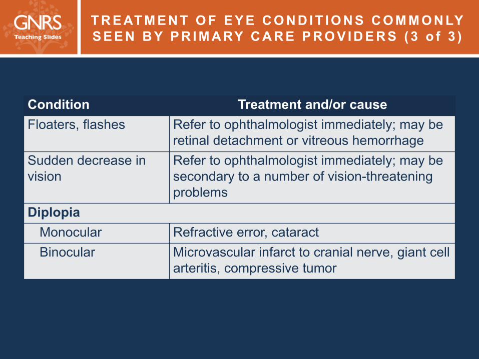
Condition Treatment and/or cause Floaters, flashes Refer to ophthalmologist immediately; may be
retinal detachment or vitreous hemorrhage Sudden decrease in vision
Refer to ophthalmologist immediately; may be secondary to a number of vision-threatening problems
Diplopia Monocular Refractive error, cataract Binocular Microvascular infarct to cranial nerve, giant cell
arteritis, compressive tumor
12
KERATITIS SICCA (DRY EYES)
• Tear production decreases with age
• Characteristics: redness, foreign body sensation, and reflex tearing
• Management: artificial tears during daytime and ointment at bedtime
• Topical cyclosporin A (0.2%) eye drops in severe cases to treat underlying inflammatory causes
• Treat accompanying blepharitis
13
LID ABNORMALITIES
• Common among older adults
• Elasticity and tensile strength are gradually lost with age
• Blepharochalasis (drooping of the brow) and blepharoptosis (drooping of the eyelid) may cause cosmetic deformity and, if severe, impair vision
• Lid ectropion (eversion) or entropion (inversion) may cause discomfort
• Treatment: various surgical procedures

14
HERPES ZOSTER OPHTHALMICUS
• Painful reactivation of varicella zoster virus
• Dermatomal distribution of weeping vesicles affecting the ophthalmic branch of the trigeminal nerve
• Hutchinson’s sign: lesions on the tip of the nose
• Oral acyclovir or famciclovir may shorten the course
• Post-herpetic neuralgia may be debilitating Ø Treat with local ointments (capsaicin, lidocaine) but not in eye,
OR Ø Treat with systemic medications (off-label): narcotics, tricyclic
antidepressants, gabapentin, pregabalin
15
REFRACTIVE ERROR
• Leading cause of visual impairment worldwide, along with cataracts
• Treatment: eyeglasses, contact lenses, laser refractive surgery
• Ametropia Ø Myopia (nearsightedness) Ø Hyperopia (farsightedness) Ø Astigmatism (visual distortion)
• Presbyopia (↓ ability to focus on near objects) Ø Begins after age 40 Ø Caused by gradual hardening of the lens and decreased
muscular effectiveness of the ciliary body

16
CATARACT (1 of 2)
• Symptoms include ↑ glare, ↓ contrast sensitivity, ↓ visual acuity, change in color perception
• Risk factors: ↑ age, ↓ vitamin intake, light (ultraviolet B) exposure, smoking, alcohol use, long-term corticosteroid use, diabetes mellitus
20%
50%
>65 years >75 years
Percentage of population with cataracts
17
CATARACT (2 of 2)
Treatment: surgical extraction
• 90% of patients achieve vision ≥ 20/40
• 3 million surgeries are performed annually in US
• Local or topical anesthesia, small-incision sonographic breakdown and aspiration of the lens, placement of an artificial lens
18 AGE-RELATED MACULAR DEGENERATION (1 of 2)
• Most common cause of irreversible blindness among older adults in developed world
• Risk factors: age, genetics, smoking, hypertension, fair skin
• Diagnosis: presence of drusen (dry form) or of choroidal neovascularization (CNV) (wet form)
• Treatment Ø Vitamin C, vitamin E, zinc, lutein, zeaxanthin—to decrease
risk of CNV in dry forms Ø Intravitreal injections of vascular endothelial growth factor
(VEGF) inhibitors—to treat CNV in wet form
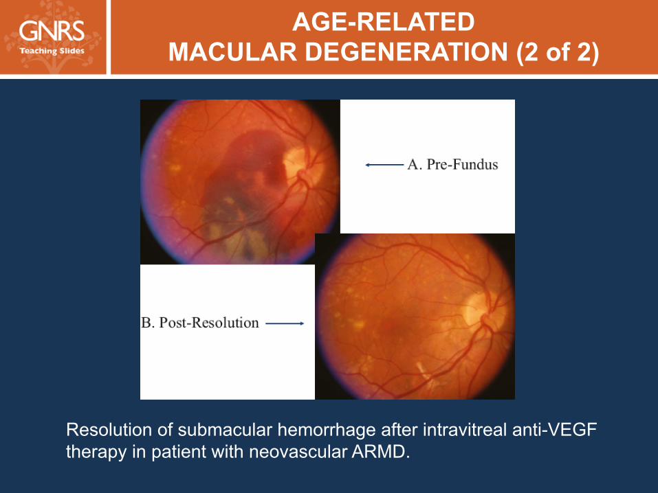
19 AGE-RELATED MACULAR DEGENERATION (2 of 2)
Resolution of submacular hemorrhage after intravitreal anti-VEGF therapy in patient with neovascular ARMD.

20
DIABETIC RETINOPATHY
• Epidemiology: Among people who have had type 2 diabetes at least 10 years: Ø 70% show retinopathy Ø Nearly 10% show proliferative disease
• Most important risk factors: Duration of disease, control of blood sugar and BP
• Prevention: Tight glucose control and BP control; however, targets should be individualized for geriatric patients
• Treatment: Laser treatment and intravitreal injections; control of blood glucose and BP
21
DIABETIC RETINOPATHY STAGES
• Nonproliferative
• Preproliferative (severe nonproliferative)
• Proliferative
22 DIABETIC RETINOPATHY: NONPROLIFERATIVE (1 of 2)
• Microaneurysms
• Intraretinal hemorrhages
• Exudates
• Macular edema

23 DIABETIC RETINOPATHY: PREPROLIFERATIVE (1 of 2)
• Multiple intraretinal hemorrhages
• Venous caliber changes
• Intraretinal microvascular abnormalities (capillary shunting)
• Capillary nonperfusion or ischemia

24 DIABETIC RETINOPATHY: PREPROLIFERATIVE (2 of 2)
Severe nonproliferative diabetic retinopathy with macular edema before and after anti-VEGF therapy.
25 DIABETIC RETINOPATHY: PROLIFERATIVE (1 of 2)
• Neovascularization of the retina
• Neovascularization of the disc
• Visual loss due to vitreous hemorrhage or traction retinal detachment

26 DIABETIC RETINOPATHY: PROLIFERATIVE (2 of 2)
Resolution of severe neovascularization of the disc after intravitreal anti-VEGF therapy in proliferative diabetic retinopathy
27
OVERVIEW OF GLAUCOMA
• Defined as characteristic optic nerve head damage and visual field loss
• Affects >2.25 million Americans 40+ years old
• Second most common cause of irreversible blindness worldwide; most common cause among black Americans
• >3 million office visits each year
• Elevated intraocular pressure is a major risk factor, but many patients with glaucoma have “normal” pressures
28 PRIMARY OPEN-ANGLE GLAUCOMA
• Most common form of glaucoma
• Slow aqueous drainage leads to chronically elevated intraocular pressure (IOP)
• Patients are asymptomatic and may suffer substantial visual field loss before consulting an ophthalmologist
• Causes are multifactorial and polygenic

29 ACUTE ANGLE-CLOSURE GLAUCOMA
• Precipitous increase in IOP
• Redness and pain with acute vision loss and often headache, nausea and vomiting
• Emergent ophthalmologic referral required

30
GLAUCOMA MANAGEMENT
• Intraocular pressure–lowering medications (local and systemic) Ø Aqueous suppressants
Ø Aqueous outflow facilitators
• Laser trabeculoplasty
• Filtering surgery ± antimetabolite
• Drainage devices
• Ciliary body destructive procedures

31 ANTERIOR ISCHEMIC OPTIC NEUROPATHY (1 of 2)
• Microvascular occlusion of the blood supply to the optic nerve
• Due to atherosclerotic vascular disease or inflammation (temporal arteritis)
• Results in acute vision or field loss

32 ANTERIOR ISCHEMIC OPTIC NEUROPATHY (2 of 2)
Pallid swelling of the optic nerve head in an older adult patient with anterior ischemic optic neuropathy
33
CHARLES BONNETT SYNDROME
• Visual hallucinations experienced by patients with significant visual impairment
• May be elementary shapes or complex such as children, animals
• Patients have a clear sensorium, are aware that visions are not real
34
LOW-VISION REHABILITATION
• Available to patients with acuity < 20/60
• Improve lighting and provide reading material with bold, enlarged fonts and accentuated black-on-white contrast
• Magnification: high-plus spectacles, magnifiers, closed-circuit TV, telescopic devices
• Eccentric viewing for patients with ARMD with central macular pathology: training to use off-center fixation
• Talking devices or Braille for those who have lost vision altogether
• Smartphone apps can provide magnification, money recognition, and dictation functions
35
CHOOSING WISELY®
• Do not perform preoperative medical tests for eye surgery without specific indications.
• Most cases of acute conjunctivitis have a viral etiology.
Do not treat viral infections with antibiotics; if diagnosis is uncertain, patients may be followed closely for resolution.
• Do not place temporary or permanent punctal plugs for
mild dry eye syndrome before trying other medical treatment.
36
SUMMARY
• Visual loss occurs commonly among older adults and may lead to reduced quality of life, high medical care costs, and loss of independence
• Primary care providers should routinely screen older adults for visual loss
• Treatment options are available for many types of visual loss

37
CASE 1 (1 of 3)
• A 68-year-old woman describes vision problems.
Ø Progressive blurring of distance vision
Ø Increased difficulty with nighttime driving, which she attributes to worsening glare and haloes around lights
Ø She no longer needs glasses for reading.
• History: hypertension, hypercholesterolemia
38
CASE 1 (2 of 3)
Which one of the following is the most likely cause of the changes in her vision? A. Uncontrolled hypertension B. Poorly controlled blood glucose C. Choroidal neovascularization D. Cataract progression E. Increased intraocular pressure
39
CASE 1 (3 of 3)
Which one of the following is the most likely cause of the changes in her vision? A. Uncontrolled hypertension B. Poorly controlled blood glucose C. Choroidal neovascularization D. Cataract progression E. Increased intraocular pressure
40
CASE 2 (1 of 3)
• A 72-year-old black man has right shoulder trauma after he fell in a parking lot. He states that he was walking to his car when he suddenly tripped and fell forward onto his arm. He subsequently learned that he had tripped over a curb.
• History: hypertension, high cholesterol, glaucoma

41
CASE 2 (2 of 3)
Which one of the following most likely contributed to his fall?
A. Acute glaucoma
B. Failure to use glaucoma drops that morning
C. Poor peripheral vision
D. Retinal embolus from a carotid plaque

42
CASE 2 (3 of 3)
Which one of the following most likely contributed to his fall?
A. Acute glaucoma
B. Failure to use glaucoma drops that morning
C. Poor peripheral vision
D. Retinal embolus from a carotid plaque

43
GNRS5 Teaching Slides Editor: Barbara Resnick, PhD, CRNP, FAAN, FAANP, AGSF
GNRS5 Teaching Slides modified from GRS9 Teaching Slides
based on chapter by JoAnn A. Giaconi, MD
and David Sarraf, MD and questions by Holly B. Hindman, MD, MPH
Managing Editor: Andrea N. Sherman, MS
Copyright © 2016 American Geriatrics Society