06 CHRONIC VISUAL LOSS -...
187
CHRONIC CHRONIC VISUAL LOSS VISUAL LOSS Wasu Supakornthanasarn, MD.
Transcript of 06 CHRONIC VISUAL LOSS -...
CHRONICCHRONICVISUAL LOSSVISUAL LOSS
Wasu Supakornthanasarn, MD.
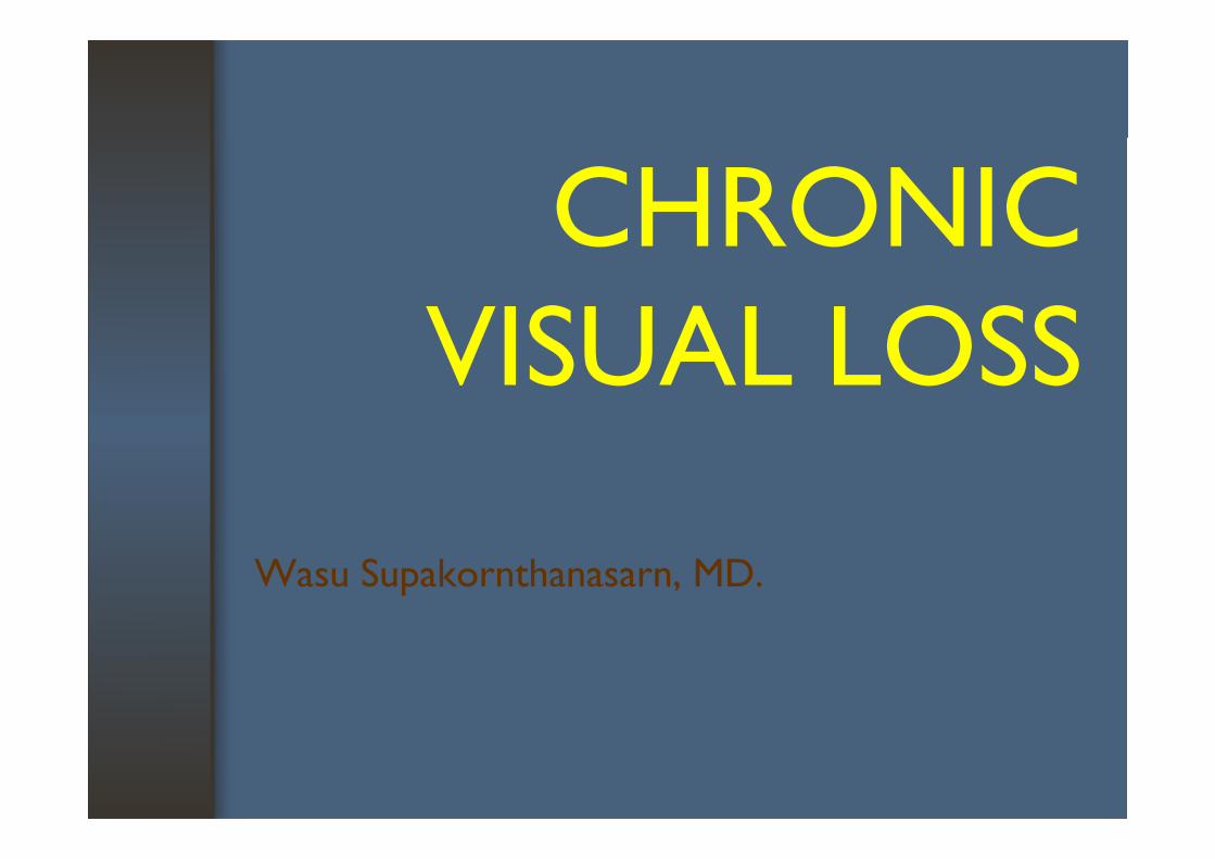
Visual loss
Refractive errors Cloudy of ocular media Sensory pathway Functional visual lossRefractive errors Cloudy of ocular media y p yabnormalities Functional visual loss
1.1 myopia 2 1 acute/subacute 3 1 acute/subacuteAmblyopiaMalingering
1 2 hyperopia
2.1 acute/subacute
2 2 gradual/chronic
3.1 acute/subacute
3 2 gradual/chronic
MalingeringHysteria
1.2 hyperopia 2.2 gradual/chronic 3.2 gradual/chronic
1.3 astigmatism 3.3 transient
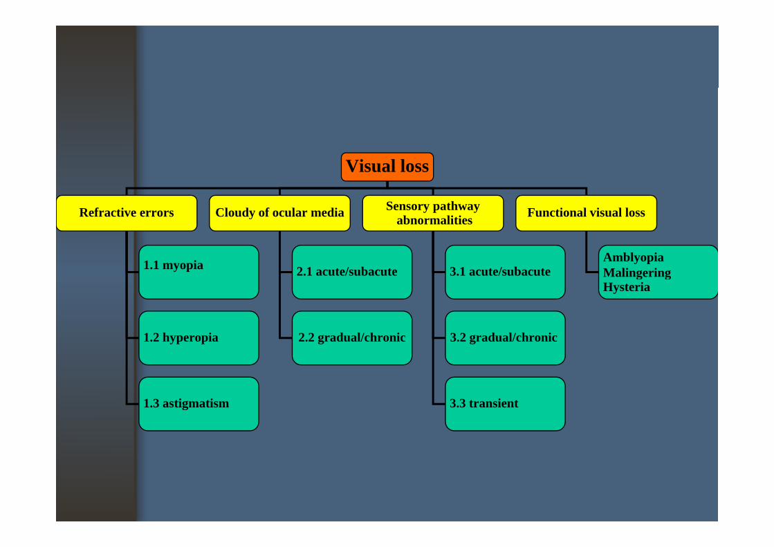
Refractive errorsRefractive errors
• Myopia• Hyperopia• Hyperopia• Astigmatism
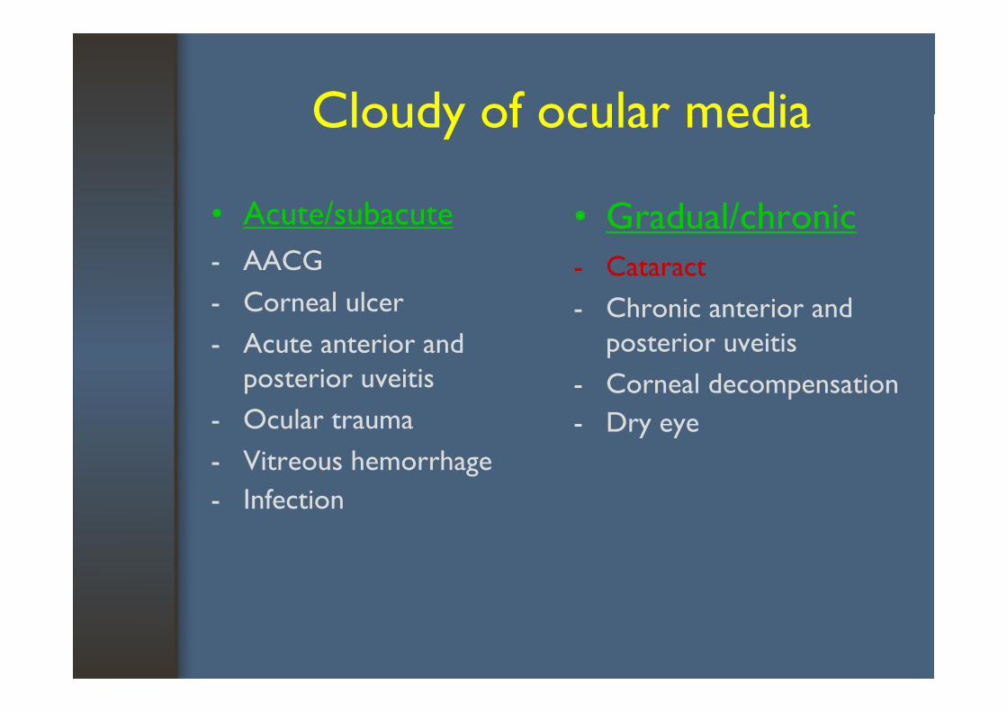
Cloudy of ocular mediaCloudy of ocular media
• Acute/subacute- AACG
• Gradual/chronic- Cataract
- Corneal ulcer- Acute anterior and
Cataract- Chronic anterior and
posterior uveitisposterior uveitis
- Ocular trauma
p- Corneal decompensation- Dry eye
- Vitreous hemorrhage- Infection
y y
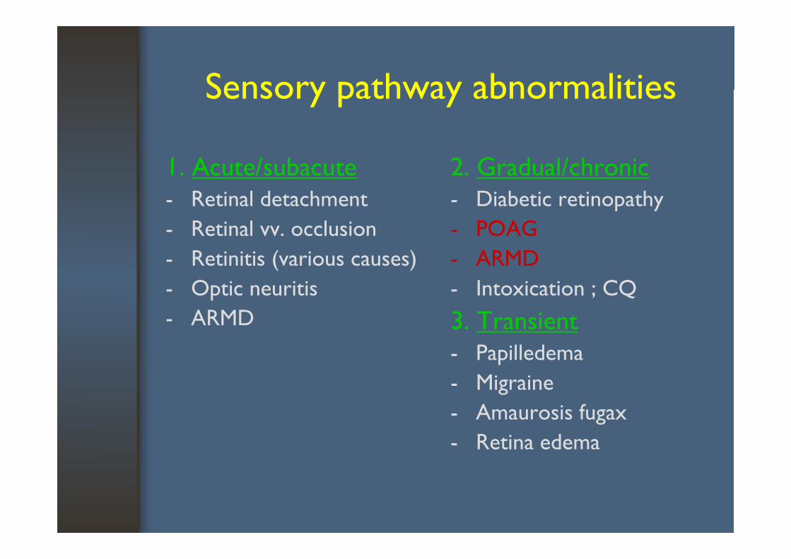
Sensory pathway abnormalitiesSensory pathway abnormalities
1. Acute/subacute- Retinal detachment
2. Gradual/chronic- Diabetic retinopathy
- Retinal vv. occlusion- Retinitis (various causes)
- POAG- ARMD
- Optic neuritis- ARMD
- Intoxication ; CQ3. Transient- Papilledema- Migraine
A i f- Amaurosis fugax- Retina edema

Visual loss
Refractive errors Cloudy of ocular media Sensory pathway Functional visual lossRefractive errors Cloudy of ocular media y p yabnormalities Functional visual loss
1.1 myopia 2 1 acute/subacute 3 1 acute/subacuteAmblyopiaMalingering
1 2 hyperopia
2.1 acute/subacute
2 2 gradual/chronic
3.1 acute/subacute
3 2 gradual/chronic
MalingeringHysteria
1.2 hyperopia 2.2 gradual/chronic 3.2 gradual/chronic
1.3 astigmatism 3.3 transient
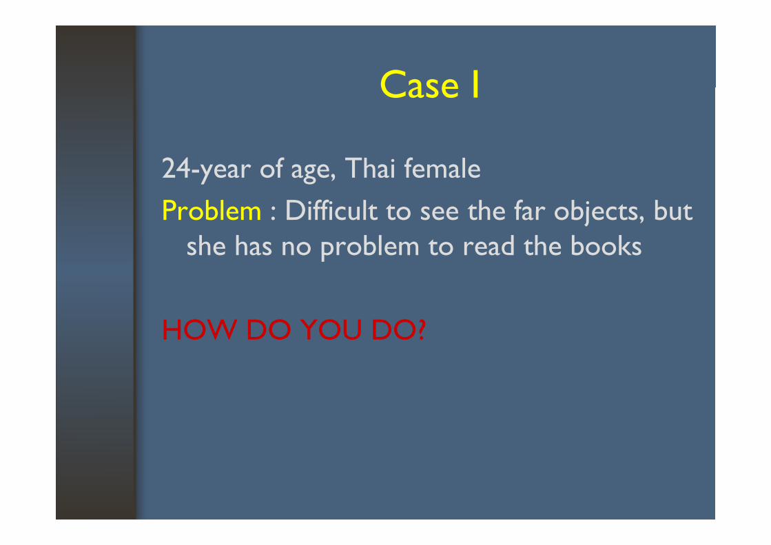
Case ICase I
24-year of age, Thai femaleProblem : Difficult to see the far objects but Problem : Difficult to see the far objects, but
she has no problem to read the books
HOW DO YOU DO?HOW DO YOU DO?
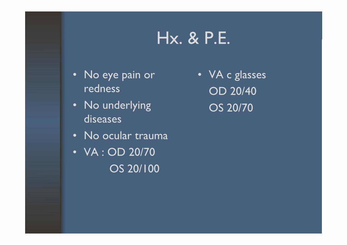
Hx & P EHx. & P.E.
• No eye pain or redness
• VA c glasses OD 20/40
• No underlying diseases
OD 20/40OS 20/70
diseases• No ocular trauma
VA OD 20/70 • VA : OD 20/70 OS 20/100
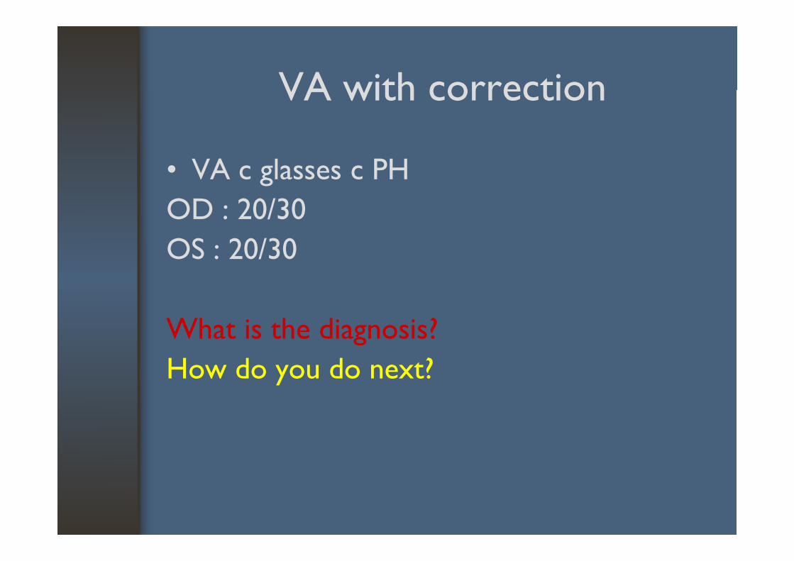
VA with correctionVA with correction
• VA c glasses c PH OD : 20/30OD : 20/30OS : 20/30
What is the diagnosis?gHow do you do next?
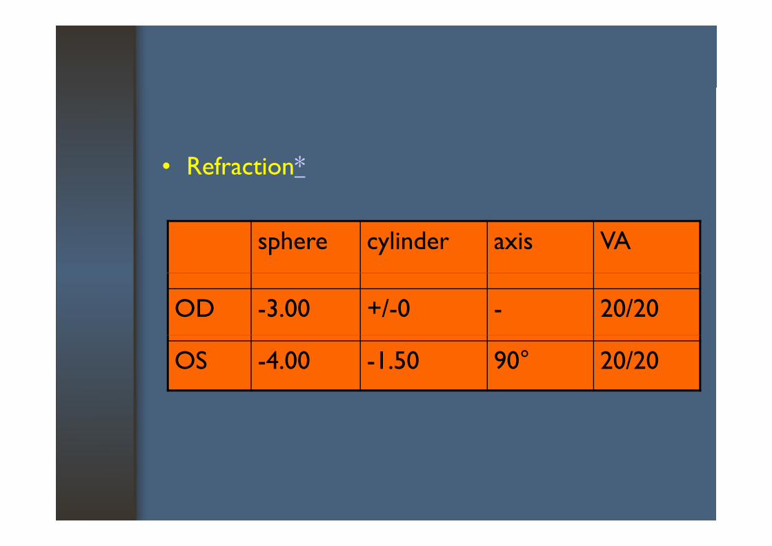
• Refraction*
sphere cylinder axis VA
OD -3.00 +/-0 - 20/20
OS -4.00 -1.50 90° 20/20
• Eye examination must be careful!!!
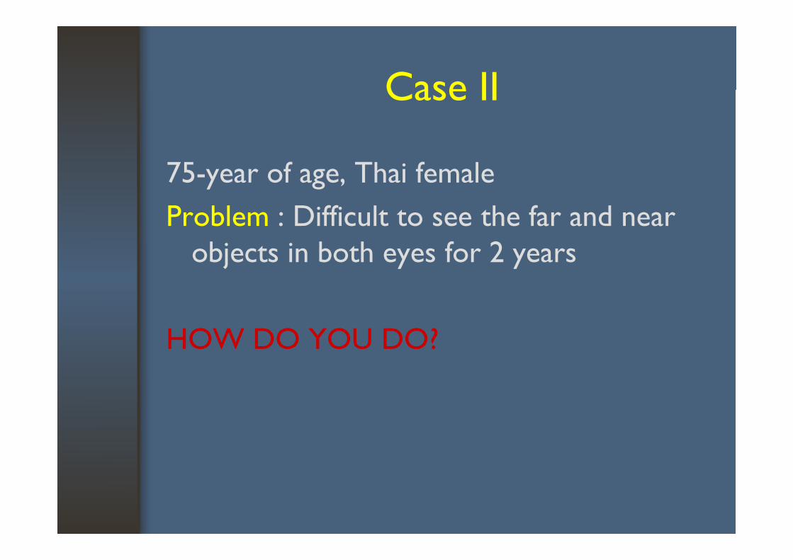
Case IICase II
75-year of age, Thai femaleProblem : Difficult to see the far and near Problem : Difficult to see the far and near
objects in both eyes for 2 years
HOW DO YOU DO?HOW DO YOU DO?

• History- Slow progressive of
• Eye examinationVA c glasses :Slow progressive of
blur vision- Painless
VA c glasses :OD : 20/200 OS HM- Painless
- No underlying di
OS : HM
diseases
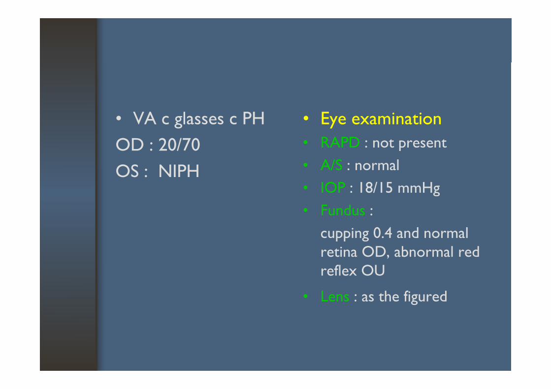
• VA c glasses c PH OD : 20/70
• Eye examination• RAPD : not presentOD : 20/70
OS : NIPH
p• A/S : normal• IOP : 18/15 mmHgg• Fundus :
cupping 0.4 and normal pp gretina OD, abnormal red reflex OU
• Lens : as the figured
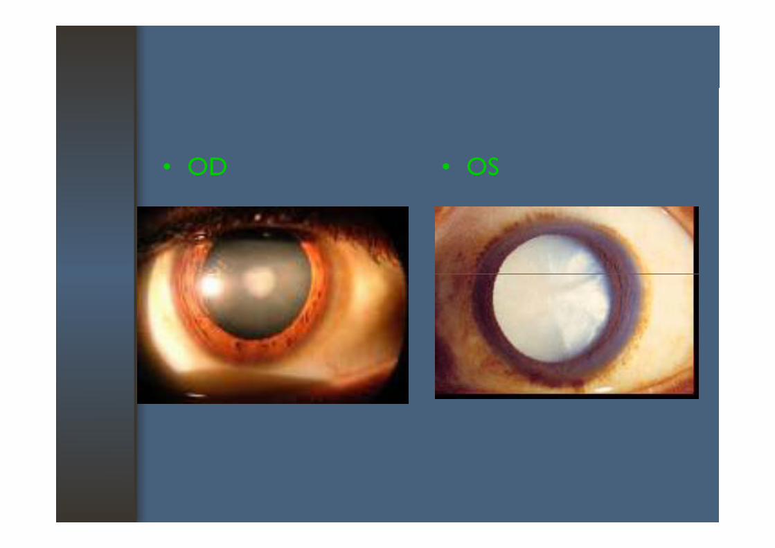
• OD • OS
• What is/are the diagnosis?• How do you do?*• How do you do?*• How is the prognosis in this case?

Case IIICase III
75-year of age, Thai femaleProblem : Difficult to see the far and near Problem : Difficult to see the far and near
objects in both eyes for 2 years
HOW DO YOU DO?HOW DO YOU DO?
• History- Slow progressive of
• Eye examinationVA c glasses :Slow progressive of
blur vision- Painless
VA c glasses :OD : 20/200 OS CF 2’- Painless
- No underlying di
OS : CF 2’
diseases
• VA c glasses c PH OD : 20/70
• Eye examination• RAPD : not presentOD : 20/70
OS : 20/100
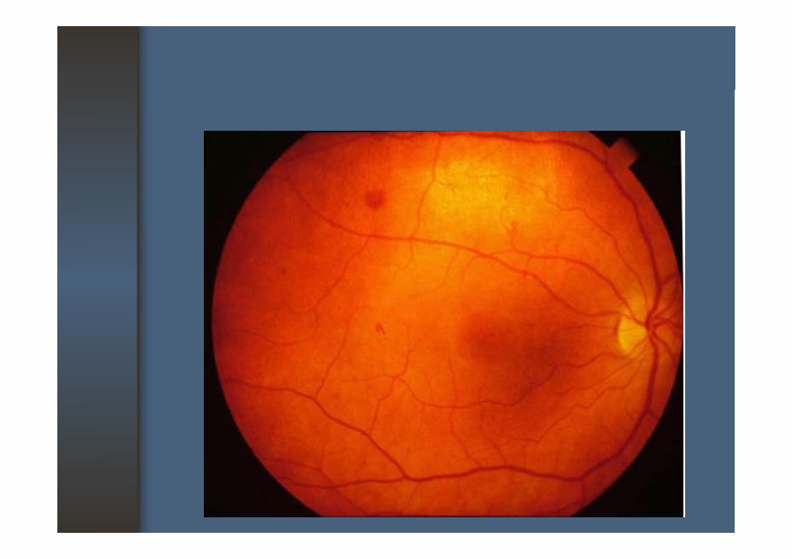
p• A/S : normal• IOP : 25/30 mmHgg• Lens : NS 2+• Fundus : normal macula
,cupping as the figured
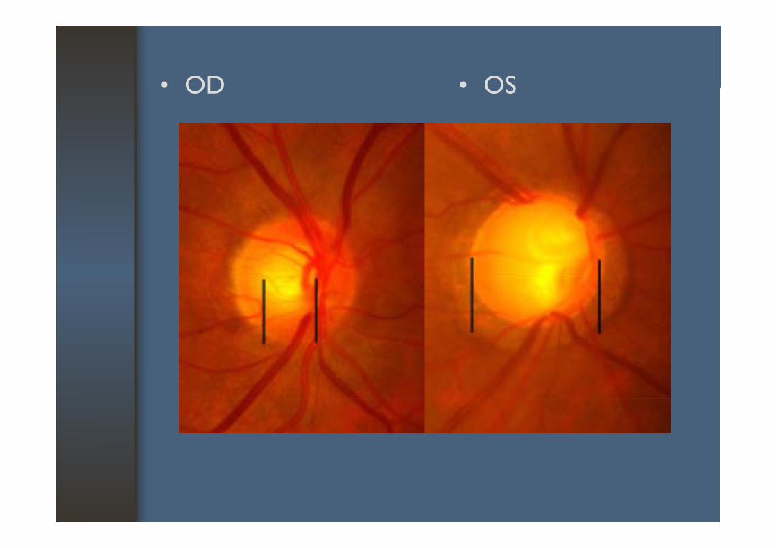
• OD • OS • OD • OS

• What is/are the diagnosis?• How do you do?+• How do you do?+
Case IVCase IV
80-year of age, Thai maleProblem : Difficult to see the far and near Problem : Difficult to see the far and near
objects in both eyes for 1 year
HOW DO YOU DO?HOW DO YOU DO?
• History- Slow progressive,
• Eye examinationVA c glasses :Slow progressive,
painless blur vision, especially central
VA c glasses :OD : CF 2’OS 20/200especially central
vision- Farmer
OS : 20/200
- Farmer- Smoking• No underlying
diseases
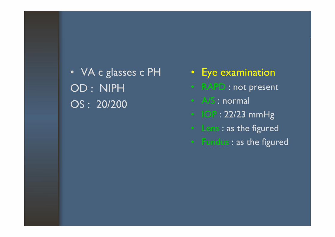
• VA c glasses c PH OD : NIPH
• Eye examination• RAPD : not presentOD : NIPH
OS : 20/200
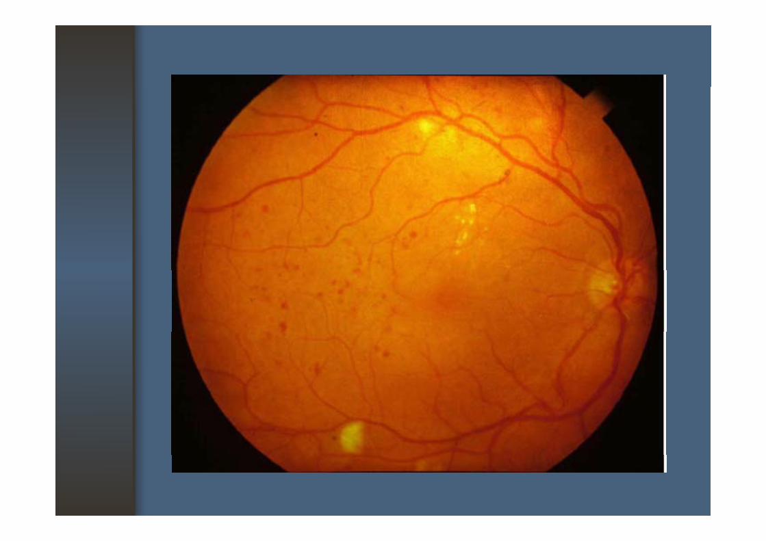
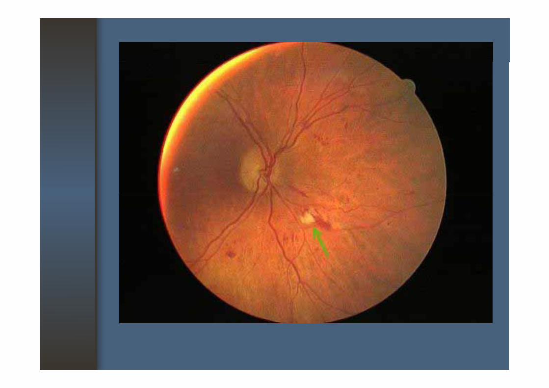
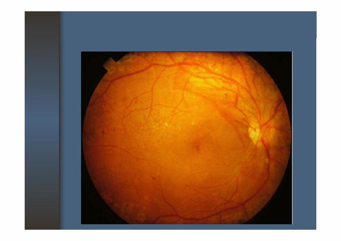
p• A/S : normal• IOP : 22/23 mmHgg• Lens : as the figured• Fundus : as the figuredg
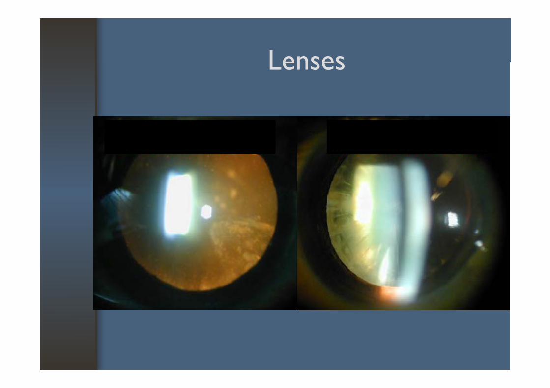
LensesLenses

FundusFundus
• What is/are the diagnosis?*• How do you do?• How do you do?
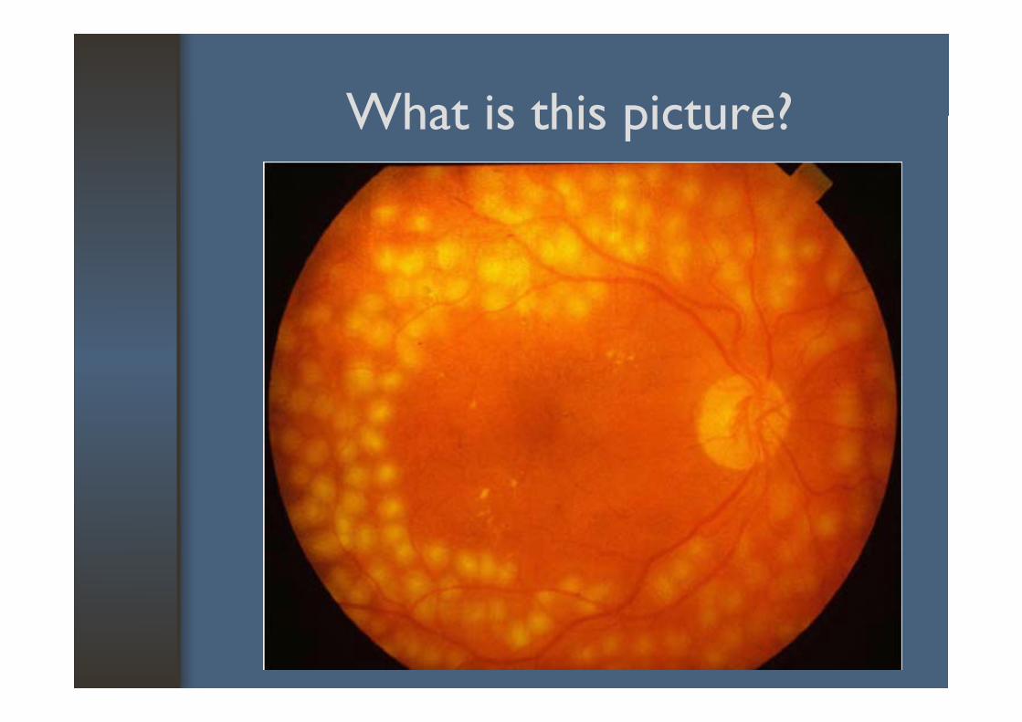
What is this picture? What is this picture?
ObjectivesObjectives
• Characteristics of the optic disc, determining normal or abnormal optic disc
• Recognize a cataract and to determine its approximate potential effect on the patient’s approximate potential effect on the patient s vision, determine whether a cataract is the only cause of a patient’s visual decrease
• Examine the macula with the ophthalmoscope and recognize the signs and symptoms of maculopathy
Glaucoma
RelevanceRelevance
• Significant cause of irreversible blindness, butthe blindness can be prevented
• Most patients are asymptomatic, majority of patients lack of pain, ocular inflammation, or h lhalo
• Peripheral vision can be lost before central i ivision
• Visual field defects are characterized by specific h d d i f i h l shaped scotoma and contraction of peripheral
field
RelevanceRelevance
• Early detection of glaucoma is important• Usually involves elevation of IOP above the Usually involves elevation of IOP above the
statistically normal range• Prolonged elevation of IOP can lead to optic • Prolonged elevation of IOP can lead to optic
nerve damageR ti IOP t i l bl • Routine IOP measurement is a valuable means of screening of glaucoma
RelevanceRelevance
• In some cases, glaucomatous optic nerve changes in normal IOPg
• Other disorders, such as brain tumor, can also cause changes in optic nervecause changes in optic nerve
Basic informationBasic information
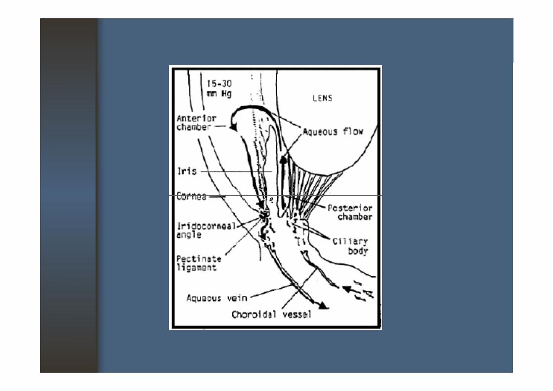
Factors effect to IOP1. Aqueous production : ciliary body epithelium (Non 1. Aqueous production : ciliary body epithelium (Non
pigmented epithelium)2. Resistance to outflow : conventional route (TM) , ( )
unconventional route (Uveoscleral)3. Episcleral venous pressure
Basic informationBasic information
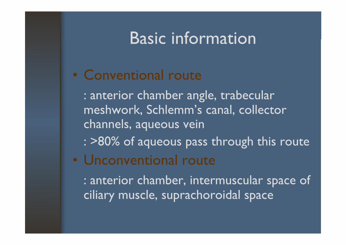
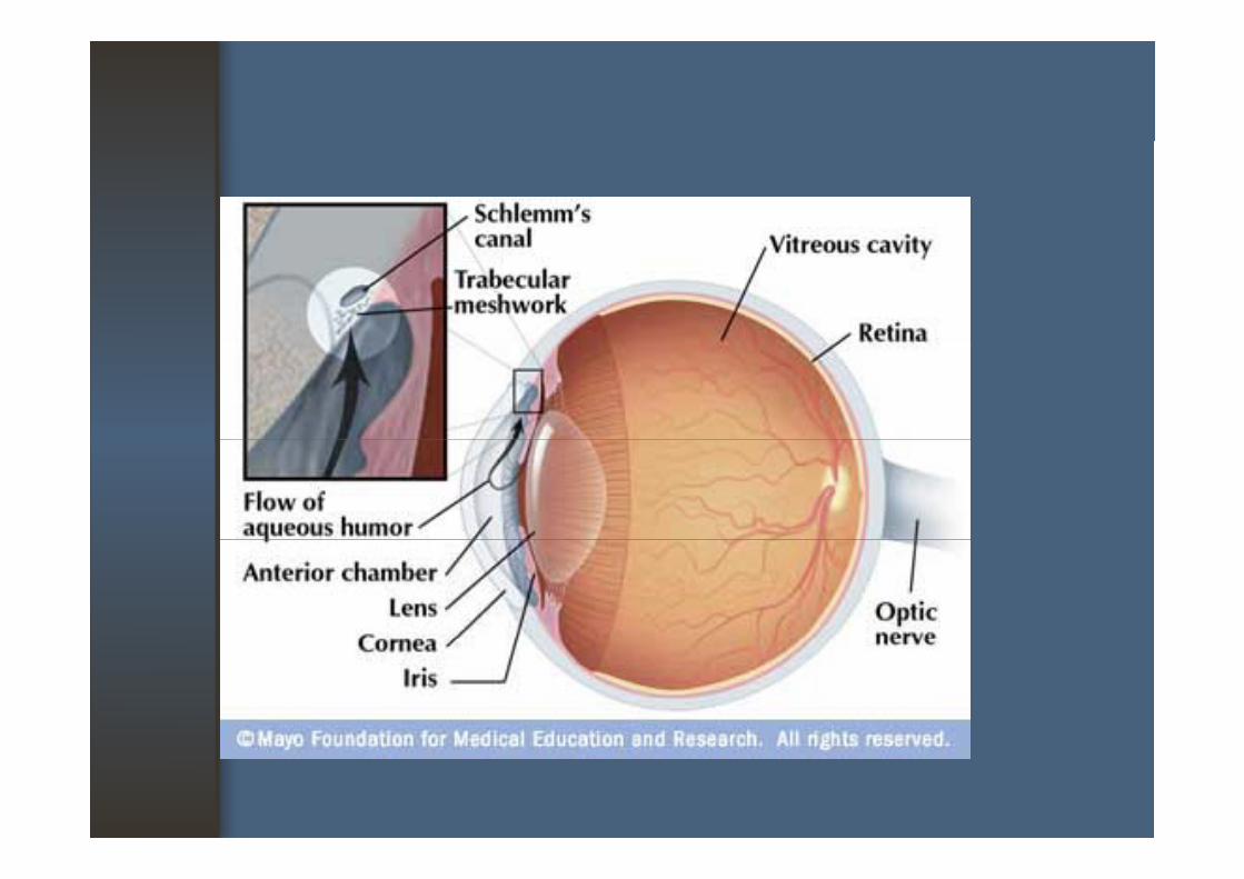
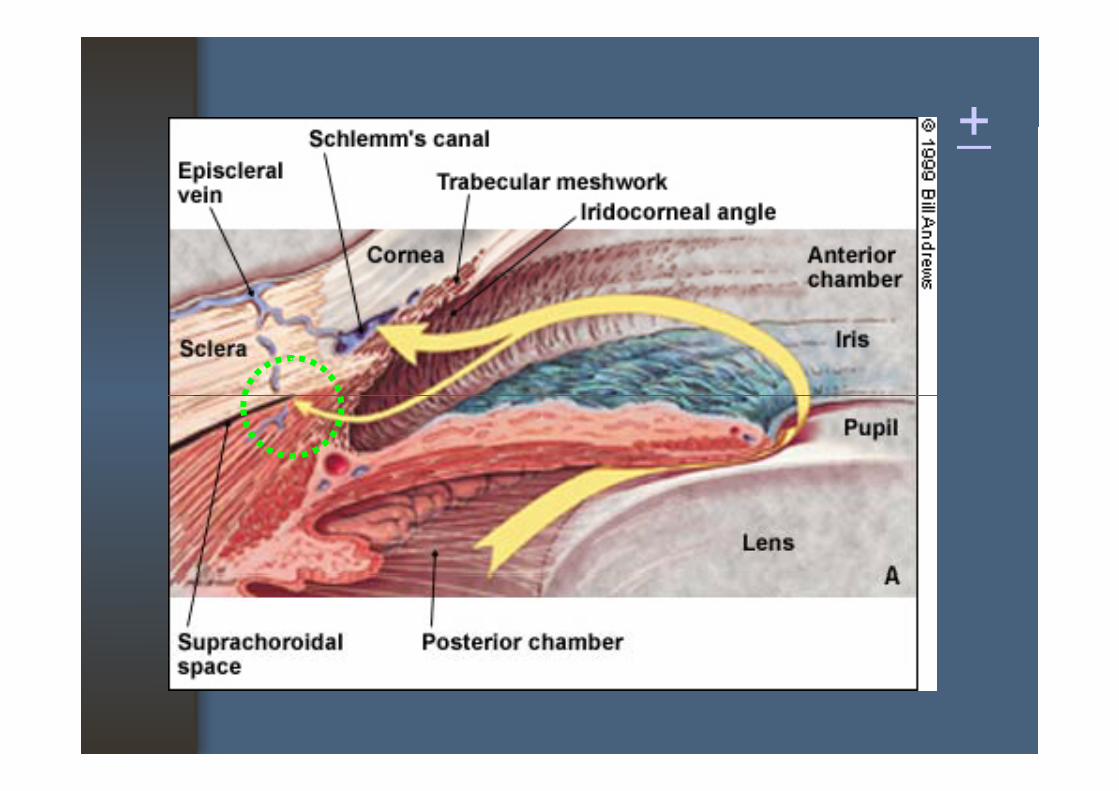
• Conventional route: anterior chamber angle trabecular : anterior chamber angle, trabecular meshwork, Schlemm’s canal, collector channels aqueous veinchannels, aqueous vein: >80% of aqueous pass through this route
• Unconventional route t i h b i t l f : anterior chamber, intermuscular space of ciliary muscle, suprachoroidal space
++
Basic informationBasic information
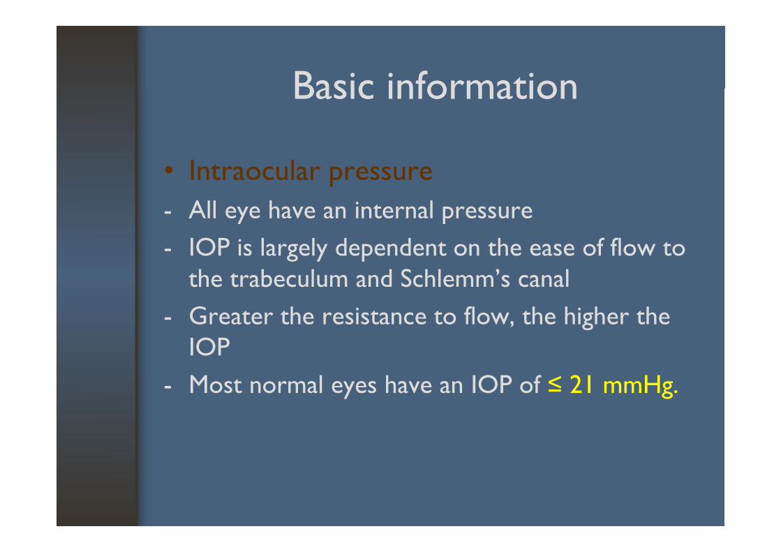
• Intraocular pressure- All eye have an internal pressure- All eye have an internal pressure- IOP is largely dependent on the ease of flow to
the trabeculum and Schlemm’s canalthe trabeculum and Schlemm s canal- Greater the resistance to flow, the higher the
IOPIOP- Most normal eyes have an IOP of ≤ 21 mmHg.
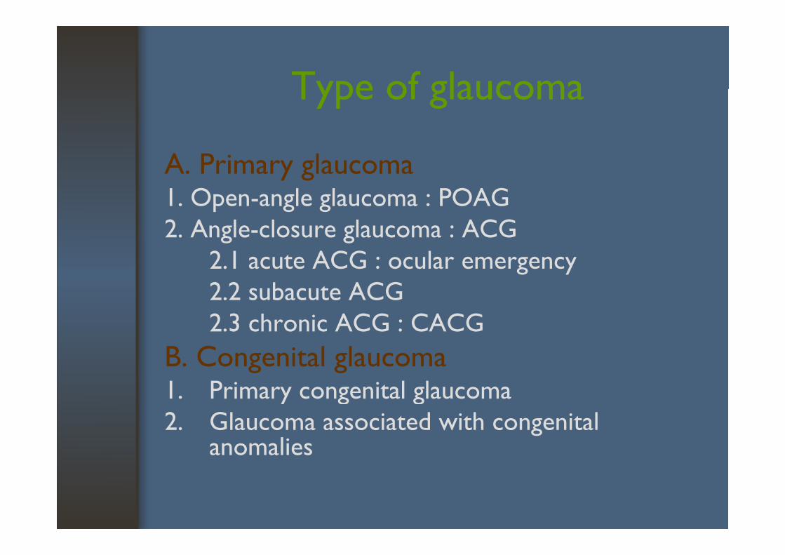
Type of glaucomaType of glaucoma
A. Primary glaucoma1. Open-angle glaucoma : POAG 2. Angle-closure glaucoma : ACG
2.1 acute ACG : ocular emergency2.2 subacute ACG2.3 chronic ACG : CACG
B. Congenital glaucoma1. Primary congenital glaucomay g g2. Glaucoma associated with congenital
anomalies
Type of glaucomaType of glaucoma
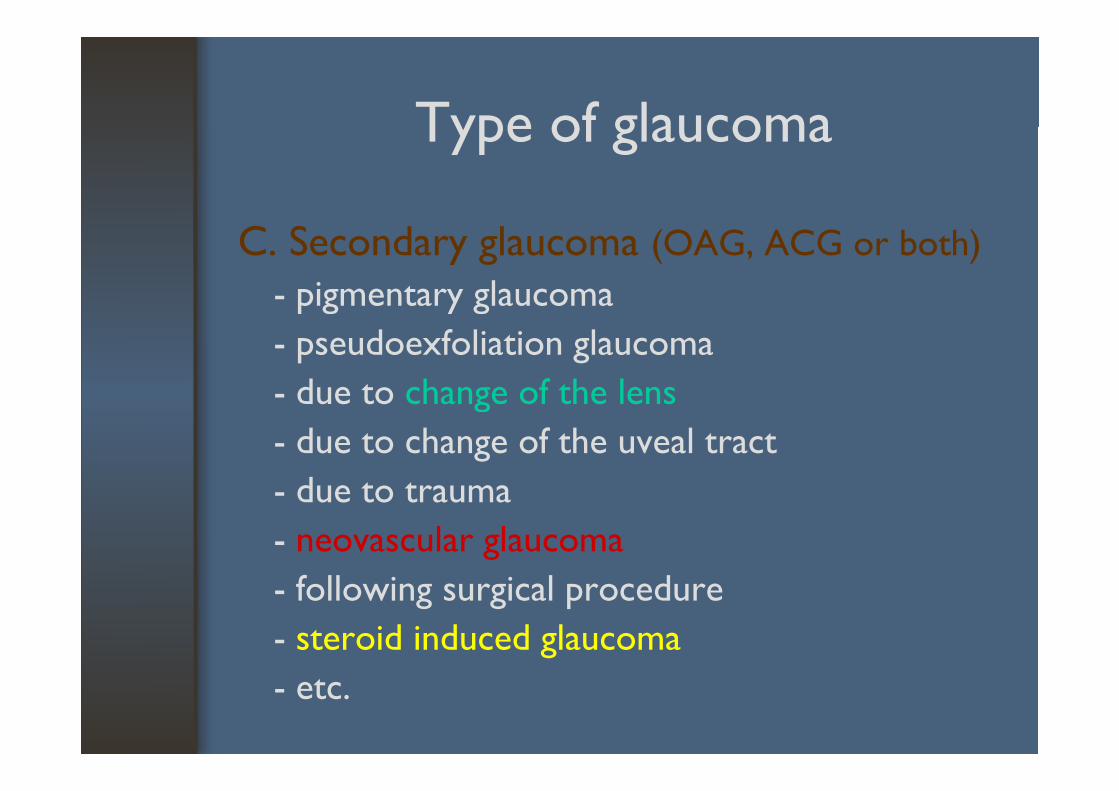
C. Secondary glaucoma (OAG, ACG or both)- pigmentary glaucomap g e ta y g auco a- pseudoexfoliation glaucoma- due to change of the lens- due to change of the lens- due to change of the uveal tract
d e t tra ma- due to trauma- neovascular glaucoma
f ll l d- following surgical procedure- steroid induced glaucoma- etc.
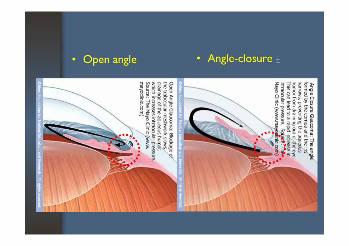
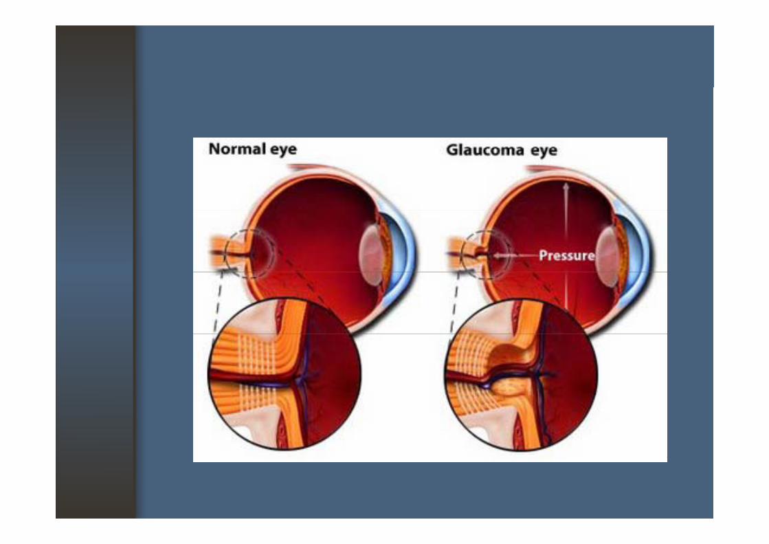
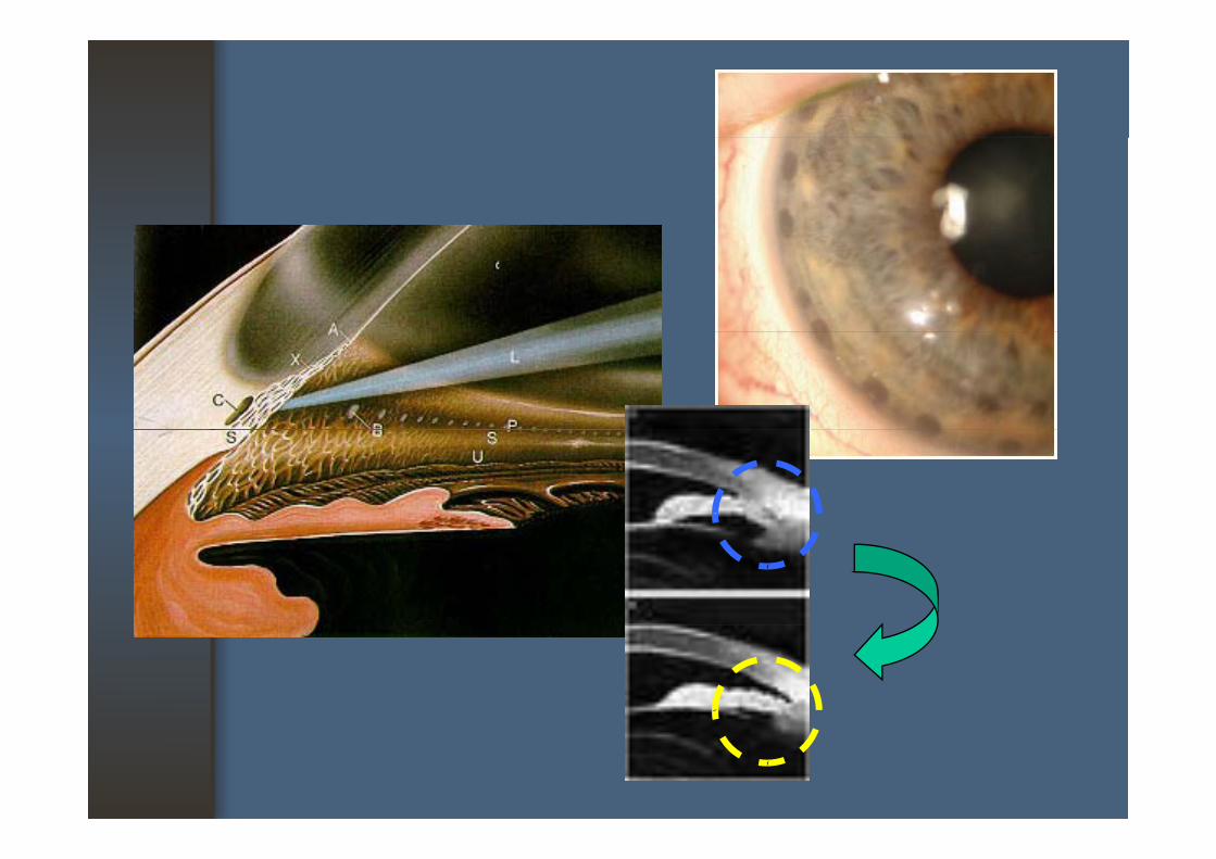
• Open angle • Angle-closure +
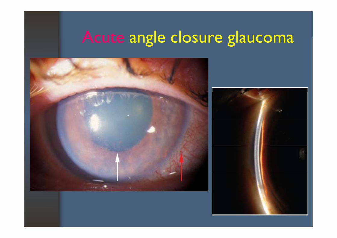
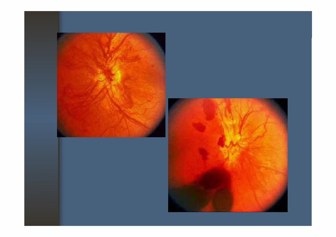
Acute angle closure glaucomaAcute angle closure glaucoma
Basic informationBasic information
• Optic nerve- Composed of more than 1 2 million nerve fibers- Composed of more than 1.2 million nerve fibers- Nerve fibers originate in the ganglion cells of the
retinaretina- At the point of origin, the nerve is called the
d ll d ll d h optic disc, small depression in it called the cup of the optic disc
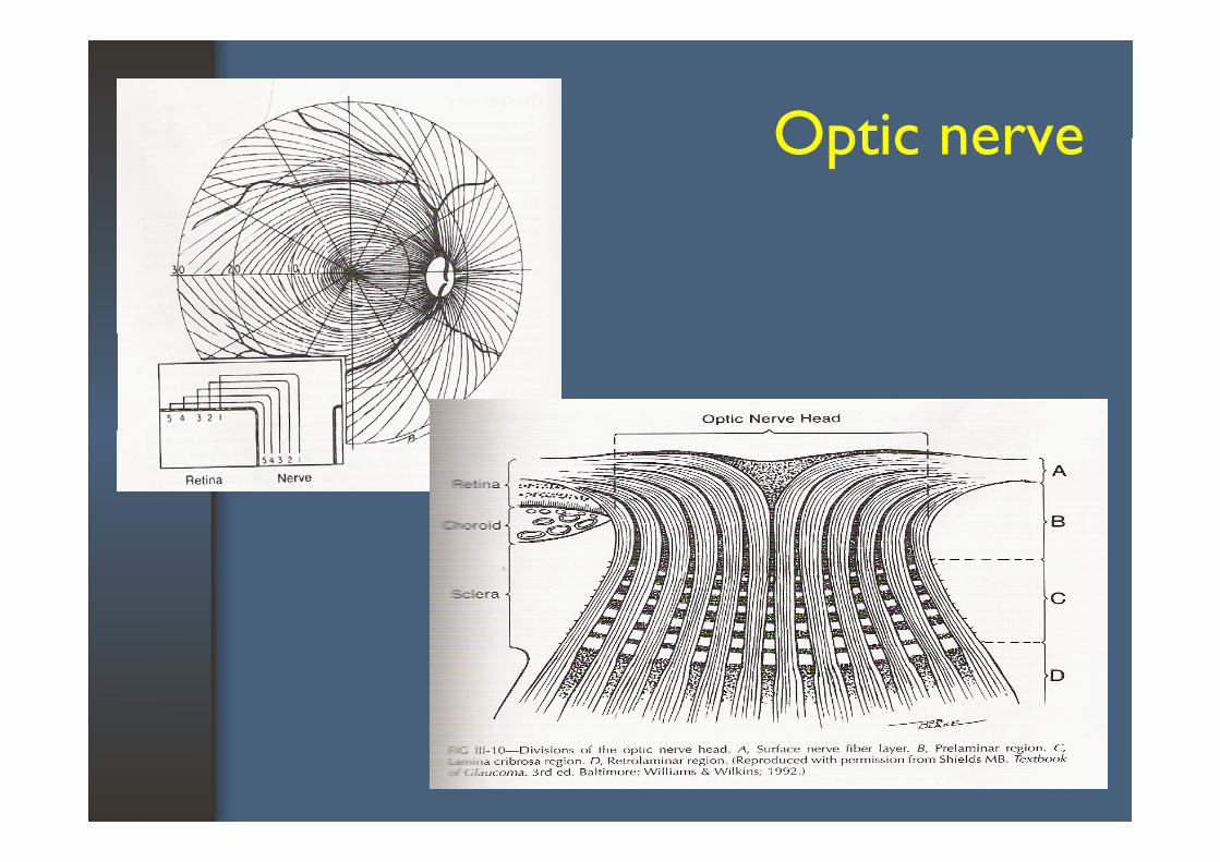
Optic nerveOptic nerve
Basic informationBasic information
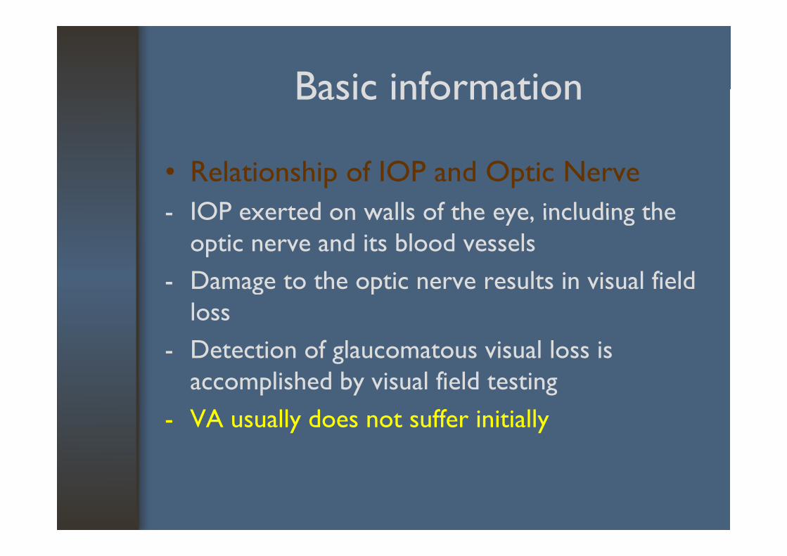
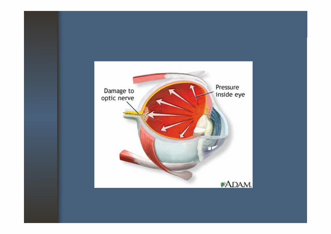
• Relationship of IOP and Optic Nerve- IOP exerted on walls of the eye including the - IOP exerted on walls of the eye, including the
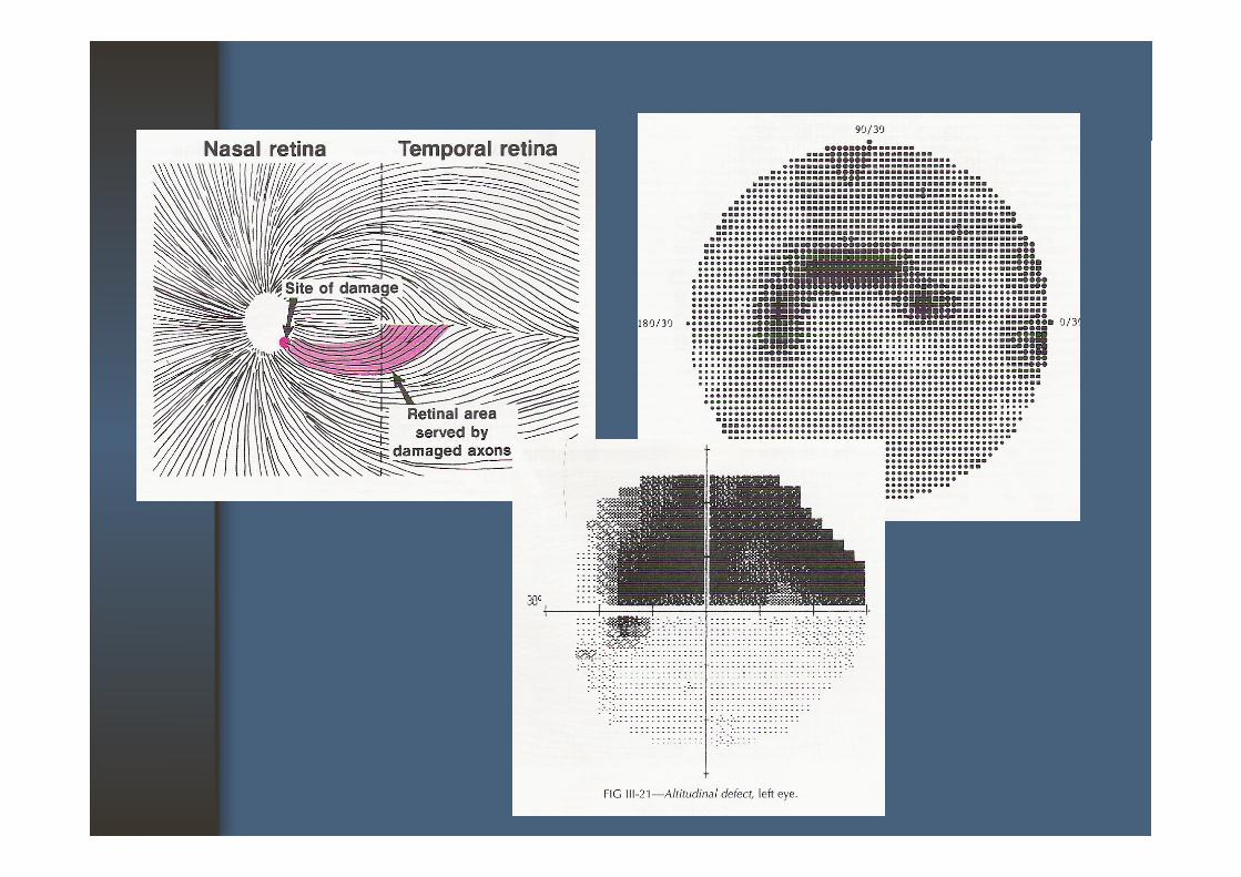
optic nerve and its blood vesselsDamage to the optic nerve results in visual field - Damage to the optic nerve results in visual field lossD f l l l - Detection of glaucomatous visual loss is accomplished by visual field testing
- VA usually does not suffer initially
When to ExamineWhen to Examine
Ophthalmoscopy• AAO recommends a glaucoma screening every AAO recommends a glaucoma screening every
2 to 4 years past age 40• Incidence of the disease increases with • Incidence of the disease increases with
age,family history and race Af A h k f • African-Americans have an greater risk for development of glaucoma
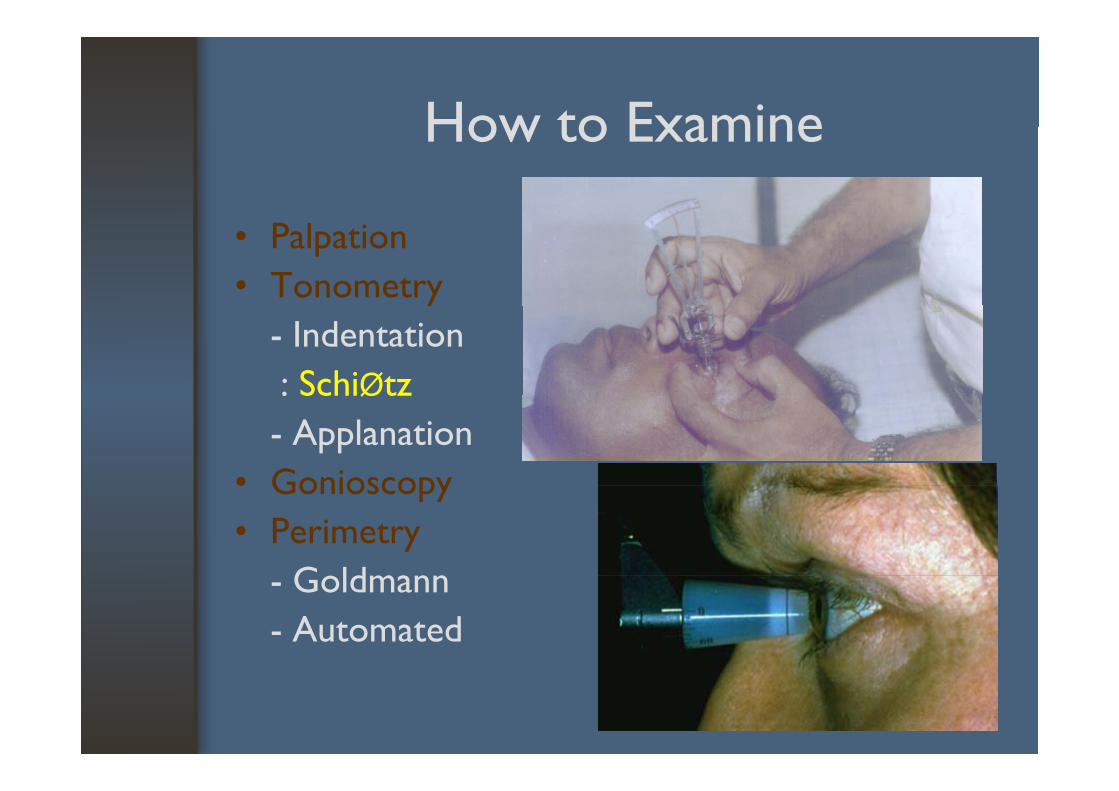
How to ExamineHow to Examine
• Palpation• Tonometryy
- Indentation: SchiØtz: SchiØtz- Applanation
• Gonioscopy• Gonioscopy• Perimetry
G ld- Goldmann- Automated
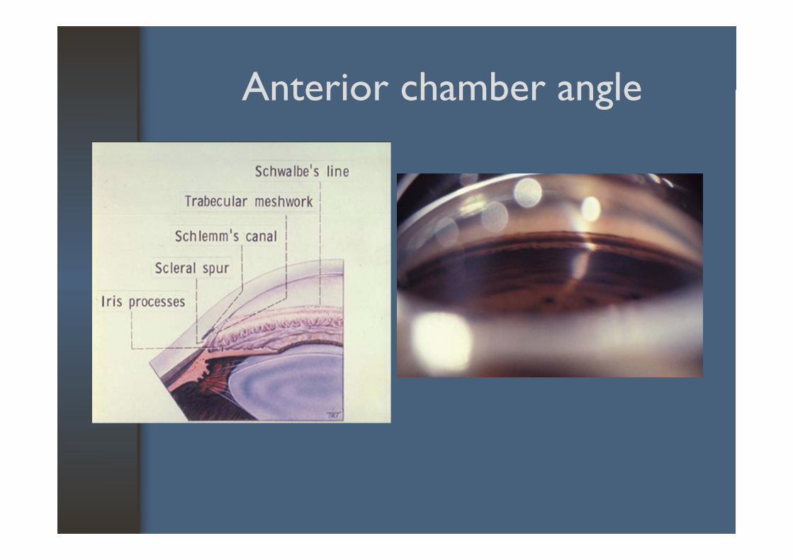
Anterior chamber angle Anterior chamber angle
How to interpret the findingsHow to interpret the findings
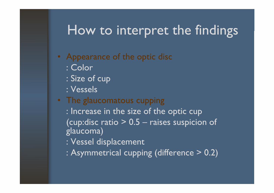
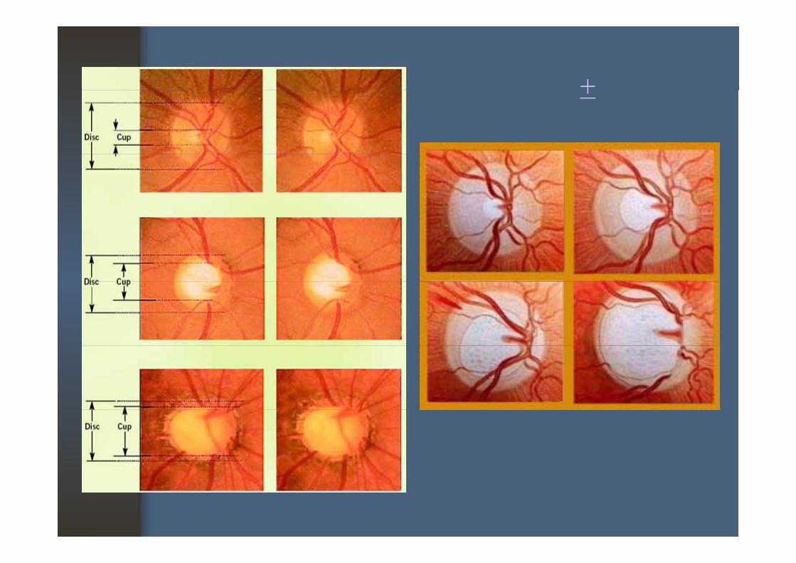
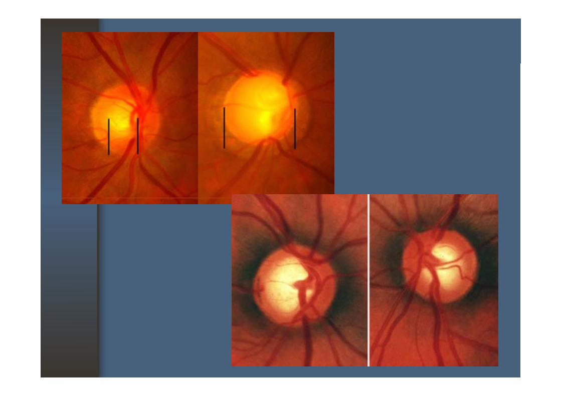
• Appearance of the optic disc: Color: Size of cup: Vessels
• The glaucomatous cupping: Increase in the size of the optic cup (cup:disc ratio > 0.5 – raises suspicion of glaucoma) V l di l t: Vessel displacement: Asymmetrical cupping (difference > 0.2)
+ +
Primary open angle glaucoma (POAG)Primary open angle glaucoma (POAG)
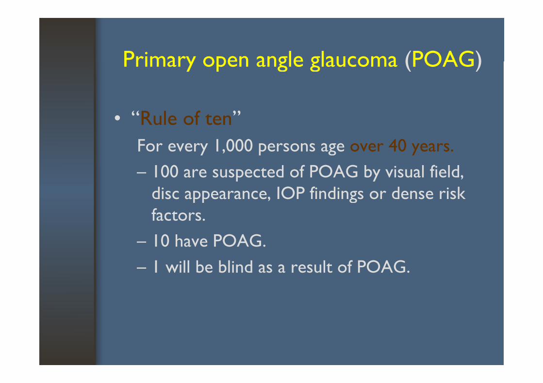
• “Rule of ten”For every 1 000 persons age over 40 yearsFor every 1,000 persons age over 40 years.– 100 are suspected of POAG by visual field,
disc appearance IOP findings or dense risk disc appearance, IOP findings or dense risk factors.10 h POAG– 10 have POAG.
– 1 will be blind as a result of POAG.
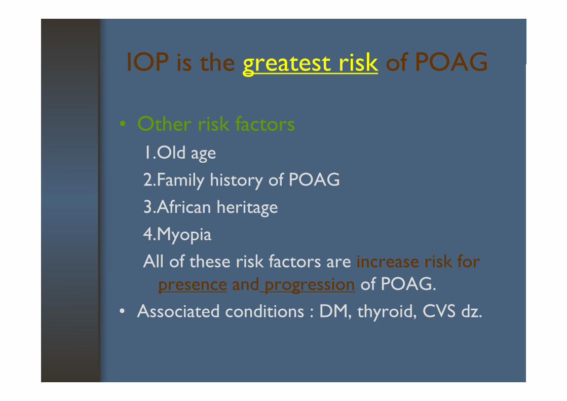
IOP is the greatest risk of POAGIOP is the greatest risk of POAG
• Other risk factors1 Old age1.Old age2.Family history of POAG3 Af h3.African heritage4.MyopiaAll of these risk factors are increase risk for
presence and progression of POAG.p p g• Associated conditions : DM, thyroid, CVS dz.
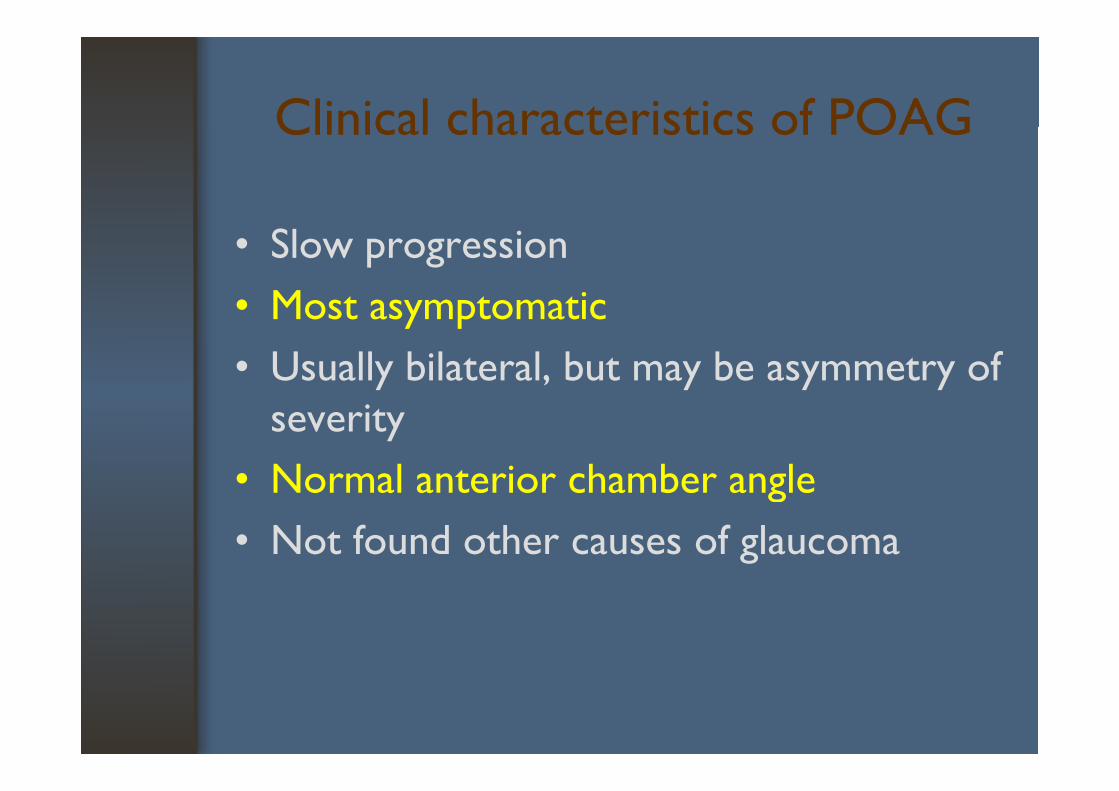
Clinical characteristics of POAGClinical characteristics of POAG
• Slow progression• Most asymptomatic• Most asymptomatic• Usually bilateral, but may be asymmetry of
severity• Normal anterior chamber angleNormal anterior chamber angle• Not found other causes of glaucoma
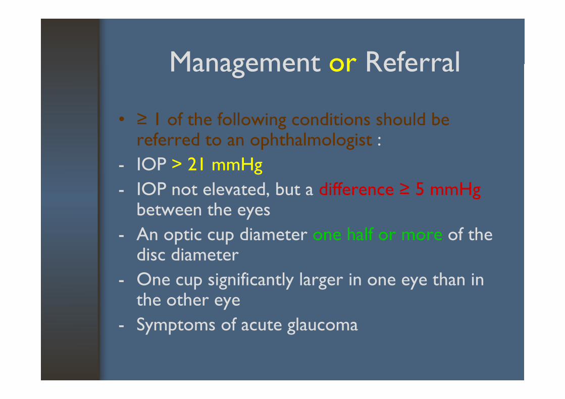
Management or ReferralManagement or Referral
• ≥ 1 of the following conditions should be referred to an ophthalmologist :
- IOP > 21 mmHg- IOP not elevated, but a difference ≥ 5 mmHg
between the eyes- An optic cup diameter one half or more of the
disc diameter- One cup significantly larger in one eye than in
the other eye- Symptoms of acute glaucoma
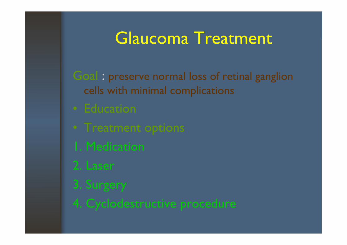
Glaucoma TreatmentGlaucoma Treatment
Goal : preserve normal loss of retinal ganglion cells with minimal complicationsp
• Education • Treatment options1. Medication 1. Medication 2. Laser 3. Surgery 4 Cyclodestructive procedure4. Cyclodestructive procedure
Anti-glaucoma drugsAnti-glaucoma drugs
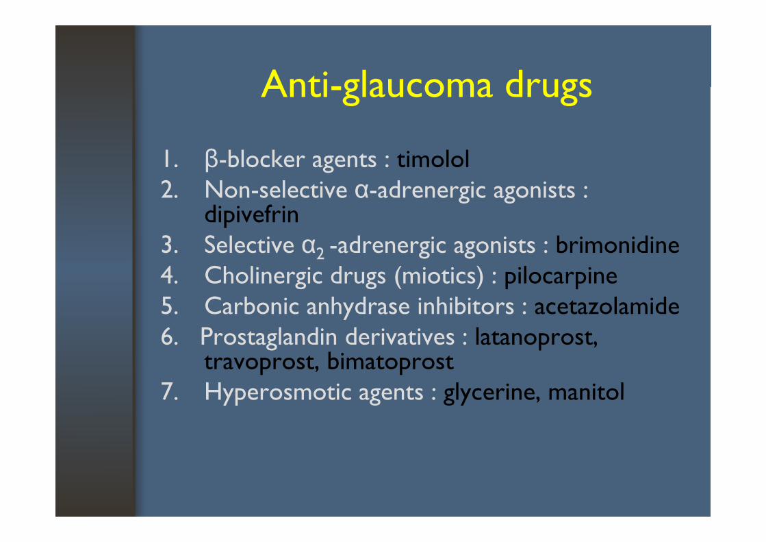
1. β-blocker agents : timolol2. Non-selective α-adrenergic agonists :
di i f idipivefrin3. Selective α2 -adrenergic agonists : brimonidine4 Ch li i d ( i ti ) il i4. Cholinergic drugs (miotics) : pilocarpine5. Carbonic anhydrase inhibitors : acetazolamide6 P t l di d i ti l t t6. Prostaglandin derivatives : latanoprost,
travoprost, bimatoprost7 Hyperosmotic agents : glycerine manitol7. Hyperosmotic agents : glycerine, manitol
Anti-glaucoma drugsAnti-glaucoma drugs
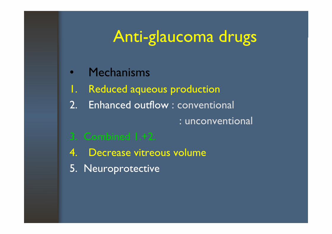
• Mechanisms 1 Reduced aqueous production1. Reduced aqueous production2. Enhanced outflow : conventional
l: unconventional3. Combined 1.+2.4. Decrease vitreous volume5 Neuroprotective5. Neuroprotective
Anti-glaucoma drugsAnti-glaucoma drugs
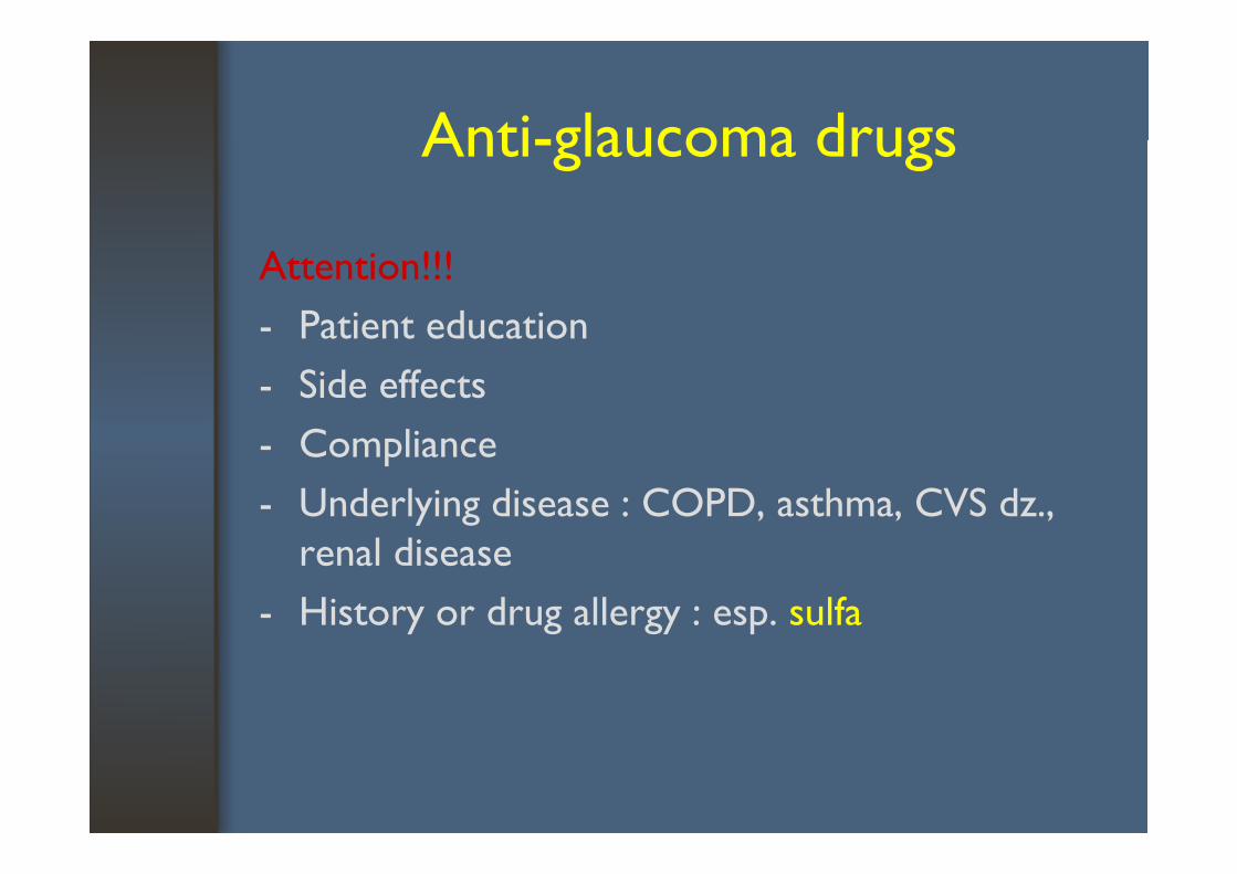
Attention!!!- Patient educationPatient education- Side effects
C li - Compliance - Underlying disease : COPD, asthma, CVS dz.,
renal disease- History or drug allergy : esp. sulfa
Laser treatmentLaser treatment
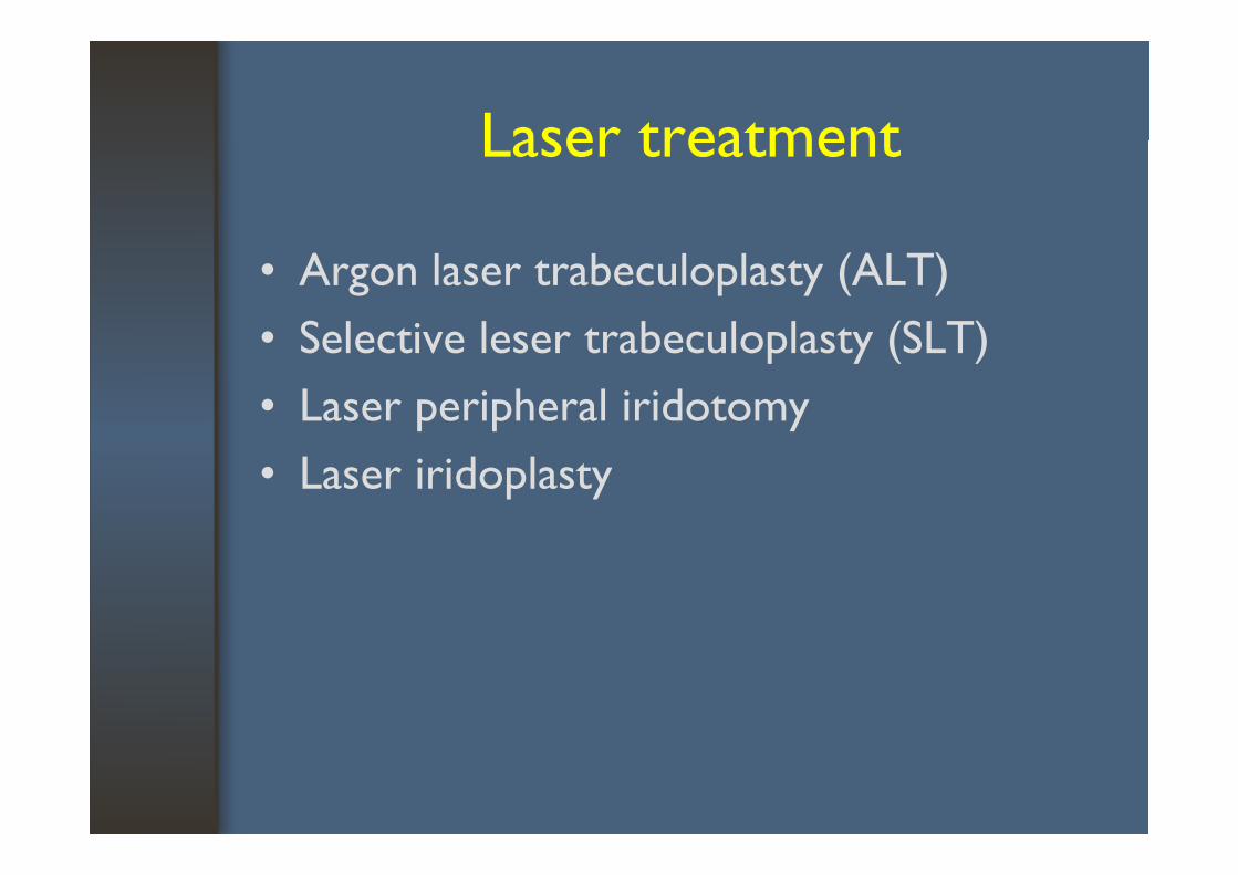
• Argon laser trabeculoplasty (ALT)• Selective leser trabeculoplasty (SLT)• Selective leser trabeculoplasty (SLT)• Laser peripheral iridotomy• Laser iridoplasty
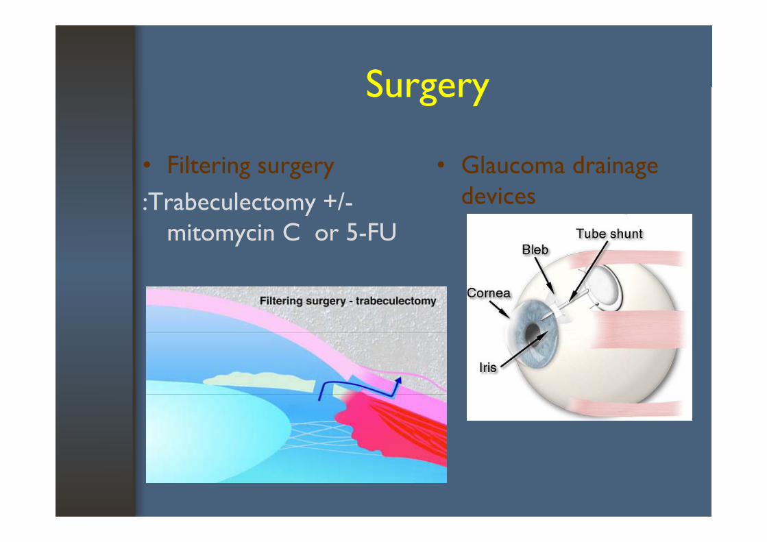
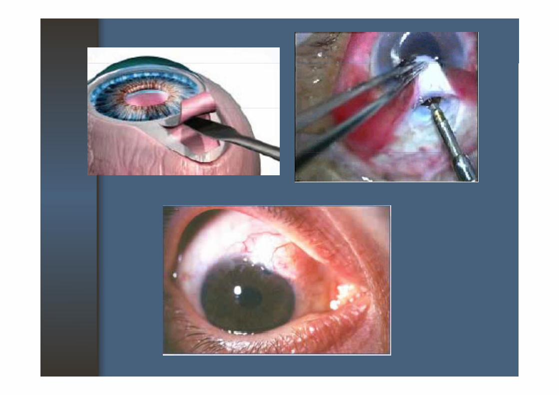
SurgerySurgery
• Filtering surgery:Trabeculectomy +/-
• Glaucoma drainage devices:Trabeculectomy /
mitomycin C or 5-FU
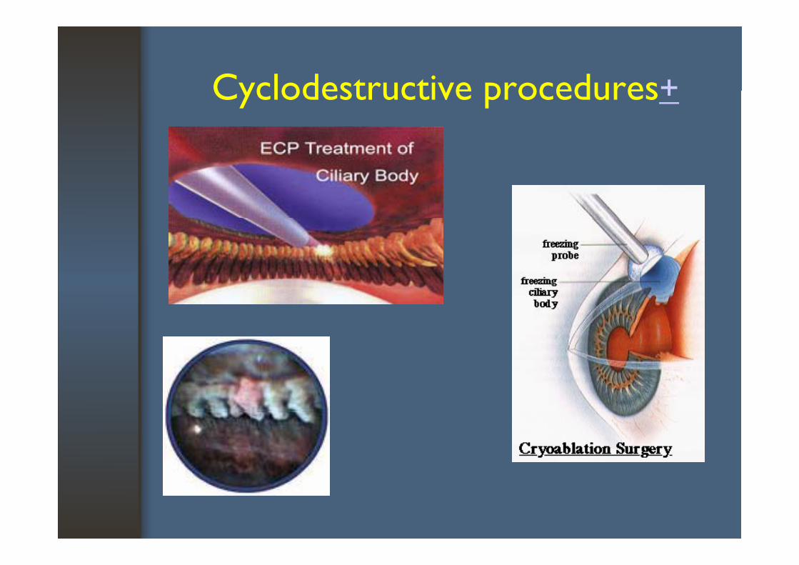
Cyclodestructive procedures+Cyclodestructive procedures+

Cataract
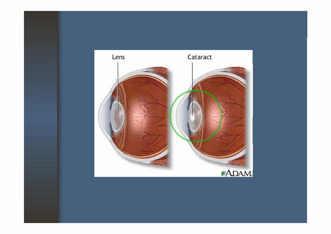
RelevanceRelevance
• Congenital, genetic anomaly, various diseases, or with increasing age (most common cause)
• Age-related cataract occurs in about 50% of people between ages 65 and 74
• One of the most successfully treated conditions in all of surgery
• Usually with intraocular lens implantation• If an implant is not used, visual rehabilitation is
still possible with a contact lens or aphakic glasses
Basic InformationBasic Information
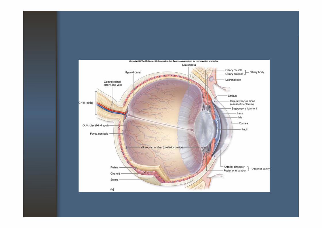
• Lens Function :
- refraction : refractive power +20 D- accommodation- protective function : U.V. , physical barrierAnatomy : y
- transparent, biconvex shape - thickness ~4 mm. , width ~ 9 mm.- capsule, cortex, nucleus
Basic InformationBasic Information
• LensSuspended by thin filamentous zonules - Suspended by thin filamentous zonules (transparent collagen fibers) from the ciliary bodybody
- Contraction of the ciliary muscle permits f i f h lfocusing of the lens
- The lens is encloses in a capsule (elastic semipermeable basement membrane)
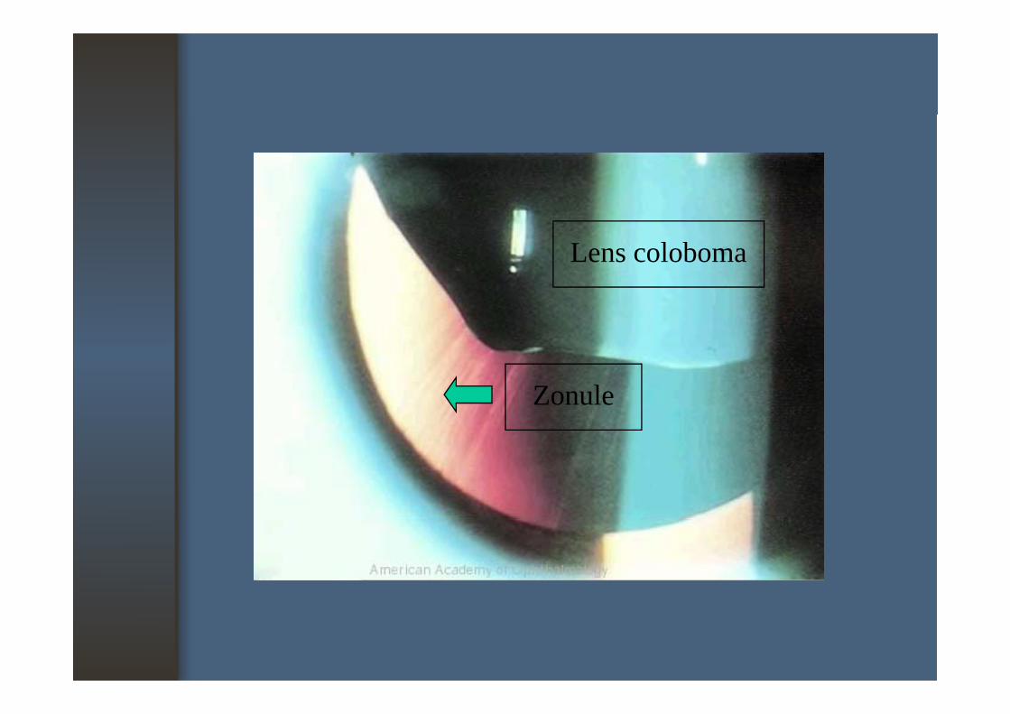
Lens coloboma
Zonule
Basic InformationBasic Information
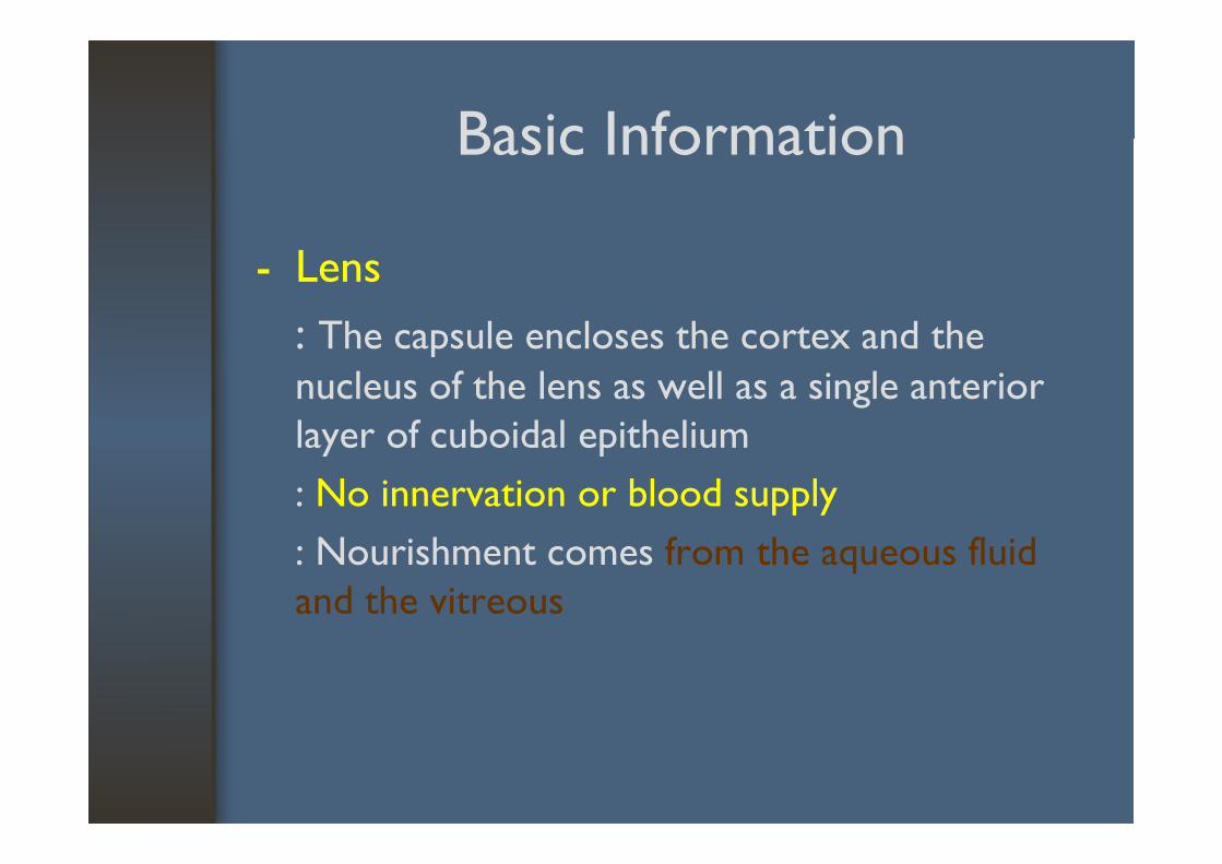
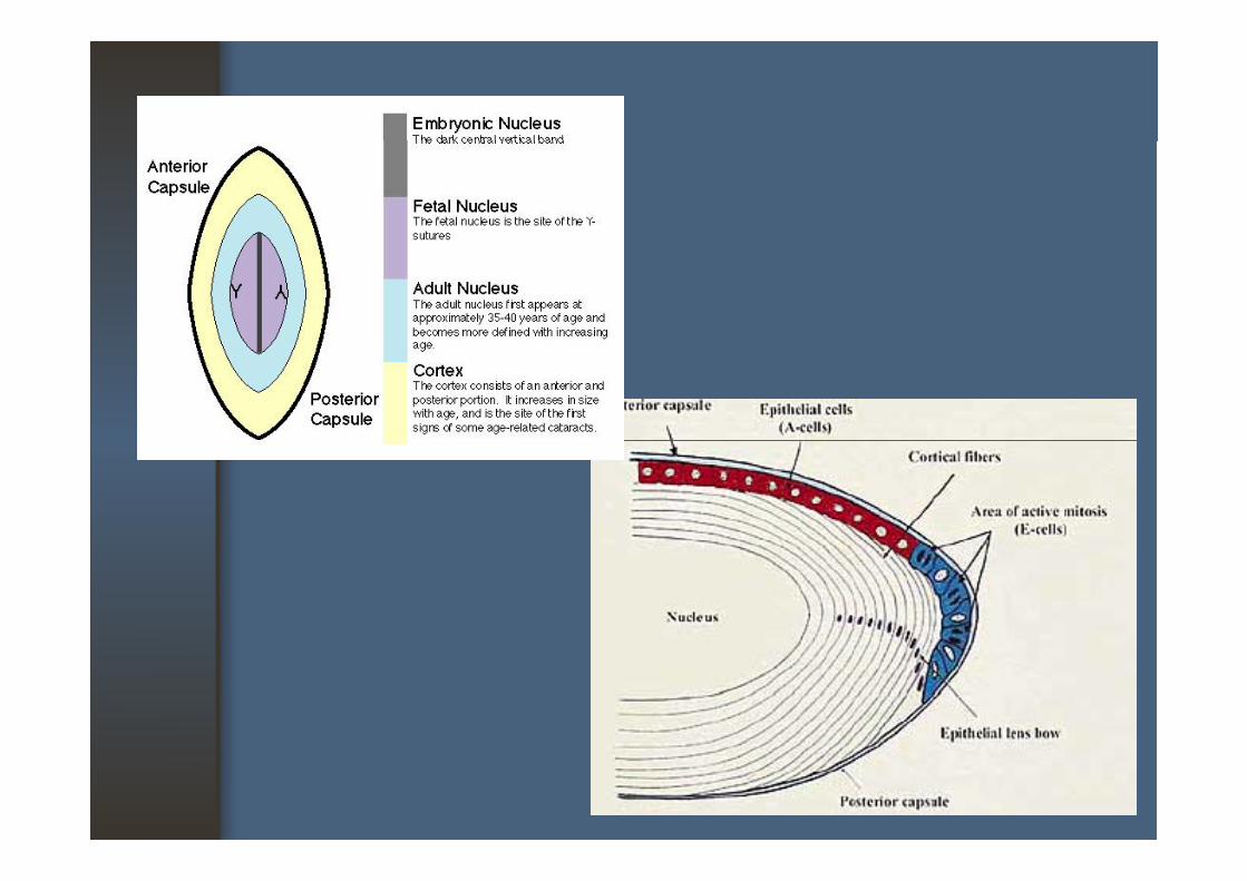
- Lens: The capsule encloses the cortex and the : The capsule encloses the cortex and the nucleus of the lens as well as a single anterior layer of cuboidal epitheliumlayer of cuboidal epithelium: No innervation or blood supply: Nourishment comes from the aqueous fluid and the vitreous
Basic InformationBasic Information
• Lens: Continues to grow throughout life: Continues to grow throughout life: Epithelial cells continue to produce new
i l l fibcortical lens fibers: Consists of 35% protein, ~ 60% water by mass: Percentage of insoluble protein increases as the lens ages and as a cataract developsg p
Basic informationBasic information
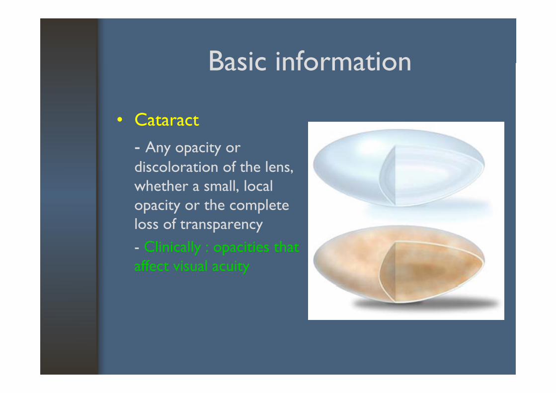
• Cataract- Any opacity or Any opacity or discoloration of the lens, whether a small, local opacity or the complete loss of transparency
Cl ll h - Clinically : opacities that affect visual acuity
Basic informationBasic information
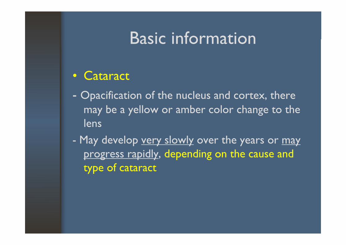
• CataractOpacification of the nucleus and cortex there - Opacification of the nucleus and cortex, there may be a yellow or amber color change to the lenslens
- May develop very slowly over the years or may idl d di h d progress rapidly, depending on the cause and
type of cataract
ClassificationClassification
• Primary cataract- congenital
• Secondary cataract- extraocular disorder- congenital
- juvenilel
- extraocular disorder- intraocular disorder
- presenile- senile
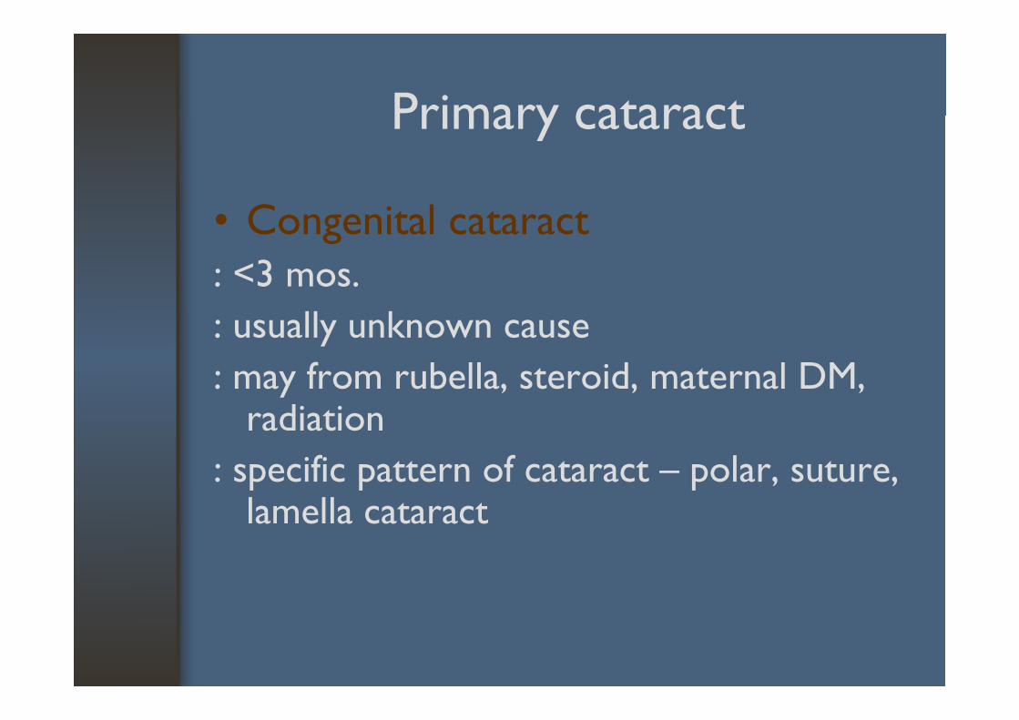
Primary cataractPrimary cataract
• Congenital cataract: <3 mos : <3 mos. : usually unknown cause: may from rubella, steroid, maternal DM,
radiation: specific pattern of cataract – polar, suture,
lamella cataracta e a cata act
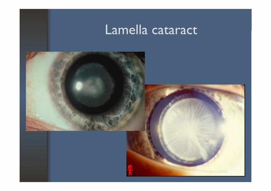
Lamella cataractLamella cataract
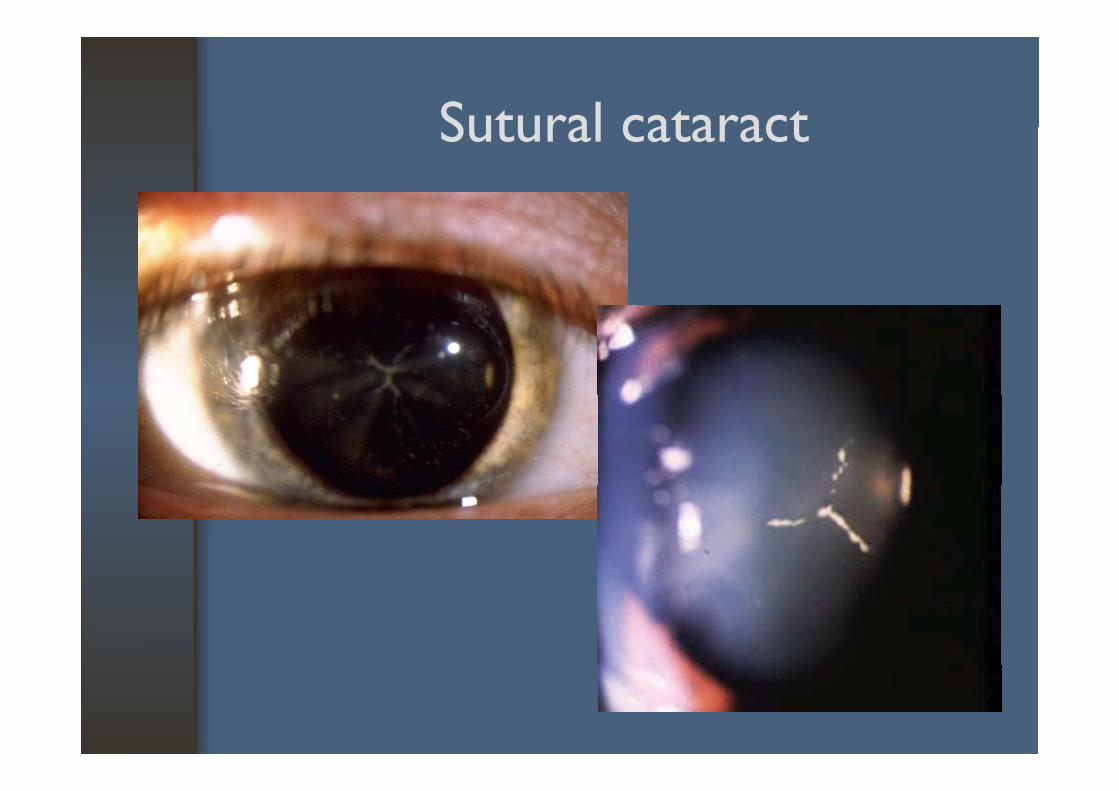
Sutural cataractSutural cataract

Anterior polar cataractAnterior polar cataract
Primary cataractPrimary cataract
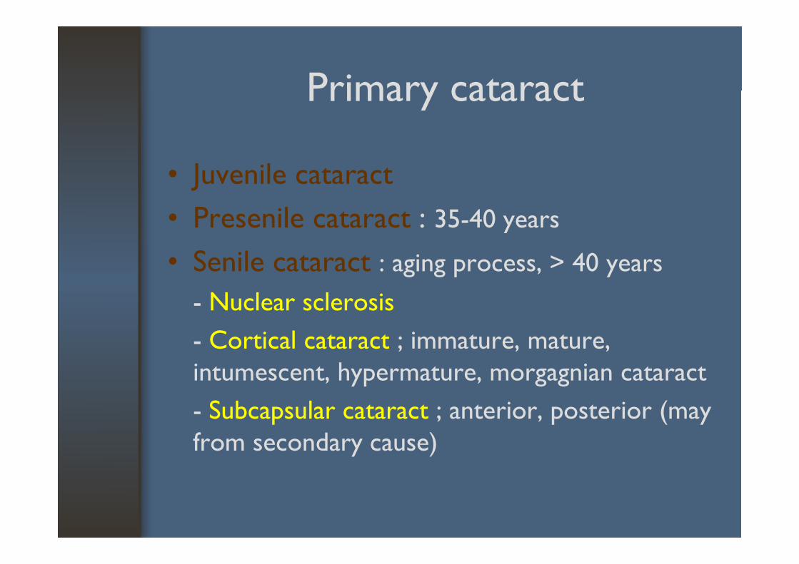
• Juvenile cataract• Presenile cataract : 35 40 years• Presenile cataract : 35-40 years
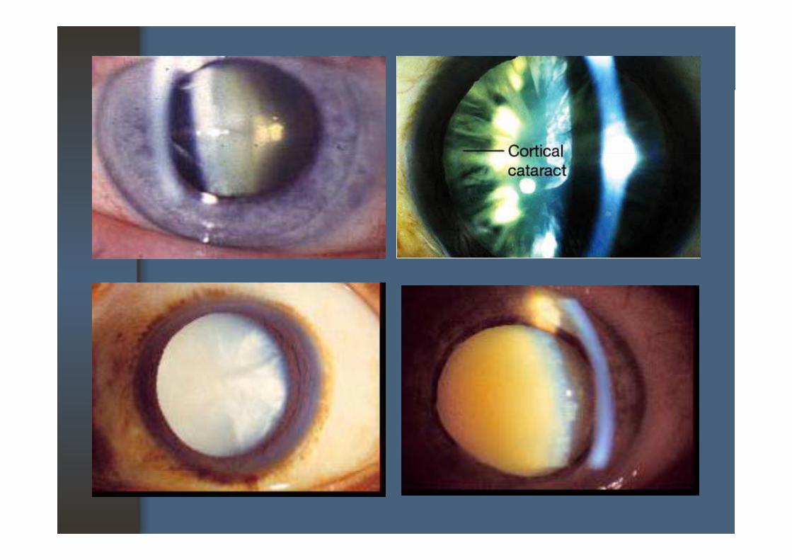
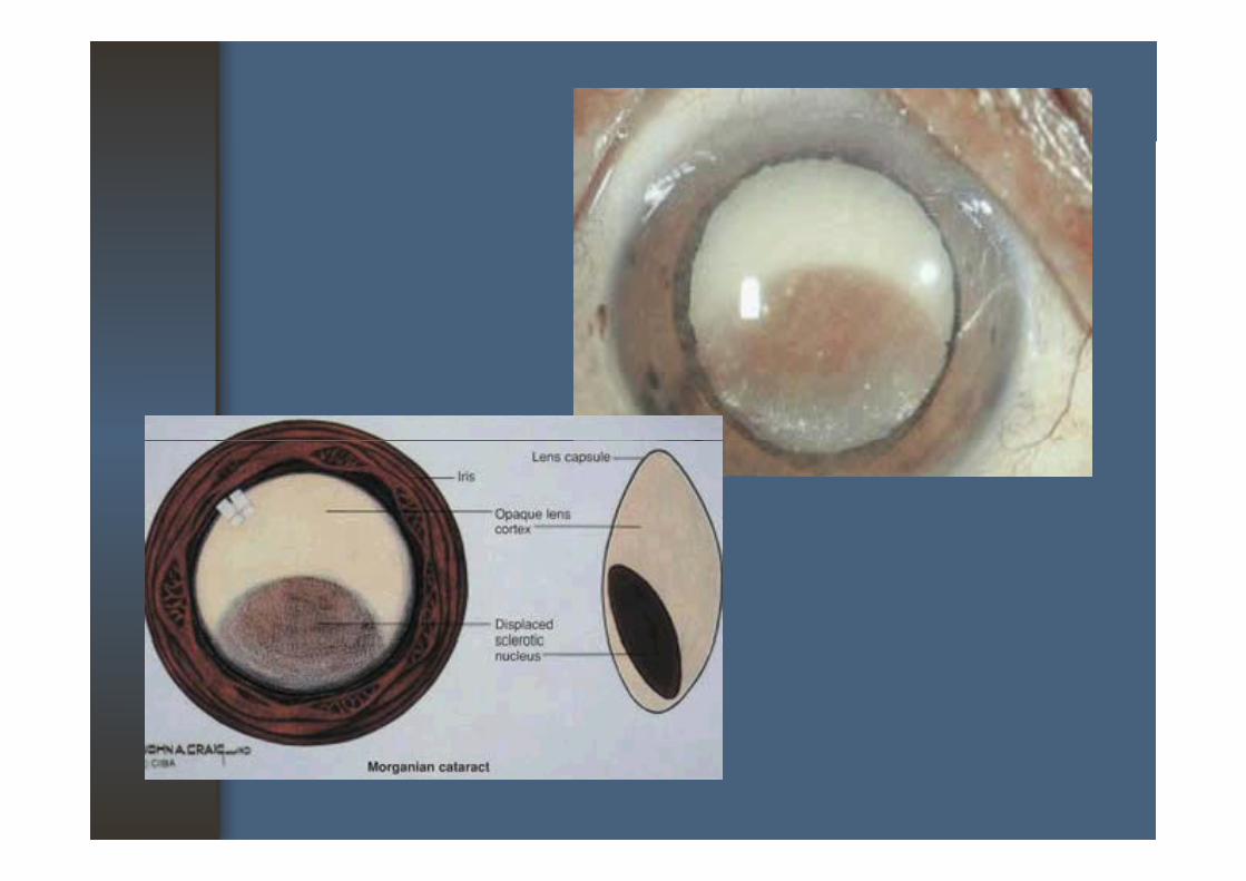
• Senile cataract : aging process, > 40 years- Nuclear sclerosis- Cortical cataract ; immature, mature, Cortical cataract ; immature, mature, intumescent, hypermature, morgagnian cataract
Subcapsular cataract ; anterior posterior (may - Subcapsular cataract ; anterior, posterior (may from secondary cause)
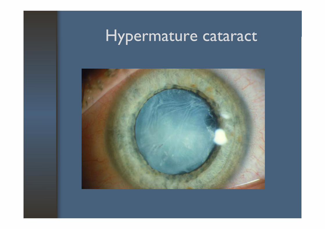
Hypermature cataractHypermature cataract
Secondary cataract Secondary cataract
• Extraocular disorderTraumatic : mechanical physical- Traumatic : mechanical, physical
- Metabolic : DM (fluctuation of vision, myopia), Wil ’ di (ASC)Wilson’s disease (ASC)- Toxic : steroid, echothiophate iodide, phenothiazines- Systemic disease : hyperparathyroidism, y yp p ymyotonic dystrophy, galactosemia, Down’s syndrome, trisomy 18, trisomy 13y y y
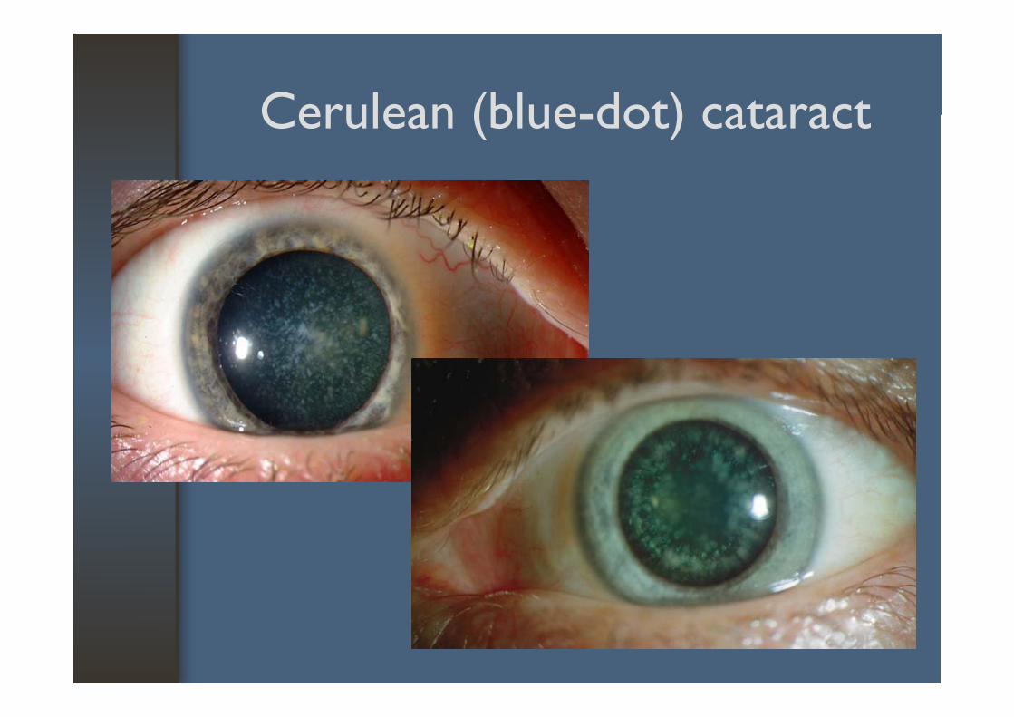
Cerulean (blue-dot) cataractCerulean (blue-dot) cataract
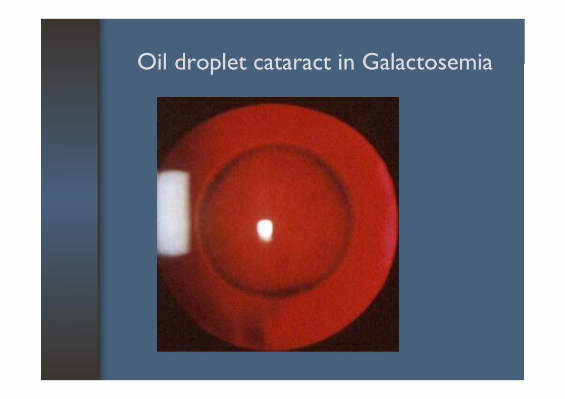
Oil droplet cataract in GalactosemiaOil droplet cataract in Galactosemia
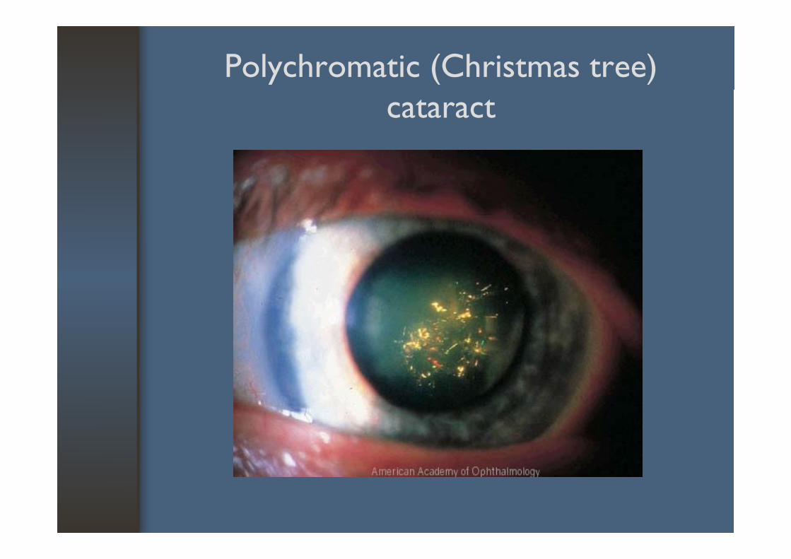
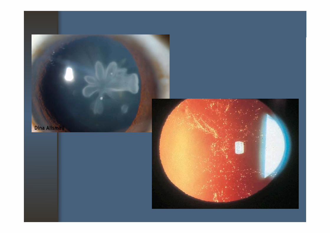
Polychromatic (Christmas tree) cataract
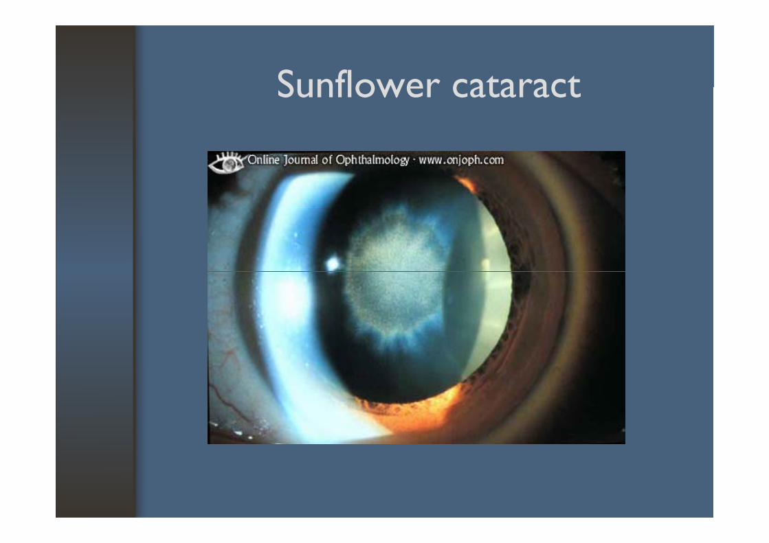
Sunflower cataractSunflower cataract
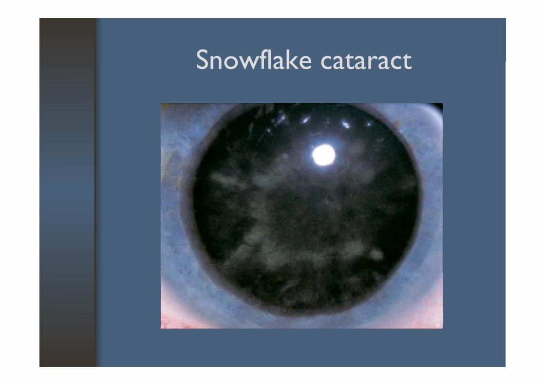
Snowflake cataractSnowflake cataract
Secondary cataractSecondary cataract
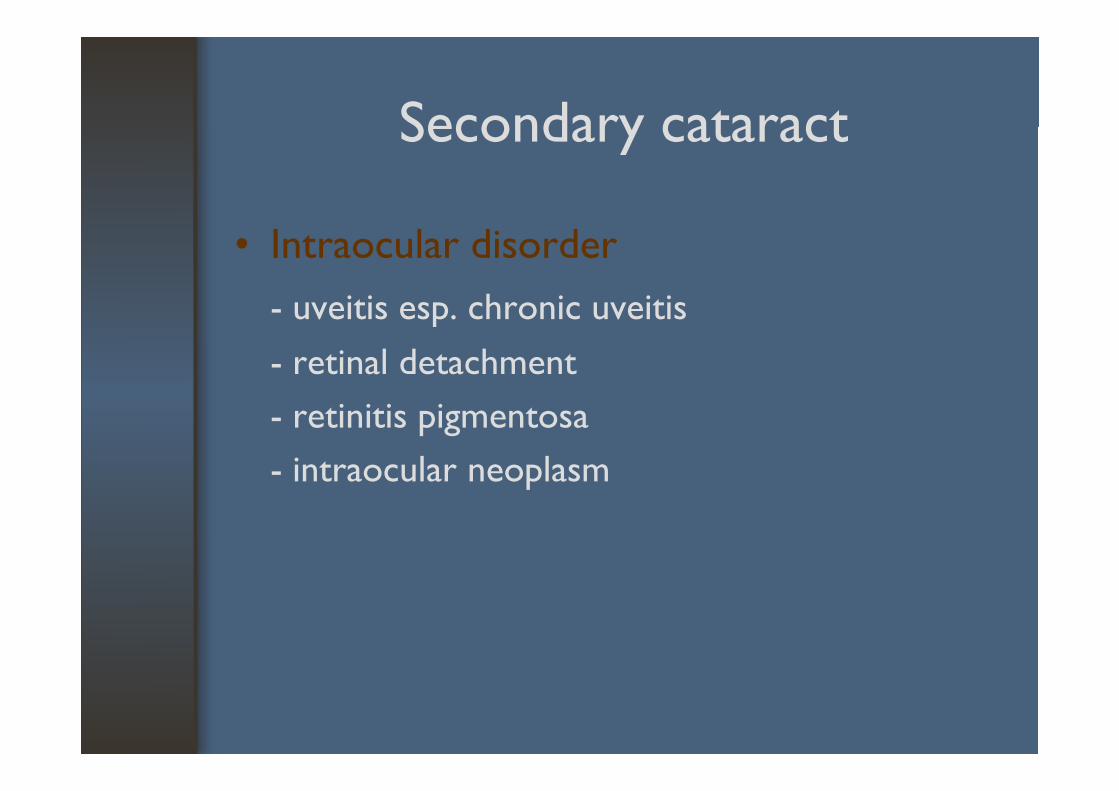
• Intraocular disorderuveitis esp chronic uveitis- uveitis esp. chronic uveitis
- retinal detachment- retinitis pigmentosa- intraocular neoplasmp
Basic informationBasic information
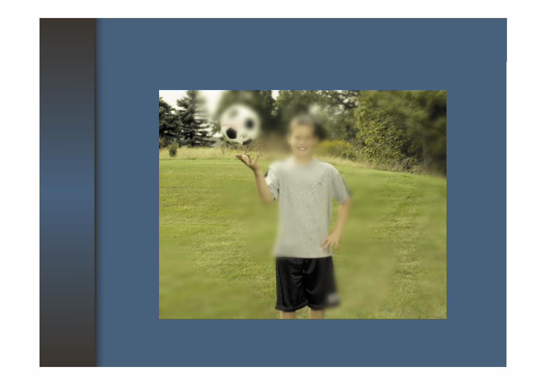
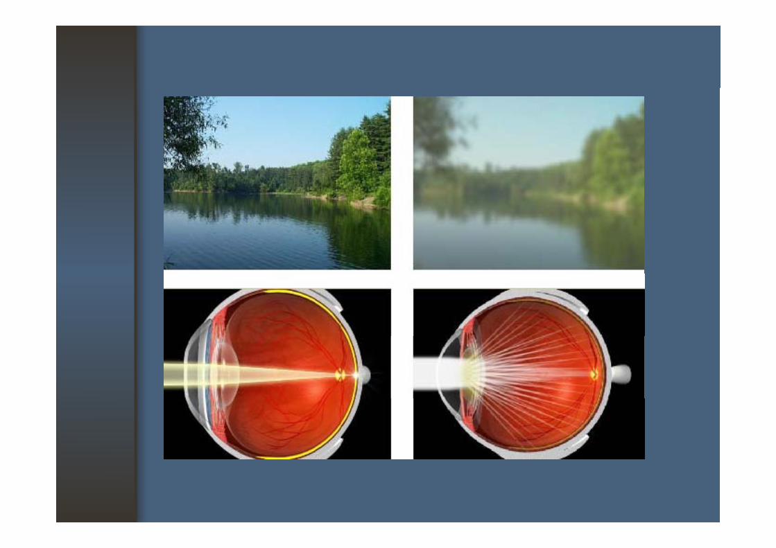
• Symptoms of cataract- Image blur : depends on the size and locationg pof opacity
: Axial opacities cause much more disabling visual loss than peripheral opacities
: Disturbance of vision, diminution, failure of vision
: Nuclear sclerosis may become progressively more myopic
Basic informationBasic information
S f ( )• Symptoms of cataract (cont.)- NS may develop a phenomenon called Second sightsight- Monocular double or multiple images, due to irregular refraction, prismatic effect within the irregular refraction, prismatic effect within the lens - Posterior subcapsular cataract (PSC) may note p ( ) ya relatively rapid decrease in vision (esp. near vision), with glare as well as image blur and distortiondistortion
: PSC is frequently associated with metabolic causes : DM, steroid usemetabolic causes : DM, steroid use
When to ExamineWhen to Examine
• A patient with decreasing vision• Important to demonstrate that the retina and Important to demonstrate that the retina and
optic nerve are healthy• If the lens is densely cataract• If the lens is densely cataract
- the risk of performing surgery for cataract ith t th without the assurance
- RAPD, color test, 4-quadrants light projection
How to ExamineHow to Examine
• Visual acuity• Pupillary responses : advanced cataract would Pupillary responses : advanced cataract would
not produce a RAPD• Anterior segment examination• Anterior segment examination• Ophthalmoscopy
How to Interpret the FindingsHow to Interpret the Findings
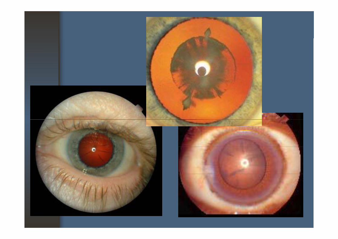
• Early cataract is not visible to the unaided eye• Very dense cataract may appear as a white pupil, Very dense cataract may appear as a white pupil,
or leukocoria• Ophthalmoscopy with plus-lens setting• Ophthalmoscopy with plus-lens setting
- partial cataract : black against the red reflex, th d flpoorer the red reflex
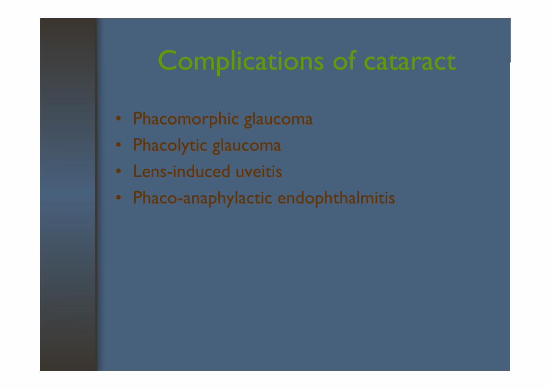
Complications of cataractComplications of cataract
• Phacomorphic glaucoma• Phacolytic glaucomaPhacolytic glaucoma• Lens-induced uveitis
Ph h l ti d hth l iti• Phaco-anaphylactic endophthalmitis
Management or ReferralManagement or Referral
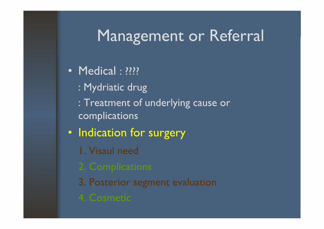
• Medical : ???? : Mydriatic drug: Mydriatic drug: Treatment of underlying cause or complicationscomplications
• Indication for surgery1. Visaul need2 Complications2. Complications3. Posterior segment evaluation4. Cosmetic
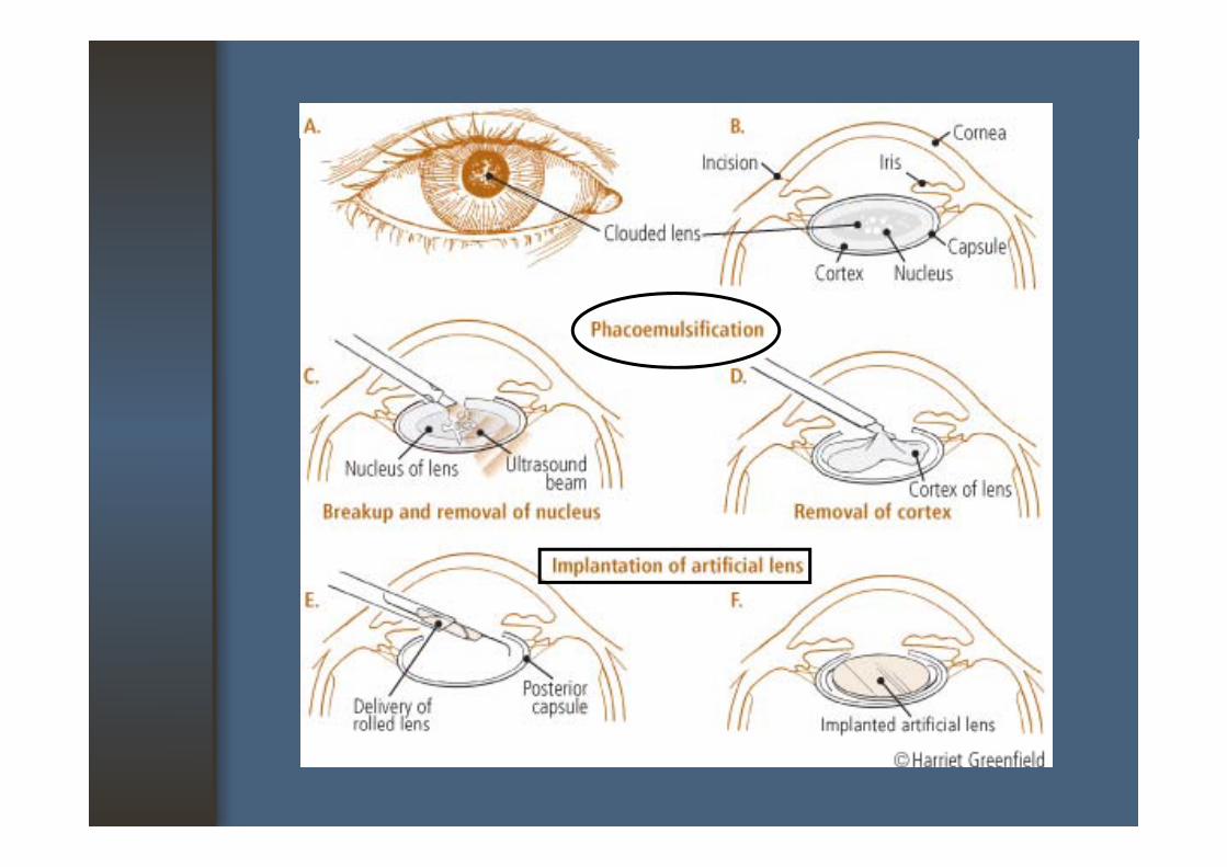
Cataract surgeryCataract surgery
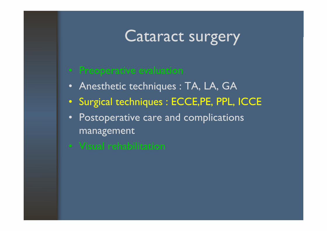
• Preoperative evaluation• Anesthetic techniques : TA, LA, GAAnesthetic techniques : TA, LA, GA• Surgical techniques : ECCE,PE, PPL, ICCE
P t ti d li ti • Postoperative care and complications management
• Visual rehabilitation
Surgical techniquesSurgical techniques
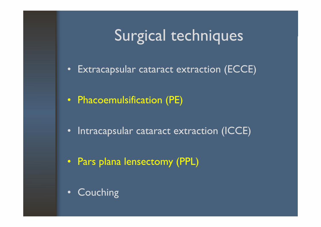
• Extracapsular cataract extraction (ECCE)
• Phacoemulsification (PE)
• Intracapsular cataract extraction (ICCE)
• Pars plana lensectomy (PPL)p y ( )
• C chin• Couching

CouchingCouching
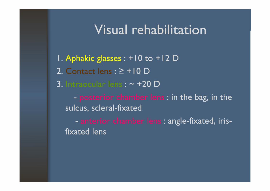
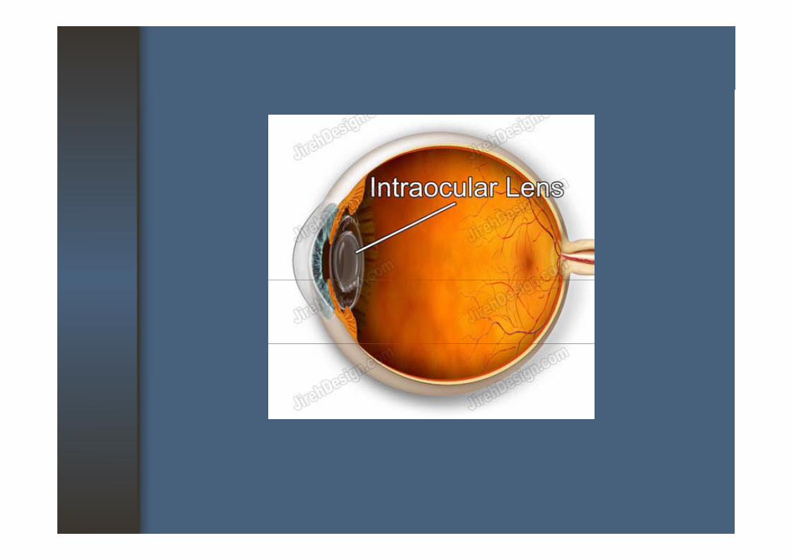
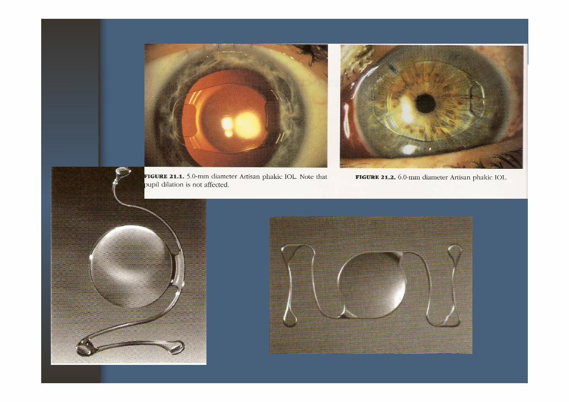
Visual rehabilitationVisual rehabilitation
1. Aphakic glasses : +10 to +12 D2. Contact lens : ≥ +10 D2. Contact lens : 10 D3. Intraocular lens : ~ +20 D
t i h b l i th b i th - posterior chamber lens : in the bag, in the sulcus, scleral-fixated
- anterior chamber lens : angle-fixated, iris-fixated lens
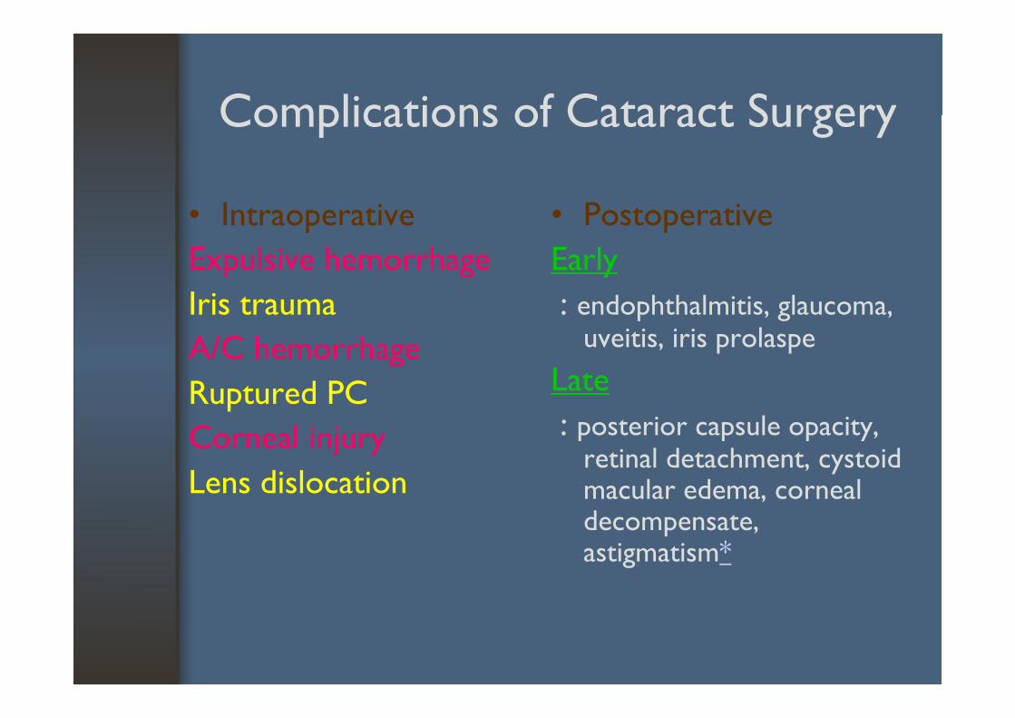
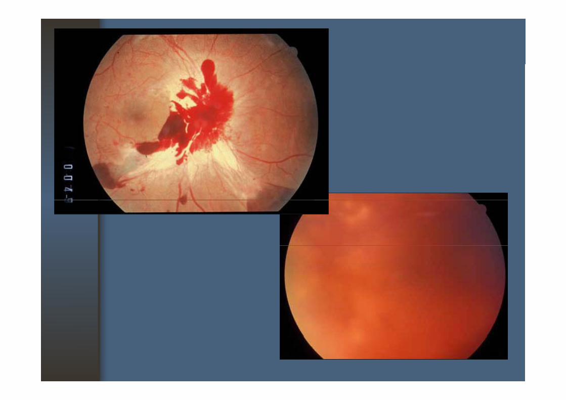
Complications of Cataract SurgeryComplications of Cataract Surgery
• IntraoperativeExpulsive hemorrhage
• PostoperativeEarly p g
Iris traumaA/C hemorrhage
y: endophthalmitis, glaucoma,
uveitis, iris prolaspeA/C hemorrhageRuptured PCCorneal injury
Late: posterior capsule opacity, Corneal injury
Lens dislocation
p p p yretinal detachment, cystoid macular edema, corneal decompensate decompensate, astigmatism*

Macular Macular De enerati nDegeneration
RelevanceRelevance
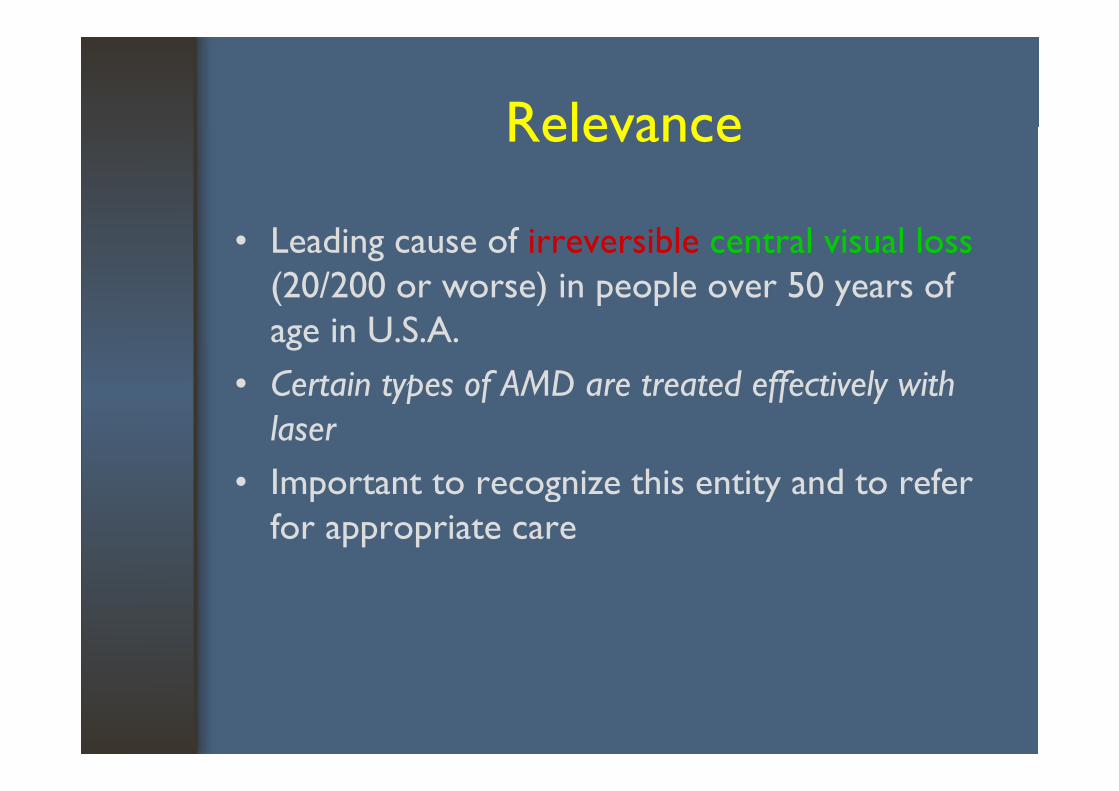
• Leading cause of irreversible central visual loss (20/200 or worse) in people over 50 years of ( ) p p yage in U.S.A.
• Certain types of AMD are treated effectively with Certain types of AMD are treated effectively with laser
• Important to recognize this entity and to refer • Important to recognize this entity and to refer for appropriate care
Basic InformationBasic Information
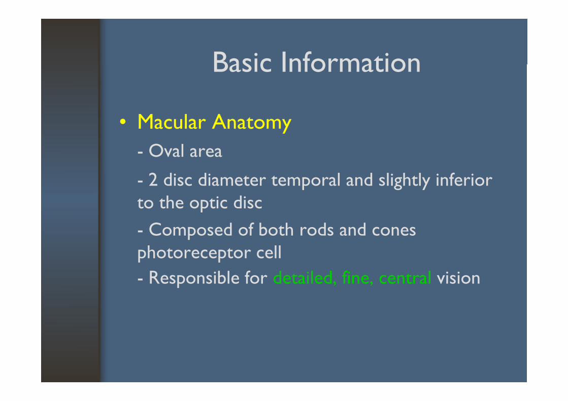
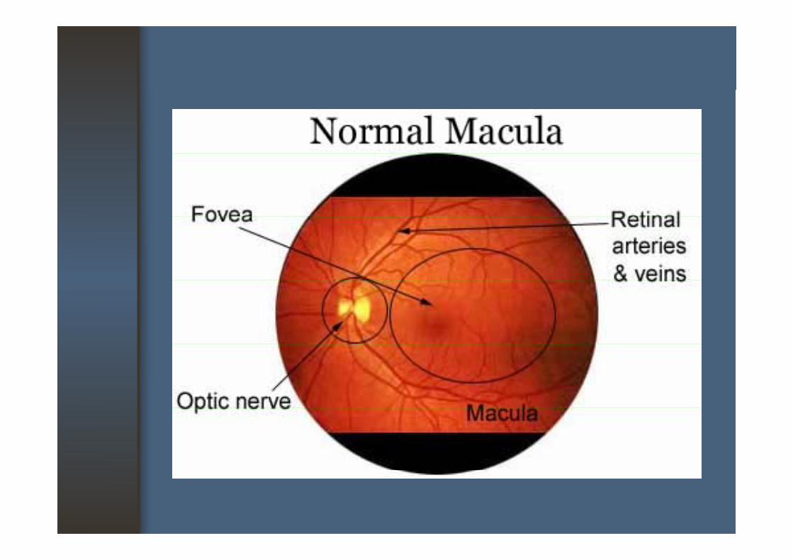
• Macular Anatomy- Oval area Oval area
- 2 disc diameter temporal and slightly inferior to the optic discto the optic disc- Composed of both rods and cones h llphotoreceptor cell
- Responsible for detailed, fine, central vision
Basic InformationBasic Information
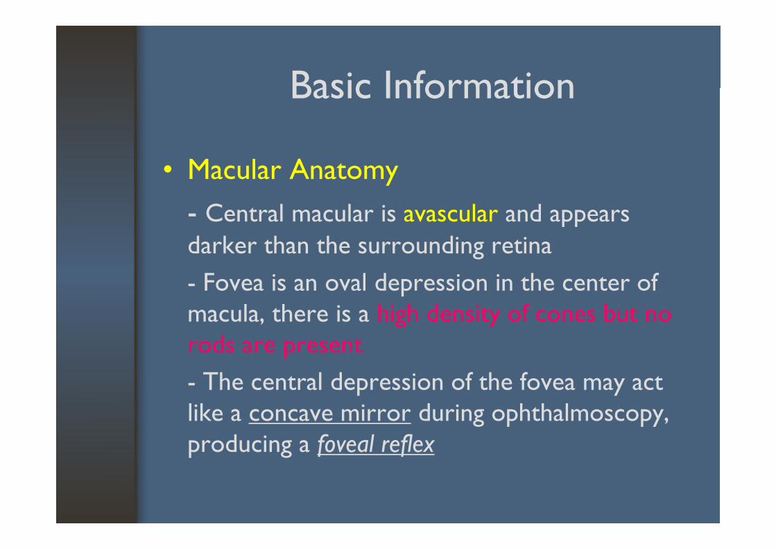
• Macular AnatomyCentral macular is avascular and appears - Central macular is avascular and appears
darker than the surrounding retinaF i l d i i h f - Fovea is an oval depression in the center of
macula, there is a high density of cones but no d rods are present
- The central depression of the fovea may act like a concave mirror during ophthalmoscopy, producing a foveal reflex
Basic InformationBasic Information
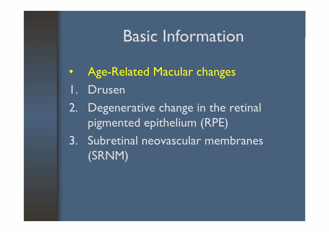
• Age-Related Macular changes1 Drusen1. Drusen2. Degenerative change in the retinal
pigmented epithelium (RPE)3 Subretinal neovascular membranes 3. Subretinal neovascular membranes
(SRNM)
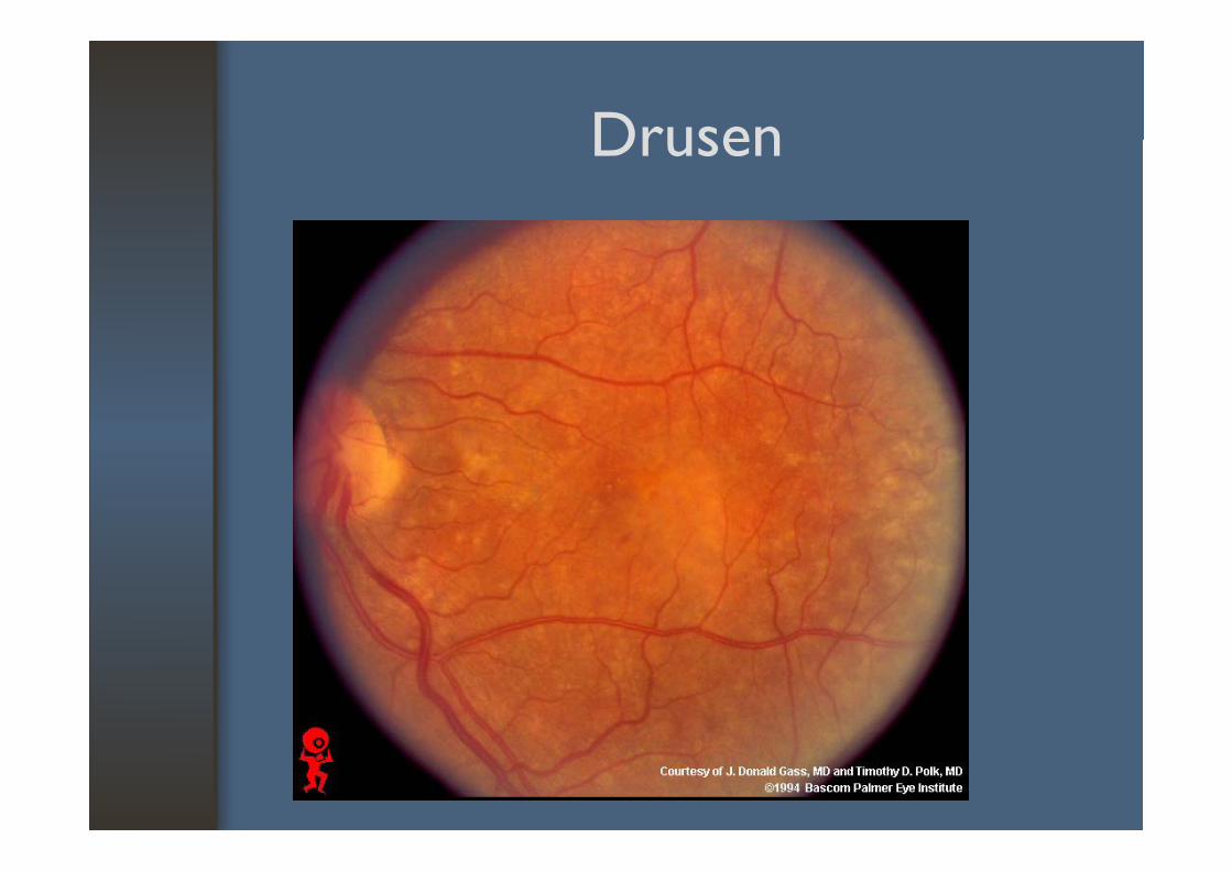
DrusenDrusen
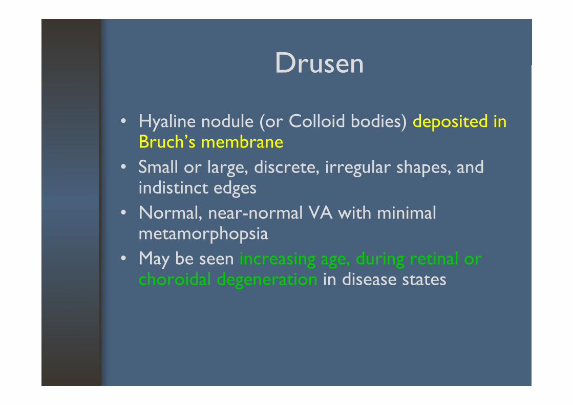
• Hyaline nodule (or Colloid bodies) deposited in Bruch’s membrane
• Small or large, discrete, irregular shapes, and indistinct edges
• Normal, near-normal VA with minimal metamorphopsia
• May be seen increasing age, during retinal or choroidal degeneration in disease states
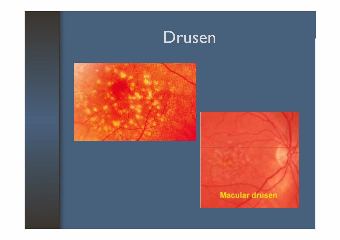
DrusenDrusen
DrusenDrusen
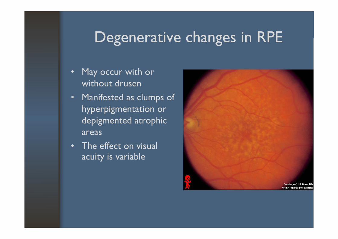
Degenerative changes in RPEDegenerative changes in RPE
• May occur with or without drusen
• Manifested as clumps of hyperpigmentation or depigmented atrophic depigmented atrophic areas
• The effect on visual • The effect on visual acuity is variable
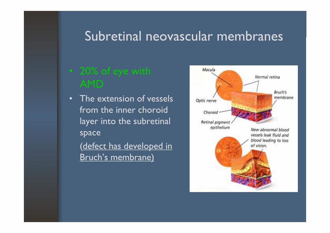
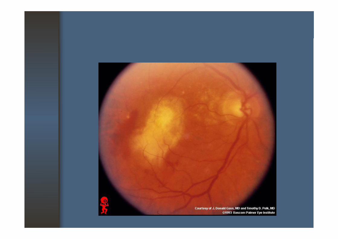
Subretinal neovascular membranesSubretinal neovascular membranes
• 20% of eye with AMD
• The extension of vessels from the inner choroid layer into the subretinal space (d f h d l d i (defect has developed in Bruch’s membrane)
Subretinal neovascular membranesSubretinal neovascular membranes
• Associated with subretinal hemorrhage, fibrosis, RPE degeneration, photoreceptor atrophyg p p p y
• Hemorrhage or subretinal fluid may result in acute visual lossacute visual loss
• Larger the membrane and the closer to thecenter of the fovea the worse prognosis for center of the fovea, the worse prognosis for good central vision
Subretinal neovascular membranesSubretinal neovascular membranes
Other causes of SRNM1. High myopia1. High myopia2. Angioid streaks3 P d l hi t l i3. Presumed ocular histoplasmosis4. Traumatic choroidal rupture
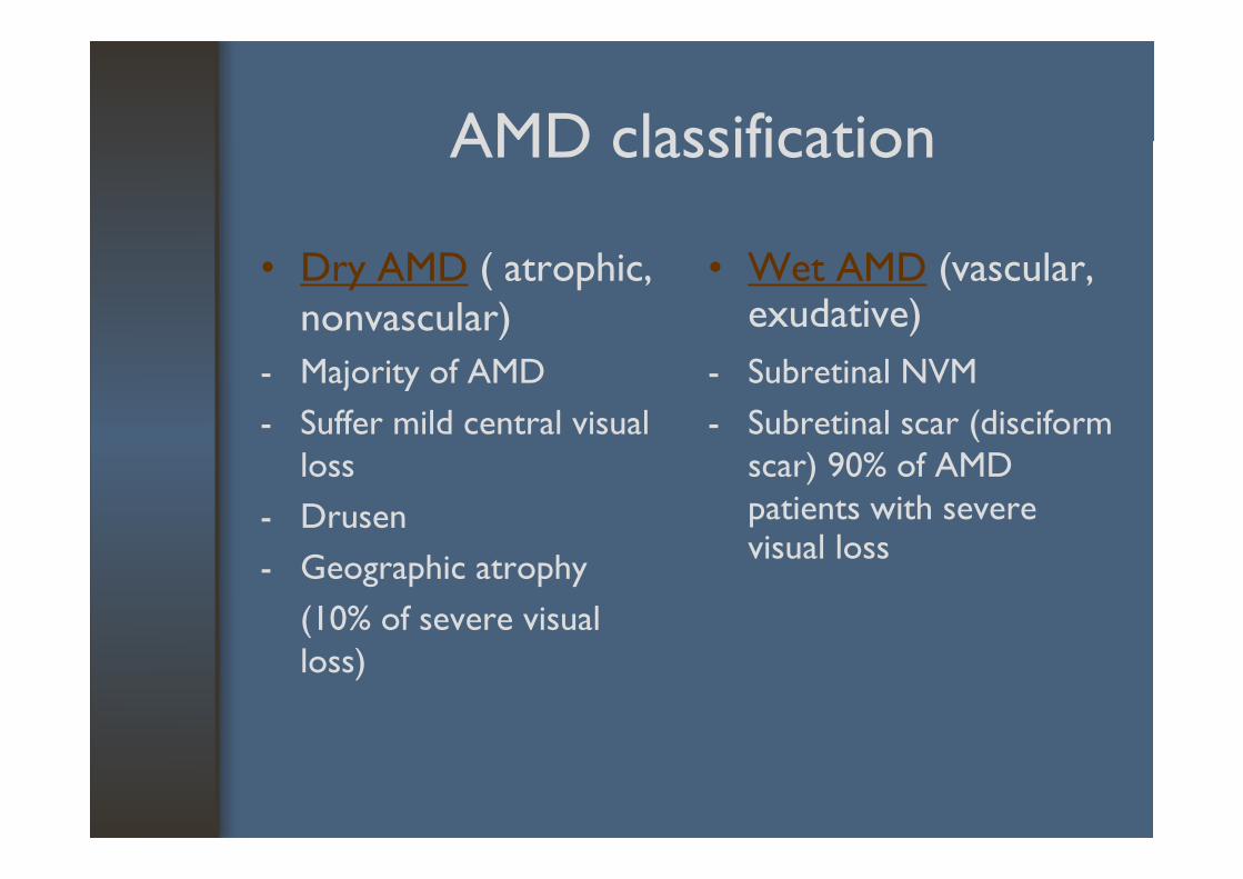
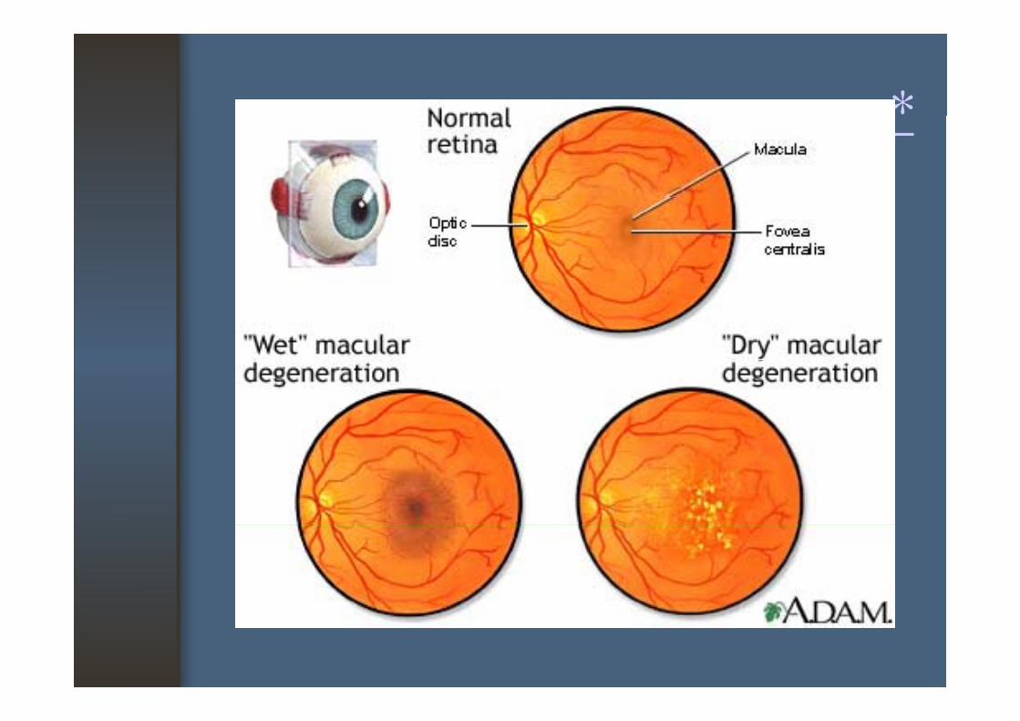
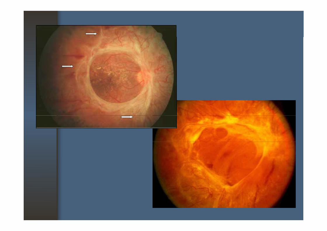
AMD classificationAMD classification
• Dry AMD ( atrophic, nonvascular)
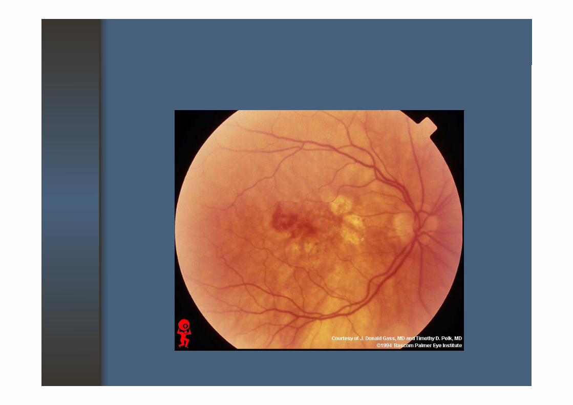
• Wet AMD (vascular, exudative))
- Majority of AMD- Suffer mild central visual
- Subretinal NVM- Subretinal scar (disciform
loss- Drusen
(scar) 90% of AMD patients with severe i l l- Geographic atrophy
(10% of severe visual
visual loss
loss)
*
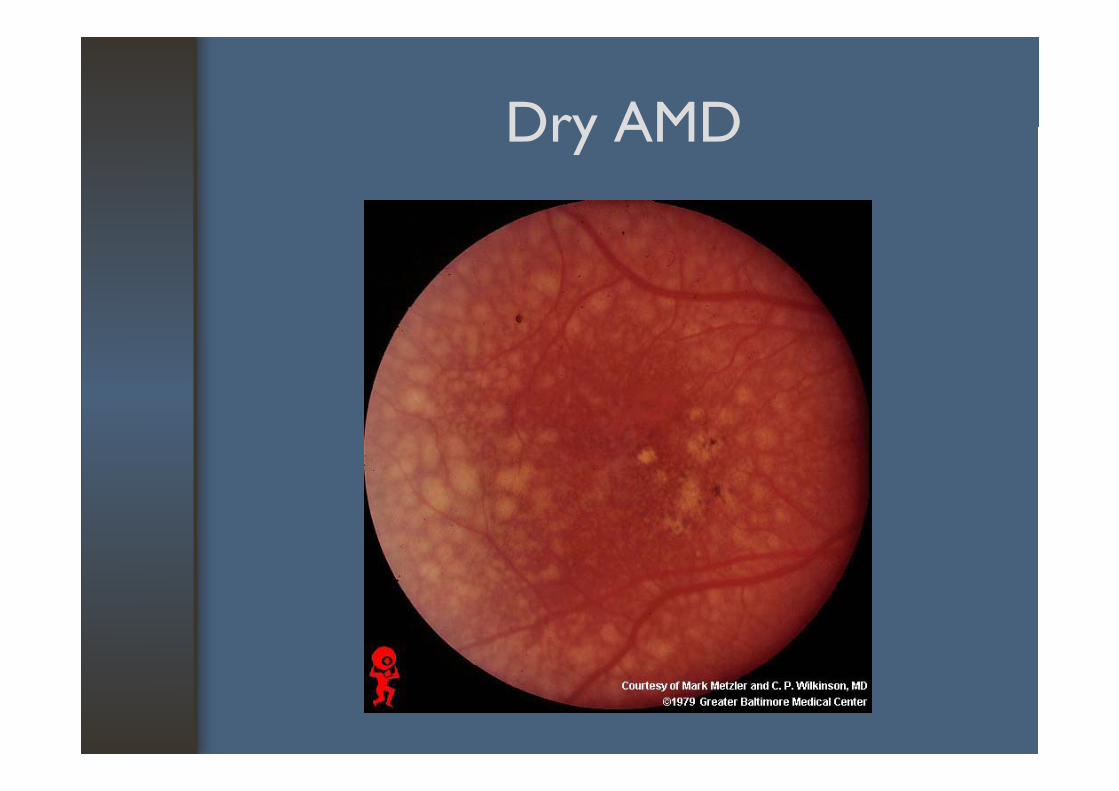
Dry AMDDry AMD
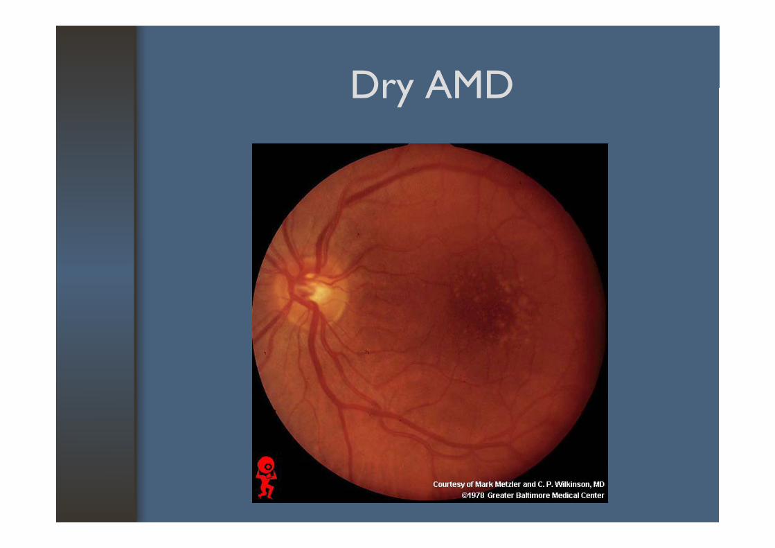
Dry AMDDry AMD
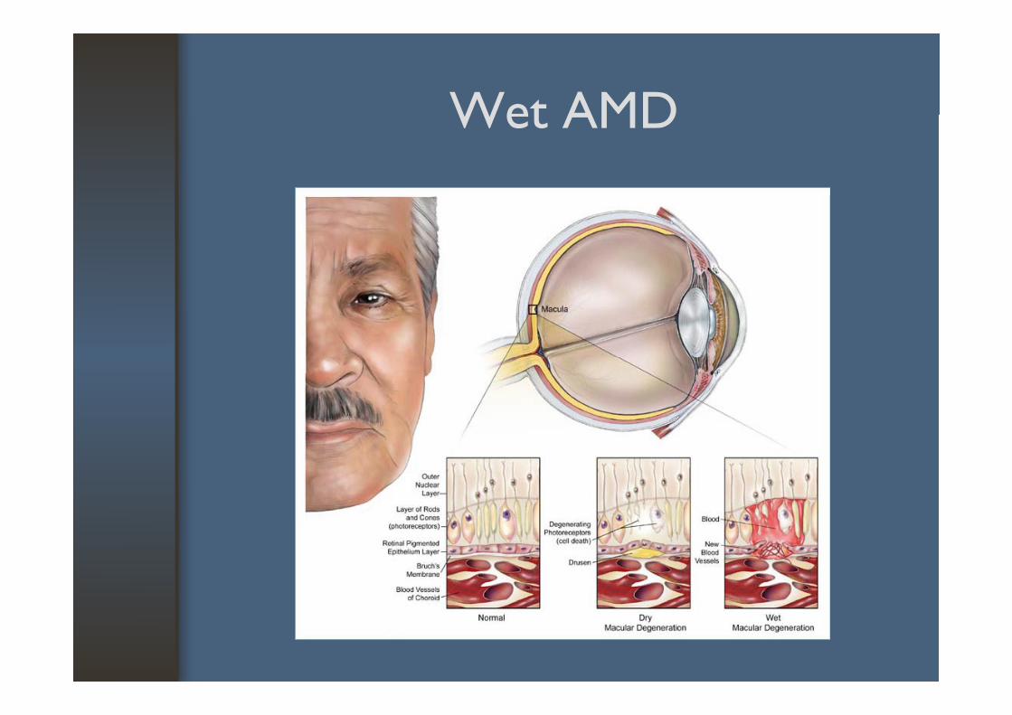
Wet AMDWet AMD
When to ExamineWhen to Examine
• Any patient with decreasing vision• Patient with decreased or distorted• Patient with decreased or distorted
central vision should be examine the lmacula
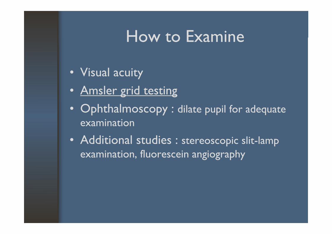
How to ExamineHow to Examine
• Visual acuity• Amsler grid testing• Amsler grid testing• Ophthalmoscopy : dilate pupil for adequate
examination
• Additional studies : stereoscopic slit-lamp Additional studies : stereoscopic slit lamp examination, fluorescein angiography
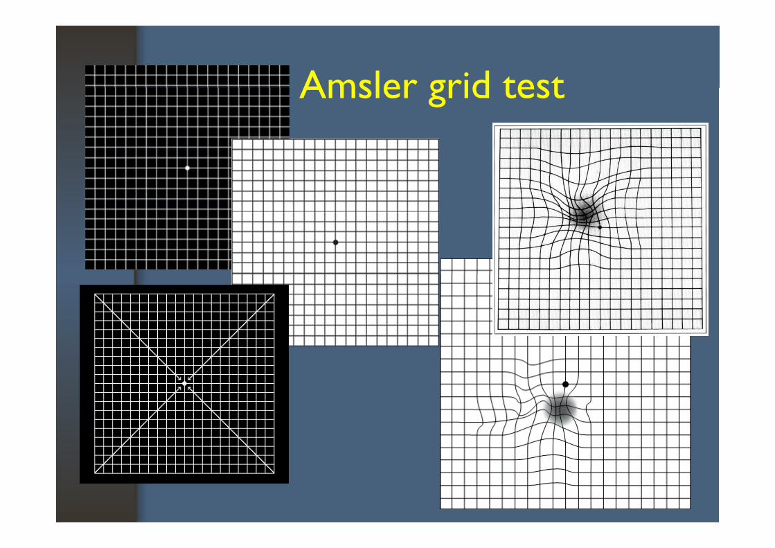
Amsler grid testAmsler grid test
Amsler gridAmsler grid
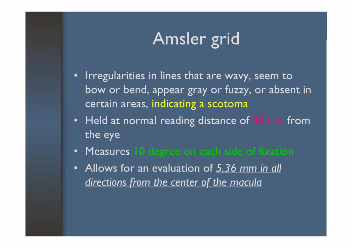
• Irregularities in lines that are wavy, seem to bow or bend, appear gray or fuzzy, or absent in pp g y ycertain areas, indicating a scotoma
• Held at normal reading distance of 30 cm. from Held at normal reading distance of 30 cm. from the eye
• Measures 10 degree on each side of fixation• Measures 10 degree on each side of fixation• Allows for an evaluation of 5.36 mm in all
di ectio s f o the ce te of the ac ladirections from the center of the macula
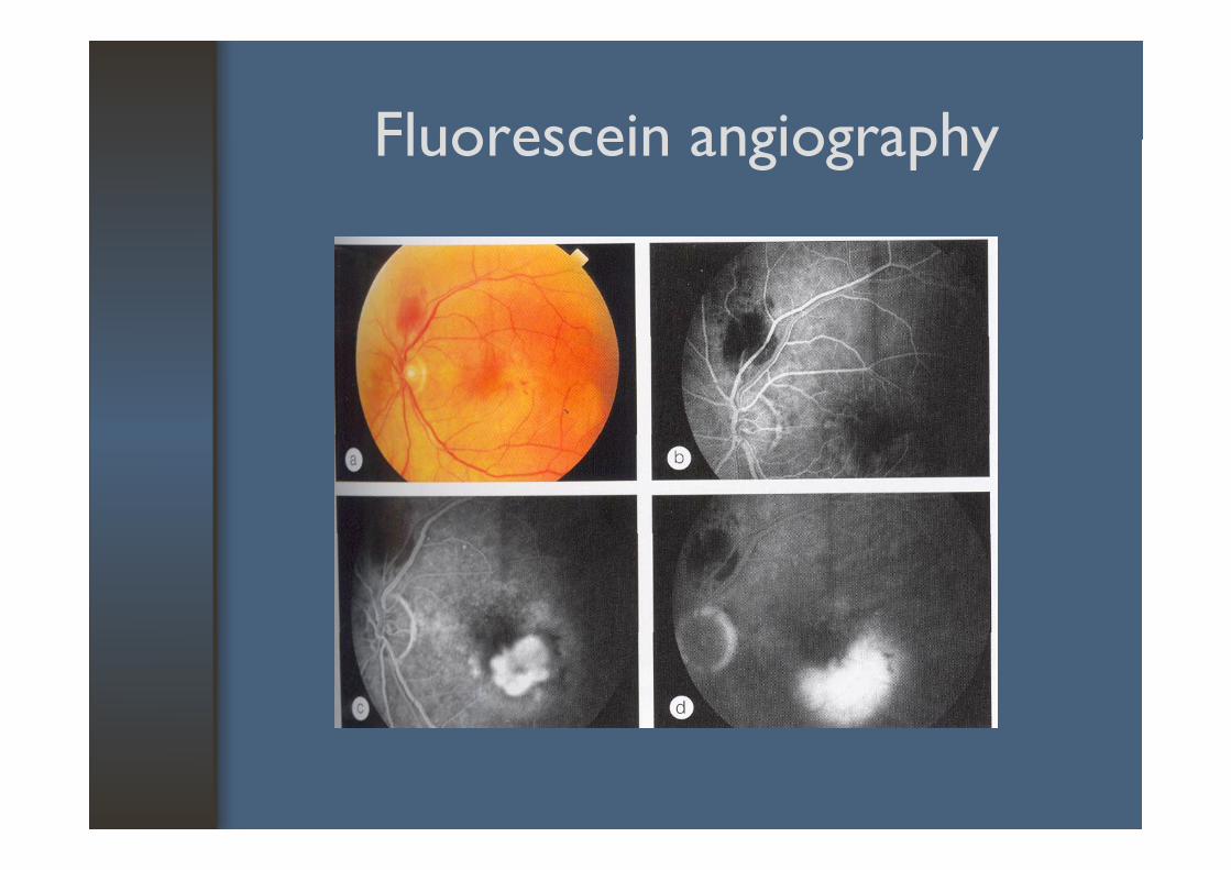
Fluorescein angiographyFluorescein angiography
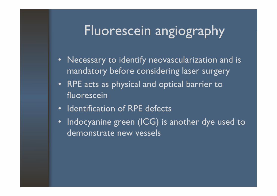
• Necessary to identify neovascularization and is mandatory before considering laser surgeryy g g y
• RPE acts as physical and optical barrier to fluoresceinfluorescein
• Identification of RPE defectsI d i (ICG) i th d d t • Indocyanine green (ICG) is another dye used to demonstrate new vessels
Fluorescein angiographyFluorescein angiography
How to Interpret the FindingsHow to Interpret the Findings
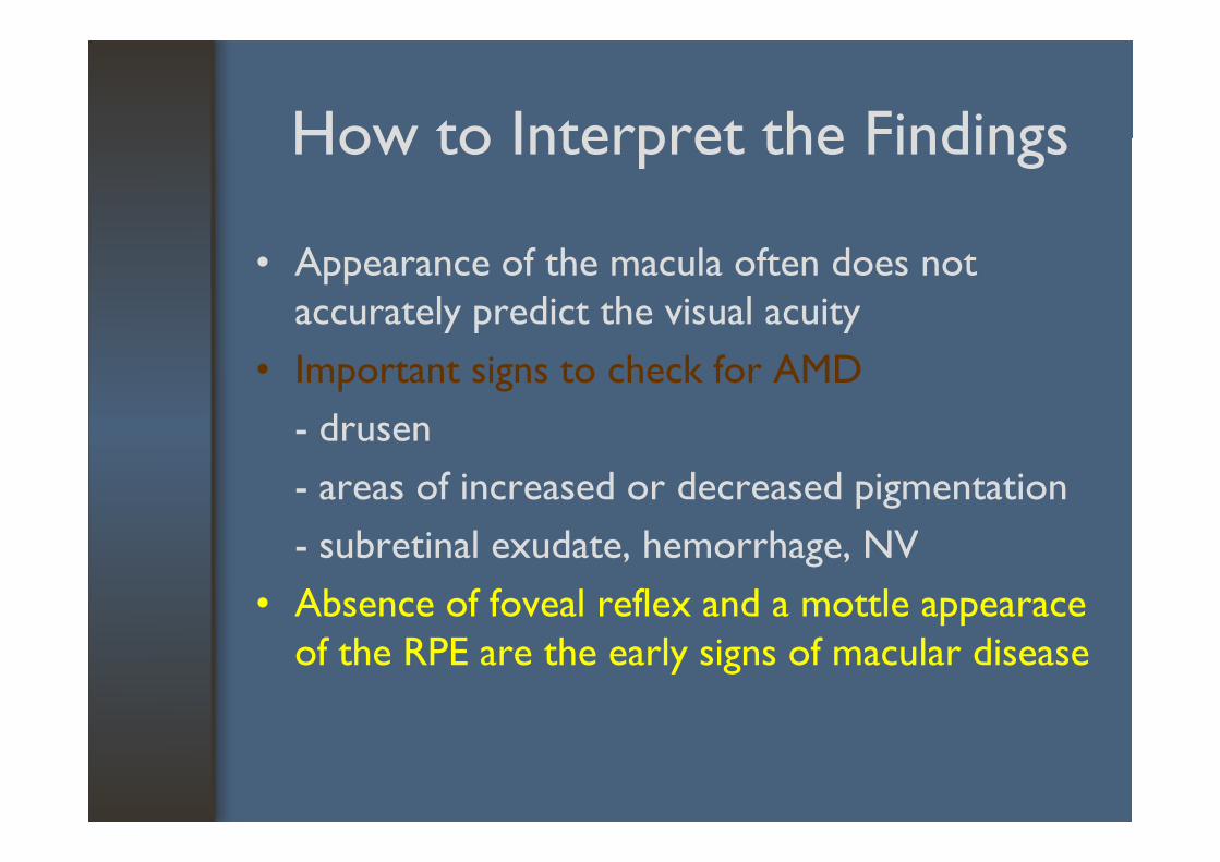
• Appearance of the macula often does not accurately predict the visual acuityy p y
• Important signs to check for AMD- drusen- drusen- areas of increased or decreased pigmentation- subretinal exudate, hemorrhage, NV
• Absence of foveal reflex and a mottle appearace of the RPE are the early signs of macular disease
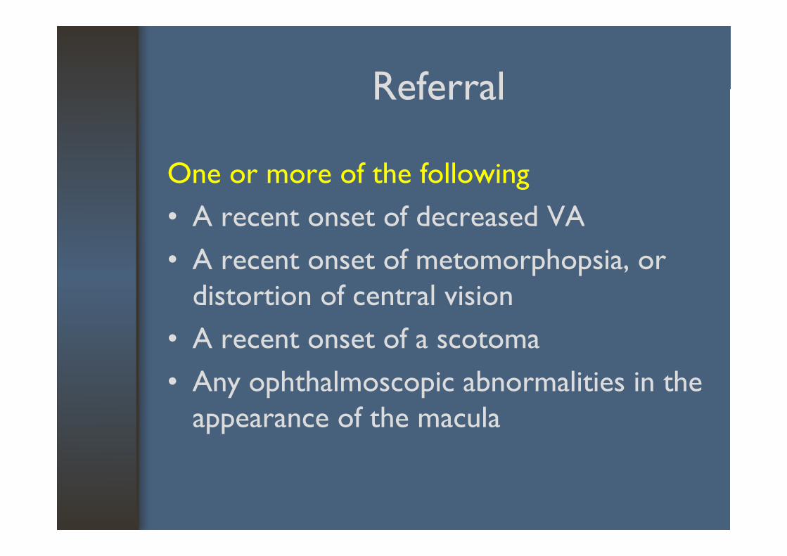
ReferralReferral
One or more of the following• A recent onset of decreased VA• A recent onset of decreased VA• A recent onset of metomorphopsia, or
distortion of central vision• A recent onset of a scotoma• A recent onset of a scotoma• Any ophthalmoscopic abnormalities in the
appearance of the macula
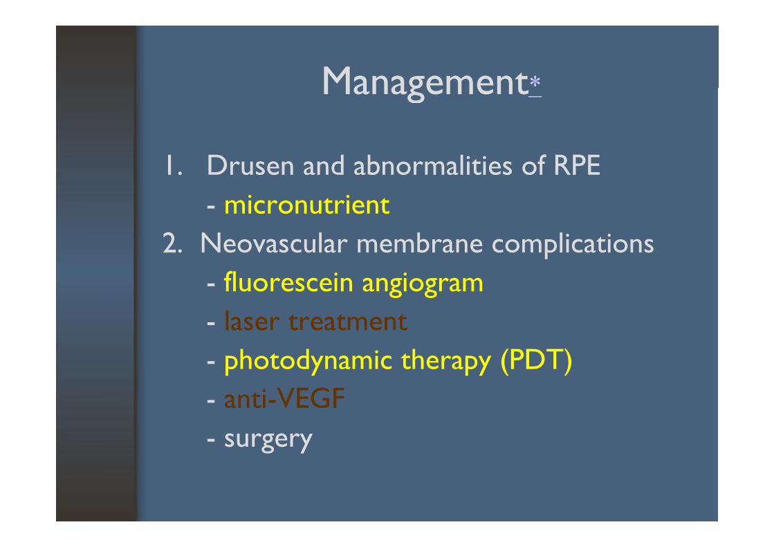
Management*Management
1. Drusen and abnormalities of RPE- micronutrientmicronutrient
2. Neovascular membrane complicationsfl i i- fluorescein angiogram
- laser treatment- photodynamic therapy (PDT)
anti VEGF- anti-VEGF- surgery
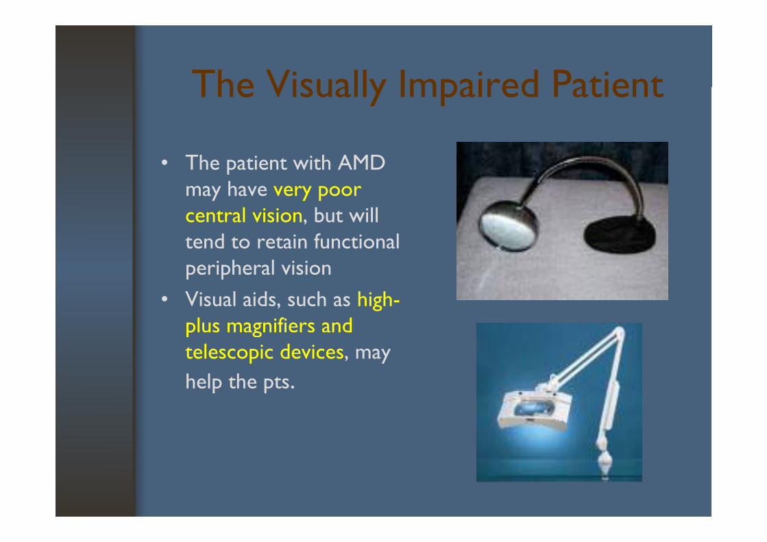
The Visually Impaired PatientThe Visually Impaired Patient
• The patient with AMD may have very poor
l i i b ill central vision, but will tend to retain functional peripheral visionperipheral vision
• Visual aids, such as high-plus magnifiers and p gtelescopic devices, may help the pts.
Diabetic Diabetic Retin athRetinopathy
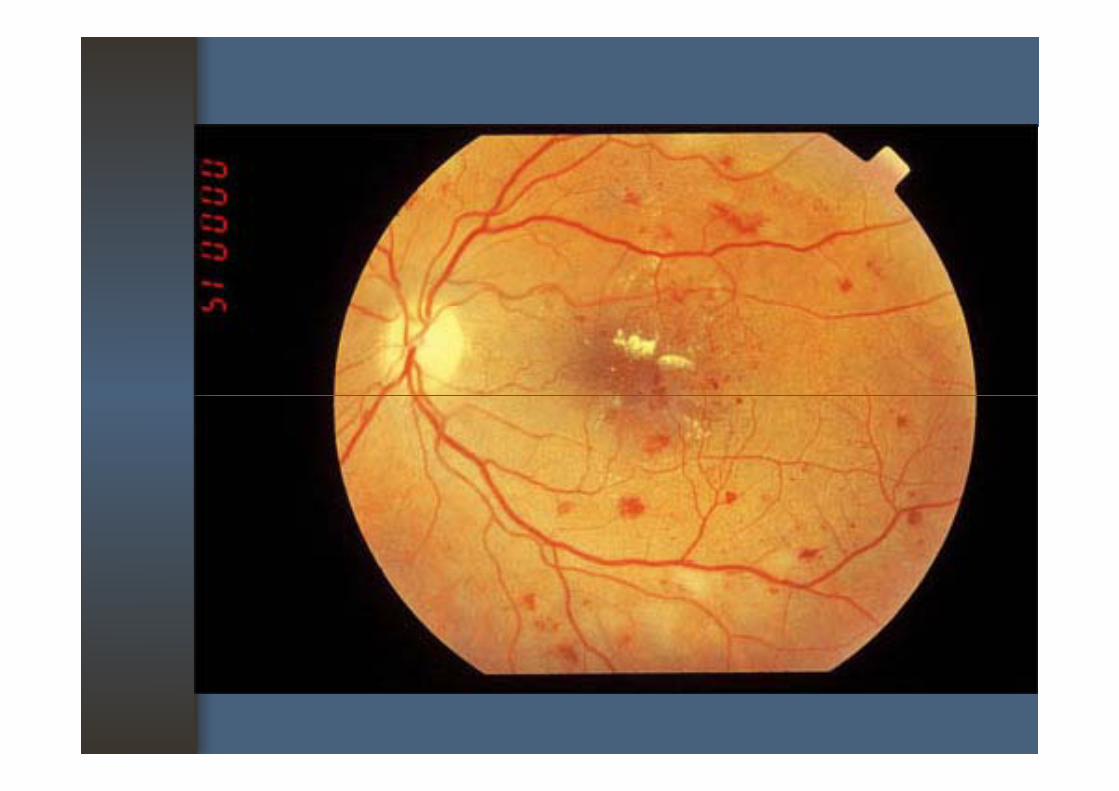
ClassificationClassification
• Non proliferative DR (NPDR)mild- mild
- moderate- severe
• Proliferative DR (PDR)• Proliferative DR (PDR)
Signs & SymptomsSigns & Symptoms
• NPDR- No symptoms
• PDR
- No symptomsNo symptoms- Vision loss
l d
No symptoms- Vision loss
NPDR: lens edema: macular edema
: as NPDR: VH
: CSME: cataract
: TRD +/- RRD: NVG: macular ischemia
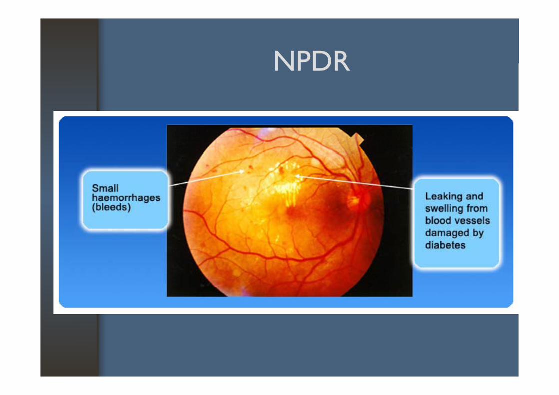
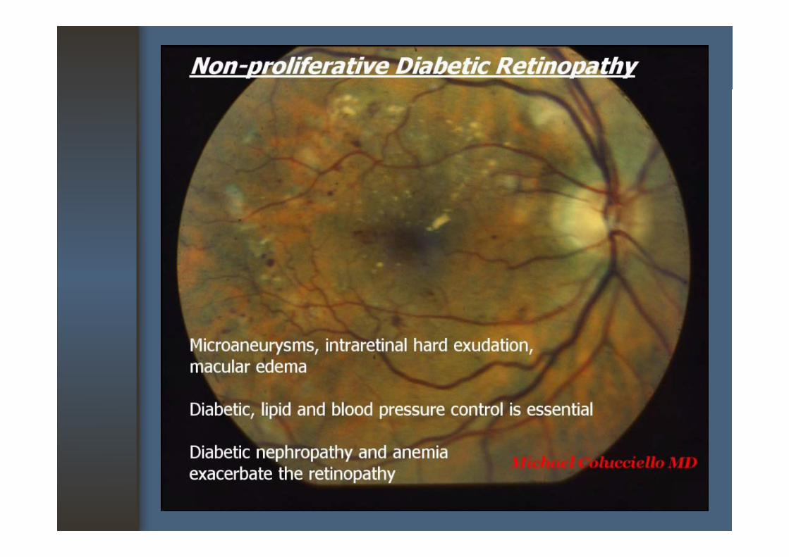
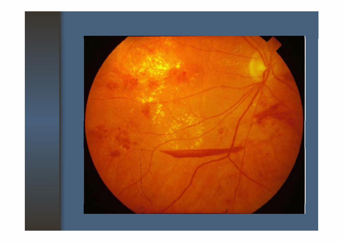
NPDRNPDR
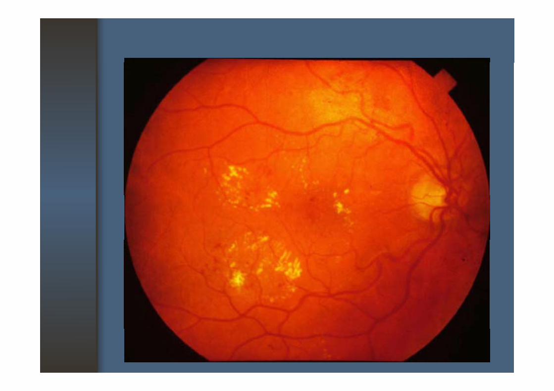
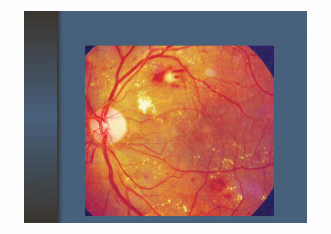
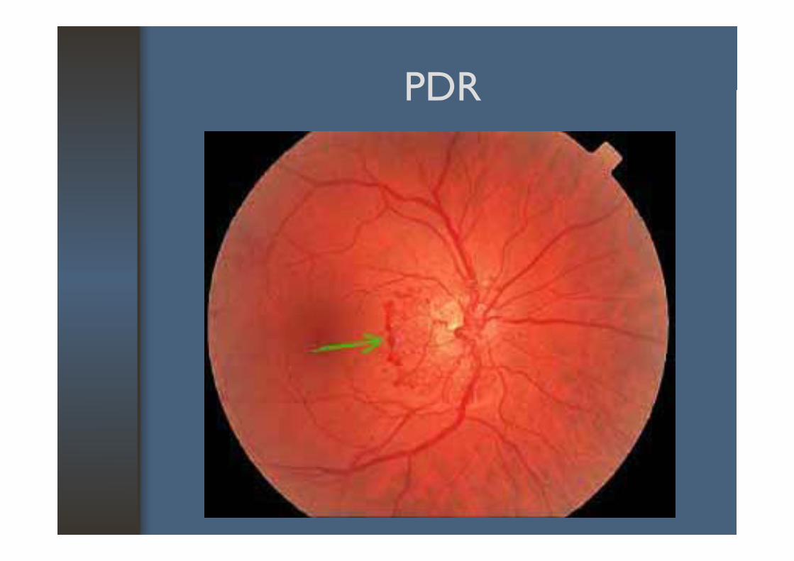
PDRPDR
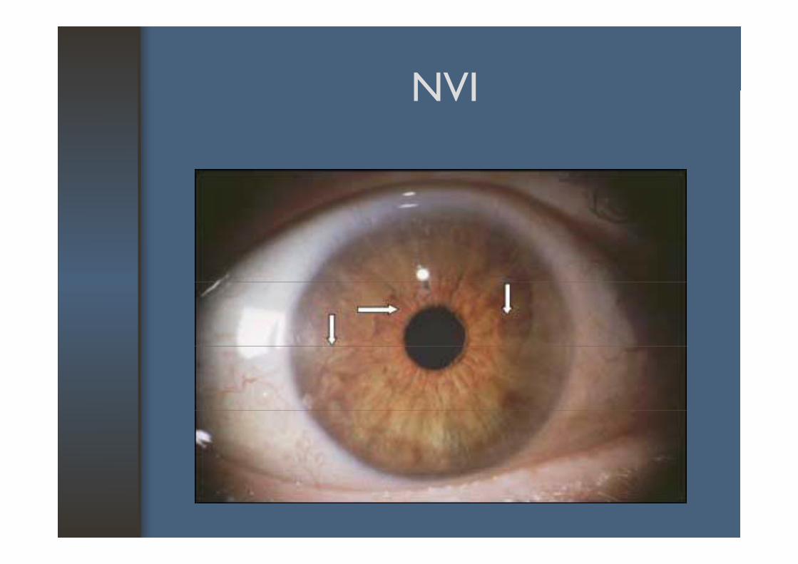
NVINVI
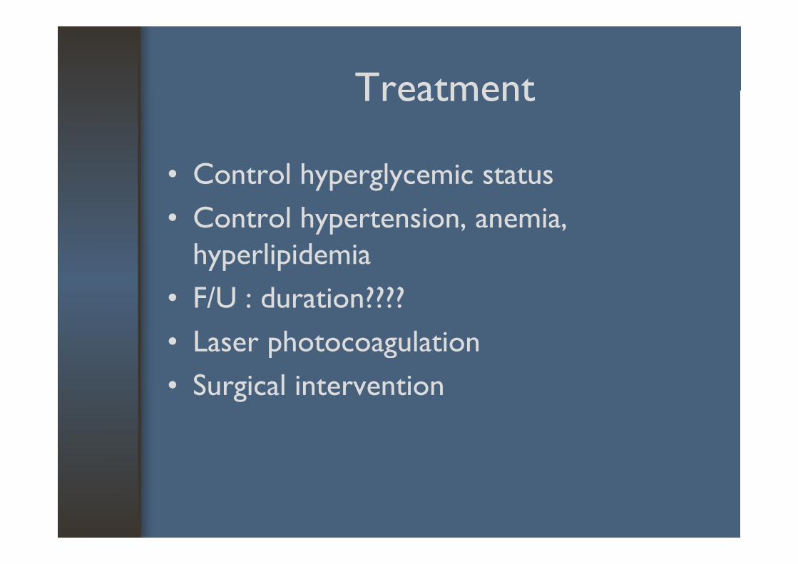
TreatmentTreatment
• Control hyperglycemic status• Control hypertension anemia • Control hypertension, anemia,
hyperlipidemia• F/U : duration????• Laser photocoagulationLaser photocoagulation• Surgical intervention
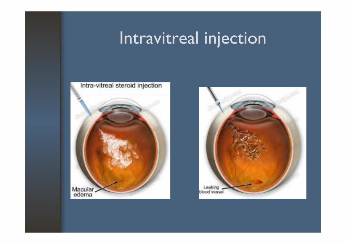
Intravitreal injectionIntravitreal injection
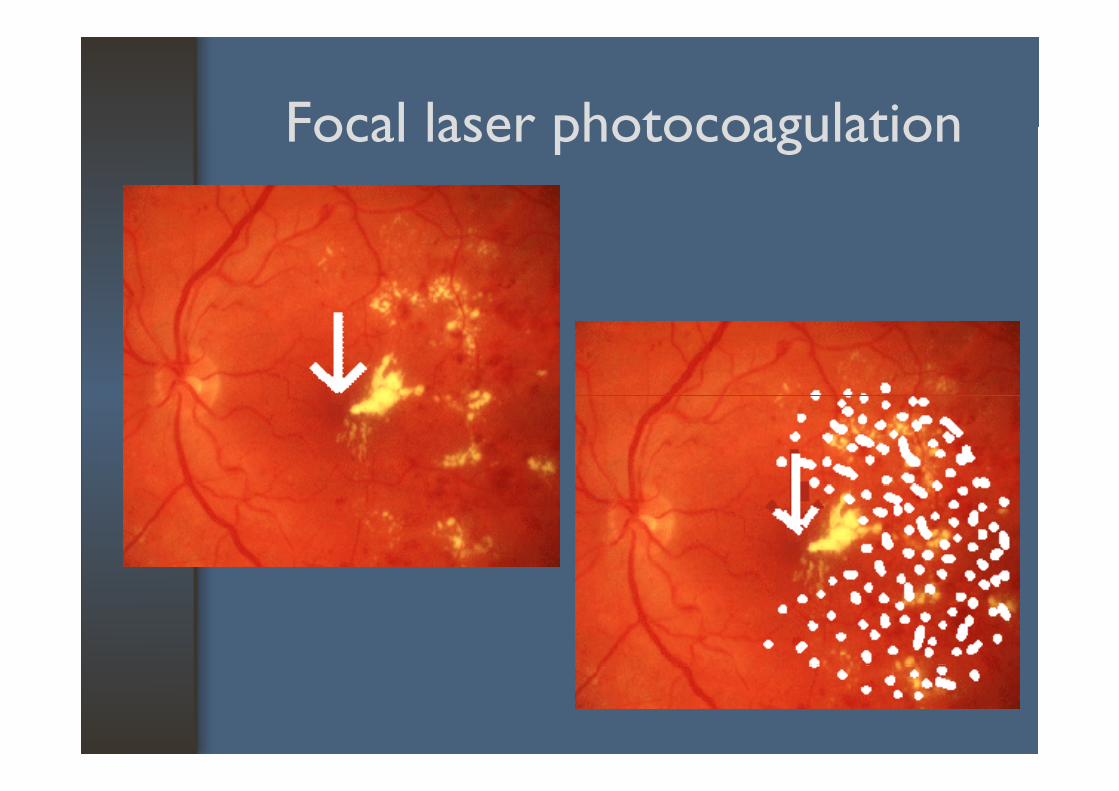
Focal laser photocoagulationFocal laser photocoagulation
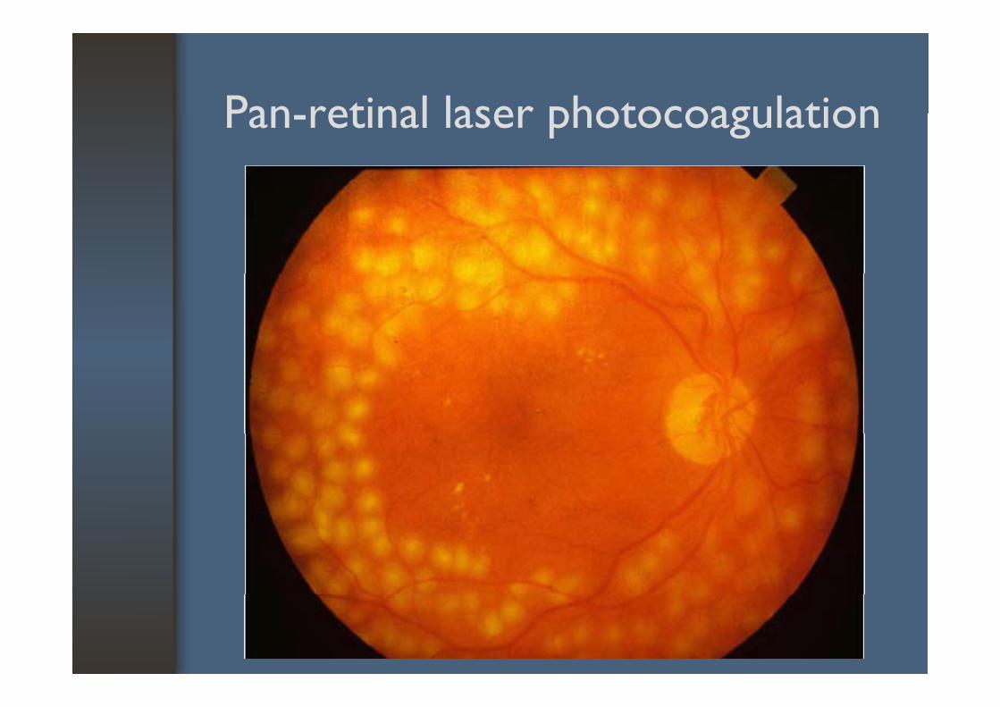
Pan retinal laser photocoagulationPan-retinal laser photocoagulation
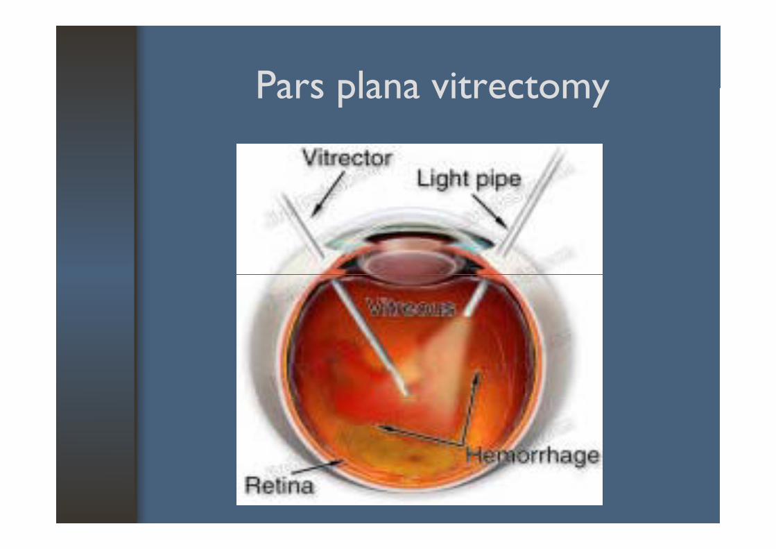
Pars plana vitrectomyPars plana vitrectomy
Refractive Errors & Refractive Errors & PresbyopiaPresbyopia
Formation of VisionFormation of Vision
• Light from object• Refraction by optical element• Refraction by optical element• Image formation on retina• Conversion into neural signals• Perception by the brain• Perception by the brain
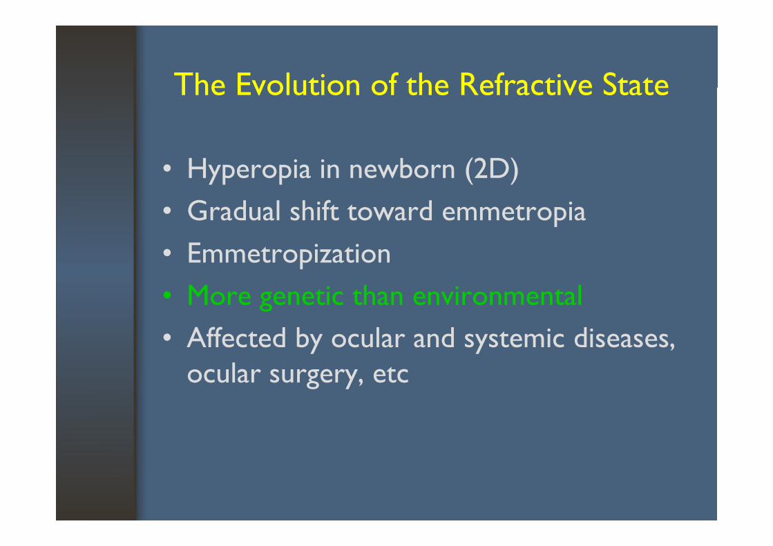
The Evolution of the Refractive StateThe Evolution of the Refractive State
• Hyperopia in newborn (2D)• Gradual shift toward emmetropia• Gradual shift toward emmetropia• Emmetropization• More genetic than environmental• Affected by ocular and systemic diseases • Affected by ocular and systemic diseases,
ocular surgery, etc
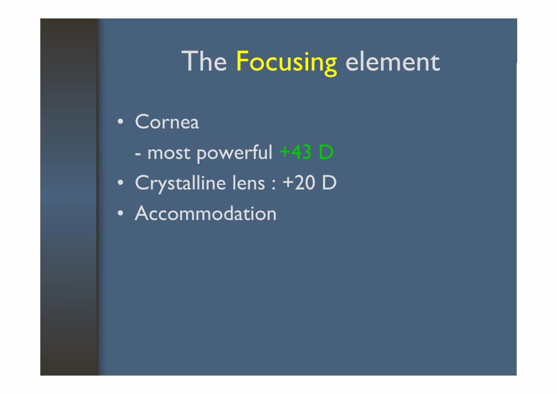
The Focusing elementThe Focusing element
• Cornea most powerful +43 D- most powerful +43 D
• Crystalline lens : +20 D • Accommodation
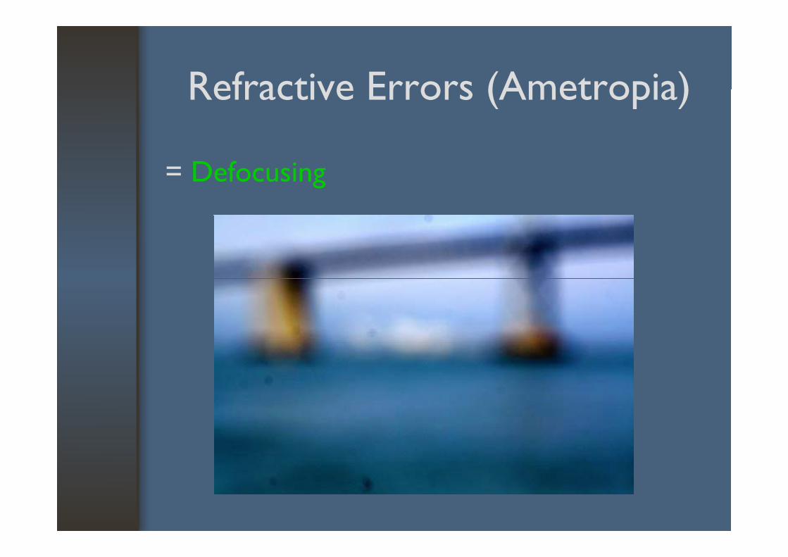
Refractive Errors (Ametropia)Refractive Errors (Ametropia)
= Defocusing
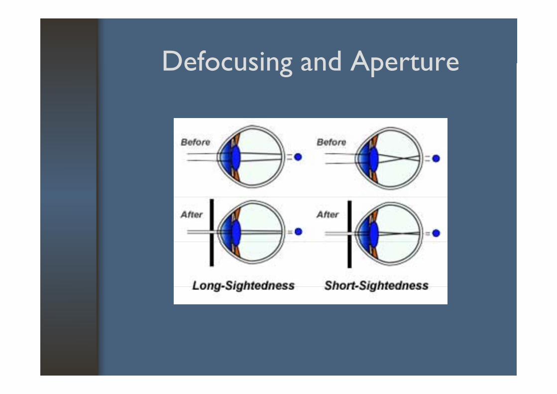
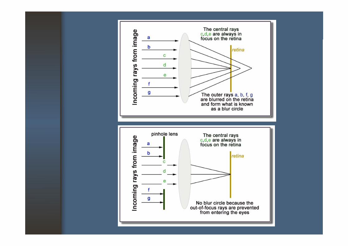
Defocusing and ApertureDefocusing and Aperture
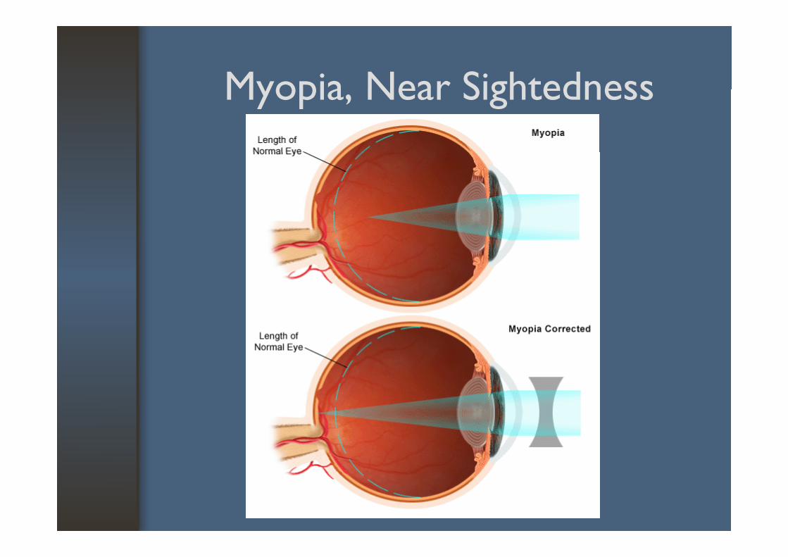
Myopia Near Sightedness Myopia, Near Sightedness
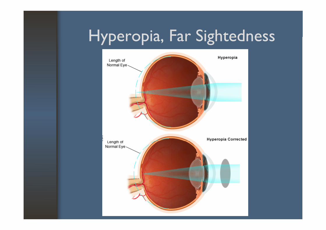
Hyperopia Far SightednessHyperopia, Far Sightedness
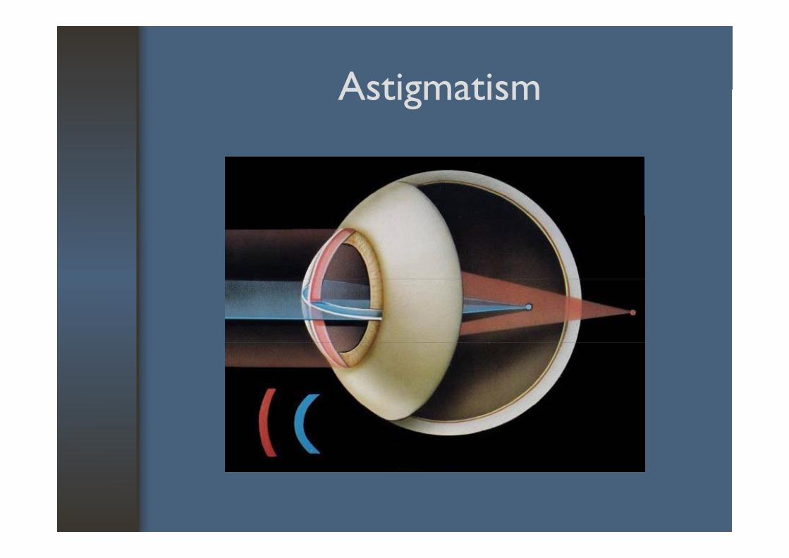
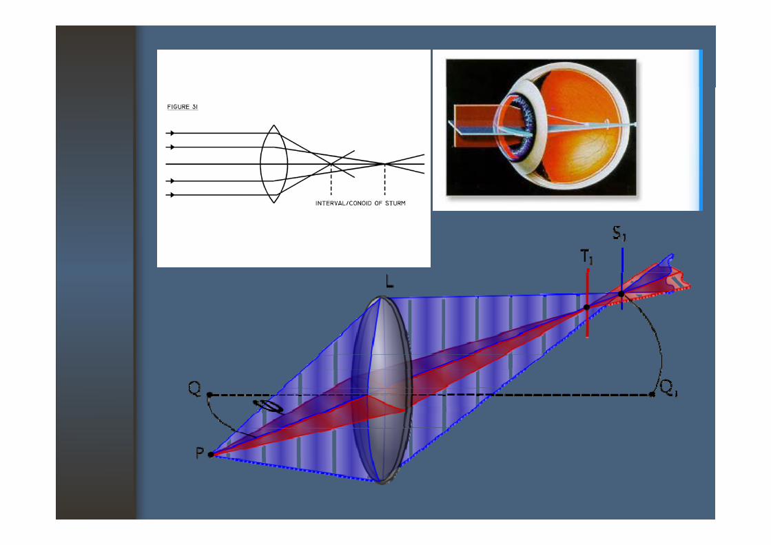
AstigmatismAstigmatism
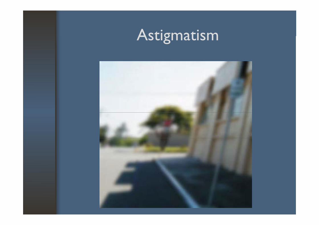
AstigmatismAstigmatism
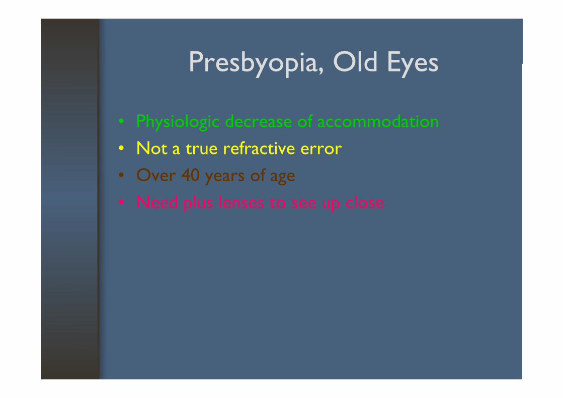
Presbyopia Old EyesPresbyopia, Old Eyes
• Physiologic decrease of accommodation• Not a true refractive errorNot a true refractive error• Over 40 years of age
N d l l t l• Need plus lenses to see up close
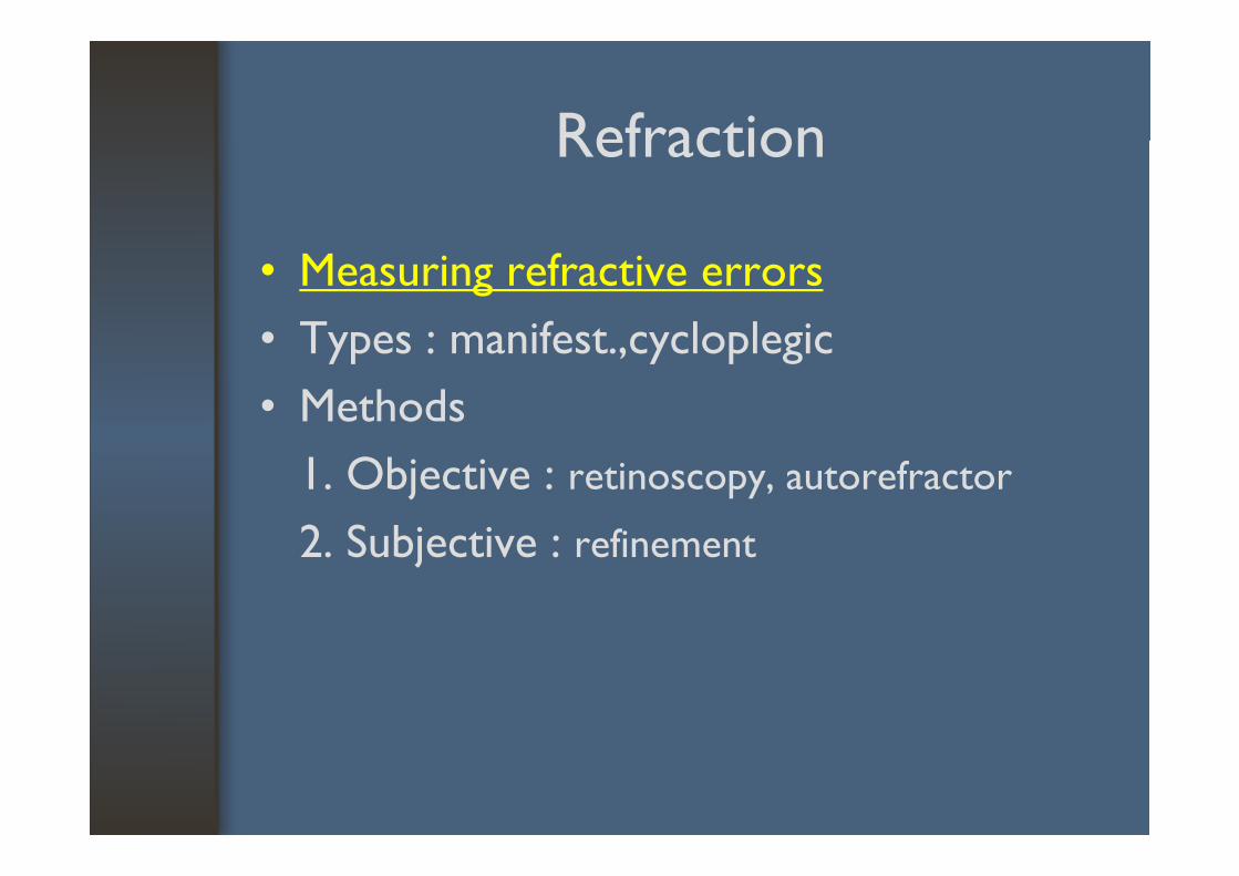
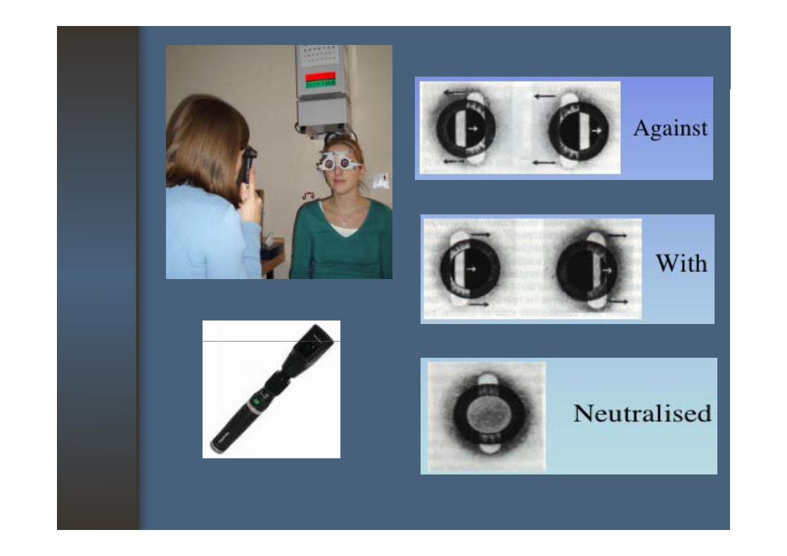
RefractionRefraction
• Measuring refractive errors• Types : manifest cycloplegic• Types : manifest.,cycloplegic• Methods
1. Objective : retinoscopy, autorefractor
2 Subjective : fi t2. Subjective : refinement
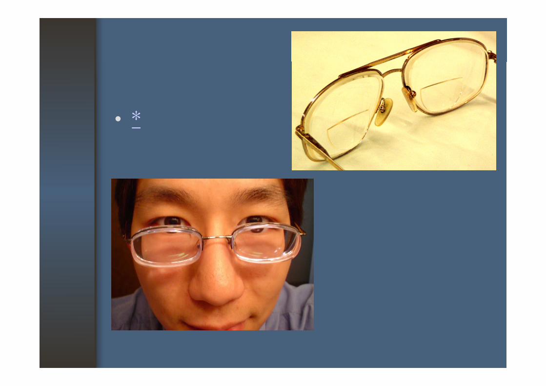
Correcting Refractive ErrorsCorrecting Refractive Errors
• Spectacles• Contact lenses• Contact lenses• Surgery (refractive surgery)
• *