The Patient with Visual Loss: Localization of ...
25
1 Steven A. Kane, M.D., Ph.D. The Edward S. Harkness Eye Institute The Patient with Visual Loss: Localization of Neuropathologic Disease and Select Diseases of Neuropathologic Interest • Eye and brain develop from neuro-ectoderm • Their functions and responses to disease are related • Blood ocular/brain barriers • The eye is a window into the brain and systemic disease Shared embryology
Transcript of The Patient with Visual Loss: Localization of ...

1
Steven A. Kane, M.D., Ph.D.The Edward S. Harkness Eye Institute
The Patient with Visual Loss: Localization of Neuropathologic Disease and Select Diseases of
Neuropathologic Interest
• Eye and brain develop from neuro-ectoderm
• Their functions and responses to disease are related
• Blood ocular/brain barriers
• The eye is a window into the brain and systemic disease
Shared embryology

2
• Unique example of structure supporting function
• Optics
• Neuro-transduction
• Neuro-transmission
Ocular anatomy
Normal left ocular fundus
• Optic disc
• Retinal vessels
• Transparent retina
• Macula
• Retinal pigment epithelium
• Choroid

3
Retinal nerve fiber layer anatomy
• Papillomacular bundle begins the macular-cortical projection
• Ganglion cells and axons respect the horizontal raphe
Retro-bulbar visual anatomy• Optic nerves carry
information from each eye
• Axons from the nasal retinas cross at the optic chiasm
• Optic tracts carry right and left sided visual information
• Thalamus
• Optic radiations
4
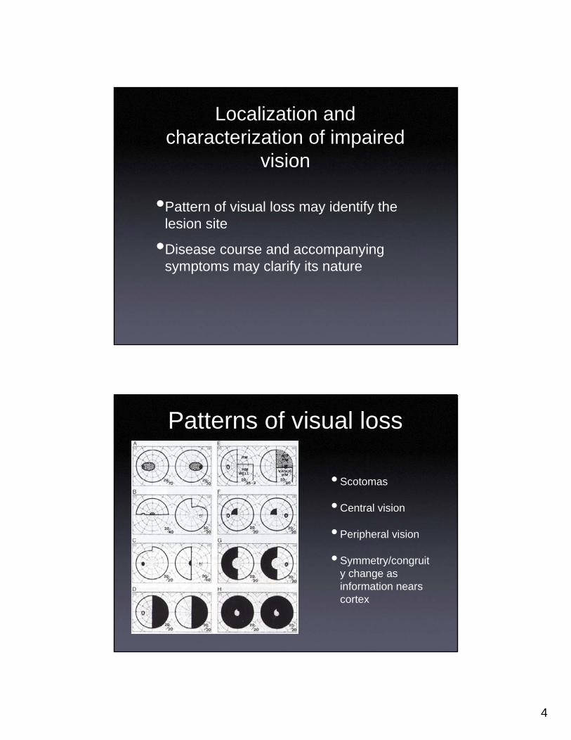
•Pattern of visual loss may identify the lesion site
•Disease course and accompanying symptoms may clarify its nature
Localization and characterization of impaired
vision
Patterns of visual loss
• Scotomas
• Central vision
• Peripheral vision
• Symmetry/congruity change as information nears cortex

5
•Refractive error
•Media opacity
•Retinal disease
•Optic nerve disease
Ocular causes of impaired vision

6
• Most common intraocular malignancy in childhood
• Leukocoria and strabismus
• 13 q14 mutation
• Spreads along the optic nerve into the brain
Retinoblastoma
Flexner-Wintersteiner rosettes

7
Retinal causes of impaired vision
• Symptoms
• Age-related macular degeneration is the most common cause of visual loss > 65 years
• Diabetic retinopathy is the most common cause of visual loss < 65 years
•Blurred vision
•Dimming of vision with decreased color perception
•Decreased pupillary response to light
•Centrocecal, and arcuate scotomata
Symptoms and signs of optic nerve disease

8
Centrocecal scotomas

9
Bilateral optic atrophy with centrocoecal
scotoma•Hereditary (dominant, Leber’s)
•Toxic (medications, methanol, heavy metals)
•Nutritional (folate, B12)
•Demyelinating (optic neuritis, multiple sclerosis)

10
•Ischemic (anterior ischemic optic neuropathy, retinal occlusive disease)
•Compressive (orbital, anterior fossa)
•Inflammatory (demyelinating, infectious, rheumatologic)
Unilateral optic nerve disorders
• Patients usually > 50
• Sudden, usually stable visual loss
• Altitudinal scotoma
• Optic atrophy in 4-6 wk
• Causes
• Idiopathic (anatomic)
• Giant cell arteritis
AION

11
Giant cell arteritis• Senior citizens
• Subacute, granulomatous, stenosing arterial disease
• Headache, amaurosis fugax, arthralgia, myalgia, weight loss
• Brain, cardiac, eye, skin, muscle end artery damage
• Progressive scotoma
• Initially normal disc
• Signs of atrophy
• Decrease in color
• Decrease in vessels
• Decrease in NFL
Compressive optic neuropathy
12
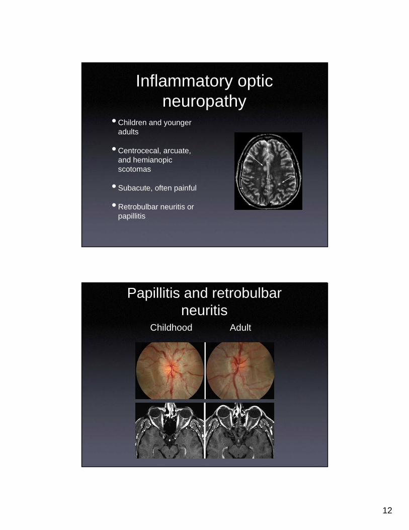
• Children and younger adults
• Centrocecal, arcuate, and hemianopic scotomas
• Subacute, often painful
• Retrobulbar neuritis or papillitis
Inflammatory optic neuropathy
Papillitis and retrobulbar neuritis
Childhood Adult

13
• Glaucoma
• Secondary to retinal degeneration
• Central retinal artery obstruction
• Post-papilledema
• Congenital anomalies: hypoplasia, coloboma
Other causes of optic atrophy
Glaucoma• Common, usually
bilateral, often asymmetric optic neuropathy
• Initial selective damage to branching axons

14
Retinal degeneration• Photoreceptor and/or
retinal pigment epithelium disturbance
• Vascular narrowing is earliest sign
• Pigment released from damaged RPE cells clumps or migrates into the retina
• Many causes
Central retinal artery obstruction

15
• versus other disc swelling
• Intracranial mass
• Pseudotumor cerebri
• Hydrocephalus
• Intracranial hemorrhage
• Venous thrombosis
• Meningitis
Papilledema

16
Papilledema in a 12 year old with idiopathic intracranial
hypertension
• Optic neuritis
• Anterior ischemic optic neuropathy
• Central retinal vein occlusion
• Diabetic papillopathy
• Infiltrative disorders
• Hypertension
• Pseudopapilledema
Other causes of disc swelling

17
•Usually compressive
•Pediatric
•Hypothalamic glioma
•Craniopharyngioma
•Adult
•Pituitary adenoma
•Meningioma
•Craniopharyngioma
•Aneurysm
Lesions of the chiasm

18
• Hemianopic scotoma
• Grossly incongruous field defects
• Small afferent defect
• Children: neoplasm > vascular > trauma
• Adults: vascular > neoplasm > trauma
Retrochiasmal lesions
• Normal pupils, nerves unless perinatal
• Superior hemianopia: temporal lobe
• Inferior hemianopia: parietal lobe
• More posterior, more congruity
Retrogeniculate lesions
19
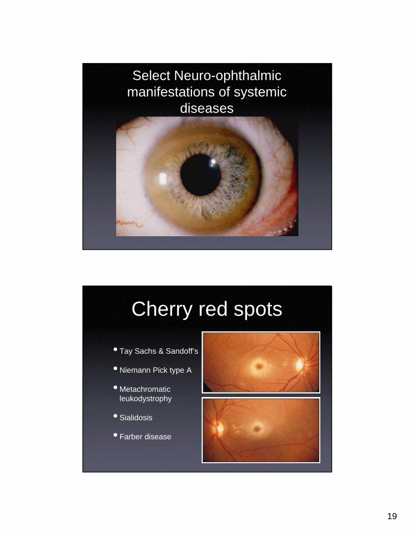
Select Neuro-ophthalmic manifestations of systemic
diseases
• Tay Sachs & Sandoff’s
• Niemann Pick type A
• Metachromatic leukodystrophy
• Sialidosis
• Farber disease
Cherry red spots
20
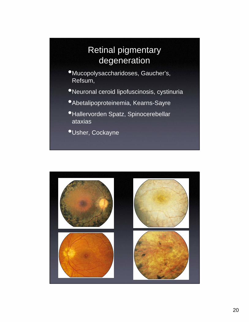
•Mucopolysaccharidoses, Gaucher’s, Refsum,
•Neuronal ceroid lipofuscinosis, cystinuria
•Abetalipoproteinemia, Kearns-Sayre
•Hallervorden Spatz, Spinocerebellar ataxias
•Usher, Cockayne
Retinal pigmentary degeneration

21
•Krabbe, Metachromatic leukodystrophy
•Adrenoleukodystrophy, Alexander
•Spinocerebellar ataxia type I
•Friedreich’s ataxia, Canavan’s,
•Pelizaeus-Merzbacher, Alper’s
Optic atrophy
• Clinical background and pre-proliferative disease
• Proliferative disease
• Diabetic papillitis
• Neovascular glaucoma
• Cataract
Ocular manifestions of
diabetes
22
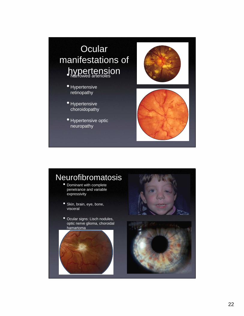
• Narrowed arterioles
• Hypertensive retinopathy
• Hypertensive choroidopathy
• Hypertensive optic neuropathy
Ocular manifestations of
hypertension
Neurofibromatosis• Dominant with complete
penetrance and variable expressivity
• Skin, brain, eye, bone, visceral
• Ocular signs: Lisch nodules, optic nerve glioma, choroidal hamartoma
23
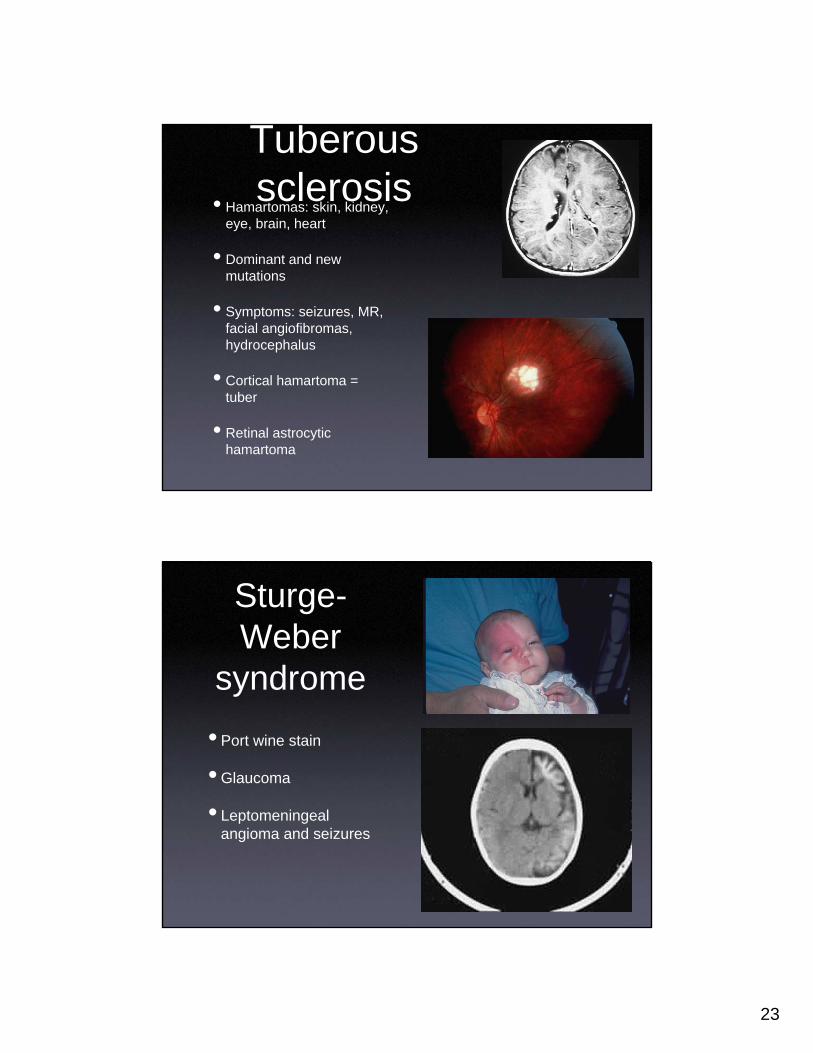
Tuberous sclerosis• Hamartomas: skin, kidney,
eye, brain, heart
• Dominant and new mutations
• Symptoms: seizures, MR, facial angiofibromas, hydrocephalus
• Cortical hamartoma = tuber
• Retinal astrocytic hamartoma
Sturge-Weber
syndrome• Port wine stain
• Glaucoma
• Leptomeningeal angioma and seizures

24
von Hippel-Lindau disease
•Dominant
•incomplete penetrance
•variable expressivity
•VHL1 on 3p
•Retina, CNS, viscera: angiomas and capillary hemangioblastoma
Wyburn-Mason
syndrome•AVMS
•Locations
•Retina
•Orbit
•Nasopharynx
•Midbrain

25
Summary•Visual loss can be understood when
knowledge of neuropathophysiology is combined with knowledge of ocular embryology and anatomy
•The pattern of visual loss can localize and identify neuropathologic disease
•The number of systemic diseases having neuro-ophthalmic manifestation is legion