Ultrasound of the biliary system - kosmos-host.co.uk · Chapter 4 (biliary) F_Layout 1 01/02/2012...
19
Chapter 4 (biliary) F_Layout 1 01/02/2012 10:07 Page 105 Chapter 4 Ultrasound of the biliary system Ana Paula Barreiros 1 , Alina Popescu 2 , Julie Walton 3 , Cui Xinwu 4 , Christoph F. Dietrich 4 1 Department of Internal Medicine 1, Johannes Gutenberg-University Mainz, Germany 2 Department of Gastroenterology, University of Medicine and Pharmacy Timişoara, Romania. 3 Faculty of Health and Life Sciences, University of Liverpool, UK. 4 Department of Internal Medicine 2, Caritas Krankenhaus Bad Mergentheim, Germany Acknowledgment: The authors thank Michael Hocke for peer review of the manuscript. 105
Transcript of Ultrasound of the biliary system - kosmos-host.co.uk · Chapter 4 (biliary) F_Layout 1 01/02/2012...

Chapter 4 (biliary) F_Layout 1 01/02/2012 10:07 Page 105
Chapter 4
Ultrasound of the biliary system
Ana Paula Barreiros1, Alina Popescu2, Julie Walton3, Cui Xinwu4,
Christoph F. Dietrich4
1Department of Internal Medicine 1, Johannes Gutenberg-University Mainz, Germany
2Department of Gastroenterology, University of Medicine and Pharmacy Timişoara, Romania.
3Faculty of Health and Life Sciences, University of Liverpool, UK.
4Department of Internal Medicine 2, Caritas Krankenhaus Bad Mergentheim, Germany
Acknowledgment: The authors thank Michael Hocke for peer review of the manuscript.
105

Chapter 4 (biliary) F_Layout 1 01/02/2012 10:07 Page 106
Ultrasound of the biliary system Ultrasound of the biliary system
Content
Keywords:
Gallbladder, cholelithiasis, cholecystitis, cholangiocellular carcinoma, bile duct
106
Introduction
Biliary system diseases are common in medical practice. After obtaining the patient’s history and
performing a physical examination, conventional B-mode ultrasound and colour Doppler imaging
(CDI) are the imaging methods of choice in, for example, the evaluation of right upper quadrant
pain and obstructive jaundice in patients with elevated cholestasis indicating enzymes and many
other hepatobiliary diseases. In the hands of an experienced ultrasound practitioner ultrasound
has become a diagnostic tool equal in importance to endoscopy. Primarily, ultrasound is the
method of choice in the confirmation or exclusion of dilated bile ducts, cysts and calcifications.
Ultrasound is widely accepted for the diagnosis of biliary system disease. It has the greatest sen-
sitivity for the diagnosis of cholecystolithiasis (approximately 100%) when compared with other
imaging modalities. It is also of great help in the diagnosis of the spectra of appearances in acute
and chronic cholecystitis and in the diagnosis of intra- and extrabile duct dilation. However, clar-
ifying the aetiology of bile duct dilatation is a far more difficult question to answer, but careful
evaluation of the clinical presentation together with the level and extent of obstruction can assist
in determining the cause on ultrasound. Gallbladder polyps are sonographically easy to detect.
Recently, ultrasound contrast agents have proven to be useful for the clarification of biliary tu-
mours. Gallbladder carcinomas are usually detected by ultrasound at a late stage by which time
the liver is already infiltrated and metastases can be detected; this because the disease presents in
advanced age with few early symptoms.
Ultrasound is a routine examination in daily practice and it is the first line imaging modality of
choice in many clinical presentations (e.g. abdominal pain) as well as in asymptomatic patients
as a screening tool [(1)]. It is an accurate, safe, non-invasive, inexpensive, accessible, repeatable
imaging modality, which is highly sensitive and specific for the detection of many biliary tree
diseases and it can frequently demonstrate an alternative diagnosis as the cause of the patient’s
symptoms if the biliary system is normal [(2;3)]. However, it is a highly operator dependent im-
aging modality and its diagnostic success is also influenced by the situation, such as non-fasting,
obesity, presence of surgical dressings and distended abdomen owing to intestinal gas.
Topography of the gallbladder
Topographically, the gallbladder typically lies on the inferior surface of the liver, and is commonly
located in the mid-clavicular line, just below the lower costal margin on the anterior abdominal
wall. Its function is to store and expel bile into the duodenum. It is situated in the gallbladder
fossa of the posterior right hepatic lobe, lateral to the second part of the duodenum and anterior
to the right kidney and transverse colon. It comprises a fundus, body, infundibulum (Hartman’s
pouch, which is the portion of body that joins the neck) and neck including the spiral valve of
Heister in the region of the neck. The cystic duct arises from the superior aspect of the gallbladder
neck, goes to the main biliary duct (MBD) and is usually 2–6cm long. Its lumen contains a series
of mucosal folds and the spiral valves of Heister, which prevent collapse or overdistension owing
to sudden position changes. This structure is important to recognise on ultrasound as it can cause
acoustic shadowing, which may sometimes be mistaken for a calculus in the neck of the gall-
bladder. The gallbladder often lies obliquely within the abdomen, which is important to appreciate
for positioning the transducer correctly to image the long- and short-axes of the gallbladder. How-
ever, its position is dependent on the patient’s body habitus. The four typical body habitus types
are hypersthenic, sthenic, hyposthenic and asthenic. The position of the gallbladder can vary, as
shown in Table 1.
107
Chapter 4 (biliary) F_Layout 1 01/02/2012 10:07 Page 108
108 109
Ultrasound of the biliary system Ultrasound of the biliary system
Table 1 Variable positions of the gallbladder dependent upon body habitus type
Body habitus type Typical gallbladder position and orientation
Hypersthenic The diaphragm, liver and gallbladder tend to lie high in the abdomen in the right upper quadrant,
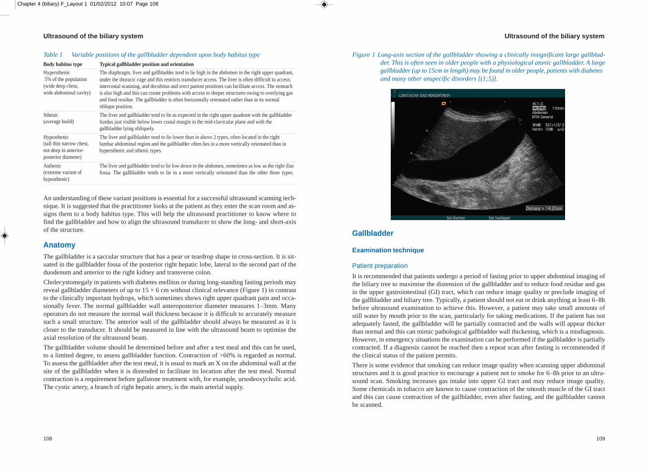
Figure 1 Long-axis section of the gallbladder showing a clinically insignificant large gallblad-
der. This is often seen in older people with a physiological atonic gallbladder. A large
gallbladder (up to 15cm in length) may be found in older people, patients with diabetes
5% of the population
(wide deep chest,
wide abdominal cavity)
under the thoracic cage and this restricts transducer access. The liver is often difficult to access;
intercostal scanning, and decubitus and erect patient positions can facilitate access. The stomach
is also high and this can create problems with access to deeper structures owing to overlying gas
and food residue. The gallbladder is often horizontally orientated rather than in its normal
oblique position.
and many other unspecific disorders [(1;5)].
Sthenic The liver and gallbladder tend to lie as expected in the right upper quadrant with the gallbladder
(average build) fundus just visible below lower costal margin in the mid-clavicular plane and with the
gallbladder lying obliquely.
Hyposthenic The liver and gallbladder tend to lie lower than in above 2 types, often located in the right
(tall thin narrow chest,
not deep in anterior-
posterior diameter)
lumbar abdominal region and the gallbladder often lies in a more vertically orientated than in
hypersthenic and sthenic types.
Asthenic The liver and gallbladder tend to lie low down in the abdomen, sometimes as low as the right iliac
(extreme variant of
hyposthenic)
fossa. The gallbladder tends to lie in a more vertically orientated than the other three types.
An understanding of these variant positions is essential for a successful ultrasound scanning tech-
nique. It is suggested that the practitioner looks at the patient as they enter the scan room and as-
signs them to a body habitus type. This will help the ultrasound practitioner to know where to
find the gallbladder and how to align the ultrasound transducer to show the long- and short-axis
of the structure.
Anatomy
The gallbladder is a saccular structure that has a pear or teardrop shape in cross-section. It is sit-
uated in the gallbladder fossa of the posterior right hepatic lobe, lateral to the second part of the
duodenum and anterior to the right kidney and transverse colon.
Cholecystomegaly in patients with diabetes mellitus or during long-standing fasting periods may
reveal gallbladder diameters of up to 15 × 6 cm without clinical relevance (Figure 1) in contrast
to the clinically important hydrops, which sometimes shows right upper quadrant pain and occa-
sionally fever. The normal gallbladder wall anteroposterior diameter measures 1–3mm. Many
operators do not measure the normal wall thickness because it is difficult to accurately measure
such a small structure. The anterior wall of the gallbladder should always be measured as it is
closer to the transducer. It should be measured in line with the ultrasound beam to optimise the
axial resolution of the ultrasound beam.
The gallbladder volume should be determined before and after a test meal and this can be used,
to a limited degree, to assess gallbladder function. Contraction of >60% is regarded as normal.
To assess the gallbladder after the test meal, it is usual to mark an X on the abdominal wall at the
site of the gallbladder when it is distended to facilitate its location after the test meal. Normal
contraction is a requirement before gallstone treatment with, for example, ursodeoxycholic acid.
The cystic artery, a branch of right hepatic artery, is the main arterial supply.
Gallbladder
Examination technique
Patient preparation
It is recommended that patients undergo a period of fasting prior to upper abdominal imaging of
the biliary tree to maximise the distension of the gallbladder and to reduce food residue and gas
in the upper gastrointestinal (GI) tract, which can reduce image quality or preclude imaging of
the gallbladder and biliary tree. Typically, a patient should not eat or drink anything at least 6–8h
before ultrasound examination to achieve this. However, a patient may take small amounts of
still water by mouth prior to the scan, particularly for taking medications. If the patient has not
adequately fasted, the gallbladder will be partially contracted and the walls will appear thicker
than normal and this can mimic pathological gallbladder wall thickening, which is a misdiagnosis.
However, in emergency situations the examination can be performed if the gallbladder is partially
contracted. If a diagnosis cannot be reached then a repeat scan after fasting is recommended if
the clinical status of the patient permits.
There is some evidence that smoking can reduce image quality when scanning upper abdominal
structures and it is good practice to encourage a patient not to smoke for 6–8h prior to an ultra-
sound scan. Smoking increases gas intake into upper GI tract and may reduce image quality.
Some chemicals in tobacco are known to cause contraction of the smooth muscle of the GI tract
and this can cause contraction of the gallbladder, even after fasting, and the gallbladder cannot
be scanned.

Chapter 4 (biliary) F_Layout 1 01/02/2012 10:07 Page 110
110 111
Ultrasound of the biliary system Ultrasound of the biliary system
Patient history and physical examination
It is recommended that a short patient history is taken and that the abdomen is examined or pal-
pated before the ultrasound examination commences. This complements the ultrasound informa-
tion with clinical data and ensures the clinical question is addressed.
Ultrasound examination of the gallbladder
Routinely, a convex high multifrequency (5–7MHz) transducer is used for the evaluation of the
gallbladder. However, lower frequencies may be used when an increased depth of penetration is
required, for example in more obese patients or when the gallbladder is deep (e.g. hypersthenic
patients). In addition, in very slim patients (e.g. asthenic or hyposthenic types) where the gall-
bladder is very superficial, one should consider the use of a linear superficial imaging transducer
to optimise image quality.
The edge of the right hepatic lobe and the liver hilum are useful landmarks for the evaluation of
the gallbladder . In the right subcostal oblique section the landmark structure to be used is the in-
terlobar fissure and the gallbladder is found by aligning the probe with the fissure and then tilting
it. The gallbladder will be located inferiorly or laterally to the fissure (between liver segments IV
and V).
Conventional real-time ultrasound produces images of thin slices of the biliary tree on the
screen.To be entirely convinced that the entire volume of the liver tissue and structures have been
imaged it is essential that the operator scans the entire organ systematically, in at least two anatom-
ical planes. The operator must then use this two-dimensional information to envisage a three-di-
mensional map of the individual patient’s biliary tree anatomy and pathology. This requires good
hand-eye-brain coordination.
The gallbladder and biliary tree can be examined initially with the patient in a supine position.
This is to be encouraged as a first line approach to minimise the risks of operator repetitive strain
injury owing to over-reaching. Successful examination of the gallbladder and biliary tree often
requires the patient to be examined in a left posterior-oblique or left-lateral decubitus position.
These latter positions cause the liver/gallbladder to rotate anteromedially under the influence of
gravity and this may optimise the use of the liver to image the gallbladder through an acoustic
window or make the gallbladder more readily accessible below the thoracic cage.
In patients with sludge or stones, movement is essential to assess whether the pathology moves
as the patient moves. Erect imaging is particularly useful to assess whether gallstones are mobile
because they will descend into the dependent part of the gallbladder (fundus) when the patient
erect.
In a typical sthenic patient, the transducer can be placed in the mid-clavicular line on the anterior
abdominal wall and the transducer position is adjusted until the gallbladder is located. The oper-
ator should try to use the liver as an acoustic window and avoid scanning through bowel by an-
gling cranially. The patient may be asked to take a suspended full inspiration to cause the
gallbladder to descend below the lower costal margin. The transducer is then rotated over the
gallbladder until the true long-axis section of the gallbladder is achieved.
It is essential to image the gallbladder in the entire long-axis section and to angle the transducer
so that the ultrasound beam is swept through the structure to ensure that the whole gallbladder
has been imaged in its entire long-axis as this is a three-dimensional structure.
The normal gallbladder wall measures <3 mm in the anteroposterior diameter. The anterior wall
of the gallbladder should always be measured as it is closer to the transducer. It should be meas-
ured in line with the axial resolution of the machine. After scanning the gallbladder in long-axis,
the transducer should be rotated over the gallbladder through 90° to image the gallbladder in its
true short-axis section. Again, the transducer should be angled (cranial-caudal) to image the gall-
bladder in its entirety.
The demonstration of the cystic duct is easiest in deep inspiration with the patient in supine or
left-lateral decubitus. It can be visualised beginning from the infundibulum of the gallbladder.
The distal segment of the cystic duct is best seen with the patient in supine, in the plane through
the hepatic portal and anterior to the portal vein.
Imaging the intrahepatic biliary tree is described in the liver chapter of this course book.
An acronym has shown to be didactically helpful [“SSOTM”] when thinking about interpreting
ultrasound images:
• S = size
• M = measurement
• S = shape
• O = outline
• T = texture
Size: See anatomy. The size of the gallbladder should be subjectively assessed. In a fasted
state it should measure 10 × 2–4 × 2–4 cm, but this depends on the volume of bile pres-
ent. Typical bile volume is normally 40–60ml, measured by a rotating ellipsoid). How-
ever gallbladder volume estimation is highly unreliable as it shows a wide intra and inter
operator variability [(4;5)].
Shape: The gallbladder is a saccular structure which has a pear or teardrop shape in long-axis
cross section when distended.
Outline: The normal gallbladder wall is thin, smooth and mildly echogenic, measuring 1–3 mm
in thickness in the normal state. There is no pericholic fluid around the gallbladder in
the normal state.
Texture: The normal gallbladder lumen should contain bile and should not have any space occu-
pying lesions. Normal bile appears anechoic (i.e. is completely black) and devoid of any
internal echoes.
Congenital anomalies of the gallbladder
There are a wide variety of congenital abnormalities that are rarely encountered in adult patients
on sonography and are predominantly found in the paediatric setting. If congenital abnormalities
are found during routine ultrasound examination the findings are, in the clinical context, mainly
without consequences to patient management.
Agenesis, hypoplasia and microgallbladder
Agenesia, hypoplasia and numerical anomalies of the gallbladder have to be considered. Agenesis
(absence) of the gallbladder is rare and normally without clinical significance. Approximately
50% of gallbladder agenesis cases are discovered at autopsy and they are often associated with
duodenal atresia and other congenital anomalies. Hypoplasia is associated with extrahepatic bil-
iary atresia. A microgallbladder is defined as being less than 2–3cm long, 0.5–1.5cm wide and
Chapter 4 (biliary) F_Layout 1 01/02/2012 10:07 Page 112
112 113
Ultrasound of the biliary system Ultrasound of the biliary system
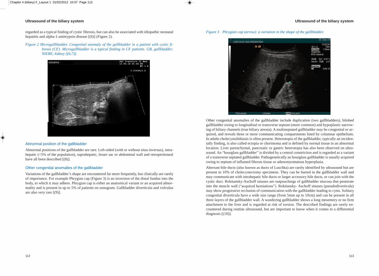
regarded as a typical finding of cystic fibrosis, but can also be associated with idiopathic neonatal
hepatitis and alpha-1-antitrypsin disease [(6)] (Figure 2).
Figure 2 Microgallbladder. Congenital anomaly of the gallbladder in a patient with cystic fi-
brosis (CF). Microgallbladder is a typical finding in CF patients. GB, gallbladder;
NIERE, kidney [(6;7)].
Abnormal position of the gallbladder
Abnormal positions of the gallbladder are rare. Left-sided (with or without situs inversus), intra-
hepatic (<5% of the population), suprahepatic, lesser sac or abdominal wall and retroperitoneal
have all been described [(8)].
Other congenital anomalies of the gallbladder
Variations of the gallbladder’s shape are encountered far more frequently, but clinically are rarely
of importance. For example Phrygian cap (Figure 3) is an inversion of the distal fundus into the
body, to which it may adhere. Phrygian cap is either an anatomical variant or an acquired abnor-
mality and is present in up to 5% of patients on sonogram. Gallbladder diverticula and volvulus
are also very rare [(9)].
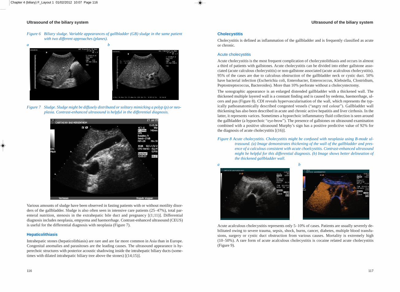
Figure 3 Phrygian cap (arrow): a variation in the shape of the gallbladder.
Other congenital anomalies of the gallbladder include duplication (two gallbladders), bilobed
gallbladder owing to longitudinal or transverse septum (more common) and hypoplastic narrow-
ing of biliary channels (true biliary atresia). A multiseptated gallbladder may be congenital or ac-
quired, and reveals three or more communicating compartments lined by columnar epithelium.
In adults cholecystolithiasis is often present. Heterotopia of the gallbladder, typically an inciden-
tally finding, is also called ectopia or choristoma and is defined by normal tissue in an abnormal
location. Liver parenchymal, pancreatic or gastric heterotopia has also been observed on ultra-
sound. An “hourglass gallbladder” is divided by a central constriction and is regarded as a variant
of a transverse septated gallbladder. Pathogenetically an hourglass gallbladder is usually acquired
owing to septum of inflamed fibrous tissue or adenomyomatous hyperplasia.
Aberrant bile ducts (also known as ducts of Luschka) are rarely identified by ultrasound but are
present in 10% of cholecystectomy specimens. They can be buried in the gallbladder wall and
may communicate with intrahepatic bile ducts or larger accessory bile ducts, or can join with the
cystic duct. Rokitansky-Aschoff sinuses are outpouchings of gallbladder mucosa that penetrate
into the muscle wall (“acquired herniations”). Rokitansky- Aschoff sinuses (pseudodiverticula)
may show progressive occlusion of communication with the gallbladder leading to cysts. Solitary
congenital diverticula have a wide size range (from 5mm up to 10cm) and can be present in all
three layers of the gallbladder wall. A wandering gallbladder shows a long mesentery or no firm
attachment to the liver and is regarded at risk of torsion. The described findings are rarely en-
countered during routine ultrasound, but are important to know when it comes to a differential
diagnosis [(10)].

Chapter 4 (biliary) F_Layout 1 01/02/2012 10:07 Page 114
114 115
Ultrasound of the biliary system Ultrasound of the biliary system
Cholelithiasis
The term cholelithiasis describes the presence of gallstones in the biliary tract. Depending on the
localisation of the concrement a further distinction between cholecystolithiasis (in the gallbladder)
(Figure 4 and 5) and choledocholithiasis (in the bile duct) is used. In 5–15% of patients a com-
bination of both conditions can be seen.
Figure 4 Typical cholecystolithiasis: isolated calculus in the gallbladder lumen. Acoustic shad-
owing (SS) is typically seen posterior to a calculus as a result of reflection of ultra-
sound and the lack of deeper echoes reaching the transducer.
Figure 5 CT image of cholecystolithiasis (courtesy Dr Baum, Bad Mergentheim).
Cholecystolithiasis is the most common disease of the biliary system. It is estimated that 10% of
the adult population have gallbladder stones, and that a third of people over 70 years will have
gallbladder stones [(3)]. Of the patients with gallstones, 35% will, in time, become symptomatic
and require surgery [(11)].
Cholecystolithiasis can be detected by transabdominal ultrasound with a high sensitivity, whereas
choledocholithiasis is more difficult to detect [(12;13)]. The literature has recently been sum-
marised [(1)] and transabdominal ultrasound is the first line imaging method of choice for any
kind of gallbladder stones. It has the additional benefits of not using ionising radiation and is rel-
atively cheap and safe. The accuracy of ultrasonography for the diagnosis of gallstones is up to
96% for experienced operators.
The number, size, echotexture, acoustic shadowing and mobility of gallbladder stones should be
recorded and analysed. The classic ultrasound appearance of the gallbladder stone is a hypere-
choic/echogenic structure located within the gallbladder lumen with posterior acoustic shadowing.
Calculi typically lie on the dependent wall of the gallbladder under the influence of gravity, but
this is dependent on the density of bile. To optimise acoustic shadowing and detection of small
calculi, it is essential to insonate the dependent wall of the gallbladder at 90°. The gallstones
should demonstrate mobility in response to movement of the patient (the “rolling stone” sign).
Calculi are more difficult to see when they are small in size (2–3mm) and in number. The gall-
bladder filled with stones (the “shell” sign) can be easily confused with air in the digestive tract
if the examiner does not have sufficient experience. Occasionally stones become impacted in the
gallbladder infundibulum, which creates a hydrops. This is not always an easy diagnosis and can
sometimes be missed because this region can be difficult to visualise in some patients. The close
anatomical proximity to the gas containing gastrointestinal tract can cause problems when imaging
this region.
A sonographically conclusive diagnosis of stone origin is not possible at present. Non-calcified
cholesterin/cholesterol derived stones do not typically show shadowing and may “float” in the
gallbladder lumen in contrast to calcified stones. So called “pigment stones”, which are rarely
found in Europe (in less than 10% of the population), are often multiple with complete acoustic
shadowing [(12)]. Very small stones can be overlooked, but a routinely performed examination
of the left lateral decubitus and a standing position improves the detection rate to almost 100%
for an experienced operator. “Mirizzi-syndrome” with stone impaction in the cystic duct, which
can cause an obstructive jaundice, may be more difficult to visualise.
Biliary sludge
Biliary sludge is sometimes the precursor of gallstones. It represents precipitate formed in the
bile and the ultrasound appearance is of homogenous echogenic material in the gallbladder lumen,
with no posterior acoustic shadowing (Figures 6 a and b). It typically forms a straight horizontal
line superficially, with the sludge normally collecting on the dependent wall of the gallbladder
because of gravity. It moves slowly with a change in patient position and it is recommended that
the patient is moved during the scan to demonstrate the motion of the sludge. If the sludge com-
pletely fills the gallbladder it can be difficult to distinguish it from the adjacent liver parenchyma.
Sometimes the sludge is organised in a round shape, is hypoechoic, with no posterior acoustic
shadowing and is often called “ball-like” or “tumour-like” sludge (Figures 6).
Chapter 4 (biliary) F_Layout 1 01/02/2012 10:07 Page 116
116 117
Ultrasound of the biliary system Ultrasound of the biliary system
Figure 6 Biliary sludge. Variable appearances of gallbladder (GB) sludge in the same patient
with two different approaches (planes).
a b
Figure 7 Sludge. Sludge might be diffusely distributed or solitary mimicking a polyp (p) or neo-
plasia. Contrast-enhanced ultrasound is helpful in the differential diagnosis.
Various amounts of sludge have been observed in fasting patients with or without motility disor-
ders of the gallbladder. Sludge is also often seen in intensive care patients (25–47%), total par-
enteral nutrition, stenosis in the extrahepatic bile duct and pregnancy [(1;11)]. Differential
diagnosis includes neoplasia, empyema and haemorrhage. Contrast-enhanced ultrasound (CEUS)
is useful for the differential diagnosis with neoplasia (Figure 7).
Hepaticolithiasis
Intrahepatic stones (hepaticolithiasis) are rare and are far more common in Asia than in Europe.
Congenital anomalies and parasitoses are the leading causes. The ultrasound appearance is hy-
perechoic structures with posterior acoustic shadowing inside the intrahepatic biliary ducts (some-
times with dilated intrahepatic biliary tree above the stones) [(14;15)].
Cholecystitis
Cholecystitis is defined as inflammation of the gallbladder and is frequently classified as acute
or chronic.
Acute cholecystitis
Acute cholecystitis is the most frequent complication of cholecystolithiasis and occurs in almost
a third of patients with gallstones. Acute cholecystitis can be divided into either gallstone asso-
ciated (acute calculous cholecystitis) or non-gallstone associated (acute acalculous cholecystitis).
95% of the cases are due to calculous obstruction of the gallbladder neck or cystic duct. 50%
have bacterial infection (Escherichia coli, Enterobacter, Enterococcus, Klebsiella, Clostridium,
Peptostreptococcus, Bacteroides). More than 10% perforate without a cholecystectomy.
The sonographic appearance is an enlarged distended gallbladder with a thickened wall. The
thickened multiple layered wall is a constant finding and is caused by oedema, haemorrhage, ul-
cers and pus (Figure 8). CDI reveals hypervascularisation of the wall, which represents the typ-
ically pathoanatomically described congested vessels (“angry red colour”). Gallbladder wall
thickening has also been described in acute and chronic active hepatitis and liver cirrhosis. In the
latter, it represents varices. Sometimes a hypoechoic inflammatory fluid collection is seen around
the gallbladder (a hypoechoic “eye-brow”). The presence of gallstones on ultrasound examination
combined with a positive ultrasound Murphy’s sign has a positive predictive value of 92% for
the diagnosis of acute cholecystitis [(16)].
Figure 8 Acute cholecystitis. Cholecystitis might be confused with neoplasia using B-mode ul-
trasound. (a) Image demonstrates thickening of the wall of the gallbladder and pres-
ence of a calculous consistent with acute choelcystitis. Contrast-enhanced ultrasound
might be helpful for this differential diagnosis. (b) Image shows better delineation of
the thickened gallbladder wall.
a b
Acute acalculous cholecystitis represents only 5–10% of cases. Patients are usually severely de-
bilitated owing to severe trauma, sepsis, shock, burns, cancer, diabetes, multiple blood transfu-
sions, surgery or cystic duct obstruction from various causes. Mortality is extremely high
(10–50%). A rare form of acute acalculous cholecystitis is cocaine related acute cholecystitis
(Figure 9).
Chapter 4 (biliary) F_Layout 1 01/02/2012 10:07 Page 118
118 119
Ultrasound of the biliary system Ultrasound of the biliary system
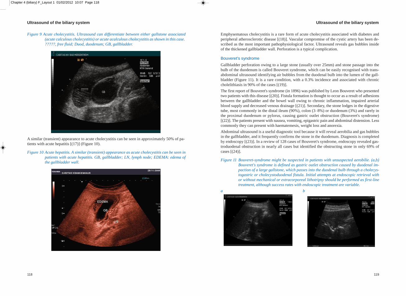
Figure 9 Acute cholecystitis. Ultrasound can differentiate between either gallstone associated
(acute calculous cholecystitis) or acute acalculous cholecystitis as shown in this case.
?????, free fluid; Duod, duodenum; GB, gallbladder.
A similar (transient) appearance to acute cholecystitis can be seen in approximately 50% of pa-
tients with acute hepatitis [(17)] (Figure 10).
Figure 10 Acute hepatitis. A similar (transient) appearance as acute cholecystitis can be seen in
patients with acute hepatitis. GB, gallbladder; LN, lymph node; EDEMA: edema of
the gallbladder wall.
Emphysematous cholecystitis is a rare form of acute cholecystitis associated with diabetes and
peripheral atherosclerotic disease [(18)]. Vascular compromise of the cystic artery has been de-
scribed as the most important pathophysiological factor. Ultrasound reveals gas bubbles inside
of the thickened gallbladder wall. Perforation is a typical complication.
Bouveret's syndrome
Gallbladder perforation owing to a large stone (usually over 25mm) and stone passage into the
bulb of the duodenum is called Bouveret syndrome, which can be easily recognised with trans-
abdominal ultrasound identifying air bubbles from the duodenal bulb into the lumen of the gall-
bladder (Figure 11). It is a rare condition, with a 0.3% incidence and associated with chronic
cholelithiasis in 90% of the cases [(19)].
The first report of Bouveret's syndrome (in 1896) was published by Leon Bouveret who presented
two patients with this disease [(20)]. Fistula formation is thought to occur as a result of adhesions
between the gallbladder and the bowel wall owing to chronic inflammation, impaired arterial
blood supply and decreased venous drainage [(21)]. Secondary, the stone lodges in the digestive
tube, most commonly in the distal ileum (90%), colon (3–8%) or duodenum (3%) and rarely in
the proximal duodenum or pylorus, causing gastric outlet obstruction (Bouveret's syndrome)
[(22)]. The patients present with nausea, vomiting, epigastric pain and abdominal distension. Less
commonly they can present with haematemesis, weight loss and anorexia.
Abdominal ultrasound is a useful diagnostic tool because it will reveal aerobilia and gas bubbles
in the gallbladder, and it frequently confirms the stone in the duodenum. Diagnosis is completed
by endoscopy [(23)]. In a review of 128 cases of Bouveret's syndrome, endoscopy revealed gas-
troduodenal obstruction in nearly all cases but identified the obstructing stone in only 69% of
cases [(24)].
Figure 11 Bouveret-syndrome might be suspected in patients with unsuspected aerobilie. (a,b)
Bouveret's syndrome is defined as gastric outlet obstruction caused by duodenal im-
paction of a large gallstone, which passes into the duodenal bulb through a cholecys-
togastric or cholecystoduodenal fistula. Initial attempts at endoscopic retrieval with
or without mechanical or extracorporeal lithotripsy should be performed as first-line
treatment, although success rates with endoscopic treatment are variable.
a b

Chapter 4 (biliary) F_Layout 1 01/02/2012 10:07 Page 120
120 121
Ultrasound of the biliary system Ultrasound of the biliary system
Chronic cholecystitis
Chronic cholecystitis is sonographically characterised by an irregular thickened gallbladder wall,
mainly caused by chronic inflammation and intermittent obstruction of the gallbladder neck or
cystic duct by gallstones, often causing biliary colic. The gallbladder is frequently small and con-
tracted, and care must be taken when imaging this region so as not to confuse the bowel with a
small contracted gallbladder. 95% are associated with cholecystolithiasis or at least sludge (Figure
12).
Figure 12 Chronic cholecystitis. (a) Sometimes a sludge filled gallbladder may present as a tu-
mour like lesion using B-mode ultrasound. (b) Contrast-enhanced ultrasound is helpful
for the correct diagnosis because the ventral wall thickening does not take up the con-
trast enhancer and can therefore be differentiated from a carcinoma.
a b
Bacteria are present in up to a third of patients, and include similar organisms to acute cholecys-
titis. There are many different forms of chronic cholecystitis that cannot be differentiated by ul-
trasound.
Diffuse lymphoplasmacytic acalculous cholecystitis is a relatively sensitive sign for primary scle-
rosing cholangitis (PSC). An association with lymphoplasmacytic sclerosing pancreatitis has also
been described.
Some other forms are acquired immune deficiency syndrome (AIDS) related. Acalculous chole-
cystitis can be caused by opportunistic infections such as cryptosporidia, cytomegalovirus (CMV)
and microsporidia. Eosinophilic cholecystitis is seen in Churg-Strauss syndrome. In addition fol-
licular cholecystitis (also called lymphoid polyp) can occasionally be observed [(25)(26)].
Other rare forms of cholecystitis include: granulomatous cholecystitis in patients with tuberculosis
and xanthogranulomatous cholecystitis (as a result of rupture of the Rokitansky-Aschoff sinuses
with extravasation of bile or ulceration of gallbladder mucosa).
Gangrenous cholecystitis occurs in 15% of acute cholecystitis cases with mural infarction, and
with perforation in more than 25%. Typically, air is found in the gallbladder (known as pneumo-
bilia). Clostridium perfringes appears to be of pathophysiological importance.
Porcelain gallbladder is found in 0.5% of cholecystectomies. An association (>20%) with gall-
bladder carcinoma is well known [(1)]. Cholecystectomy is therefore indicated when a porcelain
gallbladder is diagnosed on ultrasound. Sonographically the calcified wall can be easily detected.
It is characterised by an intramural shell-like calcification that may affect the entire wall or parts
of it.
“Limy bile”, in which there is a pathological accumulation of calcium carbonate in the gallbladder,
is a very rare condition. The diagnosis is made on a plain abdominal radiograph, where the gall-
bladder appears as an opaque pear-shaped structure.
Miscellaneous non-neoplastic disorders
Miscellaneous non-neoplastic disorders are various. Adenomyomatous hyperplasia is also called
adenomyomatosis or diverticular disease of the gallbladder. Commonly ultrasound can only
demonstrate a thickened wall. Adenomyomatous hyperplasia (generalised, segmental or localised)
is relatively common (in up to 10% of cholecystectomy specimens) and it is usually asympto-
matic. In the generalised form, diffuse wall thickening (>10mm) is found with intramural diver-
ticula resembling cystic spaces within the wall. In the segmental form focal thickening of the
gallbladder wall can usually be seen in the body, which gives it an hourglass configuration. In
the localised form the fundus shows 0.5–3cm nodules with grey-white cut surface containing
multiple cysts. The latter may cause gallbladder inversion and is also called adenomyoma.
Asymptomatic cholesterolosis is mostly characterised by cholesterol infiltration (sonographically
shown as comet-tail artefacts) in an otherwise normal gallbladder wall. Cholesterolosis is present
in up to 20% of cholecystectomy specimens, usually found in adult multiparous women. Choles-
terolosis is associated with bile supersaturation with cholesterol, but not with increased serum
cholesterol. Cholesterol infiltration is due to an accumulation of cholesterol esters and triglyc-
erides in subepithelial macrophages and gallbladder epithelium. Macroscopically focal or diffuse
yellow flat deposits are seen on the mucosal surface, which may have speckled appearance
(“strawberry gallbladder”). Association with cholesterol polyps is reported in 20%. A similar (un-
specific) irregular gallbladder wall can be seen in amyloidosis (Figure 13) [(27)].
Figure 13 Amyloidosis of the gallbladder may show irregular deposits (white arrow) [(27)].
Gallbladder varices as a cause of gallbladder thickening can be excluded using CDI.

Chapter 4 (biliary) F_Layout 1 01/02/2012 10:07 Page 122
122 123
Ultrasound of the biliary system Ultrasound of the biliary system
Benign gallbladder tumours (polyps)
Benign gallbladder tumours are divided into adenoma of the gallbladder (typically demonstrating
central vessels penetrating the polyp using CDI) (Figure 14), adenomyosis, cholesterol polyps
(containing cholesterol deposits and therefore no or few vessels), hyperplastic/metaplastic polyps,
granular cell tumour (often associated with similar lesions in extrahepatic bile ducts), inflamma-
tory polyps and villous papilloma.
Figure 14 Adenoma (7 mm) of the gallbladder typically demonstrating central vessels penetrating
the polyp using colour Doppler imaging.
Adenoma of the gallbladder are mainly single (90%), rarely multiple (10%) and contain, by def-
inition, at least low grade dysplastic epithelium. An increased prevalence is found with familial
adenomatous polyposis or Peutz-Jeghers syndrome. Invasive carcinoma is rarely reported in le-
sions under 10mm. The treatment is total excision respective cholecystecomy.
Adenomyosis is caused by hyperplasia of muscularis propria with intramural hyperplastic or cys-
tically dilated glands mainly in the fundus. They represent 15–25% of benign polyps and appear
on ultrasound as hypoechoic round structures, attached to the gallbladder wall, with no posterior
acoustic shadowing. They do not move with a change in patient position, which can be used for
a differential diagnosis with cholecystolithiasis.
Cholesterol polyps are the most common benign polyps (50–90%). Inflammatory polyps are as-
sociated with chronic cholecystitis and are described as 3–15mm, usually sessile and single
polyps, macroscopically red/grey/brown in colour. Multiple adenoma are often encountered (Fig-
ure 15).
Figure 15 Multiple adenoma of the gallbladder (an example is shown between the callipers (+)).
Growing gallbladder polyps or polyps larger than 10mm should be surgically removed owing to
the potential malignant transformation.
Gallbladder carcinoma
Gallbladder carcinoma is a rare but highly fatal malignancy. It is associated, in almost 100% of
the cases, with cholecystolithiasis and occurs more frequently in patients over 60 years old. The
risk of developing gallbladder cancer in a patient with gallbladder stones is 0.3% over a 30 year
period and published data suggest a much higher cancer risk in stones larger than 3cm.
Carcinomas of the gallbladder can be easily recognised using transabdominal ultrasound, whereas
correct staging is much more difficult and underestimation of the extent of the disease is possible.
Complementary imaging such as CT and/or MRI is often required. The majority of cases are
found incidentally in patients with cholelithiasis, and in 1–2% of these cases a gallbladder carci-
noma can be found [(28)]. As with other tumours of the upper GI tract, a definite sonographic
distinction between inflammatory and neoplastic alterations or changes in connective tissue is
not possible. In some cases a sonographic diagnosis of adenomyomatosis, cholesterol polyps and
other pathologies of the gallbladder can be made. The sonographic signs of a gallbladder carci-
noma are irregular wall thickening, a poorly defined polypoid mass protruding in the gallbladder
lumen, or, if the disease is more advanced, the replacement of the gallbladder by a solid, normally
hypoechoic, mass that completely fills the gallbladder. The presence of a gallstone in relation to
this mass suggests the diagnosis of gallbladder carcinoma. The symptoms of patients with gall-
bladder carcinoma are often non-specific and vague; therefore, the diagnosis is made when the
disease is in an advanced stage, hence the poor prognosis and the name “silent tumour”. The most
common symptom of patients in the advanced stage of the disease is pain followed by anorexia.
CEUS can be useful for the diagnosis of gallbladder carcinoma (Figure 16) and can help in the
differentiation of normal and infiltrated areas. It can also help in the assessment of liver metas-
tases.

Chapter 4 (biliary) F_Layout 1 01/02/2012 10:07 Page 124
124 125
Ultrasound of the biliary system Ultrasound of the biliary system
Figure 16 Gallbladder carcinoma. (a) Carcinomas of the gallbladder can usually only be recog-
nised using conventional B-mode transabdominal ultrasound. (b) Contrast-enhanced
ultrasound is helpful in delineating tumour infiltration.
a b
Sometimes other morphology may imitate gallbladder neoplasia, e.g. hyper-regenerative nodules
in liver cirrhosis (Figure 17) or even polyps.
Figure 17 (a) Differential diagnosis of gallbladder neoplasia (between callipers (+)) may be an
atypical liver appearance. (b) Demonstration in two anatomical levels is helpful for
the correct diagnosis.
a b
Cholangio(cellular) carcinoma (including Klatskin tumours)
Cholangio(cellular) carcinomas (CCCs), which are histologically mainly adenocarcinoma, are
more frequently detected using transabdominal ultrasound with the newer high-resolution ultra-
sound systems. They arise from the epithelial cells of the intrahepatic and extrahepatic bile ducts.
Carcinomas of the bile duct are much more common extrahepatically but can also be found in-
trahepatically (<10%).
Perihilar CCCs of the bile ducts are called Klatskin tumours and are classified by Bismuth-Cor-
lette I-IV depending on the localisation of the tumour and involvement of the hepatic ducts (Table
2).
Table 2 Bismuth-Corlette classification.
Bismuth-Corlette Description
classification
I Tumours below the confluence of the left and right hepatic ducts.
II Tumours reaching the confluence
III Tumours occluding the common hepatic duct and either the right or left hepatic duct (Type a or b).
IV Tumours that are multicentric or involve the confluence and both the right or left hepatic duct
Bismuth-Corelette Type IV can be easily confused on sonography with hepatocellular carcinoma.
Mixed forms have been observed. Sonographically dilated bile ducts proximal of the stenosis is
a typical finding. Delineation of the tumour is more difficult. Intrahepatic CCCs can often be
found subcapsular, polygonal and diffusely delineated. Risk factors for the development of a CCC
are PSC, infection of the hepatobiliary tract with liver fluke (e.g. Clonorchis sinensis), congenital
abnormalities of the hepatobiliary tract (e.g. biliary atresia, choledochal cysts). Bile duct adenoma
are rarely encountered [(29)].
CEUS has improved the detection and characterisation of CCC but prospective studies are lacking.
Peritoneal metastases are common but are difficult to visualise reliably using imaging methods
without laparoscopy.
CCCs can occur along the bile duct as Klatskin tumours (hilar CCC are more common), but they
may also appear as primary solid tumours in the liver (peripheral CCC). For the peripheral type
there are no typical sonographic characteristics and the diagnosis is usually made incidentally
within the framework of a biopsy of an unclear liver lesion. Ultrasound examination shows a
solid mass that can have any echogenicity and exhibits signs of a malignant growth. Typically
the liver metastases of a peripheral CCC are situated like satellites around the primary focus.
For reasons that are still unclear, the incidence of intrahepatic cholangiocarcinoma has been rising
over the past decade all over the world, while rates of extrahepatic cholangiocarcinoma are de-
clining [(30;31)].
Extrahepatic bile duct
The extrahepatic bile duct system can be easily displayed using transabdominal ultrasound with
a patient in a left-posterior oblique or left-lateral decubitus position (Figures 18 and 19). This po-
sition causes the liver to rotate anteromedially, and thus the liver can be used as an acoustic win-
dow for imaging the extrahepatic biliary tree. The main extrahepatic bile duct often lies obliquely
and it is superiorly more lateral so it can usually be imaged by placing the transducer below the
right costal margin in the region of the mid-clavicular line; an oblique position is required to align
the transducer parallel to the long-axis of the bile duct. The MBD appears as a tube situated in
front of the portal vein. When there is a good acoustic window, the MBD can be followed into
the retropancreatic portion to the papilla of Vateri.
Often, the bile duct is imaged with the transducer parallel to the midline. In this section the hepatic
artery will normally appear as a round structure between the MBD and the portal vein. Frequently
the duct is measured at this point; however, a single measurement of the bile duct at this level
can be misleading as the duct may be normal at this point, but distended lower down in early ob-
structive jaundice and it is therefore recommended that the duct be imaged along the entire length
and measured at several points, including near to the head of pancreas. The duct should be eval-

Chapter 4 (biliary) F_Layout 1 01/02/2012 10:07 Page 126
126 127
Ultrasound of the biliary system Ultrasound of the biliary system
uated for size (normal is up to 6mm), wall thickness and content. After colecystectomy the normal
size of the MBD may increase. In some patients the papilla of Vateri at the end of the CBD can
be displayed using transabdominal ultrasound.
Figure 18 Papilla of Vateri. (a) The papilla of Vateri at the distal end of the common bile duct .
(b) The main pancreatic duct is displayed next to the papilla. DW, main pancreatic
duct; DHC, common bile duct.
a b
Figure 19 Slightly dilated main bile duct (10mm) next to the papilla. DP, pancreatic duct; DHC,
common bile duct
Patients with jaundice
Patients with jaundice should be examined sonographically as early as possible, ideally as soon
as they present. In some cases it is better to scan the patient without fasting to assess the case and
then repeat after fasting if necessary. For example, dilated intrahepatic bile ducts can be seen
without fasting and may account for the jaundice. Ultrasound can differentiate jaundice caused
by obstructive and hepatocellular origin in almost all patients by focusing and analysing the di-
ameter of the main bile duct. Figure 19 shows a mildly distended main bile duct, but microlithiasis
might be overlooked. The left lateral decubitus position is helpful for adequate visualisation of
the liver hilum [(32-34)] and should be performed consistently in all patients. The papilla Vateri
is less reliably displayed using the transabdominal approach.
Congenital disorders
Choledochal cysts
In infants choledochal cysts are the most common cause of obstructive jaundice, but they may be
found at any age. They are not actually cysts but a dilatation of the CBD, which may secondarily
obstruct other biliary ducts or the duodenum (Figure 20). Choledochal cysts (classification of To-
dani Type 1–5 (Table 3)) are often associated with other hepatobiliary tract abnormalities, e.g.
anomalous pancreaticobiliary duct junction [(35)]. Choledochal cysts may rupture spontaneously
[(36–38)].
Table 3 Todani classification for choledochal cyst [(35)].
Type Description
Type 1 Characterised by a segmental or diffuse fusiform dilation of CBD (50–90% of all cases).
Type 2 Diverticulum of CBD
Type 3 Dilation of intraduodenal CBD (choledochocele)
Type 4 Multiple cysts of extrahepatic bile ducts with (4a) or without (4b) cysts of intrahepatic ducts
Type 5 One or more cysts of intrahepatic ducts (Caroli’s disease)
CBD, common bile duct
It is importance to note that the mean age to develop biliary tract carcinoma for 2–10% of patients
is 35 years. Carcinomas may develop within the wall of the cyst, the gallbladder or bile ducts.
Therapeutically complete cyst removal with biliary reconstruction, usually with Roux-en-Y he-
paticojejunostomy should be performed when a carcinoma is diagnosed in these patients.
The role of ultrasound techniques is not yet fully determined because of a low incidence of this
disease in adults and limited clinical experience. MRI and endoscopic retrograde cholangiography
(ERC) are the diagnostic methods of choice. Endoscopic ultrasound (EUS) should be considered
because of the excellent resolution of the CBD.

Chapter 4 (biliary) F_Layout 1 01/02/2012 10:07 Page 128
128 129
Ultrasound of the biliary system Ultrasound of the biliary system
Figure 20 Choledochal cysts are the most common cause of obstructive jaundice in infants and
beyond infancy, but can be found at any age. This image shows tortuous distension of
the main bile duct (D). Typically gallstones and sludge are displayed (S).
Choledocholithiasis
Cholecystolithiasis can be detected by transabdominal ultrasound with high sensitivity (Table 4),
whereas choledocholithiasis is more difficult to detect (Table 5). Dietrich et al have summarised
the literature [(39)]. In contrast to the very helpful shadowing exhibited by gallbladder stones,
primary choledocholithiasis does not often show shadowing, especially when the stones are very
small. Even with the most modern ultrasound equipment the sensitivity for choledocholithiasis
is still largely dependent on the expertise of the ultrasound practitioner with a large difference of
between 25% and 100%.
In contrast to the transabdominal approach EUS and miniprobe endosonography (extraductal ul-
trasound (EDUS) or EUS) are more efficient. EUS shows a detection rate of 94–100% while
EDUS diagnosis of choledocholithiasis was confirmed to be correct in 33 out of 34 patients (97%).
As expected, EDUS failed to detect peripheral lesions [(40)]. Parasites also have to be considered,
e.g. Ascarias particularly in certain geographical regions [(14;41)].
Table 4 Detection of cholecystolithiasis by transabdominal ultrasound – a review of the liter-
ature [(39)]
Sensitivity (%) Specificity (%) Reference
98 94–98 (12)
70 100 (42)
97 92 (43)
91 99 (44)
98 nm (45)
91 100 (46)
87 93 (47)
nm, not mentioned
Table 5 Detection of choledocholithiasis by transabdominal ultrasound – review of the litera-
ture [(39)].
Patients (n) Sensitivity (%) Specificity (%) NPV (%) PPV (%) Gold standard
62 25 100 56 100 ERCP with or without EST or IOC
52 80 94 nm nm ERCP/EST or surg. expl.
nm 38 100 nm nm No results
35 47 90 nm nm ERCP/EST
142 63 95 nm nm ERCP with or without EST or surg. expl.
36 50 100 74 100 ERCP/EST
50 100 97 92 100 ERCP/PTC
132 68 nm nm nm ERCP/EST
29 38 100 nm nm ERCP
nm, not mentioned; NPV, negative predictive value; PPV, positive predictive value; ERCP, endoscopic retrograde cholan-
giopancreaticography; EST, endoscopic sphincterotomy; surg. expl., surgical exploration; IOC, intraoperative cholangiography;
PTC, percutaneous transhepatic cholangiography.
Endoscopic retrograde cholangiopancreatography (ERCP) was considered the diagnostic gold
standard with a reported success rate of 90–96%. However, the value of diagnostic ERCP might
be grossly overestimated, because the rate of correctly diagnosed choledocholithiasis seems to
be much lower, especially because small gallstones (<3mm) with normal or even dilated bile
ducts are easily overlooked with all diagnostic techniques, even ERCP. The combination of ERCP
with endoscopic spincterotomy (EST), including stone extraction using the dormia basket or bal-
loon, is now the therapeutic method of choice in patients with choledocholithiasis, but it is an in-
vasive technique with a significant risk of complication for the patient [(40)]. The reported results
using magnetic resonance cholangiopancreaticography (MRCP) and CT are less convincing, es-
pecially in small stones without MBD dilatation. However, these imaging techniques are improv-
ing and it is quite likely that these methods will become more important in the next few years,
particularly because they are non-invasive. The method of choice to exclude choledocholithiasis
without sphincterotomy is EUS.
Figure 21 Choledocholithiasis (between callipers (×)). In contrast to the shadowing exhibited
by gallbladder stones (seen in this image) primary choledocholithiasis does not often
show shadowing and is, therefore, more difficult to diagnose.
Chapter 4 (biliary) F_Layout 1 01/02/2012 10:07 Page 130
130 131
Ultrasound of the biliary system Ultrasound of the biliary system
Cholangitis
Sonographic features of cholangitis (Figure 22) are enlarged extrahepatic bile ducts with more
or less symmetrical thickening of the wall in contrast to asymmetric thickening in PSC.
Figure 22 Cholangitis. Image shows a thickened bile duct wall (between callipers (×)). CBD:
common bile duct. Pap: Papilla Vateri.
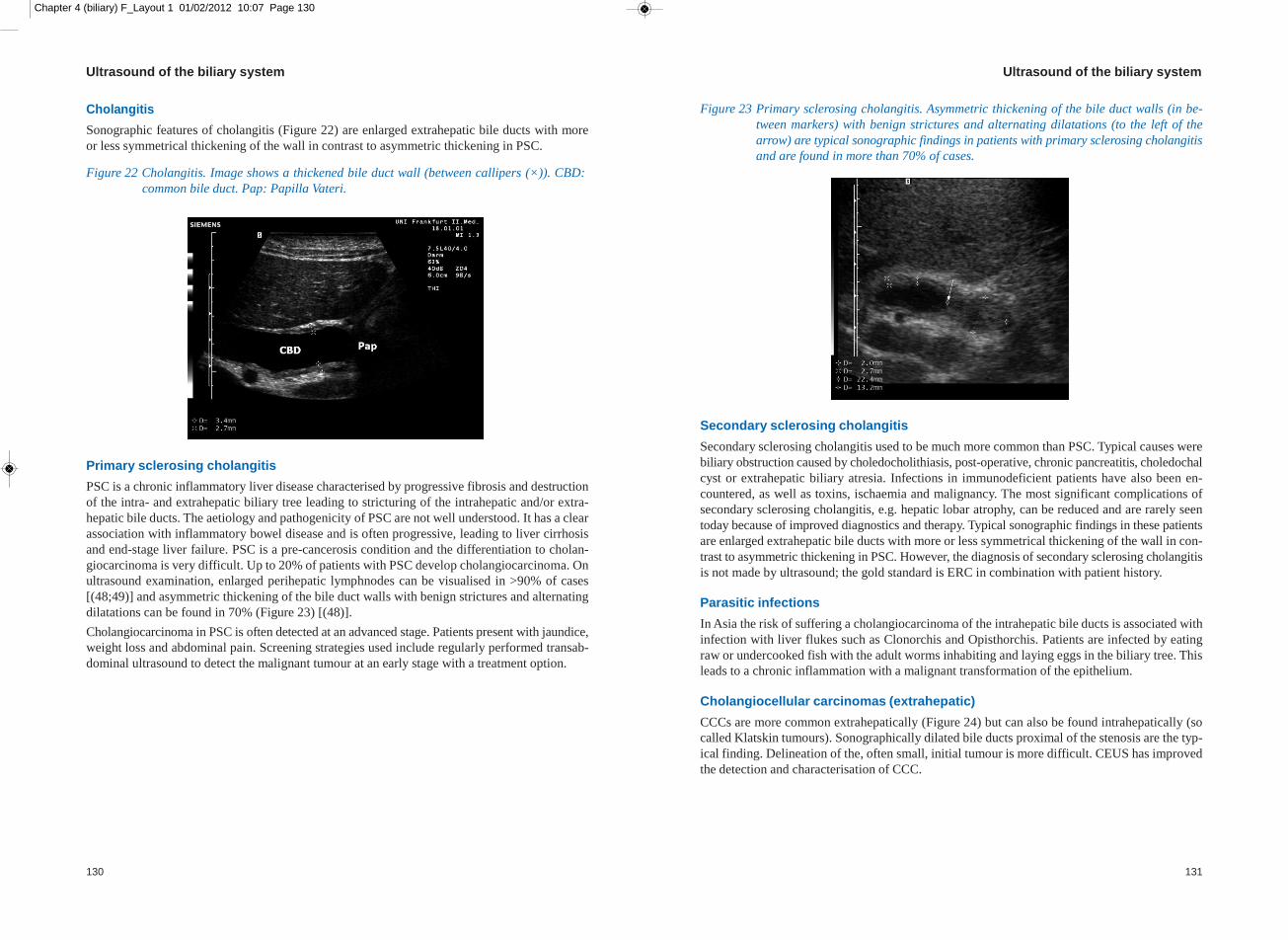
Primary sclerosing cholangitis
PSC is a chronic inflammatory liver disease characterised by progressive fibrosis and destruction
of the intra- and extrahepatic biliary tree leading to stricturing of the intrahepatic and/or extra-
hepatic bile ducts. The aetiology and pathogenicity of PSC are not well understood. It has a clear
association with inflammatory bowel disease and is often progressive, leading to liver cirrhosis
and end-stage liver failure. PSC is a pre-cancerosis condition and the differentiation to cholan-
giocarcinoma is very difficult. Up to 20% of patients with PSC develop cholangiocarcinoma. On
ultrasound examination, enlarged perihepatic lymphnodes can be visualised in >90% of cases
[(48;49)] and asymmetric thickening of the bile duct walls with benign strictures and alternating
dilatations can be found in 70% (Figure 23) [(48)].
Cholangiocarcinoma in PSC is often detected at an advanced stage. Patients present with jaundice,
weight loss and abdominal pain. Screening strategies used include regularly performed transab-
dominal ultrasound to detect the malignant tumour at an early stage with a treatment option.
Figure 23 Primary sclerosing cholangitis. Asymmetric thickening of the bile duct walls (in be-
tween markers) with benign strictures and alternating dilatations (to the left of the
arrow) are typical sonographic findings in patients with primary sclerosing cholangitis
and are found in more than 70% of cases.
Secondary sclerosing cholangitis
Secondary sclerosing cholangitis used to be much more common than PSC. Typical causes were
biliary obstruction caused by choledocholithiasis, post-operative, chronic pancreatitis, choledochal
cyst or extrahepatic biliary atresia. Infections in immunodeficient patients have also been en-
countered, as well as toxins, ischaemia and malignancy. The most significant complications of
secondary sclerosing cholangitis, e.g. hepatic lobar atrophy, can be reduced and are rarely seen
today because of improved diagnostics and therapy. Typical sonographic findings in these patients
are enlarged extrahepatic bile ducts with more or less symmetrical thickening of the wall in con-
trast to asymmetric thickening in PSC. However, the diagnosis of secondary sclerosing cholangitis
is not made by ultrasound; the gold standard is ERC in combination with patient history.
Parasitic infections
In Asia the risk of suffering a cholangiocarcinoma of the intrahepatic bile ducts is associated with
infection with liver flukes such as Clonorchis and Opisthorchis. Patients are infected by eating
raw or undercooked fish with the adult worms inhabiting and laying eggs in the biliary tree. This
leads to a chronic inflammation with a malignant transformation of the epithelium.
Cholangiocellular carcinomas (extrahepatic)
CCCs are more common extrahepatically (Figure 24) but can also be found intrahepatically (so
called Klatskin tumours). Sonographically dilated bile ducts proximal of the stenosis are the typ-
ical finding. Delineation of the, often small, initial tumour is more difficult. CEUS has improved
the detection and characterisation of CCC.

Chapter 4 (biliary) F_Layout 1 01/02/2012 10:07 Page 132
132 133
Ultrasound of the biliary system Ultrasound of the biliary system
Figure 24 Cholangiocellular carcinoma. Adenoma of the papilla and extrahepatic cholangiocel-
lular carcinoma are drained by stents that can be easily displayed by ultrasound. In
this patient with severe deficits after stroke, who had refused operation, an adenoma
of the papilla was treated and followed up for more than 8 years when a carcinoma fi-
nally developed.
Figure 25 Adenomas of the papilla Vateri (between markers) are rarely encountered on ultra-
sound. Sometimes it is difficult to differentiate sludge from neoplasia. Contrast-en-
hanced ultrasound techniques and elastography are helpful in delineating vascularised
and depth infiltration. (a) This image sequence shows an adenoma shown by transab-
dominal B-mode ultrasound, (b) transabdominal contrast-enhanced ultrasound, (c)
endoscopic elastography [(50)] and (d) contrast enhanced endoscopic ultrasound
using low mechanical index technique (CELMI EUS) [(51;52)]. (e) In contrast to ade-
noma in (harder) carcinoma infiltration of deeper layers can be delineated using elas-
tography, shown in a patient with malignant infiltration.
a b
Colour Doppler imaging c
The majority of circumscribed CCCs are slightly hyperperfused in the native colour Doppler but
CDI findings vary widely. d
Contrast-enhanced ultrasound
CEUS can be very helpful and displays a variable, but mainly hyperperfused, perfusion picture
in the arterial phase. In the late portal venous phase CCCs are contrasted as punched-out defects.
This behaviour is not always easy to demonstrate in the case of the Klatskin tumours, which often
exhibit an appreciable pericholangitic component. As far as differential diagnoses are concerned,
in the case of the hilar type of CCC, inflammatory bile duct alterations should be considered, e.g.
cholangitis. However, stratification of the bile ducts can be preserved and may actually be ac-
centuated in the sonographic image. For the detection of CCCs the examination technique in the
liver specific late phase has proved to be diagnostically useful in patients showing normal CT,
MRI and MRCP results, but so far there have been no conclusive studies on differential diagnosis
of PSC and CCCs. d
Other tumours of extrahepatic bile ducts
Adenomas represent only 10% of the incidence of carcinoma and are much more common in the
gallbladder than in the extrahepatic biliary tree (Figure 25). Other forms of benign tumours of
extraheptic bile ducts are rare, e.g. carcinoid, and show no pathognomonic ultrasound patterns.
Even rarer is the so called benign hepatobiliary papillomatosis now described as intraductal pap-
illary mucinous neoplasia of the bile ducts.
Chapter 4 (biliary) F_Layout 1 01/02/2012 10:07 Page 134
134 135
Ultrasound of the biliary system Ultrasound of the biliary system
Carcinomas of extrahepatic bile ducts account for 95% of adenocarcinoma of all extrahepatic
bile duct malignancies (bile duct carcinoma and cholangiocarcinoma). Klatskin (hilar) tumours
comprise 70% of tumours and arise at the confluence of right and left hepatic ducts at liver hilus.
Mostly they are slow growing with infrequent distant metastases.
Tumour, node, metastasis (TNM) staging for extrahepatic bile duct carcinoma applies to carci-
nomas arising above ampulla of Vater, including carcinomas in congenital choledochal cysts and
intrapancreatic portion of the CBD. The classification excludes sarcomas and carcinoid tumours.
Features to report from a surgical point of view include obstruction, bile duct wall thickness (as
a sign of infection), stones, tumour location and size, depth of invasion, tumour extension to ad-
jacent structures and regional lymph nodes.
After endoscopic therapy of a biliary obstruction with papillotomy and stent placement, the po-
sition of the stent can be easily controlled by ultrasound (Figure 26).
Figure 26 Bile duct carcinoma drained by stents can be easily displayed on ultrasound. (a) A
stent is shown using panoramic imaging. (b) Stents can sometimes be better delineated
using low mechanical index harmonic imaging techniques.
a b
Pneumobilia (also known as aerobilia)
Pneumobilia can be caused by a variety of diseases, e.g. perforation of stones of the biliary duct
system into the GI tract. However, the most common reason of pneumobilia can be found after
papillotomy with installation of a biliary stent. A very rare, but alarming, cause for pneumobilia
is a fulminant cholangitis with aerogenic bacteria. Pneumobilia is normally present after endo-
scopic sphincterotomy and surgical biliodigestive anastomosis.
Characteristic signs that can be found on ultrasound examination are linear jerks with typical re-
verberations (“ring-down-artefact”) and in contrast to sessile calcifications air is movable in the
bile ducts if the patient position is changed (Figure 27).
Figure 27 (a) Pneumobilia of peripheral bile ducts after papillotomy. Pneumobilia can be helpful
in the differentiation of (b) sufficiently and (c) insufficiently drained liver lobes.
a b
e
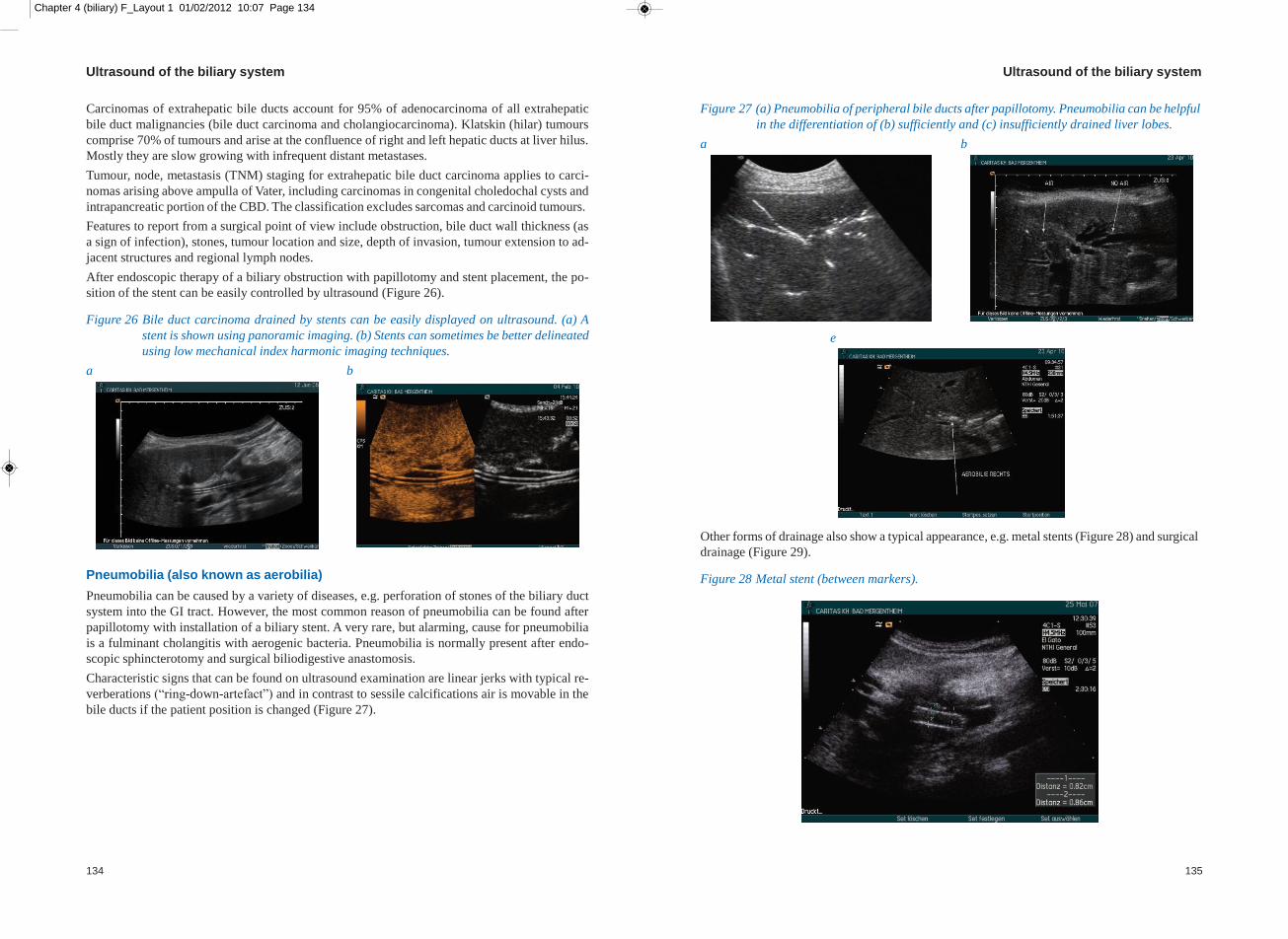
Other forms of drainage also show a typical appearance, e.g. metal stents (Figure 28) and surgical
drainage (Figure 29).
Figure 28 Metal stent (between markers).

Chapter 4 (biliary) F_Layout 1 01/02/2012 10:07 Page 136
136 137
Ultrasound of the biliary system Ultrasound of the biliary system
Figure 29 Surgical drainage (DRAINAGE) displayed by panoramic ultrasound imaging [(53)].
GB-BETT, gallbladder fissure of the liver.
New techniques
CEUS has been introduced into daily clinical procedure for many presentations when imaging
the liver, pancreas and kidneys and for monitoring local ablative treatment, mainly of the liver,
but also of other organs. New applications have also been introduced, e.g. the application of BR1
(SonoVue®, Bracco, Italy) into the bile ducts via a conventional ERC. Percutaneous transhepatic
cholangiography and drainage (PTCD) is a procedure for diagnosis and treatment of dilated bile
ducts in both malignant and benign biliary obstruction. PTCD has some limitations owing to the
blind puncture technique of peripherally located intrahepatic bile ducts. Severe (major) compli-
cations have been described in approximately 2% of cholangiography and up to 10% of transcu-
taneous interventions, e.g. sepsis, haemorrhage, abscesses, peritonitis and haematobilia the latter
especially true when puncturing close to the liver hilum. Nevertheless, the complication rate
varies with patients co-morbidity and investigators experience. Ultrasound-guided PTCD has
been described for dilated and non-dilated bile ducts and several studies have shown a reduction
in complications and a faster access to the biliary ducts. CEUS-PTCD has recently been described.
Future studies in a larger numbers of patients are necessary to evaluate this new technique con-
cerning the optimal dosage, the limitations and additional indications [(52)]. Other hepatobiliary
interventions have been recently summarised [(54;55)].
Figure 30 Choledocholithiasis (arrow) diagnosed by contrast enhanced ultrasound and contrast-
enhanced percutaneous cholangiography and treated by contrast-enhanced ultrasound
guided cholangiodrainage [(52)]. (a) Choledocholithiasis shown by B-mode ultra-
sound may be confused with tumour like lesions but intravascular (b) and extra vas-
cular contrast-enhanced ultrasound can confirm this differential diagnosis.
a b
e

Chapter 4 (biliary) F_Layout 1 01/02/2012 10:07 Page 138
138 139
Ultrasound of the biliary system Ultrasound of the biliary system
References
1. Nuernberg D, Ignee A, Dietrich CF. [Ultrasound in gastroenterology. Biliopancreatic system]. Med
Klin (Munich) 2007; 102(2):112-126.
2. Hanbidge AE, Buckler PM, O'Malley ME, Wilson SR. From the RSNA refresher courses: imaging
evaluation for acute pain in the right upper quadrant. Radiographics 2004; 24(4):1117-1135.
3. Freitas ML, Bell RL, Duffy AJ. Choledocholithiasis: evolving standards for diagnosis and management.
World J Gastroenterol 2006; 12(20):3162-3167.
4. Dietrich CF, Braden B. Sonographic assessments of gastrointestinal and biliary functions. Best Pract
Res Clin Gastroenterol 2009; 23(3):353-367.
5. Nuernberg D, Braden B, Ignee A, Schreiber-Dietrich DG, Dietrich CF. [Functional ultrasound in gas-
troenterology]. Z Gastroenterol 2008; 46(9):883-896.
6. Dietrich CF, Chichakli M, Hirche TO, Bargon J, Leitzmann P, Wagner TO et al. Sonographic findings
of the hepatobiliary-pancreatic system in adult patients with cystic fibrosis. J Ultrasound Med 2002;
21(4):409-416.
7. Bargon J, Stein J, Dietrich CF, Muller U, Caspary WF, Wagner TO. [Gastrointestinal complications of
adult patients with cystic fibrosis]. Z Gastroenterol 1999; 37(8):739-749.
8. Naganuma S, Ishida H, Konno K, Hamashima Y, Hoshino T, Naganuma H et al. Sonographic findings
of anomalous position of the gallbladder. Abdom Imaging 1998; 23(1):67-72.
9. Erdogmus B, Yazici B, Safak AA, Ozdere BA. Multiseptate gallbladder with acute acalculous chole-
cystitis. J Clin Ultrasound 2004; 32(8):423-424.
10. Shaikh AA, Charles A, Domingo S, Schaub G. Gallbladder volvulus: report of two original cases and
review of the literature. Am Surg 2005; 71(1):87-89.
11. Schirmer BD, Winters KL, Edlich RF. Cholelithiasis and cholecystitis. J Long Term Eff Med Implants
2005; 15(3):329-338.
12. Cooperberg PL, Burhenne HJ. Real-time ultrasonography. Diagnostic technique of choice in calculous
gallbladder disease. N Engl J Med 1980; 302(23):1277-1279.
13. Wermke W. Ultrasonic diagnosis of bile duct calculi. A prospective study regarding the effects and the
objective and subjective factors on accuracy in choledocholithiasis. Ultraschall Med 1992; 13(6):246-
254.
14. Sandouk F, Anand BS, Graham DY. The whirlpool jet technique for removal of pancreatic duct ascaris.
Gastrointest Endosc 1997; 46(2):180-182.
15. Sandouk F, Haffar S, Zada MM, Graham DY, Anand BS. Pancreatic-biliary ascariasis: experience of
300 cases. Am J Gastroenterol 1997; 92(12):2264-2267.
16. Ralls PW, Colletti PM, Lapin SA, Chandrasoma P, Boswell WD, Jr., Ngo C et al. Real-time sonography
in suspected acute cholecystitis. Prospective evaluation of primary and secondary signs. Radiology
1985; 155(3):767-771.
17. Braden B, Faust D, Ignee A, Schreiber D, Hirche T, Dietrich CF. Clinical relevance of perihepatic lym-
phadenopathy in acute and chronic liver disease. J Clin Gastroenterol 2008; 42(8):931-936.
18. Konno K, Ishida H, Naganuma H, Sato M, Komatsuda T, Sato A et al. Emphysematous cholecystitis:
sonographic findings. Abdom Imaging 2002; 27(2):191-195.
19. Duzgun AP, Ozmen MM, Ozer MV, Coskun F. Internal biliary fistula due to cholelithiasis: a single-
centre experience. World J Gastroenterol 2007; 13(34):4606-4609.
20. Bouveret L. Stenose du pylore adherent a la vesicule. Revue Medicale (Paris) 1896; 16:1-16.
21. Langhorst J, Schumacher B, Deselaers T, Neuhaus H. Successful endoscopic therapy of a gastric outlet
obstruction due to a gallstone with intracorporeal laser lithotripsy: a case of Bouveret's syndrome. Gas-
trointest Endosc 2000; 51(2):209-213.
22. Kavuturu S, Parithivel V, Cosgrove J. Bouveret's syndrome: a rare presentation of gallstone disease.
OPUS 12 Scientist 2008; 2:26.
23. Doycheva I, Limaye A, Suman A, Forsmark CE, Sultan S. Bouveret's syndrome: case report and review
of the literature. Gastroenterol Res Pract 2009; 2009:914951.
24. Cappell MS, Davis M. Characterization of Bouveret's syndrome: a comprehensive review of 128 cases.
Am J Gastroenterol 2006; 101(9):2139-2146.
25. Sohn VY, Arthurs ZM, Martin MJ, Sebesta JA, Branch JB, Champeaux AL. Incidental pathologic find-
ings in open resectional gastric bypass specimens with routine cholecystectomy and appendectomy.
Surg Obes Relat Dis 2008; 4(5):608-611.
26. Storr M, Weigert N, Fellbaum C, Classen M. [Polyposis of the gastrointestinal tract as a manifestation
of diffuse follicular lymphatic hyperplasia]. Dtsch Med Wochenschr 1998; 123(12):347-352.
27. Barreiros AP, Otto G, Ignee A, Galle P, Dietrich CF. Sonographic signs of amyloidosis. Z Gastroenterol
2009; 47(8):731-739.
28. A prospective analysis of 1518 laparoscopic cholecystectomies. The Southern Surgeons Club. N Engl
J Med 1991; 324(16):1073-1078.
29. Ignee A, Piscaglia F, Ott M, Salvatore V, Dietrich CF. A benign tumour of the liver mimicking malignant
liver disease--cholangiocellular adenoma. Scand J Gastroenterol 2009; 44(5):633-636.
30. Jepsen P, Vilstrup H, Tarone RE, Friis S, Sorensen HT. Incidence rates of intra- and extrahepatic cholan-
giocarcinomas in Denmark from 1978 through 2002. J Natl Cancer Inst 2007; 99(11):895-897.
31. Welzel TM, McGlynn KA, Hsing AW, O'Brien TR, Pfeiffer RM. Impact of classification of hilar cholan-
giocarcinomas (Klatskin tumors) on the incidence of intra- and extrahepatic cholangiocarcinoma in the
United States. J Natl Cancer Inst 2006; 98(12):873-875.
32. Dietrich CF, Leuschner MS, Zeuzem S, Herrmann G, Sarrazin C, Caspary WF et al. Peri-hepatic lym-
phadenopathy in primary biliary cirrhosis reflects progression of the disease. Eur J Gastroenterol He-
patol 1999; 11(7):747-753.
33. Dietrich CF, Zeuzem S. Sonographic detection of perihepatic lymph nodes: technique and clinical value.
Z Gastroenterol 1999; 37(2):141-151.
34. Dietrich CF, Stryjek-Kaminska D, Teuber G, Lee JH, Caspary WF, Zeuzem S. Perihepatic lymph nodes
as a marker of antiviral response in patients with chronic hepatitis C infection. AJR Am J Roentgenol
2000; 174(3):699-704.
35. Cheng SP, Yang TL, Jeng KS, Liu CL, Lee JJ, Liu TP. Choledochal cyst in adults: aetiological consid-
erations to intrahepatic involvement. ANZ J Surg 2004; 74(11):964-967.
36. Todani T, Watanabe Y, Toki A, Morotomi Y. Classification of congenital biliary cystic disease: special
reference to type Ic and IVA cysts with primary ductal stricture. J Hepatobiliary Pancreat Surg 2003;
10(5):340-344.
37. Todani T, Watanabe Y, Toki A, Ogura K, Wang ZQ. Co-existing biliary anomalies and anatomical vari-
ants in choledochal cyst. Br J Surg 1998; 85(6):760-763.
38. Todani T, Urushihara N, Morotomi Y, Watanabe Y, Uemura S, Noda T et al. Characteristics of chole-
dochal cysts in neonates and early infants. Eur J Pediatr Surg 1995; 5(3):143-145.
39. Dietrich C.F., Gouder S, Hocke M, Schuessler G, Ignee A. Endosonographie der Choledocholithiasis
und ihrer Differentialdiagnosen. Endoskopie Heute 2004; 17:160-166.
40. Seifert H, Wehrmann T, Hilgers R, Gouder S, Braden B, Dietrich CF. Catheter probe extraductal EUS
reliably detects distal common bile duct abnormalities. Gastrointest Endosc 2004; 60(1):61-67.
41. Sandouk F, Haffar S, Zada MM, Graham DY, Anand BS. Pancreatic-biliary ascariasis: experience of
300 cases. Am J Gastroenterol 1997; 92(12):2264-2267.
42. Di Nardo R, Urbano D, Drudi FM, Chianta GL, Tortora A, De Simone P et al. [Ultrasonography in the
preoperative assessment of candidates for laparoscopic cholecystectomy: examination technique and
results]. Radiol Med (Torino) 1996; 92(5):605-609.

Chapter 4 (biliary) F_Layout 1 01/02/2012 10:07 Page 140
140
Ultrasound of the biliary system
43. Fang R, Pilcher JA, Putnam AT, Smith T, Smith DL. Accuracy of surgeon-performed gallbladder ultra-
sound. Am J Surg 1999; 178(6):475-479.
44. Krook PM, Allen FH, Bush WH, Jr., Malmer G, MacLean MD. Comparison of real-time cholecys-
tosonography and oral cholecystography. Radiology 1980; 135(1):145-148.
45. Seitz K, Hege-Blank U, Holzinger H. [10 years of sonographic diagnosis of gallstones--what do surgical
statistics tell us about its reliability?]. Ultraschall Med 1987; 8(3):121-125.
46. Silidker MS, Cronan JJ, Scola FH, Moore MM, Schepps B, Thompson W et al. Ultrasound evaluation
of cholelithiasis in the morbidly obese. Gastrointest Radiol 1988; 13(4):345-346.
47. Goodman AJ, Neoptolemos JP, Carr-Locke DL, Finlay DB, Fossard DP. Detection of gall stones after
acute pancreatitis. Gut 1985; 26(2):125-132.
48. Hirche TO, Russler J, Braden B, Schuessler G, Zeuzem S, Wehrmann T et al. Sonographic detection of
perihepatic lymphadenopathy is an indicator for primary sclerosing cholangitis in patients with inflam-
matory bowel disease. Int J Colorectal Dis 2004; 19(6):586-594.
49. Hirche TO, Russler J, Schroder O, Schuessler G, Kappeser P, Caspary WF et al. The value of routinely
performed ultrasonography in patients with Crohn disease. Scand J Gastroenterol 2002; 37(10):1178-
1183.
50. Hirche TO, Ignee A, Barreiros AP, Schreiber-Dietrich D, Jungblut S, Ott M et al. Indications and lim-
itations of endoscopic ultrasound elastography for evaluation of focal pancreatic lesions. Endoscopy
2008; 40(11):910-917.
51. Dietrich CF. Contrast-enhanced low mechanical index endoscopic ultrasound (CELMI-EUS). En-
doscopy 2009; 41 Suppl 2:E43-E44.
52. Ignee A, Baum U, Schuessler G, Dietrich CF. Contrast-enhanced ultrasound-guided percutaneous
cholangiography and cholangiodrainage (CEUS-PTCD). Endoscopy 2009; 41(8):725-726.
53. Dietrich CF, Caspary WF. SieScape--panorama imaging. Clinical value? Internist (Berl) 2000; 41(1):24-
28.
54. Gottschalk U, Ignee A, Dietrich CF. [Ultrasound-guided interventions and description of the equipment].
Z Gastroenterol 2010; 48(11):1305-1316.
55. Gottschalk U, Ignee A, Dietrich CF. [Ultrasound guided interventions, part 1, diagnostic procedures].
Z Gastroenterol 2009; 47(7):682-690.