Biliary Disease. Gallbladder and Biliary Tree Imaging Studies Available –X-ray –Computed...
16
Biliary Disease
-
Upload
prosper-york -
Category
Documents
-
view
224 -
download
1
Transcript of Biliary Disease. Gallbladder and Biliary Tree Imaging Studies Available –X-ray –Computed...

Biliary Disease
Gallbladder and Biliary Tree
• Imaging Studies Available– X-ray– Computed tomography– Radioisotope scan (hepatobiliary scan)– Ultrasound: the most sensitive study for
gallstone
Gallstones
• 81% consist of cholesterol and are radiolucent (cannot be seen on plain films)
• 19% contain calcium and are radio-opaque (can be seen on plain films)
Gallstones
• Size: can be very small (1-2 mm) to very large (several cm)
• Shape: single stone – round, large• Multiple stones – small, faceted
Gallstones
• Complications include– Obstruction of bile duct: commonly cystic duct
and common bile duct– Cholecystitis (infection/ inflammation of the
gallbladder)– Abscess (complication of cholecystistis)– Perforation:
• peritonitis, inflammation infection of peritoneum• gallstone ileus, stone erodes into GI tract and
causes obstruction
Gallbladder
• Ultrasound– Stones all appear ECHOGENIC (white)
• May be either radiolucent or radio-opaque
– Beyond stone is an acoustic shadow– Bile is HYPOECHOIC (black)– Polyps, neoplasm typically are isoechoic
(equal echo density) to adjacent soft tissues
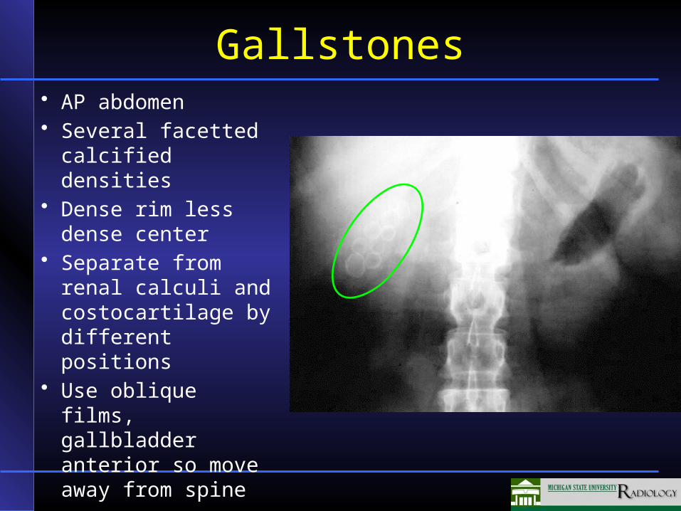
Gallstones• AP abdomen• Several facetted
calcified densities• Dense rim less
dense center• Separate from
renal calculi and costocartilage by different positions
• Use oblique films, gallbladder anterior so move away from spine
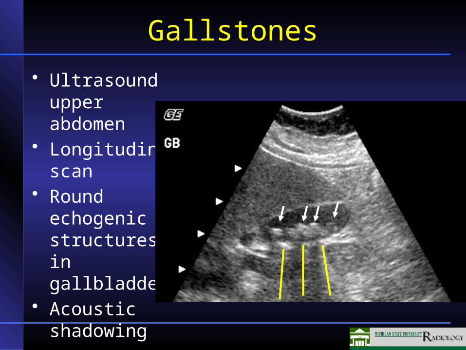
Gallstones
• Ultrasound upper abdomen
• Longitudinal scan
• Round echogenic structures in gallbladder
• Acoustic shadowing
Dilation of Bile Ducts
• Causes include– Stone (most common)– Carcinoma
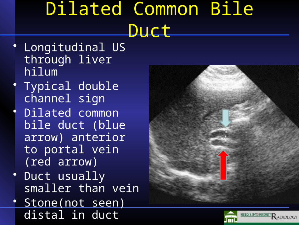
Dilated Common Bile Duct• Longitudinal US
through liver hilum• Typical double
channel sign• Dilated common bile
duct (blue arrow) anterior to portal vein (red arrow)
• Duct usually smaller than vein
• Stone(not seen) distal in duct
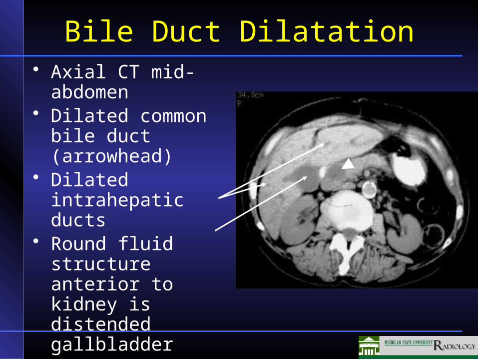
Bile Duct Dilatation• Axial CT mid-
abdomen• Dilated common
bile duct (arrowhead)
• Dilated intrahepatic ducts
• Round fluid structure anterior to kidney is distended gallbladder
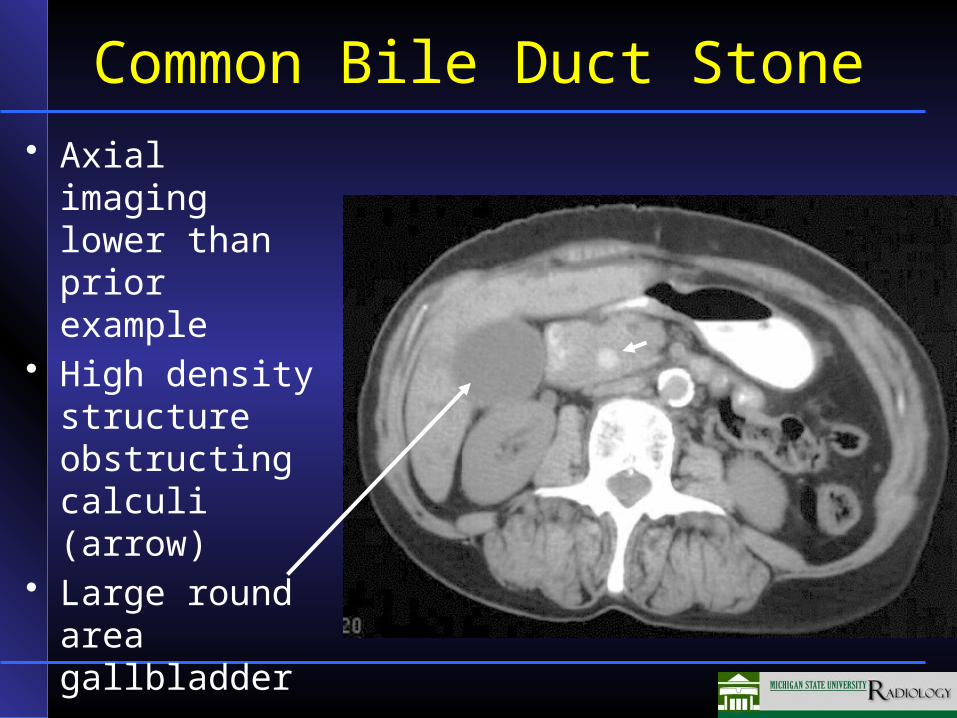
Common Bile Duct Stone• Axial imaging
lower than prior example
• High density structure obstructing calculi (arrow)
• Large round area gallbladder
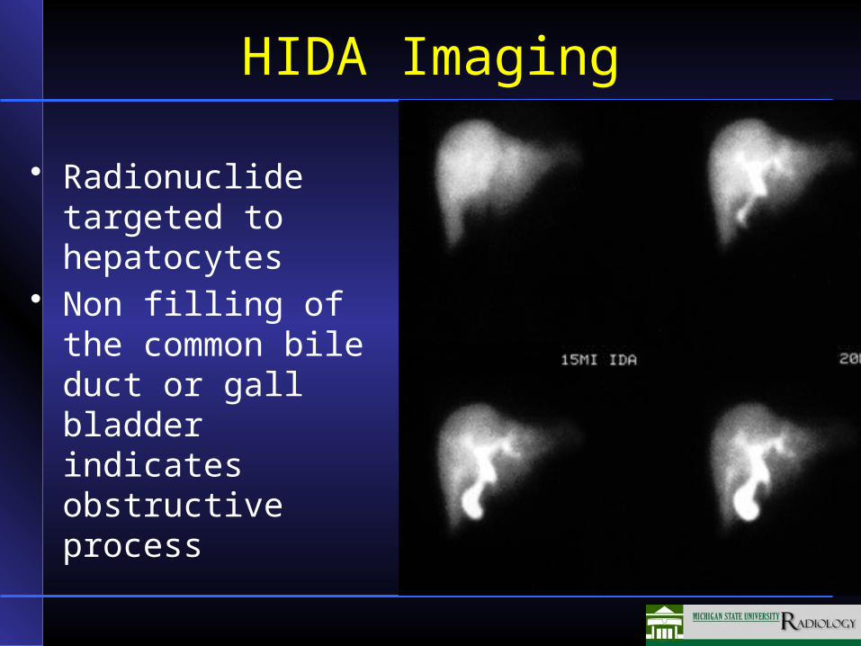
HIDA Imaging
• Radionuclide targeted to hepatocytes
• Non filling of the common bile duct or gall bladder indicates obstructive process

Acute Cholecystitis
• No filling of the gallbladder 60 minutes after injection of isotope
• Rapid accumulation within the liver and bile ducts with spill into the doudenum
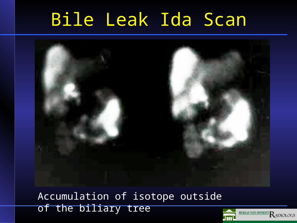
Bile Leak Ida Scan
Accumulation of isotope outside of the biliary tree
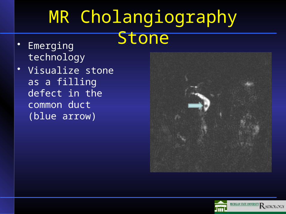
MR Cholangiography Stone
• Emerging technology
• Visualize stone as a filling defect in the common duct (blue arrow)