
ubc_2010_spring_jafarpour_bita
153
Characterization of the X4 protein of Tomato ringspot virus and analysis of its variability among virus isolates by Bita Jafarpour M.Sc. Ferdowsi University of Mashhad, College of Agriculture, 2000 B.Sc. Ferdowsi University of Mashhad, College of Agriculture, 1997 A THESIS SUBMITTED IN PARTIAL FULFILMENT OF THE REQUIREMENTS FOR THE DEGREE OF DOCTOR OF PHILOSOPHY in The Faculty of Graduate Studies (Botany) The University of British Columbia (Vancouver) March 2010 ©Bita Jafarpour 2010
-
Upload
bita-jafarpour -
Category
Documents
-
view
40 -
download
0
Transcript of ubc_2010_spring_jafarpour_bita

Characterization of the X4 protein of Tomato ringspot
virus and analysis of its variability among virus isolates
by
Bita Jafarpour
M.Sc. Ferdowsi University of Mashhad, College of Agriculture, 2000
B.Sc. Ferdowsi University of Mashhad, College of Agriculture, 1997
A THESIS SUBMITTED IN PARTIAL FULFILMENT OF
THE REQUIREMENTS FOR THE DEGREE OF
DOCTOR OF PHILOSOPHY
in
The Faculty of Graduate Studies
(Botany)
The University of British Columbia
(Vancouver)
March 2010
©Bita Jafarpour 2010

ii
Abstract
The genome of Tomato ringspot virus (nepovirus) encodes a unique X4 protein of unknown
function. X4 is absent from the genome of many other nepoviruses and does not have
significant sequence homology with other proteins in available databases.
I have studied the possible function(s) of the ToRSV-X4 protein. I used three ToRSV
isolates, Rasp2, Rasp1 and PYB. Sequence analysis of the X4 region from these isolates
showed a surprising level of sequence variability. The central region of the X4 protein
contains two series of amino acid sequence tandem repeats. The number of these repeats
varies among ToRSV isolates. N. benthamiana plants inoculated with the ToRSV-Rasp1
recover from infection at temperatures equal to or above 27oC. At 21oC, recovery does not
occur and plants eventually die. In ToRSV-PYB1 infection, plants recover from infection at
all temperature tested. Since RNA silencing is a ubiquitous plant defence response and was
observed in nepovirus infected plants, I used agroinfiltration assays to test the effect of the
X4 protein on the induction of RNA silencing directed at a green fluorescent protein reporter
gene (GFP). Co-expression of X4-Rasp1 or X4-Rasp2 proteins with GFP in N. benthamiana
plants transiently enhanced the expression of GFP. To detect the ToRSV X4 protein in virus
infected plants, I used polyclonal antibodies raised against the C-terminal region of the X4-
Rasp2 protein. This antibody detected a 60 kDa protein in ToRSV-Rasp1 infected plants.
The cross-reactivity of this X 4 (64-65) antibody with the CP suggested that the 60 kDa
protein corresponds to the CP. However, X4(3743-44) and X4 (3741-42) antibodies derived
from the X4 sequence, did not detect this 60 kDa protein. The predicted 82 kDa X4 full-
length protein was not detected by any of the anti-X4 antibodies. This suggests that X4 may
be an unstable protein.
In conclusion, the variability of the X4 protein among ToRSV isolates combined with
different symptomatology and the preliminary evidence that X4 may act as a suppressor of
silencing in plants, suggest that X4 may be a multifunctional protein that is involved in
protein–protein interaction, host specificity, symptomatology and/or interaction with host
defence responses.
iii
Table of Contents
Abstract ....................................................................................................................................ii
Table of Contents....................................................................................................................iii
List of Tables ..........................................................................................................................vii
List of Figures .......................................................................................................................viii
List of Abbreviations ...............................................................................................................x
Dedications ............................................................................................................................xvi
Co-Authorship Statement ...................................................................................................xvii
Chapter 1 ..................................................................................................................................1
Literature review .....................................................................................................................1
1. Introduction .........................................................................................................................2
1.1 Virus structure .................................................................................................................2
1.2 Nepoviruses .....................................................................................................................3
1.2.1 Nepovirus structure...................................................................................................4
1.3 Replication cycle of plant viruses within the cell ............................................................8
1.3.1 Translation of viral proteins......................................................................................9
1.3.2 Polyprotein processing............................................................................................10
1.3.3 Assembly of the viral replication complex.............................................................11
1.3.3.1 Replication and translation of nepoviruses......................................................12
1.3.3.2 Replication of RNA2 in nepoviruses...............................................................13
1.4 Movement of plant viruses ............................................................................................14
1.4.1 Plasmodesmata .......................................................................................................15
1.4.2 Types of movement from cell to cell by plant viruses............................................16
1.4.2.1 Movement of virus-like particles.....................................................................16
1.4.2.1.1 Cell-to-cell movement of comoviruses, nepoviruses and related viruses.16
1.5 Virus transmission .........................................................................................................17
1.5.1 Nematode transmission...........................................................................................18
1.5.1.1 Tobraviruses ....................................................................................................18
1.5.1.2 Nepoviruses .....................................................................................................18
1.6 ToRSV isolates ..............................................................................................................19
1.6.1 Virus evolution, diversity and sources of variation ................................................20
1.6.1.1 Mutation, insertion and deletion ......................................................................20
1.6.1.2 Recombination.................................................................................................20

iv
1.6.1.2.1 Protein domain repeats .............................................................................21
1.6.1.3 Serial passages of viruses, a source of creating sequence diversity ................21
1.7 Virion associated proteins..............................................................................................23
1.7.1 CaMV P3 virion associated protein........................................................................23
1.7.2 HC-Pro Helper component proteinase....................................................................24
1.7.3 Tobravirus 2b nematode transmission helper protein.............................................25
1.7.4 Closterovirus virion associated proteins.................................................................26
1.7.5 Potexvirus virion associated protein.......................................................................26
1.8 RNA silencing and viral suppressors of silencing and silencing activity in Nepoviruses
.............................................................................................................................................27
1.8.1 Mechanisms of RNA silencing...............................................................................27
1.8.2 Viral suppressors of silencing.................................................................................29
1.8.3 Host recovery from virus infection and the role of RNA silencing in nepoviruses33
1.9 The X4 protein of ToRSV ............................................................................................34
1.10 Computer predictions based on ToRSV X4 amino acid sequences. ...........................35
1.10.1 PEST motifs..........................................................................................................35
1.10.2 Phosphorylation sites in ToRSV isolates..............................................................38
1.10.3 Topology prediction of membrane proteins. ........................................................39
1.10.4 Secondary structure prediction in different ToRSV isolates ................................42
1.11 Research hypothesis and objectives.............................................................................43
1.12 Bibliography ................................................................................................................44
Chapter 2 ................................................................................................................................58
Insertion of large amino acid repeats and point mutations contribute to a high degree of
sequence diversity in the X4 protein of tomato ringspot virus..........................................58
2.1 Introduction....................................................................................................................59
2.2 Materials and methods...................................................................................................60
2.2.1 Provenance of the virus material ............................................................................60
2.3 Results............................................................................................................................61
2.3.1 Sequence properties ................................................................................................61
2.4 Discussion......................................................................................................................65
2.5 Bibliography ..................................................................................................................66
Chapter 3 ................................................................................................................................68
Characterization of the X4 protein in ToRSV-infected plants..........................................68
3.1 Introduction....................................................................................................................69

v
3.1.1 Research objective and hypothesis .........................................................................71
3.2 Materials and methods...................................................................................................72
3.2.1 Production of antibodies against the C-terminal and the N-terminal region of X4 72
3.2.2 Construction of plasmids used in the expression of full-length or truncated forms
of X4, CP and MP in vitro translations or in E. coli........................................................73
3.2.3 Preparation of plant extracts and subcellular fractionations...................................76
3.2.4 Purification of ToRSV particles .............................................................................76
3.2.5 Immunoblotting ......................................................................................................76
3.2.6 Immuno-electron microscopy.................................................................................77
3.2.7 Trypsin digestion of ToRSV particles ....................................................................77
3.2.8 Liquid chromatography-mass spectrometry with peptide mass fingerprinting (LC-
MS/MS) ...........................................................................................................................78
3.3 Results............................................................................................................................78
3.3.1 Preparation of antibodies against the X4 protein....................................................78
3.3.2 Detection of a 60 kDa protein by the X4(64-65) antibodies in ToRSV-infected
plant extracts....................................................................................................................81
3.3.3 Co-fractionation of the 60 kDa protein detected by the X4(64-65) antibodies with
empty and full virus particles in sucrose gradient fractionation assay ............................83
3.3.4 Co-purification of the 60 kDa protein detected by the X4(64-65) antibodies with
the virus particles.............................................................................................................85
3.3.5 Immunogold labelling of purified virus particles with X4(64-65) antibodies........87
3.3.6 Susceptibility of the 60 kDa protein recognized by the X4(64-65) antibodies to
trypsin digestion ..............................................................................................................88
3.3.7 The X4 protein is not detected in association with virions using liquid
chromatography-mass spectrometry with peptide mass fingerprinting (LC-MS/MS) ....89
3.3.8 Re-examination of the specificity of the X4(64-65) antibodies and the α-virion
antibodies.........................................................................................................................89
3.3.9 In vitro co-immunoprecipitation of X4 and CP proteins with the X4(64-65)
antibodies.........................................................................................................................92
3.3.9.1 The MP did not co-immunoprecipitate with the CP in vitro ...........................93
3.3.10. Production of antibodies from different regions of the X4 and the CP...............94
3.4 Discussion....................................................................................................................100
3.4.1. Summary and conclusion.....................................................................................106
3.5 Bibliography ................................................................................................................107

vi
Chapter 4 ..............................................................................................................................111
Preliminary evidence that the X4 protein may act as a silencing suppressor in plants 111
4.1. Introduction.................................................................................................................112
4.1.1. Research hypothesis and objectives.....................................................................113
4.2 Materials and methods.................................................................................................114
4.2.1 PCR amplification of a region of the RNA2 open reading frame coding for a
portion of X3, the entire X4 and a portion of MP for the Chickadee isolates. ..............114
4.2.2 Symptom evaluation in plants infected with Rasp1 and PYB ToRSV isolates....114
4.2.3. Plasmid construction............................................................................................115
4.2.4 Agroinfiltration of N. benthamiana plants and immunoblotting ..........................115
4.3 Results..........................................................................................................................116
4.3.1 RT-PCR amplification of ToRSV Chickadee isolate and symptom development in
ToRSV–infected Rasp1 and PYB ToRSV isolates .......................................................116
4.3.2 Preliminary evidence that the X4 protein may act as a silencing suppressor in
plants..............................................................................................................................118
4.4 Discussion....................................................................................................................120
4.4.1 Summary and conclusion......................................................................................122
4.5 Bibliography ................................................................................................................124
Chapter 5 ..............................................................................................................................127
General discussion and future projects .............................................................................127
5.1 General discussion .......................................................................................................128
5.2 Future projects .............................................................................................................131
5.2.1 Potential impact of this research on the future on molecular virology research ..134
5.3 Bibliography ................................................................................................................135
vii
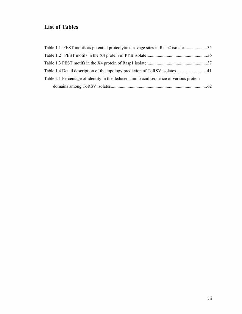
List of Tables
Table 1.1 PEST motifs as potential proteolytic cleavage sites in Rasp2 isolate ....................35
Table 1.2 PEST motifs in the X4 protein of PYB isolate......................................................36
Table 1.3 PEST motifs in the X4 protein of Rasp1 isolate......................................................37
Table 1.4 Detail description of the topology prediction of ToRSV isolates ………………...41
Table 2.1 Percentage of identity in the deduced amino acid sequence of various protein
domains among ToRSV isolates......................................................................................62

viii
List of Figures
Fig. 1.1 A simplified representation of the structure of TRSV. ...............................................6
Fig. 1.2 Pseudo-T = 3 capsid of Picornavirales. ........................................................................8
Fig. 1. 3 Schematic representation of the replication cycle of nepoviruses within the cell.......9
Fig. 1. 4 Genome organisation of ToRSV. ..............................................................................11
Fig. 1.5 Schematic representation of a plasmodesmata transversal section ............................16
Fig. 1.6 Functional regions of the helper component proteinase (HC-Pro).............................25
Fig. 1.7 The silencing pathway................................................................................................29
Fig. 1.8 Mechanism of the silencing suppression activity of p19. ..........................................31
Fig.1.9 Comparison of the genomic structure of tomato ringspot virus and grapevine fanleaf
virus (nepoviruses)...........................................................................................................34
Fig. 1.10 Predicted phosphorylation sites................................................................................39
Fig.1.11 Topology prediction of ToRSV isolate. ....................................................................41
Fig.1.12 Secondary structure prediction in different ToRSV isolates.....................................42
Fig. 2. 1 Comparison of the X4 protein domain in the ToRSV Rasp1 and Rasp2 isolates ....63
Fig. 3.1 Genome organisation of ToRSV and GFLV and production of the X4(64-65)
antibodies.........................................................................................................................79
Fig. 3.2 Specificity of the X4(64-65) and "-virion antibodies tested by immunoprecipitation
assays. ..............................................................................................................................81
Fig. 3.3 Detection of a 60 kDa protein by the X4(64-65) antibodies in infected plant extracts.
.........................................................................................................................................82
Fig. 3.4 Immunoblot analysis and electron microscopy of subcellular fractionation of proteins
by sucrose gradient. .........................................................................................................84
Fig. 3.5 Co-purification of the 60 kDa protein detected by the X4(64-65) antibodies with
purified virus particles. ....................................................................................................86
Fig. 3.6 Immunogold labelling of purified virus particles using the X4(64-65) antibodies. ...88
Fig. 3.7 Susceptibility of the 60 kDa protein to trypsin digestions. .......................................89
Fig. 3.8 Immunoblot analyses of CP, CP-150, CP-360, X4(64-65), X4-TM (Rasp2) proteins.
.........................................................................................................................................91
Fig. 3.9 Immunoprecipitation of the CP and the X4 protein. .................................................93
Fig. 3.10 In vitro protein-protein interaction assays................................................................94
Fig. 3.11 X4 protein: Alignment of 3 different isolates of ToRSV (Rasp1, Rasp2 and PYB).
.........................................................................................................................................95

ix
Fig. 3.12 CP protein: Alignment of 3 different isolates of ToRSV (Rasp1, Rasp2 and PYB).
.........................................................................................................................................96
Fig. 3.13 Immunoblot analysis of the infected (I) and healthy (H) N. benthamiana extracts
using the anti-virion, anti-CP (3739-40) and anti-CP (3737-38) antibodies. ..................98
Fig. 3.14 Immunoblot analysis of the infected (Inf) and healthy (H) N. benthamiana extracts
using the X4-N, X4(3743-44) and X4 (3741-42) antibodies...........................................99
Fig. 3.15. Sequence alignment between the X4 protein and the CP of ToRSV. ...................104
Fig. 4.1 PCR amplification of a region of the RNA2 open reading frame coding for a portion
of X3, the entire X4 and a portion of MP in Rasp1, PYB and Chickadee isolates. ......117
Fig. 4.2 Symptom development in ToRSV–infected Rasp1 and PYB ToRSV isolates........117
Fig. 4.3 Expression levels of GFP in a transient expression assay conducted in the presence
or absence of X4. ...........................................................................................................119
Fig. 4.4 Analysing the expression level of the X4 protein in infiltrated patches by
immunoblotting. ............................................................................................................120

x
List of Abbreviations
"-MP Anti movement protein antibodies "-virion Anti –virion antibodies X4-C Recombinant protein a.a Amino acid Abs Antibodies AFM Atomic force microscopy ALSV Apple latent spherical virus ArMV Arabis mosaic virus B Bottom component BLCV Beet leaf curl virus BLMV Blueberry leaf mottle virus BRAV Blackcurrant reversion associated virus BRV Blackcurrant reversion virus BSA Bovine serum albumin
BYV Beet yellow virus C. quinoa Chenopodium quinoa C. sativus Cucumis sativus CaMV Cauliflower mosaic virus CI Cylindrical inclusion CMV Cucumber mosaic virus
CP Coat protein CPm Minor capsid protein CPMV Cow pea mosaic virus CPS Small coat protein CsCL Cesium chloride CVYV Cucumber vein yellowing virus DNA Deoxyribonucleic acid dpi Days post infiltration dpi Days post inoculation dsRNA Double stranded RNA ELISA Enzyme-linked immunosorbent assay

xi
EM Electron microscopy
ER Endoplasmic reticulum Fig Figure
GFLV Grapevine fanleaf virus HA Poly-histidine tail HC-Pro Helper component proteinase HCRSV Hibiscus chlorotic ringspot virus Hsp70 Heat-Shock cognate 70-kDa Protein Hsp70h Hsp70 homologue IRES Internal ribosome binding site L Large LC-MS/MS Liquid chromatography-mass spectrometry with
Peptide mass fingerprinting LIYV Lettuce infection yellow virus LMV Lettuce mosaic virus M Morphological subunits m7GTP Methyl-guanidine triphosphate mg Milligram MP Movement protein mRNA Messenger RNA MSDB Protein sequence database designed for
massspectrometry applications
N. benthamiana Nicotiana benthamiana
NIa Nuclear inclusion a nm Nanometre nt Nucleotide NTB Nucleotide triphosphate binding protein P1 Polyproteinencoded by ToRSV RNA1 P1 Serine protease in the family potyviridae
p19 19 kDa protein of Tomato bushy stunt virus
PAZ PIWI Argonaut and Zwille PBS Phosphate buffered saline PCR Polymerase chain reaction PEBV Pea early browning virus

xii
PIPO Pretty interesting potyviridae ORF PIWI P-element induced wimpy testis in Drosophila
Pol Polymérase PPV Plum pox virus
Pro Proteinase PVIP Potyvirus VPg-interacting protein PVX Potato virus X PYB Peach yellow bud isolate RASP Raspberry isolate RdRp RNA dependent RNA polymerase RISC RNA-induced silencing complex RNA Ribonucleic acid RT-PCR Reverse transcription-polymerase chain reaction
S Small
S Sedimentation coefficient S3 Subcellular fractionation S30 Soluble protein-enriched fraction
SDS-PAGE Sodium dodecyl sulfate polyacrylamide gel
electrophoresis SEL Size exclusion limit siRNA Short interfering RNA SLRSV Strawberry latent ringspot virus
ssRNA Single stranded RNA T Top component TBRV Tomato blackring virus
TBSV Tomato bushy stunt virus
TCV Turnip crinkle virus
TEM Transmission electron microscope
TGB Tripple gene block TM Tranmembrane domain TRSV Tobacco ringspot virus TRV Tobacco rattle virus TSWV Tomato spotted wilt virus TVMV Tobacco vein mottling virus

xiii
UTR Untranslated region V
vol/vol Voltage
Voulume per volume VAP Virion associated protein VPg Genome linked viral protein
VPg-Pro-Pol ToRSV precursor protein of VPg,Pro,Pol
wt:vol Weight per volume
X2-NTB-VPg ToRSV precursor protein of X2, NTB and VPg
xg Unit of rotational speed based on gravitational
force
μg
Microgram
xiv
Acknowledgments
I would like to thank my supervisor Dr. Helene Sanfaçon whose expertise, understanding and
enthusiasm added considerably to my graduate experience. She spent a lot of time explaining
the research projects from the very early stage of this research as well as putting a great effort
in helping me with the proposal writing, preparing presentations, preparing for the
comprehensive exam and also writing the manuscript. I always enjoyed our scientific
discussions throughout my PhD. She gave me the chance to attend several international
conferences, which had a great impact on my knowledge and gave me extraordinary
experiences through out the work. I would like to thank my co-supervisor Dr. Carl Douglas
and my committee members Dr. Francois Jean and Dr. Steven Lund for their encouragement,
support and their valuable suggestions and scientific discussions, advices and critical review
of this thesis. I am very grateful to have them as my Ph.D. committee members. I would like
to thank Dr. Janet K. Chantler for the valuable suggestions and critical review of this thesis.
Also I would like to thank the staff members of the department of Botany especially
Mrs.Veronica Oxtoby (Graduate Secretary) for her assistance.
I would like to thank our lab members Mrs. Joan Chisholm and Mrs. Melanie Walker for
their assistance, friendship and technical help and also for obtaining the required lab supplies.
I would like to also thank Dr. Juan Jovel for his support and encouragement during the
research.
I wish to thank Dr. D’Ann Rochon, for scientific discussions and help during the research. I
also like to thank Mr. Michel Weis for his help with the electron microscopy. I would like to
thank Dr. Giuseppe (Joe) Mazza and Dr. Farah Hossenian for their encouragement and
support. I was lucky to do the research part of my Ph.D. in Summerland and learn more
about the Canadian culture in the beautiful Okanagan Valley. This enriched my life
experience. I would like to thank the friendly people of Agriculture and Agri- Food Canada
for their kindness and help and also for the generous permission to use their facilities.
I would like to thank Mrs. Bell Kendrick, Mr. and Mrs. Peter and Zhila Schofield and Ms.
Julie Boulé for their friendship and kindness during my stay in Penticton. Also I would like
to thank all my good friends at UBC Vancouver for their friendship and support.
Ultimately, I would like to express my deepest gratitude to my dear parents, my father Dr.
Behrooz Jafarpour who opened my eyes to the wonderful world of virology research and my
mother Mrs. Behnaz Jafarpour for her amazing support. I thank my brother Dr. Behnam

xv
Jafarpour for the good times we had in Penticton. I am very grateful for their encouragement
and support.

xvi
Dedications
I dedicate this thesis to my parents

xvii
Co-Authorship Statement
Below is a statement of my contribution to each chapter of this thesis. I wrote each chapter
and they were subsequently edited by Dr. H. Sanfacon. I entered corrections and was
responsible for the final version of each chapter. My overall contribution for each chapter is
estimated as follows: Chapter 1 (100%), Chapter 2 (75%), Chapter 3 (90%), Chapter 4 (25%)
and Chapter 5 (100%).
In Chapter 2, I extracted the RNA and made cDNA for different ToRSV isolates. Also I was
responsible for the initial identification of protein repeats, the clones design and production,
the primer design and the entire sequencing of the Rasp1 isolate. I also sequenced a portion
of the PYB isolate. Dr. H. Sanfaçon sequenced approximately half of the PYB isolate. I was
actively involved in the preparation of a research paper describing results presented in this
chapter under the guidance of Dr. H. Sanfaçon.
[Jafarpour and Sanfaçon (2009) Insertion of large amino acid repeats and point mutations contribute to a high
degree of sequence diversity in the X4 protein of tomato ringspot virus (genus Nepovirus). Archives of
Virology, in press (DOI: 10.1007.s00705-009-0497-3). ]
In Chapter 3, Mrs. Joan Chisholm produced the X4(64-65) antibodies and conducted
preliminary sucrose gradient fractionation (shown in Fig. 3.4A). I repeated the sucrose
gradient fractionation several times. Fig. 3.8A, Fig.3.13 and Fig. 3.14 are also a courtesy of
Mrs. Joan Chisholm. I conducted the rest of the experiments.
In Chapter 4, I extracted the RNA and made cDNA to produce the pCITE-X4-HA plasmids
containing full-length or truncated version (deletion mutants) of X4-Rasp1 or X4-Rasp2.
The transfer of the X4 fragments contained in these plasmids into the pBIN-X4 vector and
the symptomatology analysis was done by Dr. H. Sanfaçon (Fig. 4.2). She also conducted
preliminary agroinfiltration and immunoblotting experiments with GFP and HA Abs (Fig.
4.3A). I repeated the agroinfiltration experiments and immunoblotting assays using the GFP
(Fig 4.3B) and the HA antibodies. Mrs. Joan Chisholm also repeated immunoblotting with
the HA Abs and contributed the Fig. 4.4.
1
Chapter 1
Literature review

2
1. Introduction
A virus is a sub-microscopic infectious agent that is unable to grow or reproduce itself
outside of a host cell. Viruses depend on cells for every step of their replication cycle.
Replication begins with the release of the viral genome within the host cell. Once it is
released, the viral genome is used as a template for synthesising viral proteins by the
translational machinery of the host. The viral genome is replicated by viral enzymes and the
host factors. The newly replicated genome is then encapsidated by the viral coat protein to
form the progeny virus particles. The virus can then be transmitted from one cell to another
or from one host to another.
In this review, I will begin with a brief overview of virus replication cycle. Viruses have
limited genetic information, their protein are often multifunctional and play important roles
in key steps of the replication cycle. In this thesis, I have characterized the X4 protein of
Tomato ringspot virus (ToRSV), a virus belonging to the genus Nepovirus in the order
Picornavirales (Le Gall et al., 2008). The function of the X4 protein is unknown. I will
focus this section of the literature review on the replication cycle of nepoviruses and the
function of various viral proteins in these steps. When appropriate, I will also give some
examples using well-characterized proteins from other plant viruses. Finally, I will provide
some computer prediction based on the protein sequence of the X4 protein of tomato ringspot
virus (ToRSV) at the end of this section.
1.1 Virus structure
Viruses come in many shapes and sizes. Their genome consists of one or several molecules
of nucleic acid. The genetic material of the virus is surrounded by a capsid shell made up of
virus-encoded proteins. The nucleic acid can be deoxyribonucleic acid or ribonucleic acid
and can be single or double stranded, linear or circular. The term capsid has been proposed
for the closed shell or tube of viruses. The mature virus has been termed the virion (infective
virus particle). The coat protein (CP) has an early function in disassembly of parental virus
and a late function in assembly of progeny virus. The CP may play a role in many other steps
of the infection cycle between the early and the late function such as viral movement in the
host and transmission of the virus from host to host (Caspar and Klug, 1962; Knipe et al.,
2001).

3
The capsid provides a protein shell in which the chemically labile viral genome can be
maintained in a stable environment. The primary structure of the viral coat proteins and
nucleic acids depends on covalent bonds. However, in the final structure of the simple
geometric virus, these two major components are held together by non-covalent bonds. It has
been suggested that three kinds of interaction can be involved in the assembly or stability of
the virions: protein-protein, protein-RNA and RNA-RNA interactions. In addition, small
molecules such as divalent metal ions (e.g. Ca2+) influence or enhance the stability of some
virus particles. Caspar and Klug (1962) proposed the theory of quasi-equivalence, which
means that not all chemical subunits in the shell need to be arranged in an exact equivalent
way (Hull, 2002).
In icosahedral viruses such as nepoviruses, the basic icosahedron has 20 faces with three
subunits in identical positions on each face, giving 60 structural subunits in an icosahedron
(Hull, 2002).
1.2 Nepoviruses
Nepoviruses are classified in the order Picornavirales and together with other plant viruses of
the order have recently been reassigned to the family Secoviridae, sub-family Comovirinae
(Le Gall et al., 2007; Sanfacon et al., 2009). All picornavirales have a single strand positive
sense RNA genome that may be monopartite or bipartite. They have small icosahedral
particles (25-30 nm) with a pseudo T=3 symmetry. The CP is made up of jelly-rolls that can
be present in one large CP, or divided among two or three smaller CPs. Each genome
encodes a large polyprotein, which is cleaved by the viral protease. The genome contains a
replication block that includes a helicase, a 3C like protease and a RNA dependent RNA
polymerase. Picornavirales includes viruses that infect vertebrates, arthropods, higher plants,
fungi and algae (Le Gall et al., 2007; Sanfacon et al., 2009).
There are currently 32 different species of nepoviruses which makes the genus Nepovirus the
largest genus of plant picorna-like viruses (Rochon and Sanfacon, 2001). By definition,
nepoviruses are a group of viruses with an icosahedral structure that are transmitted from
plant to plant by soil nematodes (Longidorridae) in a semi-persistent manner. However,
there are nepoviruses with other types of vectors. For example, blackcurrant reversion virus
4
is transmitted by mites. Several nepoviruses are transmitted by pollen and/or by seed (Le
Gall et al., 2007; Sanfacon et al., 2009).
Tomato ringspot virus (ToRSV, nepovirus) is a major pathogen of small fruit crops and fruit
trees in North America. There are three nepovirus subgroups termed A, B and C. Subgroup
C nepoviruses have an additional protein domain (the X4 protein) on their RNA2. ToRSV
which is the focus of this study belongs to the subgroup C of nepoviruses. ToRSV can be
transmitted by the nematode Xiphinema americanum (Dorylamidae). It can also be
transmitted by mechanical inoculation, grafting, by seeds (demonstrated in Rubus idaeus,
Nicotiana tabacum, Glycine max and Fragaria x ananassa) and also by pollen to seed (Brunt
et al., 1996).
1.2.1 Nepovirus structure
As I mentioned above, nepoviruses have isometric particles, which contain 60 molecules of
a single coat protein (CP) with a molecular mass of 53-60 kDa (Sanfacon, 2008) and are
members of the order Picornavirales (previously referred to as picorna-like viruses or
members of the picornavirus-like superfamily or supergroup) that share many common
properties. In all members of the order Picornavirales, the capsid is composed of three jelly
roll domains (Le Gall et al., 2008). Within the plant picornavirales branch, several lineages
are formed based on their hierarchical clustering of the Pro–Pol amino acid sequence. These
lineages correspond to the different genera and have been regrouped in the family
Secoviridae. The family Secoviridae includes the genera Comovirus, Fabavirus, Nepovirus,
Sequivirus, Waikavirus, Cheravirus, Sadwavirus. The genus Torradovirus with the type
species Tomato torrado virus is also included in Secoviridae family. Nepoviruses,
fabaviruses and comoviruses are closely related to each other and are grouped together in the
subfamily Comovirinae within the family Secoviridae.
Nepoviruses have a bipartite single-strand RNA genome. The two genome segments are
encapsulated separately into two different icosahedral particles. In nepoviruses, the two
RNA molecules of ToRSV are polyadenylated at the 3' end and are covalently linked to a
small viral protein (VPg) at their 5' end (Sanfacon, 1995). Eeach RNA codes for a large
polyprotein. The polyprotein is cleaved by the viral protease (Pro) to mature and
intermediate proteins. In addition to the coding region, each RNA has a long untranslated

5
region at its 5' and 3' ends. In subgroup A nepoviruses, the 5' and 3' untranslated region of
RNA1 and RNA2 share 70-79% sequence identity. In ToRSV, the 5' untranslated region of
RNA1 and RNA2 shares 100% identity and this region of sequence identity extends into the
coding region. The 3' non-coding region of RNA1 and RNA2 is almost identical. The
sequence identity of the 5' and 3' ends of ToRSV RNA might be the cause of a recombination
event during the replication of the virus (Rott et al., 1991a). The genus Nepovirus is divided
into three sub-groups: A, B and C based on the length of RNA2, serological properties and
the similarity of their genome sequence (Wang et al., 2004). Members of the genus
Nepovirus vary in the number of processing sites, polyproteins and in the specificity of their
proteinase.
In animal picornaviruses and in the plant cheraviruses and torradoviruses, the capsid protein
precursor is cleaved at two sites to yield three subunits. Each subunits fold into a single β-
barrel domain. In comoviruses, fabaviruses and sadwaviruses, the CP precursor is cleaved at
one site to give rise to two subunits of the CP, one which contain one β-barrel domain (CPS,
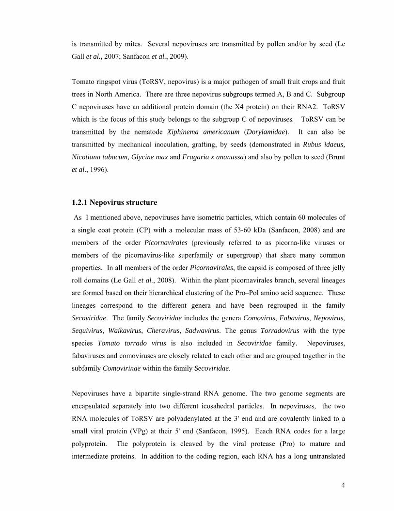
small CP) and the other which is composed of two β-barrels (CPL, large CP). In nepoviruses
the capsid protein is a single large protein that contains three covalently linked β-barrels
(Chandrasekar and Johnson, 1998). The three β-barrels are named C, B and A domain from
the N-terminus to the C- terminus. The B and C domains lay side by side around the three
fold axis of symmetry. The A domain lay around the five-fold axes (Fig.1.1 and Fig.1.2)
(Chandrasekar and Johnson, 1998).
In nepoviruses, cesium chloride (CsCL) equilibrium centrifugation of purified virus particles
typically reveals the presence of three types of viral particles. T-particles are empty virus
particles without an RNA component, and sediment at 50S. B-particles contain a single
molecule of RNA1 and sediment at 115-134S. M-particles sediment at 86-128S and contain
a single molecule of RNA2. In ToRSV, particles separate in two peaks on the gradients. The
top component (between 50-55S) consists of empty virus particles. The bottom component
(between 115-130S) is composed of two (B1 and B2) nucleoprotein components, which
contain either RNA1 or RNA2. Because RNA1 and RNA2 are very similar in size in
ToRSV, the two components are difficult to separate (Allen and Dias, 1977).
6
A
C B
Fig. 1.1 A simplified representation of the structure of TRSV. TRSV capsid protein and the
three domains in the capsid protein (C, B and A) are shown.
The structure of tobacco ringspot virus (TRSV, nepovirus of subgroup A) and blackcurrant
reversion virus (BRV, nepovirus of subgroup C) has been resolved (Chandrasekar and
Johnson, 1998; Seitsonen et al., 2008). Preparations of nepovirus particles purified from
indicator plants (C. quinoa or N. benthamiana) are composed of two forms of the unique coat
protein with identical N-termini (Lemmetty et al., 1997). The slightly smaller coat protein
bands can be separated from the full-length CP on sodium dodecyl sulfate polyacrylamide gel
(SDS–PAGE). In the case of BRV particles the two coat protein forms are 54 and 55 kDa in
size (Latvala et al., 1998; Lemmetty et al., 1997) with identical N-termini.
The atomic model of TRSV (Chandrasekar and Johnson, 1998) was fitted into the BRV
reconstruction and the difference map was calculated. The BRV homology model fitted the
cryo EM reconstruction of BRV well. One major difference of BRV and TRSV is a C-
terminal extension of 19 amino acids which is not present in TRSV. Based on the homology
model of BRV, it is predicted that the C-terminal 14 amino acid residues of BRV projects out
of the surface of the virus particle. The model also predicts that the N-terminal domain of the
BRV capsid protein extends into the capsid interior to interact with the RNA (Seitsonen et
al., 2008). The homology modelling of the BRV capsid was also used to identify potential
sequences that could be used for mite interaction. One of the most obvious regions is
predicted to be the C-terminal 19 residues extension. The C-terminal 19 residues are some of
the least conserved in the sequence alignment among other nepoviruses. Because the C-
terminus is extended from the virion surface, thus it is also a suitable region for antibody
generation. It has previously been shown that virus preparations containing the shorter form

7
of the capsid protein are infectious by mechanical inoculation, resulting in symptoms
identical to BRV symptoms (Lemmetty et al., 1997). The resulting progeny viruses contain
both protein forms (Latvala et al., 1998). The shorter form of CP is due to truncation of the
C-terminal extension. Thus, the C-terminus is not important for the infectivity of the virus
but may indeed serve as a determinant for mite transmission (Seitsonen et al., 2008). Also,
two forms of coat protein (59 and 57 kDa) were detected in tomato black ring virus (TBRV,
Nepovirus). The C-terminal extension of the larger coat protein is lost in vivo late in TBRV
infection and during virus purification. Proteins were extracted from infected plant extracts
(N. clevelandii) at various time lines and were immunoblotted with antiserum raised against
purified virus particles. In samples extracted from leaves 3 or 5 days post inoculation, the 59
kDa CP was predominant, but samples taken at later time contained the 57 kDa and 59 kDa
CP in approximately equal amounts (Demangeat et al., 1992). In vitro translation of TBRV
RNA yields only the 59 kDa CP. Partially purified virus contained both the 57 kDa and 59
kDa CP, while highly purified virus contained only the 57 kDa protein. The 59 and 57 kDa
protein shared the same N-terminus suggesting that similarly to BRV, the 57 kDa protein
arise by the loss of the C-terminal amino acids (Demangeat et al., 1992). It is known that
proteases can remove amino acids from the C-termini of the coat proteins of tobacco mosaic
virus (Harris and Knight, 1952), potato virus X (Koenig et al., 1978) and potyviruses (Shukla
et al., 1988). Presumably, as with these viruses, the C-terminal amino acids of the TBRV
coat protein protrude from the virus particle surface and can be removed without disrupting
the virion. This exposed detachable fragment may play a significant role in TBRV biology as
does the protruding N-terminal fragment of potyvirus coat proteins in their transmission by
aphids (Atreya et al., 1990). The C-terminal extensions have been reported for comoviruses,
strawberry latent ringspot virus (SLRSV), and tomato black ring virus (TBRV-S) (Le Gall et
al., 1995). Based on the multiple alignment of nepovirus coat protein, a C- terminal
extension of 54 amino acids was also predicted for ToRSV. There is no amino acid sequence
similarity between these C-terminal extensions (Latvala et al., 1998).

8
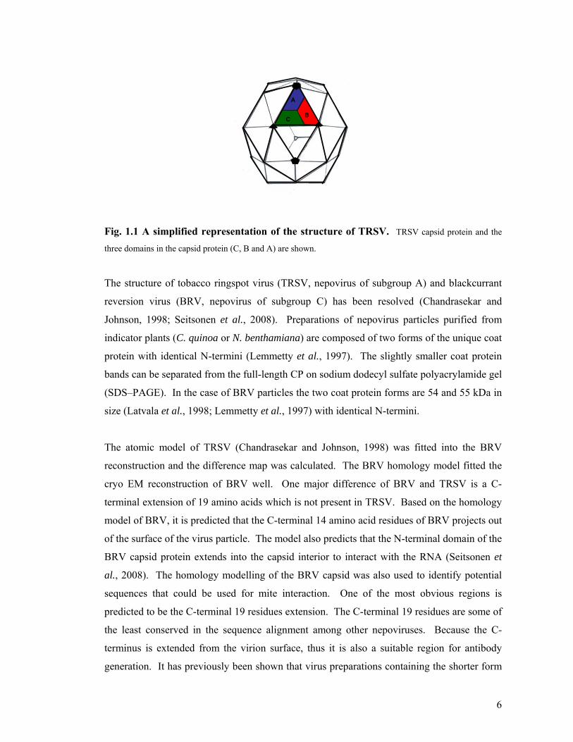
Fig. 1.2 Pseudo-T = 3 capsid of Picornavirales. The beta-barrel or jelly-roll structure is shown in the
left. The three jelly-rolls are assembled into individual capsomers as shown in the middle. Sixty of these
protomers are assembled to form the icosahedral capsid (right of the figure). The three jelly-roll are separated in
three CP subunits in most taxa, including Enterovirus, Rhinovirus, Cardiovirus, Aphthovirus, Cripavirus (VP1,
VP2 and VP3, corresponding to the CP1, CP2 and CP3 domains, respectively, as shown at the top of the figure).
The genera Comovirus, Fabavirus and Sadwavirus (S and L) encode two subunits (one large, L, and one small,
S) that contain two and one jelly rolls, respectively (shown in the middle). A single large CP subunit contains
all three jelly rolls in the genus Nepovirus (CP, shown at the bottom of the figure) (Picture courtesy of Olivier
Le Gall, reprinted with permission from Le Gall et al., 2008).
1.3 Replication cycle of plant viruses within the cell
Since the function of the X4 protein of ToRSV is unknown, I will briefly explain the
replication cycle of plant viruses and the function of the viral proteins in different steps of
this cycle with a focus on ToRSV and nepoviruses. All viruses share the same basic
replication cycle which involves the following steps: (1) virus entry into the cell, (2) viral
gene expression and genome replication and (3) capsid formation and virion assembly (Fig.
1.3).

9
Translation
Proteolytic processing
Membrane associationReplication complex assembly
EncapsidationMembrane vesicles
Cell to cell movement
(tubular structure)
Nucleus
Nematode vector penetrating the cell
Fig. 1. 3 Schematic representation of the replication cycle of nepoviruses within the cell. The virus (depicted with the yellow hexagons) is released by the nematode vector into the plant cell, the viral
RNA (shown by the thick black line) is uncoated and the (+) strand RNA genome is translated to polyprotein
using the host translational machinery (ribosomes are shown in red). The polyprotein (blue rectangle) is post
and co-translationally cleaved by viral encoded proteases (scissors). The next step is assembly of the viral
replication complex which is associated with the ER membrane (shown in purple) followed by viral replication.
The final stage is capsid formation and virion assembly, the progeny viral RNAs are packaged and translocated
from cell to cell.
1.3.1 Translation of viral proteins
Most plant viruses, including nepoviruses, have a positive-sense single-stranded [(+)-strand]
RNA genome. The first steps of the replication cycle following cell entry and uncoating of
the virus particle are translation and replication of the viral genome. A single strand RNA
genome with a positive polarity means that the genomic RNA can act as a messenger RNA
(mRNA) immediately as it enters the cell.
RNA viruses code for their own replication protein, the RNA–dependent RNA polymerase
(RdRp). Viruses do not encode translation factors or ribosomes. Therefore they must use the
host translation machinery to translate their protein. The highjacking of translation factors by
viruses often results in host translation shutdown (Sarnow, 2003). Cellular mRNAs contain
a cap structure or methyl-guanidine triphosphate (m7GTP) at their 5' end and a poly A tail at

10
their 3' end. Both elements are essential to recruit host translation factors and form the
translation complex (Sonenberg and Dever, 2003). In nepoviruses and other picorna-like
viruses, a small protein, termed genome-linked viral protein (VPg), is covalently linked to the
5' end of the RNA, therefore replacing the cap structure. The 5' untranslated region (UTR) in
picornaviruses, potyviruses and possibly comoviruses contain internal ribosome entry site
(IRES) which are composed of stem loop structures and are necessary for recruiting the host
translation factors and allow the ribosomes to initiate translation effectively on these regions
in the absence of the cap structure (Gallie, 2001; Hellen and Sarnow, 2001; Verver et al.,
1991). In Blackcurrant reversion virus (nepovirus), sequences located at the 5' and 3'
untranslated region of both genomic RNA facilitate the translation of these RNAs using
internal ribosome entry site (Karetnikov and Lehto, 2008).
1.3.2 Polyprotein processing
This approach allows the synthesis of multiple protein products from a single RNA. The
large polyprotein is often not detected in infected cells since it is processed as soon as the
protease coding sequence has been translated (Knipe et al., 2001). In picornaviruses, a
single polyprotein is first cleaved at three sites to produce P1, P2 and P3 intermediate
polyprotein. P1 contain structural proteins, while P2 and P3 contain RNA replication
proteins. The polyprotein is cleaved into smaller functional proteins by viral proteases (Knipe
et al., 2001).
Nepoviruses encode a single proteinase which is related to the 3CPro of picornaviruses. The
catalytic triad contains histidine, aspartic acid and cysteine (Sanfacon, 2008).
The proteinase also has a substrate-binding pocket that determines its cleavage site
specificity. The subgroup C nepovirus proteinase recognizes a glutamine, asparagine or
aspartate at -1 position of its cleavage site. In contrast to subgroup C, the proteinase for the
subgroup A and B nepovirus recognise different cleavage sites which have lysine, cysteine,
arginine or glycine at the -1 position. The RNA1 encoded polyprotein is cleaved
intramolecularly (cis-cleavage) and the RNA2 encoded polyprotein is cleaved in trans by the
viral protease. ToRSV P1 polyprotein contains the domain for the replication proteins which
are RNA–dependent RNA polymerase, the proteinase, genome linked viral protein (VPg),
putative helicase (also termed NTB) which has a putative nucleoside triphosphate binding

11
activity and X1 and X2 for which the functions are unknown (Rott et al., 1995; Wang and
Sanfacon, 2000b). RNA2 encodes the structural proteins which include the coat protein (CP)
and movement protein (MP), as well as the X4 and X3 proteins of unknown function (Carrier
et al., 2001; Hans and Sanfacon, 1995). The cleavage sites within the ToRSV polyproteins
consist of Q/G (glutamine/glycine) or Q/S (glutamine/serine) dipeptides. Site-directed
mutagenesis of two ToRSV cleavage sites showed that efficient processing by the viral
protease requires the presence of a Q at position -1 and a S or G at position +1 of the viral
cleavage site (Carrier et al., 1999) (Fig. 1.4).
(A)VPg X1 X2 NTB Pro Pol
VPg
(A)
VPg X3 X4 MP CP
Fig. 1. 4 Genome organisation of ToRSV. The polyprotein of nepoviruses are shown (P1 and P2). P1
is cleaved by the viral protease (Pro) at 5 sites to release 6 mature proteins and several intermediate precursors.
P2 is cleaved at 3 sites to release 4 protein domain and possible precursors.
1.3.3 Assembly of the viral replication complex
All RNA viruses whether they infect mammalian, insect or plant cells induce the proliferation
of membrane vesicles often but not always in the perinuclear area. Electron microscopy
(EM) observations made more than 40 years ago described clusters of heterogeneously sized
vesicles of 70–400 nm in diameter that were present in the perinuclear regions of poliovirus
infected cells (Dales et al., 1965). Other examples of (+)-RNA viruses that are well known
for replicating their genomes on intracellular membranes, include members of the
Picornaviridae, Flaviviridae, Togaviridae, Coronaviridae and Arteriviridae families, the
insect viruses of the Nodaviridae family and many plant viruses, such as Tobacco mosaic
virus. As mentioned above, one of the best-documented examples of a virus that induces

12
membrane alterations is the human pathogen poliovirus, a member of the Picornaviridae
family. Poliovirus-induced vesicle clusters are probably derived from the endoplasmic
reticulum (ER). Other members of the Picornaviridae family have also been shown to
replicate their RNA genomes on modified membranes that accumulate in the cytosol of
infected cells (Wessels et al., 2006). (+)RNA viruses that belong to the Flaviviridae family
and the Nidovirales order typically induce the formation of double-membrane vesicles that
are spherical membrane structures 50–400 nm in diameter and composed of two closely
apposed membrane bilayers (Mackenzie, 2005; Miller and Krijnse-Locker, 2008).
Viruses probably induce proliferation or reorganization of cellular membranes and vesicles in
order to increase the surface area for RNA replication. The viral replication complex is
associated with the intracellular membranes and includes viral and host proteins with the
template RNA. The formation of this compartment may provide a protecting environment
against RNA degradation (Sanfacon, 2005). In picornaviruses, RNA replication complex are
associated with clusters of smooth vesicles which accumulate in the cytoplasm (Bienz et al.,
1992). Cowpea mosaic virus (Comovirus) infection in plants induces the production of
vesicles and massive proliferation of the endoplasmic reticulum (ER) membranes (Carette et
al., 2000). Viral and host protein are brought to the replication complex via protein-
membrane and protein-protein interactions. Some replication proteins are brought to the
replication complex as polyprotein precursors which include the domains for the viral
membrane anchor (Bedard and Semler, 2004).
1.3.3.1 Replication and translation of nepoviruses
As for other related viruses such as picornaviruses and comoviruses, nepovirus infection
induces membrane proliferation and morphological changes in the ER membrane (Han and
Sanfacon, 2003). Replication proteins and replication intermediates of comoviruses and
nepoviruses are found in association with the ER derived membranous vesicles (Carette et
al., 2000; Han and Sanfacon, 2003; Ritzenthaler et al., 2002; Schaad et al., 1997). Viral
proteins act as membrane anchors for the replication complex. The membrane anchors
associate with the ER and other viral and host proteins that are brought to the replication
complex through protein–protein interaction with the membrane anchor protein. In ToRSV,
the putative nucleoside triphosphate binding protein (NTB) has a stretch of hydrophobic
residues at its C-terminal end which is proposed to anchor the replication complex on the
13
membrane (Han and Sanfaçon 2003). NTB-VPg associates with microsomal membranes in
vitro and the VPg domain is translocated in the lumen of the membranes (Wang et al., 2004).
In picornaviruses the VPg acts as a primer for viral replication. Since replication takes place
in the cytoplasmic side, the ToRSV NTB-VPg is unlikely active in the replication of the viral
RNA, suggesting that another form of the VPg may be the active primer for replication
(Chisholm et al., 2007; Wang, 2004). The N-terminus of NTB can also be translocated in the
lumen at least in vitro. This translocation is dependent on the presence of putative
amphipathic helix, suggesting that at least two distinct elements may play a key role in the
insertion of NTB-VPg in the membranes: a C-terminal transmembrane helix and an N-
terminal amphipathic helix (Zhang et al., 2005).
In the RNA1 ToRSV, X2 protein has a highly hydrophobic protein domain located upstream
of the NTB domain in the RNA1-encoded polyprotein. X2 has conserved sequence motifs
with the comovirus 32 kDa protein which is an ER-targeted protein implicated in the viral
replication complex assembly. Based on mutagenesis studies and confocal microscopy it was
suggested that X2 is targeted to the ER membranes. This raises the possibility that it might
act as a second membrane anchor for the viral replication complexes (Zhang and Sanfacon,
2006). Also a subpopulation of VPg-Pro-Pol which contains the truncated RNA-dependent
RNA polymerase (Pol), the proteinase (Pro) and the VPg was peripherally associated with the
ER-derived membranes active in viral replication (Chisholm et al., 2007). This suggests that
the peripheral association of the ToRSV VPg-Pro-Pol polyprotein with the ER-derived
membranes may be mediated by its interaction with one or several viral membrane proteins
(X2 or NTB-VPg) (Han and Sanfacon, 2003; Zhang and Sanfacon, 2006; Zhang et al., 2005).
1.3.3.2 Replication of RNA2 in nepoviruses
Using grapevine fanleaf virus, it was shown previously that RNA1 replicates independently
of RNA2. RNA2 requires the RNA1 replication machinery for its own replication. The
RNA2 contains three protein domains named 2A, 2B (MP) and the 2C (CP). The lack of
replication of GFLV RNA2 mutant, in which the 2A-coding sequence was deleted, strongly
indicates that 2A or its coding sequence is essential for RNA2 replication. However, protein
2A alone is not enough and additional RNA or protein sequences downstream of 2A are
essential (a minimum length of RNA2 exceeding the 2A coding sequence) for RNA2

14
replication. It was suggested that the requirement for downstream stabilizing sequences is
related to the fact that 2A is an unstable protein at least in vitro (Margis et al., 1993). Also in
cowpea mosaic virus (CPMV), the B-RNA contains the replication proteins and the M-RNA
contains the movement protein and the coat protein. It was shown that the N-terminal
domain of the 58 kDa protein encoded by the M-RNA is required for the replication of M-
RNA (Van Bokhoven et al., 1993). The RNA2-encoded 58 kDa protein of CPMV contains
both the 2A and movement protein domains (Van Bokhoven et al., 1993). It was suggested
that the CPMV 58 kDa protein could be involved in binding RNA2 to form a
ribonucleoprotein complex that would be recognized, on its own or together with unknown
cellular factors, by the RNA1-encoded replicative machinery. In nepoviruses, it was
suggested that the 2A protein is not active in replication as a mature protein but rather as a
precursor form such as the 2AB or polyprotein P2. Thus the 2AB precursor protein in GFLV
would be the functional equivalent of the 58 kDa protein in CPMV (Gaire et al., 1999). The
following model is suggested for RNA2 replication in GFLV. After viral inoculation, RNA1
and RNA2 which were encapsidated in separate virus particles are released in the cytoplasm.
Both RNAs are then translated into the P1 and P2 polyproteins. Translation of RNA1 and
self-processing of the polyprotein P1 provide virus-encoded replication proteins that most
likely co-assemble with host factors and membranes to form the viral replication complex,
first as punctate structures and afterwards as juxtanuclear aggregated structures. RNA2
depends on RNA1 for its replication and polyprotein processing. Therefore, RNA2 needs to
integrate within the replication complex initiated by RNA1-encoded proteins. Since 2A is
required for RNA2 replication, it is suggested that the 2A domain within the nascent
polyprotein either directs RNA2 to the replication site or interacts with the same cellular
structure as RNA1-derived proteins to the juxtanuclear location. Another possibility is that
the P2-polyprotein bound to RNA2 would be recruited by the replication complex (Gaire et
al., 1999).
1.4 Movement of plant viruses
In animals, the spread of viral infection from cell to cell is by means of endocytosis or by
fusion of the viral envelope with plasma membrane. A fundamental difference between plant
cell and animal cells is that each plant cell is surrounded by a rigid cell wall. Plant viruses
must overcome the barrier of the plant cell wall through cytoplasmic connections between
adjacent cells, the plasmodesmata. Plant viruses encode proteins that assist their movement

15
from cell to cell. These movement proteins interact with the plasmodesmata and modify the
plasmodesmal structure and function. Within the cell, the viral genome must be moved from
the site of replication to the plasmodesmata. For viruses replicating in the cytoplasm, this
often involves association with elements of the cytoskeleton. Cell to cell or short distance
movement is defined as the movement of the virus from primarily infected cells (epidermal
or mesophyll cells) to vascular bundle. Long distance transport of the virus occurs through
vascular tissue, usually the phloem sieve tubes.
1.4.1 Plasmodesmata
Plasmodesmata are tubular extension of plasma membrane (40-50 nm in diameter) that
traverse the cell wall and connect the cytoplasm of adjacent cells. The plasmodesmata are
specialized channels that allow the intercellular movement of water, sugar, nutrients, and
other molecules. They have a complex internal structure that regulates macromolecular
traffic from cell to cell. Each plasmodesmata contains a long narrow tubule of the endoplasmic reticulum (ER) called desmotubule which is continuous with the ER of the
adjacent cells. The cytoplasmic sleeve is a narrow space between the desmotubule and the
plasma membrane. The trafficking of the macromolecules via plasmodesmata occurs in the
cytoplasmic sleeve (Aaziz et al., 2001). In the cytoplasmic sleeve both the desmotubule and
the plasma membrane globular proteins arrange in helical rows (Fig. 1.5). The size exclusion
limit (SEL) of plasmodesmata is about 2.0nm. The SEL is not fixed and can be regulated.
Plant virus particles or viral genome are too large to pass through the plasmodesmata.
However, it has been suggested that many viral movement proteins increase the
plasmodesmata SEL The mechanism for regulating the SEL is poorly understood (Lincoln
Taiz, 2006).

16
Cell wall
Endoplasmic reticulum
Cell1
Cell 2
Desmotubule
Plasma membrane
Cytoplasmicsleeve
Fig. 1.5 Schematic representation of a plasmodesmata transversal section. The
plasmodesmata traverses the cell wall. The endoplasmic reticulum is present within the plasmodesmata and
traverses the cell wall through the plasmodesmata. Globular protein particles (pink) cover the inside of the
plasmodesmata.
1.4.2 Types of movement from cell to cell by plant viruses
Two types of cell to cell movement of plant viruses with a (+) strand RNA genome have been
characterized: movement of viral RNA as a nucleoprotein complex and movement of virus-
like particles. Since nepoviruses move from cell to cell as a virus–like particle, I will
explain this type of virus movement in more details.
1.4.2.1 Movement of virus-like particles
Plasmodesmata are modified by insertion of tubular structures that allows the transport of
virus-like particles through them (tubule-guided virion movement). Such tubular structures
have been described for viruses of different genera including Caulimovirus, Nepovirus,
Bromovirus and Tospovirus.
1.4.2.1.1 Cell-to-cell movement of comoviruses, nepoviruses and related viruses
In CPMV, cell-to-cell movement is characterized by transport of mature virions through
tubules that are assembled inside the plasmodesmal pore. The viral MP is a structural
component of the tubular structures (Pouwels et al., 2004). Similar tubular structures are
17
formed at the surface of CPMV infected protoplasts. In protoplasts and in plant tissue; virus-
like particles appear in a single and continuous row within the tubules. Tubule assembly does
not depend on the presence of virions or capsid proteins, as expression of MP alone in
protoplasts also leads to the formation of (empty) tubules (Angell et al., 1996; Carvalho et al.,
2003; Kasteel et al., 1993; Van Lent, 1991; Wellink, 1989). The CPMV MP binds to intact
virions and to the large CP subunit (Carvalho et al., 2003). The various steps leading to the
formation of CPMV induced tubular structures have been studied. Dimeric or multimeric
MP are first targeted to the plasma membrane. At the plasma membrane the MP accumulates
in peripheral punctuate spots from which tubule formation begins. The C-terminus of MP
interacts with the virus particle in the tubule and the N-terminal region of the MP is involved
in tubule formation (Pouwels et al., 2003).
As with comoviruses, nepovirus-infected cells are characterized by the formation of tubular
structures containing virus-like particles and traversing the cell wall. The movement protein
of two nepoviruses, GFLV and ToRSV, was shown to be a structural component of the
tubular structures (Ritzenthaler et al., 1995b; Wieczorek and Sanfacon, 1993). Tubular
structures were formed both in plants and protoplasts infected with GFLV (Ritzenthaler et
al., 1995a). The MP of GFLV may use the secretory pathway and the cytoskeleton for
intracellular targeting and tubule assembly (Laporte et al., 2003; Taliansky et al., 2008). In
ToRSV infected plants, MP is associated with the tubular structure containing virus particles.
The tubular structures are often found protruding through the cell wall in to the cytoplasm of
adjacent cells (Wieczorek and Sanfacon, 1993). The CP and the MP have been detected in
infected plants and protoplasts (Sanfacon et al., 1995; Wieczorek and Sanfacon, 1993). In
infected ToRSV protoplasts the MP was less stable than the CP (Sanfacon et al., 1995).
1.5 Virus transmission
Most plant viruses depend on a vector for transmission from plant to plant. Insects are the
most common vectors (e.g. aphids), although other vectors, such as nematodes and fungi, are
also important. The virus–vector interaction is very specific. Nepoviruses are transmitted by
nematode therefore in this section I will focus on the transmission of plant viruses by
nematodes.
18
1.5.1 Nematode transmission
There are two genera of plant viruses which are transmitted by nematodes: (1) Tobraviruses
which are transmitted by species of the genera Trichodorus and Paratrichodorus and (2)
Nepoviruses which are transmitted by species of the genus Xiphinema (Hull, 2002).
1.5.1.1 Tobraviruses
Pea early browning virus is a rod shape virus with bipartite (+)-strand RNA genome.
Deletion of a gene encoding a 29 kDa protein (2b protein) abolished its nematode
transmission without affecting the virus particle formation (MacFarlane, 1996). Comparison
of the highly transmissible isolate and a poorly transmissible isolate showed two amino acid
substitutions in the 2b protein (Vellios, 2002). In another tobravirus, Tobacco rattle virus, a
strong interaction was detected between a 40 kDa protein and the CP. The 40 kDa protein is
required for transmission of the virus by the nematode vector. This suggested that the 40
kDa protein of Tobacco rattle virus could act as a helper protein in nematode transmission
(Visser and Bol, 1999).
1.5.1.2 Nepoviruses
ToRSV and Tobacco ringspot virus (another nepovirus of subgroup A) infect a wide range of
fruit crops and woody plants in North America and are transmitted by X. americanum sensu
strico Cobb, an ectoparasitic nematode. This nematode also transmits two other nepoviruses:
Cherry rasp leaf virus and Peach rosette mosaic virus. For nematode transmission to occur,
the virus must first dissociate from its retention site, a specific area in the nematode food
canal. The dissociated viruses are injected to the plant root during the nematode feeding.
Transmission of nepovirus by its nematode species is specific. It has been suggested that
during nematode feeding the change of pH induced by the nematode salvation glands,
releases the virus from its retention site (Wang, 2002). Based on electron microscopy and
immunofluorescent labelling of the nematode X. americanum, the retention sites for TRSV
are localized at the inner lining of the stylet extension and the esophageal lumen (Wang,
1998; Wang and Gergerich, 1998). In contrast, ToRSV particles are retained in the triradiate
lumen of the esophageal bulb. The different retention sites suggest significant differences in
the mechanism of virus release from the nematode vector. Immunofluorescent labelling with
ToRSV CP antibodies did not detect the virus in the vector nematode. However, TRSV CP

19
antibodies readily labelled TRSV within the nematode vector. This suggests that ToRSV
virions might be coated with other components from the nematode or from the host plant
blocking the physical binding between virus particle and the antibody. Based on
transmission assays, ToRSV is transmitted more efficiently (~100%) than TRSV (75%),
although few virus particles were observed in the nematode vector. This suggests that ToRSV
is more readily released from the retention site while TRSV might remain strongly attached
to the nematode receptor (Wang, 2002). The determinants for the specificity of the retention
site and the efficiency of transmission of these viruses remain to be determined.
Grapevine fanleaf virus is transmitted from plant to plant by Xiphinema index. The viral
determinants responsible for nematode transmission were studied by engineering chimeric
constructs in which the coding region of various proteins of GFLV was replaced with their
counterparts from Arabis mosaic virus (ArMV, Nepovirus). All hybrid viruses with the CP
of GFLV were transmitted by X. index. Other hybrid viruses with the CP of ArMV were not
transmitted by this nematode. These results indicated that the CP of GFLV is the sole viral
determinants for the specificity of the nematode vector (Andret-Link, 2004).
1.6 ToRSV isolates
ToRSV is a major pathogen of small fruit crops and fruit trees in North America, it also
occurs in Australia, Bulgaria, Canada, Chile, China, Cyprus, Germany, Italy, Japan,
Korea, New Zealand , Peru, Puerto Rico, Turkey, the United States of America, the USSR
(former), Yugoslavia. The susceptible host species are found in the Family Amaranthaceae,
Caryophyllaceae, Chenopodiaceae, Cucurbitaceae, Geraniaceae, Leguminosae,
Papilionoideae, Rosaceae, Solanaceae and Umbelliferae (Brunt et al., 1996).
The complete nucleotide sequence of isolate Rasp2, a raspberry isolate from lower mainland,
B.C., Canada has been determined (Rott et al., 1995; Rott et al., 1991b). The nucleotide and
amino acid sequence for the VPg, Pro, Pol and CP of other ToRSV isolates were analyzed
and compared with the published sequence (Rasp2). These isolates were Rasp1 (originally
from raspberry plants in Washington state), GYV, grape yellow vein isolate (originally from
infected grapevines in California), PYB-1, peach yellow bud-1 (originally from infected
peach orchards in California) and T392 isolate from an unknown origin. The VPg sequence

20
and conserved amino acid motifs within the Pro, Pol and the CP domain were identical in all
isolates (Wang and Sanfacon, 2000a).
In Chapter 2, I will explain the insertion of large amino acid repeats and point mutations
contribute to a high degree of sequence diversity in the X4 protein of ToRSV (Jafarpour and
Sanfaçon, 2009). Therefore, I will briefly explain below the general principles of virus
evolution and possible sources of variation in the viral genome.
1.6.1 Virus evolution, diversity and sources of variation
1.6.1.1 Mutation, insertion and deletion
In theory, RNA viruses have a large population of diversity since they have an error-prone
replication and a short generation time. The genetically diverse populations of RNA viruses
are referred to as quasispecies (Roossinck, 1997). Maintaining diverse quasispecies is
advantageous because when the virus is exposed to a new environment or a new selective
regiment, the variant in the population that is more fit to the new environment can survive
(Roossinck, 2003; Schneider and Roossinck, 2001). In nature, vector transmission can
potentially transmit viruses to a variety of hosts and host adaptability is essential for the
survival of plant viruses. Deletion and/or insertion may include single or several nucleotides.
Some mutations will only have an effect in the nucleotide sequence and will therefore be
silent at the amino acid level. But some mutations can result in amino acid change and
frame-shift mutations that cause global changes.
1.6.1.2 Recombination
Recombination is a process in which segments of genes are swapped between different
genetic variant or different strains during viral replication. It is a general phenomenon and it
plays an essential role in the genetic variability and evolution of plant viruses (Roossinck,
2003; Schneider and Roossinck, 2001). In tobraviruses, sequencing of the RNA2 from more
than 12 tobravirus isolates showed extensive diversity in the nucleotide sequence which is the
result of recombination between nonhomologus regions of RNA1 and RNA2. In 8 of the 12
isolates the 3'-terminal sequence is replaced with those of RNA1 sequence (Vassilakos et al.,
2001).

21
1.6.1.2.1 Protein domain repeats
Repetitive nucleotide sequences are frequently found in eukaryotic genomes. These regions
are often hypermutable, rapidly gaining and losing repeats during the course of evolution.
They are usually found in non-coding genomic regions but they can also be found within the
genes. The repeats might be as short as three amino acid residues tandem repeats to
repetition of homologous domains of 100 amino acid or more residues (Marcotte, 1998). It
has been suggested that they arise through internal tandem duplication and recombination
within a single gene. However the exact mechanism behind this phenomenon is not fully
understood (Andrade, 2001; Bjorklund et al., 2006). Domain repeats are often involved in
interaction with proteins or other ligands such as DNA or RNA. They are found in proteins
with highly diverse functions (Bjorklund et al., 2006).
1.6.1.3 Serial passages of viruses, a source of creating sequence diversity
High level of genetic diversity and rapid evolution of viral RNA genomes are well
documented (Kurath and Dodds, 1995). The majority of these changes are seen during serial
passaging of viruses. Few studies have characterized the rate and nature of ongoing genetic
changes over time under controlled experimental conditions in plant hosts by monitoring
multiple serial passage lines of a virus populations for changes in their consensus sequences
(Kurath and Dodds, 1995). Evidence for genetic variation of plant viruses was reported as
early as 1926 (Agudelo-Romero et al., 2008). It was derived from the possibility of isolating
different symptom variants from the same virus source. These variants were first obtained by
isolation of symptom mutants from areas that showed atypical symptoms in systemically
infected plants (McKinney, 1935). Also, it was soon observed that serial passaging under
different conditions could alter viral properties. Most often these experiments involved a host
shift (Garcia-Arenal et al., 2001). Viruses adapt to their hosts by evading their defense
mechanisms and taking over the host cellular metabolism for their own benefit. Changing
the cell metabolism as well as side-effects of antiviral responses contribute to symptoms
development and virulence. Sometimes, a virus may move from its usual host species into a
new one, or might fail to successfully infect and further transmit the next host. During serial
passaging of the virus by mechanical inoculation, some functions such as vector transmission
may be completely lost. In cucumber necrosis virus defective interfering particles are
generated after at least eight serial passages (Rochon, 2008; Rochon, 1991). Defective
interfering particles are virus particles that encapsidate defective genomes that are grossly

22
altered genetically, usually by significant deletion of essential function. Defective viral
RNAs keep their replication origins and packaging signals allowing their encapsidation
(Knipe et al., 2001). In potyviruses, most characterized virus proteins are multifunctional,
and under different selective constraints may evolve differently corresponding to various
functions. For instance, the helper component for the transmission of the potyviruses also
has a role in the proteolytic processing of the genome translation product and in systemic
movement. The fact that its function in aphid transmission is easily lost upon mechanical
passage suggests that trade offs occur for the optimization of its other functions (reviewed by
(Garcia-Arenal et al., 2001). Below are a few documented examples of virus evolution in
response to serial passaging in a new host.
In Tobacco etch virus (Potyvirus) a virus infected plant was allowed to infect native hosts.
After 17 serial passages, the viral genome has evolved only five amino acid changes. An
amino acid substitution in the viral VPg protein was responsible for the appearance of
symptoms, while one substitution in the viral P3 protein contributed to increase in the
symptom severity (Agudelo-Romero et al., 2008).
The coat protein (CP) of Hibiscus chlorotic ringspot virus (HCRSV) was identified as a
strong RNA-silencing suppressor. CP mutants resulting from serial passage of HCRSV in its
local lesion host showed a significantly reduced silencing suppression function, indicating
that host-induced mutations lead to avirulence of HCRSV in plant that correlates with its
reduced ability to suppress post-transcriptional gene silencing (Meng et al., 2006).
Plum pox virus (PPV), populations from peaches are able to adapt consistently to herbaceous
hosts. They are characterized by reduction in time to symptom development, increase in
inoculation efficiency and increase in viral titres. Two isolates of PPV from peaches were
inoculated and passaged ten times in peas. Sequence analyses of the PPV population lines
inoculated (pea-adapted strains of PPV), identified a specific mutation occurring consistently
in the nuclear inclusion bodies gene when compared with the same PPV isolates passaged in
parallel in peach. The mutation allowed PPV to replicate up to 20 times faster in its new host
(Wallis et al., 2007).
Two isolates of plum pox virus, PPV-D and PPV-R were different in their host range. Some
isolates lost their ability to infect their natural prunus hosts after repeated passages in

23
herbaceous hosts. The changes were introduced in PPV-R isolate after long term replication
in N. clevelandii. Based on pathogenicity experiments of the two PPV isolates, PPV-R and
PPV-D, the determinants for host specificity were located at the N-terminal region of the CP
(Salvador et al., 2008).
In tobacco rattle virus (TRV, a tobravirus) the genome is very flexible and based on selection
pressure; nematode transmissibility and encapsidation, which are encoded by RNA2, were
abolished from the genome of the PPK20 isolate when this isolate was serially passaged in
tobacco by mechanical inoculation (Hernandez et al., 1996).
1.7 Virion associated proteins
I have briefly discussed virus structure above (section 1.1). Although the main component
of the virion is the coat protein(s) (CP), other minor virus-encoded protein components have
also been found in association with plant virus particles. In Chapter 3, I will discuss the
possibility that the X4 protein of ToRSV may be a virion associated protein (VAP).
Therefore in this section, I will explain what virion associated proteins are and bring some
examples of well studied VAP in plant viruses.
Virion associated protein (VAP) are proteins associated (directly or indirectly) with the
capsid protein in the virion shell. Because they are exposed on the virus particle, they are
readily available for interaction with host or vector components. VAP plays an important
role in the virus replication cycle, including cell-to-cell or long distance movement in the
plant, suppression of RNA silencing, stability of virus particles, regulation of the translation
of the viral genome and vector transmission.
1.7.1 CaMV P3 virion associated protein
In cauliflower mosaic virus (Caulimoviridae), the virion-associated protein (P3 protein) is a
small 15 kDa protein with a bipolar functional structure. The P3 protein is important for
aphid transmission of the virus and acts as a bridge between the CP and the aphid
transmission factor (P2). Also it has been suggested that the virion bound P3, participates in
the cell to cell or long distance movement of the virus inside the host (Plisson et al., 2003;
Plisson et al., 2005). P3 interacts with the virus particle through its C-terminal region. Its N-

24
terminal region has a high α-helical content and contains a coiled-coil motif. The coiled-coil
domain interacts with the aphid transmission factor (P2 protein). The N-terminal region of
P2 interacts with the aphid stylet and its C-terminal domain interacts with the virion
associated protein (P3). The MP oligomerizes as a trimer through a C-terminally located
coiled-coil domain. The coiled-coil domain of MP also interacts with the coiled-coil domain
of the VAP located at the virion surface. The MP-VAP interaction is necessary for the spread
of virus in the plant but dispensable for replication of the virus (Stavolone et al., 2005).
1.7.2 HC-Pro Helper component proteinase
The potyvirus genome encodes a single polyprotein which is processed by three viral
proteinases, P1, HC-Pro (Helper component proteinase) and NIa (nuclear inclusion), to
release all viral proteins required for virus infection. P1 and HC-Pro are responsible for two
distinct cleavages within the N-terminal region of the polyprotein. NIa carries out the
remaining cleavages at the C-terminal two-third of the poly protein (Carrington et al., 1993).
HC-Pro is a virion-associated protein and a multifunctional protein (Kasschau and
Carrington, 2001). It was first recognized as a helper factor for vector transmission
(Thornbury, 1985). Its proteinase activity was later discovered (Oh and Carrington, 1989).
The HC-Pro protein contains three regions: an N-terminal region essential for vector
transmission, a C-terminal region which contains the proteinase activity and also overlaps
with the cell-to-cell movement function and a central region implicated in all other functions
(Plisson et al., 2003). The central region of HC-Pro is responsible for genome amplification
(IGN motif), synergism with other viruses, systemic movement within the host plant (CC/SC
motif) and silencing suppression (Kasschau et al., 1997) (Fig. 1.6).

25
Vector binding RNA binding Capsid binding
Transmission Movement
Amplification/PTGS Protease
N-terminus C-terminusCentral region
RNA bindingA B
KITC FRNK IGN CC/SC
A
B
C
Fig. 1.6 Functional regions of the helper component proteinase (HC-Pro). (A) HC-Pro can
be divided into three regions. (B) Location of the putative vector-binding, RNA-binding, and viral capsid-
binding regions in the HC-Pro. (C) Biological functions of different regions within the HC-Pro and location of
its conserved motifs.
Potato virus A (PVA, another potyvirus) produces pinwheel shape cytoplasmic inclusion
bodies, which are made of the inclusion protein (Cl). The Cl of several potyviruses anchors
to the cell wall or to the close proximity of the plasmodesmata. The ability to form a pin
wheel structure and to assist the cell to cell movement of the virus depends on the ability of
its self interaction (Lopez, 2001). The Cl of PVA is associated with a sub-population of the
virus particles, possibly by interaction with the virion-bound HC-Pro. Both HC-Pro and CI
are localized at the protruding tip at the end of the virus particle (Gabrenaite-Verkhovskaya,
2008).
1.7.3 Tobravirus 2b nematode transmission helper protein
As mentioned above, tobraviruses such as tobacco rattle virus (TRV) and pea early browning
virus (PEBV) are transmitted by trichodorid vector nematodes. The 2b protein is a non
structural protein encoded by RNA2. The 2b protein interacts with the CP, forming a bridge
between the virus particle and the internal surface of the nematode vector feeding tube.
Immunogold labelling of thin sections of PEBV infected plants probed with 2b specific
antibodies showed that 2b is localized with the virus particle (Vellios et al., 2002). The

26
interacting domains were investigated by yeast two-hybrid experiments. A central domain of
the CP was identified that promote CP: CP interaction. Deletion of a conserved central
coiled-coil domain of the 2b protein prevented its interaction with the CP, while deletion of
N-and C-terminal domains of the 2b protein greatly enhanced its interaction with the CP. A
C-terminal domain of the CP was shown to be important for interaction with the 2b protein
(Holeva and Macfarlane, 2006).
1.7.4 Closterovirus virion associated proteins
Closteroviruses (family Closteroviridae) are long filamentous virus particle with a long body
and a short tail. In Beet yellows virus, the virion is composed of two principal components,
the minor CP assembles at one end of the virion and the major CP forms the long virion
body. In addition there are three VAPs presented at the virion tail: the viral Hsp70
homologue (Hsp70h), a 64 kDa protein and a 20 kDa protein (Peremyslov et al., 2004).
Comparative genomic analyses revealed that Hsp70h and p64-like proteins are conserved in
all known viruses of the family Closteroviridae (Dolja et al., 2006). Hsp70h and p64 are
each required for efficient assembly of the closterovirus virion tail (Alzhanova et al., 2007;
Satyanarayana et al., 2000). The tail is narrower than the body (8 nm vs. 12 nm). Thus, the
virions exhibit a peculiar three-segment architecture with the pointed tip segment likely
formed by p20 (Peremyslov et al., 2004). The p20 is required for long-distance transport of
the virus via the phloem. In Closteroviruses, particle assembly is a prerequisite for Beet
yellows virus transport of the large RNA genome (Alzhanova, 2007).
Another member of the family Closteroviridae, lettuce infection yellow virus (LIYV) is
transmitted from plant to plant by whiteflies (Bemesia tabaci). Virion preparation of LIYV
showed that HSP70 and p59 (a protein of unknown function) are associated with the virus
particle. LIYV minor CP antibody neutralizes LIYV transmission by B. tabaci. This
suggests that the minor CP may be important for virus transmission (Tian, 1999).
1.7.5 Potexvirus virion associated protein
In potexviruses such as PVX, the triple-gene block proteins (TGBp1, TGBp2 and TGBp3)
together with the CP are involved in cell to cell movement of PVX. The 25 kDa TGBp1 is
multifunctional virion associated protein (VAP) protein involved in many activities including
RNA helicase activity and RNA binding activity. TGBp1 has been suggested to increase the

27
plasmodesmal size exclusion limit and is also a suppressor of RNA silencing (Verchot-
Lubicz, 2005). It has been suggested that the binding of the 25-kDa MP to the viral RNA
molecule mediates the translation of the viral RNA. The 25 kDa protein binds selectively to
one end of the virus particle. Binding of the TGBp1 to the end of the virus particle causes
conformational changes in the terminal subunits and alters the stability of the 5'-terminus of
the RNA. It has been suggested that binding of TGBp1 renders the viral RNA more
accessible to the ribosome and that it triggers co-translation disassembly of PVX RNA
(Atabekov et al., 2000; Karpova et al., 2006).
1.8 RNA silencing and viral suppressors of silencing and silencing activity
in Nepoviruses
In Chapter 4 I will explain the possible role of the X4 protein as a suppressor of silencing.
Therefore, in the section below, I will briefly explain the mechanism of RNA silencing in
plants and the mode of action of suppressor of silencing encoded by plant viruses.
1.8.1 Mechanisms of RNA silencing
RNA silencing is an ancient defence mechanism. The first line of research towards
recognition of RNA silencing as an important mechanism of defence against viruses led to
the discovery of transgene-induced RNA silencing. It was found that over-expressing
endogenous genes by introducing additional transgenic copies resulted in turning off the
endogenous gene as well as the transgene (Napoli et al., 1990; Roth et al., 2004; Smith et al.,
1990). The next piece of information came from studies of pathogen-derived resistance in
which RNA silencing directed against a viral transgene provided resistance to the virus
carrying the targeted sequence (Baulcombe, 1996; Goodwin et al., 1996). It was later found
that RNA silencing is also induced as an antiviral response to virus invasion, even in the
absence of transgenes (Ratcliff et al., 1999). Therefore, viruses are targets of RNA silencing.
RNA silencing is triggered by double-stranded RNA (dsRNA). The dsRNA trigger can be
derived from inverted–repeat transcripts or from complementary RNAs that anneal by base
pairing. In the case of viral RNA, it has been proposed that double-stranded RNA formed as
a replication intermediate or specific secondary structures in the viral RNA (such as large
hairpins) can be recognized by the RNA silencing machinery (Baulcombe, 2005; Roth et al.,
2004). Silencing is initiated by the cleavage of dsRNA or extensive folded RNAs by an

28
RNase Ш-like protein, called Dicer to 21-28 nucleotides short RNA (siRNA) with a 2-nt
overhang at the 3' ends. The production of siRNA by Dicer is ATP-dependent. The two
strands of the double stranded siRNA are separated by a helicase and one of the strands is
recruited as the guide RNA to the RNA–induced silencing complex (RISC), which contain a
member of the Argonaute (Ago) protein family. Agos have a siRNA-binding PAZ domain
(PIWI Argonaut and Zwille) and also contain a PIWI domain (homologous to P-element
induced wimpy testis in Drosophila)(Cerutti et al., 2000). Argonaute proteins are
evolutionary conserved and are composed of two distinguishing domains PAZ and PIWI,
while PIWI domain is limited to Argonaute proteins, the PAZ domain is also found in the
DICER family of proteins (Song et al., 2003). The PIWI domain provides endonucleolytic
activity to RISC complex to cleave the target RNAs and specifically inhibit the expression of
the target gene (Baulcombe, 2005; Brodersen and Voinnet, 2006; Lózsa R, 2008) (Fig. 1.7).
Host RNA-dependent RNA polymerases (RdRp) are also involved in the silencing pathway
(Baulcombe, 2007).
Virus infection starts from entering through a small wound. The virus replicates in the cell
and spreads from cell to cell until it reaches the vascular system. At this stage, the virus can
rapidly move throughout the plant. In response to the virus, host plants initiate RNA
silencing against the viral RNA and produce mobile silencing signals. If the virus moves
faster than the silencing signal, a viral infection is established. If the silencing signal enters
the cell first, then the infection fails to become systemic (Roth et al., 2004).

29
ATPADP+P
P
PPOH OH
OH
POH
ATPADP+P
POH
POHmRNA
mRNA degradation
DICER-like RNase Ш complex
siRNA
siRNA/protein complex
siRNA unwinding
Activated RISC (RNA induced silencing complex)
Sequence-specific target recognition
Virus or transgenedsRNA
Fig. 1.7 The silencing pathway. Double-stranded RNA formed as a replication intermediate or specific
secondary structures in the viral RNA can be recognized by the RNA silencing machinery. The silencing
pathway is initiated by the cleavage of dsRNA or extensive folded RNAs by Dicer into 21-28 nucleotides short
RNA (siRNA). The two strands of the double stranded siRNA are separated by the helicase and one of the
strands is recruited as the guide RNA to the RNA–induced silencing complex (RISC). Within the activated
RISC, single strand siRNAs bring the complex into contact with target RNAs (such as mRNA) in a sequence
specific manner and specifically cause their degradation. Therefore, the expression of the target gene is
inhibited.
1.8.2 Viral suppressors of silencing
To counteract the RNA silencing–mediated defence response of plants, many viruses express
proteins with silencing suppressor activity. These proteins are not highly conserved and the
mode of action of these proteins against RNA silencing is diverse. Theoretically, viruses can
combat RNA-silencing mediated defence in at least three ways: preventing the generation of
siRNAs, inhibiting the incorporation of siRNAs into effector complexes and interfering with
one of the effector complexes.
Synergistic viral diseases caused by certain pairs of co-infecting viruses like (e.g., coinfection
of tobacco plants with Potato virus X (PVX) and any of several members of the genus
Potyvirus) causes a synergistic disease characterized by a dramatic increase in symptom

30
severity correlated with a 3 to 10 fold increase in the accumulation of PVX in the first
systemically infected leaves. Based on these studies, a viral protein called helper component
proteinase (HC-Pro) was suggested to mediate one class of viral synergistic disease (Shi et
al., 1997; Vance et al., 1995). Expression of HC-Pro in transgenic plants allowed a broad
range of heterologous viruses to accumulate beyond the normal level, suggesting that HC-Pro
blocks a general plant defence mechanism (Pruss et al., 1997). The mechanism blocked by
HC-Pro was found to be RNA silencing (Anandalakshmi et al., 1998; Brigneti et al., 1998;
Kasschau and Carrington, 1998). Since the initial demonstration that HC-Pro blocks RNA
silencing (silencing suppressors) many other plant viral suppressors of silencing have been
identified.
It has been proposed that the P1 protein, which is a serine protease (Potyviridae), enhance the
suppressor activity of HC-Pro in several potyviruses (Rajamaki et al., 2005). Interestingly,
the HC-Pro protein is not encoded by cucumber vein yellowing virus (Ipomovirus). In this
virus, P1 is duplicated (P1a and P1b, arranged in tandem) and the second copy, P1b, has an
RNA silencing suppression activity (Valli et al., 2006).
The 19 kDa protein (p19) of tomato bushy stunt virus (genus tombusvirus) is another well
studied plant virus suppressors of silencing. The p19 protein was first identified as a
symptom determinant and subsequently, as a suppressor of silencing protein that binds to
the siRNAs in vitro (Silhavy et al., 2002). The crystal structures of carnation Italian ringspot
virus and tomato bushy stunt virus p19 proteins bound to the siRNA duplexes was elucidated
(Vargason, 2003; Ye, 2003). It was suggested that p19 interferes with the assembly of RISC
by preventing the RNA helicase from gaining access to the unpaired ends of a siRNA duplex
thereby preventing strand separation and RISC assembly (Fig. 1.8).

31
Fig. 1.8 Mechanism of the silencing suppression activity of p19. The normal silencing pathway
is shown on the left of the figure. The mechanism of inhibition of p19 is shown on the right. The tombusvirus
p19 protein (shown in green) binds to the double-stranded siRNAs and prevents their loading into the RISC
complex.
In turnip crinkle virus (TCV), a small ssRNA icosahedral virus, the CP strongly suppresses
gene silencing induced by sense, antisense and double-stranded RNAs. It prevents systemic
silencing in N. benthamiana plants and prevents the formation of siRNAs (Qu et al., 2003).
As mentioned above, CPMV (Comovirus) codes for two viral coat protein: the large (L) and
the small (S) CP. The small coat protein is a suppressor of post-transcription gene silencing,
although it has a weak activity compared to other suppressors like HC-Pro (Cañizares, 2004;
Voinnet et al., 1999). The suppressor activity lies within the C-terminal 24 a.a of the S
protein (a.a 198-213). This sequence is exposed on the surface of the virus particle and
would be instantly available when the virus enters the cell (Cañizares, 2004).
In potexviruses (family Flexiviridae), three overlapping open reading frames, known as triple
gene block (TGB), are required for virus cell to cell movement. One of these proteins,
DICER
siRNA
RNA Helicase p19
RISC
RNA silencing
No silencing
RNA Helicase
dsRNA

32
TGBp1, binds to one end of the virion (single-tailed particles) (Atabekov et al., 2000).
Although siRNAs were found in inoculated tobacco leaves, the accumulation of these siRNA
is apparently controlled by the TGBp1 protein. When the TGBp1 coding region was deleted
from the PVX genome, the 25 nt siRNAs accumulated at higher levels and systemic silencing
occurred. Therefore TGBp1 is a suppressor of systemic RNA silencing, acting by regulating
the accumulation of the 25 nt siRNAs (Hamilton et al., 2002; Verchot-Lubicz et al., 2007; Ye
et al., 2009).
Citrus tristeza virus (Closterovirus) encodes at least three suppressor of RNA silencing (p23
and p20 proteins and the CP). The CP targets the intercellular spread of the silencing signal
but does not suppress intracellular silencing (Lu et al., 2004).
Cucumber mosaic virus (CMV) 2b protein encoded by the RNA2 of the tripartite viral RNA
genome is one of the first identified RNA silencing suppressors (Brigneti et al., 1998). It is
believed that the 2b protein inhibits siRNA-directed systemic silencing and local silencing
(Goto, 2007; Guo and Ding, 2002; Qi et al., 2004). It has been shown that the 2b protein
from a severe CMV strain interacts with the AGO1 protein and blocks the AGO1 slicer
activity (Argonaute protein AGO1, is the core component of the RISC complex) (Zhang et
al., 2006). Based on RNA sequence data CMV strains can be classified into three subgroups:
IA, IB (severe subgroup) and II (mild subgroup). It has been recently reported that the severe
subgroup (SD-CMV) has a stronger 2b suppressor activity than the mild subgroup (Q-CMV).
Also it has been suggested that the variable domain in SD2b which is absent in Q2b is the
major contributor to the stronger activity of SD2b (Ye et al., 2009).
Most plant viruses suppress silencing by binding to the long double strand RNA or to the
siRNA. There are two exceptions known so far. One of these exceptions is the 2b protein of
Cucumoviruses which binds to the AGO1 effector protein and inactivates its activity. The
second example is the P0 RNA silencing suppressor of the poleroviruses (family
Luteoviridae). P0 is an F box protein that interacts with Arabidopsis homologs of the S-
phase kinase-related protein 1 (SKP1) ASK1 and ASK2. P0 does not have RNA binding
activity. SKP1 is a core subunit of multicomponent SCF (SKP1/Cullin/F-box/RBX1) E3
ubiquitin ligase. It was suggested that P0, ASK1 and ASK2, RBX1 and CULLIN1 would
form an SCF-like E3 ubiquitin ligase involved in the specific ubiquitination and possibly
subsequent degradation of a host protein implicated in RNA silencing.

33
E3 ubiquitin ligase is present in diverse protein complexes that direct ubiquitination of
specific target proteins as a signal for their degradation to the 26S proteasome. The F box
proteins are components of E3 ubiquitin ligase complexes that add polyubiquitin tracts on
selected lysine residues and mark the protein for degradation (Baumberger et al., 2007).
Below I will briefly discus this degradation pathway and provide an example of plant virus
proteins studied so far that uses this pathway.
The most widely use and general degradation pathway in both nucleus and cytoplasm is the
26S proteasome, which is conserved in all eukaryotes. Proteins are targeted to 26S
proteasome mostly by covalent linking of ubiquitin chains. This process is catalyzed by
ubiquitin-acting enzyme (E1), ubiquitin-conjugating enzymes and an ubiquitin protein ligase
(E3). In animals and yeast the ubiquitination/26S proteasome regulate the degradation of
short-lived enzymes and regulatory proteins (Karsies et al., 2001). A well-studied plant virus
that uses the degredation pathway is tobacco mosaic virus (TMV). The TMV movement
protein is targeted through degradation through the ubiquitin-proteasome pathway (Reichel
and Beachy, 2000). The CaMV capsid protein precursor expressed in plant protoplasts is
also very unstable (Leclerc et al., 1999). Degradation signals within both terminal domains
of the cauliflower mosaic virus capsid protein precursor (p44) have been identified, The N-
terminus of p44 contains a degradation motif that is characterized by proline, glutamate,
aspartate, serine and threonine residues (PEST-rich sequence). PEST–rich sequences are
potential degradation signals that direct proteins to the ubiquitin/proteasome pathway. The
PEST signal in the CaMV CP protein can be inactivated by mutation of glutamic acid
residues to alanines (Karsies et al., 2001). It has been reported that phosphorylation of serine
or threonine residues can activate latent PEST sequences (Hericourt et al., 2000).
1.8.3 Host recovery from virus infection and the role of RNA silencing in
nepoviruses
After initial symptomatic infection, the upper leaves of plants infected with certain viruses,
including nepoviruses, become symptom free. These leaves are said to be recovered. In
tobacco ringspot virus (a nepovirus of subgroup A)-infected plants, recovered leaves are
protected against secondary infection with the same virus (Wingard, 1928). In plants
infected with another nepovirus (tomato black ring virus), a tobravirus (tobacco rattle virus),
a caulimovirus and a geminivirus (pepper golden mosaic virus), the recovery phenotype was

34
shown to be accompanied by a decrease in the virus titre and induction of virus-specific RNA
silencing (Covey et al., 1997; Rodriguez-Negrete et al., 2009). Recovery of N. benthamiana
plants from the necrotic symptoms induced by ToRSV is also associated with RNA silencing.
However, the virus titre is not significantly reduced in recovered leaves (Jovel et al., 2007).
Similarly, in tobacco streak virus, recovery of host can be initiated by minimal genetic
changes in a viral genome and it can occur in the absence of virus clearance (Xin and Ding,
2003). The role of RNA silencing in recovery from infection by ToRSV and Tobacco streak
virus remains to be clarified.
1.9 The X4 protein of ToRSV
The ToRSV genome consists of two molecule of single-stranded RNA. As I discussed
earlier, the RNA2 of ToRSV encodes the coat protein (CP), movement protein (MP), as well
as X4 and X3 for which the function is unknown (Carrier et al., 2001; Hans and Sanfacon,
1995). The ToRSV X3 protein shares conserved sequence motifs with the 2a protein of
subgroup A and B nepoviruses (Rott et al., 1991c). As discussed above, the 2a protein of
grapevine fanleaf virus has been shown to play a role in the replication of RNA2 (Gaire et
al., 1999). The ToRSV X4 protein does not have significant sequence identity with any other
proteins available in the database and does not have a functional equivalent in the genome of
nepoviruses of subgroup A and B (e.g. grapevine fanleaf virus) (Fig 1.9). The objective of
this thesis is to provide new information to increase our understanding of the possible role of
the X4 protein in ToRSV replication cycle. In order to understand the nature of X4, different
computer prediction programmes and the protein sequence of three isolate of ToRSV were
used. The Rasp2 isolate was sequenced previously (Rott et al., 1991b). The Rasp1 and PYB
isolate were sequenced during this project (Jafarpour and Sanfaçon, 2009).
(A)VPg X3 X4 MP CP
(A)VPg 2a MP CP
ToRSV
GFLV
Fig.1.9 Comparison of the genomic structure of tomato ringspot virus and grapevine
fanleaf virus (nepoviruses).

35
The following sections are computer predictions based on the protein sequence of X4. As I
mentioned before, based on blast search, X4 has no significant homology with other proteins
in the database. Based on the protein sequence of X4, I have listed a few computer
predictions that may help understand the possible function this protein.
1.10 Computer predictions based on ToRSV X4 amino acid sequences.
1.10.1 PEST motifs
As mentioned before, PEST motifs play a role as protein degradation signal (Karsies et al.,
2001; Rogers, 1986). The deduced amino acid sequence from the X4- Rasp1 has 13 poor
PEST motifs. X4-Rasp2 has 10 PEST motifs and the deduced amino acid sequence from the
X4- PYB has 9 PEST motifs (Tables 1.1., 1.2 and 1.3). Below is a more detailed description
of this prediction. Phosphorylation of serine or threonine residues can activate latent PEST
sequences (Fig.1.10) (Hericourt et al., 2000) (http://mobyle.pasteur.fr/cgi-
bin/portal.py?form=epestfind and http://www.cbs.dtu.dk/services/NetPhos/).
Table 1.1 PEST motifs as potential proteolytic cleavage sites in Rasp2 isolate. 10 PEST
motifs were identified in X4 (Rasp2) isolate from amino acid positions 1 to 634 and sorted by score.
Rasp2 PEST motif amino acids a.a PEST
Score PEST
motif
1 HQEEEPEMVPAVLEAADSVGDITEAFFDDLECESFYDSYSDEEEAEWAE
VPR
407-458 8.04 Potenti
al
2 RAVPQIVQQPTMTEELEFEVPSSWSSPLPLFANFK 232-266 -3.97 Poor
3 HEYPEISSAPVQSSLFSR 596 -613
-4.68
Poor
4 RVVLPDECMDLLSLFEDQLPEGPLPSFSWSSPLPLFASFK 333 - 372 -4.89 Poor
5 RVVLPDECMDLLSLFEDQLPEGPLPSFSWSSPLPLFANFK
280-319 -5.76 Poor
6 RQAVCPVEMPSCATSVSEK
169-187 -7.79 Poor
7 RQEACSVVAAPPIVEPVLWVPPLSEYANDFPK
84-115 -8.05 Poor

36
8 RLVTYLQSFEEPLYSSR
488-504 -12.2 Poor
9 KVVSDEFMDVLPFLFSPLVSH
387-407 -15.9 Poor
10 HDYVEGCMASTVLGCAGPVVQR
63-84
-21.0 Poor
Table 1.2 PEST motifs in the X4 protein of PYB isolate. 9 PEST motifs were identified in
PYB isolate from positions 1 to 635 and sorted by score.
PYB PEST motif amino acids a.a PEST
Score PEST
motif
1 RMVPAVLEAANSIDDVTEAFFDDLECESFYDSYSDEEEAEWAEVPR
413-458 3.40 Poor
2 RIVQQPTMTEELDFEVPSSWSSPLPLFANFK 236-266 -3.69 Poor
3 HEYPEIPSAPVQSSLFSR
596-613 -4.25 Poor
4 KESCAVVAVPPSGEFTSWVPSCTEYVSNFPK
85-115 -4.29 Poor
5 RVVLPDECMDLLSLFEDQLSEGPLPPFSWSSPLPLFASFK
280-319 -4.89 Poor
6 RVVLPDECMDLLSLFEDQLSEGPLPPFSWSSPLPLFASFK
333-372 -4.89 Poor
7 RVVPDEFMDVSPFLTTPLVLH
387-407 -11.28 Poor
8 HEYVEGSLASTILECAGPVVQSK
63-85 -11.48 Poor
9 REMLPCAVFVSEK
175-187 -22.00 Poor

37
Table 1.3 PEST motifs in the X4 protein of Rasp1 isolate. 13 PEST motifs were identified in X4
protein Rasp1 isolate from positions 1 to 732 and sorted by score.
Rasp1 PEST motif amino acids a.a PEST
Score PEST
motif
1 RMVPAVLEAANSIDDVTEAFFDDLECESFYDSYSCEEEAEWAEVPR 510-
555
1.52 Poor
2 HQEEEPEMAPAVLEAATTPLVLH
460-
482
-1.34 Poor
3 HQEEEPEMAPAVLEAVTTPLVLH
482-
504
-2.25 Poor
4 RVVLPDECMDLLSLFEDQLPEGPLPPFSWSSPLPLFTSFK
386-
425
-3.30 Poor
5 RIVQQPTMTEELEFEVPSSWSSPLPLFANFK
236-
266
-3.46 Poor
6 KESCAVVAVPPSEELISWVPSCTEYASNFPR
85-115 -3.74 Poor
7 HEYPEIPSAPVQSSLFSR
693-
710
-4.25 Poor
8 RVVLPDECMDLLSLFEDQLPEGPLPPFSWSSPLPLFANFK
280-
319
-5.58 Poor
9 RVVLPDECMDLLSLFEDQLPEGPLPPFSWSSPLPLFANFK
333-
372
-5.58 Poor
10 HEYVEGSLASTILECAGPVVQSK
63-85 -11.48 Poor
11 KSLVPLDEWSFYLR
187-
200
-18.63 Poor
12 RVVLDEFMDVLPFLLSPPVLH
440-
460
-18.94 Poor
13 REMLPCAVFVSEK 175-
184
-22.00 Poor

38
1.10.2 Phosphorylation sites in ToRSV isolates
A
B

39
C
Fig. 1.10 Predicted phosphorylation sites. Phosphorylation sites in 732 amino acid sequence of X4-
Rasp1 isolate, based on the sequence. There are 22 Serine, 5 Threonine and 7 Tyrosine phosphorylation sites
predicted in the sequence (A). Predicted phosphorylation sites in 640 amino acid sequences of X4-PYB isolate,
based on the a.a sequence. There are 22 Serine, 4 Threonine and 9 Tyrosine phosphorylation sites predicted in
the sequence (B). Predicted phosphorylation sites in 640 amino acid sequence of X4-Rasp2 isolate, based on the
sequence. There are 24 Serine, 5 Threonine and 8 Tyrosine phosphorylation sites predicted in the sequence (C).
1.10.3 Topology prediction of membrane proteins.
The topology prediction of membrane proteins (TopPred), based on the amino acid sequence
of X4 from various ToRSV isolate is shown below (Fig.1.11 and table 1.4)
(http://mobyle.pasteur.fr/cgi-bin/portal.py?form=toppred). There are 4-5 candidate
membrane-spanning segments in the X4 protein. The hydrophobicity value is low however
further studies are needed to test the possibility of X4 being a membrane protein. The X4
secondary structures as predicted by the Hnn program show high helix probability and high
sheet probability in the X4 protein. The secondary structure prediction suggested two helix-
rich regions that are separated by a less structured hinge region. The predicted structural
domains may facilitate further understanding of the structural and functional characterization
of the X4 protein (Fig.1.12).
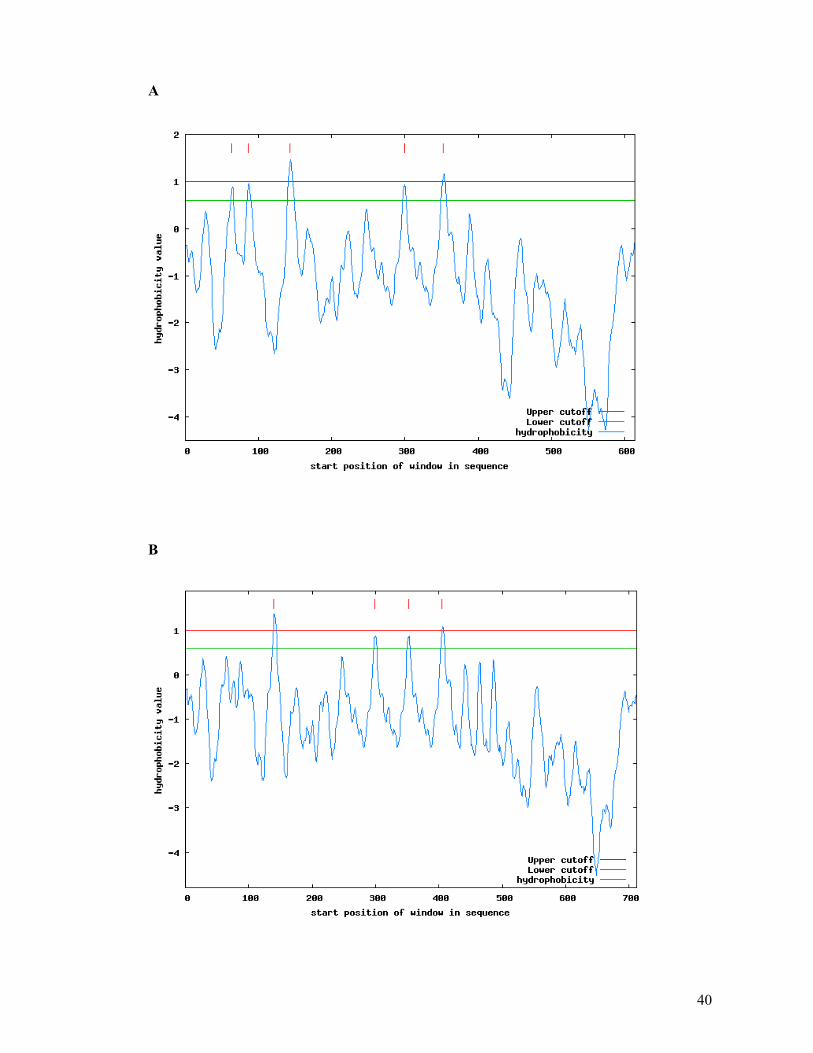
40
A
B

41
C
Fig.1.11 Topology prediction of ToRSV isolate. (A) Rasp2 isolate has 5 candidate membrane-
spanning segments. (B) X4-Rasp1 has 4 candidate membrane-spanning segments and (C) X4-PYB has 4
segments. Score details are listed in the table belo
Table 1.4 Detail description of the topology prediction of ToRSV isolates.
Isolate Helix Begin - End Score Certainity
Rasp2 64 - 84 a.a 0.884 Putative
87 - 107 a.a 0.969 Putative
144 - 164 a.a 1.459 Certain
300 - 320 a.a 0.934 Putative
353 - 373 a.a 1.159 Certain

42
Rasp1 Helix Begin - End Score Certanity 140 - 160 a.a. 1.392 Certain
300 - 320 a.a 0.884 Putative
353 - 373 a.a 0.884 Putative
406 – 426 a.a 1.089 Certain
PYB 87 - 107 a.a 0.829 Putative
143 - 163 a.a 1.718 Certain
300 - 320 a.a. 1.109 Certain
353 - 373 a.a 1.109 Certain
1.10.4 Secondary structure prediction in different ToRSV isolates
A
B
Fig.1.12 Secondary structure prediction in different ToRSV isolates. A) Hierarchical Neural
Network result for X4-Rasp2 isolate. Sequence length is 640 amino acids. Alpha helix (Hh) : 244 is
38.49%, 310 helix (Gg) : 0 is 0.00%, Pi helix (Ii) : 0 is 0.00%, Beta bridge (Bb) : 0 is
0.00%, Extended strand (Ee) : 69 is 10.88%, Beta turn (Tt) : 0 is 0.00%, Bend region (Ss) : 0 is
0.00%, Random coil (Cc) : 321 is 50.63%, Ambigous states (?) : 0 is 0.00%, Other states : 0
is 0.00%. B) Hierarchical Neural Network result for X4-Rasp1 isolate. Sequence length is 732 amino acids.
Alpha helix (Hh) : 278 is 37.98%,310 helix (Gg) : 0 is 0.00%, Pi helix (Ii) : 0 is 0.00%,
Beta bridge (Bb) : 0 is 0.00%, Extended strand (Ee) : 104 is 14.21%, Beta turn (Tt) : 0 is 0.00%,

43
Bend region (Ss) : 0 is 0.00%, Random coil (Cc) : 350 is 47.81%, Ambigous states (?) : 0 is
0.00%, Other states : 0 is 0.00%
1.11 Research hypothesis and objectives
As discussed above, the ToRSV X4 protein does not have significant sequence identity with
any other proteins available in the database. Also, it is absent in the genome of other
nepoviruses. The X4 protein could be involved in functions such as the specificity of
nematode transmission or interaction of the virus with host defence responses. Also it is
possible that the X4 plays an accessory role in cell to cell movement or the RNA2
replication. The goal of this thesis is to characterize the X4 protein at the molecular level.
The specific objectives of this thesis are as follows:
1- Characterize possible interactions between the X4 protein and other viral proteins.
2- Analyse the variability of X4 among other ToRSV isolate.
3- Investigate the possible function of the X4 protein.

44
1.12 Bibliography
Aaziz, R., Dinant, S., and Epel, B. L. (2001). Plasmodesmata and plant cytoskeleton. Trends
Plant Sci 6(7), 326-30.
Agudelo-Romero, P., Carbonell, P., Perez-Amador, M. A., and Elena, S. F. (2008). Virus
adaptation by manipulation of host's gene expression. PLoS ONE 3(6), e2397.
Allen, R. W., and Dias, H. F. (1977). Properties of the single protein and two nucleic acids of
tomato ringspot virus. Canadian Journal of Botany 55, 1028-1037.
Alzhanova, D. V., Prokhnevsky, A. I., Peremyslov, V. V., and Dolja, V. V. (2007). Virion
tails of Beet yellows virus: Coordinated assembly by three structural proteins.
Virology 359(1), 220-6.
Alzhanova, D. V., Prokhnevsky, A. I.,Peremyslov, V. V.,Dolja, V. V (2007). Virion tails of
Beet yellows virus: Coordinated assembly by three structural proteins. Virology
359(1), 220-6.
Anandalakshmi, R., Pruss, G. J., Ge, X., Marathe, R., Mallory, A. C., Smith, T. H., and
Vance, V. B. (1998). A viral suppressor of gene silencing in plants. Proc Natl Acad
Sci U S A 95(22), 13079-84.
Andrade, M. A., Perez-Iratxeta, C., Ponting, C.P (2001). Protein repeats: structures,
functions, and evolution. J Struct Biol 134, 117-31.
Andret-Link, P., Schmitt-Keichinger, C.,Demangeat, G.,Komar, V and Fuchs, M. (2004).
The specific transmission of Grapevine fanleaf virus by its nematode vector
Xiphinema index is solely determined by the viral coat protein. Virology 320(1), 12-
22.
Angell, S. M., Davies, C., and Baulcombe, D. C. (1996). Cell-to-cell movement of potato
virus X is associated with a change in the size-exclusion limit of plasmodesmata in
trichome cells of Nicotiana clevelandii. Virology 216(1), 197-201.
Atabekov, J. G., Rodionova, N. P., Karpova, O. V., Kozlovsky, S. V., and Poljakov, V. Y.
(2000). The movement protein-triggered in situ conversion of potato virus X virion
RNA from a nontranslatable into a translatable form. Virology 271(2), 259-63.
Atreya, C. D., Raccah, B., and Pirone, T. P. (1990). A point mutation in the coat protein
abolishes aphid transmissibility of a potyvirus. Virology 178(1), 161-5.
Baulcombe, D. (2005). RNA silencing. Trends Biochem Sci 30(6), 290-3.
Baulcombe, D. C. (1996). Mechanisms of pathogen-derived resistance to viruses in
transgenic plants. Plant Cell 8(10), 1833-1844.

45
Baulcombe, D. C. (2007). Molecular biology. Amplified silencing. Science 315(5809), 199-
200.
Baumberger, N., Tsai, C. H., Lie, M., Havecker, E., and Baulcombe, D. C. (2007). The
Polerovirus Silencing Suppressor P0 Targets ARGONAUTE Proteins for
Degradation. Curr Biol 17(18), 1609-1614.
Bedard, K. M., and Semler, B. L. (2004). Regulation of picornavirus gene expression.
Microbes Infect 6(7), 702-13.
Bjorklund, A. K., Ekman, D., and Elofsson, A. (2006). Expansion of protein domain repeats.
PLoS Comput Biol 2(8), e114.
Brigneti, G., Voinnet, O., Li, W. X., Ji, L. H., Ding, S. W., and Baulcombe, D. C. (1998).
Viral pathogenicity determinants are suppressors of transgene silencing in Nicotiana
benthamiana. Embo J 17(22), 6739-46.
Brodersen, P., and Voinnet, O. (2006). The diversity of RNA silencing pathways in plants.
Trends Genet 22(5), 268-80.
Brunt, A. A., Crabtree, K., Dallwitz, M. J., Gibbs, A. J., and Watson, L., Eds. (1996). Viruses
of Plants- Descriptions and Lists from the VIDE Database, . Wallingford: CAB
International,.
Cañizares, M. C., Taylor, K.M., Lomonossoff, G.P (2004). Surface-exposed C-terminal
amino acids of the small coat protein of Cowpea mosaic virus are required for
suppression of silencing. J Gen Virol 85(11), 3431-5.
Carette, J. E., Stuiver, M., Van Lent, J., Wellink, J., and Van Kammen, A. (2000). Cowpea
mosaic virus infection induces a massive proliferation of endoplasmic reticulum but
not Golgi membranes and is dependent on de novo membrane synthesis. J Virol
74(14), 6556-63.
Carrier, K., Hans, F., and Sanfacon, H. (1999). Mutagenesis of amino acids at two tomato
ringspot nepovirus cleavage sites: effect on proteolytic processing in cis and in trans
by the 3C-like protease. Virology 258(1), 161-75.
Carrier, K., Xiang, Y., and Sanfacon, H. (2001). Genomic organization of RNA2 of Tomato
ringspot virus: processing at a third cleavage site in the N-terminal region of the
polyprotein in vitro. J Gen Virol 82(Pt 7), 1785-90.
Carrington, J. C., Haldeman, R., Dolja, V. V., and Restrepo-Hartwig, M. A. (1993). Internal
cleavage and trans-proteolytic activities of the VPg-proteinase (NIa) of tobacco etch
potyvirus in vivo. J Virol 67(12), 6995-7000.

46
Carvalho, C. M., Wellink, J., Ribeiro, S. G., Goldbach, R. W., and Van Lent, J. W. (2003).
The C-terminal region of the movement protein of Cowpea mosaic virus is involved
in binding to the large but not to the small coat protein. J Gen Virol 84(Pt 8), 2271-7.
Caspar, D. L. D., and Klug, A. (1962). Physical principles in the construction of regular
viruses. Cold Spring Harb Symp Quant Biol 27, 1-24.
Cerutti, L., Mian, N., and Bateman, A. (2000). Domains in gene silencing and cell
differentiation proteins: the novel PAZ domain and redefinition of the Piwi domain.
Trends Biochem. Sci 25(10), 481-2.
Chandrasekar, V., and Johnson, J. E. (1998). The structure of tobacco ringspot virus: a link in
the evolution of icosahedral capsids in the picornavirus superfamily. Structure 6(2),
157-71.
Chisholm, J., Zhang, G., Wang, A., and Sanfacon, H. (2007). Peripheral association of a
polyprotein precursor form of the RNA-dependent RNA polymerase of Tomato
ringspot virus with the membrane-bound viral replication complex. Virology 368(1),
133-144.
Covey, S. N., Al-Kaff, N. S., Langara, A., and Turner, D. S. (1997). Plants combat infection
by gene silencing. Nature 385, 781-782.
Dales, S., Eggers, H. J., Tamm, I., and Palade, G. E. (1965). Electron microscopic study of
the formation of poliovirus. Virology 26, 379–389.
Demangeat, G., Hemmer, O., Reinbolt, J., Mayo, M. A., and Fritsch, C. (1992). Virus-
specific proteins in cells infected with tomato black ring nepovirus: evidence for
proteolytic processing in vivo. J Gen Virol 73(7), 1609-14.
Dolja, V. V., Kreuze, J. F., and Valkonen, J. P. (2006). Comparative and functional genomics
of closteroviruses. Virus Res 117(1), 38-51.
Gabrenaite-Verkhovskaya, R., Andreev, I. A.,Kalinina, N. O.,Torrance, L.,Taliansky, M.
E.,Makinen, K. (2008). Cylindrical inclusion protein of potato virus A is associated
with a subpopulation of particles isolated from infected plants. J Gen Virol 89(Pt 3),
829-38.
Gaire, F., Schmitt, C., Stussi-Garaud, C., Pinck, L., and Ritzenthaler, C. (1999). Protein 2A
of grapevine fanleaf nepovirus is implicated in RNA2 replication and colocalizes to
the replication site. Virology 264(1), 25-36.
Gallie, D. R. (2001). Cap-independent translation conferred by the 5' leader of tobacco etch
virus is eukaryotic initiation factor 4G dependent. J Virol 75(24), 12141-52.

47
Garcia-Arenal, F., Fraile, A., and Malpica, J. M. (2001). Variability and genetic structure of
plant virus populations. Annu Rev Phytopathol 39, 157-86.
Goodwin, J., Chapman, K., Swaney, S., Parks, T. D., Wernsman, E. A., and Dougherty, W.
G. (1996). Genetic and biochemical dissection of transgenic RNA-mediated virus
resistance. Plant Cell 8(1), 95-105.
Goto, K., Kobori, T., Kosaka, Y., Natsuaki, T. and Masuta, C. (2007). Characterization of
silencing suppressor 2b of cucumber mosaic virus based on examination of its small
RNA-binding abilities. Plant Cell Physiol 48, 1050–1060.
Guo, H. S., and Ding, S. W. (2002). A viral protein inhibits the long range signaling activity
of the gene silencing signal. Embo J 21(3), 398-407.
Hamilton, A., Voinnet, O., Chappell, L., and Baulcombe, D. (2002). Two classes of short
interfering RNA in RNA silencing. Embo J 21(17), 4671-9.
Han, S., and Sanfacon, H. (2003). Tomato ringspot virus proteins containing the nucleoside
triphosphate binding domain are transmembrane proteins that associate with the
endoplasmic reticulum and cofractionate with replication complexes. J Virol 77(1),
523-34.
Hans, F., and Sanfacon, H. (1995). Tomato ringspot nepovirus protease: characterization and
cleavage site specificity. J Gen Virol 76(4), 917-27.
Harris, J. I., and Knight, C. A. (1952). Action of carboxypeptidase on tobacco mosaic virus.
London 170, 613(170).
Hellen, C. U., and Sarnow, P. (2001). Internal ribosome entry sites in eukaryotic mRNA
molecules. Genes Dev 15(13), 1593-612.
Hericourt, F., Blanc, S., Redeker, V., and Jupin, I. (2000). Evidence for phosphorylation and
ubiquitinylation of the turnip yellow mosaic virus RNA-dependent RNA polymerase
domain expressed in a baculovirus-insect cell system. Biochem J 349(Pt 2), 417-25.
Hernandez, C., Carette, J. E., Brown, D. J., and Bol, J. F. (1996). Serial passage of tobacco
rattle virus under different selection conditions results in deletion of structural and
nonstructural genes in RNA 2. J Virol 70(8), 4933-40.
Holeva, R. C., and Macfarlane, S. A. (2006). Yeast two-hybrid study of tobacco rattle virus
coat protein and 2b protein interactions. Arch Virol.
Hull, R. (2002). "Matthew's plant virology." 4th ed. Academic Press, San Diego.
Jafarpour, B., and Sanfaçon, H. (2009). Insertion of large amino acid repeats and point
mutations contribute to a high degree of sequence diversity in the X4 protein of
tomato ringspot virus (genus Nepovirus). Archives of Virology 154(10), 1713-1717.

48
Jovel, J., Walker, M., and Sanfacon, H. (2007). Recovery of Nicotiana benthamiana plants
from a necrotic response induced by a nepovirus is associated with RNA silencing but
not with reduced virus titer. J Virol 81(22), 12285-12297.
Karetnikov, A., and Lehto, K. (2008). Translation mechanisms involving long-distance base
pairing interactions between the 5' and 3' non-translated regions and internal
ribosomal entry are conserved for both genomic RNAs of Blackcurrant reversion
nepovirus. Virology 371(2), 292-308.
Karpova, O. V., Zayakina, O. V., Arkhipenko, M. V., Sheval, E. V., Kiselyova, O. I.,
Poljakov, V. Y., Yaminsky, I. V., Rodionova, N. P., and Atabekov, J. G. (2006).
Potato virus X RNA-mediated assembly of single-tailed ternary 'coat protein-RNA-
movement protein' complexes. J Gen Virol 87(Pt 9), 2731-40.
Karsies, A., Hohn, T., and Leclerc, D. (2001). Degradation signals within both terminal
domains of the cauliflower mosaic virus capsid protein precursor. The Plant Journal
27(4), 335-43.
Kasschau, K. D., and Carrington, J. C. (1998). A counterdefensive strategy of plant viruses:
suppression of posttranscriptional gene silencing. Cell 95(4), 461-70.
Kasschau, K. D., and Carrington, J. C. (2001). Long-distance movement and replication
maintenance functions correlate with silencing suppression activity of potyviral HC-
Pro. Virology 285(1), 71-81.
Kasschau, K. D., Cronin, S., and Carrington, J. C. (1997). Genome amplification and long-
distance movement functions associated with the central domain of tobacco etch
potyvirus helper component-proteinase. Virology 228(2), 251-62.
Kasteel, D., Wellink, J., Verver, J., van Lent, J., Goldbach, R., and van Kammen, A. (1993).
The involvement of cowpea mosaic virus M RNA-encoded proteins in tubule
formation. J Gen Virol 74 ( Pt 8), 1721-4.
Knipe, D. M., Howley, P. H., Griffin., D. E., Lamb, R. A., Martin, M. A., Roizman, B., and
Straus, S. E. (2001). Fields virology, p3063.
Koenig, R., Tremaine, J. H., and Sheperd, J. F. (1978). In situ degradation of the protein
chain of potato virus X at the N- and C-termini. Journal of General Virology 38, 329-
337.
Kurath, G., and Dodds, J. A. (1995). Mutation analyses of molecularly cloned satellite
tobacco mosaic virus during serial passage in plants: evidence for hotspots of genetic
change. RNA 1(5), 491–500.
49
Laporte, C., Vetter, G., Loudes, A. M., Robinson, D. G., Hillmer, S., Stussi-Garaud, C., and
Ritzenthaler, C. (2003). Involvement of the secretory pathway and the cytoskeleton in
intracellular targeting and tubule assembly of Grapevine fanleaf virus movement
protein in tobacco BY-2 cells. Plant Cell 15(9), 2058-75.
Latvala, S., Susi, P., Kalkkinen, N., and Lehto, K. (1998). Characterization of the coat protein
gene of mite-transmitted blackcurrant reversion associated nepovirus. Virus Res
53(1), 1-11.
Le Gall, O., Candresse, T., and Dunez, J. (1995). A multiple alignment of the capsid protein
sequences of nepoviruses and comoviruses suggests a common structure. Arch Virol
140(11), 2041-2053.
Le Gall, O., Christian, P., Fauquet, C. M., King, A. M., Knowles, N. J., Nakashima, N.,
Stanway, G., and Gorbalenya, A. E. (2008). Picornavirales, a proposed order of
positive-sense single-stranded RNA viruses with a pseudo-T = 3 virion architecture.
Arch Virol 153(4), 715-727.
Le Gall, O., Sanfacon, H., Ikegami, M., Iwanami, T., Jones, T., Karasev, A., Lehto, K.,
Wellink, J., Wetzel, T., and Yoshikawa, N. (2007). Cheravirus and Sadwavirus: two
unassigned genera of plant positive-sense single-stranded RNA viruses formerly
considered atypical members of the genus Nepovirus (family Comoviridae). Arch
Virol 159, 1767-1774.
Leclerc, D., Chapdelaine, Y., and Hohn, T. (1999). Nuclear targeting of the cauliflower
mosaic virus coat protein. J Virol 73(1), 553-60.
Lemmetty, A., Latvala, S., Jones, A. T., Susi, P., McGavin, W. J., and Lehto, K. (1997).
Purification and properties of a new virus from black currant, its affinities with
nepoviruses, and its close association with black currant reversion disease.
Phytopathology 87(4), 404-13.
Lincoln Taiz, E. Z. (2006). Plant physiology Sunderland, Mass.,Sinauer Associates.
Lopez, L., Urzainqui, A.,Dominguez, E.,Garcia, J. A. (2001). Identification of an N-terminal
domain of the plum pox potyvirus CI RNA helicase involved in self-interaction in a
yeast two-hybrid system. J Gen Virol 82(Pt 3), 677-86.
Lózsa R, C. T., Lakatos L, Burgyán J (2008). Inhibition of 3' modification of small RNAs in
virus-infected plants require spatial and temporal co-expression of small RNAs and
viral silencing-suppressor proteins. Nucleic Acids Res 36(12), 4099-107.
50
Lu, R., Folimonov, A., Shintaku, M., Li, W. X., Falk, B. W., Dawson, W. O., and Ding, S.
W. (2004). Three distinct suppressors of RNA silencing encoded by a 20-kb viral
RNA genome. Proc Natl Acad Sci U S A 101(44), 15742-7.
MacFarlane, S. A., Wallis, C. V.,Brown, D. J. (1996). Multiple virus genes involved in the
nematode transmission of pea early browning virus. Virology 219(2), 417-22.
Mackenzie, J. (2005). Wrapping things up about virus RNA replication. Traffic 6(11), 967-
77.
Marcotte, E. M., Pellegrini, M.,Yeates, T.O and Eisenberg, D (1998). A Census of Protein
Repeats. J. Mol. Biol 293, 151-160.
Margis, R., Ritzenthaler, C., Reinbolt, J., Pinck, M., and Pinck, L. (1993). Genome
organization of grapevine fanleaf nepovirus RNA2 deduced from the 122K
polyprotein P2 in vitro cleavage products. J Gen Virol 74(9), 1919-26.
McKinney, H. (1935). Evidenceof virus mutation in the common mosaic of tobacco. J. Agric.
Res. 51, 951-81.
Meng, C., Chen, J., Peng, J., and Wong, S. M. (2006). Host-induced avirulence of hibiscus
chlorotic ringspot virus mutants correlates with reduced gene-silencing suppression
activity. J Gen Virol 87(Pt 2), 451-9.
Miller, S., and Krijnse-Locker, J. (2008). Modification of intracellular membrane structures
for virus replication. Nature Reviews Microbiology 6, 363-374.
Napoli, C., Lemieux, C., and Jorgensen, R. (1990). Introduction of a chimeric chalcone
sythase gene into Petunia results in reversible co-suppression of homologous gene in
trans. Plant Cell 2, 279-289.
Oh, C. S., and Carrington, J. C. (1989). Identification of essential residues in potyvirus
proteinase HC-Pro by site-directed mutagenesis. Virology 173(2), 692-9.
Peremyslov, V. V., Andreev, I. A., Prokhnevsky, A. I., Duncan, G. H., Taliansky, M. E., and
Dolja, V. V. (2004). Complex molecular architecture of beet yellows virus particles.
Proc Natl Acad Sci U S A 101(14), 5030-5.
Plisson, C., Drucker, M., Blanc, S., German-Retana, S., Le Gall, O., Thomas, D., and Bron,
P. (2003). Structural characterization of HC-Pro, a plant virus multifunctional protein.
J Biol Chem 278(26), 23753-61.
Plisson, C., Uzest, M., Drucker, M., Froissart, R., Dumas, C., Conway, J., Thomas, D., Blanc,
S., and Bron, P. (2005). Structure of the mature P3-virus particle complex of
cauliflower mosaic virus revealed by cryo-electron microscopy. J Mol Biol 346(1),
267-77.

51
Pouwels, J., Kornet, N., Van Bers, N., Guighelaar, T., Van Lent, J., Bisseling, T., and
Wellink, J. (2003). Identification of distinct steps during tubule formation by the
movement protein of Cowpea mosaic virus. J Gen Virol 84(Pt 12), 3485-94.
Pouwels, J., van der Velden, T., Willemse, J., Borst, J. W., van Lent, J., Bisseling, T., and
Wellink, J. (2004). Studies on the origin and structure of tubules made by the
movement protein of Cowpea mosaic virus. J Gen Virol 85(Pt 12), 3787-96.
Pruss, G., Ge, X., Shi, X. M., Carrington, J. C., and Bowman Vance, V. (1997). Plant viral
synergism: the potyviral genome encodes a broad-range pathogenicity enhancer that
transactivates replication of heterologous viruses. Plant Cell 9(6), 859-68.
Qi, Y., Zhong, X., Itaya, A., and Ding, B. (2004). Dissecting RNA silencing in protoplasts
uncovers novel effects of viral suppressors on the silencing pathway at the cellular
level. Nucleic Acids Res 32(22), e179.
Qu, F., Ren, T., and Morris, T. J. (2003). The coat protein of turnip crinkle virus suppresses
posttranscriptional gene silencing at an early initiation step. J Virol 77(1), 511-22.
Rajamaki, M. L., Kelloniemi, J., Alminaite, A., Kekarainen, T., Rabenstein, F., and
Valkonen, J. P. (2005). A novel insertion site inside the potyvirus P1 cistron allows
expression of heterologous proteins and suggests some P1 functions. Virology 342,
88-101.
Ratcliff, F. G., MacFarlane, S. A., and Baulcombe, D. C. (1999). Gene silencing without
DNA. RNA-mediated cross-protection between viruses. Plant Cell 11(7), 1207-16.
Reichel, C., and Beachy, R. N. (2000). Degradation of tobacco mosaic virus movement
protein by the 26S proteasome. J Virol 74(7), 3330-7.
Ritzenthaler, C., Laporte, C., Gaire, F., Dunoyer, P., Schmitt, C., Duval, S., Piequet, A.,
Loudes, A. M., Rohfritsch, O., Stussi-Garaud, C., and Pfeiffer, P. (2002). Grapevine
fanleaf virus replication occurs on endoplasmic reticulum-derived membranes. J Virol
76(17), 8808-19.
Ritzenthaler, C., Pinck, M., and Pinck, L. (1995a). Grapevine fanleaf nepovirus P38 putative
movement protein is not transiently expressed and is a stable final maturation product
in vivo. J Gen Virol 76 ( Pt 4), 907-15.
Ritzenthaler, C., Schmit, A. C., Michler, P., Stussi-Garaud, C., and Pinck, L. (1995b).
Grapevine fanleaf nepovirus P38 putative movement protein is located on tubules in
vivo. Mol Plant Microbe Interact 8(3), 379-387.

52
Rochon, D. (2008). Molecular insight into plant virus-vector interactions. In "Biotechnology
and plant disease management" (Z. K. Punja, S. H. de Boer, and H. Sanfacon, Eds.),
pp. 532-565. CAB International, Wallingford, UK.
Rochon, D., and Sanfacon, H. (2001). Nepoviruses. In "Encyclopedia of Plant Pathology" (O.
C. Maloy, and T. D. Murray, Eds.), pp. 704-708. John Wiley & Sons, Inc.
Rochon, D. M. (1991). Rapid de novo generation of defective interfering RNA by cucumber
necrosis virus mutants that do not express the 20-kDa nonstructural protein. Proc Natl
Acad Sci U S A 88(24), 11153-7.
Rodriguez-Negrete, E. A., Carrillo-Tripp, J., and Rivera-Bustamante, R. F. (2009). RNA
silencing against geminivirus: complementary action of posttranscriptional gene
silencing and transcriptional gene silencing in host recovery. J Virol 83(3), 1332-40.
Rogers, S., Wells, R., Rechsteiner ,M (1986). Amino acid sequences common to rapidly
degraded proteins: the PEST hypothesis. Science 234(4774), 364-8.
Roossinck, M. J. (1997). Mechanisms of plant virus evolution. Annu Rev Phytopathol 35,
191-209.
Roossinck, M. J. (2003). Plant RNA virus evolution. Curr Opin Microbiol 6(4), 406-9.
Roth, B. M., Pruss, G. J., and Vance, V. B. (2004). Plant viral suppressors of RNA silencing.
Virus Res 102(1), 97-108.
Rott, M. E., Gilchrist, A., Lee, L., and Rochon, D. (1995). Nucleotide sequence of tomato
ringspot virus RNA1. J Gen Virol 76(2), 465-73.
Rott, M. E., Tremaine, J. H., and Rochon, D. M. (1991a). Comparison of the 5' and 3' termini
of tomato ringspot virus RNA1 and RNA2: evidence for RNA recombination.
Virology 185(1), 468-72.
Rott, M. E., Tremaine, J. H., and Rochon, D. M. (1991b). Nucleotide sequence of tomato
ringspot virus RNA-2. J Gen Virol 72(7), 1505-14.
Salvador, B., Delgadillo, M. O., Saenz, P., Garcia, J. A., and Simon-Mateo, C. (2008).
Identification of Plum pox virus pathogenicity determinants in herbaceous and woody
hosts. Molecular Plant Microbe Interactions 21(1), 20-29.
Sanfacon, H. (1995). Nepoviruses. In "Pathogenesis and host specificity in plant diseases,
Vol. III. Viruses and Viroids" (R. P. Singh, U. S. Singh, and K. Kohmoto, Eds.), pp.
129-141. Pergamon Press, Oxford.
Sanfacon, H. (2005). Replication of positive-strand RNA viruses in plants: Contact points
between plant and virus components. Can J Bot 83(12), 1529-1549.
53
Sanfacon, H. (2008). Nepovirus. In "Encyclopedia of Virology, Third Edition" (B. W. J.
Mahy, and M. H. Van Regenmortel, Eds.), Vol. 3, pp. 405-413. 5 vols. Elsevier,
Oxford.
Sanfacon, H., Wieczorek, A., and Hans, F. (1995). Expression of the tomato ringspot
nepovirus movement and coat proteins in protoplasts. J Gen Virol 76(9), 2299-303.
Sanfaçon, H., Wellink, J., Le Gall, O., Karasev, A.V., van der Vlugt, R., and Wetzel, T.
(2009). "Secoviridae: a proposed family of plant viruses within the order Picornavirales that
combines the families Sequiviridae and Comoviridae, the unassigned genera Cheravirus and
Sadwavirus, and the proposed genus Torradovirus.", Archives of Virology, 154(5), pp. 899-
907
Sarnow, P. (2003). Viral internal ribosome entry site elements: novel ribosome-RNA
complexes and roles in viral pathogenesis. J Virol 77(5), 2801-6.
Satyanarayana, T., Gowda, S., Mawassi, M., Albiach-Marti, M. R., Ayllon, M. A.,
Robertson, C., Garnsey, S. M., and Dawson, W. O. (2000). Closterovirus encoded
HSP70 homolog and p61 in addition to both coat proteins function in efficient virion
assembly. Virology 278(1), 253-65.
Schaad, M. C., Jensen, P. E., and Carrington, J. C. (1997). Formation of plant RNA virus
replication complexes on membranes: role of an endoplasmic reticulum-targeted viral
protein. Embo J 16(13), 4049-59.
Schneider, W. L., and Roossinck, M. J. (2001). Genetic diversity in RNA virus quasispecies
is controlled by host-virus interactions. J Virol 75(14), 6566-71.
Seitsonen, J. J., Susi, P., Lemmetty, A., and Butcher, S. J. (2008). Structure of the mite-
transmitted Blackcurrant reversion nepovirus using electron cryo-microscopy.
Virology 378(1), 162-8.
Shi, X. M., Miller, H., Verchot, J., Carrington, J. C., and Vance, V. B. (1997). Mutations in
the region encoding the central domain of helper component-proteinase (HC-Pro)
eliminate potato virus X/potyviral synergism. Virology 231(1), 35-42.
Shukla, D. D., Thomas, J. E., McKern, N. M., Tracy, S. L., and Ward, C. W. (1988). Coat
protein of potyviruses. 4. Comparison of biological properties, serological
relationships, and coat protein amino acid sequences of four strains of potato virus Y.
Arch Virol 102(3-4), 207-19.
Silhavy, D., Molnar, A., Lucioli, A., Szittya, G., Hornyik, C., Tavazza, M., and Burgyan, J.
(2002). A viral protein suppresses RNA silencing and binds silencing-generated, 21-
to 25-nucleotide double-stranded RNAs. Embo J 21(12), 3070-80.

54
Smith, C. J., Watson, C. F., Bird, C. R., Ray, J., Schuch, W.,, and Grierson, D. (1990).
Expression of a truncated tomato polygalacturonase gene inhibits expression of the
endogenous gene in transgenic plants. Mol.Gen. Genet 224, 477–481.
Sonenberg, N., and Dever, T. E. (2003). Eukaryotic translation initiation factors and
regulators. Curr Opin Struct Biol 13(1), 56-63.
Song, J. J., Liu , J., Tolia, N. H., Schneiderman , J., Smith , S. K., Martienssen, R. A.,
Hannon, G. J., and Joshua-Tor, L. (2003). The crystal structure of the Argonaute2
PAZ domain reveals an RNA binding motif in RNAi effector complexes. Nat Struct
Biol 10(12), 1026-32.
Stavolone, L., Villani, M. E., Leclerc, D., and Hohn, T. (2005). A coiled-coil interaction
mediates cauliflower mosaic virus cell-to-cell movement. Proc Natl Acad Sci U S A
102(17), 6219-24.
Taliansky, M., Torrance, L., and Kalinina, N. O. (2008). Role of plant virus movement
proteins. Methods Mol Biol 451, 33-54.
Thornbury, D. W., G.M. Hellmann, R.E, Rhoads and .T.P. Pirone (1985). Purification and
characterization of potyvirus helper component. Virology 144, 260-267.
Tian, T., Rubio.L.,Yeh, H. H .,Crawford, B ., Falk, B. W (1999). Lettuce infectious yellows
virus: in vitro acquisition analysis using partially purified virions and the whitefly
Bemisia tabaci. Journal of General Virology 80, 1111-1117.
Valli, A., Martin-Hernandez, A. M., Lopez-Moya, J. J., and Garcia, J. A. (2006). RNA
silencing suppression by a second copy of the P1 serine protease of Cucumber vein
yellowing ipomovirus, a member of the family Potyviridae that lacks the cysteine
protease HCPro. J Virol 80(20), 10055-63.
Van Bokhoven, H., Le Gall, O., Kasteel, D., Verver, J., Wellink, J., and Van Kammen, A. B.
(1993). Cis- and trans-acting elements in cowpea mosaic virus RNA replication.
Virology 195(2), 377-86.
Van Lent, J., Storms, M., Van Der Meer, F., Wellink, J. and Goldbach, R. (1991). Tubular
structures involved in movement of cowpea mosaic virus are also formed in infected
cowpea protoplasts. J Gen Virol 72, 2615–2624.
Vance, V. B., Berger, P. H., Carrington, J. C., Hunt, A. G., and Shi, X. M. (1995). 5'
proximal potyviral sequences mediate potato virus X/potyviral synergistic disease in
transgenic tobacco. Virology 206(1), 583-90.
Vargason, J. M. e. a. (2003). Crystal structure of CIRV p19 bound to siRNA. Cell 115(799-
811).

55
Vassilakos, N., Vellios, E. K., Brown, E. C., Brown, D. J., and MacFarlane, S. A. (2001).
Tobravirus 2b protein acts in trans to facilitate transmission by nematodes. Virology
279(2), 478-87.
Vellios, E., Brown, D.J., MacFarlane, S. A (2002). Substitution of a single amino acid in the
2b protein of Pea early-browning virus affects nematode transmission. J Gen Virol 83,
1771-1775.
Vellios, E., Duncan, G., Brown, D., and MacFarlane, S. (2002). Immunogold localization of
tobravirus 2b nematode transmission helper protein associated with virus particles.
Virology 300(1), 118-24.
Verchot-Lubicz, J. (2005). A new cell-to-cell transport model for Potexviruses. Mol Plant
Microbe Interact 18(4), 283-90.
Verchot-Lubicz, J., Ye, C. M., and Bamunusinghe, D. (2007). Molecular biology of
potexviruses: recent advances. J Gen Virol 88(Pt 6), 1643-55.
Verver, J., Le Gall, O., van Kammen, A., and Wellink, J. (1991). The sequence between
nucleotides 161 and 512 of cowpea mosaic virus M RNA is able to support internal
initiation of translation in vitro. J Gen Virol 72 ( Pt 10), 2339-45.
Visser, P. B., and Bol, J. F. (1999). Nonstructural proteins of Tobacco rattle virus which have
a role in nematode-transmission: expression pattern and interaction with viral coat
protein. J Gen Virol 80 ( Pt 12), 3273-80.
Voinnet, O., Pinto, Y. M., and Baulcombe, D. C. (1999). Suppression of gene silencing: a
general strategy used by diverse DNA and RNA viruses of plants. Proc Natl Acad Sci
U S A 96(24), 14147-52.
Wallis, C. M., Stone, A. L., Sherman, D. J., Damsteegt, V. D., Gildow, F. E., and Schneider,
W. L. (2007). Adaptation of plum pox virus to a herbaceous host (Pisum sativum)
following serial passages. J Gen Virol 88(Pt 10), 2839-45.
Wang, A., Han, S., and Sanfacon, H. (2004). Topogenesis in membranes of the NTB-VPg
protein of Tomato ringspot nepovirus: definition of the C-terminal transmembrane
domain. J Gen Virol 85(Pt 2), 535-45.
Wang, A., S.Han, and H.Sanfacon (2004). Topogenesis in membranes of the NTB-VPg
protein of Tomato ringspot nepovirus: definition of the C-terminal transmembrane
domain. Journal of General Virology 85, 535-545.
Wang, A., and Sanfacon, H. (2000a). Diversity in the coding regions for the coat protein,
VPg, protease and putative RNA-dependent RNA polymerase among tomato ringspot
nepovirus isolates. Can J Plant Pathol 22, 145-149.

56
Wang, A., and Sanfacon, H. (2000b). Proteolytic processing at a novel cleavage site in the N-
terminal region of the tomato ringspot nepovirus RNA-1-encoded polyprotein in
vitro. J Gen Virol 81(Pt 11), 2771-81.
Wang, S., and Gergerich, R. C (1998). Immunofluorescent localization of tobacco ringspot
nepovirus in vector nematode Xiphinema americanum. Phytopathology 88, 885-889.
Wang, S., Gergerich ,R.C., Wickizer,S.L and Kim K.K (2002). Localization of transmissible
and nontransmissible viruses in the vector nematode Xiphinema americanum.
Phytopathology 92(6), 646-653.
Wang, S., and Gergerich, R. C. (1998). Immunofluorescent localization of Tobacco ringspot
nepovirus in the vector nematode Xiphinema americanum. Phytopathology 88(9),
885-889.
Wellink, J., van Kammen, A. (1989). Cell-to-cell transport of cowpea mosaic virus requires
both the 58K/48K proteins and the capsid proteins. J Gen Virol 70, 2279-2286.
Wessels , E., Duijsings, D., Niu, T. K., S., N., Oorschot, V. M., de Lange, F., Lanke, K. H.,
J., K., Henke, A., Jackson, C. L., Melchers, W. J., and van Kuppeveld , F. J. (2006). A
viral protein that blocks Arf1-mediated COP-I assembly by inhibiting the guanine
nucleotide exchange factor GBF1. Dev. Cell 11, 191–201.
Wieczorek, A., and Sanfacon, H. (1993). Characterization and subcellular localization of
tomato ringspot nepovirus putative movement protein. Virology 194(2), 734-42.
Wingard, S. A. (1928). Hosts and symptoms of ring spot, a virus disease of plants. J. Agric.
Res. 37, 127-153.
Xin, H. W., and Ding, S. W. (2003). Identification and molecular characterization of a
naturally occurring RNA virus mutant defective in the initiation of host recovery.
Virology 317(2), 253-62.
Ye, J., Qu, J., Zhang, Y. J., and Fang, G. R. (2009). A critical domain of the Cucumber
mosaic virus 2b protein for RNA silencing suppressor activity. FEBS Letters 583(1),
101-106.
Ye, K. e. a. (2003). Recognition of small interfering RNA by a viral suppressor of RNA
Silencing. Nature 426, 874–878.
Zhang, G., and Sanfacon, H. (2006). Characterization of membrane-association domains
within the Tomato ringspot nepovirus X2 protein, an endoplasmic reticulum-targeted
polytopic membrane protein. J Virol 80(21), 10847-10857.
Zhang, S. C., Zhang, G., Yang, L., Chisholm, J., and Sanfacon, H. (2005). Evidence that
insertion of Tomato ringspot nepovirus NTB-VPg protein in endoplasmic reticulum

57
membranes is directed by two domains: a C-terminal transmembrane helix and an N-
terminal amphipathic helix. J Virol 79(18), 11752-65.
Zhang, X., Yuan, Y. R., Pei, Y., Lin, S. S., Tuschl, T., Patel, D. J., and Chua, N. H. (2006).
Cucumber mosaic virus-encoded 2b suppressor inhibits Arabidopsis Argonaute1
cleavage activity to counter plant defense. Genes Dev 20(23), 3255-68.

58
Chapter 2
Insertion of large amino acid repeats and point mutations
contribute to a high degree of sequence diversity in the X4 protein
of tomato ringspot virus
A version of this chapter has been published. Jafarpour, B. and Sanfacon, H. (2009)
Insertion of large amino acid repeats and point mutations contribute to a high degree of
sequence diversity in the X4 protein of tomato ringspot virus (genus Nepovirus).
Archives of Virology, 154, 1713-1717

59
2.1 Introduction
Tomato ringspot virus (ToRSV) is a serious pathogen of small fruits and fruit trees in North
America (Sanfacon et al., 2006). Several isolates have been described that differ in their
natural host range and in the intensity of symptoms they induce in herbaceous hosts (Bitterlin
and Gonsalves, 1988; Wang and Sanfacon, 2000). Tomato ringspot virus is a member of the
genus Nepovirus (Sanfacon, 2008), which has recently been reassigned to the proposed new
family “secoviridae” (sub-family “comovirinae”) within the order Picornavirales (Sanfacon
et al., 2009). Nepoviruses have a bipartite positive-strand RNA genome. Each RNA encodes
a polyprotein that is cleaved by the viral proteinase (Pro) at specific cleavage sites. RNA1
encodes replication proteins while RNA2 codes for the coat protein (CP), movement protein
(MP) and other protein(s) of less defined function. Using in vitro processing assays, three
proteinase cleavage sites have been identified in the RNA2-encoded polyprotein of ToRSV (a
subgroup C nepovirus), allowing the definition of four protein domains (X3, X4, MP and CP)
(Carrier et al., 1999; Carrier et al., 2001). In contrast, only three protein domains are present
in the polyprotein of subgroups A and B nepoviruses (2a, MP and CP, see Fig. 2.1A). The
ToRSV X3 protein shares conserved sequence motifs with the 2a protein of subgroup A and
B nepoviruses. It has been suggested that the 2a protein plays a role in the replication of
RNA2 (Gaire et al., 1999; Rott et al., 1991). The ToRSV X4 protein does not have
significant sequence identity with proteins available in the database and does not have a
functional equivalent in the genome of nepoviruses of subgroups A and B (Fig. 2.1A). The
only other nepovirus of subgroup C for which the entire nucleotide sequence is available is
blackcurrant reversion virus (BRV). The extent of sequence identity between ToRSV and
BRV is very low in the deduced amino acid (a. a.) sequence of the X4 protein (8 %) but
higher in the deduced a. a. sequence of the CP (29 %) or the VPg (viral protein linked to the
genome), Pro and polymerase (Pol) (36 %). In this study, we examined the extent of
sequence diversity in the X4 protein among closely related ToRSV isolates. We show that
there is a high degree of sequence diversity in the X4 protein, which is due in part to the
insertion of multiple copies of two types of large amino acid repeats.

60
2.2 Materials and methods
2.2.1 Provenance of the virus material
The complete nucleotide sequence of a ToRSV raspberry isolate from British Columbia
(isolate Rasp2) has been previously determined (Genbank accession number NP_60762) and
cDNA clones of RNA1 and RNA2 are available, although they are not infectious (Rott et al.,
1995; Rott et al., 1991). The original Rasp2 isolate was lost and is not available for
inoculations. A different ToRSV raspberry isolate (Rasp1) is available to our laboratory and
has been maintained in cucumber for several years. Rasp1 was originally isolated by Dr. R.
R. Martin (USDA-ARS, Corvallis) from raspberry fields in Washington State. Sequence
analysis of the CP, X2, VPg, Pro and Pol coding regions of Rasp1 revealed that it is closely
related to Rasp2 [sequence identity of 97-100 % at the nucleotide (nt) level and 98-100 % at
the a. a. level] (Wang and Sanfacon, 2000; Zhang and Sanfacon, 2006). We have previously
shown that N. benthamiana plants inoculated with Rasp1 recover from infection at
temperatures equal to or above 27° C and young leaves become symptom-free after an initial
symptomatic systemic infection (Jovel et al., 2007). At lower temperatures (21° C), recovery
does not occur and the plants eventually die as a result of the necrosis induced upon ToRSV
infection (results not shown). Isolate PYB1 (peach yellow bud) was originally collected from
a peach orchard in California and was obtained from Dr. A. Rowhani (UC-Davis). PYB1 is
closely related to Rasp1 and Rasp2 as revealed by high level of sequence identity in the CP,
VPg, Pro and Pol coding regions (93-98 % identity at the nt level and 97-100 % at the a. a.
level) (Wang and Sanfacon, 2000). In N. benthamiana plants infected with PYB1, symptoms
are milder than those observed in Rasp1-infected plants and PYB1-infected plants recover
from infection even when cultivated at 21° C (data not shown).
Leaves from cucumber plants infected with Rasp1 or from N. benthamiana plants infected
with PYB1 were ground in liquid nitrogen and total RNA was extracted using the TRIzol
reagent (Invitrogen). Because of the high degree of sequence conservation previously
observed between Rasp1, Rasp2 and PYB1, I designed primers that were based on the
published Rasp2 nucleotide sequence. First–strand cDNA was synthesised using 1 μg of
total RNA as a template and primer CP-R (5'-GTCAAGCTTGCCACGCCCGAAAGGAT-3',
complementary to nts 5723-5707 of the RNA2 of ToRSV isolate Rasp2) (Rott et al., 1991)
and Superscript Π Reverse Transcriptase (Invitrogen). For the PCR reaction, I used primers
F208 (5'-GAGGCCGAATTGGCCTCAAAG-3', corresponding to nts 801-822 of Rasp2

61
RNA2, approximately 180 nts upstream of the X4 coding region) and R207 (5'-
GCACCCGCATCAGAGGATC-3', complementary to nts 2998-2980 of Rasp2 RNA2,
approximately 120 nts downstream of the X4 coding region) (Fig. 2.1A). As expected, a
fragment of approximately 2200 nts was amplified when these primers were used in a direct
PCR reaction with a cDNA clone derived from Rasp2 RNA2 or in an RT-PCR reaction with
total RNA extracted from PYB1-infected plants (Fig. 2.1B). However, a significantly larger
cDNA fragment (approximately 2500 nts in length) was amplified by RT-PCR with total
RNA extracted from Rasp1-infected plants (Fig. 2.1B). Sequencing reactions were initially
conducted directly on amplified cDNA fragments, using primers R207 and F208. Other
primers were designed as the sequence of the Rasp1 and PYB1 isolates were determined. The
sequence was confirmed by partial sequencing of several individual cDNA clones. Each
nucleotide was sequenced at least twice in each direction. Sequences were assembled using
vector NTI advance software (Invitrogen). Multiple sequence alignments were completed
using Clustal W2 (Chenna et al., 2003). The sequences for the X4 coding regions of ToRSV-
PYB and ToRSV-Rasp1 were deposited in the GenBank database under accession numbers
GQ325249 and GQ325250, respectively.
2.3 Results
2.3.1 Sequence properties
The previously identified X3-X4 and X4-MP cleavage sites were conserved in the Rasp1 and
PYB1 isolates (Fig. 2.1C). The Rasp1 X4 coding region was 2190 nts in length compared to
1899 nts for the X4 coding region from Rasp2 and PYB1. The predicted size of the Rasp1-
X4 protein is 730 a. a. (calculated Mr of 82 kDa) compared to 633 a. a. for the Rasp2 and
PYB1 X4 protein (calculated Mr of 71 kDa). The degree of sequence identity in the X4
protein among the three ToRSV isolates was 75-87 % at the a. a. level (78-89 % at the nt
level), a value significantly lower than that previously observed for the CP or VPg-Pro-Pol
protein domains (Table 2.1). This high degree of diversity was due in part to the insertion of
sequences in the X4 protein from Rasp1. However, even when gaps were excluded from the
analysis, the degree of sequence identity in the X4 protein was lower than that observed for
other protein domains (Table 2.1). Analysis of individual cDNA clones of the X4 coding
region of Rasp1 revealed at least two sites of sequence heterogeneity in the virus population.
One of these mutations was silent, while the other would cause a significant change in the
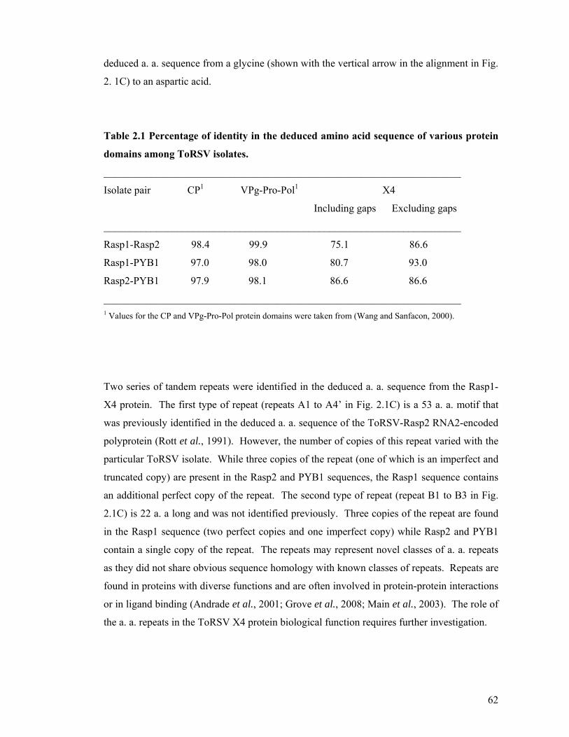
62
deduced a. a. sequence from a glycine (shown with the vertical arrow in the alignment in Fig.
2. 1C) to an aspartic acid.
Table 2.1 Percentage of identity in the deduced amino acid sequence of various protein
domains among ToRSV isolates.
____________________________________________________________________
Isolate pair CP1 VPg-Pro-Pol1 X4
Including gaps Excluding gaps
____________________________________________________________________
Rasp1-Rasp2 98.4 99.9 75.1 86.6
Rasp1-PYB1 97.0 98.0 80.7 93.0
Rasp2-PYB1 97.9 98.1 86.6 86.6
____________________________________________________________________ 1 Values for the CP and VPg-Pro-Pol protein domains were taken from (Wang and Sanfacon, 2000).
Two series of tandem repeats were identified in the deduced a. a. sequence from the Rasp1-
X4 protein. The first type of repeat (repeats A1 to A4’ in Fig. 2.1C) is a 53 a. a. motif that
was previously identified in the deduced a. a. sequence of the ToRSV-Rasp2 RNA2-encoded
polyprotein (Rott et al., 1991). However, the number of copies of this repeat varied with the
particular ToRSV isolate. While three copies of the repeat (one of which is an imperfect and
truncated copy) are present in the Rasp2 and PYB1 sequences, the Rasp1 sequence contains
an additional perfect copy of the repeat. The second type of repeat (repeat B1 to B3 in Fig.
2.1C) is 22 a. a long and was not identified previously. Three copies of the repeat are found
in the Rasp1 sequence (two perfect copies and one imperfect copy) while Rasp2 and PYB1
contain a single copy of the repeat. The repeats may represent novel classes of a. a. repeats
as they did not share obvious sequence homology with known classes of repeats. Repeats are
found in proteins with diverse functions and are often involved in protein-protein interactions
or in ligand binding (Andrade et al., 2001; Grove et al., 2008; Main et al., 2003). The role of
the a. a. repeats in the ToRSV X4 protein biological function requires further investigation.

63
Fig. 2. 1 Comparison of the X4 protein domain in the ToRSV Rasp1 and Rasp2 isolates (A) Schematic representation of the X4 protein domain of ToRSV-Rasp1, Rasp2 and PYB1 isolates. The
RNA2-encoded polyprotein from nepoviruses of subgroups A and B and of ToRSV is shown at the top of the
figure with the vertical lines representing the proteinase cleavage sites. The lower portion of the figure
represents the X4 protein domain of Rasp1, Rasp2 and PYB1. The series of repeats are shown with arrows. The
approximate position of two primers used for RT-PCR is shown above the schematic representation of the
ToRSV RNA2-encoded polyprotein. (B) PCR amplification of a region of the RNA2 open reading frame
coding for a portion of X3, the entire X4 and a portion of MP. PCR products were amplified by direct PCR
using available cDNA clones for Rasp2, or by RT-PCR using total RNA purified from cucumber or N.
benthamiana plants infected with ToRSV Rasp1 or PYB1. Primer pair R207/F208 was used for the
amplification. (C) Alignment of the deduced amino acid sequence of the X4 protein from Rasp1, Rasp2 and
PYB1 isolates. The sequence of Rasp2 was previously published (Rott et al., 1991). The previously identified
X3-X4 and X4-MP cleavage sites (Carrier et al., 1999; Carrier et al., 2001) are shown with the boxes. Amino
acid sequence repeats are indicated with the horizontal arrows above the sequence. Repeats A1-A3 and repeats
B1-B2 are perfect copies of the first and second sets of repeats, respectively. Repeat A4’ is an imperfect and
truncated copy of the first set of repeats and repeat B3 is a full-length imperfect copy of the second set of
repeats. The site of sequence heterogeneity in the virus population that would result in an amino acid change
(from glycine to aspartic acid) is underlined in the Rasp1 sequence and shown by the vertical arrow.
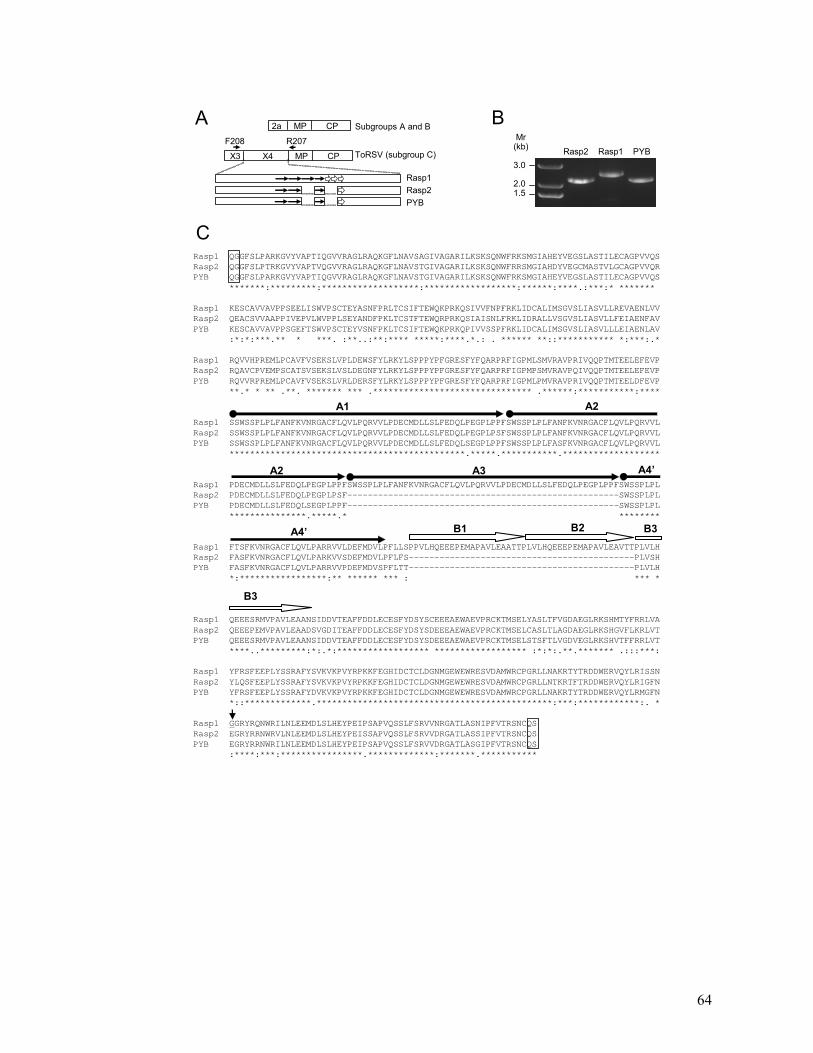
64
Mr(kb)
3.0
2.01.5
Rasp2 Rasp1 PYB
A
X3 X4 MP CP
2a MP CP Subgroups A and B
ToRSV (subgroup C)
Rasp1Rasp2PYB
BF208 R207
Rasp1 QGGFSLPARKGVYVAPTIQGVVRAGLRAQKGFLNAVSAGIVAGARILKSKSQNWFRKSMGIAHEYVEGSLASTILECAGPVVQS Rasp2 QGGFSLPTRKGVYVAPTVQGVVRAGLRAQKGFLNAVSTGIVAGARILKSKSQNWFRRSMGIAHDYVEGCMASTVLGCAGPVVQRPYB QGGFSLPARKGVYVAPTIQGVVRAGLRAQKGFLNAVSTGIVAGARILKSKSQNWFRKSMGIAHEYVEGSLASTILECAGPVVQS
*******:*********:*******************:******************:******:****.:***:* *******
Rasp1 KESCAVVAVPPSEELISWVPSCTEYASNFPRLTCSIFTEWQKPRKQSIVVFNPFRKLIDCALIMSGVSLIASVLLREVAENLVV Rasp2 QEACSVVAAPPIVEPVLWVPPLSEYANDFPKLTCSTFTEWQRPRKQSIAISNLFRKLIDRALLVSGVSLIASVLLFEIAENFAVPYB KESCAVVAVPPSGEFTSWVPSCTEYVSNFPKLTCSIFTEWQKPRKQPIVVSSPFRKLIDCALIMSGVSLIASVLLLEIAENLAV
:*:*:***.** * ***. :**..:**:**** *****:****.*.: . ****** **::*********** *:***:.*
Rasp1 RQVVHPREMLPCAVFVSEKSLVPLDEWSFYLRKYLSPPPYPFGRESFYFQARPRFIGPMLSMVRAVPRIVQQPTMTEELEFEVP Rasp2 RQAVCPVEMPSCATSVSEKSLVSLDEGNFYLRKYLSPPPYPFGRESFYFQARPRFIGPMPSMVRAVPQIVQQPTMTEELEFEVP PYB RQVVRPREMLPCAVFVSEKSLVRLDERSFYLRKYLSPPPYPFGRESFYFQARPRFIGPMLPMVRAVPRIVQQPTMTEELDFEVP
**.* * ** .**. ******* *** .******************************* .******:***********:****
Rasp1 SSWSSPLPLFANFKVNRGACFLQVLPQRVVLPDECMDLLSLFEDQLPEGPLPPFSWSSPLPLFANFKVNRGACFLQVLPQRVVL Rasp2 SSWSSPLPLFANFKVNRGACFLQVLPQRVVLPDECMDLLSLFEDQLPEGPLPSFSWSSPLPLFANFKVNRGACFLQVLPQRVVLPYB SSWSSPLPLFANFKVNRGACFLQVLPQRVVLPDECMDLLSLFEDQLSEGPLPPFSWSSPLPLFASFKVNRGACFLQVLPQRVVL
**********************************************.*****.***********.*******************
Rasp1 PDECMDLLSLFEDQLPEGPLPPFSWSSPLPLFANFKVNRGACFLQVLPQRVVLPDECMDLLSLFEDQLPEGPLPPFSWSSPLPL Rasp2 PDECMDLLSLFEDQLPEGPLPSF-----------------------------------------------------SWSSPLPLPYB PDECMDLLSLFEDQLSEGPLPPF-----------------------------------------------------SWSSPLPL
***************.*****.* ********
Rasp1 FTSFKVNRGACFLQVLPARRVVLDEFMDVLPFLLSPPVLHQEEEPEMAPAVLEAATTPLVLHQEEEPEMAPAVLEAVTTPLVLH Rasp2 FASFKVNRGACFLQVLPARKVVSDEFMDVLPFLFS--------------------------------------------PLVSHPYB FASFKVNRGACFLQVLPARRVVPDEFMDVSPFLTT--------------------------------------------PLVLH
*:*****************:** ****** *** : *** *
Rasp1 QEEESRMVPAVLEAANSIDDVTEAFFDDLECESFYDSYSCEEEAEWAEVPRCKTMSELYASLTFVGDAEGLRKSHMTYFRRLVA Rasp2 QEEEPEMVPAVLEAADSVGDITEAFFDDLECESFYDSYSDEEEAEWAEVPRCKTMSELCASLTLAGDAEGLRKSHGVFLKRLVTPYB QEEESRMVPAVLEAANSIDDVTEAFFDDLECESFYDSYSDEEEAEWAEVPRCKTMSELSTSFTLVGDVEGLRKSHVTFFRRLVT
****..*********:*:.*:****************** ****************** :*:*:.**.******* .:::***:
Rasp1 YFRSFEEPLYSSRAFYSVKVKPVYRPKKFEGHIDCTCLDGNMGEWEWRESVDAMWRCPGRLLNAKRTYTRDDWERVQYLRISSN Rasp2 YLQSFEEPLYSSRAFYSVKVKPVYRPKKFEGHIDCTCLDGNMGEWEWRESVDAMWRCPGRLLNTKRTFTRDDWERVQYLRIGFNPYB YFRSFEEPLYSSRAFYDVKVKPVYRPKKFEGHIDCTCLDGNMGEWEWRESVDAMWRCPGRLLNAKRTYTRDDWERVQYLRMGFN
*::*************.**********************************************:***:************:. *
Rasp1 GGRYRQNWRILNLEEMDLSLHEYPEIPSAPVQSSLFSRVVNRGATLASNIPFVTRSNCQS Rasp2 EGRYRRNWRVLNLEEMDLSLHEYPEISSAPVQSSLFSRVVDRGATLASSIPFVTRSNCQSPYB EGRYRRNWRILNLEEMDLSLHEYPEIPSAPVQSSLFSRVVDRGATLASGIPFVTRSNCQS
:****:***:****************.*************:*******.***********
A1 A2
A2 A3 A4’
A4’ B1 B2 B3
B3
C

65
2.4 Discussion
The high variability of the X4 protein sequence suggests that it is not involved in a core
process, such as virus replication. Serial passage of ToRSV isolates by mechanical
inoculation would eliminate selection pressure for the maintenance of sequences important
for transmission of the virus by the Xiphinema americanum nematode, the natural vector for
ToRSV (Sanfacon, 2008; Sanfacon et al., 2006). Although the CP has been suggested to be
the sole determinant for the specificity of nematode transmission in a subgroup A nepovirus
(Andret-Link et al., 2004), it is possible that X4 plays a role in the specificity and/or
efficiency of nematode transmission in the case of ToRSV. The X4 protein could also be
involved in host specificity, symptomatology and/or interaction with host defence responses.
Herbaceous plants infected with nepoviruses often recover from an initial symptomatic
infection. Recovery from tomato black ring nepovirus (TBRV, a subgroup B nepovirus) has
been associated with the induction of RNA silencing and a concurrent dramatic reduction in
virus titer in the recovered leaves (Ratcliff et al., 1997). We have recently shown that
ToRSV (Rasp1 isolate) accumulates to high titre in recovered leaves, in spite of active RNA
silencing directed at ToRSV sequences (Jovel et al., 2007). One possible interpretation for
these results is that ToRSV encodes a suppressor of silencing, thereby allowing the virus to
accumulate in recovered leaves. The X4 protein, which is absent from the TBRV genome is
an attractive candidate for the silencing suppression function. The availability of two closely
related ToRSV isolates with diverging X4 sequences and differences in symptomatology will
provide us with an invaluable tool to address this question.

66
2.5 Bibliography
Andrade, M. A., Perez-Iratxeta, C., and Ponting, C. P. (2001). Protein repeats: structures,
functions, and evolution. J Struct Biol 134(2-3), 117-31.
Andret-Link, P., Schmitt-Keichinger, C., Demangeat, G., Komar, V., and Fuchs, M. (2004).
The specific transmission of Grapevine fanleaf virus by its nematode vector
Xiphinema index is solely determined by the viral coat protein. Virology 320(1), 12-
22.
Bitterlin, M. W., and Gonsalves, C. (1988). Serological grouping of Tomato ringspot virus
isolates: Implications for diagnosis and cross-protection. Phytopathology 78(3), 278-
285.
Carrier, K., Hans, F., and Sanfacon, H. (1999). Mutagenesis of amino acids at two tomato
ringspot nepovirus cleavage sites: effect on proteolytic processing in cis and in trans
by the 3C-like protease. Virology 258(1), 161-75.
Carrier, K., Xiang, Y., and Sanfacon, H. (2001). Genomic organization of RNA2 of Tomato
ringspot virus: processing at a third cleavage site in the N-terminal region of the
polyprotein in vitro. J Gen Virol 82(Pt 7), 1785-90.
Chenna, R., Sugawara, H., Koike, T., Lopez, R., Gibson, T. J., Higgins, D. G., and
Thompson, J. D. (2003). Multiple sequence alignment with the Clustal series of
programs. Nucleic Acids Res 31(13), 3497-500.
Gaire, F., Schmitt, C., Stussi-Garaud, C., Pinck, L., and Ritzenthaler, C. (1999). Protein 2A
of grapevine fanleaf nepovirus is implicated in RNA2 replication and colocalizes to
the replication site. Virology 264(1), 25-36.
Grove, T. Z., Cortajarena, A. L., and Regan, L. (2008). Ligand binding by repeat proteins:
natural and designed. Curr Opin Struct Biol 18(4), 507-15.
Jovel, J., Walker, M., and Sanfacon, H. (2007). Recovery of Nicotiana benthamiana plants
from a necrotic response induced by a nepovirus is associated with RNA silencing but
not with reduced virus titer. J Virol 81(22), 12285-12297.
Main, E. R., Jackson, S. E., and Regan, L. (2003). The folding and design of repeat proteins:
reaching a consensus. Curr Opin Struct Biol 13(4), 482-9.
Ratcliff, F., Harrison, B. D., and Baulcombe, D. C. (1997). A similarity between viral
defense and gene silencing in plants. Science 276, 1558-1560.
Rott, M. E., Gilchrist, A., Lee, L., and Rochon, D. (1995). Nucleotide sequence of tomato
ringspot virus RNA1. J Gen Virol 76(2), 465-73.

67
Rott, M. E., Tremaine, J. H., and Rochon, D. M. (1991). Nucleotide sequence of tomato
ringspot virus RNA-2. J Gen Virol 72(7), 1505-14.
Sanfacon, H. (2008). Nepovirus. In "Encyclopedia of Virology, Third Edition" (B. W. J.
Mahy, and M. H. Van Regenmortel, Eds.), Vol. 3, pp. 405-413. 5 vols. Elsevier,
Oxford.
Sanfacon, H., Zhang, G., Chisholm, J., Jafarpour, B., and Jovel, J. (2006). Molecular biology
of Tomato ringspot nepovirus, a pathogen of ornamentals, small fruits and fruit trees.
In "Floriculture, Ornamental and Plant Biotechnology: Advances and Topical Issues
(1st Edition)" (J. Teixeira da Silva, Ed.), Vol. III, pp. 540-546. Global Science Books,
London, UK.
Sanfaçon, H., Wellink, J., Le Gall, O., Karasev, A.V., van der Vlugt, R., and Wetzel, T.
(2009). "Secoviridae: a proposed family of plant viruses within the order Picornavirales that
combines the families Sequiviridae and Comoviridae, the unassigned genera Cheravirus and
Sadwavirus, and the proposed genus Torradovirus.", Archives of Virology, 154(5), pp. 899-
907
Wang, A., and Sanfacon, H. (2000). Diversity in the coding regions for the coat protein, VPg,
protease and putative RNA-dependent RNA polymerase among tomato ringspot
nepovirus isolates. Can J Plant Pathol 22, 145-149.
Zhang, G., and Sanfacon, H. (2006). Characterization of membrane-association domains
within the Tomato ringspot nepovirus X2 protein, an endoplasmic reticulum-targeted
polytopic membrane protein. J Virol 80(21), 10847-10857.

68
Chapter 3
Characterization of the X4 protein in ToRSV-infected plants
A version of this chapter will be submitted for publication. Jafarpour, B., Chisholm, J
and Sanfacon, H. (2010) Characterization of the ToRSV X4 protein.

69
3.1 Introduction
Most plant viruses, encapsidate their small genome in a non-enveloped virion that has an
icosahedral or helical structure. Nepovirus particles are made of 60 copies of the coat protein
(CP) and have a pseudo T=3 structure. The capsid protein is made up of three β-barrel
domains. The observation that transgenic plants expressing the CP from Arabis mosaic virus
form empty virus particles, similar to those observed in natural infection, suggested that other
viral proteins are not required for virion assembly (Bertioli et al., 1991). The CP of ToRSV
is a polypeptide of 562 amino acids and shows the highest similarity to Blueberry leaf mottle
virus (BLMV) and Blackcurrant reversion virus (BRV), two members of the nepovirus
subgroup C. This subgroup is characterized by a larger RNA2 component (Rochon and
Sanfacon, 2001).
Preparations of nepovirus particles purified from indicator plants (C. quinoa or N.
benthamiana) are composed of two forms of the unique coat protein (Latvala et al., 1998;
Lemmetty et al., 1997). Similarly, two forms of the small comovirus CP are also often
detected (Taylor et al., 1999). The structure of tobacco ringspot virus (TRSV, a subgroup A
nepovirus) and BRV have been resolved. Most preparations of BRV particles purified from
indicator plants (C. quinoa or N. benthamiana) are composed of two forms of the single coat
protein of 54 and 55 kDa in size with identical N-termini (Latvala et al., 1998; Lemmetty et
al., 1997). Using antibodies specific for various regions of the CP, it was determined that the
smaller form of the coat protein is a truncated version of the CP, which has lost a C-terminal
extension of 19 amino acids. This truncation may be the result of processing occurring
during the course of virus infection or during the various steps of virus purification. As for
BRV, two forms of the coat protein have also been detected in tomato black ring virus
(TBRV, a subgroup B nepovirus). Purified BRV virion containing only the smaller form of
the CP (54 kDa) can be used to infect indicator plants (C. quinoa) by mechanical inoculation
resulting in symptoms identical to BRV symptoms (Lemmetty et al., 1997). However, the
resulting progeny viruses contain both protein forms. Thus, the C-terminus is not important
for the infectivity of the virus. It was proposed that the C-terminal extension may serve as a
determinant for mite transmission however this needs to be confirmed experimentally.
Sequence alignments of the CP from various nepoviruses, revealed that the C-terminal
extension is very variable in sequence and varies in length from one nepovirus to another
(Seitsonen et al., 2008). As mentioned above the BRV coat protein has a C-terminal

70
extension of 19 amino acids. In contrast the tobacco ringspot virus coat protein does not
include an apparent C-terminal extension. In the case of tomato ringspot virus (ToRSV), a
possible C-terminal extension is 54 amino acids long (Seitsonen et al., 2008). Based on
homology modeling of the BRV and TRSV structures and also based on sequence alignment
of the coat proteins, it has been predicted that the C-terminal 14 residues of BRV project out
from the surface of the BRV virus. Since it is extended from the virion surface it is also a
potential specific antigenic site (Seitsonen et al., 2008).
Although the main component of the virion is the coat protein(s) (CP), other minor virus-
encoded protein components have also been found in association with plant virus particles.
These virion-associated proteins (VAP) interact directly or indirectly with the CP and are
often loosely associated with the virus particle. Because they are exposed on the surface of
the virion, VAP are readily available for interaction with host or vector components. They
may play important roles in the virus replication cycle, including cell-to-cell or long distance
movement in the plant, suppression of RNA silencing, stability of virus particles, regulation
of translation of the viral genome and vector transmission. Several VAP have been identified
in association with helical viruses. The potyvirus helper component proteinase (HC-Pro) is a
multifunctional protein that was first recognized as an essential factor for aphid transmission,
but was later found to act as a proteinase, an enhancer of genome amplification and of cell-
to-cell and systemic movement of the virus in the plant, a determinant of symptom formation
and a suppressor of silencing (reviewed in Maia et al., 1996; Roth et al., 2004; Urcuqui-
Inchima et al., 2001).
The Cauliflower mosaic virus (CaMV) pIII protein is a well studied VAP of a spherical virus.
This protein is indispensable for aphid transmission and cell-to-cell movement but is not
required for virus encapsidation or replication (Kobayashi et al., 2002; Leh et al., 1999). The
pIII protein interacts directly with the CP through its C-terminal domain (Leclerc et al., 2001;
Leh et al., 2001). The pIII protein co-purifies with virus particles under certain conditions,
although it can easily dissociate from the purified virions (Dautel et al., 1994). Elucidation
of the structure of the reconstituted pIII-virion complex showed that pIII is exposed on the
surface of the virion with its C-terminus anchored in the inner shell of the virion (Plisson et
al., 2005). The N-terminal region of the protein includes a coiled-coiled domain, which may
be essential for dimerization and for interaction with other viral proteins. One of these

71
interactions is with the pII protein. An aphid transmission factor that itself interacts with the
aphid stylet (Leh et al., 1999; Uzest et al., 2007). Another interaction is between the coiled-
coil domain of pIII and a coiled-coil domain of the movement protein (MP) of CaMV
(Stavolone et al., 2005). The viral MP is a structural component of the tubular structures that
contain virus-like particles, traverse the cell wall and probably enable cell-to-cell movement
of the virus (Perbal et al., 1993 and references therein).
As mentioned above, members of subgroup C have a larger RNA2 than members of
subgroup A and B. Analysis of the polyprotein processing of ToRSV, a subgroup C
nepovirus, revealed the presence of three proteinase cleavage sites allowing the definition of
four protein domains, termed X3, X4, MP and CP (Carrier et al., 2001). In contrast, only
three protein domains are present in the polyprotein of subgroups A and B nepoviruses
termed 2a, MP (or 2b) and CP (or 2c), see Fig. 3.1A. The ToRSV X3 protein has some
conserved sequence motifs with the 2a protein of nepoviruses of subgroup A and B (Rott et
al., 1991). The grapevine fanleaf virus (GFLV, a subgroup A nepovirus) 2a protein was
suggested to play a role in the replication of RNA2 (Gaire et al., 1999). The MP of ToRSV
and GFLV is a structural component of tubular structures that are very similar to those
observed in CaMV and cowpea mosaic virus (CPMV, a comovirus) infected plant tissues in
that they contain virus-like particles and are found traversing the cell wall (Ritzenthaler et al.,
1995; Wieczorek and Sanfacon, 1993 and references therein). In grapevine fanleaf virus
(GFLV) analysis of chimeric viruses in which the MP and CP domains were exchanged
suggested that the CP and MP interact with each other, either directly or indirectly to promote
cell-to-cell movement (Belin et al., 1999). In addition, similar analyses of chimeric viruses
suggested that the CP is the sole determinant for the specificity of transmission of GFLV by
its nematode vector (Andret-Link, 2004).
3.1.1 Research objective and hypothesis
The ToRSV X4 protein does not have significant sequence identity with any other proteins
available in the database and does not have a functional equivalent in the genome of
nepoviruses of subgroup A and B (Fig. 3.1A). In this chapter, I will describe the detection of
a 60 kDa protein in ToRSV-Rasp1 infected plants using a polyclonal antibody X4(64-65),
raised against a recombinant protein corresponding to the C-terminal portion of the X4
protein. Subsequent analysis revealed that this protein is associated with empty and full virus

72
particles. The initial hypothesis was that this protein (60 kDa) may be a virion-associated
protein corresponding to a truncated form of X4. I conducted a series of experiments to
investigate the nature of the 60 kDa protein recognized by the X4(64-65) antibodies and its
possible interaction with the virus particle. The results indicated that the epitope recognized
by the X4(64-65) antibodies is exposed at the surface of the viral particle. It was later found
that the X4(64-65) antibodies cross reacted with the CP. Therefore the 60 kDa protein
detected by this antibody was the full length ToRSV CP. Two additional antibodies raised
against the X4 protein did not detect this 60 kDa protein confirming the suggestion that the
60 kDa protein is not derived from the X4 coding region. A protein corresponding to the full
length X4 (82 kDa in Rasp1) was not detected in plant extracts by any of the X4 antibodies
suggesting that X4 may be an unstable protein or that the concentration of the protein was
lower than the detection level of the antibodies. Although smaller (e.g. 45 kDa) or larger
~120 kDa proteins were detected in infected plant extracts by the X4 antibodies, the nature of
these proteins needs to be further investigated.
3.2 Materials and methods
3.2.1 Production of antibodies against the C-terminal and the N-terminal region
of X4
When this part of my research was initiated, the sequence of Rasp1 and PYB isolates were
not available to our laboratory. The sequences of CP, VPg, Pro and Pol region were highly
conserved among closely related ToRSV isolates. Therefore, recombinant proteins or
peptides for antibody production were initially designed based on the available Rasp2
sequence. Later after sequencing the X4 region from a few isolates, we observed a high
degree of sequence diversity in the X4 protein (Chapter 2).
The C-terminal portion of the X4-Rasp2 coding region was amplified by PCR from the
cDNA clone of ToRSV RNA2 (Rott et al., 1991) using primers 64 (5´-
AACGCCATGGGCCTTGTGACTTACCTTCAG-3´, corresponding to nts 2440-2459 of
ToRSV-Rasp2 RNA2) and 65 (5´-AACCCTCGAGCTGTACTGGGGCAGACG-3´,
complementary to nts 2798-2782 of ToRSV-Rasp2 RNA2). The amplified fragment was
cleaved by NcoІ and XhoІ and inserted into the corresponding site of the pET21d (Novagen)

73
expression vector to create plasmid pET-X4-C (Rasp2). This plasmid was transformed into
E. coli BL21 (DE3). Bacteria were grown until the OD600 of the culture reached
approximately 0.6. Protein expression was induced by adding 1 mM IPTG for 2 h at 37°C.
Cells were harvested and purification of the recombinant X4-C His-tagged 15 kDa X4
protein was carried out under denaturing conditions (8 M urea) using a Ni+2 resin column as
recommended by the manufacturer (Qiagen). After purification, the urea was eliminated by
gradual dialysis against sodium phosphate buffer [50 mM NaPO4 (pH 7.5), 0.3 M NaCl]
containing decreasing concentration of urea. Rabbits were injected with the purified protein
for antibody production.
Another antibody was raised against the N-terminal end of the X4 protein using a synthetic
peptide KSKSQNWFRRSMGIAHDYVEG corresponding to the coding region of Rasp2
sequence (nts 1119-1182 of RNA2). The antibody was produced commercially (Bioworld
Molecular Tools and Laboratory Essentials, Dublin, OH, USA).
3.2.2 Construction of plasmids used in the expression of full-length or truncated
forms of X4, CP and MP in vitro translations or in E. coli
To express proteins in vitro I used coupled in vitro transcription/translation system
(Invitrogen). Plasmid pCITE-X4-Rasp2 and pCITE-CP-Rasp2 were constructed as follows.
A cDNA fragment containing the entire X4 coding region from the Rasp2 isolate was
amplified using primers FX4-Rasp2 [5´-GATTCCATGGGGGGCTTCTCCCTCCCTA-3´,
corresponding to nts 981-999 of ToRSV-Rasp2 RNA2 and containing an NcoІ site
(underlined)] and RX4-Rasp2 [5´-AATCCTCGAGTCACTGGCAGTTAGAGCG-3´,
complementary to nts 2865-2879 of ToRSV-Rasp2 RNA2 and containing an XhoІ site
(underlined)]. The amplified cDNA fragment was digested by NcoІ and XhoІ and inserted
into the corresponding sites of plasmid pCITE-4a (+) (Novagen) to create pCITE-X4-Rasp2.
Plasmid pCITE-CP-Rasp2 was constructed in a similar manner using primers FCP [5´-
GATTCCATGGGCGGGTCCTGGCAAGAAG-3´, corresponding to nts 4338-4356 of
ToRSV-Rasp2 RNA2 and containing an NcoI site (underlined)] and RCP-2 [5´-
TATCCTCGAGTCAGCCACGCCCGAAAGG-3´, complementary to nts 5708-5723 of
ToRSV-Rasp2 RNA2 and containing an XhoI site (underlined)]. Plasmids pCITE-CP-Rasp2
and pCITE-X4-Rasp2 include a stop codon at the end of the viral sequence preventing fusion
to the His tail present in the plasmid. However, the viral sequences would be fused in frame
74
with an N-terminal S-tag present in the vector. Plasmid pCITE-X4-Rasp2-HA and plasmid
pCITE-X4-Rasp1-HA were constructed as follows: to create plasmid pCITE-X4-Rasp2-HA,
a cDNA fragment containing the entire X4 coding region from the Rasp2 isolate fused to the
HA tag was amplified using primers F-X4-Rasp2 [5'-
ATATCCATGGGGGGCTTCTCCCTCCCT-3'] and R6-X4-Rasp2 [5'-
TAGAAGATCTTACGCGTAATCAGGAACGTCGTACGGATACTGGCAGTTAGAGCG
AGTAAC-3', complementary to nts 2859-2879 of ToRSV-Rasp2 and containing the HA tag
and a BglII site (underlined)]. Similarly, a cDNA fragment containing the entire X4 coding
region from the Rasp1 isolate fused to the HA tag was amplified using primers F1-X4-Rasp1
[5’-ATATCCATGGGGGGTTTTTTCCTTCCTGCC-3’, corresponding to the 5’ end of the
X4-Rasp1 coding region and containing an NcoI site (underlined)] and R6-X4-Rasp1 [5'-
TAGAAGATCTTACGCGTAATCAGGAACGTCGTACGGATACTGGCAGTTAGAACG
AGTGAC-3' complementary to the 3’ end of the X4-Rasp1 coding region and containing the
HA tag and a BglII site (underlined)] to create plasmid pCITE-X4-Rasp1-HA. The fragments
were digested by NcoI and BglII and inserted in the NcoI-BamHI sites of plasmid pCITE-
4a(+). To construct plasmid pCITE-MP, a cDNA fragment containing the entire coding
region for the full length movement protein (MP) was amplified by PCR from the cDNA
clone of ToRSV RNA2 (Rott et al., 1991) using primers FMP [5´-
GATTCCATGGTCTCTTCTCTAGGAAC-3´, corresponding to nts 2880-2893 of ToRSV-
Rasp2 (RNA2] and RMP [5´- GATTCTCGAGCTGAACAGAAGAATTC-3´,
complementary to nts 4021-4037 of ToRSV-Rasp2 RNA2]. The amplified fragment was
cleaved by NcoІ and XhoІ and inserted into the corresponding site of pCITE-4a (+)
(Novagen) vectors and the pET21d (Escherichia coli expression vectors).
The plasmids were used as a template for coupled in vitro transcription/translation reactions
using the TNT® Rabbit Reticulocyte lysate system following the manufacturer's protocol
(Promega). The labelled [35S] methionine translation products were separated by SDS-
PAGE. Immunoprecipitation of the in vitro translation products were conducted as previously
described (Hans and Sanfacon, 1995).
To express proteins in E. coli, the following plasmids were constructed. Plasmid pET-CP-
His and pET-X4-TM-His (a truncated form of X4 lacking the N-terminal region including a
putative transmembrane domain) were constructed as follows. For pET-X4-TM-His plasmid:
a PCR fragment was amplified from the cDNA clone of ToRSV RNA2 (Rott et al., 1991)

75
using primers FX4-TM1502 [5´- CATTCCATGGAAATGCCGTCTTGCGCAACC -3´,
corresponding to nts 1502-1523 of ToRSV-Rasp2 RNA2] and RX4-TM [5´-
CGGCTCGAGCTGGCAGTTAGAGCGAGTAAC -3´, complementary to nts 2858-2879 of
ToRSV-Rasp2 RNA2]. For pET-CP-His, a PCR fragment was amplified from the cDNA
clone of ToRSV RNA2 (Rott et al., 1991) using primers FCP-pET4038 [5´-
CATGCCATGGGCGGGTCCTGGCAAGAAGGT -3´, corresponding to nt 4038-4058 of
ToRSV-Rasp2 RNA2] and RCP-pET [5´- GCCCTCGAGGCCACGCCCGAAAGGATTT
-3´, complementary to nts 5705- 5723 of ToRSV-Rasp2 RNA2]. Plasmid pET-CP-150-His
(predicted C-terminal extension in the CP, nts 5571-5723 of ToRSV RNA2) and pET-CP-
360-His (predicted exposed loop in the CP, nts 5198-5558 of ToRSV RNA2) were
constructed using the following primers: for pET-CP-360-His: primers F145 [5´-
CGAACCATGGCTAGGCAACATCCTTTATCTCTG -3´, corresponding to nts 5198-5219
of ToRSV-Rasp2 RNA2] and RCP [5´-
GAACTCGAGACTAGGTCCATAGAAAGAAAATC -3´, complementary to nts 5558-5535
of ToRSV-Rasp2 RNA2] and for pET-CP-150-His: primers F147 [5´-
CGAACCATGGCTAAGAAAGAAGTCGGCACGCCTAG -3´, corresponding to nts 5571-
5593 of ToRSV-Rasp2 RNA2] and R148 [5´-
CGAACTCGAGGCCACGCCCGAAAGGATTTTTTCC -3´, complementary to nts 5723-
5699 of ToRSV-Rasp2 RNA2]. The amplified fragments were cleaved by NcoІ and XhoІ
enzymes and inserted into the corresponding site of the pET24d (Novagen) expression
vector. In all these plasmids, the coding regions for the ToRSV proteins were fused in frame
with a poly-histidine tail. We also produced a version of the CP in which a stop codon was
introduced at the end of the CP coding region. As a result, the CP was not fused to a poly-
histidine tail. This plasmid, termed pET-CP, was constructed by transferring the Nco-XhoI
fragment containing the CP coding region with a stop codon from pCITE-CP into the
corresponding site of the pET24d vector. Plasmids were transformed into E. coli BL21
(DE3). Bacteria were grown until the OD600 of the culture reached approximately 0.6.
Protein expression was induced by adding 1 mM IPTG for 2 h at 37°C. Purification of the
recombinant X4-TM His-tagged ~53 kDa protein (Rasp2) was carried out under denaturing
conditions as recommended by the manufacturer (Qiagen).

76
3.2.3 Preparation of plant extracts and subcellular fractionations
ToRSV-Rasp1 was propagated routinely in Cucumis sativus var. Straight Eight. Plants were
inoculated at leaf stage 5 to 7 using 40 μl of fresh leaf extracts of ToRSV-infected cucumber
plants diluted 1:10 (w:v) in phosphate buffered saline (PBS). To produce post-nuclear plant
extracts, one g of leaf tissue from healthy and ToRSV-infected C. sativus was collected 7
days post-inoculation (dpi) and briefly ground in 4 ml of homogenization buffer (Schaad et
al., 1997). After centrifugation at 3,700xg for 10 min, the supernatant was collected (S3
fraction). For subsequent subcellular fractionation, the S3 fraction was overlaid on a 20 to 45
% sucrose gradient and centrifuged at 34,000xg for 4 h as described previously (Han and
Sanfacon, 2003). Protein content in the S3 fraction or in the sucrose gradient fractions were
separated by sodium dodecyl sulfate-polyacrylamide gel electrophoresis (SDS-PAGE) and
analyzed by immunoblotting.
3.2.4 Purification of ToRSV particles
Virus particles were purified from ToRSV-Rasp1-infected tissues essentially as described
(Stace-Smith, 1966). Briefly, harvested symptomatic leaves from approximately 200
ToRSV-infected cucumber plants were ground in 0.5 M borate buffer (pH 6.8). After
freezing overnight at -20 C, the extract was centrifuged at 10,000 x g for 30 min to eliminate
large debris. The supernatant was further clarified using an ammonium sulphate precipitation
step (15 g ammonium sulphate per 100 ml of extract). After centrifugation at 10,000 x g for
30 min, the supernatant was collected. Virus particles were pelleted from this clarified
extract by centrifugation at 140,000 x g for 2 h. The pellets were resuspended in 9 mls of 50
mM borate buffer (pH 6.8) and CsCl was added to a density of 0.68 g/ml. The CsCl
gradients were formed by an overnight centrifugation at 190,500 x g. Virus particles were
extracted from the gradient and dialysed overnight against 20 mM borate buffer (pH 6.8).
The purified virus preparation was diluted (1:100 phosphate buffer saline pH 7.4) and the
protein content was analysed by SDS-PAGE. Proteins were visualized by silver staining
(Invitrogen) or immunoblotting.
3.2.5 Immunoblotting
Proteins were separated by SDS-PAGE and transferred to polyvinylidene fluoride (PVDF)
membranes (Bio-Rad). Membranes were incubated with the rabbit polyclonal antibodies

77
raised against the C-terminal region of X4 (X4(64-65), used at 1:5000 dilution, this study),
denatured purified viral particles ("-virion, used at 1:5000 dilution) (Sanfacon et al., 1995),
the central region of the movement protein ("-MP antibodies, used at 1:5000 dilution)
(Wieczorek and Sanfacon, 1993) or the proteinase ("-pro antibodies, used at 1:5000 dilution)
(Chisholm et al., 2007). Rabbit polyclonal antibodies raised against the Bip protein (donated
by M. Chrispeels) were also used. The secondary antibody was goat anti-rabbit conjugated
with horseradish peroxydase (1:15000 dilution, Bio/Can). Membranes were developed using
an ECL-development kit (Amersham Bioscience).
3.2.6 Immuno-electron microscopy
Highly purified ToRSV-Rasp1 particles (2 μg/ml) were diluted 1:10 in phosphate buffer
saline (buffer supplemented with 0.1% Tween-40, PBST buffer). Formvar/carbon coated
grids were allowed to float on 10 μl of purified virions for 2 min. Grids were incubated with
blocking buffer (PBS, 1% BSA, 0.1% Tween-20) for 10 min and transferred to 10 μl of
X4(64-65) antiserum or pre-immune antiserum (diluted 1:100 in PBST) for 1 hour. Grids
were rinsed once in PBST buffer, and 5 times in distilled water and blocked again for 30 min
in blocking buffer. The samples were washed (as described above) and incubated with anti-
rabbit secondary antibody conjugated with 10 nm gold particles (1:30 in blocking buffer,
Sigma) for 1h. Grids were incubated in blocking buffer for 30 min, rinsed and stained with 2
% uranyl acetate. Samples were viewed at 27,000 to 80,000 X magnification using a JEOL
100CX transmission electron microscope (TEM) operated at 80 kV.
For immunocapture experiment, formvar/carbon coated grids were allowed to float on "-
virion antibody (diluted 1:30 in PBST) for 1 hour. After the incubation, grids were rinsed
once in PBST buffer and 5 times in distilled water. Grids were incubated for 30 min in
blocking buffer. The samples were washed (as described above) and incubated with 10 μl of
fraction 6 or fraction 12 from the sucrose gradient for 1 hour. Grids were rinsed and stained
with 2 % uranyl acetate. Samples were viewed at 27,000 to 80,000 X magnification using a
JEOL 100CX transmission electron microscope (TEM) operated at 80 kV.
3.2.7 Trypsin digestion of ToRSV particles
TPCK-Trypsin (Sigma) stock solution was prepared at a concentration of 2 mg/ml in 0.001 N
HCl, and serial dilutions were made in phosphate buffer 67 mM (NaH2PO4, pH 7.6).

78
Purified ToRSV particles (800 ng/ μl) were incubated with various dilutions of Trypsin (0,
0.6, 3, 16, 80, 400 ng/μl) in a final volume of 50 μl reaction for two hours at 37°C. The
digestion reactions were stopped by adding 2X SDS-PAGE protein loading buffer. The
protein content of each sample was examined by immunoblotting after SDS-PAGE.
3.2.8 Liquid chromatography-mass spectrometry with peptide mass
fingerprinting (LC-MS/MS)
ToRSV was purified as discussed above (Stace-Smith, 1966; Stace-Smith, 1984) and the
proteins were separated by 12% sodium dodecyl sulfate polyacrylamide gel (SDS-PAGE)
followed by staining with mass spectrometry-compatible silver staining polyacrylamide gel
(SilverQuest, Invitrogen). Two fragments were carefully cut: fragment 1, which contained
doublet protein bands of 53, 55 kDa and fragment 2 which contained a 60 kDa protein band.
The in–gel samples were sent for mass spectrometry. In a separate experiment a few μg of
purified virus prep were sent for mass spectrometry. The samples were digested with trypsin.
The extracted peptides were analyzed by LC-MS/MS using an Applied Biosystems QStarI
mass spectrometer, which is a quadrupole time-of-flight hybrid instrument equipped with
nano-electrospray ionization source. The sample was first separated by reverse phase
chromatography over a two hour gradient before spraying into the mass spectrometer. The
MS/MS data were analyzed using a protein identification search engine algorithm called:
MASCOT (http://www.matrixscience.com). The data was searched against the heterologous
non-identical MSDB database, which is a protein sequence database designed specifically for
mass spectrometry applications.
3.3 Results
3.3.1 Preparation of antibodies against the X4 protein
To characterize the ToRSV-Rasp1 X4 protein in infected plants, we initially raised rabbit
polyclonal antibodies against a His-tagged recombinant protein that correspond to the C-
terminal 15 kDa X4 protein of the ToRSV-Rasp2 (Fig. 3.1A,"-X4(64-65)). This region of
the protein was later found to be relatively well conserved between the Rasp1 and Rasp2
isolates (Fig.3.11). Immunoblotting experiments confirmed that the X4(64-65) antibodies
recognized the X4-C recombinant protein (Fig. 3.1B).

79
A
31
45
21
100 50 10 5 0.5 100 50 10 5 0.5 pmoles C-X4 purified protein
stain "-X4(64-65)
Immunoblotting
B
X3 X4 MP CP
2a MP CP Subgroups A and B (eg.GFLV)
ToRSV (subgroup C)
PCR fragment (Rasp2)
X4-C Protein expressed for antibody production
Mr
Fig. 3.1 Genome organisation of ToRSV and GFLV and production of the X4(64-65)
antibodies. (A) Genome organization of ToRSV protein domains (subgroup C) and GFLV (subgroup A)
nepovirus. GFLV lacks the X4 protein. The lower portion of the figure represents the X4 protein domain of
Rasp2 isolate. The blue box at the C-terminal end of the protein represents the region of the ToRSV-Rasp2 X4
protein, which was used to produce polyclonal antibodies. The yellow box at the C termini X4, represents the
protein that was expressed for X4 (64-65) antibody production. (B) Specificity of the X4(64-65) antibody tested
by immunobloting. Immunoblot analysis was conducted using the purified recombinant X4-C protein and the
X4 (64-65) antibodies. Molecular marker (Mr).
As I discussed in Chapter 2, the predicted size for the X4 protein differs among ToRSV
isolates in vivo. The X4 proteins from the Rasp1 and Rasp2 isolates were expressed in vitro
using coupled in vitro transcription/translation system. The apparent molecular mass of the
proteins on SDS-PAGE was consistent with their calculated sizes. As expected the X4-
Rasp1-HA protein (calculated molecular mass of 87.4 kDa, taking into account a 4 kDa N-
terminal S-tag and a 1 kDa C-terminal HA-tag) migrated significantly slower than the X4-
Rasp2 protein (calculated molecular mass of 76 kDa, with the N-terminal S-tag and C-
terminal HA-tag) (data not shown). The untagged X4-Rasp2 (calculated molecular mass 75

80
kDa, including the N-terminal S-tag) and coat protein (calculated molecular mass of 66 kDa
for the CP including the N-terminal S-tag) were also synthesized in vitro.
To test the specificity of the antibodies, immunoprecipitation was conducted using in vitro
translated X4-Rasp2 and CP with different sets of antibodies specific for X4, CP and MP.
The X4(64-65) antibody immunoprecipitated the X4 protein but did not immunoprecipitate
the CP protein (Fig.3. 2A lanes 3 and 5), showing its specificity for the X4 protein in the
immunoprecipitation assay. In addition X4(64-65) antibodies immunoprecipitated the X4-
Rasp1–HA and the X4-Rasp2-HA translated proteins.
I also tested the previously described "-virion antibodies, which were raised against a
denatured preparation of purified virus particles (Sanfacon et al., 1995). As expected, the "-
virion antibodies recognized the CP (Fig. 3.2A lane 4). The "-virion antibodies also
recognized the X4 protein (Fig.3.2A lane 6), suggesting that the X4 protein may have been
present in the purified virus preparation used as an antigen or that the X4 protein and the CP
may share a common epitope. Two other "-virion antibodies which were used in enzyme-
linked immunosorbent assay (polyclonal antibody for ELISA test, Agdia) were tested against
the CP. These antibodies were produced against native virus particles. The antibodies did not
immunoprecipitated in vitro synthesized CP (Fig 3.2B, lane 3 and 4), suggesting that they
may recognize conformational epitope presented in intact virions but not in the CP subunit.
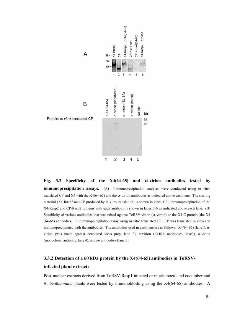
81
97
66
1 2 3 4 5 6
X4-R
asp2
CP
X4-R
asp2
+ α
-X4(
64-6
5)
CP
+ α
-viri
on
CP
+ α
-X4(
64-6
5)
X4-R
asp2
+ α
-viri
on
A
α-X4
(64-
65)
α-v
irion
(den
atur
ed)
α–v
irion
(ELI
SA)
α-v
irion
(mon
o)
No
Abs
Protein: In vitro translated CP 6645
B
1 2 3 4 5
Mr
Mr
Fig. 3.2 Specificity of the X4(64-65) and "-virion antibodies tested by
immunoprecipitation assays. (A) Immunoprecipitation analyses were conducted using in vitro
translated CP and X4 with the X4(64-65) and the "-virion antibodies as indicated above each lane. The starting
material (X4-Rasp2 and CP produced by in vitro translation) is shown in lanes 1-2. Immunoprecipitation of the
X4-Rasp2 and CP-Rasp2 proteins with each antibody is shown in lanes 3-6 as indicated above each lane. (B)
Specificity of various antibodies that was raised against ToRSV virion ("-virion) or the X4-C protein (the X4
(64-65) antibodies), in immunoprecipitation assay using in vitro translated CP. CP was translated in vitro and
immunoprecipitated with the antibodies. The antibodies used in each lane are as follows: X4(64-65) (lane1), α-
virion (was made against denatured virus prep, lane 2), α-virion (ELISA antibodies, lane3), α-virion
(monoclonal antibody, lane 4), and no antibodies (lane 5).
3.3.2 Detection of a 60 kDa protein by the X4(64-65) antibodies in ToRSV-
infected plant extracts
Post-nuclear extracts derived from ToRSV-Rasp1 infected or mock-inoculated cucumber and
N. benthamiana plants were tested by immunoblotting using the X4(64-65) antibodies. A

82
predominant protein with an apparent molecular mass of approximately 60 kDa was detected
by the X4(64-65) antibodies in extracts derived from ToRSV-Rasp1 infected plants but not in
healthy plant extracts (Fig. 3.3A). The protein migrated faster on SDS-PAGE than expected
for the full-length X4 protein (calculated molecular mass of 82.6 kDa for X4-Rasp1). I will
refer to this protein as the 60 kDa protein detected by the X4(64-65) antibodies for the
remainder of this chapter. The X4(64-65) antibodies also detected a faint protein band of
~58-60 kDa in the PYB isolate (the band is not visible in the scanned Fig. 3.3B). Antibodies
against the N-terminal region of X4 did not detect any specific band corresponding to the full
length X4-Rasp2 (Carrier et al., 2001) or the 60 kDa protein detected by X4(64-65)
antibodies (data not shown). All subsequent experiments with ToRSV infected plants were
conducted using the Rasp1 isolate and the X4(64-65) antibodies.
Inf H Inf HA
1 2 3 4
4565
31
C. sativus N. benthamiana
45
6697
116
31
1 2 3 4 5 6 7 8 9 10 11 12
α-X4(64-65)α-virionBMr
Fig. 3.3 Detection of a 60 kDa protein by the X4(64-65) antibodies in infected plant
extracts. (A) Immunoblot analysis of healthy (H) or ToRSV-Rasp1 infected (Inf) C. sativus and N.
benthamiana plant extracts. Proteins were separated by SDS-PAGE (12%) and detected by immunobloting
using X4(64-65) antibodies. (B) Immunoblot analysis of plant extracts derived from plants infected with
various isolates. The blots were probed using "-virion antibodies (denatured) (lanes 1-6) or X4(64-65)
antibodies (lanes 7-12). Plants were infected with the following ToRSV isolates: lane 1 Rasp1 (Cucumber),
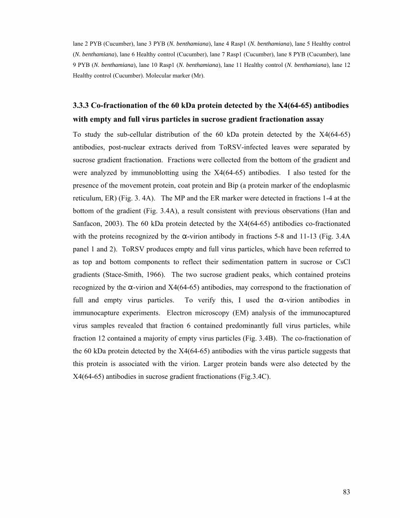
83
lane 2 PYB (Cucumber), lane 3 PYB (N. benthamiana), lane 4 Rasp1 (N. benthamiana), lane 5 Healthy control
(N. benthamiana), lane 6 Healthy control (Cucumber), lane 7 Rasp1 (Cucumber), lane 8 PYB (Cucumber), lane
9 PYB (N. benthamiana), lane 10 Rasp1 (N. benthamiana), lane 11 Healthy control (N. benthamiana), lane 12
Healthy control (Cucumber). Molecular marker (Mr).
3.3.3 Co-fractionation of the 60 kDa protein detected by the X4(64-65) antibodies
with empty and full virus particles in sucrose gradient fractionation assay
To study the sub-cellular distribution of the 60 kDa protein detected by the X4(64-65)
antibodies, post-nuclear extracts derived from ToRSV-infected leaves were separated by
sucrose gradient fractionation. Fractions were collected from the bottom of the gradient and
were analyzed by immunoblotting using the X4(64-65) antibodies. I also tested for the
presence of the movement protein, coat protein and Bip (a protein marker of the endoplasmic
reticulum, ER) (Fig. 3. 4A). The MP and the ER marker were detected in fractions 1-4 at the
bottom of the gradient (Fig. 3.4A), a result consistent with previous observations (Han and
Sanfacon, 2003). The 60 kDa protein detected by the X4(64-65) antibodies co-fractionated
with the proteins recognized by the "-virion antibody in fractions 5-8 and 11-13 (Fig. 3.4A
panel 1 and 2). ToRSV produces empty and full virus particles, which have been referred to
as top and bottom components to reflect their sedimentation pattern in sucrose or CsCl
gradients (Stace-Smith, 1966). The two sucrose gradient peaks, which contained proteins
recognized by the "-virion and X4(64-65) antibodies, may correspond to the fractionation of
full and empty virus particles. To verify this, I used the "-virion antibodies in
immunocapture experiments. Electron microscopy (EM) analysis of the immunocaptured
virus samples revealed that fraction 6 contained predominantly full virus particles, while
fraction 12 contained a majority of empty virus particles (Fig. 3.4B). The co-fractionation of
the 60 kDa protein detected by the X4(64-65) antibodies with the virus particle suggests that
this protein is associated with the virion. Larger protein bands were also detected by the
X4(64-65) antibodies in sucrose gradient fractionations (Fig.3.4C).

84
B
Fraction 6 Fraction 12
1 2 3 4 5 6 7 8 9 10 11 12 13 14 15 16
bottom top
"-virion
"-MP
A
ER marker
"-X4(64-65)
C "-X4(64-65) antibodies
66
45
31
21
97116120
1 2 3 4 5 6 7 8 9 10 11 12 13 14 15Mr
60 kDa protein
Fig. 3.4 Immunoblot analysis and electron microscopy of subcellular fractionation of
proteins by sucrose gradient. (A) Immunoblot analysis of proteins in the sucrose gradient fractionation.
Infected plant extracts were prepared and fractionated on a 20-45% sucrose density gradient. The proteins from
each fraction were separated by SDS-PAGE (12%) and detected by immunoblotting using the X4(64-65), "-
virion, "-MP and "-Bip antibodies. Bip is a resident protein of the ER and is labelled as ER marker in the
figure. (B) Electron micrographs of empty and full virus particles from fraction 6 and 12 obtained after sucrose
gradient fractionation. (C) Immunoblot analysis of proteins in the sucrose gradient fractionation. In this
experiment the proteins were fractionated in 15 fractions, the X4(64-65) antibodies also detected higher protein

85
mass (80-120 kDa) In the viral fractions and lower protein mass on top of the gradient, the nature of these
proteins needs further investigation. The 60 kDa detected by X4(64-65) antibodies in sucrose gradient is shown
by the arrows. Molecular marker (Mr).
3.3.4 Co-purification of the 60 kDa protein detected by the X4(64-65) antibodies
with the virus particles
To further investigate the possibility that the 60 kDa protein detected by the X4(64-65)
antibodies is associated with the virions, I purified virus particles from ToRSV-infected
cucumber using differential centrifugation, an ammonium sulphate clarification step and
CsCl gradient centrifugation. The protein content of this preparation was separated by SDS-
PAGE and analyzed by silver staining and immunoblot analysis (Fig. 3.5A). Silver staining
detected a predominant band corresponding to a doublet protein with an estimated molecular
mass of 53-55 kDa, as well as minor bands of apparent molecular masses of 60 kDa and 120
kDa (Fig. 3.5 panel 1). The "-virion antibodies recognized all proteins detected by silver
staining (Fig.3. 5A panel 2 and Fig. 3.5B). This was expected since a similar preparation was
originally used to produce this antibody. The X4(64-65) antibodies detected a 60 kDa
protein, which co-migrated with the 60 kDa band detected by silver staining and with the "-
virion antibodies (Fig. 3.5A panel 3). These antibodies also detected a protein of
approximately ~120 kDa, which may correspond to dimeric forms of the 60 kDa protein.
Another possibility is that this protein corresponds to a putative X3-X4 intermediate
precursor of the mature X4 protein. The cleavage site between X3 and X4 (Rasp2) is a
suboptimal cleavage site as described before (Carrier et al., 2001). The presence of the 60
kDa protein in highly purified virus preparation suggests that it is associated with virions.
Other viral proteins (e.g. proteinase, movement protein) were not detected in association with
the purified virus particles (Fig. 3.5A, panel 4 and data not shown).

86
suc. grad. highly storedfraction 6 purified virus virus
1 2 3 4 5 6 7 8
"-v
irion
"-X
4 (6
4-65
)
"-v
irion
"-X
4
"-v
irion
"-X
4(64
-65)
"-v
irion
"-X
4(64
-65)
B
stai
n
"-v
irion
"-X
4 (6
4-65
)
"-M
P
97
66
45
1 2 3 4
116
60
55
53
A
Fig. 3.5 Co-purification of the 60 kDa protein detected by the X4(64-65) antibodies with
purified virus particles. (A) Protein content of a highly purified virus preparation was separated by SDS-
PAGE (12%) and examined by silver staining (lane 1) or by immunoblotting using X4(64-65), "-virion and "-
MP antibodies (lanes 2-4). (B) Examination of the relative proportion of the 60 kDa protein detected by the
X4(64-65) antibodies in a sucrose gradient fraction (lanes 1-2), two separate highly purified virus preparation
(lanes 3-6) and after extensive storage of a highly purified virus preparation at 4 °C (lanes 7-8). Proteins were
separated by SDS-PAGE as above and detected by immunoblotting using "-virion and X4(64-65) antibodies.
The 53 and 55 kDa protein doublet, which was recognized by "-virion antibodies but not by
the X4(64-65) antibodies, likely corresponds to the CP. However, the apparent molecular
masses of these proteins were smaller than those expected for the full-length CP (calculated
molecular mass of 62 kDa), raising the possibility that they may correspond to truncated
forms of the CP lacking the C-terminal portion of the protein as previously described for
other nepoviruses (Seitsonen et al., 2008 and references therein). It is possible that the 60
kDa protein band detected by both "-virion antibodies and the X4(64-65) antibodies
correspond to a mixture of the two proteins each recognized by a single antibody (Fig. 3.5

87
A). Alternatively, the 60 kDa protein band could contain a single protein that is recognized
by both antibodies.
The relative intensity of the 53, 55 kDa doublet bands detected by the "-virion antibodies
and of the 60 kDa band detected by the "-virion and X4(64-65) antibodies varied from one
virus preparation to another (Fig. 3.5B). In crude extracts and partially purified virus
(sucrose gradient fraction), the 60 kDa band appeared as predominant (Fig. 3.5B, lanes 1-2,
and Fig. 3.3B). In highly purified virus preparation, the relative intensity of the 60 kDa band
was much lower compared to the 53, 55 kDa bands and varied from one preparation to
another (Fig. 3.5B. lanes 3-6). After prolonged storage of purified virus preparation at 4° C,
the 60 kDa band was not detectable by either antibody (Fig. 3.5B. lanes 7-8). It should be
noted that many intact virus particles were detected in older virus preparations by EM, in
spite of the apparent absence of the 60 kDa protein band (data not shown). The possible
nature of this protein band was investigated further below.
3.3.5 Immunogold labelling of purified virus particles with X4(64-65) antibodies
Immunogold labelling experiments were conducted using the X4(64-65) antibodies and
purified virus particles. The X4(64-65) antibodies decorated both empty and full virus
particles (Fig. 3.6A panels 1-3). A pre-immune rabbit antibody did not bind to the virions
(panel 4). Antibodies specific for the ToRSV movement protein (MP) or proteinase did not
label the virions (data not shown) confirming that the MP does not co-purify with the virus
particle. This result suggests that the 60 kDa protein detected by the X4(64-65) antibodies is
associated with the virion and indicates that the epitope(s) recognized by the X4(64-65)
antibodies is exposed on the surface of the virions.

88
2 3
1
4
Fig. 3.6 Immunogold labelling of purified virus particles using the X4(64-65) antibodies. Immunogold labelling of ToRSV particles using X4(64-65) antibodies (panel 1-3). The preimmune serum
showed no specific labelling (panel 4).
3.3.6 Susceptibility of the 60 kDa protein recognized by the X4(64-65) antibodies
to trypsin digestion
To further test the topology of the 60 kDa protein which is associated with the virions, I
conducted limited trypsin digestion experiments using the purified virus preparation. This
preparation was digested with various concentrations of trypsin. The partially digested
proteins were separated by SDS-PAGE and detected by immunoblotting using "-virion
antibodies (Fig. 3.7). Several fragments were apparently protected from the digestion when
lower concentrations of trypsin were used (Fig. 3. 7, lanes 2-4). These fragments likely
correspond to portions of the proteins which are buried within the structure of the virion after
digestion. Alternatively, they could also correspond to highly structured regions in the
protein. The 60 kDa protein detected by the X4(64-65) antibodies was resistant to trypsin
digestion at lower concentration of trypsin (lane 7-8). At higher concentration of trypsin,
smaller fragments were not detected with these antibodies (lane 9-12). This result suggests
that the portion of the 60 kDa protein, which is recognized by the X4(64-65) antibodies, is
exposed at the surface of the virion.

89
66
45
31
21.5
α-virion "-X4(64-65)
0 0.6 3 16 80 400 0 0.6 3 16 80 400 [trypsin]
1 2 3 4 5 6 7 8 9 10 11 12
Mr
Fig. 3. 7 Susceptibility of the 60 kDa protein to trypsin digestions. Immunoblot analysis of
purified virus preparations was carried out using the X4(64-65) and the α-virion antibodies. Trypsin digestion of
highly purified virus preparations were conducted as described in Material and Methods. The concentration of
trypsin used is indicated above each lane in ng/:l.
3.3.7 The X4 protein is not detected in association with virions using liquid
chromatography-mass spectrometry with peptide mass fingerprinting (LC-
MS/MS)
To obtain additional information on the nature of the 60 kDa protein recognized by the X4
antibodies, LC-MS/MS experiments were conducted. In a first experiment, the 60 kDa
protein band was cut out from the SDS-PAGE gel and sent for analysis. In a second
experiment, a preparation of purified virus was sent for analysis. In both cases, the protein
fragments detected by the LC-MS/MS corresponded predominately to the ToRSV coat
protein. The Rubisco protein, a high abundance protein in plants, was also detected. The X4
protein was not detected in the samples, suggesting that it was absent from the virus
preparation or that its concentration was below the detection threshold of this method. It is
possible that the high concentration of the two dominant proteins (CP and Rubisco) competed
for the ionization process with low abundance proteins.
3.3.8 Re-examination of the specificity of the X4(64-65) antibodies and the α-
virion antibodies
Since X4 was not detected in LC-MS/MS and since the 60 kDa protein found in association
with purified virus particles was recognized by both "-virion and "-X4 antibodies in
immunoblotting, I decided to re-examine the specificity of the X4(64-65) and "-virion
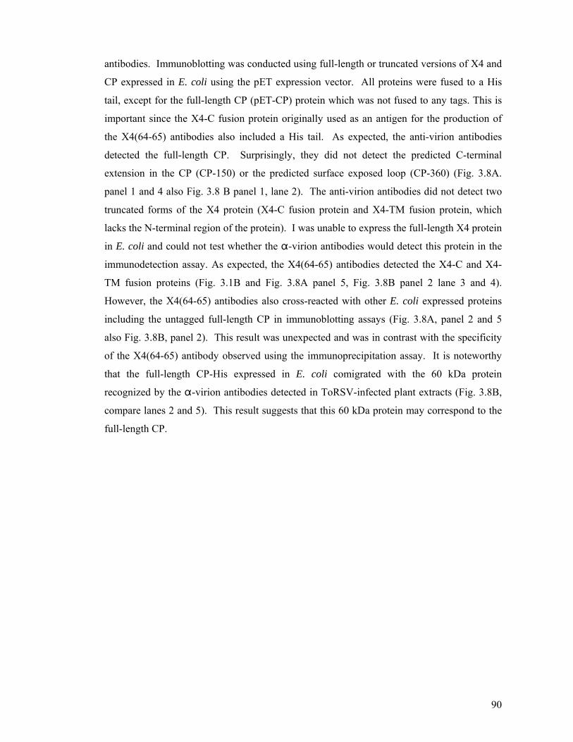
90
antibodies. Immunoblotting was conducted using full-length or truncated versions of X4 and
CP expressed in E. coli using the pET expression vector. All proteins were fused to a His
tail, except for the full-length CP (pET-CP) protein which was not fused to any tags. This is
important since the X4-C fusion protein originally used as an antigen for the production of
the X4(64-65) antibodies also included a His tail. As expected, the anti-virion antibodies
detected the full-length CP. Surprisingly, they did not detect the predicted C-terminal
extension in the CP (CP-150) or the predicted surface exposed loop (CP-360) (Fig. 3.8A.
panel 1 and 4 also Fig. 3.8 B panel 1, lane 2). The anti-virion antibodies did not detect two
truncated forms of the X4 protein (X4-C fusion protein and X4-TM fusion protein, which
lacks the N-terminal region of the protein). I was unable to express the full-length X4 protein
in E. coli and could not test whether the "-virion antibodies would detect this protein in the
immunodetection assay. As expected, the X4(64-65) antibodies detected the X4-C and X4-
TM fusion proteins (Fig. 3.1B and Fig. 3.8A panel 5, Fig. 3.8B panel 2 lane 3 and 4).
However, the X4(64-65) antibodies also cross-reacted with other E. coli expressed proteins
including the untagged full-length CP in immunoblotting assays (Fig. 3.8A, panel 2 and 5
also Fig. 3.8B, panel 2). This result was unexpected and was in contrast with the specificity
of the X4(64-65) antibody observed using the immunoprecipitation assay. It is noteworthy
that the full-length CP-His expressed in E. coli comigrated with the 60 kDa protein
recognized by the "-virion antibodies detected in ToRSV-infected plant extracts (Fig. 3.8B,
compare lanes 2 and 5). This result suggests that this 60 kDa protein may correspond to the
full-length CP.
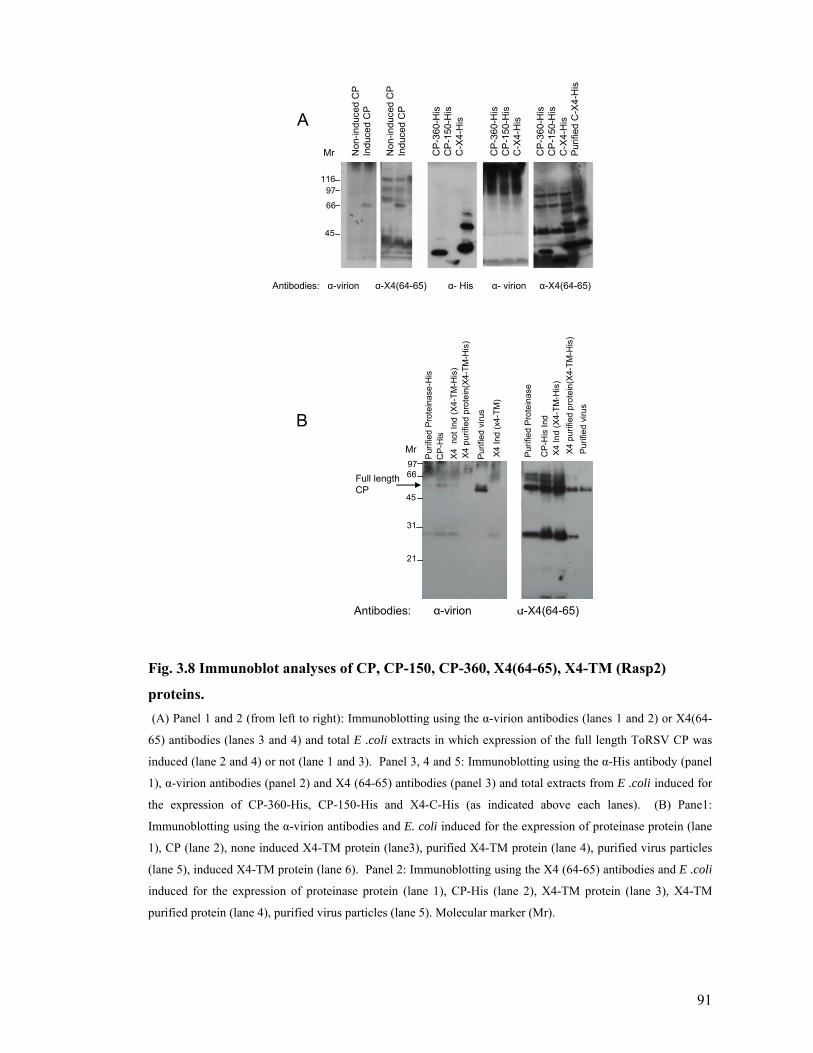
91
Non
-indu
ced
CP
Indu
ced
CP
Non
-indu
ced
CP
Indu
ced
CP
CP
-360
-His
CP
-150
-His
C-X
4-H
is
CP
-360
-His
CP
-150
-His
C-X
4-H
is
CP
-360
-His
CP
-150
-His
C-X
4-H
isP
urifi
ed C
-X4-
His
Antibodies: α-virion α-X4(64-65) α- His α- virion α-X4(64-65)
Antibodies: α-virion α-X4(64-65)
9766
45
31
21
Pur
ified
Pro
tein
ase-
His
CP
-His
X4 n
ot In
d(X
4-TM
-His
)X4
pur
ified
pro
tein
(X4-
TM-H
is)
Purif
ied
viru
s
X4 In
d(x
4-TM
)
CP-
His
Ind
X4 In
d(X
4-TM
-His
)X4
pur
ified
pro
tein
(X4-
TM-H
is)
Purif
ied
viru
s
11697
66
45
A
B
Purif
ied
Prot
eina
se
Full length CP
Mr
Mr
Fig. 3.8 Immunoblot analyses of CP, CP-150, CP-360, X4(64-65), X4-TM (Rasp2)
proteins. (A) Panel 1 and 2 (from left to right): Immunoblotting using the α-virion antibodies (lanes 1 and 2) or X4(64-
65) antibodies (lanes 3 and 4) and total E .coli extracts in which expression of the full length ToRSV CP was
induced (lane 2 and 4) or not (lane 1 and 3). Panel 3, 4 and 5: Immunoblotting using the α-His antibody (panel
1), α-virion antibodies (panel 2) and X4 (64-65) antibodies (panel 3) and total extracts from E .coli induced for
the expression of CP-360-His, CP-150-His and X4-C-His (as indicated above each lanes). (B) Pane1:
Immunoblotting using the α-virion antibodies and E. coli induced for the expression of proteinase protein (lane
1), CP (lane 2), none induced X4-TM protein (lane3), purified X4-TM protein (lane 4), purified virus particles
(lane 5), induced X4-TM protein (lane 6). Panel 2: Immunoblotting using the X4 (64-65) antibodies and E .coli
induced for the expression of proteinase protein (lane 1), CP-His (lane 2), X4-TM protein (lane 3), X4-TM
purified protein (lane 4), purified virus particles (lane 5). Molecular marker (Mr).

92
3.3.9 In vitro co-immunoprecipitation of X4 and CP proteins with the X4(64-65)
antibodies
Because the X4(64-65) antibodies recognized X4 but not CP in the immunoprecipitation
assay, we used this assay to investigate possible protein-protein interactions between X4 and
CP. Co-immunoprecipitation was conducted using the in-vitro translated CP (Rasp2) and X4
(Rasp2) proteins and also the X4(64-65) and "-virion antibodies. As shown before, the
X4(64-65) antibody recognized X4 but not CP (Fig. 3.9A, lanes 3 and 5) while the α-virion
antibody recognized both proteins (Fig. 3.9A, lane 7). The CP was co-immunoprecipitated
with X4 by the X4(64-65) antibodies, suggesting an interaction (direct or indirect) between
the CP and X4 proteins (Fig. 3.9A, lanes 8). I also used HA tags to test for possible
interactions between X4 and CP. Full-length or truncated version of X4 were tagged with the
HA epitope and mixed with an untagged version of the coat protein. Protein complexes were
immunoprecipitated using anti-HA conjugated beads. The HA antibodies pulled down the
X4-Rasp1-HA and X4-Rasp2-HA proteins (Fig.3.9B, lanes 8 and 9). However, the amount
of CP was similar in the presence or absence of X4-HA (compare lanes 8-9 to lane 7).
Therefore there was no clear evidence of a protein–protein interaction between the X4 and
the CP in this assay.
X4 CP X4
and α-
X4(
64-6
5)
CP
and
α-v
irion
CP
and
α-X
4(64
-65)
X4
and α-
virio
n
X4+
CP
with
α-v
irion
X4+C
P w
ith α
-X4(
64-6
5)
A
1 2
1 2 3 4 5 6 7 8
Full length X4-Rasp2Full length CP-Rasp2

93
B
Translation Unbound Co-immunoprecipitation
CP
CP+
X4-
RA
SP1
CP
+X4-
RA
SP2
CP
CP
CP+
X4-R
ASP1
CP+
X4-
RAS
P2
CP
+X4-
-RAS
P2
CP
+X4-
RA
SP1
CPRasp2Rasp1
1 2 3 4 5 6 7 8 9
Fig. 3.9 Immunoprecipitation of the CP and the X4 protein. (A) Panel 1: in vitro translation of
X4 full length Rasp2 (X4) and CP Rasp2 (CP). Panel 2: Immunoprecipitation assays. In order to verify the
specificity of the antibodies, single in vitro translated (CP and X4) proteins were incubated with the X4 (64-65)
or "-virion antibodies as indicated above each lane. X4 (64-65) antibodies immunoprecipitated the X4 protein
(lane 3) but not the CP (lane 5). The "-virion antibodies immunoprecipitated both the CP (lane 4) and the X4
proteins (lane 6). In co-immunoprecipitation assays, proteins were mixed together and incubated with a single
antibody as indicated above each lane (lanes 7-8). The "-virion antibodies immunoprecipitated both the CP and
the X4 proteins (lane 7). X4 (64-65) antibodies immunoprecipitated the CP and the X4 proteins (lane 8). (B)
In vitro translated full length CP (lane 1), mixture of full length Rasp1-X4 protein and CP (lane 2), mixture of
Rasp2 -X4 and CP (lane 3). Rasp1 and Rasp2 plasmids were fused to an HA tag at their C-terminal end. The
proteins were incubated with the anti-HA conjugated beads and after centrifugation the unbound top fraction
was loaded on the gel. The pellet fraction containing the immunoprecipitated proteins by the HA- beads were
shown in lanes 7-9.
3.3.9.1 The MP did not co-immunoprecipitate with the CP in vitro
In a separate experiment I tested the possible interaction of the CP subunits with the MP by
co-immunoprecipitation assay. In Cowpea mosaic virus, the MP was shown to bind
specifically to intact CPMV virions and to the large CP, but not to the small CP (Carvalho et
al., 2003). The entire coding region for the MP (Rasp2, ToRSV) was cloned into an
expression vector to allow its expression by coupled in vitro transcription/translation system.
The MP was fused to a poly-histidine tail and an S-tag. The CP was also expressed in vitro
(Fig. 3.10, lane 1 and 2). The "-MP antibodies immunoprecipitated the in vitro translated
MP (Fig. 3.10, lane3), but not the in vitro translated CP (lane 5). The "-virion antibodies
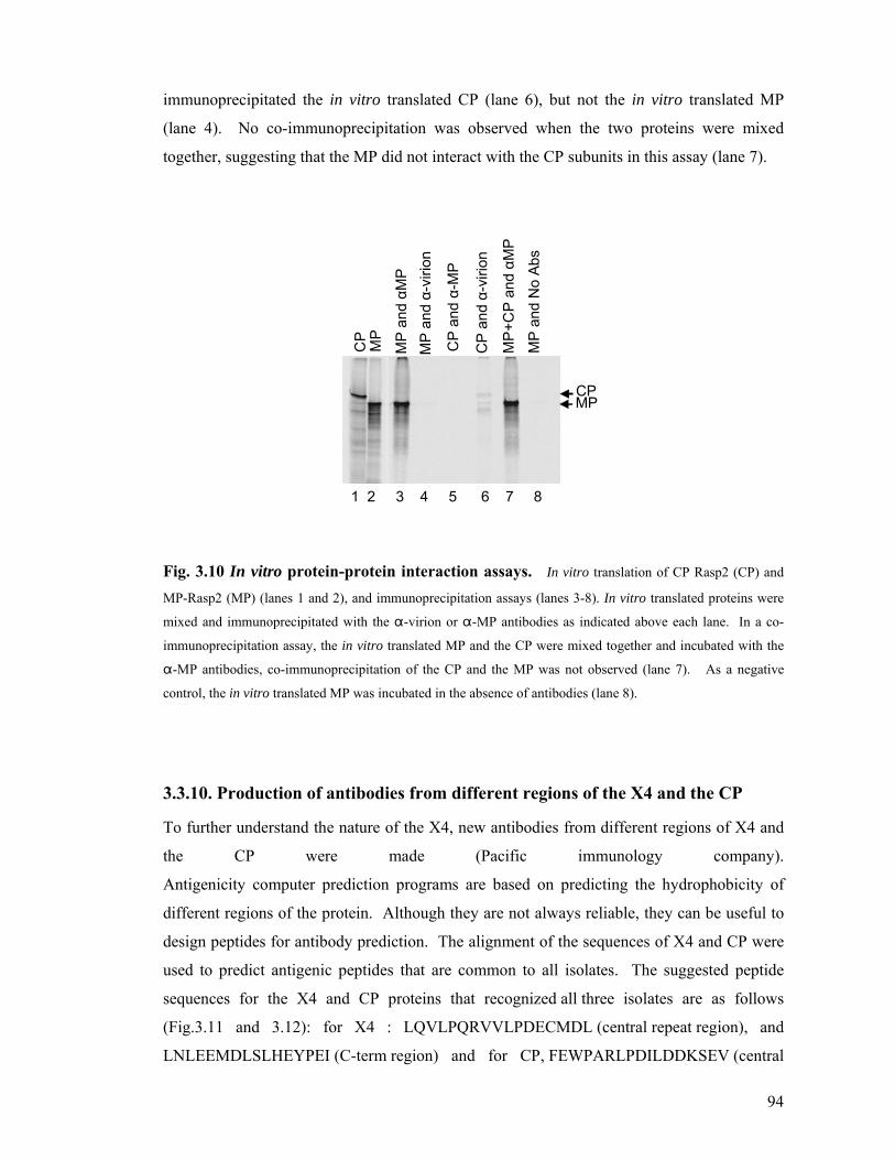
94
immunoprecipitated the in vitro translated CP (lane 6), but not the in vitro translated MP
(lane 4). No co-immunoprecipitation was observed when the two proteins were mixed
together, suggesting that the MP did not interact with the CP subunits in this assay (lane 7).
CP
MP
MP
and αM
P
MP
and α-
virio
n
CP
and α-
MP
CP
and α-
virio
n
MP
+CP
and αM
P
MP
and
No
Abs
1 2 3 4 5 6 7 8
CPMP
Fig. 3.10 In vitro protein-protein interaction assays. In vitro translation of CP Rasp2 (CP) and
MP-Rasp2 (MP) (lanes 1 and 2), and immunoprecipitation assays (lanes 3-8). In vitro translated proteins were
mixed and immunoprecipitated with the "-virion or "-MP antibodies as indicated above each lane. In a co-
immunoprecipitation assay, the in vitro translated MP and the CP were mixed together and incubated with the
"-MP antibodies, co-immunoprecipitation of the CP and the MP was not observed (lane 7). As a negative
control, the in vitro translated MP was incubated in the absence of antibodies (lane 8).
3.3.10. Production of antibodies from different regions of the X4 and the CP
To further understand the nature of the X4, new antibodies from different regions of X4 and
the CP were made (Pacific immunology company).
Antigenicity computer prediction programs are based on predicting the hydrophobicity of
different regions of the protein. Although they are not always reliable, they can be useful to
design peptides for antibody prediction. The alignment of the sequences of X4 and CP were
used to predict antigenic peptides that are common to all isolates. The suggested peptide
sequences for the X4 and CP proteins that recognized all three isolates are as follows
(Fig.3.11 and 3.12): for X4 : LQVLPQRVVLPDECMDL (central repeat region), and
LNLEEMDLSLHEYPEI (C-term region) and for CP, FEWPARLPDILDDKSEV (central

95
region) and NNPVGRPPENV (C-terminal extension). Immunoblotting were conducted
using the X4 (3741-42, C-terminal region) and X4 (3743-44, repeat region) antibodies
(Fig.3.11).
Rasp1 GGFSLPARKGVYVAPTIQGVVRAGLRAQKGFLNAVSAGIVAGARILKSKSQNWFRKSMGIAHEYVEGSLASTILECAGPVVQS Rasp2 GGFSLPTRKGVYVAPTVQGVVRAGLRAQKGFLNAVSTGIVAGARILKSKSQNWFRRSMGIAHDYVEGCMASTVLGCAGPVVQRPYB GGFSLPARKGVYVAPTIQGVVRAGLRAQKGFLNAVSTGIVAGARILKSKSQNWFRKSMGIAHEYVEGSLASTILECAGPVVQS
******:*********:*******************:******************:******:****.:***:* *******
Rasp1 KESCAVVAVPPSEELISWVPSCTEYASNFPRLTCSIFTEWQKPRKQSIVVFNPFRKLIDCALIMSGVSLIASVLLREVAENLVV Rasp2 QEACSVVAAPPIVEPVLWVPPLSEYANDFPKLTCSTFTEWQRPRKQSIAISNLFRKLIDRALLVSGVSLIASVLLFEIAENFAVPYB KESCAVVAVPPSGEFTSWVPSCTEYVSNFPKLTCSIFTEWQKPRKQPIVVSSPFRKLIDCALIMSGVSLIASVLLLEIAENLAV
:*:*:***.** * ***. :**..:**:**** *****:****.*.: . ****** **::*********** *:***:.*
Rasp1 RQVVHPREMLPCAVFVSEKSLVPLDEWSFYLRKYLSPPPYPFGRESFYFQARPRFIGPMLSMVRAVPRIVQQPTMTEELEFEVP Rasp2 RQAVCPVEMPSCATSVSEKSLVSLDEGNFYLRKYLSPPPYPFGRESFYFQARPRFIGPMPSMVRAVPQIVQQPTMTEELEFEVP PYB RQVVRPREMLPCAVFVSEKSLVRLDERSFYLRKYLSPPPYPFGRESFYFQARPRFIGPMLPMVRAVPRIVQQPTMTEELDFEVP
**.* * ** .**. ******* *** .******************************* .******:***********:****
Rasp1 SSWSSPLPLFANFKVNRGACFLQVLPQRVVLPDECMDLLSLFEDQLPEGPLPPFSWSSPLPLFANFKVNRGACFLQVLPQRVVL Rasp2 SSWSSPLPLFANFKVNRGACFLQVLPQRVVLPDECMDLLSLFEDQLPEGPLPSFSWSSPLPLFANFKVNRGACFLQVLPQRVVLPYB SSWSSPLPLFANFKVNRGACFLQVLPQRVVLPDECMDLLSLFEDQLSEGPLPPFSWSSPLPLFASFKVNRGACFLQVLPQRVVL
**********************************************.*****.***********.*******************
Rasp1 PDECMDLLSLFEDQLPEGPLPPFSWSSPLPLFANFKVNRGACFLQVLPQRVVLPDECMDLLSLFEDQLPEGPLPPFSWSSPLPL Rasp2 PDECMDLLSLFEDQLPEGPLPSF-----------------------------------------------------SWSSPLPLPYB PDECMDLLSLFEDQLSEGPLPPF-----------------------------------------------------SWSSPLPL
***************.*****.* ********
Rasp1 FTSFKVNRGACFLQVLPARRVVLDEFMDVLPFLLSPPVLHQEEEPEMAPAVLEAATTPLVLHQEEEPEMAPAVLEAVTTPLVLH Rasp2 FASFKVNRGACFLQVLPARKVVSDEFMDVLPFLFS--------------------------------------------PLVSHPYB FASFKVNRGACFLQVLPARRVVPDEFMDVSPFLTT--------------------------------------------PLVLH
*:*****************:** ****** *** : *** *
Rasp1 QEEESRMVPAVLEAANSIDDVTEAFFDDLECESFYDSYSCEEEAEWAEVPRCKTMSELYASLTFVGDAEGLRKSHMTYFRRLVA Rasp2 QEEEPEMVPAVLEAADSVGDITEAFFDDLECESFYDSYSDEEEAEWAEVPRCKTMSELCASLTLAGDAEGLRKSHGVFLKRLVTPYB QEEESRMVPAVLEAANSIDDVTEAFFDDLECESFYDSYSDEEEAEWAEVPRCKTMSELSTSFTLVGDVEGLRKSHVTFFRRLVT
****..*********:*:.*:****************** ****************** :*:*:.**.******* .:::***:
Rasp1 YFRSFEEPLYSSRAFYSVKVKPVYRPKKFEGHIDCTCLDGNMGEWEWRESVDAMWRCPGRLLNAKRTYTRDDWERVQYLRISSN Rasp2 YLQSFEEPLYSSRAFYSVKVKPVYRPKKFEGHIDCTCLDGNMGEWEWRESVDAMWRCPGRLLNTKRTFTRDDWERVQYLRIGFNPYB YFRSFEEPLYSSRAFYDVKVKPVYRPKKFEGHIDCTCLDGNMGEWEWRESVDAMWRCPGRLLNAKRTYTRDDWERVQYLRMGFN
*::*************.**********************************************:***:************:. *
Rasp1 GGRYRQNWRILNLEEMDLSLHEYPEIPSAPVQSSLFSRVVNRGATLASNIPFVTRSNCQ Rasp2 EGRYRRNWRVLNLEEMDLSLHEYPEISSAPVQSSLFSRVVDRGATLASSIPFVTRSNCQPYB EGRYRRNWRILNLEEMDLSLHEYPEIPSAPVQSSLFSRVVDRGATLASGIPFVTRSNCQ
:****:***:****************.*************:*******.**********
X4-C protein
Abs3: C-terminal region
Abs 2: central repeat region
Fig. 3.11 X4 protein: Alignment of 3 different isolates of ToRSV (Rasp1, Rasp2 and
PYB). The three peptides designed for antibody production are from the central repeat region and C-terminal
region of the X4 protein (in colored boxes). Also the region of the X4 protein which was used to produce the
X4(64-65) antibodies (X4-C peptide) at the C-terminal region of the protein (last 200 amino acids) is shown by
the arrow.

96
Rasp2 GGSWQEGTEAAYLGKVTCAKDAKGGTLLHTLDIIKECKSQNLLRYKEWQRQGFLHGKLRL 60 Rasp1 GGSWQEGTEAAFLGKVTCAKDAKGGTLLHTLDIIKECKSQNLLRYKEWQRQGFLHGKLRL 60 PYB1 GGSWQEGTEAAFLGKVTCAKDAKGGTLLHTLDIIKECKSQNLLRYKEWQRQGFLHGKLRL 60
***********:************************************************
Rasp2 RCFIPTNIFCGHSMMCSLDAFGRYDSNVLGASFPVKLASLLPTEVISLADGPVVTWTFDI 120 Rasp1 RCFIPTNIFCGHSMMCSLDAFGRYDSSVLGASFPVKLASLLPTEVISLADGPVVTWTFDI 120 PYB1 RCFIPTNIFCGHSMMCSLDAFGRYDSNVLGASFPVKLASLLPTEVISLADGPMVTWTFDI 120
**************************.*************************:*******
Rasp2 GRLCGHGLYYSEGAYARPKIYFLVLSDNDVPAEADWQFTYQLLFEDHTFSNSFGAVPFIT 180 Rasp1 GRLCGHGLYYSEGAYARPKIYFLILSDNDVPAEADWQFTYQLLFEDHTFSNSFGAVPFIT 180 PYB1 GRLCGHGLYYSEGAYAKPKIYFLILSDNDVPAEADWQFTYQLLFEDHTFSNSFGAVPFIT 180
****************:******:************************************
Rasp2 LPHIFNRLDIGYWRGPTEIDLTSTPAPNAYRLLFGLSTVISGNMSTLNANQALLRFFQGS 240 Rasp1 LPHIFNRLDIGYWRGPTEIDLTSTPAPNAYRLLFGLSTVISGNMSTLNANQALLRFFQGS 240 PYB1 LPHIFNKLDIGYWRGPTEIDLTSTPAPNAYRLLFGLSTVISGNMSTLNANQALLRFFQGS 240
******:*****************************************************
Rasp2 NGTLHGRIKKIGTALTTCSLLLSLRHKDASLTLETAYQRPHYILADGQGAFSLPISTPHA 300 Rasp1 NGTLHGRIKKIGTALTTCSLLLSLRHKDADLTLETAYQRPHYILANGQGAFSLPISTPHA 300 PYB1 NGTLHGRIKKIGTALSTCSLLLSLRHKDASLTLETAYQRPHYILADGQGAFSLPISTPHA 300
***************:*************.***************:**************
Rasp2 ATSFLEDMLRLEIFAIAGPFSPKDNKAKYQFMCYFDHIELVEGVPRTIAGEQQFNWCSFR 360 Rasp1 ATSFLEDMLRLEIFAIAGPFSPKDNNAKYQFMCYFDHIELVEGVPRTIAGEQQFNWCSFR 360 PYB1 ATSFLEDMLRLEIFAIAGPFSPKDNKAKYQFMCYFDHIELVEGVPRTLAGEQQFNWCSFS 360
*************************:*********************:***********
Rasp2 NFKIDDWKFEWPARLPDILDDKSEVLLRQHPLSLLISSTGFFTGRAIFVFQWGLNTTAGN 420 Rasp1 NFKIDDWRFEWPARLPDILDDKSEVLLRQHPLSLLISSTGFFTGRAIFVFQWGLNTTAGN 420 PYB1 NFKIDDWKFEWPARLPDILDDKSEVLLRQHPLSLLISSTGFFTGRAIFVFQWGLNTTAGN 420
*******:****************************************************
Rasp2 MKGSFSARLAFGKGVEEIEQTSTVQPLVGACEARIPVEFKTYTGYTTSGPPGSMEPYIYV 480 Rasp1 MKGSFSARLAFGKGVEEIEQTSTVQPLVGACEARIPVEFKTYTGYTTSGPPGSMEPYIYV 480 PYB1 MKGSFSARLAFGKGVEEIEQTSTVQPLVGACEARIPVEFKTYTGYTTSGPPGSMEPYIYV 480
************************************************************
Rasp2 RLTQAKLVDRLSVNVILQEGFSFYGPSVKHFKKEVGTPSATLGTNNPVGRPPENVDTGGP 540 Rasp1 RLTQAKLVDRLSVNVILQEGFSFYGPSIKHFKKEVGTPSATLETNNPVGRPPENVDTGGP 540 PYB1 RLTQAKLVDRLSVNVILQEGFSFYGPSVKHFKKEVGRPSATLGVNNPVGRPPENVNTGGP 540
***************************:******** ***** .***********:****
Rasp2 GGQYAAALQAAQQAGKNPFGRG 562 Rasp1 GGQYAAALQAAQQAGKNPFGRG 562 PYB1 GGQYAAALQAAQQAGENPFGRG 562
***************:******
C-terminal extension
Fig. 3.12 CP protein: Alignment of 3 different isolates of ToRSV (Rasp1, Rasp2 and
PYB). The two peptides designed for antibody production are from the central region, CP (3739-40) and the
C-terminal extension of the coat protein, CP (3737-38) (in colored boxes).

97
To test the specificity of the CP (3737-38) antibodies, immunoblotting were conducted using
the antibodies against the C-terminal extension of the CP (polypeptide,
TLETNNPVGRPPENVD). This antibody reacted specifically with the E. coli expressed CP
but not with the X4-Rasp1 protein (data not shown). The CP-2 antibody was made against a
different region of CP (polypeptide, PARLPDILDDKSEV). This antibody also detected the
E. coli expressed CP and did not recognise the X4-Rasp1 protein (data not shown). Both
antibodies detected the full-length CP in infected plant extracts (60 kDa protein). The
truncated CP (53-55 kDa) was detected in infected plant extracts by the CP (3739-40)
antibodies but not by the CP (3737-38) antibodies, confirming that it is missing the C-
terminal extension of the CP (Fig. 3.13).
The X4(3743-44) antibody (in the B3 repeat TPLVLHQEESRMV) reacted specifically with
the E. coli expressed X4-Rasp1 protein but not with the E. coli expressed CP (data not
shown). The X4 (3741-42) antibody (a polypeptide against the C-terminal region of X4
PGRLLNAKRTYTRDD) also recognized the E. coli expressed X4-Rasp1 but not CP (data
not shown). This is in contrast with the previous X4(64-65) antibodies that cross-reacted
with the CP. The X4(3743-44) and X4(3741-42) antibodies did not detect the full length X4
or the 60 kDa protein that was detected by the X4(64-65) antibodies in infected extracts.
Therefore the 60 kDa protein recognized by the X4(64-65) antibodies is not derived from the
X4 coding region and probably corresponds to the full length CP. As shown above, the anti
CP (3737-38) antibodies against the C- terminal extension recognized the 60 kDa protein
confirming that this protein is the full length CP. Both new antibodies did detect a 45 kDa
protein (Fig. 3.14B). A 45 kDa protein was also detected by the N-X4 antibodies. The nature
of this protein needs further investigation. Also post-nuclear extracts derived from ToRSV-
infected leaves were separated by sucrose gradient fractionation (as described in section
3.2.3). Fractions were collected from the bottom of the gradient and were analyzed by
immunoblotting using the X4(3741-42) and X4(3743-44) antibodies (Fig.3.14B). In the
sucrose gradient fractionation a 45 kDa protein was detected on top of the gradient,
suggesting that it may be a soluble protein (Fig 3.14 B).

98
5 6 5 6 5 6 5 6 5 6 5 6 (Fraction number from sucrose gradient)
97
66
45
31
H I H I H I H I H I H I
Anti-virion CP(3739-40) CP(3737-38) Antibodies
middle region C-terminal extention
Mr
Fig. 3.13 Immunoblot analysis of the infected (I) and healthy (H) N. benthamiana
extracts using the anti-virion, anti-CP (3739-40) and anti-CP (3737-38) antibodies. Sucrose gradient fractions from healthy and infected plant extracts were prepared and fraction number 5 and 6
containing virions were used for immunoblotting and tested against various CP antibodies. The full length CP
(60 kDa) was detected by all CP antibodies, the CP (3737-38), against the C-terminal extension) only detected
the upper band corresponding to the full length CP.

99
H I nf H I nf H Inf
X4(N-term) X4 (3741-42) X4(3743-44)
31
45
6697
116
21
Mr
Antibodies:
A
45
6697
31
Mr
Fraction number: 1 2 3 4 5 6 7 8 9 10 11 12 13 14 1 2 3 4 5 6 7 8 9 10 11 12 13 14 1 2 3 4 5 6 7 8 9 10 11 1213 14
HealthyAnti-X4(3743-44)
B3 repeat region
Anti-X4(3741-42)
C-terminal
B
Fig. 3.14 Immunoblot analysis of the infected (Inf) and healthy (H) N. benthamiana
extracts using the X4-N, X4(3743-44) and X4 (3741-42) antibodies. (A) Post-nuclear fractions
(S3 fractions) were tested against various X4 antibodies. The full length X4 (expected molecular mass of 82
kDa) or the 60 kDa protein previously detected by the X4(64-65) antibodies were not detected in infected plant
extracts by the X4(3743-44) or X4(3741-42) antibodies. A faint 45 kDa protein (shown by arrow) was detected
that may correspond to a truncated form of the X4 protein. (B) Immunoblott detection of the X4 protein in
sucrose gradient fractions in healthy and infected plant extracts with two new X4 antibodies, X4(3741-42) and
X4(3743-44). The antibodies detected a 45 kDa protein and also degradation products at the top of the gradient
.Molecular marker (Mr).
100
3.4 Discussion
The objective of this section of my research was to characterize the ToRSV X4 protein in
planta. Towards this objective, I have used the X4(64-65) antibodies (a polyclonal
antibody). The X4(64-65) antibodies were raised against a recombinant protein
corresponding to the C-terminal 15 kDa of X4 (from the Rasp2 isolate) fused to a His tag.
The X4(64-65) antibody recognized a 60 kDa protein in ToRSV infected plant extracts but
not in healthy plant extracts. The 60 kDa protein co-fractionated with empty and full virus
particles after sucrose gradient fractionation of infected plant extracts and co-purified with
the virus particles. The results of trypsin digestion experiments and immunogold labelling
suggest that the epitope of the 60 kDa protein recognized by the X4(64-65) antibody is
exposed on the surface of the virus particle. The 60 kDa protein was in high concentration in
plant virus extracts but in lower concentration after virus purification. After long storage of
purified virus the 60 kDa protein was not detected by the X4(64-65) antibodies.
The "-virion antibody detected a 53-55 kDa doublet as well as a 60 kDa protein, which co-
migrated with the 60 kDa protein recognized by the X4(64-65) antibody. These proteins were
detected in plant extract as well as in purified virus preparations. As I explained in the
introduction two forms of the CP are often observed in nepoviruses: the full-length form and
a smaller form which has a C-terminal truncation. While the full-length form is predominant
early in infection, the truncated form becomes predominant later in infection or during
purification process as shown for BRV (Latvala et al., 1998) and for TBRV (Demangeat et
al., 1992). In the case of ToRSV, the C-terminal extension is predicted to be 54 amino acids
(approximately 6 kDa). The difference in the apparent molecular mass between the 60 kDa
and 53-55 kDa proteins detected by the "-virion antibodies would be consistent with the 60
kDa protein being the full-length CP and the 53-55 kDa protein corresponding to a truncated
form of the protein lacking the C-terminal extension. Consistent with this suggestion, a full-
length recombinant CP-His expressed in E. coli comigrated with the 60 kDa protein
recognized by the "-virion antibodies (Fig. 3.8B). Also, the 60 kDa protein was recognized
by all ToRSV CP antibodies available in the lab. In contrast, the antibodies raised against the
C-terminal extension did not recognize the 53-55 kDa proteins confirming that they
correspond to truncated forms of CP (Fig.3.13). The relative ratio of the 53-55 kDa and 60
kDa proteins recognized by the "-virion antibodies varied. The 60 kDa protein was
predominant in crude infected plant extracts. In purified virus preparation, the ratio of the 60

101
kDa protein to the 53-55 kDa doublets was lower. After extended storage of purified virus
preparation, the 60 kDa protein was not detectable by immunoblotting. The pattern of
detection of the 60 kDa recognized by the "-virion antibody was similar to that observed for
the 60 kDa recognized by the X4(64-65) antibody. This raised the possibility that the two
antibodies recognized the same protein. Alternatively, two distinct 60 kDa proteins present in
infected plant tissues are recognized by the two different antibodies.
The results from the mass spectrometry suggested that purified virus preparations contained
predominately the coat protein and the Rubisco. Sequences corresponding to the X4 protein
were not detected, although these results do not exclude the possibility that small amounts of
the X4 protein were present in the preparations. These results raise the question of the nature
of the 60 kDa protein detected by X4(64-65) antibodies.
There are two possible interpretations of the results. The first possible interpretation is that
the 60 kDa protein recognized by the X4(64-65) antibody is a truncated form of the X4
protein. In this interpretation of the result, the 60 kDa protein recognized by the X4(64-65)
antibody is a distinct protein that co-migrates with the 60 kDa full-length CP. The specificity
of the X4(64-65) antibodies for the X4 protein but not for the CP protein in the
immunoprecipitation assays is consistent with this interpretation of the results (Fig. 3.2).
This interpretation of the results would suggest that the 60 kDa X4 protein is a virion-
associated protein that becomes disassociated from the virions during the course of infection
or during the course of virus purification. In addition to this protein there was also a ~120
kDa protein band detected by the X4(64-65) antibodies. The nature of this protein needs
further investigation. If we assume that the 60 kDa protein is a truncated form of X4, one
possibility is that the 120 kDa protein is an aggregated form (dimer) of the 60 kDa protein
detected by the X4(64-65) antibodies. However, based on computer-based prediction of
hydrophobicity (Chapter 1, Fig.1.11), X4 is not very hydrophobic. Another possibility is that
the 120 kDa protein corresponds to a possible X3-X4 protein, a predicted intermediate
precursor of the mature X4 protein that may be associated with the virion. This hypothesis
would also suggest that the 60 kDa (or the 120 kDa protein) detected by the X4(64-65)
antibodies are exposed at the surface of the virions. The fact that the "-virion antibodies
recognized the in vitro synthesized X4 protein in the immunoprecipitation assay could
suggest that the 60 kDa or the 120 kDa protein detected by the X4(64-65) antibodies was

102
present in the original virus preparation used for the production of these antibodies (Fig 3.5
A).
The second possible interpretation of the results is that the X4(64-65) antibody cross-react
with the full-length 60 kDa CP but not with the truncated 53-55 kDa form of the CP. The
cross-reactivity between the X4(64-65) antibodies and the E. coli expressed full-length CP in
immunoblotting experiments is consistent with this suggestion. The lack of detection of X4
sequences in mass spectrometry using purified virus preparations is also consistent with this
hypothesis. This interpretation of the results implies that the X4(64-65) antibody may
recognizes an epitope present on the C-terminal extension of the CP and that this epitope is
exposed at the surface of the virions. To reach a clear conclusion about the nature of the X4
protein and further understand the role of this protein in virus infection more antibodies were
made from different regions of the CP and the X4. The anti-CP (3737-38) and CP (3739-40)
antibodies detected the full length CP (60 kDa protein) in plant extracts. However, the anti-
X4(3741-42) and (3743-44) antibodies did not detect either the 60 kDa protein or the
predicted 82 kDa full length X4. The results from these antibodies were consistent with the
results obtained by the N-terminus X4 antibodies. Also, preliminary results of the expression
of X4-GFP protein in plants by biolistic delivery of the plasmid into N. benthamiana
followed by immunodetection by the GFP antibodies did not allow the detection of the
predicted full-length X4-GFP (data not shown). The observations that the X4 protein was not
detected by four different antibodies in extracts derived from ToRSV-infected or transiently
transfected plants (GFP antibodies, two new X4(3741-42) and X4(3743-44) antibodies and
the N-terminal X4 antibodies) support the second interpretation in which the 60 kDa protein
recognized by the X4(64-65) antibodies probably is the full length CP and this antibodies had
cross reacted with the CP.
As mentioned above, the most likely interpretation of my results is that the X4(64-65)
antibodies recognize an epitope within the C-terminal extension of the full-length CP. This
interpretation of the results would imply that this C-terminal extension is exposed at the
surface of the virion. This is consistent with the presence of an exposed C-terminal extension
in other nepoviruses and comoviruses as I discussed in the introduction. The C-terminal
extension can dissociate from the virus particle during purification or prolonged storage of
the virus, therefore the X4(64-65) would not be able to detect the C-terminal extension in
those preparations.

103
As I discussed in Chapter 1, nematodes acquire and retain virus particles but release the
particle during their feeding process to allow plant-to-plant transmission. The CP C-terminal
extension can be a potential site for interaction with the nematode receptor, since it can be
released from the virus particle. In tomato black ring virus (TBRV) the C-terminal extension
of the coat protein protrude from the virus particle surface and can be removed without
disrupting the virion. This exposed detachable fragment may play a significant role in TBRV
biology as does the protruding N-terminal fragment of potyvirus coat proteins in their
transmission by aphids (Atreya et al., 1990).
The C-terminal extension in ToRSV is longer in size than the BRV C-terminal extension.
This part of CP has no sequence homology with the CP of other nepoviruses or comoviruses
sequenced so far (Seitsonen et al., 2008). Since the sequence is specific, the C-terminal
extension is valuable for making valid antigenic targets for diagnostic and quick indication
of ToRSV disease. The development of sensitive and reliable antibodies against ToRSV
surface exposed CP sequences combined with regular tests would allow the recognition of
the disease long before the symptoms become evident. Early detection might help in
controlling the disease which may alleviate the economic impact of ToRSV on fruit crops all
over the world.
In immunoprecipitation assays, the "-virion antibodies detect the full length X4 from Rasp2
or Rasp1 (Fig. 3.2A and data not shown). Also the X4(64-65) antibodies cross-reacted with
the full length E. coli expressed CP in immunoblotting experiments. One possible
explanation for these results is that the CP and X4 proteins share a common epitope.
Alignment of the CP and the X4 did not show significant sequence homology (Fig. 3.12)
although the possibility of a structural epitope can not be excluded.

104
CP GGSWQEGTEAAYLG-----KVTCAKDAKGGTLLHTLD------IIKECKSQNLLRYKEWQ 49
X4 GGFSLPTRKGVYVAPTVQGVVRAGLRAQKGFLNAVSTGIVAGARILKSKSQNWFRRSMGI 60
** :..*:. * .. *: * * . * :.**** :* .
CP RQGFLHGKLRLRCFIPTNIFCGHSMMCSLDAFGEYDSNVLGASFPVKLASLLPTEVIS-- 107
X4 AHDYVEGCMASTVLGCAGPVVQRQEACSVVAAPPIVEPVLWVPPLSEYANDFPKLTCSTF 120
:.::.* : : :. . :. **: * . ** .. : *. :*. . *
CP ----LADGPVVTWTFDIGRLCGHGLYYSEGAYARPKIYFLVLS---DNDVPAEADWQFTY 160
X4 TEWQRPRKQSIAISNLFRKLIDRALLVSGVSLIASVLLFEIAENFAVRQAVCPVEMPSCA 180
. :: : : :* .:.* * : . : * : . .:. . .:
CP QLLFEDHTFSNSFG----AVPFITLPHIFNRLDIGYWRGPTEIDLTSTPAPNAYRLLFGL 216
X4 TSVSEKSLVSLDEGNFYLRKYLSPPPYPFGRESFYFQARPRFIGPMPSMVRAVPQIVQQP 240
: *. .* . * : . *: *.* .: : * *. .: . . :::
CP STVISGNMSTLNANQALLRFFQGSNG--------TLHGRIKKIGTALTTCSLLLSLRHKD 268
X4 TMTEELEFEVPSSWSSPLPLFANFKVNRGACFLQVLPQRVVLPDECMDLLSLFEDQLPEG 300
: . . ::.. .: .: * :* . : .* *: . .: **: . :.
CP ASLTLETAYQRPHYILADGQGAFSLPISTPHAATSFLEDMLRLEIFAIAGPFSPKDNKAK 328
X4 PLPSFSWSSPLPLFANFKVNRGACFLQVLPQRVVLPDECMDLLSLFEDQLPEGPLPSFSW 360
. ::. : * : . : . .: *: .. * * *.:* * .* . :
CP YQFMCYFDHIELVEG-------VPRTIAGEQQFNWCSFRNFKIDDWKFEWPARLPDILDD 381
X4 SSPLPLFASFKVNRGACFLQVLPARKVVSDEFMDVLPFLFSPLVSHQEEEPEMVPAVLEA 420
. : * ::: .* .*.:..:: :: .* : . : * * :* :*:
CP KSEVLLRQHPLSLLISSTGFFTGRAIFVFQWGLNTTAGNMKGSFSARLAFGKGVEEIEQT 441
X4 ADSVGDITEAFFDDLECESFYDSYSDEEEAEWAEVPRCKTMSELCASLTLAGDAEGLRKS 480
..* ..: :.. .*: . : :.. : ..:.* *::. ..* :.::
CP S--TVQPLVGACEARIPVEFKTYTGYTTSGPPGSMEPYIYVRLTQAKLVD--------RL 491
X4 HGVFLKRLVTYLQSFEEPLYSSRAFYSVKVKP-VYRPKKFEGHIDCTCLDGNMGEWEWRE 539
:: ** :: :.: : *:.. * .* : :.. :* *
CP SVNVILQEGFSFYGPSVKHFKKEVG-TPSATLGTNN-----------------PVGRPPE 533
X4 SVDAMWRCPGRLLNTKRTFTRDDWERVQYLRIGFNEGRYRRNWRVLNLEEMDLSLHEYPE 599
**:.: : : ... .. :.: . :* *: .: . **
CP NVDTGGPGGQYAAALQ-AAQQAGKNPFGRG---- 562
X4 ISSAPVQSSLFSRVVDRGATLASSIPFVTRSNCQ 633
.: .. :: .:: .* *.. **
Fig.3.15. Sequence alignment between the X4 protein and the CP of ToRSV. There is no
significant homology suitable for a common epitope between the two sequences.

105
The X4 coding region is present in the RNA2 from all ToRSV infected isolates that were
sequenced so far. However the X4 full length protein (e.g 82 kDa in Rasp1) is not detectable
in plant extracts. Other ToRSV viral proteins studied so far are readily detected by
antibodies in infected plant extracts.
In ToRSV, each viral RNA codes for a large polyprotein. The polyprotein is cleaved by the
viral protease (Pro) to mature and intermediate proteins. Therefore theoretically the cleavage
products should have a ratio of 1:1. The absence of detection of the full-length X4 protein in
plant extracts could be due to a shorter half-life of the X4 protein compared to other viral
proteins like the MP or the CP, which are readily detected in infected plant extracts. This
suggests that X4 may be subject to degradation.
Cauliflower mosaic virus (CaMV) particles are stable although its capsid protein (CP)
precursor is very unstable in plant protoplasts when it is expressed outside the viral context
(Leclerc et al., 1999). The capsid is assembled from three related proteins p44, p39 and p37.
These proteins are all derived from bi-terminal processing of the 57 kDa CP precursor. The
N-terminus of p44 contains a PEST motif (degradation motif, Chapter 1). It is suggested that
the PEST signal within the N-terminus of the precursor targets the protein to the proteasome
in plants. It is suggested that PEST related signals may act as degradation signals in plants
(Baumberger et al., 2007), however the algorithm was developed for mammalian proteins
and needs to be adapted for the plant system ( Chapter 1 section 1.10.1) (Karsies et al., 2001).
Little is known about the degradation pathways in either the nucleus or the cytoplasm of
plant cells (Karsies et al., 2001). Viruses can use the degradation pathways of their host for
their own benefit. Poleroviruses (family Luteoviridae) suppress RNA silencing activity in N.
benthamiana plants using the viral P0 protein which targets the Argonaute protein for
degradation (Baumberger et al., 2007). Phosphorylation often plays a role in regulation of
PEST signal target degradation. There are a few predicted phosphorylation sites in the X4
region of ToRSV isolates and there are a few predicted PEST motifs (Chapter 1). The
number of predicted PEST motifs was greater in the X4 protein from the Rasp1 isolate. The
PEST motifs were present within the protein repeats, therefore the greater number of the
PEST motifs in Rasp1 may correlate with protein instability.
106
As discussed above, our results suggest that the full length X4 protein is an unstable protein.
Another possibility is that since the cleavage between X3 and X4 is a suboptimal cleavage
site, the protein may not has been cleaved from its precursor as efficiently as other viral
proteins like the MP or the CP. Larger proteins are occasionally detected in ToRSV-plant
extracts by all of the X4 antibodies but the nature of these proteins requires further
investigation. It would be interesting to investigate the possibility of X3 as a stabilizing
sequence for the X4 protein.
X4 is also a highly diverse protein among ToRSV isolates that contains tandem repeats and
an extensive number of point mutations (Chapter 2). What is the possible function(s) of X4?
In particular, why does the virus code for a protein that is absent from the genome of many
other nepoviruses? Indeed, the coding region for X4 is retained in the virus genome even
after extensive passages of the virus on herbaceous hosts. As mentioned in Chapter 2, a
possible role in symptomatology or protein–protein interaction could be considered. The titre
of ToRSV viral RNA remains at high concentration in recovered leaves in contrast with other
nepoviruses. In Chapter 4, I will discuss preliminary results suggesting that X4 may be a
silencing suppressor.
3.4.1. Summary and conclusion
Using X4(64-65) antibodies raised against the C-terminal region of X4, I have detected a 60
kDa protein in infected plants. A series of experiments aimed at analyzing the nature of this
60 kDa protein and its possible interaction with the virus particles allowed me to formulate
two possible interpretations for the results. (1) The 60 kDa protein (or the possible 120 kDa
precursor protein) recognized by the X4(64-65) antibody is a truncated form of the X4
protein, which is associated with virus particles and (2) the X4 antibodies recognize the C-
terminal extension of the full-length CP. Recent new antibodies raised against different
regions of X4 and CP, supported the second hypothesis. Based on the results from the X4
(3741-42), X4(3743-44) and the N-X4 antibodies, the predicted full length X4 (82 kDa)
protein was not detected in ToRSV-infected plant extracts or the protein concentration was
lower than the detection level by immunoblotting in infected plant extracts. This suggests
that the full length X4 protein may be an unstable protein although further experiments are
required to address this question.

107
3.5 Bibliography
Andret-Link, P., Schmitt-Keichinger, C.,Demangeat, G.,Komar, V and Fuchs, M. (2004).
The specific transmission of Grapevine fanleaf virus by its nematode vector
Xiphinema index is solely determined by the viral coat protein. Virology 320(1), 12-
22.
Atreya, C. D., Raccah, B., and Pirone, T. P. (1990). A point mutation in the coat protein
abolishes aphid transmissibility of a potyvirus. Virology 178(1), 161-5.
Baumberger, N., Tsai, C. H., Lie, M., Havecker, E., and Baulcombe, D. C. (2007). The
Polerovirus Silencing Suppressor P0 Targets ARGONAUTE Proteins for
Degradation. Curr Biol 17(18), 1609-1614.
Belin, C., Schmitt, C., Gaire, F., Walter, B., Demangeat, G., and Pinck, L. (1999). The nine
C-terminal residues of the grapevine fanleaf nepovirus movement protein are critical
for systemic virus spread. J Gen Virol 80 (6), 1347-56.
Bertioli, D. J., Harris, R. D., Edwards, M. L., Cooper, J. I., and Hawes, W. S. (1991).
Transgenic plants and insect cells expressing the coat protein of arabis mosaic virus
produce empty virus-like particles. J Gen Virol 72 ( Pt 8), 1801-9.
Carrier, K., Xiang, Y., and Sanfacon, H. (2001). Genomic organization of RNA2 of Tomato
ringspot virus: processing at a third cleavage site in the N-terminal region of the
polyprotein in vitro. J Gen Virol 82(Pt 7), 1785-90.
Carvalho, C. M., Wellink, J., Ribeiro, S. G., Goldbach, R. W., and Van Lent, J. W. (2003).
The C-terminal region of the movement protein of Cowpea mosaic virus is involved
in binding to the large but not to the small coat protein. J Gen Virol 84(Pt 8), 2271-7.
Chisholm, J., Zhang, G., Wang, A., and Sanfacon, H. (2007). Peripheral association of a
polyprotein precursor form of the RNA-dependent RNA polymerase of Tomato
ringspot virus with the membrane-bound viral replication complex. Virology 368(1),
133-144.
Dautel, S., Guidasci, T., Pique, M., Mougeot, J. L., Lebeurier, G., Yot, P., and Mesnard, J. M.
(1994). The full-length product of cauliflower mosaic virus open reading frame III is
associated with the viral particle. Virology 202(2), 1043-5.
Demangeat, G., Hemmer, O., Reinbolt, J., Mayo, M. A., and Fritsch, C. (1992). Virus-
specific proteins in cells infected with tomato black ring nepovirus: evidence for
proteolytic processing in vivo. J Gen Virol 73(7), 1609-14.

108
Gaire, F., Schmitt, C., Stussi-Garaud, C., Pinck, L., and Ritzenthaler, C. (1999). Protein 2A
of grapevine fanleaf nepovirus is implicated in RNA2 replication and colocalizes to
the replication site. Virology 264(1), 25-36.
Han, S., and Sanfacon, H. (2003). Tomato ringspot virus proteins containing the nucleoside
triphosphate binding domain are transmembrane proteins that associate with the
endoplasmic reticulum and cofractionate with replication complexes. J Virol 77(1),
523-34.
Hans, F., and Sanfacon, H. (1995). Tomato ringspot nepovirus protease: characterization and
cleavage site specificity. J Gen Virol 76(4), 917-27.
Karsies, A., Hohn, T., and Leclerc, D. (2001). Degradation signals within both terminal
domains of the cauliflower mosaic virus capsid protein precursor. The Plant Journal
27(4), 335-43.
Kobayashi, K., Tsuge, S., Stavolone, L., and Hohn, T. (2002). The cauliflower mosaic virus
virion-associated protein is dispensable for viral replication in single cells. J Virol
76(18), 9457-64.
Latvala, S., Susi, P., Kalkkinen, N., and Lehto, K. (1998). Characterization of the coat protein
gene of mite-transmitted blackcurrant reversion associated nepovirus. Virus Res
53(1), 1-11.
Leclerc, D., Chapdelaine, Y., and Hohn, T. (1999). Nuclear targeting of the cauliflower
mosaic virus coat protein. J Virol 73(1), 553-60.
Leclerc, D., Stavolone, L., Meier, E., Guerra-Peraza, O., Herzog, E., and Hohn, T. (2001).
The product of ORF III in cauliflower mosaic virus interacts with the viral coat
protein through its C-terminal proline rich domain. Virus Genes 22(2), 159-65.
Leh, V., Jacquot, E., Geldreich, A., Haas, M., Blanc, S., Keller, M., and Yot, P. (2001).
Interaction between the open reading frame III product and the coat protein is
required for transmission of cauliflower mosaic virus by aphids. J Virol 75(1), 100-6.
Leh, V., Jacquot, E., Geldreich, A., Hermann, T., Leclerc, D., Cerutti, M., Yot, P., Keller, M.,
and Blanc, S. (1999). Aphid transmission of cauliflower mosaic virus requires the
viral PIII protein. Embo J 18(24), 7077-85.
Lemmetty, A., Latvala, S., Jones, A. T., Susi, P., McGavin, W. J., and Lehto, K. (1997).
Purification and properties of a new virus from black currant, its affinities with
nepoviruses, and its close association with black currant reversion disease.
Phytopathology 87(4), 404-13.
109
Maia, I. G., Haenni, A., and Bernardi, F. (1996). Potyviral HC-Pro: a multifunctional protein.
J Gen Virol 77 ( Pt 7), 1335-41.
Perbal, M. C., Thomas, C. L., and Maule, A. J. (1993). Cauliflower mosaic virus gene I
product (P1) forms tubular structures which extend from the surface of infected
protoplasts. Virology 195(1), 281-5.
Plisson, C., Uzest, M., Drucker, M., Froissart, R., Dumas, C., Conway, J., Thomas, D., Blanc,
S., and Bron, P. (2005). Structure of the mature P3-virus particle complex of
cauliflower mosaic virus revealed by cryo-electron microscopy. J Mol Biol 346(1),
267-77.
Ritzenthaler, C., Schmit, A.-C., Michler, P., Stussi-Garaud, C., and Pinck, L. (1995).
Grapevine fanleaf nepovirus P38 putative movement protein is located on tubules in
vivo. Mol Plant Microbe Interact 8(3), 379-387.
Rochon, D., and Sanfacon, H. (2001). Nepoviruses. In "Encyclopedia of Plant Pathology" (O.
C. Maloy, and T. D. Murray, Eds.), pp. 704-708. John Wiley & Sons, Inc.
Roth, B. M., Pruss, G. J., and Vance, V. B. (2004). Plant viral suppressors of RNA silencing.
Virus Res 102(1), 97-108.
Rott, M. E., Tremaine, J. H., and Rochon, D. M. (1991). Nucleotide sequence of tomato
ringspot virus RNA-2. J Gen Virol 72(7), 1505-14.
Sanfacon, H., Wieczorek, A., and Hans, F. (1995). Expression of the tomato ringspot
nepovirus movement and coat proteins in protoplasts. J Gen Virol 76(9), 2299-303.
Schaad, M. C., Jensen, P. E., and Carrington, J. C. (1997). Formation of plant RNA virus
replication complexes on membranes: role of an endoplasmic reticulum-targeted viral
protein. Embo J 16(13), 4049-59.
Seitsonen, J. J., Susi, P., Lemmetty, A., and Butcher, S. J. (2008). Structure of the mite-
transmitted Blackcurrant reversion nepovirus using electron cryo-microscopy.
Virology 378(1), 162-8.
Stace-Smith, R. (1966). Purification and properties of tomato ringspot virus and an RNA-
deficient component. Virology 29(2), 240-7.
Stavolone, L., Villani, M. E., Leclerc, D., and Hohn, T. (2005). A coiled-coil interaction
mediates cauliflower mosaic virus cell-to-cell movement. Proc Natl Acad Sci U S A
102(17), 6219-24.
Taylor, K. M., Spall, V. E., Butler, P. J., and Lomonossoff, G. P. (1999). The cleavable
carboxyl-terminus of the small coat protein of cowpea mosaic virus is involved in
RNA encapsidation. Virology 255(1), 129-37.

110
Urcuqui-Inchima, S., Haenni, A. L., and Bernardi, F. (2001). Potyvirus proteins: a wealth of
functions. Virus Res 74(1-2), 157-75.
Uzest, M., Gargani, D., Drucker, M., Hebrard, E., Garzo, E., Candresse, T., Fereres, A., and
Blanc, S. (2007). A protein key to plant virus transmission at the tip of the insect
vector stylet. Proc Natl Acad Sci U S A 104(46), 17959-17964.
Wieczorek, A., and Sanfacon, H. (1993). Characterization and subcellular localization of
tomato ringspot nepovirus putative movement protein. Virology 194(2), 734-42.

111
Chapter 4
Preliminary evidence that the X4 protein may act as a silencing
suppressor in plants
A version of this chapter will be submitted for publication. Jafarpour, B., Chisholm, J
and Sanfacon, H. (2010) Preliminary evidence that the X4 protein may act as a silencing
suppressor in plants.
112
4.1. Introduction
As I discussed in Chapter 1, RNA silencing is one of the natural plant defence mechanisms
against virus infection. RNA silencing generally relies on a set of core reactions that are
triggered by dsRNA, which is processed by the RNase III enzyme Dicer into small RNA
duplexes that are 21–24 bp in length (Ding, 2000; Voinnet, 2005). The helicase selects one
strand of the viral siRNA duplex to be the guide RNA in the mature RNA–induced silencing
complex (RISC) (Schwarz et al., 2003). This strand of the siRNA duplexesis then
incorporated into the RISC complex. A core component of the RISC complex is an
Argonaute protein (Ago), which is an RNase H-like enzyme (Baumberger and Baulcombe,
2005; Zhang et al., 2006) that guides sequence-specific degradation of the complementary
RNA (Ding and Voinnet, 2007; Dunoyer and Voinnet, 2008; Lu, 2003).
The sequence-specificity of this process, together with the fact that dsRNA is a common
product of virus replication, prompted the idea that virus-induced gene silencing is an
antiviral defence response. The dsRNA in virus-infected cells is thought to be the replication
intermediate that are produced during virus replication. Secondary structures within a
specific strand of viral genomic RNA could also be recognized as dsRNA by the silencing
machinery. As I explained above eventually this causes the siRNA/RNase complex to target
the viral single-stranded RNA, and virus accumulation would slow down. Many plant
viruses encode proteins that are suppressors of this RNA silencing process (Brigneti et al.,
1998; Voinnet et al., 1999). These proteins influence the final steady-state level of virus
accumulation (Lu, 2003). This, together with the discovery of siRNAs in virus-infected plant
cells (Hamilton and Baulcombe, 1999) provided further evidence that RNA silencing is an
antiviral defence system of plants. Viral suppressor proteins that counteract RNA silencing
were first identified in plant cells (Anandalakshmi et al., 1998). It was later found that
viruses of insect and mammalian cells, such as influenza virus also encode silencing
suppressor proteins (Li, 2002; Li et al., 2004). This suggests that RNA silencing could have
been an ancient antiviral defence mechanism in primitive eukaryotes and that its function has
been conserved during the evolution of plants and animals (Baulcombe and Molnar, 2004).
As discussed in Chapter 1, one of the well studied plant virus suppressors of silencing is the
19 kDa protein (p19) from tomato bushy stunt virus (genus tombusvirus) (Silhavy et al.,
2002). HC-Pro (potyviral helper component protease) was also one of the first identified

113
silencing suppressors (reviewed in Maia et al., 1996; Maia and Haenni, 1994; Roth et al.,
2004; Urcuqui-Inchima et al., 2001). Viral silencing suppressor proteins are not highly
conserved and the mode of action of these proteins against RNA silencing is diverse.
Theoretically, viruses can combat RNA-silencing mediated defence in at least three ways:
preventing the generation of siRNAs, inhibiting the incorporation of siRNAs into effector
complexes and interfering with one of the effector complexes.
The recovery of plants from virus infection was first described for a nepovirus in 1928
(Wingard, 1928). Initial symptomatic infection of herbaceous plants of the Solanaceae
family by tobacco ringspot virus was shown to be followed by attenuation or elimination of
the symptoms in newly emerging leaves. Characterization of the recovery phenotypes
observed in some natural virus infections provided additional experimental evidence for a
link between RNA silencing and an antiviral defence mechanism (Covey et al., 1997; Ratcliff
et al., 1997; Ratcliff et al., 1999). Plants infected with nepoviruses and caulimoviruses
exhibit a response very similar to the virus-induced recovery. In plants infected with tomato
black ring virus (TBRV, a nepovirus of subgroup B), the recovered leaves exhibit homology-
dependent resistance to secondary infections and the virus titre was dramatically reduced in
the recovered leaves (Covey et al., 1997; Ratcliff et al., 1997; Ratcliff et al., 1999). It has
been demonstrated that recovery of Nicotiana benthamiana plants from the necrotic symptom
induced by ToRSV is associated with RNA silencing. Unlike TBRV-infected plants,
recovered leaves from ToRSV-infected plants did not show reduced virus titre (Jovel et al.,
2007). ToRSV is different from TBRV in that it has a larger coding region in the two RNAs.
The X4 domain in the N-terminal region of the ToRSV RNA-2 encoded polyprotein is absent
in TBRV (Carrier et al., 2001).
4.1.1. Research hypothesis and objectives
The X4 is a variable protein among ToRSV isolates (Chapter 2). The high concentration of
virus in ToRSV infected plants (Jovel et al., 2007) differs from what was previously
observed with other nepoviruses and suggest that ToRSV has the ability to counteract or
evade RNA silencing. Since the X4 protein is absent from the genome of other nepoviruses,
we hypothesized that X4 may suppress silencing in plants. In this study, we tested the
recovery phenotype and symptomatology of different ToRSV isolates. We used a transient
expression assay and the green fluorescent reporter protein (GFP) to determine whether co-

114
expression of X4 and GFP could prevent the induction of RNA silencing directed at GFP.
We show that when X4 is co-expressed with GFP in plants, the concentration of the GFP
protein is higher than when the GFP is expressed alone.
4.2 Materials and methods
4.2.1 PCR amplification of a region of the RNA2 open reading frame coding for
a portion of X3, the entire X4 and a portion of MP for the Chickadee isolates.
Chickadee isolate was obtained from Dr. Zongrang Liu (West Virginia). The exact origin of
this specific Chickadee isolate is not known. However, a previously described Chickadee
isolate was reported to have been introduced to New York State in rooted MM.106 apple
layers (Bitterlin and Gonsalves, 1988). The Chickadee isolate was passaged in cucumber
extensively before it was used in our laboratory. Therefore, it may have adapted to the
cucumber host.
As I discussed in Chapter 2, leaves from N. benthamiana plants infected with Chickadee
isolate were ground in liquid nitrogen and the total RNA was extracted using TRIzol reagent
(Invitrogen). First–strand cDNA was synthesised using 1 μg of total RNA as a template and
primer CP-R (5'-GTCAAGCTTGCCACGCCCGAAAGGAT-3', complementary to nts 5723-
5707 of the RNA2 of ToRSV isolate Rasp2 and Superscript Π Reverse Transcriptase
(Invitrogen). For the PCR reaction, I used primers F208 (5'-
GAGGCCGAATTGGCCTCAAAG-3', corresponding to nts 801-822 of Rasp2 RNA2, and
R207 (5'-GCACCCGCATCAGAGGATC-3', complementary to nts 2998-2980 of Rasp2
RNA2.
4.2.2 Symptom evaluation in plants infected with Rasp1 and PYB ToRSV isolates
ToRSV-Rasp1 and ToRSV-PYB were inoculated on Nicotiana benthamiana plants. An
abrasive (carborundum powder) was dusted on the leaves prior to inoculation. Plants were
inoculated at leaf stage 5 to 7 by inoculating two leaves with approximately 40 μl of fresh
leaf extracts of ToRSV-infected plants diluted 1:4 (w:v) in inoculation buffer (0.1 M
phosphate buffer pH 7.4). The plants were grown in conviron climate chamber at 21°C and
27°C for symptom development and were assessed at regular interval for 22° days post
inoculation.
115
4.2.3. Plasmid construction
Several agroinfiltration vectors were constructed. The pCITE-X4-Rasp1-HA and pCITE-
X4-Rasp2-HA (containing the coding region for the X4 protein from Rasp1 and Rasp2 fused
to an HA tag at the C-terminal end of the encoded protein) were described in Chapter 3.
These plasmids were digested with NcoΙ and BglΠ. The resulting X4 (Rasp1 and Rasp2)
fragments were inserted into the corresponding sites of an intermediate vector pBBI525,
which contains a duplicated 35S promoter, the alfalfa mosaic virus translation enhancer and
the nos polyadenylation signal (Sun et al., 2001). The plasmids were then digested with
HindШ and EcoRΙ for the X4-Rasp2-HA plasmid and with KpnΙ and EcoRΙ for the X4-
Rasp1-HA plasmid. The resulting fragments were inserted into the corresponding sites of a
binary vector pBINplus (clontech Laboratory Inc.) resulting in pBIN-X4-Rasp1 and pBIN-
X4-Rasp2. Plasmids pBIN-GFP allowing the expression of GFP and pBIN-p19 containing
the coding region for the tomato bushy stunt virus (TBSV) suppressor of gene silencing were
described previously (Zhang et al., 2005).
4.2.4 Agroinfiltration of N. benthamiana plants and immunoblotting
The binary vectors containing X4-Rasp1-HA, X4-Rasp2-HA, GFP and p19 were transferred
into Agrobacterium tumefaciens LBA4044 (Invitrogen) by electroporation. Colonies
confirmed to contain the X4 (Rasp1 and Rasp2) binary vector were used for agroinfiltration
assays as described (Voinnet et al., 2003). The expression of GFP was tested in the presence
or absence of the X4 protein or p19. TBSV p19 was used as a positive control for silencing
suppression activity (Voinnet et al., 2003). After agroinfiltration, the plants were grown in a
conviron climate chamber at 27°C for 6 days. The infiltrated area was collected for
immunoblot analysis at 1-6 days post infiltration (dpi). One hundred mg of each leaf tissue
was stored at -80°C until further use. Protein extracts were prepared by grinding the leaf
under liquid nitrogen in the presence of extraction buffer (10mM KCl, 5 mM MgCl2, 400
mM sucrose, 100mM Tris-HCl, pH 8. 0, 10% [vol/vol] glycerol). Cell debris was removed by
centrifugation at 3,700 X g at 4°C for 10 min. The supernatant was resolved on sodium
dodecyl sulfate-polyacrylamide gels (12% acrylamide) and electroblotted to a nitrocellulose
membrane. Immunodetection was carried out using a mouse monoclonal anti-GFP antibody
(BD Bioscience). The secondary antibody was a goat anti mouse immunoglobulin G
conjugated to horseradish peroxidase (Bio/Can). The immunostained protein was visualized
by enhanced chemiluminescence detection with ECL kit (Amersham) according to the
116
manufacturer’s instructions. Coomassie staining of rubisco protein was shown as a loading
control. To detect the X4-HA protein, immunoblotting was carried out using the anti-HA
antibodies (Rat monoclonal IgG, Roche) followed by a secondary antibody, a goat anti
mouse immunoglobulin G conjugated to horseradish peroxidase (Bio/Can) and visualized by
enhanced chemiluminescence detection as described above.
4.3 Results
4.3.1 RT-PCR amplification of ToRSV Chickadee isolate and symptom
development in ToRSV–infected Rasp1 and PYB ToRSV isolates
As I discussed earlier sequence analysis of the X4 region from (Rasp1 and PYB) isolates
showed a surprising level of sequence variability. In Chapter 2, I have shown that the X4
protein varies in size among ToRSV isolates (Rasp1, Rasp2 and PYB) due to the insertion of
a variable number of amino acid repeats. Similarly, after RT-PCR amplification of an
additional isolate (Chickadee) a fragment that was larger in size than PYB or Rasp1 isolates
was observed (Fig 4.1). The sequence of the X4 coding region in the Chickadee isolate has
not yet been elucidated. The focus of this chapter was Rasp1, Rasp2 and PYB isolates of
ToRSV. The X4 region of Chickadee isolate was not sequenced or inserted in the binary
vector and was not agroinfiltered or expressed in plants.
Here I present a detailed comparison of the symptoms induced by the two isolates that the X4
sequence is available: Rasp1 and PYB. The Rasp1 and PYB isolates of ToRSV induced
symptoms on inoculated and systemic leaves in N. benthamiana. In plants inoculated with
the Rasp1 isolate, recovery from symptomatic infection occurred at 27oC after 14 days post-
infection. However, at lower temperatures (21oC), recovery did not occur and the plants died
after 22 days post inoculation (dpi) as a result of necrosis induced upon ToRSV infection
(Fig. 4.2C). In N. benthamiana plants infected with PYB1, symptoms were milder than those
observed in Rasp1-infected plants and PYB1-infected plants recovered from infection even
when cultivated at 21oC (Fig. 4.2F).

117
1.5
3.02.0
Mr
(Kb) Rasp1 PYB Chickadee
Fig. 4.1 PCR amplification of a region of the RNA2 open reading frame coding for a
portion of X3, the entire X4 and a portion of MP in Rasp1, PYB and Chickadee isolates. PCR fragments were amplified by RT-PCR using total RNA purified from N. benthamiana plants infected with
ToRSV Rasp1, PYB1 and Chickadee isolates. Primer pair R207/F208 was used for the amplification (the RT-
PCR product concentration of the lanes are not equal). RT-RCR reactions using total RNA extracted from
Chickadee-infected plants resulted in the amplification of an even larger fragment (the RT-PCR product
concentration of the lanes are not equal).
A
1 2
3 4
B
5
C
6
D
7
8
E
F
10 11
10
8 9
Fig. 4.2 Symptom development in ToRSV–infected Rasp1 and PYB ToRSV isolates. ToRSV induced symptoms in inoculated and systemic leaves. The viral symptoms in Nicotiana benthamiana
plants infected with Rasp1 (panels A-C) and PYB isolates (panels D-F) are shown after 6 (panels A and D), 13
(panels B and E) and 22 days post inoculation (panels C and F) at 21°C (panels 1, 3, 6, 8, 10) and 27°C (panels

118
2, 4, 5, 7, 9, 11). Individual leaves shown in panels A and D after 6 days post-inoculation are inoculated leaves.
Other panels show the entire plant. A picture is not shown for the 22 dpi time-point at 21° C for Rasp1, because
at this time points all plants were dead and had been discarded.
4.3.2 Preliminary evidence that the X4 protein may act as a silencing suppressor
in plants
To investigate the function of X4 as a silencing suppressor, we agro-infiltrated leaves of N.
benthamiana with various combinations of agrobacteria carrying binary vectors allowing the
expression of GFP, p19, X4-Rasp1-HA and X4-Rasp2-HA. The expression level of GFP
alone was used as a control. The expression of GFP was decreased after 6 days as evidenced
by the low level of GFP protein detected by immunoblotting. It was shown previously that
transient expression of GFP in N. benthamiana plants after agroinfiltration, usually peaks at
60–72 hours post-infiltration and declines rapidly afterwards because of the induction of
GFP-specific RNA silencing (Voinnet et al., 2003). However, when plants are co-infiltrated
with two strains of Agrobacterium allowing co-expression of GFP with a viral suppressor of
silencing, silencing of GFP is inhibited and the expression level of GFP is increased. Co-
expression of GFP with a virus suppressor of silencing (e.g., the p19 protein) allows
sustained expression of GFP up to 10-14 days post agroinfiltration (Voinnet et al., 2003).
This assay is commonly used to assess silencing suppression activity of viral proteins.
As expected, co-expression of p19 and GFP resulted in enhanced and sustained expression of
GFP compared to expression of GFP alone (Fig. 4.3 A and B). This was observed in two
separate experiments: after 5dpi or in a time-course experiment (2-6) dpi. When pBIN-X4-
Rasp2 was co-agro-infiltrated with pBIN-GFP, expression of GFP was enhanced after 5dpi.
However, the protein expression level of GFP was not enhanced after 5dpi when pBIN-GFP
was co-agro-infiltrated with pBIN-X4-Rasp1 (Fig. 4.3A). In a time course experiment, the
GFP protein expressed individually was detectable at 2 and 4 dpi but was not detectable at 6
dpi, suggesting efficient induction of RNA silencing directed at GFP (Fig. 4.3B). When
leaves were agro-infiltrated with both the pBIN-GFP and pBIN-p19 vectors, the expression
of GFP was sustained and the concentration of GFP was higher at 4 to 6 days post infiltration
(dpi) than at 2 dpi. In leaves infiltrated with a mixture of agrobacteria carrying pBIN-GFP
and pBIN-X4 (Rasp1 and Rasp2), the concentration of GFP at 2 and 4 dpi was much stronger
than in leaves infiltrated with GFP alone (Fig. 4.3A and B). However, this effect was

119
transient and the concentration of GFP dropped after 6 dpi. One possible interpretation for
this result is that X4 transiently interferes with the induction of silencing directed at GFP.
- p19 X4-Rasp1 X4-Rasp2
GFP Abs
Coomassie blue stain
2 dpi 4 dpi 6 dpi
A
B
GFP Abs
Coomassie blue stain
_ p19
Ras
p1
Ras
p2
_ p19
p19
Ras
p1
Ras
p1
_Ras
p2
Ras
p2
Fig. 4.3 Expression levels of GFP in a transient expression assay conducted in the
presence or absence of X4. (A) The expression level of GFP detected 5 days post-agroinfiltration. GFP
was expressed alone (-) or in combination with p19 or X4 (Rasp1 and Rasp2) in wild-type Nicotiana
benthamiana plants as indicated above each lane. Plants agroinfiltrated with the X4-Rasp2 isolate show high
level of GFP expression, however this was not observed in plants infiltrated with X4-Rasp1. (B) X4 transiently
enhance the expression of GFP in Nicotiana benthamiana plants. GFP was expressed alone (-) or with p19 or
X4 (Rasp2 and Rasp1) in wild-type Nicotiana benthamiana and samples were taken 2-6 dpi (top panel). Both,
X4-Rasp1 and X4-Rasp2 enhance the GFP expression at 2 dpi. The lower panel in both A and B show the
Coomassie blue staining of Rubisco as a loading control.
To determine whether the X4-Rasp1-HA and X4–Rasp2-HA proteins were stably expressed
in the agroinfiltrated leaves, immunoblotting analysis was carried out using HA and X4(64-
65) antibodies. The expected size for the full-length X4 is 82 kDa for Rasp1 and 71 kDa for
Rasp2. The X4(64-65) antibodies did not detect any protein of the expected size for X4 in
the agro-infiltrated areas (data not shown). However the HA antibodies did detect a smaller

120
protein band ~20 kDa one day post infiltration (dpi) in leaves expressing the X4 protein from
the Rasp1 isolate (Fig. 4.4).
Rasp2 Rasp1 Rasp2 Rasp1 Rasp2 Rasp1 Rasp2 Rasp1 Rasp2 Rasp1 Rasp2 Rasp1 Rasp2 Rasp1 Rasp2 Rasp1 x x x x
- p19 - p19 - p19 - p19
1 dpi 2 dpi 3 dpi 4 dpi
31
45
66
97
Fig. 4.4 Analysing the expression level of the X4 protein in infiltrated patches by
immunoblotting. Immunoblotting was carried out using plant extracts of leaf patches infiltrated with
agrobacteria carrying pBIN (p19), pBIN(X4-Rasp1-HA) and pBIN(X4-Rasp2-HA) as indicated above each
lane. (-) the first two lanes in each panel, represent leaves that were infiltered with agrobacteria carrying
pBIN(X4-Rasp1-HA) and pBIN(X4-Rasp2-HA). (P19) the third and the fourth lanes in each panel represents
leaves that were infiltered with agrobacteria carrying pBIN(X4-Rasp1-HA) and pBIN(X4-Rasp2-HA) plus
agrobacteria carrying pBIN (p19). Plant extracts were collected at 1, 2, 3, and 4 dpi and the proteins were
detected using the HA antibodies. A protein band of ~20 kDa was present in Rasp1 isolate at 1dpi. pBIN(X4-
Rasp2-HA) was shown by Rasp2 and pBIN(X4-Rasp1-HA) was shown by Rasp1 above each lane.
4.4 Discussion
We have previously shown that N. benthamiana plants inoculated with Rasp1 recover from
infection at temperatures equal to or above 27oC and young leaves become symptom-free
after an initial symptomatic systemic infection (Jovel et al., 2007). It was shown previously
that at low temperature both virus and transgene triggered RNA silencing are inhibited.
Therefore, in cold temperatures, plants become more susceptible to viruses. At low
temperature, the level of virus or transgene-derived siRNAs is dramatically reduced. In
contrast, RNA silencing is activated and the amount of siRNAs gradually increases with
temperature rising (Szittya et al., 2003). Recovery from tomato black ring virus (TBRV, a
subgroup B nepovirus) has been associated with the induction of RNA silencing and a
concurrent dramatic reduction in virus titre in the recovered leaves (Ratcliff et al., 1997).
ToRSV (Rasp1 isolate) accumulates to high titre in recovered leaves, in spite of active RNA

121
silencing directed at ToRSV sequences (Jovel et al., 2007). Although ToRSV-specific RNA
silencing was detected in recovered leaves, the concentration of ToRSV-derived siRNAs was
low and degradation of a sensor construct containing a ToRSV fragment was incomplete
(Jovel et al., 2007). One possible interpretation for these results is that ToRSV encodes a
suppressor of silencing, thereby allowing the virus to accumulate in recovered leaves. At
lower temperatures (21oC) recovery does not occur in Rasp1 infected plants and the plants
eventually die as a result of the necrosis induced by ToRSV infection (Fig. 4.2). PYB1 is
closely related to Rasp1 and Rasp2 (Wang and Sanfacon, 2000). However, in N.
benthamiana plants infected with PYB1, symptoms are milder than those observed in Rasp1-
infected plants and PYB1-infected plants recover from infection even when cultivated at
21oC (Fig. 4.2). The X4 protein, which is absent from the TBRV genome is an attractive
candidate for the silencing suppression function. Also the silencing suppressor activity of X4
may differ among ToRSV isolates. In cucumber mosaic virus (CMV) the severe subgroup
(SD-CMV) has a stronger 2b suppressor activity than the mild subgroup (Q-CMV). Also it
has been suggested that the variable domain in SD2b, which is absent in Q2b, is the major
contributor to the stronger suppressor activity of SD2b (Ye et al., 2009). The availability of
two closely related ToRSV isolates with diverging X4 sequences and differences in
symptomatology provides us with an invaluable tool to address this question.
Based on the agroinfiltration experiments one possible interpretation of our result is that X4
transiently suppress silencing of the GFP reporter gene (Fig. 4.3). In leaves co-infiltrated
with GFP and X4-Rasp1 at 5 dpi, the expression of GFP was not significantly enhanced
compared to the control in which GFP was expressed alone. However, the GFP expression
level was enhanced at 5 dpi when it was co-infiltrated with X4-Rasp2. In the time-course
experiment, the co-expression of X4 (Rasp1 and Rasp2) with GFP allowed enhanced
expression of GFP at early time-points. Altogether, these experiments suggest that both the
X4-Rasp1 and X4-Rasp2 protein may be active as a suppressor of silencing in Nicotiana
bentamiana plants. However, the possible silencing suppression activity of X4 will need to
be further investigated by analyzing the levels of GFP RNA and siRNA in agro-infiltrated
leaves.
The full length X4 or the 60 kDa protein was not detected in infiltrated plants with the HA
antibodies (Fig. 4.4). The HA antibodies detected a 20 kDa protein in the Rasp1 isolate
which does not correspond to the expected size for the full length X4-Rasp1 (82 kDa). The
122
nature of this 20 kDa protein needs to be further investigated. The HA tag was fused to the
C-terminus of the X4 protein. Also the X4(64-65) antibodies were raised against the C-
terminal end of the X4-Rasp2 protein did not detect the X4 protein in infiltrated plants (data
not shown). The fact that the 20 kDa protein is detected by the HA antibodies at 1dpi but not
by the X4(64-65) antibodies raised more questions regarding the nature of the 20 kDa
protein. As I discussed in Chapter 3, X4 was not detected in infected plant extracts based on
the X4-N, X4(3741-42) and X4(3743-44) antibodies. The results suggested that the X4
protein may be degraded or that the expression level of X4 protein in plants is below the
detection level of the antibodies. It is also possible that a small fragment of X4 rather than
the full length protein is active in silencing suppression. It would be interesting to test
whether the X4 protein from PYB and Rasp1 isolates have different silencing suppression
activities.
How does a protein that is not detectable and apparently is unstable in plants (Chapter 3), acts
as an active suppressor of silencing? The silencing pathways are very diverse in plants
(Brodersen and Voinnet, 2006), therefore the suppressor activity would be diverse as well.
Many viral suppressor of silencing such as the TEV HC-Pro and the tombusvirus p19 and
closterovirus p21 proteins act by binding to the ds-siRNA and prevent them from loading to
the RISC complex. Most viral suppressors bind to long dsRNA or siRNA and prevent the
production of siRNA or binding of siRNA to the AGO protein. However these are not the
only suppressor activity pathways in plants. There are other pathways that prevent the
silencing activity. One example is the 2b protein of Cucumoviruses that binds to the AGO
protein and prevents silencing activity. The P0 of Polerovirus suppresses silencing through
the proteasome mediated degradation pathway (Baumberger et al., 2007). This is a novel
pathway to suppress silencing in plants. We have suggested that the 82 kDa X4 protein is an
unstable protein (Chapter 3) and that it may act as a suppressor of silencing (this Chapter),
the possibility that the X4 protein may suppress the silencing activity trough the proteasome
pathway like the P0 protein of Polerovirus can not be excluded although further experiments
need to be done (Chapter 5).
4.4.1 Summary and conclusion
The variability of the X4 protein among ToRSV isolates combined with the differences in
symptomatology induced by these isolates and the preliminary evidence that X4 may act as
a suppressor of silencing in plants, suggest that X4 could be involved in host specificity,

123
symptomatology and/or interaction with host defence responses. These results added a new
level of understanding to the possible function of the X4 protein in ToRSV infection.

124
4.5 Bibliography
Anandalakshmi, R., Pruss, G. J., Ge, X., Marathe, R., Mallory, A. C., Smith, T. H., and
Vance, V. B. (1998). A viral suppressor of gene silencing in plants. Proc Natl Acad
Sci U S A 95(22), 13079-84.
Baulcombe, D. C., and Molnar, A. (2004). Crystal structure of p19--a universal suppressor of
RNA silencing. Trends Biochem Sci 29(6), 279-81.
Baumberger, N., and Baulcombe, D. C. (2005). Arabidopsis ARGONAUTE1 is an RNA
Slicer that selectively recruits microRNAs and short interfering RNAs. Proc Natl
Acad Sci U S A 102(33), 11928-33.
Baumberger, N., Tsai, C. H., Lie, M., Havecker, E., and Baulcombe, D. C. (2007). The
Polerovirus Silencing Suppressor P0 Targets ARGONAUTE Proteins for
Degradation. Curr Biol 17(18), 1609-1614.
Bitterlin, M. W., and Gonsalves, C. (1988). Serological grouping of Tomato ringspot virus
isolates: Implications for diagnosis and cross-protection. Phytopathology 78(3), 278-
285.
Brigneti, G., Voinnet, O., Li, W. X., Ji, L. H., Ding, S. W., and Baulcombe, D. C. (1998).
Viral pathogenicity determinants are suppressors of transgene silencing in Nicotiana
benthamiana. Embo J 17(22), 6739-46.
Brodersen, P., and Voinnet, O. (2006). The diversity of RNA silencing pathways in plants.
Trends Genet 22(5), 268-80.
Carrier, K., Xiang, Y., and Sanfacon, H. (2001). Genomic organization of RNA2 of Tomato
ringspot virus: processing at a third cleavage site in the N-terminal region of the
polyprotein in vitro. J Gen Virol 82(Pt 7), 1785-90.
Covey, S. N., Al-Kaff, N. S., Langara, A., and Turner, D. S. (1997). Plants combat infection
by gene silencing. Nature 385, 781-782.
Ding, S. W. (2000). RNA silencing. Current Opinion in Biotechnology 11, 152-156.
Ding, S. W., and Voinnet, O. (2007). Antiviral Immunity Directed by Small RNAs. Cell
130(3), 413-26.
Dunoyer, P., and Voinnet, O. (2008). Mixing and matching: the essence of plant systemic
silencing? Trends Genet 24(4), 151-4.
Hamilton, A. J., and Baulcombe, D. C. (1999). A species of small antisense RNA in
posttranscriptional gene silencing in plants. Science 286(5441), 950-2.

125
Jovel, J., Walker, M., and Sanfacon, H. (2007). Recovery of Nicotiana benthamiana plants
from a necrotic response induced by a nepovirus is associated with RNA silencing but
not with reduced virus titer. J Virol 81(22), 12285-12297.
Li, H., Li,W. X., Ding,S.W (2002). Induction and suppression of RNA silencing by an
animal virus. Science 296(1319-1321).
Li, W. X., Li, H., Lu , R., Li, F., Dus, M., Atkinson, P., Brydon E.W, Johnson K.L, García-
Sastre, A., Ball, L. A., Palese, P., and Ding, S. W. (2004). Interferon antagonist
proteins of influenza and vaccinia virsus are suppressors ofRNA silencing. Proc. Natl.
Acad. Sci 101, 1350-1355.
Lu, R., Martin-Hernandez, A. M,Peart, J. R,Malcuit, I,Baulcombe, D. C. (2003). Virus-
induced gene silencing in plants. Methods 30(4), 296-303.
Maia, I. G., Haenni, A., and Bernardi, F. (1996). Potyviral HC-Pro: a multifunctional protein.
J Gen Virol 77 ( Pt 7), 1335-41.
Maia, I. G., and Haenni, A. L. (1994). Early events in virus-plant interactions. Braz J Med
Biol Res 27(11), 2521-32.
Ratcliff, F., Harrison, B. D., and Baulcombe, D. C. (1997). A similarity between viral
defense and gene silencing in plants. Science 276, 1558-1560.
Ratcliff, F. G., MacFarlane, S. A., and Baulcombe, D. C. (1999). Gene silencing without
DNA. RNA-mediated cross-protection between viruses. Plant Cell 11(7), 1207-16.
Roth, B. M., Pruss, G. J., and Vance, V. B. (2004). Plant viral suppressors of RNA silencing.
Virus Res 102(1), 97-108.
Schwarz, D. S., Hutvagner, G., Du, T., Xu, Z., Aronin, N., and Zamore, P. D. (2003).
Asymmetry in the assembly of the RNAi enzyme complex. Cell 115(2), 199-208.
Silhavy, D., Molnar, A., Lucioli, A., Szittya, G., Hornyik, C., Tavazza, M., and Burgyan, J.
(2002). A viral protein suppresses RNA silencing and binds silencing-generated, 21-
to 25-nucleotide double-stranded RNAs. Embo J 21(12), 3070-80.
Sun, F., Xiang, Y., and Sanfacon, H. (2001). Homology-dependent resistance to tomato
ringspot nepovirus in plants transformed with the VPg-protease coding region. Can J
Plant Pathol 23, 292-299.
Szittya, G., Silhavy, D., Molnar, A., Havelda, Z., Lovas, A., Lakatos, L., Banfalvi, Z., and
Burgyan, J. (2003). Low temperature inhibits RNA silencing-mediated defence by the
control of siRNA generation. Embo J 22(3), 633-40.
Urcuqui-Inchima, S., Haenni, A. L., and Bernardi, F. (2001). Potyvirus proteins: a wealth of
functions. Virus Res 74(1-2), 157-75.

126
Voinnet, O. (2005). Induction and suppression of RNA silencing: insights from viral
infections. Nat Rev Genet 6(3), 206-20.
Voinnet, O., Pinto, Y. M., and Baulcombe, D. C. (1999). Suppression of gene silencing: a
general strategy used by diverse DNA and RNA viruses of plants. Proc Natl Acad Sci
U S A 96(24), 14147-52.
Voinnet, O., Rivas, S., Mestre, P., and Baulcombe, D. (2003). An enhanced transient
expression system in plants based on suppression of gene silencing by the p19 protein
of tomato bushy stunt virus. Plant J 33(5), 949-56.
Wang, A., and Sanfacon, H. (2000). Diversity in the coding regions for the coat protein, VPg,
protease and putative RNA-dependent RNA polymerase among tomato ringspot
nepovirus isolates. Can J Plant Pathol 22, 145-149.
Wingard, S. A. (1928). Hosts and symptoms of ring spot, a virus disease of plants. J. Agric.
Res. 37, 127-153.
Ye, J., Qu, J., Zhang, J. F., Geng, Y. F., and Fang, R. X. (2009). A critical domain of the
Cucumber mosaic virus 2b protein for RNA silencing suppressor activity. FEBS Lett
583(1), 101-106.
Zhang, S. C., Zhang, G., Yang, L., Chisholm, J., and Sanfacon, H. (2005). Evidence that
insertion of Tomato ringspot nepovirus NTB-VPg protein in endoplasmic reticulum
membranes is directed by two domains: a C-terminal transmembrane helix and an N-
terminal amphipathic helix. J Virol 79(18), 11752-65.
Zhang, X., Yuan, Y. R., Pei, Y., Lin, S. S., Tuschl, T., Patel, D. J., and Chua, N. H. (2006).
Cucumber mosaic virus-encoded 2b suppressor inhibits Arabidopsis Argonaute1
cleavage activity to counter plant defense. Genes Dev 20(23), 3255-68.

127
Chapter 5
General discussion and future projects

128
5.1 General discussion
In this thesis, I have studied the tomato ringspot virus X4 protein. The X4 protein does not
have significant sequence identity with any other proteins available in the database and does
not have a functional equivalent in the genome of nepoviruses of subgroup A and B. In
Chapter 2, I have discussed the sequence diversity of the X4 protein among ToRSV isolates
and have shown that insertion of large amino acid repeats and point mutations contribute to a
high degree of sequence variability in this protein. In Chapter 3, I have studied a 60 kDa
protein detected by the antibodies raised against the C-terminal region of X4. This antibody
cross reacted with the full length CP. Based on this and on further experiment with the recent
X4(3741-42) and X4(3743-44) antibodies, this 60 kDa protein corresponds to the full length
CP. The predicted full-length X4 (82 kDa) protein was not detected in ToRSV-infected plant
extracts. This suggests that the full length X4 protein may have a short half life. In Chapter
4, I presented preliminary results suggesting that X4 may suppress RNA silencing.
Altogether, the results discussed in this thesis bring novel ideas about the nature of the X4
protein and raise new questions for future studies.
Based on the sequencing data, two series of tandem repeats were identified in the deduced
amino acid sequence from the X4-Rasp1 protein. The first type of repeat is a 53 a. a. motif
that was previously identified in the deduced a.a. sequence of the ToRSV-Rasp2 RNA2
encoded polyprotein (Rott et al., 1991). However, the number of copies of this repeat varied
with the particular ToRSV isolate. The second type of repeat is 22 a. a long and was not
identified previously. The presence of two types of tandem repeats in the X4 protein among
ToRSV Rasp1 and PYB isolates was reported for the first time. These repeats may represent
novel classes of amino acid repeats as they did not share obvious sequence homology with
known classes of repeats. Repeats can be found in proteins with diverse functions. It is
believed that tandem repeats evolve from a common ancestor that contained a single repeat
(Andrade, 2001). Evolution of repeat-containing proteins is relatively easy and
“inexpensive”. In fact, large stable protein repeats may arise by simple gene duplication
rather than the more complex process of α-helix and β-sheet creation. However, this may not
be the only answer to the evolutionary success of protein repeats. It has been suggested that
duplication of repeats allow proteins to acquire different molecular functions (e.g. protein–
protein interaction or association with different ligands). The different number of repeats in
ToRSV isolates may influence the interaction of X4 with other proteins. Further experiment
such as co-immunoprecipitation assays of the X4 proteins (truncated forms and full length)

129
from different isolates or the yeast two hybrid system can be used to investigate the
interaction of X4 with other viral proteins or with host proteins. It has also been suggested
that an increase in the number of repeated domains might not alter the protein structure
significantly and can in fact promote protein stability (Kloss et al., 2008; Kohl et al., 2003;
Tripp and Barrick, 2004). However, there are also reports that the presence of protein repeats
can result in the generation of misfolded and/or malfunctionning proteins, which are unstable
or hypervariable (Usdin, 2008).
The full-length X4 is not detected with any of the antibody tested either in the context of
virus infection or after transient expression by agroinfiltration or biolistic delivery (Chapter
3). The X4, CP and the MP are released from the RNA2 encoded polyprotein and
theoretically the released proteins should be in a 1:1 ratio. The CP and the MP are readily
detected by the CP and the MP specific antibodies in infected plant extract, however the full
length X4 was not detected. Altogether this suggests that the X4 may be an unstable protein
when it is expressed in plants. The cleavage site between X3-X4 is also a suboptimal
cleavage site, therefore it is possible that X4 is not being cleaved from its precursor form
properly (Carrier et al., 2001). Larger proteins of ~120 kDa are also detected in plant
extracts by the X4 antibodies that may correspond to the X3-X4 intermediate precursor
polyprotein, however further experiments are required to detect the nature of this proteins.
Based on the high degree of sequence variability of the X4 protein among ToRSV isolates,
especially in the Rasp1 isolate that was extensively passaged in herbaceous hosts, I have
suggested that the X4 protein may play a role in vector transmission, host specificity,
symptomatology or interaction with host defence response such as RNA silencing (see
Chapter 2 and 4). It was previously shown that in ToRSV systemic recovered leaves, the
virus is in high concentration. Also, it was shown that RNA silencing is active during
recovery and siRNA are detected in infected leaves although at low concentration (Jovel et
al., 2007). This suggests that the virus may counteract RNA silencing by expressing a
silencing suppressor. The high degree of sequence variability in the X4 coding region among
ToRSV isolates may correlate with the different symptomatology and recovery phenotype in
these isolates (see Chapter 4). Therefore, I investigated the possible function of the X4
protein as a suppressor of silencing in N. benthamiana plants.
In Chapter 4, I showed that the X4 protein transiently enhanced the expression of GFP in N.
benthamiana plants. One possible interpretation for this result is that X4 suppresses RNA
130
silencing directed at GFP. However, we were not able to detect the expected full-length X4
protein in agroinfiltrated leaves by immunoblotting. Instead, a truncated 20 kDa protein was
detected by the HA antibodies although the nature of this protein needs further investigation.
This provides further support for the suggestion that the X4 protein is not a stable protein and
it may be targeted for degradation in planta. The possibility that X4 is an unstable protein
could also explain why it only transiently enhanced GFP expression.
Because ToRSV-specific siRNAs are detected in infected plants, the possible silencing
suppressor activity of ToRSV is not likely to function by eliminating the processing of
dsRNA by the Dicer. One possible explanation is that ToRSV does not counteract RNA
silencing completely. Another possibility is that ToRSV suppresses silencing downstream of
the formation of the double stranded siRNA. As mentioned above, two different isolates of
ToRSV, Rasp1 and PYB show differences in the strength of symptoms and recovery
phenotype. The X4 protein in these two isolates also have different number of protein
repeats. In tobacco etch virus (TEV), variants with hyposuppressor mutants were far less
virulent than wild-type TEV (Torres-Barcelo et al., 2008). In cucumber mosaic virus (Ye et
al., 2009)(CMV) the severe subgroup (SD-CMV) has a stronger 2b suppressor activity than
the mild subgroup (Q-CMV). It has been suggested that the variable domain in SD2b, which
is absent in Q2b, is the major contributor to the stronger suppressor activity of SD2b.
Therefore a possible correlation between relative strength of silencing suppression activity of
the X4 protein and the strength of the symptoms developed in ToRSV can not be excluded.
Another possible pathway for suppressing the silencing activity in plant will be to induce
degradation of a key protein of the silencing pathway. As mentioned in Chapter 1, the 2b
protein of Cucumoviruses binds to and inactivates an AGO1 effector protein of silencing.
The P0 also targets the AGO to degradation since X4 is likely to be targeted for
degradation, this raise the interesting possibility that it may also enhance degradation of other
protein interaction partners, such as the key host proteins in the silencing pathway. However
future experiment to test the decrease in the stability of these proteins in interaction with the
X4 protein is required.
Do virus derived siRNAs down regulate the host genes implicated in defence response? It is
possible that the virus derived siRNAs share homologies with plant defence genes and
therefore target these genes to degradation. In this case it would be beneficial for the virus
to only partially inhibit the silencing activity.
131
When I started this project, we only had the sequence of the Rasp2 isolate, the X4(64-65)
antibodies and preliminary evidence that these antibodies recognize a 60 kDa protein in
plants. The research presented in this thesis has increased our understanding about the
variability of the X4 protein among ToRSV Isolates and has provided preliminary evidence
that X4 may act as a suppressor of silencing in plants. However, several questions regarding
the nature of this protein and its function remain unanswered. To address these questions and
to continue the research on the X4 protein, I discuss below some possible projects and ideas.
5.2 Future projects
As discussed above several important questions were raised in this thesis. These questions
need to be addressed to provide a better understanding of the X4 protein. The first project
would be to confirm the possible function of the X4 protein as a silencing suppressor in
ToRSV infected plants. Using the GFP transient expression assay, it would be necessary to
examine the concentration of GFP mRNA and siRNA extracted from patches co-infiltrated
with agrobacteria carrying the pBIN-GFP plasmid and pBIN-X4 using Northern blot
analysis. Since the sequence of the X4 protein (especially the region containing the tandem
repeats) is different among ToRSV isolates, it would be interesting to compare the silencing
suppressor activity of each of these isolates in N. benthamiana plants. I have produced a
collection of deletion mutants of X4-Rasp1 and X4-Rasp2 (data not shown), which could be
used to identify the protein domains involved in the silencing suppression. The next step
would be to look at the ability of X4 to suppress local silencing or systemic (cell-to-cell or
long-distance) spread of the RNA silencing signal in GFP-transformed N. benthamiana line
16c (transgenic line 16c, constitutively expressing green fluorescent protein) and analyse the
effects of the X4 protein on GFP siRNA accumulation in wild-type N. benthamiana and
transgenic N. benthamiana 16c plants. The result may provide further information on the
mechanism of the X4 silencing suppressor activity in plants. It has been suggested that
tobacco etch virus HC-Pro, as well as tombusvirus p19 and closterovirus p21, act by binding
to the double-stranded siRNAs and prevent its loading into the RNA-induced silencing
complex (Lakatos et al., 2006). It would be interesting to test the binding affinity of the X4
protein with the siRNA duplexes. Duplex siRNA-binding silencing suppressors inhibit the
target RNA cleavage in planta. The X4 silencing suppressor activity possibly detected in N.
bemthamiana plants was transient, and may be correlated with the low level of expression of
the protein (Chapter 4) or its possible instability (discussed in Chapter 3, 4 and above in this

132
chapter). It may be useful to add proteasome inhibitors in combination with agroinfiltration
and minimize the possible degradation of this protein.
The second project would be to test whether the PEST signal identified in the X4 sequence
(Chapter 1) is a possible degradation motif by point mutation analysis of the predicted PEST
sequence. The X4 protein (or mutated versions of this protein) could be expressed in plant or
protoplasts and their stability could be examined (Karsies et al., 2001). It would be
interesting to look at predicted phosphorylation sites that are localized to the putative PEST
region and look at the effect of possible post translational modifications in the regulation of
the protein metabolic stability.
The third project would be to look at the suppressor activity of X4 in the degradation
pathway in plants and its activity to bind to host proteins that are responsible in the silencing
activity like the AGO1 effector protein. Also it would be interesting to look at the expression
level of AGO1 when X4 is expressed in plant (Baumberger et al., 2007; Pazhouhandeh et al.,
2006).
The fourth project would be to investigate the efficiency of nematode transmission in
different ToRSV isolates. Based on selection pressure, nematode transmissibility may be
abolished in the viral genome of ToRSV isolates after extensive passages of the virus in
herbaceous hosts. It would be interesting to compare ToRSV isolates in the lab with different
number of protein repeats (Chapter 2) with the nematode transmissible ToRSV isolate from
the field. Serial mechanical passages of the virus would eliminate the selection pressure to
keep sequences that are important for nematode transmissibility of the virus. Isolates of
tobraviruses that have lost their nematode transmissibility after serial passages of the virus on
herbaceous hosts contain RNA2 molecules of widely different lengths. Internal deletions in
RNA2 removed a large portion of the CP gene which abolished the nematode transmissibility
of the PPK20 isolate (Hernandez et al., 1996; Hernandez et al., 1997). Similarly, it would be
interesting to use ToRSV nematode transmissible isolates from the field, sequence them and
then do serial passaging by mechanical inoculation of infected extracts and look at the
evolution of the X4 protein and the possible loss of its nematode transmissibility.
The fifth project is related and would involve sequencing a larger number of ToRSV isolates,
especially the X4 region to further examine the diversity of X4 among ToRSV isolates. I

133
have already sequenced the X4 protein of the Rasp1 and PYB isolates (Chapter 2). Based on
the X4 sequence, Rasp2 and PYB had a similar protein size and Rasp1 had an extra number
of tandem repeats. Therefore, the X4-Rasp1 protein was larger in size than the other two.
The results from reverse transcription-polymerase chain reaction (RT-PCR) showed that
another isolate, Chickadee, was even larger in size compare to Rasp1 (Chapter 4). Therefore,
it would be fascinating to gather more ToRSV isolates particularly the ones from North
America. This study would provide valuable information on the evolution of ToRSV.
The sixth project would be to look at the stability of the X4 protein and the possible function
of its protein tandem repeats in the process of protein degradation. I have discussed the
possible role of tandem repeats in protein–protein or protein-ligand interaction, it would be
useful to also look at the stability of X4 protein and study the possible role of the tandem
repeats in protein degradation. The X4 protein sequence does have few predicted PEST
sequences (data not shown) (a peptide sequence which is rich in proline (P), glutamic acid
(E), serine (S), and threonine (T). This sequence is associated with proteins that have a short
intracellular half-life. It is hypothesized that the PEST sequence acts as a signal peptide for
protein degradation (Rogers et al., 1986). Studying the stability of the X4 protein can be
performed by pulse-labelling and immunoprecipitation of the X4 protein, as previously
conducted for the coat protein and the movement protein (Sanfacon et al., 1995).
The seventh project would be to look for protein-protein interaction of the X4 protein and
host factors that play a critical role in virus-host interaction using N. benthamiana yeast two
hybrid cDNA library system (Goodin et al., 2009). Since different ToRSV isolates have
different symptoms in N. benthamiana and since X4 is a variable protein among these
isolates, it would be interesting to compare the host factors interacting with X4 from various
isolates and also compare the binding efficiency of the X4 protein to its interacting partner
among the isolates.
The eighth project would be to investigate the nature of the larger proteins that were detected
by the X4(64-65) antibodies in purified virus preparations or sucrose gradient (Chapter 3) by
liquid chromatography-mass spectrometry (LC-MS-MS).
The final project would be to study the role of X4 or the X3-X 4 precursors in the RNA2
replication of ToRSV. The X4 protein may be representing a multifunctional protein in

134
ToRSV replication cycle. RNA1 replicates independently of RNA2 and RNA2 needs the
RNA1 replication machinery for its own replication in GFLV. In order to perform similar
experiment in ToRSV this requires infectious transcripts of ToRSV. The infectious
transcripts are not available for ToRSV in our laboratory. However there are infectious
transcripts for other nepoviruses. It would be interesting to replace the X3-X4 or the X4 in
their coding region and make deletion mutation of the RNA2 and look for any possible
accessory role of these proteins in the replication cycle. The 2a protein in RNA2 has an
accessory role in RNA2 replication. In GFLV the 2a protein was located into juxtanuclear
area in C. quinoa protoplasm (Gaire et al., 1999). It would be also interesting to look at the
sub cellular localization of the X4 protein in plant cells. For this purpose, fusion protein of
X4 with fluorescent protein such as GFP will be useful. I have constructed plasmids
allowing the expression of the X4-Rasp2-GFP and GFP-X4-Rasp2 fusion proteins (data not
shown). Similar expression vectors allowing the expression of X4 from different ToRSV
isolates fused to GFP could be constructed and tested using biolistic delivery or
agroinfiltration.
5.2.1 Potential impact of this research on the future on molecular virology
research
It was previously believed that nepoviruses were especially susceptible to plant RNA
silencing and that they did not encode a suppressor of silencing, explaining the symptom
recovery phenomenon observed in plants infected with nepoviruses (Chapter 4). In this
thesis, I provide preliminary evidence that suggests ToRSV X4 protein may play a role as a
silencing suppressor in N. benthamiana plants. The suppressor activity may represent a
novel class of suppressors. The presence of two types of protein repeats in the X4 protein
that may represent novel classes of a. a. repeats is also an interesting new feature of the X4
protein. The results in this thesis will enrich our understanding of the possible function(s) of
the ToRSV X4 protein and its possible protein–protein interaction with their hosts.

135
5.3 Bibliography
Andrade, M. A., Perez-Iratxeta, C., Ponting, C.P (2001). Protein repeats: structures,
functions, and evolution. J Struct Biol 134, 117-31.
Baumberger, N., Tsai, C. H., Lie, M., Havecker, E., and Baulcombe, D. C. (2007). The
Polerovirus Silencing Suppressor P0 Targets ARGONAUTE Proteins for
Degradation. Curr Biol 17(18), 1609-1614.
Carrier, K., Xiang, Y., and Sanfacon, H. (2001). Genomic organization of RNA2 of Tomato
ringspot virus: processing at a third cleavage site in the N-terminal region of the
polyprotein in vitro. J Gen Virol 82(Pt 7), 1785-90.
Gaire, F., Schmitt, C., Stussi-Garaud, C., Pinck, L., and Ritzenthaler, C. (1999). Protein 2A
of grapevine fanleaf nepovirus is implicated in RNA2 replication and colocalizes to
the replication site. Virology 264(1), 25-36.
Goodin, M., Wang, R., Masson, M., Tafelmeyer, P., B young-Eun, M., Anderson, G., and
Martin, K. (2009). A high resolution Nicotiana benthamiana yeast two hybrid library
for constructing plant-virus protein interaction maps. ASV P21-8.
Hernandez, C., Carette, J. E., Brown, D. J., and Bol, J. F. (1996). Serial passage of tobacco
rattle virus under different selection conditions results in deletion of structural and
nonstructural genes in RNA 2. J Virol 70(8), 4933-40.
Hernandez, C., Visser, P. B., Brown, D. J., and Bol, J. F. (1997). Transmission of tobacco
rattle virus isolate PpK20 by its nematode vector requires one of the two non-
structural genes in the viral RNA 2. J Gen Virol 78 ( Pt 2), 465-7.
Jovel, J., Walker, M., and Sanfacon, H. (2007). Recovery of Nicotiana benthamiana plants
from a necrotic response induced by a nepovirus is associated with RNA silencing but
not with reduced virus titer. J Virol 81(22), 12285-12297.
Karsies, A., Hohn, T., and Leclerc, D. (2001). Degradation signals within both terminal
domains of the cauliflower mosaic virus capsid protein precursor. The Plant Journal
27(4), 335-43.
Kloss, E., Courtemanche, N., and Barrick, D. (2008). Repeat-protein folding: new insights
into origins of cooperativity, stability, and topology. Arch Biochem Biophys 469(1),
83-99.
Kohl, A., Binz, H., Forrer, P. S., Stumpp, M., Plückthun, A., and Grütter, M. G. (2003).
Designed to be stable: Crystal structure of a consensus ankyrin repeat protein. Proc
Natl Acad Sci U S A 100, 1700–1705

136
Lakatos, L., Csorba, T., Pantaleo, V., Chapman, E. J., Carrington, J. C., Liu, Y. P., Dolja, V.
V., Calvino, L. F., Lopez-Moya, J. J., and Burgyan, J. (2006). Small RNA binding is a
common strategy to suppress RNA silencing by several viral suppressors. Embo J 25,
2768-2780.
Pazhouhandeh, M., Dieterle, M., Marrocco, K., Lechner, E., Berry, B., Brault, V., Hemmer,
O., Kretsch, T., Richards, K. E., Genschik, P., and Ziegler-Graff, V. (2006). F-box-
like domain in the polerovirus protein P0 is required for silencing suppressor
function. Proc Natl Acad Sci U S A 103(6), 1994-9.
Rogers, S., Wells, R., and Rechsteiner, M. (1986). Amino acid sequences common to rapidly
degraded proteins: the PEST hypothesis. Science 234(4774), 364–8.
Rott, M. E., Tremaine, J. H., and Rochon, D. M. (1991). Nucleotide sequence of tomato
ringspot virus RNA-2. J Gen Virol 72(7), 1505-14.
Sanfacon, H., Wieczorek, A., and Hans, F. (1995). Expression of the tomato ringspot
nepovirus movement and coat proteins in protoplasts. J Gen Virol 76(9), 2299-303.
Torres-Barcelo, C., Martin, S., Daros, J. A., and Elena, S. F. (2008). From hypo- to
hypersuppression: effect of amino acid substitutions on the RNA-silencing suppressor
activity of the Tobacco etch potyvirus HC-Pro. Genetics 180(2), 1039-49.
Tripp, K., and Barrick, D. (2004). The tolerance of a modular protein to duplication and
deletion of internal repeats. J Mol Biol 344, 169–178.
Usdin, K. (2008). The biological effects of simple tandem repeats: Lessons from therepeat
expansion diseases. Genome Res 18, 1011-1019.
Ye, J., Qu, J., Zhang, J. F., Geng, Y. F., and Fang, R. X. (2009). A critical domain of the
Cucumber mosaic virus 2b protein for RNA silencing suppressor activity. FEBS Lett
583(1), 101-106.


