Two- and Three photon excited fluorescence in Y-shaped...
17
Two- and Three photon excited fluorescence in Y-shaped molecules
Transcript of Two- and Three photon excited fluorescence in Y-shaped...
Two- and Three photon excited
fluorescence in Y-shaped molecules
Abstract
In this work we have studied the two- and three-photon excited
fluorescence on a new series of Y-shaped chromophores, using
pulses at 750 nm and 1400 nm from an optical parametric
amplifier pumped by 150 fs pulses from a Ti:sapphire chirped
pulse amplified system. The measured two- and three-photon
absorption cross-sections are in the order of 1000 x 10-50 cm4 s
and 5 x 10-78 cm6 s2, respectively. Besides, the two-photon
excited fluorescence signal, achieved using low energy pulses
(Ti:sapphire modelocked oscillator), were used in an
evolutionary strategy to optimize either the laser pulse or the
two-photon excited fluorescence.

Y-shaped chromophores
6543
R = H, CH3, OCH3R = H, CH3, OCH3
21
N NCH3
NS
SO2CH3
R R
N NCH3
NS
NO2
R R
Two new series of sulfonyl and nitro-based
chromophores with an imidazole-thiazole core
were synthesized in order to study their
nonlinear optical properties. The samples
were dissolved in dimethyl sulfoxide (Aldrich)
and placed in a 2-mm thick quartz cuvette to
perform linear and nonlinear optical
measurements. These compounds are stable
and can be stored at ambient temperature for
months without any degradation.
: H, CH3, OCH3
: NO2, SO2CH3

Two-photon absorption
...EEEEE:E.P )3()2()1(
I 0
)3(Im
1-photon absorption
(Linear)
2-photon absorption
(Nonlinear)
: two-photon
absorption coefficient
The rate of energy absorbed is proportional to the square of the excitation intensity,
which allows for spatial resolution in applications such as microfabrication, two-
photon fluorescence imaging, two-photon photodynamic cancer therapy and three-
dimensional (3D) optical storage.
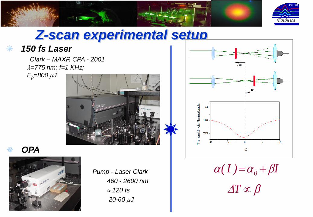
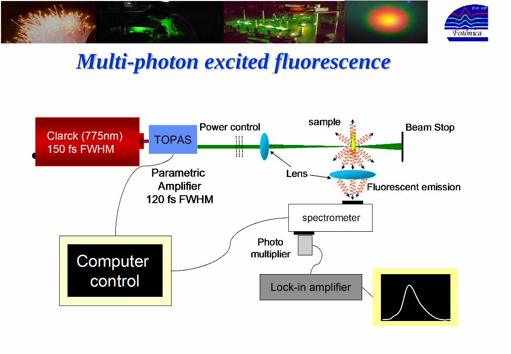
Z-scan experimental setup 150 fs Laser
Clark – MAXR CPA - 2001
=775 nm; f=1 KHz;
Ep=800 J
Pump - Laser Clark
460 - 2600 nm
120 fs
20-60 J
I)I( 0
T
OPA
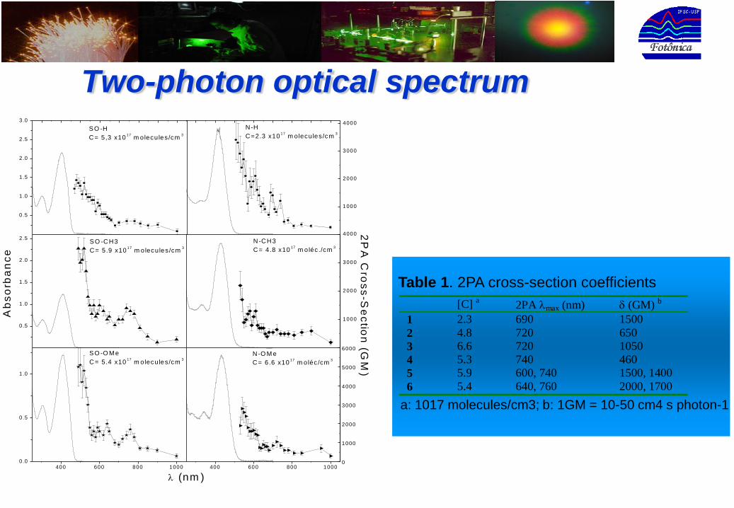
Two-photon optical spectrum
[C] a 2PA max (nm) (GM) b
1 2.3 690 1500
2 4.8 720 650
3 6.6 720 1050
4 5.3 740 460
5 5.9 600, 740 1500, 1400
6 5.4 640, 760 2000, 1700
Table 1. 2PA cross-section coefficients
a: 1017 molecules/cm3; b: 1GM = 10-50 cm4 s photon-1
0.5
1.0
1.5
2.0
2.5
3.0
0.5
1.0
1.5
2.0
2.5
400 600 800 10000.0
0.5
1.0
400 600 800 1000
S O -H
C = 5,3 x10¹⁷
m olecules/cm3
N-H
C=2.3 x10¹⁷
m olecules/cm3
1000
2000
3000
4000
S O -C H3
C = 5.9 x10¹⁷
m o lecules/cm3
Ab
so
rba
nc
e
N -C H 3
C = 4.8 x10¹⁷
m o léc ./cm3
2P
A C
ros
s-S
ec
tion
(GM
)
1000
2000
3000
4000
S O -O M e
C = 5.4 x10¹⁷
m olecules/cm3
(nm )
N -O M e
C = 6.6 x10¹⁷
m oléc /cm3
0
1000
2000
3000
4000
5000
6000
Molecular geometry
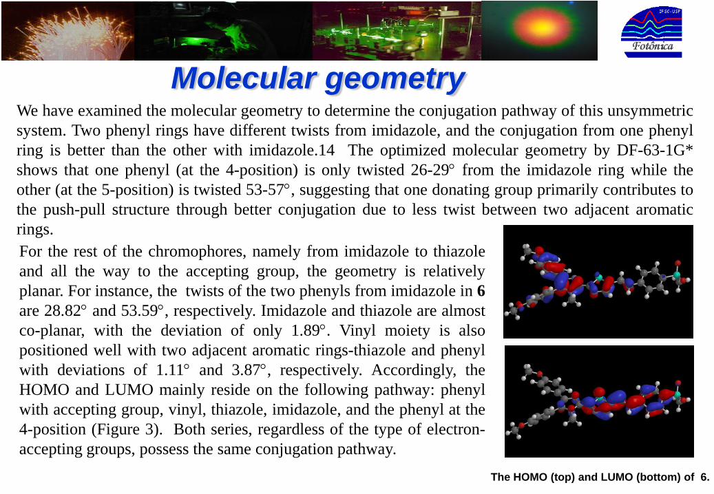
The HOMO (top) and LUMO (bottom) of 6.
We have examined the molecular geometry to determine the conjugation pathway of this unsymmetric
system. Two phenyl rings have different twists from imidazole, and the conjugation from one phenyl
ring is better than the other with imidazole.14 The optimized molecular geometry by DF-63-1G*
shows that one phenyl (at the 4-position) is only twisted 26-29 from the imidazole ring while the
other (at the 5-position) is twisted 53-57, suggesting that one donating group primarily contributes to
the push-pull structure through better conjugation due to less twist between two adjacent aromatic
rings.
For the rest of the chromophores, namely from imidazole to thiazole
and all the way to the accepting group, the geometry is relatively
planar. For instance, the twists of the two phenyls from imidazole in 6
are 28.82 and 53.59, respectively. Imidazole and thiazole are almost
co-planar, with the deviation of only 1.89. Vinyl moiety is also
positioned well with two adjacent aromatic rings-thiazole and phenyl
with deviations of 1.11 and 3.87, respectively. Accordingly, the
HOMO and LUMO mainly reside on the following pathway: phenyl
with accepting group, vinyl, thiazole, imidazole, and the phenyl at the
4-position (Figure 3). Both series, regardless of the type of electron-
accepting groups, possess the same conjugation pathway.
300 400 500 600 700 800
0
1
2
S-OM eS-CH3S-H
R = H, CH3, OCH3
N NCH3
N
S
SO2CH3
R R
S-OM eS-CH3S-H
R = H, CH3, OCH3
N NCH3
N
S
SO2CH3
R R
Absorb
ance
Wavelength (nm)
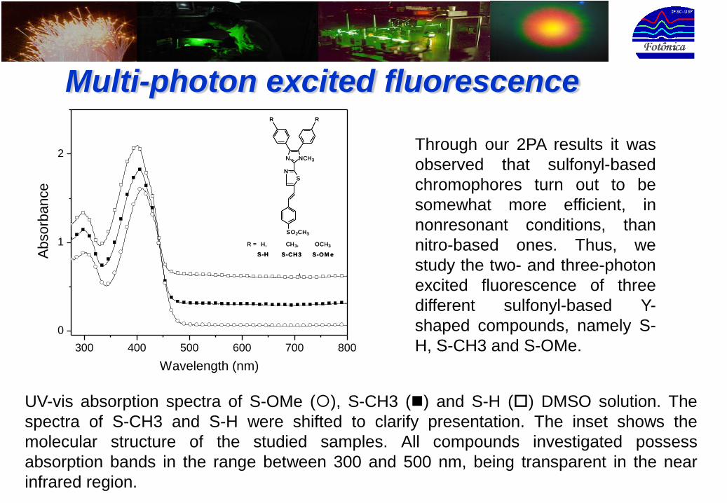
Through our 2PA results it was
observed that sulfonyl-based
chromophores turn out to be
somewhat more efficient, in
nonresonant conditions, than
nitro-based ones. Thus, we
study the two- and three-photon
excited fluorescence of three
different sulfonyl-based Y-
shaped compounds, namely S-
H, S-CH3 and S-OMe.
Multi-photon excited fluorescence
UV-vis absorption spectra of S-OMe (), S-CH3 () and S-H () DMSO solution. The
spectra of S-CH3 and S-H were shifted to clarify presentation. The inset shows the
molecular structure of the studied samples. All compounds investigated possess
absorption bands in the range between 300 and 500 nm, being transparent in the near
infrared region.
Multi-photon excited fluorescence
450 500 550 600 650 700 750 800 850
0.0
0.2
0.4
0.6
0.8
1.0
S0
S1
490 nm
750 nm
1400 nm
S0
S1
490 nm
750 nm
1400 nm
1400 nm
750 nm
490 nm
Flu
ore
sce
nce (
arb
. u
nits)
Wavelenght (nm)
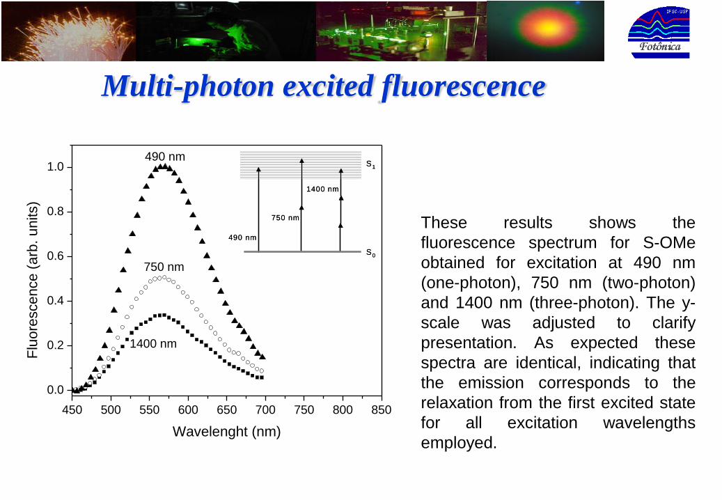
These results shows the
fluorescence spectrum for S-OMe
obtained for excitation at 490 nm
(one-photon), 750 nm (two-photon)
and 1400 nm (three-photon). The y-
scale was adjusted to clarify
presentation. As expected these
spectra are identical, indicating that
the emission corresponds to the
relaxation from the first excited state
for all excitation wavelengths
employed.
Multi-photon excited fluorescence
250 500 750
0.1
1
Flu
ore
sce
nce
(a
rb.
un
its)
250 500 750
0.1
1
I0 (GW/cm
2)I
0 (GW/cm
2)
b) three-photon
Flu
ore
sce
nce
(a
rb.
un
its)
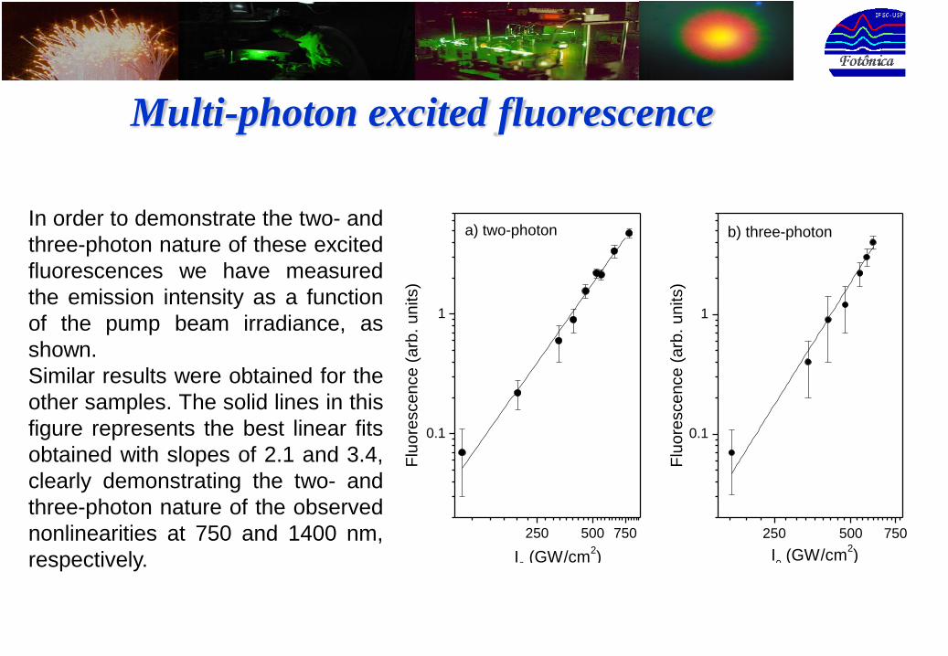
a) two-photonIn order to demonstrate the two- and
three-photon nature of these excited
fluorescences we have measured
the emission intensity as a function
of the pump beam irradiance, as
shown.
Similar results were obtained for the
other samples. The solid lines in this
figure represents the best linear fits
obtained with slopes of 2.1 and 3.4,
clearly demonstrating the two- and
three-photon nature of the observed
nonlinearities at 750 and 1400 nm,
respectively.
Multi-photon excited fluorescence
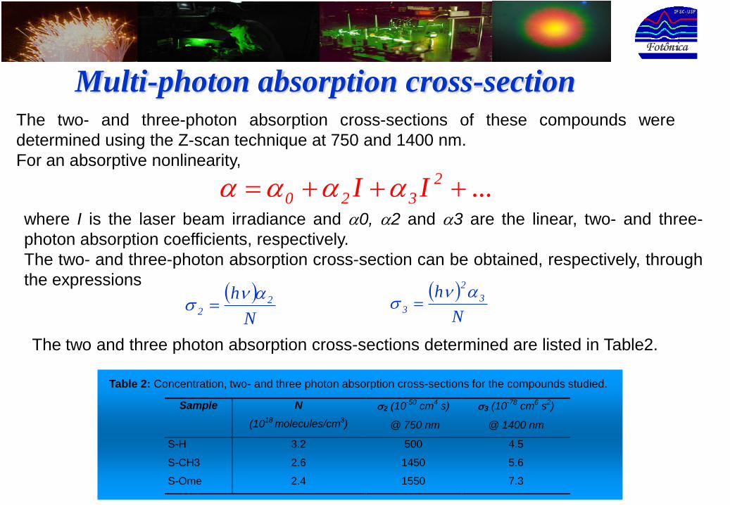
Sample N
(1018 molecules/cm3)
2 (10-50 cm4 s)
@ 750 nm
3 (10-78 cm6 s2)
@ 1400 nm
S-H 3.2 500 4.5
S-CH3 2.6 1450 5.6
S-Ome 2.4 1550 7.3
Table 2: Concentration, two- and three photon absorption cross-sections for the compounds studied.
The two- and three-photon absorption cross-sections of these compounds were
determined using the Z-scan technique at 750 and 1400 nm.
For an absorptive nonlinearity,
Multi-photon absorption cross-section
...II 2
320 where I is the laser beam irradiance and 0, 2 and 3 are the linear, two- and three-
photon absorption coefficients, respectively.
The two- and three-photon absorption cross-section can be obtained, respectively, through
the expressions
N
h 22
N
h 3
2
3
The two and three photon absorption cross-sections determined are listed in Table2.
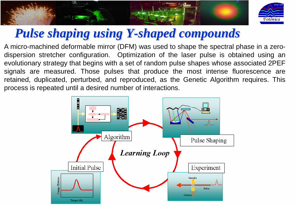
Pulse shaping using Y-shaped compoundsA micro-machined deformable mirror (DFM) was used to shape the spectral phase in a zero-
dispersion stretcher configuration. Optimization of the laser pulse is obtained using an
evolutionary strategy that begins with a set of random pulse shapes whose associated 2PEF
signals are measured. Those pulses that produce the most intense fluorescence are
retained, duplicated, perturbed, and reproduced, as the Genetic Algorithm requires. This
process is repeated until a desired number of interactions.

This figure shows the two-photon
excited fluorescence spectrum for the
S-OMe sample obtained with a KLM
Ti:sapphire oscillator, with pulses at
two distinct energies (2 and 1 nJ). As
can be seen, the 2PEF signal
increases about four times when the
pump irradiance increases twice,
exhibiting the quadratic dependence
expected for two-photon processes.400 500 600 700
1
2
3
4
Em
sis
sio
n(a
rb.
un
its)
wavelength (nm)
400 500 600 700
1
2
3
4
Em
sis
sio
n(a
rb.
un
its)
wavelength (nm)
The relatively high two-photon absorption cross-section of Y-shaped compounds
allows its application for two-photon excited fluorescence (2PEF) even without an
amplified laser system (oscillator).
Two-photon excited fluorescence with oscillator

400 500 600 700
0
1
2
3
4
0 2 4 6 8 10 12 14 16
1.00
1.05
1.10
Em
sis
sio
n (
arb
. units)
wavelength (nm)
Fitn
ess S
ign
al (a
rb.
un
its)
Interaction
Fitness signal evolutionThe fitness signal evolution as a function of the number of interaction for S-OMe is the
figure below, where a clear increase in the fitness can be observed. An increase by 10
% was observed in the 2PEF for this solution, comparing with the initial condition (flat
DFM).
Approximately the same evolution
was observed for the other
compounds. Such an optimization
corresponds to a decrease in the
pulse duration, measured using
autocorrelation, from 28 fs to 22 fs.
In this way, we were able to use the
multiphoton fluorescence process to
improve the pulse quality, thus
enhancing the fluorescence which
can be used to further improve the
performance of multiphoton imaging
systems.

ConclusionsIn summary, we have demonstrated two- and three-photon
absorption through fluorescence emission measurements as
function of the excitation irradiance in a new series of Y-
shaped chromophores at 750 and 1400 nm, respectively. The
nonlinear absorption (2PA and 3PA) cross-section values,
measured using the Z-scan technique, are comparable to the
best ones presented in the literature for organic compounds.
These findings open the door to access a new class of
materials with potential applications as fluorescent probes for
two- and three-photon microscopy. Moreover, we were able to
enhance the 2PEF signal via the laser pulse optimization
using an evolutionary strategy, which could be employed to
further improve the performance of multiphoton imaging
systems.
Support in part by U.S. Army (DAAD19-01-0746), NIH
(G12-RR03062), and NASA (NCC3-910 and NCC3-
552), from USA and by FAPESP from Brazil is gratefully
appreciated. We also thank Prof. Dr. Sergio Carlos Zilio
for helpful discussions.
Acknowledgements