TimingintheAbsenceofSupraspinalInputII:Regularly ... ·...
11
Behavioral/Systems/Cognitive Timing in the Absence of Supraspinal Input II: Regularly Spaced Stimulation Induces a Lasting Alteration in Spinal Function That Depends on the NMDA Receptor, BDNF Release, and Protein Synthesis Kyle M. Baumbauer, John R. Huie, Abbey J. Hughes, and James W. Grau Department of Psychology, Texas A&M University, College Station, Texas 77843-4325 The detection of temporal regularity allows organisms to predict the occurrence of future events. When events occur in an irregular manner, uncertainty is increased, and negative outcomes can ensue (e.g., stress). The present study shows that spinal neurons can discriminate between variable- and fixed-spaced stimulation and that the detection of regularity requires training and engages a form of NMDA receptor-mediated plasticity. The impact of stimulus exposure was assessed using a spinally mediated instrumental response, wherein spinally transected rats are given legshock whenever one hindlimb is extended. Over time, they learn to maintain the leg in a flexed position that minimizes net shock exposure. Prior exposure to 180 –900 tailshocks given in a variable (unpredictable) manner inhibited this learning. A learning deficit was not observed when 900 tailshocks were applied using a fixed (predictable) spacing. Fixed-spaced stimulation did not have a divergent effect when fewer (180) shocks were presented, implying that the abstraction of temporal regularity required repeated exposure (training). Moreover, fixed-spaced stimulation both prevented and reversed the learning deficit. The protective effect of fixed-spaced shock lasted 48 h, and was prevented by pretreatment with the NMDA receptor antagonist MK-801. Administration of the protein synthesis inhibitor cycloheximide after training blocked the long-term effect. Inhibiting BDNF function, using TrkB-IgG, also eliminated the beneficial effect of fixed-spaced stimulation. The results suggest that spinal systems can detect regularity and that this type of stimulation promotes adaptive plasticity, which may foster recovery after spinal injury. Introduction Organisms can gain an adaptive advantage by detecting regularity across time and space, allowing for the prediction of future events on the basis of past experience. For some closely spaced stimuli (e.g., sound waves between 20 and 20,000 Hz), predesigned neu- ral systems may automatically detect regularity, providing the neural cues that underlie our ability to perceive pitch and texture (Griffiths et al., 1998; Hall et al., 2005; Yau et al., 2009). As the gap between events is increased, detecting regularity becomes more difficult and may require a form of learning (temporal condition- ing) to encode that the interval between events reliably predicts the occurrence of the next event. In the absence of this learning, and other predictive (Pavlovian) cues, there is a high level of temporal uncertainty. For biologically significant events, tempo- ral uncertainty introduces a form of chaos that can enhance stress and undermine function (Overmier and Seligman, 1967; Seligman and Maier, 1967; Shors et al., 1990), thereby favoring the evolution of neural mechanisms that can detect regularity. Researchers have often attributed the abstraction of temporal relations to neural systems within the brain (Ivry and Spencer, 2004; Mauk and Buonomano, 2004; Karmarkar and Buonomano, 2007). However, recent research suggests that even simple sys- tems have some capacity to abstract temporal relations. For ex- ample, in a brain slice preparation, an extended series (900) of low-frequency (1 Hz) pulses generally yields long-term depres- sion (LTD) when the stimuli are presented in a regular manner (Dudek and Bear, 1992; Mulkey and Malenka, 1992). Presenting the same number of stimuli in a variable manner undermines the development of LTD and enhances signs of long-term potentia- tion (Perrett et al., 2001). Likewise, in the isolated spinal cord, repeated stimulation of the sciatic nerve has divergent effects on plastic function depending upon whether the stimuli occur in a fixed or variable manner; 900 shock pulses given in a variable manner disrupts subsequent learning whereas the same number of shocks given in a regular manner do not (Baumbauer et al., 2008). Interestingly, variable- and fixed-spaced shock both in- duce a learning deficit when fewer (180) shocks are given. Only after extended training (720 additional shocks) does fixed-spaced stimulation produce a divergent effect, an outcome that suggests that the underlying process may involve a form of learning. Using a spinal preparation, we now show that an extended exposure to fixed-spaced stimulation not only reverses the learn- ing deficit, it evokes a lasting protective effect that blocks the adverse consequences of variable stimulation. The protective ef- Received July 23, 2009; revised Sept. 2, 2009; accepted Sept. 21, 2009. The present work was supported by National Institute of Neurological Disorders and Stroke Grants NS51443 and NS041548, and National Institute of Child Health and Human Development Grant HD058412. We thank Dr. Michelle Hook, Denise Puga, Kevin Hoy, and Georgina Moreno for their helpful comments. Correspondence should be addressed to Kyle M. Baumbauer at the above address. E-mail: baumbauer@ tamu.edu. DOI:10.1523/JNEUROSCI.3583-09.2009 Copyright © 2009 Society for Neuroscience 0270-6474/09/2914383-11$15.00/0 The Journal of Neuroscience, November 18, 2009 • 29(46):14383–14393 • 14383
Transcript of TimingintheAbsenceofSupraspinalInputII:Regularly ... ·...
Behavioral/Systems/Cognitive
Timing in the Absence of Supraspinal Input II: RegularlySpaced Stimulation Induces a Lasting Alteration in SpinalFunction That Depends on the NMDA Receptor, BDNFRelease, and Protein Synthesis
Kyle M. Baumbauer, John R. Huie, Abbey J. Hughes, and James W. GrauDepartment of Psychology, Texas A&M University, College Station, Texas 77843-4325
The detection of temporal regularity allows organisms to predict the occurrence of future events. When events occur in an irregularmanner, uncertainty is increased, and negative outcomes can ensue (e.g., stress). The present study shows that spinal neurons candiscriminate between variable- and fixed-spaced stimulation and that the detection of regularity requires training and engages a form ofNMDA receptor-mediated plasticity. The impact of stimulus exposure was assessed using a spinally mediated instrumental response,wherein spinally transected rats are given legshock whenever one hindlimb is extended. Over time, they learn to maintain the leg in aflexed position that minimizes net shock exposure. Prior exposure to 180 –900 tailshocks given in a variable (unpredictable) mannerinhibited this learning. A learning deficit was not observed when 900 tailshocks were applied using a fixed (predictable) spacing.Fixed-spaced stimulation did not have a divergent effect when fewer (180) shocks were presented, implying that the abstraction oftemporal regularity required repeated exposure (training). Moreover, fixed-spaced stimulation both prevented and reversed the learningdeficit. The protective effect of fixed-spaced shock lasted 48 h, and was prevented by pretreatment with the NMDA receptor antagonistMK-801. Administration of the protein synthesis inhibitor cycloheximide after training blocked the long-term effect. Inhibiting BDNFfunction, using TrkB-IgG, also eliminated the beneficial effect of fixed-spaced stimulation. The results suggest that spinal systems candetect regularity and that this type of stimulation promotes adaptive plasticity, which may foster recovery after spinal injury.
IntroductionOrganisms can gain an adaptive advantage by detecting regularityacross time and space, allowing for the prediction of future eventson the basis of past experience. For some closely spaced stimuli(e.g., sound waves between 20 and 20,000 Hz), predesigned neu-ral systems may automatically detect regularity, providing theneural cues that underlie our ability to perceive pitch and texture(Griffiths et al., 1998; Hall et al., 2005; Yau et al., 2009). As the gapbetween events is increased, detecting regularity becomes moredifficult and may require a form of learning (temporal condition-ing) to encode that the interval between events reliably predictsthe occurrence of the next event. In the absence of this learning,and other predictive (Pavlovian) cues, there is a high level oftemporal uncertainty. For biologically significant events, tempo-ral uncertainty introduces a form of chaos that can enhance stressand undermine function (Overmier and Seligman, 1967; Seligmanand Maier, 1967; Shors et al., 1990), thereby favoring the evolution ofneural mechanisms that can detect regularity.
Researchers have often attributed the abstraction of temporalrelations to neural systems within the brain (Ivry and Spencer,2004; Mauk and Buonomano, 2004; Karmarkar and Buonomano,2007). However, recent research suggests that even simple sys-tems have some capacity to abstract temporal relations. For ex-ample, in a brain slice preparation, an extended series (900) oflow-frequency (1 Hz) pulses generally yields long-term depres-sion (LTD) when the stimuli are presented in a regular manner(Dudek and Bear, 1992; Mulkey and Malenka, 1992). Presentingthe same number of stimuli in a variable manner undermines thedevelopment of LTD and enhances signs of long-term potentia-tion (Perrett et al., 2001). Likewise, in the isolated spinal cord,repeated stimulation of the sciatic nerve has divergent effects onplastic function depending upon whether the stimuli occur in afixed or variable manner; 900 shock pulses given in a variablemanner disrupts subsequent learning whereas the same numberof shocks given in a regular manner do not (Baumbauer et al.,2008). Interestingly, variable- and fixed-spaced shock both in-duce a learning deficit when fewer (180) shocks are given. Onlyafter extended training (720 additional shocks) does fixed-spacedstimulation produce a divergent effect, an outcome that suggeststhat the underlying process may involve a form of learning.
Using a spinal preparation, we now show that an extendedexposure to fixed-spaced stimulation not only reverses the learn-ing deficit, it evokes a lasting protective effect that blocks theadverse consequences of variable stimulation. The protective ef-
Received July 23, 2009; revised Sept. 2, 2009; accepted Sept. 21, 2009.The present work was supported by National Institute of Neurological Disorders and Stroke Grants NS51443 and
NS041548, and National Institute of Child Health and Human Development Grant HD058412. We thank Dr. MichelleHook, Denise Puga, Kevin Hoy, and Georgina Moreno for their helpful comments.
Correspondence should be addressed to Kyle M. Baumbauer at the above address. E-mail: [email protected].
DOI:10.1523/JNEUROSCI.3583-09.2009Copyright © 2009 Society for Neuroscience 0270-6474/09/2914383-11$15.00/0
The Journal of Neuroscience, November 18, 2009 • 29(46):14383–14393 • 14383
fect evoked by fixed-spaced stimulation depends on a form ofNMDA receptor (NMDAR)-mediated plasticity, protein synthe-sis, and the neurotrophin BDNF. The results demonstrate thatthe isolated spinal cord can discriminate variable- and fixed-spaced stimulation, that these two forms of stimulation have di-vergent functional consequences, and that the abstraction ofregularity depends on neurochemical systems implicated inlearning and memory.
Materials and MethodsSubjects. Male Sprague Dawley rats obtained from Harlan were used assubjects. Rats were 70 –90 d old and weighed 350 – 400 g at the time ofspinal cord transection. They were housed in pairs with ad libitum accessto food and water, and were maintained on a 14 –10 h light– dark cycle.All experiments were performed in accordance with National Institutesof Health (NIH) standards for the care and use of laboratory animals(NIH publication No. 80-23), and were approved by the University Lab-oratory Animal Care Committee at Texas A&M University. Every effortwas made to minimize suffering and limit the number of animals used.
Spinalization surgery. Before surgery, the fur over the thoracic portionof the vertebral column was shaved and disinfected with betadine solu-tion (H-E-B). Rats were anesthetized with isoflurane gas. The rat’s headwas rendered immobile in a stereotaxic apparatus with a small (5 � 4 �2.5 cm) gauze pillow under the subject’s chest. An anterior-to-posteriorincision was made over the second thoracic vertebrae (T2), the tissueimmediately rostral to T2 was cleared using rongeurs, and the cord wasexposed and cauterized. The remaining gap in the cord was filled withGelfoam (Pharmacia), and the wound was closed with Michel clips(Fisher Scientific). Following closure of the wound, the surface ofeach leg was shaved for electrode placement. Intraperitoneal injec-tions (3 ml) of 0.9% saline solution were administered postopera-tively to prevent dehydration. Following surgery, rats were placed in atemperature-controlled environment (25.5°C) and monitored untilawake. All rats were checked every six to 8 h during the 18 –24 hpostsurgical period. During this time, hydration was maintained withsupplemental injections of saline, and the rats’ bladders and colonswere expressed as necessary.
Spinal transections were confirmed by inspecting the cord under a10� dissection scope, and observing the behavior of the subjects afterthey recovered to ensure that they exhibited paralysis below the level ofthe forepaws and did not exhibit any supraspinally mediated pain re-sponses to legshock.
Tailshock. Tailshock was applied using a shock electrode constructedfrom a modified fuse clip. The electrode was coated with electrode pasteand attached to the tail with Orthaletic tape. Leads from the fuse clip wereattached to a BRS/LVE shock generator (Model SG-903). To induce alearning deficit, 6 min of tailshock (1.5 mA AC) was administered on avariable interstimulus interval (ISI; range � 0.2–3.8 s, mean ISI of 2 s, 80ms duration). This shock procedure has been found to generate a reliabledeficit in spinal learning that lasts for at least 48 h (Crown et al., 2002). Toexamine how fixed ISI stimulation affected performance, tailshock wasadministered on a 2 s ISI (1.5 mA AC 80 ms duration). A 2 s ISI was usedto match the mean interval in the variable condition, allowing us toequate other factors (e.g., duration of restraint) across conditions. Fur-ther, prior work indicated that variable- and fixed-spaced stimulationhad divergent effects at this ISI (Baumbauer et al., 2008).
Learning procedures. The apparatus used was similar to that describedby Grau et al. (1998). Briefly, during instrumental testing all subjectswere loosely restrained in Plexiglas tubes, with their hindlimbs sus-pended above a rectangular plastic dish (11.5 cm [width (w)] � 19 cm[length (l)] � 5 cm [depth (d)]) containing a saline solution positioned7.5 cm below the restraining tube. Holes were drilled into the anteriorportion of the tubes to allow for ventilation. Two slots were cut 4 cmapart and 1.5 cm from the posterior end of the tube to allow both hindlegs to hang freely. To monitor leg position, a stainless-steel rod [7 cm (l),0.46 mm (w)] was attached to the pad of one foot (contact electrode)extending past the toes. The contact electrode was taped to the plantarsurface of the rat’s foot [Orthaletic, 1.3 cm (width); Johnson and John-
son] with the end positioned directly in front of the plantar protuber-ance. Heat-shrink tubing electrically insulated the rod from the paw. Afine wire (0.01 mm 2 [36 American wire gauge (AWG)], magnet wiresingle beldsol) was attached to the end of the rod at a point under theinsulation. This wire extended from the rear of the foot and was con-nected to a digital input board that was monitored by a Macintosh G4computer. To minimize lateral leg movements, a piece of porous tape[Orthaletic, 1.3 cm (width)] was wrapped around the leg above the tarsusand attached under the front panel of the restraining tube.
Two electrodes were then inserted into one hindleg. The first electrodewas constructed of stainless-steel wire [0.05 mm 2 (30 AWG)] and wasinserted through the skin over the tibia, 1.5 cm from the tarsus. Thesecond was made of fine wire [0.01 mm 2 (36 AWG), magnet wire singlebeldsol] and was inserted perpendicular to the leg, through the body ofthe tibialis anterior muscle, 1.7 cm above the first electrode. Legshock wasapplied by attaching one lead from a constant current AC shock genera-tor (Model SG-903; BRS/LVE) to the electrode inserted into the tibialisanterior muscle. The second lead was attached to the wire implanted inthe skin over the tibia. Shock (60 Hz, AC) intensity was adjusted for eachsubject to a level that produced a 0.4 N flexion response. This value wasdetermined before instrumental training by looping a monofilamentplastic line (“6 lb.” test strength; DuPont) around the rat’s ankle. The endof the line was attached to a strain gauge (Fort-1000; World PrecisionInstruments) fastened to a ring stand. The strain gauge output was fedthrough a calibrated multimeter that allowed for a conversion from volt-age to force in newtons. To determine the necessary flexion force, a single300 ms shock was applied to the leg and the shock intensity was adjustedso that all subjects displayed a 0.4 N flexion response. After flexion forcewas set, the monofilament line was removed from the rat’s paw and thesaline solution was adjusted so that the contact electrode sat 4 mm be-neath the surface of the salt solution. Once the animals were prepared,the 30 min instrumental testing session began. Whenever the subjects’legs were extended, the end of the rod contacted the saline solution andcompleted an electrical circuit. When the circuit was closed, shock wasdelivered to the tibialis anterior muscle, which elicited a flexion response.The flexion response broke the circuit and terminated the shock.
Drug administration. Drugs were administered intrathecally using a 10�l Hamilton syringe attached to the exposed end of each subject’s intra-thecal cannula. MK-801 and cycloheximide (Sigma-Aldrich) were eachdissolved into 10 �l of 0.9% saline, and TrkB-IgG (R&D Research) wasdissolved into 1 �l of 0.9% saline. Each compound was administered at aconstant rate over a period of several minutes. Following each injectioncannulae were flushed with 20 �l of 0.9% saline.
Behavioral measures of learning. Training and testing sessions weredivided into thirty 1 min bins to examine learning across trials. Responsenumber and response duration were collected by the computer duringthese sessions, and were separately averaged across each 1 min bin. Everytime the contact electrode left the solution, the number of responses wasincreased by one. The computer also recorded the amount of time theelectrode remained out of the solution. Response duration served as theprimary measure of learning and was calculated for each 1 min bin usingthe following equation: response duration � (time out of solution) �(response number � 1).
Statistics. Baseline shock threshold intensities, initial response dura-tions, and response duration over time were analyzed using a mixed-design ANOVA, analysis of covariance (ANCOVA), or trend analyses.Where appropriate, Tukey’s honestly significant difference (HSD) wasused to conduct post hoc analyses. Significant group differences are indi-cated in the figures with an “*.” In all cases, p � 0.05 was used to deter-mine statistical significance.
Measures of baseline behavioral reactivity. To verify that our experi-mental manipulations did not impair the subject’s capacity to exhibit aflexion response, we assessed both the shock intensity needed to elicit a0.4 N flexion force and the duration of the first flexion response at thestart of testing. The average shock intensity needed to elicit a 0.4 N flexionforce was 0.56 � 0.02 mA (mean � SE) and the mean duration of the firstresponse was 0.15 � 0.01 s. As observed in past studies, few differences inbaseline reactivity were observed across experiments and emerged with afrequency that seemed ascribable to chance, with one significant group
14384 • J. Neurosci., November 18, 2009 • 29(46):14383–14393 Baumbauer et al. • Impact of Regular Stimulation on Spinal Function
difference observed in shock intensity and another in initial flexion du-ration (both F values �3.60, p � 0.05). To assure that these baselinedifferences did not contribute to our test results, we analyzed the datafrom these experiments using an analysis of covariance (ANCOVA),treating the baseline score that yielded a significant difference as acovariate.
Response number. Elsewhere, we have shown that treatments that dis-rupt learning (as indexed by a decrease in response duration) do notundermine the subjects’ capacity to perform the target response. Indeed,subjects that fail to learn typically exhibit the highest rates of responding(Grau et al., 1998). A similar pattern was observed in the present exper-iments. For example, in the first six experiments reported below, subjectsthat had previously received variable shock exhibited little change inresponse duration over the 30 min of testing, but maintained a high levelof responding [146.24 � 37.86 (mean � SE)]. As a result, these subjectsrepeatedly experienced the response– outcome relation, but did not ex-hibit an increase in response duration (our index of instrumental learn-ing). In contrast, subjects that had received no treatment (unshockedcontrols), or 24 –30 min of fixed-spaced shock, before testing exhibited aprogressive increase in response duration and a concomitant decrease inresponse number (21.84 � 15.53) (all F values �1.88, p � 0.01). Because(1) our measure of learning (response duration) and response numbercovaried (in an inverse manner) across experiments, (2) the formeravoids some interpretative problems (see Grau et al., 1998), and (3) ouranalyses of response number yielded no surprising results, we focus oursubsequent analyses on response duration.
ResultsOur initial evidence that spinal neurons can learn about tem-poral spacing came from a series of experiments examininghow stimulation of the sciatic nerve impacts a simple form ofinstrumental conditioning, wherein optimal performance de-pends upon learning about a response– outcome relationship.In a prototypical study, one group (master) of spinally transected(spinalized) rats is given shock to one hindlimb whenever that legis extended (Grau et al., 2006). These subjects exhibit a progres-sive increase in flexion duration that reduces net shock exposure.Subjects in a second group are experimentally coupled (yoked) tothe master rats and receive shock at the same time, and for thesame duration, but independent of leg position. These subjectsdo not exhibit an increase in flexion duration. Further, exposureto uncontrollable stimulation undermines subsequent instru-mental learning, a learning deficit reminiscent of the behavioralphenomenon of learned helplessness (Overmier and Seligman,1967; Seligman and Maier, 1967).
To further analyze the mechanisms that underlie the learningdeficit, we developed a computer program that emulated the dis-tribution of shocks produced by a typical master subject duringthe first 5–10 min of training (Crown et al., 2002). During thisperiod, master rats typically received approximately thirty 80 msshocks per minute, yielding a mean ISI of 2 s. Because mastersubjects exhibit some variability in the duration of each flexionresponse, the interval between shocks varied. We emulated thisproperty by varying the ISI between 0.2 and 3.8 s (rectangulardistribution). Using these parameters, we showed that just 6 minof shock (180 pulses) to the leg or tail disrupted spinal learningfor 24 – 48 h. Further work has shown that the induction of thelearning effect depends on protein synthesis, the mGlu, GABA,and NMDA receptors, protein kinase C, and a ligand (presum-ably, substance P) that acts on the neurokinin receptor (Fergusonet al., 2003, 2006, 2008; Patton et al., 2004; Baumbauer et al.,2007).
To further explore the neural mechanisms that underlie theinduction of the learning deficit, we evaluated the consequencesof electrophysiological stimulation applied to the sciatic nerve
(Baumbauer et al., 2008). Assuming that spinal mechanisms can-not discriminate variable- and fixed-spaced stimulation, we pre-sented stimuli in a regular manner, a step we thought wouldsimplify the derivation of the effective frequency range. As a safe-guard, we verified that 180 shocks applied to the sciatic nerveinduced a learning deficit regardless of whether the stimuli occurredin a fixed or variable manner. However, when the duration of train-ing was increased fivefold (to 900 shocks), a surprising outcomeemerged. As in past studies (Crown et al., 2002; Ferguson et al.,2006; Baumbauer et al., 2008), variable stimulation produced alearning deficit, but fixed-spaced shock had no adverse effect.
The present experiments demonstrate that fixed-spaced stim-ulation does much more than reverse the learning deficit—itinduces a lasting protective effect that blocks the induction of thelearning deficit through a process that depends on the NMDAR,protein synthesis, and the neurotrophin BDNF.
Training with extended variable-, but not fixed-, spacedstimulation undermines learningOur claim that fixed and variable stimulation have distinct effectson spinal cord function, and that the consequences of fixed-spacedstimulation depend on training, rests on single experiment using aninvasive shock procedure. Here we sought evidence that a similarpattern of results could be obtained using the noninvasive shockprocedures used in prior studies. The experiment also seeks toextend the generality of the phenomenon by demonstratingthat fixed-spaced shock applied to a remote location (the tail)can impact learning when subjects are tested with legshock.Spinalized rats were given 0, 6, or 30 min of variable (ISI �0.2–3.8 s, mean � 2 s) or fixed ISI (2 s) stimulation throughcutaneous tail electrodes (n � 6 subjects/condition). We thentested subjects under common conditions in our instrumentallearning paradigm.
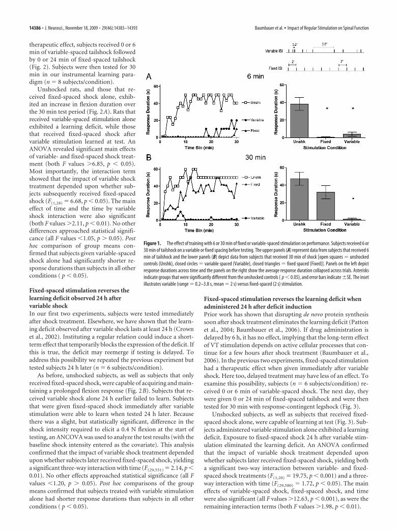
When tested with response-contingent legshock, previouslyunshocked rats exhibited a progressive increase in response du-ration, while subjects given 6 min of variable- or fixed-spacedshock (180 shocks) failed to learn (Fig. 1A). When shock ex-posure was increased fivefold (900 shocks), only variableshock produced a learning deficit (Fig. 1B). An ANOVA revealedsignificant main effects of shock treatment (unshocked, fixed, orvariable), duration of exposure (6 vs 30 min), and time bin (all Fvalues �8.10, p � 0.001). Most importantly, the impact of shocktreatment depended upon both duration of exposure (F(2,30) �4.35, p � 0.05) and time (F(58,870) � 2.78, p � 0.001). There wasalso a three-way interaction between shock treatment, duration,and time (F(58,870) � 2.01, p � 0.001). No other differences ap-proached statistical significance (F(29,870) � 1.0, p � 0.05). Posthoc comparisons of the group means demonstrated that bothunshocked groups and subjects that received fixed-spaced shockfor 30 min had significantly longer response durations than sub-jects in all other conditions ( p � 0.05).
Fixed-spaced stimulation reverses the learning deficitSix minutes (180 shocks) of intermittent shock produced a learn-ing deficit, independent of whether the interval between shockswas variable or fixed. However, when the duration of stimulationwas increased to 30 min (900 shocks), only variable-spaced shockimpaired subsequent learning. Because 6 min of fixed stimula-tion yielded a learning deficit, it appears that continued exposureto fixed-spaced shock can reverse the deficit. If this is true, thenexposure to 24 min (720 shocks) of fixed-spaced shock may re-verse the adverse effect of 180 shocks given in a variable manner.To examine whether fixed-spaced stimulation can have such a
Baumbauer et al. • Impact of Regular Stimulation on Spinal Function J. Neurosci., November 18, 2009 • 29(46):14383–14393 • 14385
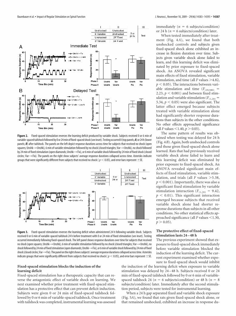
therapeutic effect, subjects received 0 or 6min of variable-spaced tailshock followedby 0 or 24 min of fixed-spaced tailshock(Fig. 2). Subjects were then tested for 30min in our instrumental learning para-digm (n � 8 subjects/condition).
Unshocked rats, and those that re-ceived fixed-spaced shock alone, exhib-ited an increase in flexion duration overthe 30 min test period (Fig. 2A). Rats thatreceived variable-spaced stimulation aloneexhibited a learning deficit, while thosethat received fixed-spaced shock aftervariable stimulation learned at test. AnANOVA revealed significant main effectsof variable- and fixed-spaced shock treat-ment (both F values �6.85, p � 0.05).Most importantly, the interaction termshowed that the impact of variable shocktreatment depended upon whether sub-jects subsequently received fixed-spacedshock (F(1,28) � 6.68, p � 0.05). The maineffect of time and the time by variableshock interaction were also significant(both F values �2.11, p � 0.01). No otherdifferences approached statistical signifi-cance (all F values �1.05, p � 0.05). Posthoc comparison of group means con-firmed that subjects given variable-spacedshock alone had significantly shorter re-sponse durations than subjects in all otherconditions ( p � 0.05).
Fixed-spaced stimulation reverses thelearning deficit observed 24 h aftervariable shockIn our first two experiments, subjects were tested immediatelyafter shock treatment. Elsewhere, we have shown that the learn-ing deficit observed after variable shock lasts at least 24 h (Crownet al., 2002). Instituting a regular relation could induce a short-term effect that temporarily blocks the expression of the deficit. Ifthis is true, the deficit may reemerge if testing is delayed. Toaddress this possibility we repeated the previous experiment buttested subjects 24 h later (n � 6 subjects/condition).
As before, unshocked subjects, as well as subjects that onlyreceived fixed-spaced shock, were capable of acquiring and main-taining a prolonged flexion response (Fig. 2B). Subjects that re-ceived variable shock alone 24 h earlier failed to learn. Subjectsthat were given fixed-spaced shock immediately after variablestimulation were able to learn when tested 24 h later. Becausethere was a slight, but statistically significant, difference in theshock intensity required to elicit a 0.4 N flexion at the start oftesting, an ANCOVA was used to analyze the test results (with thebaseline shock intensity entered as the covariate). This analysisconfirmed that the impact of variable shock treatment dependedupon whether subjects later received fixed-spaced shock, yieldinga significant three-way interaction with time (F(29,551) � 2.14, p �0.01). No other effects approached statistical significance (all Fvalues �1.20, p � 0.05). Post hoc comparisons of the groupmeans confirmed that subjects treated with variable stimulationalone had shorter response durations than subjects in all otherconditions ( p � 0.05).
Fixed-spaced stimulation reverses the learning deficit whenadministered 24 h after deficit inductionPrior work has shown that disrupting de novo protein synthesissoon after shock treatment eliminates the learning deficit (Pattonet al., 2004; Baumbauer et al., 2006). If drug administration isdelayed by 6 h, it has no effect, implying that the long-term effectof VT stimulation depends on active cellular processes that con-tinue for a few hours after shock treatment (Baumbauer et al.,2006). In the previous two experiments, fixed-spaced stimulationhad a therapeutic effect when given immediately after variableshock. Here too, delayed treatment may have less of an effect. Toexamine this possibility, subjects (n � 6 subjects/condition) re-ceived 0 or 6 min of variable-spaced shock. The next day, theywere given 0 or 24 min of fixed-spaced tailshock and were thentested for 30 min with response-contingent legshock (Fig. 3).
Unshocked subjects, as well as subjects that received fixed-spaced shock alone, were capable of learning at test (Fig. 3). Sub-jects administered variable stimulation alone exhibited a learningdeficit. Exposure to fixed-spaced shock 24 h after variable stim-ulation eliminated the learning deficit. An ANOVA confirmedthat the impact of variable shock treatment depended uponwhether subjects later received fixed-spaced shock, yielding botha significant two-way interaction between variable- and fixed-spaced shock treatments (F(1,20) � 19.75, p � 0.001) and a three-way interaction with time (F(29,580) � 1.72, p � 0.05). The maineffects of variable-spaced shock, fixed-spaced shock, and timewere also significant (all F values �12.63, p � 0.001), as were theremaining interaction terms (both F values �1.98, p � 0.01).
Figure 1. The effect of training with 6 or 30 min of fixed or variable-spaced stimulation on performance. Subjects received 6 or30 min of tailshock on a variable or fixed spacing before testing. The upper panels (A) represent data from subjects that received 6min of tailshock and the lower panels (B) depict data from subjects that received 30 min of shock [open squares � unshockedcontrols (Unshk), closed circles � variable spaced (Variable), closed triangles � fixed spaced (Fixed)]. Panels on the left depictresponse durations across time and the panels on the right show the average response duration collapsed across trials. Asterisksindicate groups that were significantly different from the unshocked controls ( p � 0.05), and error bars indicate �SE. The insetillustrates variable (range � 0.2–3.8 s, mean � 2 s) versus fixed-spaced (2 s) stimulation.
14386 • J. Neurosci., November 18, 2009 • 29(46):14383–14393 Baumbauer et al. • Impact of Regular Stimulation on Spinal Function
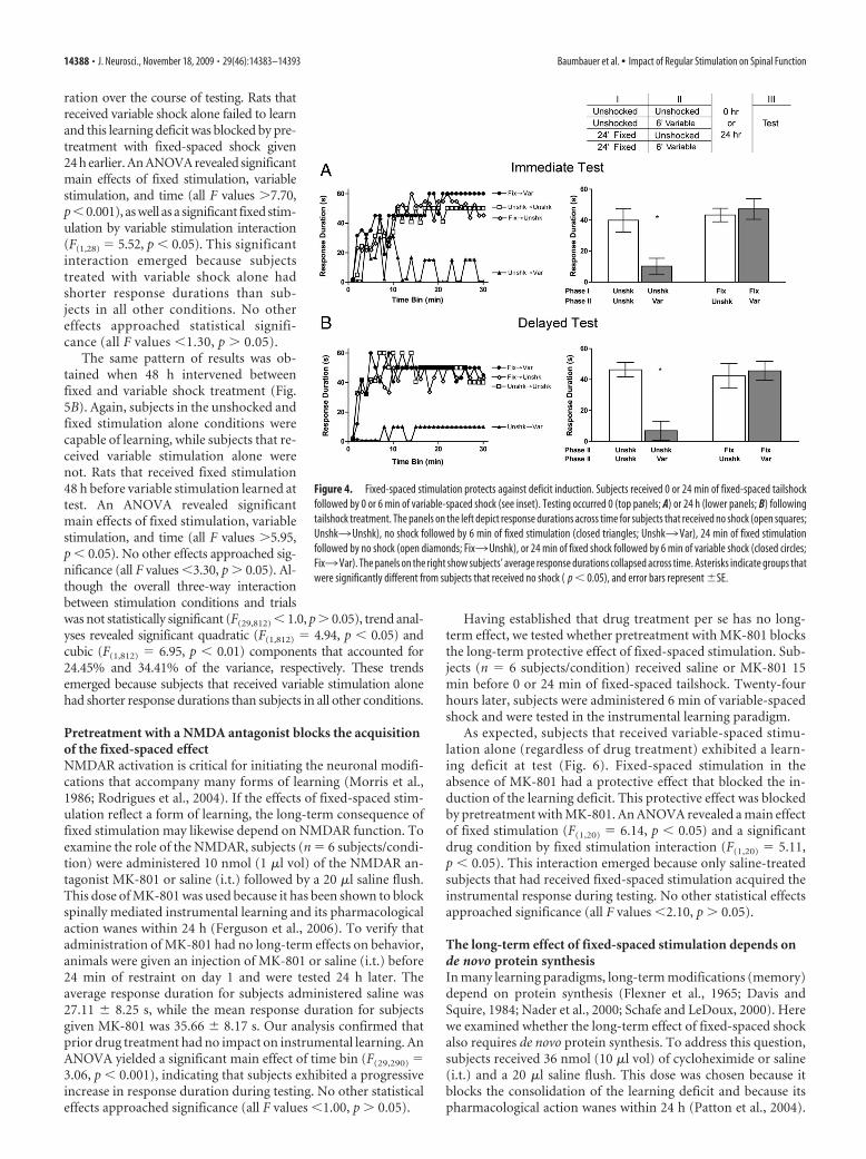
Fixed-spaced stimulation blocks the induction of thelearning deficitFixed-spaced stimulation has a therapeutic capacity that can re-verse the antagonistic effect of variable shock on learning. Wenext examined whether prior treatment with fixed-spaced stim-ulation has a protective effect that can prevent deficit induction.Subjects were given 0 or 24 min of fixed-spaced tailshock fol-lowed by 0 or 6 min of variable-spaced tailshock. Once treatmentwith tailshock was completed, instrumental learning was assessed
immediately (n � 6 subjects/condition)or 24 h (n � 6 subjects/condition) later.
When tested immediately after treat-ment (Fig. 4 A), we found that bothunshocked controls and subjects givenfixed-spaced shock alone exhibited an in-crease in flexion duration over time. Sub-jects given variable shock alone failed tolearn, and this learning deficit was elimi-nated by prior exposure to fixed-spacedshock. An ANOVA revealed significantmain effects of fixed stimulation, variablestimulation, and time (all F values �4.82,p � 0.05). The interactions between vari-able stimulation and time (F(29,580) �2.23, p � 0.001) and between fixed stim-ulation and variable stimulation (F(1,20) �5.34, p � 0.05) were also significant. Thelatter effect emerged because subjectstreated with variable stimulation alonehad significantly shorter response dura-tions than subjects in the other conditions.No other effects approached significance(all F values �1.40, p � 0.05).
The same pattern of results was ob-tained when testing was delayed for 24 h(Fig. 4B). Again, both unshocked controlsand those given fixed-spaced shock alonelearned. Rats that had previously receivedvariable shock alone failed to learn andthis learning deficit was eliminated byprior exposure to fixed-spaced shock. AnANOVA revealed significant main ef-fects of fixed stimulation, variable stim-ulation, and trials (all F values �5.38,p � 0.001). Importantly, there was also asignificant fixed stimulation by variablestimulation interaction (F(1,20) � 9.42,p � 0.01). This significant interactionemerged because subjects that receivedvariable shock alone had shorter re-sponse durations than subjects in all otherconditions. No other statistical effects ap-proached significance (all F values �1.30,p � 0.05).
The protective effect of fixed-spacedstimulation lasts 24 – 48 hThe previous experiment showed that ex-posure to fixed-spaced shock immediatelybefore variable stimulation blocked theinduction of the learning deficit. The cur-rent experiment examined whether expo-sure to fixed-spaced shock would inhibit
the induction of the learning deficit when exposure to variablestimulation was delayed by 24 – 48 h. Subjects received 0 or 24min of fixed-spaced tailshock followed by 0 or 6 min of variable-spaced tailshock 24 (n � 6 subjects/condition) or 48 h (n � 8subjects/condition) later. Immediately after the second stimula-tion period, subjects were tested for instrumental learning.
When a 24 h gap separated fixed and variable shock exposure(Fig. 5A), we found that rats given fixed-spaced shock alone, orthat remained unshocked, exhibited an increase in response du-
Figure 2. Fixed-spaced stimulation reverses the learning deficit produced by variable shock. Subjects received 0 or 6 min ofvariable-spaced tailshock followed by 0 or 24 min of fixed-spaced shock (see inset). Testing occurred 0 (top panels; A) or 24 h (lowerpanels; B) after tailshock. The panels on the left depict response durations across time for subjects that received no shock (opensquares; Unshk3Unshk), 6 min of variable stimulation followed by no shock (closed triangles; Var3Unshk), no shock followedby 24 min of fixed stimulation (open diamonds; Unshk3 Fix), or 6 min of variable shock followed by 24 min of fixed shock (closedcircles; Var3 Fix). The panels on the right show subjects’ average response durations collapsed across time. Asterisks indicategroups that were significantly different from subjects that received no shock ( p � 0.05), and error bars represent �SE.
Figure 3. Fixed-spaced stimulation reverses the learning deficit when administered 24 h following variable shock. Subjectsreceived 0 or 6 min of variable-spaced tailshock 24 h before treatment with 0 or 24 min of fixed stimulation (see inset). Testingoccurred immediately following fixed-spaced shock. The left panel shows response durations over time for subjects that receivedno shock (open squares; Unshk3Unshk), 6 min of variable stimulation followed by no shock (closed triangles; Var3Unshk), noshock followed by 24 min of fixed stimulation (open diamonds; Unshk3 Fix), or 6 min of variable shock followed by 24 min of fixedshock (closed circles; Var3 Fix). The panel on the right shows subjects’ average response durations collapsed across time. Asterisksindicate groups that were significantly different from subjects that received no shock ( p � 0.05), and error bars represent �SE.
Baumbauer et al. • Impact of Regular Stimulation on Spinal Function J. Neurosci., November 18, 2009 • 29(46):14383–14393 • 14387
ration over the course of testing. Rats thatreceived variable shock alone failed to learnand this learning deficit was blocked by pre-treatment with fixed-spaced shock given24 h earlier. An ANOVA revealed significantmain effects of fixed stimulation, variablestimulation, and time (all F values �7.70,p�0.001), as well as a significant fixed stim-ulation by variable stimulation interaction(F(1,28) � 5.52, p � 0.05). This significantinteraction emerged because subjectstreated with variable shock alone hadshorter response durations than sub-jects in all other conditions. No othereffects approached statistical signifi-cance (all F values �1.30, p � 0.05).
The same pattern of results was ob-tained when 48 h intervened betweenfixed and variable shock treatment (Fig.5B). Again, subjects in the unshocked andfixed stimulation alone conditions werecapable of learning, while subjects that re-ceived variable stimulation alone werenot. Rats that received fixed stimulation48 h before variable stimulation learned attest. An ANOVA revealed significantmain effects of fixed stimulation, variablestimulation, and time (all F values �5.95,p � 0.05). No other effects approached sig-nificance (all F values �3.30, p � 0.05). Al-though the overall three-way interactionbetween stimulation conditions and trialswas not statistically significant (F(29,812) � 1.0, p � 0.05), trend anal-yses revealed significant quadratic (F(1,812) � 4.94, p � 0.05) andcubic (F(1,812) � 6.95, p � 0.01) components that accounted for24.45% and 34.41% of the variance, respectively. These trendsemerged because subjects that received variable stimulation alonehad shorter response durations than subjects in all other conditions.
Pretreatment with a NMDA antagonist blocks the acquisitionof the fixed-spaced effectNMDAR activation is critical for initiating the neuronal modifi-cations that accompany many forms of learning (Morris et al.,1986; Rodrigues et al., 2004). If the effects of fixed-spaced stim-ulation reflect a form of learning, the long-term consequence offixed stimulation may likewise depend on NMDAR function. Toexamine the role of the NMDAR, subjects (n � 6 subjects/condi-tion) were administered 10 nmol (1 �l vol) of the NMDAR an-tagonist MK-801 or saline (i.t.) followed by a 20 �l saline flush.This dose of MK-801 was used because it has been shown to blockspinally mediated instrumental learning and its pharmacologicalaction wanes within 24 h (Ferguson et al., 2006). To verify thatadministration of MK-801 had no long-term effects on behavior,animals were given an injection of MK-801 or saline (i.t.) before24 min of restraint on day 1 and were tested 24 h later. Theaverage response duration for subjects administered saline was27.11 � 8.25 s, while the mean response duration for subjectsgiven MK-801 was 35.66 � 8.17 s. Our analysis confirmed thatprior drug treatment had no impact on instrumental learning. AnANOVA yielded a significant main effect of time bin (F(29,290) �3.06, p � 0.001), indicating that subjects exhibited a progressiveincrease in response duration during testing. No other statisticaleffects approached significance (all F values �1.00, p � 0.05).
Having established that drug treatment per se has no long-term effect, we tested whether pretreatment with MK-801 blocksthe long-term protective effect of fixed-spaced stimulation. Sub-jects (n � 6 subjects/condition) received saline or MK-801 15min before 0 or 24 min of fixed-spaced tailshock. Twenty-fourhours later, subjects were administered 6 min of variable-spacedshock and were tested in the instrumental learning paradigm.
As expected, subjects that received variable-spaced stimu-lation alone (regardless of drug treatment) exhibited a learn-ing deficit at test (Fig. 6). Fixed-spaced stimulation in theabsence of MK-801 had a protective effect that blocked the in-duction of the learning deficit. This protective effect was blockedby pretreatment with MK-801. An ANOVA revealed a main effectof fixed stimulation (F(1,20) � 6.14, p � 0.05) and a significantdrug condition by fixed stimulation interaction (F(1,20) � 5.11,p � 0.05). This interaction emerged because only saline-treatedsubjects that had received fixed-spaced stimulation acquired theinstrumental response during testing. No other statistical effectsapproached significance (all F values �2.10, p � 0.05).
The long-term effect of fixed-spaced stimulation depends onde novo protein synthesisIn many learning paradigms, long-term modifications (memory)depend on protein synthesis (Flexner et al., 1965; Davis andSquire, 1984; Nader et al., 2000; Schafe and LeDoux, 2000). Herewe examined whether the long-term effect of fixed-spaced shockalso requires de novo protein synthesis. To address this question,subjects received 36 nmol (10 �l vol) of cycloheximide or saline(i.t.) and a 20 �l saline flush. This dose was chosen because itblocks the consolidation of the learning deficit and because itspharmacological action wanes within 24 h (Patton et al., 2004).
Figure 4. Fixed-spaced stimulation protects against deficit induction. Subjects received 0 or 24 min of fixed-spaced tailshockfollowed by 0 or 6 min of variable-spaced shock (see inset). Testing occurred 0 (top panels; A) or 24 h (lower panels; B) followingtailshock treatment. The panels on the left depict response durations across time for subjects that received no shock (open squares;Unshk3Unshk), no shock followed by 6 min of fixed stimulation (closed triangles; Unshk3Var), 24 min of fixed stimulationfollowed by no shock (open diamonds; Fix3Unshk), or 24 min of fixed shock followed by 6 min of variable shock (closed circles;Fix3Var). The panels on the right show subjects’ average response durations collapsed across time. Asterisks indicate groups thatwere significantly different from subjects that received no shock ( p � 0.05), and error bars represent �SE.
14388 • J. Neurosci., November 18, 2009 • 29(46):14383–14393 Baumbauer et al. • Impact of Regular Stimulation on Spinal Function
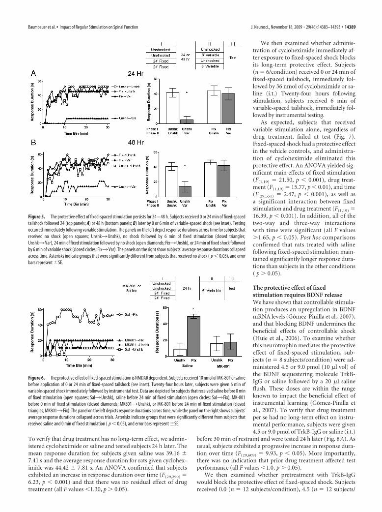
To verify that drug treatment has no long-term effect, we admin-istered cycloheximide or saline and tested subjects 24 h later. Themean response duration for subjects given saline was 39.16 �7.41 s and the average response duration for rats given cyclohex-imide was 44.42 � 7.81 s. An ANOVA confirmed that subjectsexhibited an increase in response duration over time (F(29,290) �6.23, p � 0.001) and that there was no residual effect of drugtreatment (all F values �1.30, p � 0.05).
We then examined whether adminis-tration of cycloheximide immediately af-ter exposure to fixed-spaced shock blocksits long-term protective effect. Subjects(n � 6/condition) received 0 or 24 min offixed-spaced tailshock, immediately fol-lowed by 36 nmol of cycloheximide or sa-line (i.t.) Twenty-four hours followingstimulation, subjects received 6 min ofvariable-spaced tailshock, immediately fol-lowed by instrumental testing.
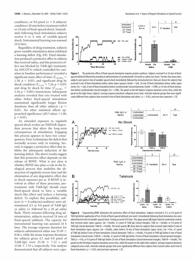
As expected, subjects that receivedvariable stimulation alone, regardless ofdrug treatment, failed at test (Fig. 7).Fixed-spaced shock had a protective effectin the vehicle controls, and administra-tion of cycloheximide eliminated thisprotective effect. An ANOVA yielded sig-nificant main effects of fixed stimulation(F(1,19) � 21.50, p � 0.001), drug treat-ment (F(1,19) � 15.77, p � 0.01), and time(F(29,551) � 2.47, p � 0.001), as well asa significant interaction between fixedstimulation and drug treatment (F(1,19) �16.39, p � 0.001). In addition, all of thetwo-way and three-way interactionswith time were significant (all F values�1.65, p � 0.05). Post hoc comparisonsconfirmed that rats treated with salinefollowing fixed-spaced stimulation main-tained significantly longer response dura-tions than subjects in the other conditions( p � 0.05).
The protective effect of fixedstimulation requires BDNF releaseWe have shown that controllable stimula-tion produces an upregulation in BDNFmRNA levels (Gomez-Pinilla et al., 2007),and that blocking BDNF undermines thebeneficial effects of controllable shock(Huie et al., 2006). To examine whetherthis neurotrophin mediates the protectiveeffect of fixed-spaced stimulation, sub-jects (n � 8 subjects/condition) were ad-ministered 4.5 or 9.0 pmol (10 �l vol) ofthe BDNF sequestering molecule TrkB-IgG or saline followed by a 20 �l salineflush. These doses are within the rangeknown to impact the beneficial effect ofinstrumental learning (Gomez-Pinilla etal., 2007). To verify that drug treatmentper se had no long-term effect on instru-mental performance, subjects were given4.5 or 9.0 pmol of TrkB-IgG or saline (i.t.)
before 30 min of restraint and were tested 24 h later (Fig. 8A). Asusual, subjects exhibited a progressive increase in response dura-tion over time (F(29,609) � 9.93, p � 0.05). More importantly,there was no indication that prior drug treatment affected testperformance (all F values �1.0, p � 0.05).
We then examined whether pretreatment with TrkB-IgGwould block the protective effect of fixed-spaced shock. Subjectsreceived 0.0 (n � 12 subjects/condition), 4.5 (n � 12 subjects/
Figure 5. The protective effect of fixed-spaced stimulation persists for 24 – 48 h. Subjects received 0 or 24 min of fixed-spacedtailshock followed 24 (top panels; A) or 48 h (bottom panels; B) later by 0 or 6 min of variable-spaced shock (see inset). Testingoccurred immediately following variable stimulation. The panels on the left depict response durations across time for subjects thatreceived no shock (open squares; Unshk3Unshk), no shock followed by 6 min of fixed stimulation (closed triangles;Unshk3Var), 24 min of fixed stimulation followed by no shock (open diamonds; Fix3Unshk), or 24 min of fixed shock followedby 6 min of variable shock (closed circles; Fix3Var). The panels on the right show subjects’ average response durations collapsedacross time. Asterisks indicate groups that were significantly different from subjects that received no shock ( p � 0.05), and errorbars represent �SE.
Figure 6. The protective effect of fixed-spaced stimulation is NMDAR dependent. Subjects received 10 nmol of MK-801 or salinebefore application of 0 or 24 min of fixed-spaced tailshock (see inset). Twenty-four hours later, subjects were given 6 min ofvariable-spaced shock immediately followed by instrumental test. Data are depicted for subjects that received saline before 0 minof fixed stimulation (open squares; Sal3Unshk), saline before 24 min of fixed stimulation (open circles; Sal3 Fix), MK-801before 0 min of fixed stimulation (closed diamonds; MK8013Unshk), or MK-801 before 24 min of fixed stimulation (closedtriangles; MK8013 Fix). The panel on the left depicts response durations across time, while the panel on the right shows subjects’average response durations collapsed across trials. Asterisks indicate groups that were significantly different from subjects thatreceived saline and 0 min of fixed stimulation ( p � 0.05), and error bars represent �SE.
Baumbauer et al. • Impact of Regular Stimulation on Spinal Function J. Neurosci., November 18, 2009 • 29(46):14383–14393 • 14389
condition), or 9.0 pmol (n � 8 subjects/condition) 30 min before treatment with 0or 24 min of fixed-spaced shock. Immedi-ately following fixed stimulation subjectsreceive 0 or 6 min of variable-spacedshock. Instrumental learning was assessed24 h later.
Regardless of drug treatment, subjectsgiven variable stimulation alone exhibiteda learning deficit (Fig. 8B). Fixed stimula-tion produced a protective effect in subjectsthat received saline, and this protective ef-fect was blocked by TrkB-IgG treatment.An ANCOVA (which controlled for vari-ation in baseline performance) revealed asignificant main effect of time (F(29,986) �1.58, p � 0.05), and significant drug byshock condition (F(1,34) � 7.26, p � 0.05)and drug by shock by time (F(29,986) �2.16, p � 0.001) interactions. Subsequentanalyses revealed that rats treated withsaline before fixed-spaced stimulationmaintained significantly longer flexiondurations than all other subjects ( p �0.05). No other statistical effects ap-proached significance (all F values �1.00,p � 0.05).
An extended exposure to regularlyspaced shock evokes an NMDAR-depen-dent process that alters the long-termconsequences of stimulation. Engagingthis process appears to have two conse-quences. First, it eliminates the deficit thatnormally accrues early in training. Sec-ond, it engages a protective effect that in-hibits the subsequent induction of thelearning deficit. The above results suggestthat this protective effect depends on therelease of BDNF. What is not clear iswhether BDNF also plays a role in the bi-ological process that underlies the ab-straction of regularity across time and theelimination of any degradative effect dueto shock exposure per se. If BDNF is in-volved in either of these processes, pre-treatment with TrkB-IgG should causefixed-spaced shock to have a variableshock like effect and induce a learningdeficit. To explore this possibility, sub-jects (n � 8 subjects/condition) were ad-ministered 4.5 or 9.0 pmol of TrkB-IgGor saline i.t. followed by a 20 �l salineflush. Thirty minutes following drug ad-ministration, subjects received 24 min offixed-spaced tailshock. The capacity forinstrumental learning was assessed 24 hlater. The average response duration forsubjects administered saline was 35.49 �8.06 s, while the mean response durationsfor subjects given 4.5 and 9.0 pmol ofTrkB-IgG were 35.38 � 7.22 s and23.06 � 7.76 s, respectively. Our analysisdemonstrated that all subjects were capa-
Figure 7. The protective effect of fixed-spaced stimulation requires protein synthesis. Subjects received 0 or 24 min of fixed-spaced tailshock followed by intrathecal administration of cycloheximide (10 nmol) or saline (see inset). Twenty-four hours later,subjects were given 6 min of variable-spaced shock immediately followed by instrumental test. Data are shown for subjects thatreceived 0 min of fixed stimulation before saline (open squares; Unshk3 Sal), 24 min of fixed stimulation before saline (opencircles; Fix3 Sal), 0 min of fixed stimulation before cycloheximide (closed diamonds; Unshk3 CXM), or 24 min of fixed stimula-tion before cycloheximide (closed triangles; Fix3 CXM). The panel on the left depicts response durations across time, while thepanel on the right shows subjects’ average response durations collapsed across trials. Asterisks indicate groups that were signifi-cantly different from subjects that received 0 min of fixed stimulation and saline ( p � 0.05), and error bars represent �SE.
Figure 8. Sequestering BDNF eliminates the protective effect of fixed stimulation. Subjects received 0, 4.5, or 9.0 pmol ofTrkB-IgG before application of 0 or 24 min of fixed-spaced tailshock (see inset). Immediately following fixed stimulation rats wereadministered 6 min of variable-spaced shock. Testing occurred 24 h later. The upper panels (A) depict data for unshocked controlsthat received saline (open squares; Sal3Unshk), 4.5 pmol of TrkB-IgG (closed triangles; TrkB Lo3Unshk), or 9.0 pmol ofTrkB-IgG (closed diamonds; TrkB Hi3Unshk). The lower panels (B) show data for subjects that received saline before 0 min offixed stimulation (open squares; Sal3Unshk), saline before 24 min of fixed stimulation (open circles; Sal3 Fix), 4.5 pmolof TrkB-IgG before 0 min of fixed stimulation (closed diamonds; TrkB Lo3Unshk), 9.0 pmol of TrkB-IgG before 0 min of fixedstimulation (closed circles; TrkB Hi3Unshk), 4.5 pmol of TrkB-IgG before 24 min of fixed stimulation (closed upward triangles;TrkB Lo3 Fix), or 9.0 pmol of TrkB-IgG before 24 min of fixed stimulation (closed downward triangles; TrkB Hi3Unshk). Thepanels on the left depict response durations across time, while the panels on the right show subjects’ average response durationscollapsed across trials. Asterisks indicate groups that were significantly different from subjects that received saline and 0 min offixed stimulation ( p � 0.05), and error bars represent �SE.
14390 • J. Neurosci., November 18, 2009 • 29(46):14383–14393 Baumbauer et al. • Impact of Regular Stimulation on Spinal Function
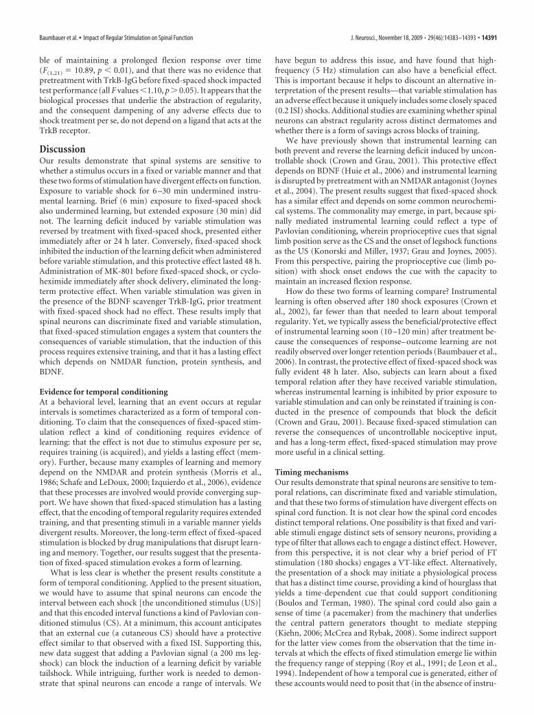
ble of maintaining a prolonged flexion response over time(F(1,21) � 10.89, p � 0.01), and that there was no evidence thatpretreatment with TrkB-IgG before fixed-spaced shock impactedtest performance (all F values �1.10, p � 0.05). It appears that thebiological processes that underlie the abstraction of regularity,and the consequent dampening of any adverse effects due toshock treatment per se, do not depend on a ligand that acts at theTrkB receptor.
DiscussionOur results demonstrate that spinal systems are sensitive towhether a stimulus occurs in a fixed or variable manner and thatthese two forms of stimulation have divergent effects on function.Exposure to variable shock for 6 –30 min undermined instru-mental learning. Brief (6 min) exposure to fixed-spaced shockalso undermined learning, but extended exposure (30 min) didnot. The learning deficit induced by variable stimulation wasreversed by treatment with fixed-spaced shock, presented eitherimmediately after or 24 h later. Conversely, fixed-spaced shockinhibited the induction of the learning deficit when administeredbefore variable stimulation, and this protective effect lasted 48 h.Administration of MK-801 before fixed-spaced shock, or cyclo-heximide immediately after shock delivery, eliminated the long-term protective effect. When variable stimulation was given inthe presence of the BDNF scavenger TrkB-IgG, prior treatmentwith fixed-spaced shock had no effect. These results imply thatspinal neurons can discriminate fixed and variable stimulation,that fixed-spaced stimulation engages a system that counters theconsequences of variable stimulation, that the induction of thisprocess requires extensive training, and that it has a lasting effectwhich depends on NMDAR function, protein synthesis, andBDNF.
Evidence for temporal conditioningAt a behavioral level, learning that an event occurs at regularintervals is sometimes characterized as a form of temporal con-ditioning. To claim that the consequences of fixed-spaced stim-ulation reflect a kind of conditioning requires evidence oflearning: that the effect is not due to stimulus exposure per se,requires training (is acquired), and yields a lasting effect (mem-ory). Further, because many examples of learning and memorydepend on the NMDAR and protein synthesis (Morris et al.,1986; Schafe and LeDoux, 2000; Izquierdo et al., 2006), evidencethat these processes are involved would provide converging sup-port. We have shown that fixed-spaced stimulation has a lastingeffect, that the encoding of temporal regularity requires extendedtraining, and that presenting stimuli in a variable manner yieldsdivergent results. Moreover, the long-term effect of fixed-spacedstimulation is blocked by drug manipulations that disrupt learn-ing and memory. Together, our results suggest that the presenta-tion of fixed-spaced stimulation evokes a form of learning.
What is less clear is whether the present results constitute aform of temporal conditioning. Applied to the present situation,we would have to assume that spinal neurons can encode theinterval between each shock [the unconditioned stimulus (US)]and that this encoded interval functions a kind of Pavlovian con-ditioned stimulus (CS). At a minimum, this account anticipatesthat an external cue (a cutaneous CS) should have a protectiveeffect similar to that observed with a fixed ISI. Supporting this,new data suggest that adding a Pavlovian signal (a 200 ms leg-shock) can block the induction of a learning deficit by variabletailshock. While intriguing, further work is needed to demon-strate that spinal neurons can encode a range of intervals. We
have begun to address this issue, and have found that high-frequency (5 Hz) stimulation can also have a beneficial effect.This is important because it helps to discount an alternative in-terpretation of the present results—that variable stimulation hasan adverse effect because it uniquely includes some closely spaced(0.2 ISI) shocks. Additional studies are examining whether spinalneurons can abstract regularity across distinct dermatomes andwhether there is a form of savings across blocks of training.
We have previously shown that instrumental learning canboth prevent and reverse the learning deficit induced by uncon-trollable shock (Crown and Grau, 2001). This protective effectdepends on BDNF (Huie et al., 2006) and instrumental learningis disrupted by pretreatment with an NMDAR antagonist (Joyneset al., 2004). The present results suggest that fixed-spaced shockhas a similar effect and depends on some common neurochemi-cal systems. The commonality may emerge, in part, because spi-nally mediated instrumental learning could reflect a type ofPavlovian conditioning, wherein proprioceptive cues that signallimb position serve as the CS and the onset of legshock functionsas the US (Konorski and Miller, 1937; Grau and Joynes, 2005).From this perspective, pairing the proprioceptive cue (limb po-sition) with shock onset endows the cue with the capacity tomaintain an increased flexion response.
How do these two forms of learning compare? Instrumentallearning is often observed after 180 shock exposures (Crown etal., 2002), far fewer than that needed to learn about temporalregularity. Yet, we typically assess the beneficial/protective effectof instrumental learning soon (10 –120 min) after treatment be-cause the consequences of response– outcome learning are notreadily observed over longer retention periods (Baumbauer et al.,2006). In contrast, the protective effect of fixed-spaced shock wasfully evident 48 h later. Also, subjects can learn about a fixedtemporal relation after they have received variable stimulation,whereas instrumental learning is inhibited by prior exposure tovariable stimulation and can only be reinstated if training is con-ducted in the presence of compounds that block the deficit(Crown and Grau, 2001). Because fixed-spaced stimulation canreverse the consequences of uncontrollable nociceptive input,and has a long-term effect, fixed-spaced stimulation may provemore useful in a clinical setting.
Timing mechanismsOur results demonstrate that spinal neurons are sensitive to tem-poral relations, can discriminate fixed and variable stimulation,and that these two forms of stimulation have divergent effects onspinal cord function. It is not clear how the spinal cord encodesdistinct temporal relations. One possibility is that fixed and vari-able stimuli engage distinct sets of sensory neurons, providing atype of filter that allows each to engage a distinct effect. However,from this perspective, it is not clear why a brief period of FTstimulation (180 shocks) engages a VT-like effect. Alternatively,the presentation of a shock may initiate a physiological processthat has a distinct time course, providing a kind of hourglass thatyields a time-dependent cue that could support conditioning(Boulos and Terman, 1980). The spinal cord could also gain asense of time (a pacemaker) from the machinery that underliesthe central pattern generators thought to mediate stepping(Kiehn, 2006; McCrea and Rybak, 2008). Some indirect supportfor the latter view comes from the observation that the time in-tervals at which the effects of fixed stimulation emerge lie withinthe frequency range of stepping (Roy et al., 1991; de Leon et al.,1994). Independent of how a temporal cue is generated, either ofthese accounts would need to posit that (in the absence of instru-
Baumbauer et al. • Impact of Regular Stimulation on Spinal Function J. Neurosci., November 18, 2009 • 29(46):14383–14393 • 14391
mental control) shock engages a physiological process that inhib-its learning, and only after extensive training is the opponenteffect engaged.
Recent studies suggest that pattern detection can occur inother physiologically reduced preparations, including the isolatedretina and visual cortex. For example, the isolated salamander andmouse retinas can detect alternating patterns of light/dark stimu-lation (Schwartz et al., 2007). After a pattern has been entrained,the ganglion cells exhibit increased spike bursting during stimu-lus omission that is precisely timed to when the missing stimulusshould have occurred. Similarly, plasticity within the visual cor-tex is modulated by temporal regularity: fixed-spaced stimuligiven at 1 Hz produces LTD, while stimuli given in a variable(Poisson) manner do not (Perrett et al., 2001). Like the isolatedretina and visual cortex, the spinal cord is also sensitive to pat-terns of stimulation. In fact, the number of stimulus presenta-tions (900) required to induce the divergent effects of variableand fixed stimulation are similar in the spinal cord and visualcortex. Further, each system requires NMDAR activation andBDNF release (Aicardi et al., 2004; Watt et al., 2004; Abidin et al.,2006; Yashiro and Philpot, 2008). What appears to set the spinalcord apart from systems like the retina is that extended training isrequired for the spinal cord to detect regularity within the stim-ulus train, whereas the retina is entrained to the pattern followinga minimal amount of stimulation.
ImplicationsIn intact subjects, exposure to an aversive stimulus can engagestress-related processes that undermine adaptive function (Overmierand Seligman, 1967; Seligman and Maier, 1967; Seligman, 1972;Shors, 2004). In many cases, the consequences of stimulation arereduced by instrumental control (Seligman et al., 1968, 1975) orintroducing a Pavlovian cue (Seligman and Binik, 1977). Withinthe brain, the effects of instrumental control and Pavlovian cuesappear to be mediated, in part, by distinct neural systems that canhave divergent effects (Amat et al., 1998, 2008; Christianson et al.,2009). Surprisingly few studies have examined how introducingtemporal regularity impacts the consequences of aversive stimu-lation. Instead, it appears that many implicitly assume that stressis enhanced by temporal uncertainty and design their experimen-tal protocols using variable ISIs. If our work generalizes to othersystems, it would suggest that introducing temporal regularitymay do more than simply lessen the adverse effects of biologicalstressors. It may foster adaptive plasticity and active coping, per-haps through common neurochemical systems (e.g., enhancedBDNF release).
We have previously shown that uncontrollable nociceptivestimulation impairs recovery after a spinal cord injury (Grau etal., 2004). This is important because spinal injury is often accom-panied by concurrent tissue damage that can provide a source ofuncontrolled nociceptive input. Further, electrical stimulation isnow used to engage motor behavior after spinal injury, both toengage motor systems to foster locomotion and to reduce muscleatrophy (Hook and Grau, 2007). Our prior work showed thatintroducing a level of instrumental control could reduce the ad-verse consequences of nociceptive stimulation. The present studydemonstrates that simply presenting stimuli in a regular mannercould lessen adverse side effects and foster adaptive plasticity thatcould promote recovery. Importantly, temporally regular stimu-lation can be applied even in cases where motor function is se-verely compromised, suggesting that this sort of stimulation mayhave considerable clinical value.
ReferencesAbidin I, Kohler T, Weiler E, Zoidl G, Eysel UT, Lessmann V, Mittmann T
(2006) Reduced presynaptic efficiency of excitatory synaptic transmis-sion impairs LTP in the visual cortex of BDNF-heterozygous mice. EurJ Neurosci 24:3519 –3531.
Aicardi G, Argilli E, Cappello S, Santi S, Riccio M, Thoenen H, Canossa M(2004) Induction of long-term potentiation and depression is reflectedby corresponding changes in secretion of endogenous brain-derived neu-rotrophin factor. Proc Natl Acad Sci U S A 101:15788 –15792.
Amat J, Matus-Amat P, Watkins LR, Maier SF (1998) Escapable and ines-capable stress differentially alter extracellular levels of 5-HT in the baso-lateral amygdala of the rat. Brain Res 812:113–120.
Amat J, Paul E, Watkins LR, Maier SF (2008) Activation of the ventral me-dial prefrontal cortex during an uncontrollable stressor reproduces boththe immediate and long-term protective effects of behavioral control.Neuroscience 154:1178 –1186.
Baumbauer KM, Young EE, Hoy KC Jr, France JL, Joynes RL (2006) Intra-thecal infusions of anisomycin impact the learning deficit but not thelearning effect observed in spinal rats that have received instrumentaltraining. Behav Brain Res 173:299 –309.
Baumbauer KM, Young EE, Hoy KC Jr, Joynes RL (2007) Neurokinin re-ceptors modulate the impact of uncontrollable stimulation on adaptivespinal plasticity. Behav Neurosci 121:1082–1094.
Baumbauer KM, Hoy KC Jr, Huie JR, Hughes AJ, Woller SA, Puga DA, SetlowB, Grau JW (2008) Timing in the absence of supraspinal input I: vari-able, but not fixed, spaced stimulation of the sciatic nerve underminesspinally-mediated instrumental learning. Neuroscience 155:1030 –1047.
Boulos Z, Terman M (1980) Food availability and daily biological rhythms.Neurosci Biobehav Rev 4:119 –131.
Christianson JP, Thompson BM, Watkins LR, Maier SF (2009) Medial pre-frontal cortical activation modulates the impact of controllable and un-controllable stressor exposure on a social exploration test of anxiety in therat. Stress 12:445– 450.
Crown ED, Grau JW (2001) Preserving and restoring behavioral potentialwithin the spinal cord using an instrumental training paradigm. J Neuro-physiol 86:845– 855.
Crown ED, Ferguson AR, Joynes RL, Grau JW (2002) Instrumental learningwithin the spinal cord: IV. Induction and retention of the behavioraldeficit observed after noncontingent shock. Behav Neurosci116:1032–1051.
Davis HP, Squire LR (1984) Protein synthesis and memory: a review. Psy-chol Bull 96:518 –559.
de Leon R, Hodgson JA, Roy RR, Edgerton VR (1994) Extensor- and flexor-like modulation within motor pools of the rat hindlimb during treadmilllocomotion and swimming. Brain Res 654:241–250.
Dudek SM, Bear MF (1992) Homosynaptic long-term depression in areaCA1 of hippocampus and the effects of NMDA receptor blockade. ProcNatl Acad Sci U S A 89:4363– 4367.
Ferguson AR, Washburn SN, Crown ED, Grau JW (2003) GABA(A) recep-tor activation is involved in noncontingent shock inhibition of instru-mental conditioning in spinal rats. Behav Neurosci 117:799 – 812.
Ferguson AR, Crown ED, Grau JW (2006) Nociceptive plasticity inhibitsadaptive learning in the spinal cord. Neuroscience 141:421– 431.
Ferguson AR, Bolding KA, Huie JR, Hook MA, Santillano DR, Miranda RC,Grau JW (2008) Group I metabotropic glutamate receptors controlmetaplasticity of spinal cord learning through a PKC-dependent mecha-nism. J Neurosci 28:11939 –11949.
Flexner LB, Flexner JB, Stellar E (1965) Memory and cerebral protein syn-thesis in mice as affected by graded amounts of puromycin. Exp Neurol13:264 –272.
Gomez-Pinilla F, Huie JR, Ying Z, Ferguson AR, Crown ED, Baumbauer KM,Edgerton VR, Grau JW (2007) BDNF and learning: evidence that instru-mental training promotes learning within the spinal cord by up-regulating BDNF expression. Neuroscience 148:893–906.
Grau JW, Joynes RL (2005) A neural-functionalist approach to learning. IntJ Comp Psychol 18:1–22.
Grau JW, Barstow DG, Joynes RL (1998) Instrumental learning within thespinal cord: I. Behavioral properties. Behav Neurosci 112:1366 –1386.
Grau JW, Washburn SN, Hook MA, Ferguson AR, Crown ED, Garcia G,Bolding KA, Miranda RC (2004) Uncontrollable stimulation under-mines recovery after spinal cord injury. J Neurotrauma 21:1795–1817.
Grau JW, Crown ED, Ferguson AR, Washburn SN, Hook MA, Miranda RC
14392 • J. Neurosci., November 18, 2009 • 29(46):14383–14393 Baumbauer et al. • Impact of Regular Stimulation on Spinal Function
(2006) Instrumental learning within the spinal cord: underlying mecha-nisms and implications for recovery after injury. Behav Cogn NeurosciRev 5:191–239.
Griffiths TD, Buchel C, Frackowiak RS, Patterson RD (1998) Analysis oftemporal structure in sound by the human brain. Nat Neurosci1:422– 427.
Hall DA, Barrett DJ, Akeroyd MA, Summerfield AQ (2005) Cortical repre-sentations of temporal structure in sound. J Neurophysiol 94:3181–3191.
Hook MA, Grau JW (2007) An animal model of functional electrical stimu-lation: evidence that the central nervous system modulates the conse-quences of training. Spinal Cord 45:702–712.
Huie JR, Hoy KC, Miranda R, Grau JW (2006) BDNF facilitates learning inthe rat spinal cord. Soc Neurosci Abstr 32:555.17.
Ivry RB, Spencer RM (2004) The neural representation of time. Curr OpinNeurobiol 14:225–232.
Izquierdo I, Bevilaqua LR, Rossato JI, Bonini JS, Medina JH, Cammarota M(2006) Different molecular cascades in different sites of the brain controlmemory consolidation. Trends Neurosci 29:496 –505.
Joynes RL, Janjua K, Grau JW (2004) Instrumental learning within the spi-nal cord: VI. The NMDA receptor antagonist, AP5, disrupts acquisitionand maintenance of an acquired flexion response. Behav Brain Res154:431– 438.
Karmarkar UR, Buonomano DV (2007) Timing in the absence of clocks:encoding time in neural network states. Neuron 53:427– 438.
Kiehn O (2006) Locomotor circuits in the mammalian spinal cord. AnnuRev Neurosci 29:279 –306.
Konorski JA, Miller SM (1937) On two types of conditioned reflex. J GenPsychol 16:264 –273.
Mauk MD, Buonomano DV (2004) The neural basis of temporal process-ing. Annu Rev Neurosci 27:307–340.
McCrea DA, Rybak IA (2008) Organization of mammalian locomotorrhythm and pattern generation. Brain Res Rev 57:134 –146.
Morris RG, Anderson E, Lynch GS, Baudry M (1986) Selective impairmentof learning and blockade of long-term potentiation by an N-methyl-D-aspartate receptor antagonist, AP5. Nature 319:774 –776.
Mulkey RM, Malenka RC (1992) Mechanisms underlying induction of ho-mosynaptic long-term depression in area CA1 of the hippocampus. Neu-ron 9:967–975.
Nader K, Schafe GE, Le Doux JE (2000) Fear memories require proteinsynthesis in the amygdala for reconsolidation after retrieval. Nature406:722–726.
Overmier JB, Seligman MEP (1967) Effects of inescapable shock upon sub-sequent escape and avoidance responding. J Comp Physiol Psychol63:28 –33.
Patton BC, Hook MA, Ferguson AR, Crown ED, Grau JW (2004) The be-havioral deficit observed following noncontingent shock in spinalized ratsis prevented by the protein synthesis inhibitor cycloheximide. Behav Neu-rosci 118:653– 658.
Perrett SP, Dudek SM, Eagleman D, Montague PR, Friedlander MJ (2001)LTD induction in adult visual cortex: role of stimulus timing and inhibi-tion. J Neurosci 21:2308 –2319.
Rodrigues SM, Schafe GE, LeDoux JE (2004) Molecular mechanisms un-derlying emotional learning and memory in the lateral amygdala. Neuron44:75–91.
Roy RR, Hutchison DL, Pierotti DJ, Hodgson JA, Edgerton VR (1991) EMGpatterns of rat ankle extensors and flexors during treadmill locomotionand swimming. J Appl Physiol 70:2522–2529.
Schafe GE, LeDoux JE (2000) Memory consolidation of auditory Pavlovianfear conditioning requires protein synthesis and protein kinase A in theamygdala. J Neurosci 20:RC96(1–5).
Schwartz G, Harris R, Shrom D, Berry MJ (2007) Detection and prediction ofperiodic patterns by the retina. Nat Neurosci 10:552–554.
Seligman M, Binik Y (1977) The safety signal hypothesis. In: Operant Pav-lovian interactions (Davis H, Hurwitz H, eds) pp 165–188. Hillsdale, NJ:Erlbaum.
Seligman ME (1972) Learned helplessness. Annu Rev Med 23:407– 412.Seligman MEP, Maier SF (1967) Failure to escape traumatic shock. J Exp
Psychol 74:1–9.Seligman MEP, Maier SF, Geer JH (1968) Alleviation of learned helplessness
in the dog. J Abnorm Psychol 73:256 –262.Seligman MEP, Rosellini RA, Kozak MJ (1975) Learned helplessness in the
rat: reversibility, time course, and immunization. J Comp Physiol Psychol88:542–547.
Shors TJ (2004) Learning during stressful times. Learn Mem 11:137–144.Shors TJ, Foy MR, Levine S, Thompson RF (1990) Unpredictable and un-
controllable stress impairs neuronal plasticity in the rat hippocampus.Brain Res Bull 24:663– 667.
Watt AJ, Sjostrom PJ, Hausser M, Nelson SB, Turrigiano GG (2004) A pro-portional but slower NMDA potentiation follows AMPA potentiation inLTP. Nat Neurosci 7:518 –524.
Yashiro K, Philpot BD (2008) Regulation of NMDA receptor subunit ex-pression and its implications for LTD, LTP, and metaplasticity. Neuro-pharmacology 55:1081–1094.
Yau JM, Olenczak JB, Dammann JF, Bensmaia SJ (2009) Temporal fre-quency channels are linked across audition and touch. Curr Biol 19:561–566.
Baumbauer et al. • Impact of Regular Stimulation on Spinal Function J. Neurosci., November 18, 2009 • 29(46):14383–14393 • 14393


![BDNF JAD re-revised final cleanlnu.diva-portal.org/smash/get/diva2:1061468/FULLTEXT01.pdf · 2017-01-17 · BDNF responsivity in older humans [Skriv text] 1 BDNF Responses in Healthy](https://static.fdocuments.net/doc/165x107/5f35cfd6915e2c06c97e2ffc/bdnf-jad-re-revised-final-1061468fulltext01pdf-2017-01-17-bdnf-responsivity.jpg)