The Use of Ultrasound in the Diagnosis of Crohn's...
9
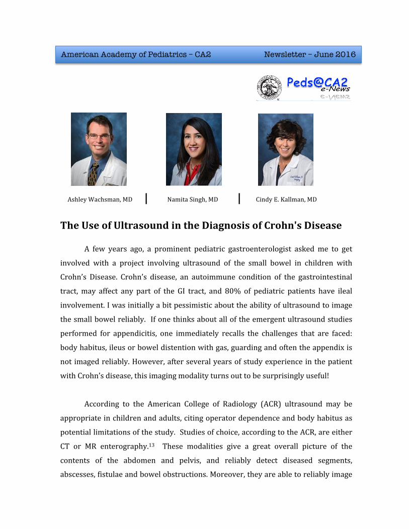
Ashley Wachsman, MD | Namita Singh, MD | Cindy E. Kallman, MD The Use of Ultrasound in the Diagnosis of Crohn's Disease A few years ago, a prominent pediatric gastroenterologist asked me to get involved with a project involving ultrasound of the small bowel in children with Crohn’s Disease. Crohn’s disease, an autoimmune condition of the gastrointestinal tract, may affect any part of the GI tract, and 80% of pediatric patients have ileal involvement. I was initially a bit pessimistic about the ability of ultrasound to image the small bowel reliably. If one thinks about all of the emergent ultrasound studies performed for appendicitis, one immediately recalls the challenges that are faced: body habitus, ileus or bowel distention with gas, guarding and often the appendix is not imaged reliably. However, after several years of study experience in the patient with Crohn’s disease, this imaging modality turns out to be surprisingly useful! According to the American College of Radiology (ACR) ultrasound may be appropriate in children and adults, citing operator dependence and body habitus as potential limitations of the study. Studies of choice, according to the ACR, are either CT or MR enterography. 13 These modalities give a great overall picture of the contents of the abdomen and pelvis, and reliably detect diseased segments, abscesses, fistulae and bowel obstructions. Moreover, they are able to reliably image American Academy of Pediatrics – CA2 Newsletter – June 2016
Transcript of The Use of Ultrasound in the Diagnosis of Crohn's...
AshleyWachsman,MD|NamitaSingh,MD|CindyE.Kallman,MDTheUseofUltrasoundintheDiagnosisofCrohn'sDisease
A few years ago, a prominent pediatric gastroenterologist asked me to get
involved with a project involving ultrasound of the small bowel in children with
Crohn’s Disease. Crohn’s disease, an autoimmune condition of the gastrointestinal
tract,may affect any part of the GI tract, and 80% of pediatric patients have ileal
involvement.Iwasinitiallyabitpessimisticabouttheabilityofultrasoundtoimage
thesmallbowelreliably. Ifonethinksaboutalloftheemergentultrasoundstudies
performed for appendicitis, one immediately recalls the challenges that are faced:
bodyhabitus,ileusorboweldistentionwithgas,guardingandoftentheappendixis
notimagedreliably.However,afterseveralyearsofstudyexperienceinthepatient
withCrohn’sdisease,thisimagingmodalityturnsouttobesurprisinglyuseful!
According to the American College of Radiology (ACR) ultrasound may be
appropriateinchildrenandadults,citingoperatordependenceandbodyhabitusas
potentiallimitationsofthestudy.Studiesofchoice,accordingtotheACR,areeither
CT or MR enterography.13 These modalities give a great overall picture of the
contents of the abdomen and pelvis, and reliably detect diseased segments,
abscesses,fistulaeandbowelobstructions.Moreover,theyareabletoreliablyimage
American Academy of Pediatrics – CA2 Newsletter – June 2016

areas that might be hindered by bowel gas even in the morbidly obese patient.
However, these screeningmethodsalsohavenotabledrawbacks suchashigh cost,
radiation (with CT), limitations in stricturing disease and significant time
requirements.
A largevolumeof literaturesupportstheuseofsmallbowelultrasoundasa
firstlineimagingtechniqueinotherpartsoftheworld.SeveralstudiesinEuropeand
Canadahavedemonstratedsmallbowelultrasoundtobeaneffectivescreeningtool
with a sensitivity of 77-95% in detecting mucosal involvement in patients with
Crohn’s disease1-4. Other studies demonstrate that it is at least as comparable to
thoseotherformsofimaging,includingCTandMR.5,6
The normal bowel wall demonstrates interfaces between the bowel lumen
and mucosa, sub mucosa, muscularis propria along with interfaces between the
mesenteric fatandserosa. InpatientswithCrohn’sdisease, thickeningand lossof
bowelstratificationcanbeseen,andabowelwallthickness>3mmonultrasoundhas
been shown to be associated with disease7. In addition, fibro-fatty proliferation
separating the diseased segments of bowel away from the normal loops and the
presenceof free fluidalsoaid in imaging.Other sonographicmarkersofdisease in
Crohn’spatientsincludestrictureordilationofintestinallumen,andextra-intestinal
abscesses,fistulas,orenlargedlymphnodes.Additionally,theuseofDopplershows
activelyinflamedsegmentsinthefastingpatient.8-11
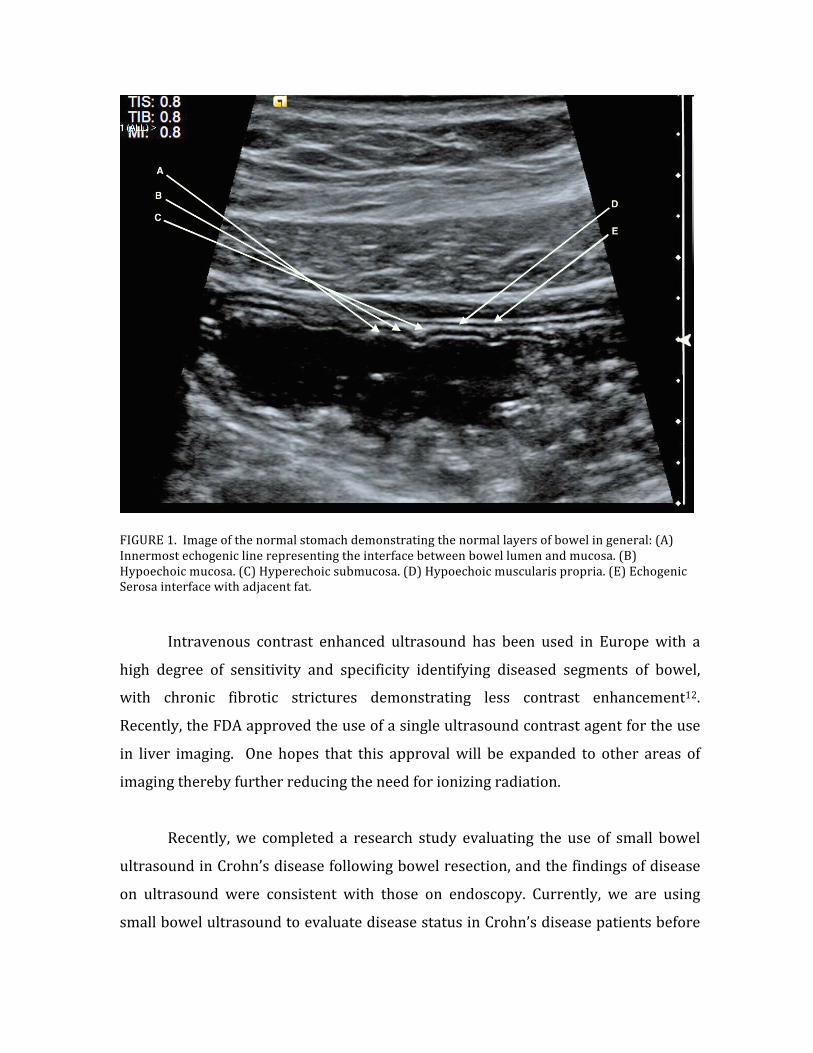
FIGURE1.Imageofthenormalstomachdemonstratingthenormallayersofbowelingeneral:(A)Innermostechogeniclinerepresentingtheinterfacebetweenbowellumenandmucosa.(B)Hypoechoicmucosa.(C)Hyperechoicsubmucosa.(D)Hypoechoicmuscularispropria.(E)EchogenicSerosainterfacewithadjacentfat.
Intravenous contrast enhanced ultrasound has been used in Europewith a
high degree of sensitivity and specificity identifying diseased segments of bowel,
with chronic fibrotic strictures demonstrating less contrast enhancement12.
Recently,theFDAapprovedtheuseofasingleultrasoundcontrastagentfortheuse
in liver imaging. One hopes that this approvalwill be expanded to other areas of
imagingtherebyfurtherreducingtheneedforionizingradiation.
Recently,we completed a research study evaluating the use of small bowel
ultrasoundinCrohn’sdiseasefollowingbowelresection,andthefindingsofdisease
on ultrasound were consistent with those on endoscopy. Currently, we are using
smallbowelultrasoundtoevaluatediseasestatusinCrohn’sdiseasepatientsbefore

and after initiating medical therapy. Following therapy with biologic therapy in
Crohn’spatients,withtheuseofultrasound,weareseeingareturnofthebowelwall
stratificationandlessfreefluid.Dopplerisroutinelyusedandshoulditprovetobe
positiveforhyperemia,portendsareturnofinflammation. Inflammatorystrictures
withdilatedloopsofbowelupstreammayalsobeseen.
Figure2.MREofpatientwithCrohn’sDisease.T2andT1fatsaturatedcoronalimagesfollowinggadoliniumenhancement.Arrowpointingtoaffectedsegmentofterminalileumwithadjacentfreefluid(A)andabnormalbowelenhancement(B&C).

Figure2.MREofpatientwithCrohn’sdisease.T2andT1fatsaturatedcoronalimagesfollowinggadoliniumenhancement.Arrowpointingtoaffectedsegmentofterminalileumwithadjacentfreefluid(A)andabnormalbowelenhancement(B&C).
Figure3.UltrasoundwithcolorDopplerofsamepatientwithCrohn’sDiseasepriortomedicaltherapy.Notelossofbowelwallsignatureandhyperemia.Thisloopofbowelissurroundedbyfreefluid.

Figure4.Week14aftermedicaltherapy.Notereturnofnormalbowelwallsignatureandtheabsenceoffreefluid.
Bowel ultrasound does have a learning curve and requires a radiologist’s
input. However, with the renewed interest of bowel imaging, our ultrasound
technologistsaremakingmoreconcertedeffortstoevaluatepatientswithabdominal
pain. They are readily seeing abnormal segments of bowel, therebymakingmore
expeditiousdiagnosesandimprovingthequalityofpatientcare. Weseetheuseof
small bowel ultrasound as complementary to first imaging studies, and as a non-
invasive,cost-effectivedisease-monitoringtoolinCrohn’sdiseasepatients.
REFERENCES
1. RutgeertsP,GeboesK,VantrappenG,BeylsJ,KerremansR,HieleM.PredictabilityofthepostoperativecourseofCrohn'sdisease.Gastroenterology.1990;99(4):956-963.
2. BlumE,KatzJA.PostoperativetherapyforCrohn'sdisease.InflammBowelDis.2009;15(3):463-472.
3. PanesJ,BouzasR,ChaparroM,etal.Systematicreview:theuseofultrasonography,computedtomographyandmagneticresonanceimagingforthediagnosis,assessmentofactivityandabdominalcomplicationsofCrohn'sdisease.AlimentPharmacolTher.2011;34(2):125-145.
4. CastiglioneF,MainentiPP,DePalmaGD,etal.NoninvasivediagnosisofsmallbowelCrohn'sdisease:directcomparisonofbowelsonographyandmagneticresonanceenterography.InflammBowelDis.2013;19(5):991-998.
5. AloiM,DiNardoG,RomanoG,etal.Magneticresonanceenterography,small-intestinecontrastUS,andcapsuleendoscopytoevaluatethesmallbowelinpediatricCrohn'sdisease:aprospective,blinded,comparisonstudy.GastrointestEndosc.2015;81(2):420-427.
6. StuartS,ConnerT,AhmedA,etal.Thesmallerbowel:imagingthesmallbowelinpaediatricCrohn'sdisease.PostgradMedJ.2011;87(1026):288-297.
7. RispoA,BucciL,PesceG,etal.BowelsonographyforthediagnosisandgradingofpostsurgicalrecurrenceofCrohn'sdisease.InflammBowelDis.2006;12(6):486-490.
8. HaberHP,BuschA,ZiebachR,DetteS,RuckP,SternM.Ultrasonographicfindingscorrespondtoclinical,endoscopic,andhistologicfindingsininflammatoryboweldiseaseandotherenterocolitides.JUltrasoundMed.2002;21(4):375-382.
9. MiaoYM,KohDM,AminZ,etal.UltrasoundandmagneticresonanceimagingassessmentofactivebowelsegmentsinCrohn'sdisease.ClinRadiol.2002;57(10):913-918.
10. GascheC,MoserG,TuretschekK,SchoberE,MoeschlP,OberhuberG.TransabdominalbowelsonographyforthedetectionofintestinalcomplicationsinCrohn'sdisease.Gut.1999;44(1):112-117.
11. ParenteF,GrecoS,MolteniM,etal.Roleofearlyultrasoundindetectinginflammatoryintestinaldisordersandidentifyingtheiranatomicallocationwithinthebowel.AlimentPharmacolTher.2003;18(10):1009-1016.
12. MigaledduV,ScanuAM,QuaiaE,etal.Contrast-enhancedultrasonographicevaluationofinflammatoryactivityinCrohn'sdisease.Gastroenterology.2009;137(1):43-52
13. KimDH.,CanucciLR.,BakerME.,et.al.ACRAppropriatenessCriteriaCrohnDisease.Availableatacsearch.acr.org/docs/69470/Narrative/.AccessedMay,2016,lastupdated2014.

ADDITIONALARTICLESOFINTEREST
1) MuradaliD.,GolbergD.,USofGastrointestinalTractDisease.RadioGraphics2015;35:50-70.
2) CalabreseE,et.al.,UltrasoundoftheSmallBowelinCrohn’sDisease,InternationalJournalofInflammationVol.2012,ArticleID964720.
3) CalabreseE,BowelUltrasoundfortheAssessmentofCrohn’sDisease.GastroenterologyandHepatologyVol.7,Iss.2,Feb,2011.
4) Strobel,D.,GoertzR.S.,BernatikT.DiagnosticsInInflammatoryBowelDisease:Ultrasound.WorldJournalofGastroenterology2011July21;17(27):3192-3197.
5) AnupindiS.A.,HalversonM.,KhwajaA.,JeckoviM.,WangX.,BellahR.D.CommonandUncommonApplicationsofBowelUltrasoundWithPathologicCorrelationinChildren.AJR2014;202:946-959.
6) SerraC.,etal.UltrasoundassessmentofvascularizationofthethickenedterminalileumwallinCrohn’sdiseasepatientsusingalow-mechanicalindexreal-timescanningtechniquewithasecondgenerationultrasoundcontrastagent.EuropeanJournalofRadiology62;(2007)114-121.
7) RipollesT.,et.al.CrohnDisease:CorrelationofFindingsatContrast-enhancedUSwithSeverityatEndoscopy.Radiology:Volume253:Number1-October2009.