The Structure and Function of Nucleic Acids - Biochemical Society
70
The Structure and Function of Nucleic Acids Revised edition C.F.A. Bryce* and D. Pacini † *Department of Biological Sciences, Napier University, Edinburgh, and † Bolton School Boy’s Division, Bolton
Transcript of The Structure and Function of Nucleic Acids - Biochemical Society
The Structure and Functionof Nucleic Acids
Revised edition
C.F.A. Bryce* and D. Pacini†
*Department of Biological Sciences, Napier University, Edinburgh, and†Bolton School Boy’s Division, Bolton
The Biochemistry Across the School Curriculum Group (BASC) was setup by the Biochemical Society in 1985. Its membership includes edu-cation professionals as well as Society members with an interest inschool science education. Its first task has been to produce this series ofbooklets, designed to help teachers of syllabuses which have a high bio-chemical content.
Other topics covered by this series include: Essential Chemistry forBiochemistry; Enzymes and their Role in Biotechnology; Metabolism;Immunology; Photosynthesis; Recombinant DNA Technology; BiologicalMembranes; and The Biochemical Basis of Disease.
More information on the work of BASC and these booklets is availablefrom the Education Officer at the Biochemical Society, 59 PortlandPlace, London W1N 3AJ.
Comments on the content of this booklet will be welcomed by the SeriesEditor Mrs D. Gull at the above address.
ISBN 0 90449 834 4
© The Biochemical Society 1998First edition published 1991
All BASC material is copyright by the Biochemical Society. Extractsmay be photocopied for classroom work, but complete reproduction ofthe entire text or incorporation of any of the material with other doc-uments or coursework requires approval by the Biochemical Society.
Printed by Holbrooks Printers Ltd, Portsmouth, U.K.

iii
Contents
Foreword................................................................................v
1 Nucleic acids and genetic information transfer ................1
Learning objectives.................................................................................1Introduction.............................................................................................1
2 Isolation and structure of nucleic acids..............................5
Learning objectives.................................................................................5Sugars........................................................................................................6Bases .........................................................................................................7Inorganic phosphate...............................................................................8Building nucleic acids from their building blocks..............................8Nucleoside di- and triphosphates .......................................................9Nucleic acids .........................................................................................10DNA isolation, base composition and structure ...........................12RNA isolation, base composition and structure............................17
3 DNA replication.................................................................21
Learning objectives ..............................................................................21Introduction ..........................................................................................21The Meselson and Stahl experiment ................................................23Mechanism of replication....................................................................25
4 The genetic code................................................................29
Learning objectives ..............................................................................29Introduction ..........................................................................................29How many bases code for an amino acid? ......................................31Evidence for a triplet code .................................................................32Deciphering the code..........................................................................33Nonsense codons ................................................................................34
iv
5 Transcription ......................................................................37
Learning objectives ..............................................................................37Introduction ..........................................................................................37Mechanism of transcription ...............................................................38
6 Translation..........................................................................41
Learning objectives ..............................................................................41Introduction ..........................................................................................41Activation of tRNA..............................................................................42Initiation .................................................................................................43Elongation ..............................................................................................44Termination ...........................................................................................45Polysomes ..............................................................................................46
7 Assessment:past examination questions and outlinesolutions..............................................................................47
Questions ..............................................................................................47Answers .................................................................................................56
8 Laboratory practicals ........................................................59
Extraction and characterization of DNA from Micrococcuslysodeikticus ............................................................................................59Purification of RNA .............................................................................61Further practicals .................................................................................62
9 Source materials ................................................................63
Textbooks ..............................................................................................63Articles ...................................................................................................63Teaching aids..........................................................................................64
Subject index......................................................................65

v
Foreword
A study of the structure and function of nucleic acids is needed to beable to understand how information controlling the characteristics of anorganism is stored in the form of genes in a cell and how these genes aretransmitted to future generations of offspring. The rapid developmentsin the area of genetic engineering and recombinant DNA technology(which are covered in Booklet 7 of the BASC series) have only beenpossible as a result of detailed understanding of the structure of DNAand RNA. It is therefore not surprising to see nucleic acids included inthe compulsory core subject matter of all of the linear and modular A-level Biology syllabuses. The major purpose of these guidance notes is toprovide an account of the role of DNA and RNA in these processes in atleast sufficient detail for A-level study. Additional information moresuited for general interest, Oxbridge preparation or first-year under-graduate study is placed in shaded boxes of text.
In order to maximize the benefit of this booklet for A-level studentsand their teachers, a set of learning objectives have been included at thestart of chapters 1–6. These objectives are based upon the relevantknowledge and understanding statements of current A-level Biologysyllabuses. Chapter 7 consists of recent A-level examination questionsand answers and this should help students get a feel for the level of detailrequired. We are grateful to the various Examinations Boards, especiallythe Northern Examinations and Assessment Board (NEAB), for theirpermission to include this material.


1
Nucleic acids and geneticinformation transfer
Learning objectives
Each student should, without reference to his or her notes, be able to:• state that the genotype describes the genetic composition of an
organism in terms of specific genes, i.e. alleles that it contains;• state that the phenotype is the collection of visible characteristics
which identify an individual organism;• state that the phenotype of an individual depends not only on its
genotype but also on the environment in which it lives; and• state that DNA, which is found in the nucleus of a cell, contains and
is responsible for the transfer of genetic information, i.e. the genotype.
Introduction
Normally, when we talk about genes and heredity we perhaps thinkimmediately of how children resemble their parents or, depending onour background knowledge, we may even think about Mendel and theheight and colour of his pea plants. This level of genetics is known asphenotypic expression, where the phenotype of an individual is the col-lection of visible and recognizable characteristics by which that indi-vidual is identified.
The phenotype is influenced by the environment as well as thegenotype; but this aspect is more appropriate to classical genetics and willnot concern us at the present time. In this section we are more concernedwith the collection of the inherited factors which determine these traits,
1
and this is a cell’s genotypic expression. The genotype is the genetic com-position of an organism in terms of the forms of specific genes, i.e. alleles,that it contains, and its study is referred to as molecular genetics.
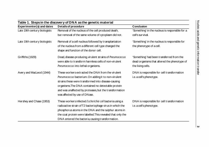
The very fact that a child can resemble one or both parents meansthat there was some way in which the genetic information was passedfrom the parent to the child. What then is the nature of the geneticmaterial? The answer to this question came from the experimentalobservations shown in Table 1.
More to heredity than DNA
In similar types of nuclear transplantation experiments, scientists have recently
revealed that the environment of the nucleus (i.e. the cytoplasm in which it finds
itself) can alter the functioning of key genes, including those which determine
body size.These have been referred to as epigenetic changes, these being
changes which affect the way DNA works rather than sequence changes.These
findings could have implications for in vitro fertilization in humans.
New Scientist (19 April 1997)
Single-cell organisms take a siesta
In 1997 a short report appeared which indicated that a study on the patterns of
DNA synthesis in single-cell organisms revealed that cells synthesized DNA from
sunrise to about noon, then shut down synthesis for between three and six
hours, then restarted before sunset.The explanation given for this phenomenon is
that this represents an evolutionary throwback to the Precambrian times when
there was no ozone layer and hence the sun’s ultraviolet light at its peak could
have damaged the DNA.This may have a future significance if the ozone layer
does continue to degrade.
New Scientist (4 January 1997)
Such studies represent the most conclusive evidence that DNA isresponsible for the transfer of genetic information, at least in micro-organisms and viruses. The evidence that this is also true in higherorganisms (plants and animals), although somewhat less conclusive,leaves no doubt that this is the case. Since some virus particles have beenfound that have no DNA, but which have RNA instead, it is morecorrect to say that ‘nucleic acids are the genetic information carriers’.
The exact roles of DNA and RNA in the complex process of thetransfer of genetic information are the subjects of subsequent sections ofthis booklet.
2 Nucleic acids: structure and function
Nucleic acids and genetic inform
ation transfer3
Table 1. Steps in the discovery of DNA as the genetic materialExperimenter(s) and dates Details of procedure ConclusionLate 19th century biologists Removal of the nucleus of the cell produced death, ‘Something’ in the nucleus is responsible for a
but removal of the same volume of cytoplasm did not. cell’s survival.
Late 19th century biologists Removal of a cell nucleus followed by transplantation ‘Something’ in the nucleus is responsible for
of the nucleus from a different cell type changed the the phenotype of a cell.
shape and function of the donor cell.
Griffiths (1929) Dead, disease-producing, virulent strains of Pneumococcus ‘Something’ had been transferred from the
were able to transform harmless cells of non-virulent dead organisms that altered the phenotype of
Pneumococcus into lethal organisms. the living cells.
Avery and MacLeod (1944) These workers extracted the DNA from the virulent DNA is responsible for cell transformation
Pneumococcus bacterium.On adding it to non-virulent i.e. a cell’s phenotype.
strains these were transformed into disease-causing
organisms.The DNA contained no detectable protein
and was unaffected by proteases, but the transformation
was affected by use of DNase.
Hershey and Chase (1953) These workers infected Escherichia coli bacteria using a DNA is responsible for cell transformation
radioactive strain of T2 bacteriophage virus in which the i.e. a cell’s phenotype.
phosphorus atoms in the DNA and the sulphur atoms in
the coat protein were labelled.This revealed that only the
DNA entered the bacteria, causing transformation.

2
Isolation and structure ofnucleic acids
Learning objectives
Each student should, without reference to his or her notes, be able to:• state that DNA is composed of phosphate, deoxyribose and the four
major bases: adenine, guanine, cytosine and thymine;• state that RNA is composed of phosphate, ribose, adenine, guanine,
cytosine and uracil;• state that DNA and RNA are polymers of nucleotide subunits;• state that a nucleotide is composed of a phosphate group, a pentose
sugar and one of the four corresponding bases;• state that the backbone of a DNA molecule is a chain of repeating
deoxyribose–phosphate units;• state that the backbone of an RNA molecule is a chain of repeating
ribose–phosphate units;• state that each molecule of DNA is usually composed of two chains;• state that in DNA, adenine will only bind with thymine on opposite
chains and guanine will only bind with cytosine on opposite chains;• state that the two linked chains in DNA are arranged in a double helix;• state that there are three principal types of RNA, these being
messenger RNA (mRNA), transfer RNA (tRNA) and ribosomal RNA (rRNA), which are all single-stranded molecules;
• state the function of the three principal types of RNA;• draw a diagram of a simple ladder-like representation of a DNA
molecule; and• state that DNA is a stable polynucleotide which contains coded
genetic information for inherited characteristics.
5

Introduction
Although the nucleic acids were first discovered in 1868, by FriedrichMiescher working with pus cells obtained from discarded surgicalbandages, it was not really until the early 1940s that the chemistry andbiology of the nucleic acids were set on firm foundations.
Basically, nucleic acids can be subdivided into two types: deoxy-ribonucleic acid (DNA) and ribonucleic acid (RNA). Both DNA andRNA have been shown to consist of three groups of molecules: pentose(5-carbon-atom) sugars; organic bases; and inorganic phosphate.
Sugars
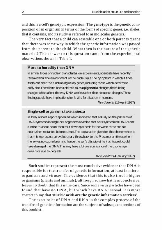
There are only two types of sugar present in nucleic acids, ribose whichis present solely in RNA (hence its name) and deoxyribose which ispresent solely in DNA (again, the sugar gives rise to the name deoxy-ribonucleic acid). The chemical structures for these compounds areshown here:
The prefix ‘deoxy’ means ‘without oxygen’, and we can see fromthe structures that the only difference between them is the absence of anoxygen in the deoxyribose sugar (see shaded area). Both sugars contain 5carbon atoms (pentose sugars) and for convenience we number these asshown in the next figure. The ‘dash’ or ‘prime’ (�) on, for example, the 5indicates the carbon in the ribose ring. The purine or pyrimidine rings(see Bases, below) are numbered without primes in order to distinguishthem.
6 Isolation and structure of nucleic acids
Ribose Deoxyribose
OHOHOH
OH OH
No OHgroup
O
H
O HOCH2HOCH2
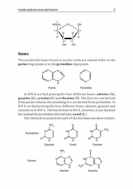
Bases
The nucleotide bases found in nucleic acids are related either to thepurine ring system or to the pyrimidine ring system.
In DNA we find principally four different bases: adenine (A),guanine (G), cytosine (C) and thymine (T). The first two are derivedfrom purine whereas the remaining two are derived from pyrimidine. InRNA we find principally four different bases; adenine, guanine andcytosine as in DNA. The fourth base in RNA, however, is not thyminebut instead the pyrimidine-derived base, uracil (U).
The chemical structures for each of the five bases are shown below.
Nucleic acids: structure and function 7
OH OH
OH
C
HC C
H
C
O
H H
HOCH2
5�
4� 1�
2�3�
Purine Pyrimidine
N N N
N
H
N N
Purines
Adenine Guanine
Cytosine Uracil Thymine
PyrimidinesN HN
HN
H2N
HNCH3
N
NH2
NH2
H
NNH
NN
NN
H
NH
NH
O O O
O
O
CN
C
O
Note: A-level students are not expected to recognize these structuralformulae. For further help in understanding the chemistry of these com-pounds, refer to BASC Booklet 1, Essential Chemistry for Biochemistry.
In addition to these major bases there is also a large range of so-called minor bases which occur less frequently than the others, e.g. 5-methyl cytosine.
Inorganic phosphate
There are phosphate residues in nucleic acids and they are of the typederived from phosphoric acid, the structure of which is shown below.
Building nucleic acids from their building blocks
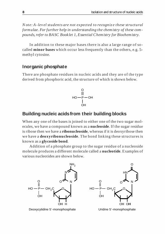
When any one of the bases is joined to either one of the two sugar mol-ecules, we have a compound known as a nucleoside. If the sugar residueis ribose then we have a ribonucleoside, whereas if it is deoxyribose thenwe have a deoxyribonucleoside. The bond linking these structures isknown as a glycoside bond.
Addition of a phosphate group to the sugar residue of a nucleosidemolecule produces a different molecule called a nucleotide. Examples ofvarious nucleotides are shown below.
8 Isolation and structure of nucleic acids
OH
OH
O
POH
Deoxycytidine 5�-monophosphate Uridine 5�-monophosphate
HO P
O
OH
OH2C HO P
O
OH
OH2C
HN
OH
NH2
H OHH OHHH
NN
N
O
O
O
O
O

Nucleotides can be regarded as the building blocks for the largernucleic acid molecules, DNA and RNA.
Nucleoside di- and triphosphates
One or two additional phosphates can be added to the first phosphategroup of a nucleoside molecule (a nucleoside monophosphate, NMP) bymeans of a pyrophosphate linkage. The molecules formed in this wayare called nucleoside diphosphates (NDPs) and nucleoside triphosphates(NTPs). The most important base involved in these compounds isadenine, forming the adenosine mono-, di- and triphosphate molecules(AMP, ADP and ATP), which fulfil vital roles in many cellular processesas you may know from your study of respiration and photosynthesis[for further details see BASC Booklets 4 (Metabolism) and 6 (Photo-synthesis)].
Nucleic acids: structure and function 9
OH OH OH
OH OH
O O O
���
O O O O
Base
NMP
NDP
NTP
H HHH
CH2P P POH

Nucleic acids
Nucleic acids are formed by the combination of nucleotide moleculesthrough sugar–phosphate bonds known as phosphodiester linkages.Because a nucleic acid is a polymer of many nucleotide molecules, DNAand RNA molecules are called polynucleotides.
The structure of a polynucleotide is shown diagrammatically above.In common with the formation of other biological polymers, e.g. starchand proteins, water molecules are produced by condensation reactionsand energy from ATP molecules, produced in cellular respiration, isused.
10 Isolation and structure of nucleic acids
3�–5�phosphodiester
linkage
3�–5�phosphodiester
linkage
3�–5�phosphodiester
linkage
3� end
5� end
Adenine (A)
Guanine (G)
Thymine (T)
Cytosine (C)
O
O
P
O
H H
N
N N
NH2
H3C
NH2
NH2
NH
OH
NH
CH2
CH2
CH2
CH2
N
N
N
N
N
N
H
H
H
H HH
H
H
H H
N
H
H
H
H HH
H
H
OO
O
O
P
O
O
O
O
P
O
OO O
O
O
P
O
OO
O O
O
O
�
�
�
�
�
5�
4�
3� 2�
5�
4�
3� 2�
5�
4�
3� 2�
5�
4�
3� 2�
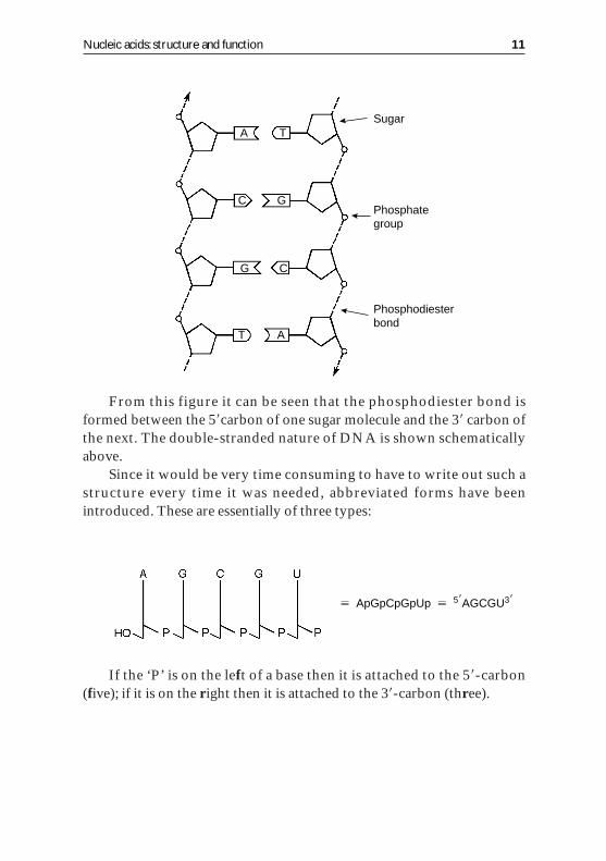
From this figure it can be seen that the phosphodiester bond isformed between the 5�carbon of one sugar molecule and the 3� carbon ofthe next. The double-stranded nature of DNA is shown schematicallyabove.
Since it would be very time consuming to have to write out such astructure every time it was needed, abbreviated forms have beenintroduced. These are essentially of three types:
If the ‘P’ is on the left of a base then it is attached to the 5�-carbon(five); if it is on the right then it is attached to the 3�-carbon (three).
Nucleic acids: structure and function 11
Sugar
Phosphategroup
Phosphodiesterbond
A T
C G
G C
T A
� ApGpCpGpUp � 5�AGCGU3�
DNA isolation,base composition and structure
Isolation
In order to study the chemistry of DNA it must be isolated and purifiedfrom the nucleus of a cell. In the isolation of DNA from cell samples, themajor contaminant is histone protein which is closely associated withDNA to form visible chromatin threads in the nucleus. To remove thiscontaminant the cell is first deproteinized by treatment with phenol andthe DNA is precipitated using ice-cold ethanol. A more detailed accountof the isolation of DNA is given in one of the practical schedules later(see Chapter 8) and in BASC Booklet 7, Recombinant DNA Technology.
Once isolated, DNA can be broken down or degraded by either: (i)treatment with perchloric acid at 100�C, which breaks down the DNAto its constituent bases; or (ii) treatment with enzymes (DNase, phos-phodiesterase, etc.) which break down the DNA to its constituentnucleotides.
The mixture of either of these products can be separated by chroma-tography or electrophoresis and the amount of each of the constituentbases can be measured.
Composition
Using the techniques outlined above it was noted by Chargaff and co-workers in the late 1940s that:(i) the base composition of an organism’s DNA is characteristic of thatorganism;(ii) different cells or tissues of the same organism have identical basecomposition;(iii) closely related organisms exhibit similar base compositions (thisproperty is used in biology as a basis for chemical taxonomy)(iv) the majority of DNA molecules exhibit certain chemical regularitiesthat are defined as Chargaff’s base pairing rules (base equivalence),namely the amount of A equals the amount of T and the amount of Gequals the amount of C, as can be seen (within the bounds of experi-mental error) from the data shown in Table 2.
In considering the data shown in Table 2, in terms of base equiv-alence it is necessary to combine the values for cytosine and 5-methylcytosine where these apply. The only exception to base equivalence is the
12 Isolation and structure of nucleic acids
DNA from the virus X174. Can you think of an explanation? (See theStructure section in this chapter for the solution.)
Structure
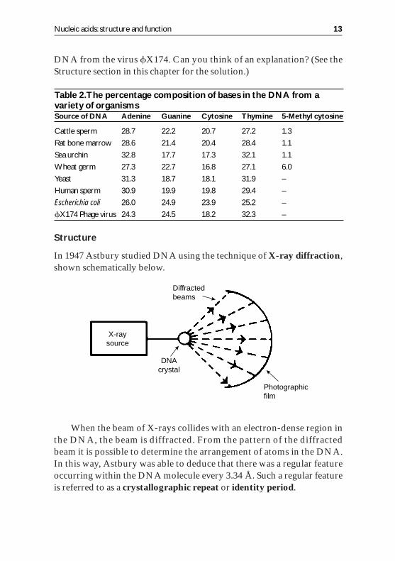
In 1947 Astbury studied DNA using the technique of X-ray diffraction,shown schematically below.
When the beam of X-rays collides with an electron-dense region inthe DNA, the beam is diffracted. From the pattern of the diffractedbeam it is possible to determine the arrangement of atoms in the DNA.In this way, Astbury was able to deduce that there was a regular featureoccurring within the DNA molecule every 3.34 Å. Such a regular featureis referred to as a crystallographic repeat or identity period.
Nucleic acids: structure and function 13
X-raysource
Diffractedbeams
Photographicfilm
DNAcrystal
Table 2.The percentage composition of bases in the DNA from avariety of organismsSource of DNA Adenine Guanine Cytosine Thymine 5-Methyl cytosine
Cattle sperm 28.7 22.2 20.7 27.2 1.3
Rat bone marrow 28.6 21.4 20.4 28.4 1.1
Sea urchin 32.8 17.7 17.3 32.1 1.1
Wheat germ 27.3 22.7 16.8 27.1 6.0
Yeast 31.3 18.7 18.1 31.9 –
Human sperm 30.9 19.9 19.8 29.4 –
Escherichia coli 26.0 24.9 23.9 25.2 –
X174 Phage virus 24.3 24.5 18.2 32.3 –
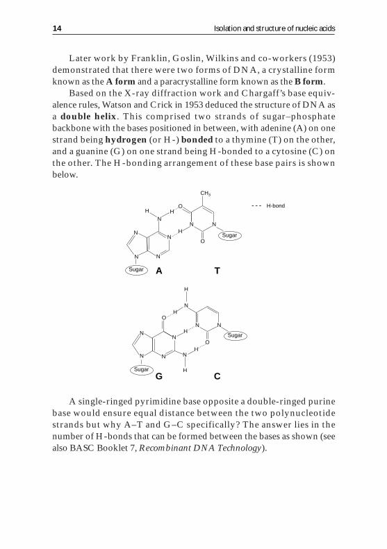
Later work by Franklin, Goslin, Wilkins and co-workers (1953)demonstrated that there were two forms of DNA, a crystalline formknown as the A form and a paracrystalline form known as the B form.
Based on the X-ray diffraction work and Chargaff’s base equiv-alence rules, Watson and Crick in 1953 deduced the structure of DNA asa double helix. This comprised two strands of sugar–phosphatebackbone with the bases positioned in between, with adenine (A) on onestrand being hydrogen (or H-) bonded to a thymine (T) on the other,and a guanine (G) on one strand being H-bonded to a cytosine (C) onthe other. The H-bonding arrangement of these base pairs is shownbelow.
A single-ringed pyrimidine base opposite a double-ringed purinebase would ensure equal distance between the two polynucleotidestrands but why A–T and G–C specifically? The answer lies in thenumber of H-bonds that can be formed between the bases as shown (seealso BASC Booklet 7, Recombinant DNA Technology).
14 Isolation and structure of nucleic acids
N
OH
H
H
N
N
N N
CH3
N N
O
N
N H
H
H
H N
N N N H
N
O
N O
A T
G C
Sugar
Sugar
Sugar
Sugar
H-bond

Molecular impostor that cannot form hydrogen bonds
In a recent study a synthetic chemical which resembled thymine but which had a
benzene ring instead of the pyrimidine ring and fluorine atoms in place of the
oxygen and nitrogen atoms was used in an assay with DNA polymerase.Much to
the researchers’ surprise, a DNA helix was produced thus raising doubts on the
role of hydrogen bonds in the DNA structure. A further study showed that mol-
ecular mimics which have the wrong shape distort the helix and that this may rep-
resent the key feature of the structural stability.
New Scientist (22 February 1997)
Because the two polynucleotide strands have ‘direction’, two con-figurations are possible, one in which both strands run in the samedirection, which is known as a parallel arrangement, and one in whichthe strands run in the opposite direction, which is known as anantiparallel arrangement. It has been shown that the DNA duplexesfound in nature have their chains in an antiparallel arrangement. This hasa profound significance when we come to consider the biologicalfunction of the molecules (see DNA replication section).
In coiling the backbone into a helical configuration, two arrange-ments in space are possible, one a right-handed helix, the other a left-handed helix. The difference between these two arrangements can beeasily demonstrated using two pipe cleaners. It has been shown that thedouble helix of DNA found in nature is in the right-handed configu-ration.
Nucleic acids: structure and function 15
Left-handed Right-handed
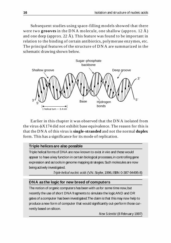
Subsequent studies using space-filling models showed that therewere two grooves in the DNA molecule, one shallow (approx. 12 Å)and one deep (approx. 22 Å). This feature was found to be important inrelation to the binding of certain antibiotics, polymerase enzymes, etc.The principal features of the structure of DNA are summarized in theschematic drawing shown below.
Earlier in this chapter it was observed that the DNA isolated fromthe virus X174 did not exhibit base equivalence. The reason for this isthat the DNA of this virus is single-stranded and not the normal duplexform. This has a significance for its mode of replication.
Triple helices are also possible
Triple helical forms of DNA are now known to exist in vivo and these would
appear to have a key function in certain biological processes, in controlling gene
expression and as tools in genome mapping strategies. Such molecules are now
being actively investigated.
Triple-helical nucleic acids (V.N. Soyfer, 1996, ISBN 0-387-94495-8)
DNA as the logic for new breed of computers
The notion of organic computers has been with us for some time now,but
recently the use of short DNA fragments to simulate the logic AND and OR
gates of a computer has been investigated.The claim is that this may now help to
produce a new form of computer that would significantly out-perform those cur-
rently based on silicon.
New Scientist (8 February 1997)
16 Isolation and structure of nucleic acids
Sugar–phosphatebackbone
Hydrogenbonds
Base
3�
3� 5�5�
Shallow groove Deep groove
1 helical turn 3.4 nm
Breast tissue traps carcinogens
From recent research work it would appear that the fatty tissue that constitutes
80% of women’s breasts is capable of absorbing fat-soluble organic molecules,
some of which are carcinogenic, causing DNA damage and mutation. It would
appear that certain foodstuffs may be the source of these carcinogens and as such
it might be impossible or difficult to avoid ingesting these.
New Scientist (7 December 1996)
RNA isolation,base composition and structure
Isolation
The isolation procedure is very similar to that for DNA isolationalthough it is essential to include an RNase inhibitor to prevent degra-dation of the RNA. Unlike DNA, which exists as a more or less homo-geneous molecule for any one cell, RNA exists as a family of molecules,each member of which has a distinctive structure and function (see latersections in this chapter). Because of this wide variety of RNA species itis necessary to separate further the initial RNA pool into its constituentmolecules. The methods employed include:• density-gradient centrifugation;• ion-exchange chromatography;
Nucleic acids: structure and function 17
Table 3.DNA structure and functionStructural feature Function
Strong, covalently bonded Preserves the base sequence of genetic
sugar–phosphate backbone information during the lifetime of the cell
and allows shortening of the DNA during
chromosome formation in cell division
Complementary purine–pyrimidine Keeps an equal distance between the two
base pairing polynucleotide strands, increasing the stability
of the double helix
Numerous but weak H-bonds Enhance stability of the helix but are easily
between complementary bases broken by enzymes to allow DNA replication
and transcription
Large, insoluble molecule Restricts DNA to the nucleus, protecting it
(molecular mass approx. 100000 units) from biochemical damage, thus preserving
the genetic code
• gel filtration;• electrophoresis.
Base composition
Purified RNA molecules can be degraded by chemical means (alkali) orby enzymes (RNase) and the base composition determined in a mannersimilar to that described for DNA (see section on DNA isolation in thischapter). One major difference between DNA and RNA is that in RNAthere are a fairly large number of minor bases. Also, in the majority ofcases there is no base equivalence, signifying that RNA molecules areusually single-stranded.
Structure
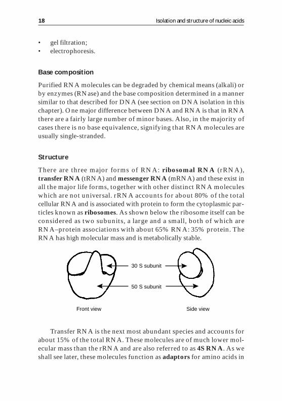
There are three major forms of RNA: ribosomal RNA (rRNA),transfer RNA (tRNA) and messenger RNA (mRNA) and these exist inall the major life forms, together with other distinct RNA moleculeswhich are not universal. rRNA accounts for about 80% of the totalcellular RNA and is associated with protein to form the cytoplasmic par-ticles known as ribosomes. As shown below the ribosome itself can beconsidered as two subunits, a large and a small, both of which areRNA–protein associations with about 65% RNA: 35% protein. TheRNA has high molecular mass and is metabolically stable.
Transfer RNA is the next most abundant species and accounts forabout 15% of the total RNA. These molecules are of much lower mol-ecular mass than the rRNA and are also referred to as 4S RNA. As weshall see later, these molecules function as adaptors for amino acids in
18 Isolation and structure of nucleic acids
Front view Side view
30 S subunit
50 S subunit
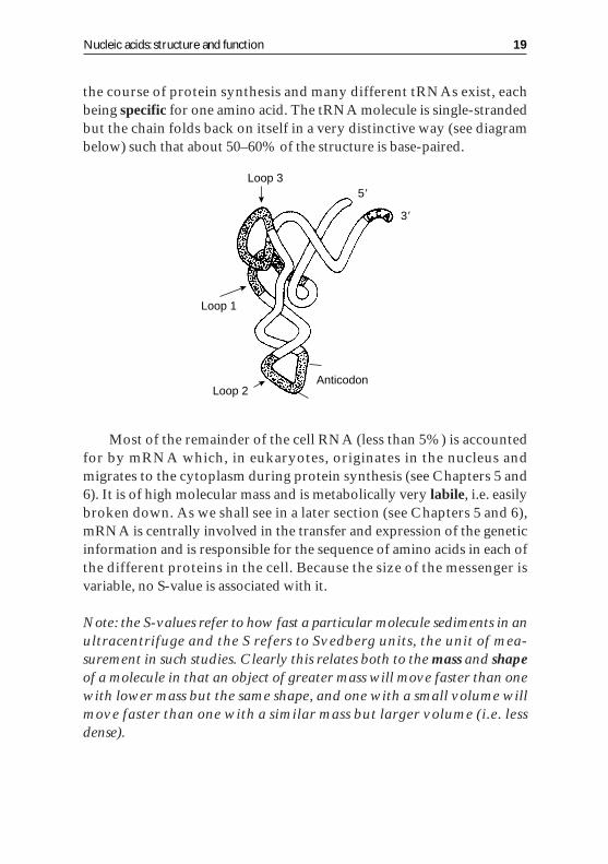
the course of protein synthesis and many different tRNAs exist, eachbeing specific for one amino acid. The tRNA molecule is single-strandedbut the chain folds back on itself in a very distinctive way (see diagrambelow) such that about 50–60% of the structure is base-paired.
Most of the remainder of the cell RNA (less than 5%) is accountedfor by mRNA which, in eukaryotes, originates in the nucleus andmigrates to the cytoplasm during protein synthesis (see Chapters 5 and6). It is of high molecular mass and is metabolically very labile, i.e. easilybroken down. As we shall see in a later section (see Chapters 5 and 6),mRNA is centrally involved in the transfer and expression of the geneticinformation and is responsible for the sequence of amino acids in each ofthe different proteins in the cell. Because the size of the messenger isvariable, no S-value is associated with it.
Note: the S-values refer to how fast a particular molecule sediments in anultracentrifuge and the S refers to Svedberg units, the unit of mea-surement in such studies. Clearly this relates both to the mass and shapeof a molecule in that an object of greater mass will move faster than onewith lower mass but the same shape, and one with a small volume willmove faster than one with a similar mass but larger volume (i.e. lessdense).
Nucleic acids: structure and function 19
Loop 3
Loop 1
Loop 2Anticodon
3�
5�
RNAs which act as enzymesIn the late 1980s it was shown that certain RNA molecules could act as enzymes,
capable of splicing out specific sequences of RNA either on itself or other RNA
strands.The name given to such molecules was ribozyme and this work earned
a Nobel Prize for one of the researchers (Tom Cech of the University of
Colorado).Two of these ribozymes are now being clinically tested as potential
treatment against HIV (see BASC Booklet 3,Enzymes and their Role in
Biotechnology, for further details).
New Scientist (7 December 1996)
A role for RNA in the treatment of debilitating disease
Specific RNA molecules are being selected from pools of randomly generated and
synthesized RNA as a means of treating the debilitating disease myasthenia gravis.
Sufferers of this disease produce auto-antibodies which block the normal signals
from nerves to muscles by blocking the acetylcholine receptor. It has been shown
that specific RNA molecules are able to bind to the antibodies, so interfering with
their binding to the receptors and hence restoring the normal nerve-to-muscle
signals (see BASC Booklets 5, Immunology, and 9,The Biochemical Basis of Disease
for further information).
New Scientist (18 January 1997)
20 Isolation and structure of nucleic acids
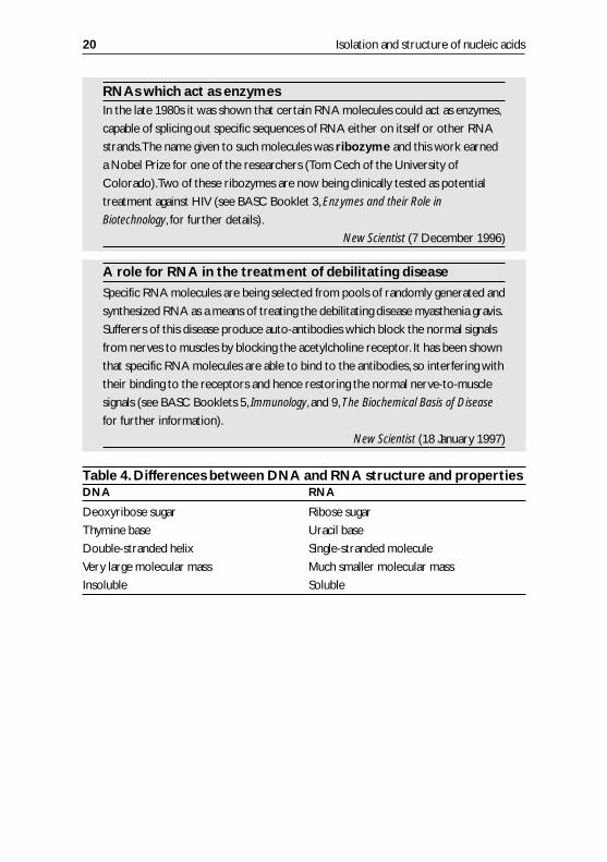
Table 4.Differences between DNA and RNA structure and propertiesDNA RNA
Deoxyribose sugar Ribose sugar
Thymine base Uracil base
Double-stranded helix Single-stranded molecule
Very large molecular mass Much smaller molecular mass
Insoluble Soluble
3
DNA replication
Learning objectives
Each student should, without reference to his or her notes, be able to:• explain the biological significance of DNA replication;• explain the suitability of DNA structure for replication;• outline the main features of semi-conservative replication of DNA;• outline the Meselson and Stahl experiment; and• name the major enzymes involved in replication.
Introduction
In the previous chapter it was shown how we knew that the geneticinformation of a cell is contained in the DNA of that cell. For the cell todivide and produce daughter cells in mitosis and meiosis it is essentialthat the DNA is copied (replicated) and an identical copy is passed to thedaughter cell. DNA is replicated during interphase of both mitosis andmeiosis.
The basic results on which our present day assumptions of themechanism of DNA replication are built are:(i) the evidence that the genetic information was contained within thenucleic acids;(ii) the fact that most DNA consists of two strands of polynucleotidechains, each of which consists of deoxyribonucleotide residues joined by3�–5� phosphodiester bonds; and(iii) the fact that H-bonds occur between the bases in the two strands,adenine in one strand is always bonded to thymine in the other andlikewise cytosine with guanine.
21
Each of these we have already considered, and thus, if we are giventhe sequence of bases in one strand, we could immediately write downthe sequence of bases in the complementary strand.
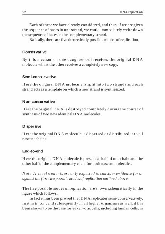
Basically, there are five theoretically possible modes of replication.
Conservative
By this mechanism one daughter cell receives the original DNAmolecule whilst the other receives a completely new copy.
Semi-conservative
Here the original DNA molecule is split into two strands and eachstrand acts as a template on which a new strand is synthesized.
Non-conservative
Here the original DNA is destroyed completely during the course ofsynthesis of two new identical DNA molecules.
Dispersive
Here the original DNA molecule is dispersed or distributed into allnascent chains.
End-to-end
Here the original DNA molecule is present as half of one chain and theother half of the complementary chain for both nascent molecules.
Note: A-level students are only expected to consider evidence for oragainst the first two possible modes of replication outlined above.
The five possible modes of replication are shown schematically in thefigure which follows.
In fact it has been proved that DNA replicates semi-conservatively,first in E. coli, and subsequently in all higher organisms as well: it hasbeen shown to be the case for eukaryotic cells, including human cells, in
22 DNA replication
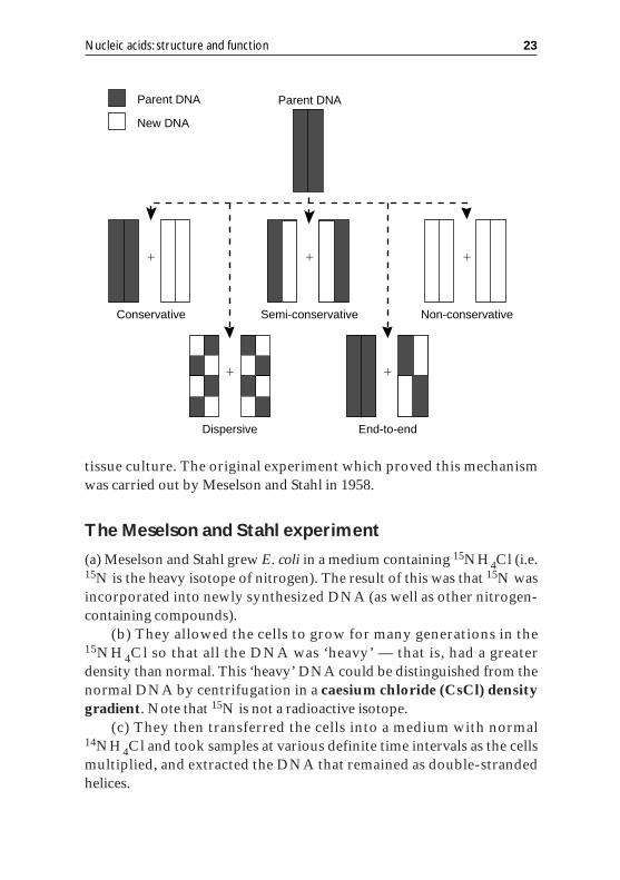
tissue culture. The original experiment which proved this mechanismwas carried out by Meselson and Stahl in 1958.
The Meselson and Stahl experiment
(a) Meselson and Stahl grew E. coli in a medium containing 15NH4Cl (i.e.15N is the heavy isotope of nitrogen). The result of this was that 15N wasincorporated into newly synthesized DNA (as well as other nitrogen-containing compounds).
(b) They allowed the cells to grow for many generations in the15NH4Cl so that all the DNA was ‘heavy’ — that is, had a greaterdensity than normal. This ‘heavy’ DNA could be distinguished from thenormal DNA by centrifugation in a caesium chloride (CsCl) densitygradient. Note that 15N is not a radioactive isotope.
(c) They then transferred the cells into a medium with normal14NH4Cl and took samples at various definite time intervals as the cellsmultiplied, and extracted the DNA that remained as double-strandedhelices.
Nucleic acids: structure and function 23
Parent DNA
New DNA
Parent DNA
�
Conservative
�
Semi-conservative
�
Non-conservative
�
End-to-end
�
Dispersive
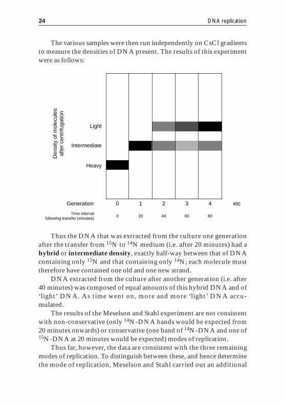
The various samples were then run independently on CsCl gradientsto measure the densities of DNA present. The results of this experimentwere as follows:
Thus the DNA that was extracted from the culture one generationafter the transfer from 15N to 14N medium (i.e. after 20 minutes) had ahybrid or intermediate density, exactly half-way between that of DNAcontaining only 15N and that containing only 14N; each molecule musttherefore have contained one old and one new strand.
DNA extracted from the culture after another generation (i.e. after40 minutes) was composed of equal amounts of this hybrid DNA and of‘light’ DNA. As time went on, more and more ‘light’ DNA accu-mulated.
The results of the Meselson and Stahl experiment are not consistentwith non-conservative (only 14N-DNA bands would be expected from20 minutes onwards) or conservative (one band of 14N-DNA and one of15N-DNA at 20 minutes would be expected) modes of replication.
Thus far, however, the data are consistent with the three remainingmodes of replication. To distinguish between these, and hence determinethe mode of replication, Meselson and Stahl carried out an additional
24 DNA replication
Light
Intermediate
Heavy
0Generation 1 2 3 4 etc
0Time interval
following transfer (minutes) 20 40 60 80
Den
sity
of m
olec
ules
afte
r ce
ntrif
ugat
ion

experiment in which they took the DNA isolated at 20 minutes aftertransfer to 14N-medium, strand separated this and ran the resultantsample on a CsCl gradient. For dispersive or end-to-end models wewould predict a single hybrid band, whereas for semi-conservative repli-cation we would expect two bands, one light, one heavy. The latter wasfound to be the case and hence DNA replication is a semi-conservativeprocess.
Mechanism of replication
Watson and Crick, in their 1953 paper in which they proposed thedouble-helical structure for DNA, conclude as follows: “It has notescaped our notice that the specific pairing we have postulated imme-diately suggests a possible copying mechanism for the genetic material”.The implication was that, owing to the strict complementary nature ofthe strands, the base sequence of one strand would determine the basesequence of the other. It could be imagined therefore that replicationcould follow a scheme as shown below.
Nucleic acids: structure and function 25
5�3�
3�
3�
3�
3�
3�
3�
3�
5�
5�
5� 5�
5�
5�
5�

The problem, however, with this scheme is that the energy requiredfor the first step, namely strand separation, would be far too high to beplausible. This led to the suggestion that unfolding took place a little at atime, so forming a replication fork as shown.
Here, as elsewhere in biochemical reactions, an enzyme is used as acatalyst. Enzymes lower the energy needed to activate a reaction, makingit more likely to occur. For E. coli this is an enzyme called DNA poly-merase III. The simplest scheme would be for polymerase molecules toattach themselves to the ends of the single-stranded DNA template andto synthesize a complimentary strand of each.
The first problem encountered with this proposal arose because thepolymerase enzyme was found to operate in one direction only, that is itsynthesized the nascent DNA strand in the direction 5�→3�. This meantthat one of the parent strands (called the leading strand) could be copiedin a continuous fashion, whereas the other (called the lagging strand)could only be copied in a discontinuous way as a series of short lengths(approx. 1000 bases long) of DNA, each of which is called an Okazakifragment (see Figure below). It was also discovered early on that theDNA polymerase enzyme could not actually synthesize a DNAmolecule from individual DNA nucleotides but instead required a shortstretch of existing DNA helix duplex on which to build. In this sense it istruly a DNA-dependent DNA polymerase III.
26 DNA replication
3�
5�
3�
3�
5�
5�
RNA primer
Helix-destabilizing protein
DNA helicase
DNA gyrase
DNA polymerase III
3�5�
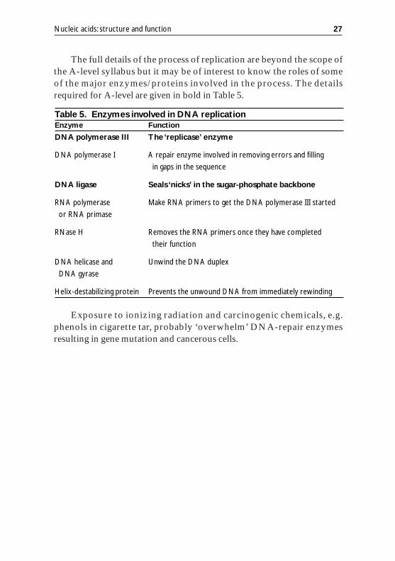
The full details of the process of replication are beyond the scope ofthe A-level syllabus but it may be of interest to know the roles of someof the major enzymes/proteins involved in the process. The detailsrequired for A-level are given in bold in Table 5.
Exposure to ionizing radiation and carcinogenic chemicals, e.g.phenols in cigarette tar, probably ‘overwhelm’ DNA-repair enzymesresulting in gene mutation and cancerous cells.
Nucleic acids: structure and function 27
Table 5. Enzymes involved in DNA replicationEnzyme Function
DNA polymerase III The ‘replicase’ enzyme
DNA polymerase I A repair enzyme involved in removing errors and filling
in gaps in the sequence
DNA ligase Seals ‘nicks’ in the sugar-phosphate backbone
RNA polymerase Make RNA primers to get the DNA polymerase III started
or RNA primase
RNase H Removes the RNA primers once they have completed
their function
DNA helicase and Unwind the DNA duplex
DNA gyrase
Helix-destabilizing protein Prevents the unwound DNA from immediately rewinding
4
The genetic code
Learning objectives
Each student should, without reference to his or her notes, be able to:• state that the unit of information is the codon — a sequence of three
bases (on DNA and mRNA) representing one amino acid;• state that since there are four bases there are 64 possible triplet
codons;• state that all codons have been assigned to amino acids or start/stop
signals;• give details of the characteristics of the genetic code; and• predict the amino acid sequence of a protein from an mRNA or
DNA base sequence (with the aid of a copy of the genetic code).
Introduction
The middle to late 1950s was a particularly active and productive phasewith respect to the research to aid our understanding of the process ofgene expression. At about this time Crick formulated the central dogmawhich, in the earliest forms, was expressed as:
Thus far, we have seen that the genetic information which dictates acell’s phenotype is stored in the DNA and this can be passed to progeny
29
Replication DNA RNA Protein
TranslationTranscription
cells via the process of replication. We will now consider how this storedinformation is expressed to yield the specific phenotype.
A key feature in this respect is the concept of a gene which has beendefined “operationally as a region of the genome that segregates as asingle unit during meiosis and gives rise to a definable phenotypic traitsuch as a red or white eye in Drosophila or a round or wrinkled seed inpeas”.
The genome is the sum of all DNA nucleotide/base sequences in anindividual organism or species.
Later work demonstrated that a gene or gene region corresponds toa length of DNA on the genome that contains the information for thesynthesis of a single protein. This led to the useful concept that one genecorresponds to one protein.
However, as a great many biologically active proteins comprisemore than one polypeptide chain, it is more correct to consider this as‘one gene — one polypeptide chain’.
Although serving as a useful memory aid, even this statement is notentirely correct since some genes code for single structural RNA mol-ecules, such as mRNA and tRNA, rather than polypeptide chains and,also, more recent studies have revealed additional complexities in termsof the structural organization of the genetic information. For thepurposes of the present section, however, it is acceptable to treat the genein terms of the ‘one gene — one polypeptide chain’ hypothesis.
Building a human chromosome
The first synthetic chromosome, that of yeast,was built about a decade ago.To
extend this to the synthesis of a human chromosome required detailed consid-
eration of further structural complexities.Thus, in addition to the structural genes
there is a need for telomeres, long repeating DNA sequences which protect the
ends of the chromosome and prevent them binding together, and centromeres,
which provide the scaffolding that enables duplicate chromosomes to split during
cell division.Using this approach, totally artificial chromosomes have been
produced and it is hoped that these may help in gene therapy in the future.
New Scientist (5 April 1997)
30 The genetic code

To transfer the genetic information from DNA to a sequence ofamino acids that constitutes a polypeptide chain involves two discretebut consecutive processes:(i) transcription, in which an mRNA strand of nucleotides is syn-thesized from one of the strands of the DNA in the gene region; and(ii) translation, in which the genetic information coded as a sequence ofbases in mRNA is translated or converted into a sequence of amino acidsto form a discrete polypeptide chain.
The gene regions specific to protein synthesis collectively only rep-resent a small part of the genome for many cells. The remaining regionsare accounted for as non-coding/‘nonsense’ sequences, multiple repeatsequences, control regions and regions specifying the synthesis of func-tional RNA molecules such as tRNA and rRNA. A summary of thesefeatures is shown below.
How many bases code for an amino acid?
In the next section we shall consider details of the mechanism by whichthe information coded in the mRNA is used to synthesize proteins of aspecific sequence. An important concept underpinning this topic is thenature of the code used to translate the sequence of bases in the mRNAmolecule into a sequence of amino acids in a polypeptide chain. Thiscode is referred to as the genetic code and in the present section we shallconsider the general properties of the code.
For mRNA to control the synthesis of specific proteins we must askhow a molecule made up from only four different kinds of base candetermine the order of about 20 different amino acids. Obviously therecannot be a 1:1 correspondence between the RNA bases and the amino
Nucleic acids: structure and function 31
Transcription
Control region for gene A Multiple copies of gene CControl region for genes B1 and B2
Translation
gene A
Protein A Protein B (dimer)Non-codingsequences
mRNA mRNA mRNA tRNA
gene B1 gene B2

acids. Similarly there cannot be a 2:1 correspondence, since this onlyallows for 16 different arrangements (4 �4). If, however, there are threebases coding for each amino acid, the number of possible triplets is 64(4 � 4 � 4), which would be enough combinations to code for eachamino acid with plenty to spare. Clear-cut evidence has been producedthat the triplet theory is correct and we will now look at the evidencefor such a view. The name given to the sequence of three nucleotides is acodon.
Evidence for a triplet code
The evidence for a triplet code comes from genetic studies by Crick andco-workers using mutations caused by acridine orange or proflavine infruit flies. The mode of action of these compounds is not fullyunderstood but the net effect is to cause the insertion of an additionalbase into a DNA strand or to delete one of the bases in a DNA strand,possibly during DNA replication.
It is easier to explain the results of these studies by considering asimple model and studying its behaviour. If we assume a DNA sequenceto be a series of triplet codes of bases, or codons, for five amino acids:
– CAT – CAT – CAT – CAT – CAT –
The insertion of a base, T:
– CAT – TCA – TCA – TCA – TCA –
Or deletion of a base, C, from the original sequence:
– CAT – ATC – ATC – ATC – ATC –
The result of a single mutation (either insertion or deletion) is tocause the genetic message/amino acid sequence everywhere to the rightof the mutation to be misread.
The result would almost certainly be a defective protein or enzyme,especially if the amino acid sequence is in or near the active site of anenzyme.
Also in these studies it was shown that if we insert an extra base,then delete another base a little further on in the sequence, the net resultis an almost identical product (demonstrate this for yourself). Theimportant point here is that the insertion and deletion are relatively close
32 The genetic code
together (why?). There are all sorts of other ways of combiningmutation, but the net results provide very strong evidence in favour of atriplet code. These results also provide additional data concerning themode of translating the code.
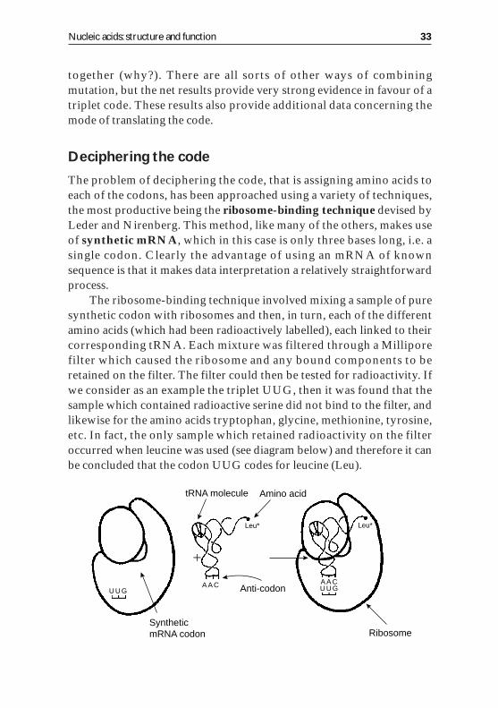
Deciphering the code
The problem of deciphering the code, that is assigning amino acids toeach of the codons, has been approached using a variety of techniques,the most productive being the ribosome-binding technique devised byLeder and Nirenberg. This method, like many of the others, makes useof synthetic mRNA, which in this case is only three bases long, i.e. asingle codon. Clearly the advantage of using an mRNA of knownsequence is that it makes data interpretation a relatively straightforwardprocess.
The ribosome-binding technique involved mixing a sample of puresynthetic codon with ribosomes and then, in turn, each of the differentamino acids (which had been radioactively labelled), each linked to theircorresponding tRNA. Each mixture was filtered through a Milliporefilter which caused the ribosome and any bound components to beretained on the filter. The filter could then be tested for radioactivity. Ifwe consider as an example the triplet UUG, then it was found that thesample which contained radioactive serine did not bind to the filter, andlikewise for the amino acids tryptophan, glycine, methionine, tyrosine,etc. In fact, the only sample which retained radioactivity on the filteroccurred when leucine was used (see diagram below) and therefore it canbe concluded that the codon UUG codes for leucine (Leu).
Nucleic acids: structure and function 33
U U G U U GA A CA A C
Leu*Leu*
RibosomeSynthetic mRNA codon
Anti-codon
Amino acidtRNA molecule
�

This process is then repeated exactly as above for a different tripletcode. Using this simple yet highly efficient technique (with some minormodifications) it was possible to assign 61 of the 64 possible tripletsunambiguously to specific amino acids, and the accepted code is asshown here.
Nonsense codons
As has just been seen, 61 of the 64 triplets have been assigned to par-ticular amino acids — what then is the function of the remaining three,UAA, UAG and UGA? These were initially labelled as the nonsensecodons but it has since been shown that they have an important functionin protein biosynthesis as chain-terminating signals. It is these codonswhich inform the protein-synthesizing system that the nascent poly-peptide chain has been completely synthesized and to thus allow it to bereleased from the ribosome.
34 The genetic code
U
U C A G
C
A
G
U
C
A
G
UUU
UUC
UUA
UUG
UCU
UCC
UCA
UCG
UAU
UAC
UAA
UAG
UGU
UGC
UGA
UGG
CUU
CUC
CUA
CUG
CCU
CCC
CCA
CCG
CAU
CAC
CAA
CAG
CGU
CGC
CGA
CGG
AUU
AUC
AUA
AUG
ACU
ACC
ACA
ACG
AAU
AAC
AAA
AAG
AGU
AGC
AGA
AGG
GUU
GUC
GUA
GUG
GCU
GCC
GCA
GCG
GAU
GAC
GAA
GAG
GGU
GGC
GGA
GGG
U
C
A
G
U
C
A
G
U
C
A
G
} } }
} }}}}
}}
} }}}}
}
}}
}
}
}
Phe
Ser
Pro
Thr
Ala GlyVal
Met
Ile
Leu
Leu
His
Gln
Asn
Lys
Ser
Arg
CysTyr
OCHRE
AMBER
?
Trp
Arg
Asp
Glu
SECOND LETTER
FIR
ST
LE
TT
ER
TH
IRD
LET
TE
R
From the experiments just described, plus other related studies, thegeneral features of the code that are important are as follows:• the code is sequential (i.e. it is read in strict sequence from one end
to the other); and• the code is degenerate.
A most important feature is that many amino acids are coded bymore than one triplet (all except methionine and tryptophan). Indeed itseems that for most amino acids only the first two bases of the codonneed be specified. A code of this type is termed degenerate. It followsfrom this that more than one kind of tRNA may code for the sameamino acid. This is in fact known to be the case.• The code is universal or ubiquitous.
At present, as a result of experimental data, it is generally acceptedthat the code is universal, e.g. Lipmann and others demonstrated usingyeast, Micrococcal or E. coli tRNA, rabbit reticulocyte ribosomes andmRNA for haemoglobin that a perfectly normal protein, indistin-guishable from wild-type, could be formed. More recently, however,some exceptions to this generalization have been identified in mito-chondria, leukaemic cells and in some ciliates, and these observations arepresently posing a challenge for evolutionary theorists.
Nucleic acids: structure and function 35
5
Transcription
Learning objectives
Each student should, without reference to his or her notes, be able to:• explain the meaning of the term transcription;• explain the role of mRNA;• explain the relationship of mRNA to the DNA template;• outline the necessity of moving the genetic information required to
synthesize proteins from the nucleus to the cytoplasm;• state that the genetic information is carried to the cytoplasm by an
mRNA strand which is complementary to the DNA template strand;
• state that only a relatively short segment of DNA coding for a specific polypeptide is transcribed as a unit from the template strand of DNA;
• state that mRNA is synthesized by RNA polymerase using ribo-nucleoside triphosphates as substrates and DNA as a template; and
• outline with the aid of simple labelled diagrams the process of trans-cription.
Introduction
Transcription involves the synthesis of an mRNA molecule comple-mentary to a gene region/specific nucleotide sequence of DNA. Thisbeing the case, we require the basic building blocks for RNA, namelyfree RNA nucleotides and ATP molecules for energy together with aDNA molecule, part of which will be copied, the latter being called aDNA template.
37
For such a process to be undertaken at an appropriate and suffi-ciently fast rate for the cell requires the help of an enzyme, which in thiscase is called DNA-dependent RNA polymerase or just RNA poly-merase. This enzyme cannot synthesize an mRNA strand from freeRNA nucleotides unless a DNA molecule is present.
It is worth remembering that enzymes have three key advantages.(i) They act as biological catalysts to speed up biochemical reactions bylowering the energy needed to activate a reaction. This means a reactionis far more likely to proceed under the conditions found in cells, e.g.moderate temperature and low pressures.(ii) They usually have high specificity in that one very specific reaction(or a few related reactions) is catalysed because of the specific three-dimensional shape of the active site.(iii) Their activity can be controlled, e.g. by their products and othercompounds.
Mechanism of transcription
The enzyme RNA polymerase, like DNA polymerase, operates in thedirection 5�→3�, that being the direction in which the mRNA is syn-thesized. Unlike DNA polymerase it only transcribes one strand ofDNA, i.e. ‘genes’ are located on a single DNA template strand.
To understand the mechanism of this process it is clearly of interestto know the way in which the start of each gene is correctly recognized.The picture which emerged from a study of the DNA sequences near thestart of specific gene regions was that of AT-rich sequences of bases.These sequences were situated a few bases upstream from the startingpoint of the mRNA transcript, as shown here.
38 Transcription
Specific gene Base sequence near the start of specific gene region
fd
T7 A2
Lac-UV-5
SV40
E. coli Tyr tRNA
CTGACTATAATAGACAGGGTAAAGACCTG
TGCAGTAAGATACAAATCCGTAGGTAACA
GCTCGTATAATGGTTACAAATAAAGCAAT
CAGCTTATAATGGTTACAAATAAAGCAAT
TTTGATATGATGCGCCCCGCTTCCCGATA
The AT base pair has two H-bonds compared with three for the GCbase pair (see Chapter 2); and consequently a region rich in AT pairstends to melt or strand separate more readily, so forming a bubble. Theadvantages of forming this bubble are that (i) it helps the RNA poly-merase to identify the start of the gene region and (ii) it allows the RNApolymerase to select more easily the template or coding strand and soallow asymmetric synthesis to take place. In fact it is the non-TATAstrand (i.e. the 3�→5� strand) that acts as the template strand in tran-scription.
Once correct initiation has been achieved the RNA polymerasemoves along the template strand synthesizing an mRNA from RNAnucleotides. The base on each successive RNA nucleotide is comple-mentary to the DNA template strand.
The mechanisms for termination of transcription vary from gene togene. For some genes an additional protein called rho (�) identifies thetermination site and causes the RNA polymerase to stop synthesis.Other mechanisms, as shown schematically below, include (i) an AT-richregion flanked on either side by a GC-rich region or (ii) a palindromicregion. In both these cases a bubble structure is formed which can beconsidered to act as a physical barrier, so causing the RNA polymeraseto terminate synthesis as shown.
Each mRNA strand then exits the nucleus through pores in thenuclear membrane and attaches to ribosomes in the cell cytoplasm.
Nucleic acids: structure and function 39
C–G rich C–G richAATATTATTATAAT
CGATAATCGGCTATTAGC
AATATTA
TTA TAAT
AATATTA
TTA TAAT
growing mRNA
RNA polymerase
growing mRNA
mRNA
mRNA
TAGC
ATCG
A
GCTA
CGAT
A
TT
TAGC
ATCG
GCTA
CGAT
(i)
(ii)

The mechanism of transcription is summarized schematically below.
40 Transcription
TATA
TATA
TATA
TATA
TATA
Sense strand RNA polymerase
Gene
Initiation site
DNA unzipped byRNA polymerase
Termination sequence
Termination sequence
Growing mRNA transcript
mRNA
Termination sequence
5�

6
Translation
Learning objectives
Each student should, without reference to his or her notes, be able to:• state that ribosomes are responsible for the translation of the genetic
information encoded as a sequence of bases in the mRNA strand into a specific sequence of amino acids in a polypeptide chain;
• outline the functions of ribosomes, mRNA, tRNA and amino acids in translation; and
• outline the processes involved in translation using the terms: codon, anticodon, peptide-bond formation and termination, using simple diagrams.
Introduction
It can be shown that no specific affinity/binding occurs between aminoacids and the mRNA, to neither the sugar–phosphate backbone nor thebases (see the Figure below). This being the case, we cannot have a direct‘read-off’ in translation as we did in replication and transcription.
The solution to this problem is to have an indirect associationachieved by coupling the specific amino acid to the mRNA via an‘adaptor molecule’, which turned out to be tRNA.
41
mRNA
Amino acid
No reaction Binds to mRNA
Binds to amino acid
mRNA
tRNA
The tRNA molecules must be activated before they can attach to aspecific amino acid and perform their role in translation of the geneticcode.
Activation of tRNA
To bind the amino acid to the tRNA molecule requires ATP energy anda specific enzyme called an amino acyl-tRNA synthetase. The reactionoccurs in two stages: in the first the amino acid is activated and in thesecond the activated amino acid is transferred to its specific tRNA asshown schematically below.
42 Translation
A
A
Amino acid Enzyme (amino acyl-tRNA synthetase)
ATP
PyrophosphatePhosphate
A
A
CC
A
tRNA
CCA
AMP
CCA
Amino acyl-tRNA(an “activated”
amino acid)
The amino acyl-tRNA synthetase enzyme, apart from catalysing thereaction, also displays high specificity in that it will only bind a par-ticular amino acid to its correct, specific tRNA molecule thus ensuringhigh accuracy/fidelity in the translation event. The amino acids arelinked to the 3� end of the tRNA (all end with the sequence CCA-OH)via their carboxyl group (COOH).
Initiation
Translation of the mRNA strand starts from the 5� end at a specific ini-tiation site or triplet codon, AUG, which normally codes for the aminoacid methionine. This initiation site AUG codon is distinguished fromall other AUG sequences as it is preceded in the mRNA sequence by anAGGA base sequence, known as the Shine–Dalgarno consensussequence, named after the discoverers.
This sequence is recognized by a UCCU complementary sequencein the tRNA of the small ribosomal subunit and so causes the ribosometo bind to the correct starting point on the mRNA as seen below.
It was also shown that this unique AUG binds a special amino acid(linked to its corresponding tRNA as described previously). This specialamino acid is a methionine residue in which the amino group has aformyl group (CHO) bound to it and is called N-formylmethionine(fMet) in prokaryotes. The advantage of this added complexity is thatthis effectively blocks the amino group and inhibits it reacting, therebydirecting the polypeptide chain to be synthesized in the N-terminal→C-terminal direction. Eukaryotes do not use the formyl group.
Nucleic acids: structure and function 43
AGGA AUG AUG AUG AUG
AGGAUCCU
AUG AUG AUG AUG
5�
5�
3�
3�
By such means an initiation complex is formed in which theribosome is bound to the correct site on the mRNA and the first aminoacid (linked to its corresponding tRNA) is in position.
Elongation
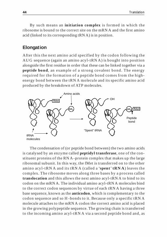
After this the next amino acid specified by the codon following theAUG sequence (again an amino acyl-tRNA) is bought into positionalongside the first residue in order that these can be linked together via apeptide bond, an example of a strong covalent bond. The energyrequired for the formation of a peptide bond comes from the high-energy bond between the tRNA molecule and its specific amino acidproduced by the breakdown of ATP molecules.
The condensation of (or peptide bond between) the two amino acidsis catalysed by an enzyme called peptidyl transferase, one of the con-stituent proteins of the RNA–protein complex that makes up the largeribosomal subunit. In this way, the fMet is transferred on to the otheramino acyl-tRNA and its tRNA (called a ‘spent’ tRNA) leaves thecomplex. The ribosome moves along three bases by a process calledtranslocation and this allows the next amino acyl-tRNA to bind to itscodon on the mRNA. The individual amino acyl-tRNA molecules bindto the correct codon sequences by virtue of each tRNA having a threebase sequence, known as the anticodon, which is complementary to thecodon sequence and so H-bonds to it. Because only a specific tRNAmolecule attaches to the mRNA codon the correct amino acid is placedin the growing polypeptide sequence. The growing chain is transferredto the incoming amino acyl-tRNA via a second peptide bond and, as
44 Translation
fMet OH
“Spent” tRNA
tRNAmolecules
Amino acids
Peptide bond
aa2 aa2 fMet
before, the ‘spent’ tRNA leaves the complex because of the relativeweakness of the H-bonds between the codon and anticodon bases.
Termination
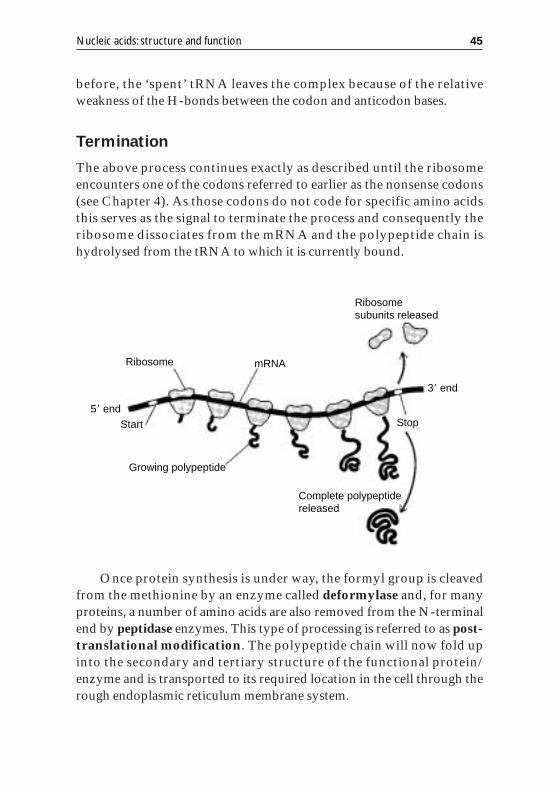
The above process continues exactly as described until the ribosomeencounters one of the codons referred to earlier as the nonsense codons(see Chapter 4). As those codons do not code for specific amino acidsthis serves as the signal to terminate the process and consequently theribosome dissociates from the mRNA and the polypeptide chain ishydrolysed from the tRNA to which it is currently bound.
Once protein synthesis is under way, the formyl group is cleavedfrom the methionine by an enzyme called deformylase and, for manyproteins, a number of amino acids are also removed from the N-terminalend by peptidase enzymes. This type of processing is referred to as post-translational modification. The polypeptide chain will now fold upinto the secondary and tertiary structure of the functional protein/enzyme and is transported to its required location in the cell through therough endoplasmic reticulum membrane system.
Nucleic acids: structure and function 45
Ribosome
Start
mRNA
3� end
5� end
Complete polypeptide released
Ribosome subunits released
Stop
Growing polypeptide
Therefore, by the processes of transcription and translation thelarge, permanent, insoluble DNA molecule, which is restricted to thenucleus, can control the expression of its genetic code by producing aprotein/enzyme molecule which can now affect a biochemical processthroughout the cell.
Polysomes
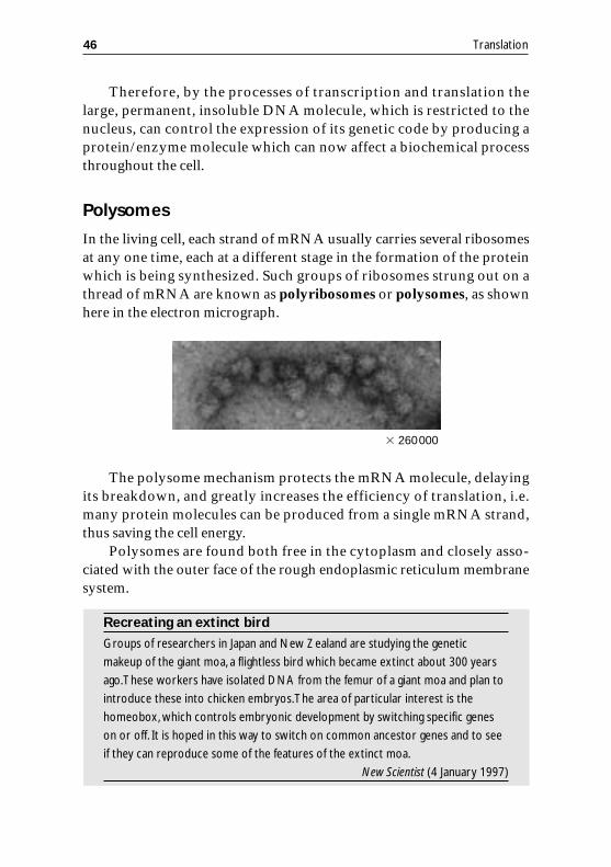
In the living cell, each strand of mRNA usually carries several ribosomesat any one time, each at a different stage in the formation of the proteinwhich is being synthesized. Such groups of ribosomes strung out on athread of mRNA are known as polyribosomes or polysomes, as shownhere in the electron micrograph.
The polysome mechanism protects the mRNA molecule, delayingits breakdown, and greatly increases the efficiency of translation, i.e.many protein molecules can be produced from a single mRNA strand,thus saving the cell energy.
Polysomes are found both free in the cytoplasm and closely asso-ciated with the outer face of the rough endoplasmic reticulum membranesystem.
Recreating an extinct bird
Groups of researchers in Japan and New Zealand are studying the genetic
makeup of the giant moa, a flightless bird which became extinct about 300 years
ago.These workers have isolated DNA from the femur of a giant moa and plan to
introduce these into chicken embryos.The area of particular interest is the
homeobox,which controls embryonic development by switching specific genes
on or off. It is hoped in this way to switch on common ancestor genes and to see
if they can reproduce some of the features of the extinct moa.
New Scientist (4 January 1997)
46 Translation
� 260000
7
Assessment:past examination questionsand outline solutions
As stated earlier, it is hoped that the inclusion of detailed learningobjectives within each section of these guidance notes will not only helpstudents to identify key topics in the syllabus but also help them togauge the level of expected outcome, and hence be of value in the formalassessment of the material.
Many of the Examination Boards publish detailed analyses of theexam-question performance and it may be worthwhile to read thesecarefully and modify the guidance notes where appropriate to reflect thevarying emphasis placed on the different themes within this broad topic.
The following questions are actual A-level Biology examinationquestions together with their solutions provided by the authors. Othersolutions and approaches may also be valid. Presented in this way theymight serve as useful source material in the review of each of the sectionsat the end of formal lessons. Once again, we are grateful to the variousExamination Boards, especially the Northern Examinations andAssessment Board, for allowing past examination questions to be used.Answer all the questions in the spaces provided.
Question 1
The diagram shows the sequence of bases on one strand of a short lengthof DNA. This sequence should be read from left to right.
CGACCCCAG
47
(a) Give:(i) the base sequence that will be produced as a result of trans-cription of the complete length of DNA shown in the diagram;. . . . . . . . . . . . . . . . . . . . . . . . . . . . . . . . . . . . . . . . . . . . . . . . . . . . . . . . . . (2)
(ii) the three bases of the tRNA that will correspond to thesequence of bases shown by the underlined bases on the diagram.. . . . . . . . . . . . . . . . . . . . . . . . . . . . . . . . . . . . . . . . . . . . . . . . . . . . . . . . . . (1)
As a result of a mutation, the first base in the length of DNA shown inthe diagram is lost (deleted).
(b) (i) Use Table 6 to identify the first two amino acids for which the mutated DNA codes;. . . . . . . . . . . . . . . . . . . . . . . . . . . . . . . . . . . . . . . . . . . . . . . . . . . . . . . . . . (1)
(ii) explain why mutations involving the deletion of a base may have greater effects than those involving substitution of one base for another.. . . . . . . . . . . . . . . . . . . . . . . . . . . . . . . . . . . . . . . . . . . . . . . . . . . . . . . . . . . . . . . . . . . . . . . . . . . . . . . . . . . . . . . . . . . . . . . . . . . . . . . . . . . . . . . . . . . . . . . . . . . . . . . . . . . . . . . . . . . . . . . . . . . . . . . . . . . . . . . . . . . . . . . . . . . . . . . . . . . . (2)
Question 2
In an investigation of DNA replication, a species of bacterium wasgrown for many generations in a medium containing the 15N isotope ofnitrogen, rather than its more common form, 14N.
DNA was isolated from the bacterial cells and separated into singlestrands by mild chemical treatment. The density of this single-strandedDNA was measured using density-gradient centrifugation. In this
48 Past examination questions
Table 6.DNA base sequences and the amino acids for which theycodeDNA base sequence Amino acid
ACC Tryptophan
CAG Valine
CCA Glycine
CCC Glycine
CGA Alanine
GAC Leucine
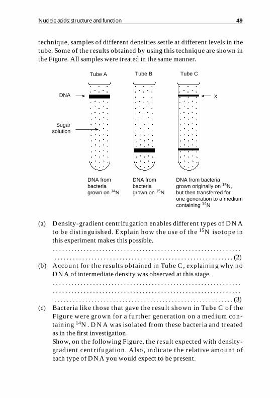
technique, samples of different densities settle at different levels in thetube. Some of the results obtained by using this technique are shown inthe Figure. All samples were treated in the same manner.
(a) Density-gradient centrifugation enables different types of DNA to be distinguished. Explain how the use of the 15N isotope in this experiment makes this possible.. . . . . . . . . . . . . . . . . . . . . . . . . . . . . . . . . . . . . . . . . . . . . . . . . . . . . . . . . . . . . . . . . . . . . . . . . . . . . . . . . . . . . . . . . . . . . . . . . . . . . . . . . . . . . . . . . . . . . . . (2)
(b) Account for the results obtained in Tube C, explaining why no DNA of intermediate density was observed at this stage.. . . . . . . . . . . . . . . . . . . . . . . . . . . . . . . . . . . . . . . . . . . . . . . . . . . . . . . . . . . . . . . . . . . . . . . . . . . . . . . . . . . . . . . . . . . . . . . . . . . . . . . . . . . . . . . . . . . . . . . . . . . . . . . . . . . . . . . . . . . . . . . . . . . . . . . . . . . . . . . . . . . . . . . . . . . . . . . . . . . . (3)
(c) Bacteria like those that gave the result shown in Tube C of the Figure were grown for a further generation on a medium con-taining 14N. DNA was isolated from these bacteria and treated as in the first investigation.Show, on the following Figure, the result expected with density-gradient centrifugation. Also, indicate the relative amount of each type of DNA you would expect to be present.
Nucleic acids: structure and function 49
Tube CTube BTube A
DNA X
DNA frombacteriagrown on 14N
DNA frombacteriagrown on 15N
DNA from bacteriagrown originally on 15N,but then transferred forone generation to a mediumcontaining 14N
Sugarsolution

. . . . . . . . . . . . . . . . . . . . . . . . . . . . . . . . . . . . . . . . . . . . . . . . . . . . . . . . . . (2)
Studies were carried out to determine the nitrogenous base compositionof DNA in this bacterium. This involved finding values for double-stranded DNA as well as for each individual (� and �) DNA strand.
The Table below gives some of the results obtained.
(d) Enter the seven ‘missing’ values in the table. (4)(e) What ratio of (A�G) to (C�T) would you expect in the band of
DNA marked as ‘X’ in Tube C in the first Figure? Explain your answer.. . . . . . . . . . . . . . . . . . . . . . . . . . . . . . . . . . . . . . . . . . . . . . . . . . . . . . . . . . . . . . . . . . . . . . . . . . . . . . . . . . . . . . . . . . . . . . . . . . . . . . . . . . . . . . . . . . . . . . . (2)
Question 3
Albino mice are completely white because they cannot produce coatpigment. They cannot produce this pigment because of a gene mutationthat affects the formation of an enzyme in the metabolic pathway that
50 Past examination questions
DNA from bacteriagrown originally on 15N,but then transferred fortwo generations to amedium containing 14N
Table 7. Base composition of DNAPercentage of base
present in DNA sample Ratio of (A�G) Ratio of (A�T)DNA sample A G C T to (C�T) to (C�G)
Double-stranded 30 20 20 … 1.00 1.5
� Strand 28 26 … 32 1.17 …
� Strand 32 … … … 0.85 …

produces the pigment. The enzyme produced by the mutant gene doesnot function effectively.
(a) (i) Explain what is meant by gene mutation;. . . . . . . . . . . . . . . . . . . . . . . . . . . . . . . . . . . . . . . . . . . . . . . . . . . . . . . . . . (1)
(ii) explain how a gene mutation could result in a change in theamino acid sequence in the enzyme produced by the coat-pigment gene.. . . . . . . . . . . . . . . . . . . . . . . . . . . . . . . . . . . . . . . . . . . . . . . . . . . . . . . . . . . . . . . . . . . . . . . . . . . . . . . . . . . . . . . . . . . . . . . . . . . . . . . . . . . . . . . . . . . . . . . . . . . . . . . . . . . . . . . . . . . . . . . . . . . . . . . . . . . . . . . . . . . . . . . . . . . . . . . . . . . . (4)
(b) Explain why mice that are heterozygous for this mutant geneare never albinos.. . . . . . . . . . . . . . . . . . . . . . . . . . . . . . . . . . . . . . . . . . . . . . . . . . . . . . . . . . . . . . . . . . . . . . . . . . . . . . . . . . . . . . . . . . . . . . . . . . . . . . . . . . . . . . . . . . . . . . . (2)
Question 4
The diagram shows part of a DNA molecule.
(a) How are the two strands of the DNA molecule held together?. . . . . . . . . . . . . . . . . . . . . . . . . . . . . . . . . . . . . . . . . . . . . . . . . . . . . . . . . . (1)
(b) What do each of the following letters on the diagram represent?W . . . . . . . . . . . . . . . . . . . . . . . . . . . . . . . . . . . . . . . . . . . . . . . . . . . . . . . . . . . X . . . . . . . . . . . . . . . . . . . . . . . . . . . . . . . . . . . . . . . . . . . . . . . . . . . . . . . . . . . Y . . . . . . . . . . . . . . . . . . . . . . . . . . . . . . . . . . . . . . . . . . . . . . . . . . . . . . . . . . . Z . . . . . . . . . . . . . . . . . . . . . . . . . . . . . . . . . . . . . . . . . . . . . . . . . . . . . . . . . (4)
Nucleic acids: structure and function 51
Y guanine
W
Z
X
thymine
Question 5
One technique used to produce human insulin by genetic engineeringinvolves inserting a gene for human insulin into the DNA of a bacterium.
(a) Name the enzyme that would be used to:(i) cut the bacterial DNA;. . . . . . . . . . . . . . . . . . . . . . . . . . . . . . . . . . . . . . . . . . . . . . . . . . . . . . . . . . (1)
(ii) insert the DNA for human insulin into the cut bacterial DNA.. . . . . . . . . . . . . . . . . . . . . . . . . . . . . . . . . . . . . . . . . . . . . . . . . . . . . . . . . . (1)
(b) There are 51 amino acids in insulin, made up of 16 amino acids out of the 20 that are coded for by DNA. What is the minimum number of different types of tRNA molecule necessary for the synthesis of insulin? Explain your answer.. . . . . . . . . . . . . . . . . . . . . . . . . . . . . . . . . . . . . . . . . . . . . . . . . . . . . . . . . . . . . . . . . . . . . . . . . . . . . . . . . . . . . . . . . . . . . . . . . . . . . . . . . . . . . . . . . . . . . . . (2)
(c) The base sequence below is part of the DNA sequence that codes for insulin.
CCATAGCAC
(i) Write down the corresponding mRNA sequence;. . . . . . . . . . . . . . . . . . . . . . . . . . . . . . . . . . . . . . . . . . . . . . . . . . . . . . . . . . (1)
(ii) A mutation occurred that replaced guanine in this DNA sequence with a different base. Explain the possible effects of this mutation on the structure of the insulin molecule.. . . . . . . . . . . . . . . . . . . . . . . . . . . . . . . . . . . . . . . . . . . . . . . . . . . . . . . . . . . . . . . . . . . . . . . . . . . . . . . . . . . . . . . . . . . . . . . . . . . . . . . . . . . . . . . . . . . . . . . . . . . . . . . . . . . . . . . . . . . . . . . . . . . . . . . . . . . . . . . . . . . . . . . . . . . . . . . . . . . . (3)
Question 6
Scientists have shown that kidney beans are resistant to cowpea weevilsand adzuki bean weevils, two of the most serious pests of African andAsian pulses (vegetables related to peas and beans). This is because thebeans produce a protein that inhibits one of the weevils’ digestiveenzymes. Weevils that eat the pulses soon starve to death. Theresearchers have identified the gene that produces the inhibitor andremoved it from the kidney bean DNA. They inserted the gene thatproduces the inhibitor into the DNA of a bacterium called Agro-bacterium tumefaciens. Using this bacterium they have been able to add
52 Past examination questions
the inhibitor gene to peas. They hope soon to be able to add the gene toAfrican and Asian pulses.
(a) Describe how scientists could:(i) remove the gene that produces the inhibitor from kidney beans;. . . . . . . . . . . . . . . . . . . . . . . . . . . . . . . . . . . . . . . . . . . . . . . . . . . . . . . . . . . . . . . . . . . . . . . . . . . . . . . . . . . . . . . . . . . . . . . . . . . . . . . . . . . . . . . . . . . . . . . (2)
(ii) insert this gene into the DNA of a bacterium.. . . . . . . . . . . . . . . . . . . . . . . . . . . . . . . . . . . . . . . . . . . . . . . . . . . . . . . . . . . . . . . . . . . . . . . . . . . . . . . . . . . . . . . . . . . . . . . . . . . . . . . . . . . . . . . . . . . . . . . (2)
(b) The DNA in the bacterium is able to replicate to produce many copies of itself for insertion into pea cells. Describe the structure of a DNA molecule and explain how this structure enables the molecule to replicate itself.. . . . . . . . . . . . . . . . . . . . . . . . . . . . . . . . . . . . . . . . . . . . . . . . . . . . . . . . . . . . . . . . . . . . . . . . . . . . . . . . . . . . . . . . . . . . . . . . . . . . . . . . . . . . . . . . . . . . . . . . . . . . . . . . . . . . . . . . . . . . . . . . . . . . . . . . . . . . . . . . . . . . . . . . . . . . . . . . . . . . . . . . . . . . . . . . . . . . . . . . . . . . . . . . . . . . . . . . . . . . . . . . . . . . . . . . . . . . . . . . . (8)
Question 7
(a) Draw a labelled diagram of a representative length of the DNA (deoxyribonucleic acid) molecule (the chemical structure of the different constituents is not required). (3)
(b) Name the repeating units that are joined to form the macro-molecule of DNA.. . . . . . . . . . . . . . . . . . . . . . . . . . . . . . . . . . . . . . . . . . . . . . . . . . . . . . . . . . (1)
Nucleic acids: structure and function 53
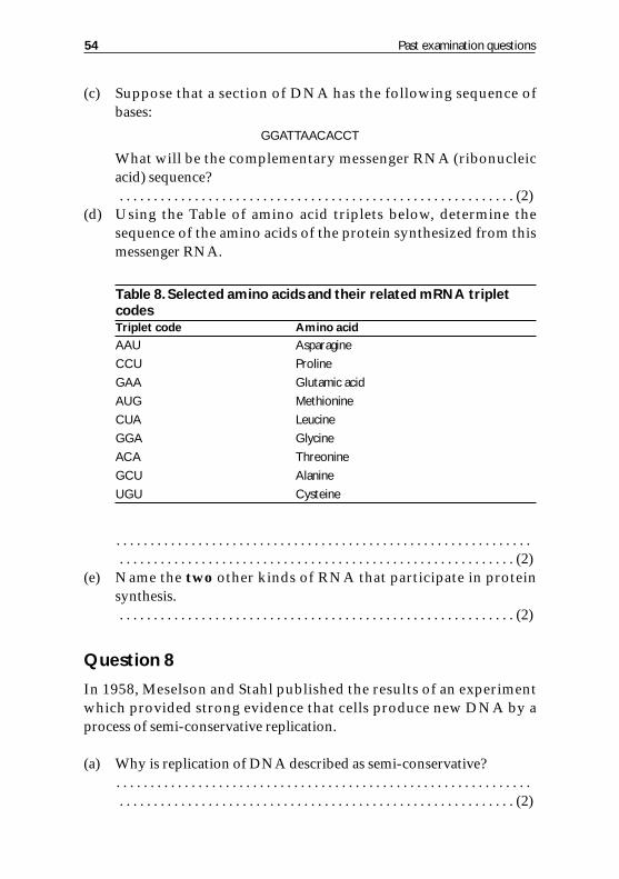
(c) Suppose that a section of DNA has the following sequence of bases:
GGATTAACACCT
What will be the complementary messenger RNA (ribonucleic acid) sequence?. . . . . . . . . . . . . . . . . . . . . . . . . . . . . . . . . . . . . . . . . . . . . . . . . . . . . . . . . . (2)
(d) Using the Table of amino acid triplets below, determine the sequence of the amino acids of the protein synthesized from this messenger RNA.
. . . . . . . . . . . . . . . . . . . . . . . . . . . . . . . . . . . . . . . . . . . . . . . . . . . . . . . . . . . . . . . . . . . . . . . . . . . . . . . . . . . . . . . . . . . . . . . . . . . . . . . . . . . . . . . . . . . . . . . (2)
(e) Name the two other kinds of RNA that participate in protein synthesis.. . . . . . . . . . . . . . . . . . . . . . . . . . . . . . . . . . . . . . . . . . . . . . . . . . . . . . . . . . (2)
Question 8
In 1958, Meselson and Stahl published the results of an experimentwhich provided strong evidence that cells produce new DNA by aprocess of semi-conservative replication.
(a) Why is replication of DNA described as semi-conservative?. . . . . . . . . . . . . . . . . . . . . . . . . . . . . . . . . . . . . . . . . . . . . . . . . . . . . . . . . . . . . . . . . . . . . . . . . . . . . . . . . . . . . . . . . . . . . . . . . . . . . . . . . . . . . . . . . . . . . . . (2)
54 Past examination questions
Table 8.Selected amino acids and their related mRNA triplet codesTriplet code Amino acid
AAU Asparagine
CCU Proline
GAA Glutamic acid
AUG Methionine
CUA Leucine
GGA Glycine
ACA Threonine
GCU Alanine
UGU Cysteine
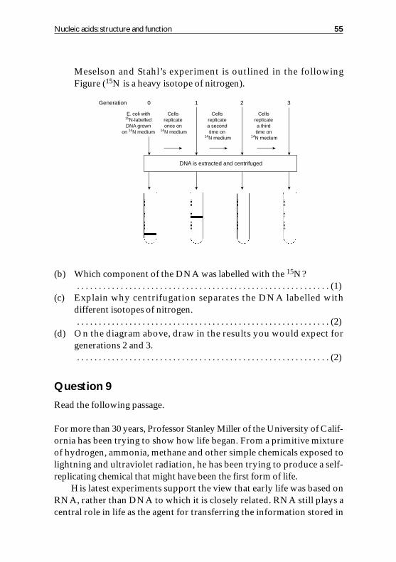
Meselson and Stahl’s experiment is outlined in the following Figure (15N is a heavy isotope of nitrogen).
(b) Which component of the DNA was labelled with the 15N?. . . . . . . . . . . . . . . . . . . . . . . . . . . . . . . . . . . . . . . . . . . . . . . . . . . . . . . . . . (1)
(c) Explain why centrifugation separates the DNA labelled with different isotopes of nitrogen.. . . . . . . . . . . . . . . . . . . . . . . . . . . . . . . . . . . . . . . . . . . . . . . . . . . . . . . . . . (2)
(d) On the diagram above, draw in the results you would expect for generations 2 and 3.. . . . . . . . . . . . . . . . . . . . . . . . . . . . . . . . . . . . . . . . . . . . . . . . . . . . . . . . . . (2)
Question 9
Read the following passage.
For more than 30 years, Professor Stanley Miller of the University of Calif-ornia has been trying to show how life began. From a primitive mixtureof hydrogen, ammonia, methane and other simple chemicals exposed tolightning and ultraviolet radiation, he has been trying to produce a self-replicating chemical that might have been the first form of life.
His latest experiments support the view that early life was based onRNA, rather than DNA to which it is closely related. RNA still plays acentral role in life as the agent for transferring the information stored in
Nucleic acids: structure and function 55
DNA is extracted and centrifuged
E. coli with15N-labelledDNA grown
on 14N medium
Cellsreplicateonce on
14N medium
Cellsreplicatea secondtime on
14N medium
Cellsreplicatea thirdtime on
14N medium
0Generation 1 2 3

DNA and used to make proteins. In early life it seems to have been moreimportant still. RNA can not only act as a store of genetic informationand a means of transferring it, but can also act as a catalyst.
Professor Miller’s latest experiments go back to an earlier stage ofthe process, seeking to show how the RNA itself might have beenproduced. He has shown that it is likely that all the components onwhich RNA is based could have been made in the conditions on theEarth 3.5 billion years ago.
Adapted from an article by Nigel Hawkes in The Times (3 July 1995).(a) In what ways are the structures of RNA and DNA alike, and in
what ways are they different?. . . . . . . . . . . . . . . . . . . . . . . . . . . . . . . . . . . . . . . . . . . . . . . . . . . . . . . . . . . . . . . . . . . . . . . . . . . . . . . . . . . . . . . . . . . . . . . . . . . . . . . . . . . . . . . . . . . . . . . . . . . . . . . . . . . . . . . . . . . . . . . . . . . . . . . . . . . . . . . . . . . . . . . . . . . . . . . . . . . . (4)
(b) Explain how RNA acts ‘as the agent for transferring the inform-ation stored in DNA that is used to make proteins’ (lines 7 and 8).. . . . . . . . . . . . . . . . . . . . . . . . . . . . . . . . . . . . . . . . . . . . . . . . . . . . . . . . . . . . . . . . . . . . . . . . . . . . . . . . . . . . . . . . . . . . . . . . . . . . . . . . . . . . . . . . . . . . . . . . . . . . . . . . . . . . . . . . . . . . . . . . . . . . . . . . . . . . . . . . . . . . . . . . . . . . . . . . . . . . . . . . . . . . . . . . . . . . . . . . . . . . . . . . . . . . . . . . . . . . . . . . . . . . . . . . . . . . . . . . . (6)
(c) Suggest why a nucleic acid molecule would have been essential in the first forms of life.. . . . . . . . . . . . . . . . . . . . . . . . . . . . . . . . . . . . . . . . . . . . . . . . . . . . . . . . . . . . . . . . . . . . . . . . . . . . . . . . . . . . . . . . . . . . . . . . . . . . . . . . . . . . . . . . . . . . . . . (2)
AnswersQuestion 1(a) (i) GCUGGGUC.(2 marks if fully correct; 1 mark if only G and C correct)
(ii) CAG. (1)(b) (i) Leucine and glycine. (1)
(ii) Deletion affects all (subsequent) triplets in the DNA sequence and somay alter entire protein; substitution may alter only a single amino acid/triplet codon; if the third base in the triplet changed, it may have no effect/degenerate code. (any 2)
Question 2(a) 15N is incorporated into (nitrogenous) base of DNA. Its presence increases
the density of the DNA. (2)(b) The DNA in Tube C consists of a dense 15N strand, the template used in
replication. Also present is a less-dense 14N strand, the newly replicated
56 Outline solutions
strand. No intermediate exists because the DNA was made single strandedbefore it was centrifuged. (3)
(c) There will be two bands present, one of lower density (top of the tube) andone of higher density. The former contains about three times as muchDNA as the latter. (2)
(e) 1.00, because it is composed of equal numbers of � and � strands. (2)
Question 3(a) (i) Change in sequence of bases/nucleotides in gene/codon. (1)
(ii) Any four of the following: (substitution) leading to different codon/bases at this point (in DNA) chain; leads to ‘wrong’ amino acid insertion at this point in polypeptide; deletion/addition alters all subsequent amino acids; leads to changed sequence of all subsequent amino acids; copy of changed (codon) in mRNA; reference to ribosomes; reference to anti-codons on tRNA; reference to specific amino acid attached to tRNA. (4)
(b) Heterozygotes always have one dominant allele, therefore one DNA chain will always produce enough functional enzyme. (2)
Question 4(a) Hydrogen bonding/specific base pairing. (1)(b) W �phosphate or phosphoric acid group; X �deoxyribose/sugar/pentose;
Y �cytosine; Z �adenine. (4)
Question 5(a) (i) Restriction enzyme/endonuclease/named enzyme e.g. EcoR1, HindIII,
BamHI. (1)(ii) DNA ligase (see BASC Booklet 7, Recombinant DNA Technology for further details). (1)
(b) 16, because each tRNA is specific to one amino acid. (2)(c) (i) GGUAUCGUG; (1)
(ii) any four of the following: might code for a stop codon; might code for a different amino acid; this would make a modified protein; protein might not function; amino acid might not alter (because the change occurs on third triplet); therefore no change to protein made. (3)
Question 6(a) (i) Restriction enzyme/endonuclease. Removes section of DNA. (2)
(ii) Two of the following: cut bacterial DNA/plasmid; ligase; splices in DNA; reference to vectors, e.g. phage. (2)(See BASC Booklet 7, Recombinant DNA Technology for further details.)
(b) Eight of the following (allow points on diagram if appropriately labelled/annotated): reference to nucleotides; four bases named; reference to sugar–phosphate ‘backbone’; reference to two (polynucleotide) strands;
Nucleic acids: structure and function 57
(d) Percentage of basepresent in DNA sample Ratio of (A�G) Ratio of (A�T)
DNA Sample A G C T to (C�T) to (C�G)Double-stranded 30 20 20 30 1.00 1.5� Strand 28 26 14 32 1.17 1.5� Strand 32 14 26 28 0.85 1.5 (4)
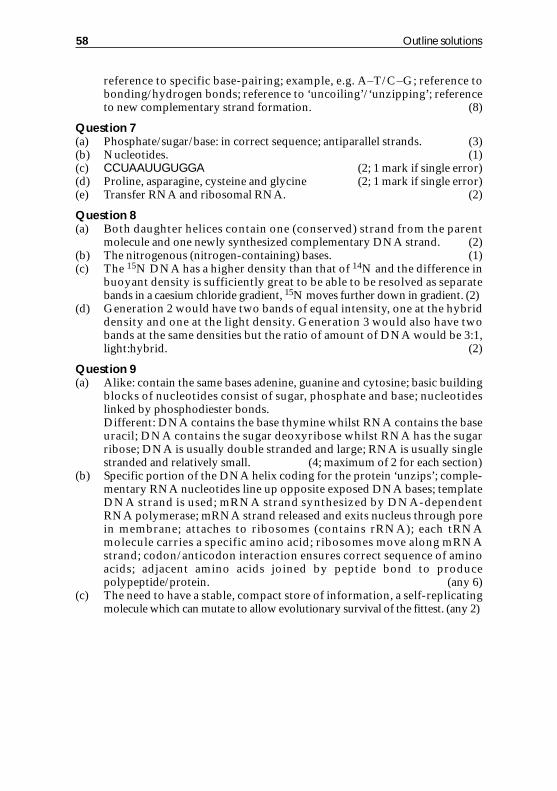
reference to specific base-pairing; example, e.g. A–T/C–G; reference to bonding/hydrogen bonds; reference to ‘uncoiling’/‘unzipping’; reference to new complementary strand formation. (8)
Question 7(a) Phosphate/sugar/base: in correct sequence; antiparallel strands. (3)(b) Nucleotides. (1)(c) CCUAAUUGUGGA (2; 1 mark if single error)(d) Proline, asparagine, cysteine and glycine (2; 1 mark if single error)(e) Transfer RNA and ribosomal RNA. (2)
Question 8(a) Both daughter helices contain one (conserved) strand from the parent
molecule and one newly synthesized complementary DNA strand. (2)(b) The nitrogenous (nitrogen-containing) bases. (1)(c) The 15N DNA has a higher density than that of 14N and the difference in
buoyant density is sufficiently great to be able to be resolved as separate bands in a caesium chloride gradient, 15N moves further down in gradient. (2)
(d) Generation 2 would have two bands of equal intensity, one at the hybrid density and one at the light density. Generation 3 would also have two bands at the same densities but the ratio of amount of DNA would be 3:1, light:hybrid. (2)
Question 9(a) Alike: contain the same bases adenine, guanine and cytosine; basic building
blocks of nucleotides consist of sugar, phosphate and base; nucleotides linked by phosphodiester bonds.Different: DNA contains the base thymine whilst RNA contains the base uracil; DNA contains the sugar deoxyribose whilst RNA has the sugar ribose; DNA is usually double stranded and large; RNA is usually single stranded and relatively small. (4; maximum of 2 for each section)
(b) Specific portion of the DNA helix coding for the protein ‘unzips’; comple-mentary RNA nucleotides line up opposite exposed DNA bases; template DNA strand is used; mRNA strand synthesized by DNA-dependent RNA polymerase; mRNA strand released and exits nucleus through pore in membrane; attaches to ribosomes (contains rRNA); each tRNA molecule carries a specific amino acid; ribosomes move along mRNA strand; codon/anticodon interaction ensures correct sequence of amino acids; adjacent amino acids joined by peptide bond to produce polypeptide/protein. (any 6)
(c) The need to have a stable, compact store of information, a self-replicating molecule which can mutate to allow evolutionary survival of the fittest. (any 2)
58 Outline solutions
8
Laboratory practicals
Extraction and characterization of DNA fromMicrococcus lysodeikticus
As DNA is a large fragile molecule, isolation methods must be mild inorder to avoid excessive breakage of DNA chains. Extreme conditions(either of pH or temperature) must be avoided and the following pro-cedure describes a method of DNA preparation from a bacterial source.Students should record carefully any changes which occur at each stageof the method and try to account for any changes.
Method (day 1,2 hours)
Suspend 0.5 g of freeze-dried Micrococcus lysodeikticus cells in 20 ml of0.15 M NaCl/0.1 M EDTA, pH 8.0. Add 20 mg of lysozyme andincubate at 37�C for 15 min. Add 1.6 ml of 25% (w/v) sodium dodecylsulphate (SDS) solution, mix and incubate for 10 min at 65�C. Cool andadd 5.5 ml of 5 M sodium perchlorate followed by 25 ml of chloroform-isoamyl alcohol (25:1 v/v). Mix thoroughly for 15 min. Centrifuge for10 min at high speed. Carefully draw off the top aqueous layer with aPasteur pipette and place the solution in a narrow beaker. Carefully layeron top an equal volume of ice-cold absolute alcohol. Using a clean glassrod collect the DNA precipitate from the interface and transfer to a cleanportion of ethanol. Wash the DNA by unwinding and rewinding ontothe rod. Transfer to a tube containing 3 ml of SSC (0.15 M NaCl/0.015Msodium citrate, pH 7.0). Leave until next practical period to dissolve.
59

Properties of DNA
HyperchromicityThis feature of DNA arises from the fact that when double-strandedDNA is denatured (i.e. strands are separated) the absorbance at 260 nmincreases substantially (up to 40%).
Base compositionThe base compositions of DNA and RNA differ, and this can be demon-strated by chromatography of hydrolysates of samples of each. Byeluting the separated bases and determining the amount of each spec-trophotometrically the base ratios can be calculated and compared.
Method (day 2,2.5 hours)
Make a 1-in-10 dilution of 1 ml of your DNA sample using SSC. If aspectrophotometer is available record the ultraviolet absorptionspectrum using SSC as blank (further dilution may be necessary). Heat asample of your dilute DNA to 100�C for 15 min and cool quickly. Re-record the spectrum.
Place approx. 20 mg of DNA and RNA provided and your DNAsolution [an A260 of 1 optical density (OD) unit is approx. 50 �g/ml] inseparate clean test tubes and add 0.5 ml of 70% HClO4. Heat in aboiling-water bath for 1 h in a fume cupboard. Cool, add an equalvolume of water, and spot 10 �l samples on to a chromatogram togetherwith 10 �l samples of thymine, adenine, guanine, cytosine and uracilsolutions. Develop the chromatogram using 65% isopropanol/16% con-centrated HCl/19% water as solvent.
Method (day 3,1.5 hours)
Examine the chromatogram under ultraviolet light and mark in pencillines around the spots (wear a face mask!). Measure RF values and cutout the separated bases with scissors (including approx. equal areas ofthe chromatograms as a blank). Transfer to clean test tubes and elute thebases by adding 10 ml of 0.1 M HCl. Heat to 45�C for 10–15 min andallow to cool. Record the ultraviolet absorption spectrum of each eluateand calculate concentrations using molar extinction coefficients.
60 Laboratory practicals
Purification of RNA
Before use in biochemistry practical classes, commercially availableRNA must first be purified and treated in the following sequence:(i) 10 g of RNA (BDH-yeast) taken in some distilled water and dissolvedby bringing to pH 6.8–7.0 with NaOH;(ii) dilute to 200 ml with distilled water;(iii) spin down insoluble residue in a centrifuge (5000 revolutions/minfor 15 min);(iv) dialyse for 48 h in three changes of 2 litres of ice-cold distilled water;(v) precipitate with 2 vols. of 95% ethanol and solid sodium acetate tomake 2% solution;(vi) spin down RNA, redissolved to 200 ml with distilled water;(vii) dialyse for 24 h against distilled water;(viii) precipitate with 2 vols. of 95% ethanol and bring to 2% with solidsodium acetate;(ix) RNA spun down and wash the precipitate twice with ethanol:ether(1:1);(x) wash precipitated RNA with ether and air dry (stirring well).Store cold and use within 8 weeks.
Further experiments relating to the physical and chemical propertiesof DNA and RNA can be performed using the DNA isolated in this wayor using material obtained from commercial sources. The choice ofexperiments is best made by the individual teacher based on such factorsas availability of specialist equipment, cost of consumables, safety andpractical time available. Titles for some practicals in this area are givenbelow together with the appropriate reference. Practicals relating tofunctional aspects of the nucleic acids that are suitable for school aresparse, largely because many depend on the use of very specialistequipment or expensive and potentially hazardous radiochemicals. Inthese cases use may be made of computer simulations and/or videotapematerial and possible titles are included below and in the list thatfollows. School teachers may also wish to take the opportunity oflinking with a neighbouring institute of higher education in order toestablish access to specialist equipment of this type. At the present timemany such institutions would welcome such an approach.
Nucleic acids: structure and function 61
Further practicals
Falconer,A.C. and Hayes, L.J. (1986) The Extraction and Partial Purification of Bacterial DNA asa Practical Exercise for GCE Advanced Level Students. Journal of Biological Education 20,25–26
Wood, P. (1984) Use of Computer Simulations in Microbial and Molecular Genetics. Journal ofBiological Education 20, 309–312
Goodridge, F. (1983) The Teaching of Protein Synthesis — a Microcomputer Based Method.Journal of Biological Education 17, 222–224
New, J. and Jolley,R. (1986) Ultraviolet Radiation Induction of Mutation in Penicillium claviforme.Journal of Biological Education 20, 155–157
Franco,R. and Canela, E.I. (1985) SIMCODE;A Program for Simulating Point Mutations inGenomic DNA.Biochemical Education 13, 66–67
Spinder, S., Siebert, P.,Coffman, F. and Jurnak, F. (1984) Isolation of Biologically Active mRNA.Biochemical Education 12, 22–25
Rosenfield, I.Z.H.E. and Van Loon,B. (1983) DNA for Beginners,Writers and Readers PublishingCo., location ISBN 0 86316 023 9
62 Laboratory practicals
9
Source materials
TextbooksBoyer,R.F. (1986) Modern Experimental Biochemistry,Addison–Wesley Publishing Co. Inc.,
Wokingham [experiments 15–20]Calladine,C.R. and Drew,H.R. (1997) Understanding DNA (2nd edition),Academic Press,
LondonCampbell,N.A. (1996) Biology (4th edition), Benjamin/Cummings Publishing Company,CaliforniaDrlica,K. (1984) Understanding DNA and Gene Cloning:A Guide for the Curious, John Wiley and
Sons,ChichesterGarrett, R.H. and Grisham,C.M. (1997) Biochemistry, Saunders College Publishing,CaliforniaMatthews,C.K. and van Holde,K.E. (1996) Biochemistry (2nd edition), Benjamin/Cummings
Publishing Company,CaliforniaMatthews,H.R., Freedland,R. and Miesfeld,R.L. (1997) Biochemistry: A Short Course, John Wiley
and Sons,ChichesterRosenfield, I., Riff, E. and Van Loon,B. (1983) DNA for Beginners,Writers and Readers Publishing
Co., ISBN 0 86316 023 9Sinden,R.R. (1994) DNA Structure and Function,Academic Press, LondonSolomon, E.P., Berg, L.R.,Martin,D.W. and Villee,C. (1996) Biology (4th edition), Saunders College
Publishing,CaliforniaVoet,D. and Voet, J.G. (1995) Biochemistry (2nd edition), John Wiley and Sons,Chichester
ArticlesDarnell Jr., J.E. (1985) RNA.Scientific American,253, 54–72Felsenfeld,G. (1985) DNA.Scientific American 253, 54–72The Molecular Basis of Evolution. (1985) Scientific American 253, 148–157Weinberg,R.A. (1985) The Molecules of Life.Scientific American 253, 34–43
63
Teaching aidsFletterick,R.J., Schroer,T. and Matela, R.J. (1985) Blackwell Molecular Models (Patent No. 437218)
together with Molecular Structure:Macromolecules in Three Dimensions, Blackwell ScientificPublications,Oxford
Parish J.H. (1986) Nucleic Acid Structure and Synthesis, IRL Press Ltd.,OxfordBleasby,A.J. and Parish J.H. (1986) The Genetic Code and Protein Synthesis, IRL Press Ltd.,OxfordLowrie,R.S.Minit Molecular Modeling System.DNA and RNA model kits.Cochranes of Oxford
Ltd.,OxfordNicholson Molecular Models.DNA and RNA model kits. Labquip,WokinghamThe Bio-Bombshell (1984) New ScientistVideo, IPC Magazines Ltd., LondonThe DNA Story, John Wiley and Sons Ltd.,ChichesterDNA - Blueprint of Life, John Wiley and Sons Ltd.,ChichesterMolecular Models Kit (DNA and RNA),Molecular Design Inc.,Academic Press, LondonPipe cleaners: useful for distinguishing left and right-handed helicesThe Biochemical Basis of Biology (video programme), volumes 2 and 3,The Biochemical Society,
LondonDNA Kit,NCBE,The University of Reading,ReadingPlant DNA Investigation Kit,NCBE,The University of Reading,ReadingDNA Model,NCBE,The University of Reading,Reading
64 Source materials
59
A form (crystalline form), of DNA, 13Adaptor molecule, 18, 41Adenine, 7Amino-acyl tRNA synthetase, 42, 43Anticodon, 44Antiparallel arrangement, of DNA, 15
B form, 13Base composition
of DNA, 12, 13of RNA, 18
Base pairing, 12, 14, 17
Caesium chloride density gradient,23, 24
Central dogma of molecular biology,29
Chain-terminating signal, 35Chargaff ’s base pairing rule, 12–14Chemical taxonomy, 12Chromatin, 11Codon, 29, 32Condensation reaction, 10, 44Conservative replication, 22Control region, 31Crick, Francis, 14, 32Crystallographic repeat, 13Cytosine, 7
Deformylase, 45Degenerate code, 35Density gradient centrifugation, 17,23, 24
Deoxyribonucleic acid (see DNA)Deoxyribonucleoside, 8Deoxyribose, 6DNA ligase, 27DNA polymerase III, 26, 27DNA
A form (crystalline form), 13antiparallel arrangement, 15base composition, 12, 13duplex form, 16
isolation, 12parallel arrangement, 15replication, 27structure, 13–17, 20, 25template, 37
Double helix, 14, 25
Enzymein DNA replication, 26, 27in post-translational modification,45
in transcription, 38in translation, 42, 43
Electrophoresis, 18Elongation, 44Epigenetic change, 2
N-Formylmethionine (fMET), 43
Gel filtration, 18Gene region, 30Genetic code, 29-31Genetic information carrier, 2Genotype, 2Glycoside bond, 8Guanine, 7
Heredity, 1, 2Histone protein, 11Hybrid density, 24Hydrogen bond, 14–16, 21, 39
Identity period, 13Initiation, 39Initiation complex, 44Initiation site, 40, 43Ion-exchange chromatography, 17Isolation
of DNA, 12of RNA, 17
Lagging strand, 26Leading strand, 26
59
Subject index
Leder and Nirenberg, 33Meselson and Stahl experiment,23, 24
Messenger RNA, 18, 19, 38, 39Minor base, 8Multiple repeat sequence, 31
Non-coding sequence, 31Nonsense codon, 34, 45Nucleoside, 8Nucleotide, 8
Okazaki fragment, 26One gene–one polypeptide theory,30
Palindromic region, 39Parallel arrangement of DNA, 15Peptidase, 45Peptide bond, 44Peptide synthesis, 30–35, 43–45Peptidyl transferase, 44Perchloric acid, 12Phenotype, 1, 3Phosphodiester linkage, 9–11Polynucleotide, 9Polysome/polyribosome, 46Post-translational modification, 45Protein (see Peptide synthesis)Purine, 7Pyrimidine, 7Pyrophosphate linkage, 9
Replicase, 27Replication, 21–27Replication fork, 26Rho protein, 39Ribonucleic acid (see RNA)Ribonucleoside, 8Ribose, 6Ribosomal RNA, 18Ribosome-binding technique, 33
Ribosome, 18, 43–46Ribozyme, 20Right-handed helix, 15RNA polymerase, 37–40RNA
base composition, 18isolation, 174S RNA, 19structure, 18–20
Semi-conservative replication, 22, 25Sequential code, 35Shine–Dalgarno consensus sequence,43
Single-stranded RNA, 16, 20‘Spent’ tRNA, 44Strand separation, 26Structure
of DNA, 13–17, 20, 25of RNA, 18–20
Sugar–phosphate backbone, 10, 14,16, 17
Svedberg (S) unit, 19Synthetic mRNA, 33
Termination, 39, 45Thymine, 7Transcription, 29, 31, 37–40Transfer RNA, 18, 19, 44Translation, 31, 41–46Translocation, 44Triple helix, 16Triplet theory, 32
Uracil, 7
Virus, 12, 16
Watson, James, 14
X-ray diffraction, 13, 14�X174, 12, 16
60 Subject index