The Skeletal System Bones, Skeleton, & Joints. Unit Objectives Identify the subdivisions of the...
41
The Skeletal System Bones, Skeleton, & Joints
-
Upload
elwin-chase -
Category
Documents
-
view
223 -
download
1
Transcript of The Skeletal System Bones, Skeleton, & Joints. Unit Objectives Identify the subdivisions of the...
The Skeletal System
Bones, Skeleton, & Joints

Unit Objectives
Identify the subdivisions of the skeleton as axial or appendicular Axial
On a skull or diagram, identify & name the bones of the skull Describe how the skull of a newborn infant differs from that of an adult, and explain the function of fontanels Name the various parts of a typical vertebra Discuss the importance of the intervertebral discs and spinal curvatures Explain how the abnormal spinal curvatures (scoliosis, lordosis, and kyphosis) differ from one another
Appendicular Identify on a skeleton or diagram the bones of the shoulder and pelvic girdles and their attached limbs Describe important differences between a male and female pelvis
List at least 3 functions of the skeletal system Name the four main kinds of bones Identify the major anatomical areas of a long bone Explain the role of bone salts and the organic matrix in making bone both hard & flexible Describe briefly the process of bone formation in the fetus and summarize the events of bone
remodeling Name & Describe the various types of fractures Name the 3 major categories of joints and compare the amount of movement allowed by each Understand the functions and differences between tendons & ligaments Identify some of the causes of bone and joint problems throughout life
Functions of Bone
Support & Give shape to the body Protects Internal Organs Helps make movements possible Stores calcium Hemopoiesis or blood cell formation

Types of Bones
Long Short Flat Irregular
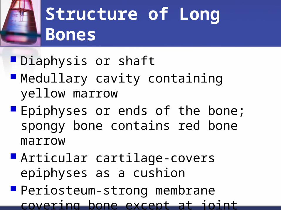
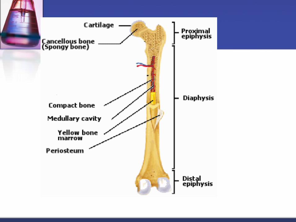
Structure of Long Bones
Diaphysis or shaft Medullary cavity containing yellow marrow Epiphyses or ends of the bone; spongy bone
contains red bone marrow Articular cartilage-covers epiphyses as a
cushion Periosteum-strong membrane covering bone
except at joint surfaces Endostuem-lines the medullary cavity
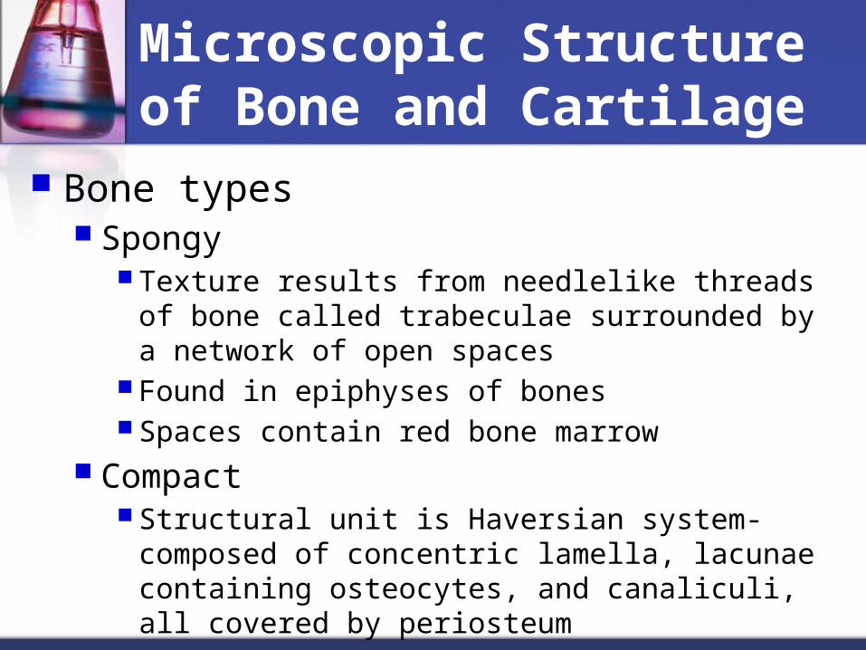
Microscopic Structure of Bone and Cartilage
Bone types Spongy
Texture results from needlelike threads of bone called trabeculae surrounded by a network of open spaces
Found in epiphyses of bones Spaces contain red bone marrow
Compact Structural unit is Haversian system-composed of
concentric lamella, lacunae containing osteocytes, and canaliculi, all covered by periosteum
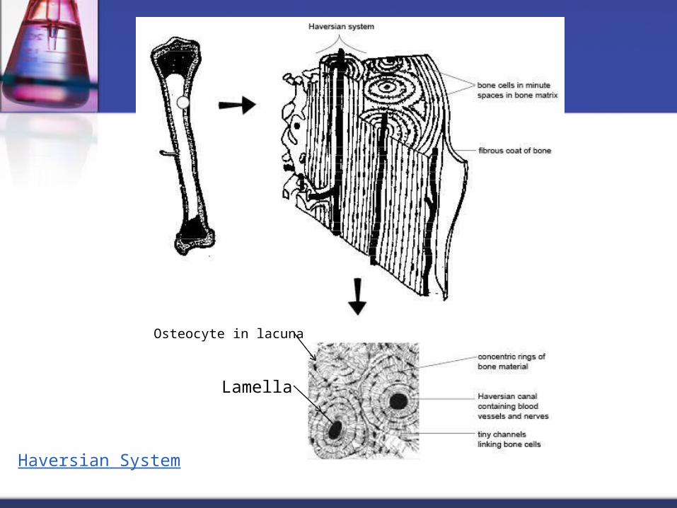
Haversian System
Lamella
Osteocyte in lacuna

Microscopic Structure of Bone and Cartilage con’t
Cartilage Cell type called chondrocyte Matrix is gel-like and lacks blood vessels

Bone formation & Growth
Sequence of development early-cartilage models replaced by calcified bone matirx The laying down of calcium salts in the gel-like
matrix of the forming bones is an ongoing process
Osteoblasts form new bone, osteoclasts reabsorb bone
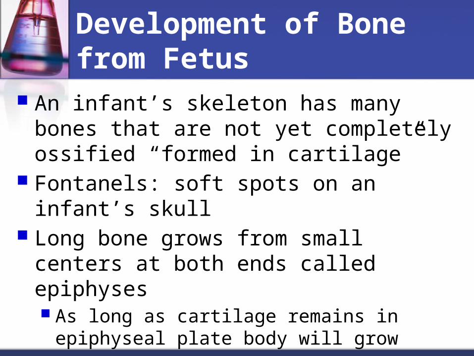
Development of Bone from Fetus
An infant’s skeleton has many bones that are not yet completely ossified “formed in cartilage”
Fontanels: soft spots on an infant’s skull Long bone grows from small centers at both
ends called epiphyses As long as cartilage remains in epiphyseal plate
body will grow

Division of Skeleton
The skeleton is composed of the following divisions & subdivisions
Axial Skeleton Skull Spine Thorax Hyoid bone
Appendicular Skeleton Upper Extremities, including shoulder girdle Lower Extremities, including hip girdle


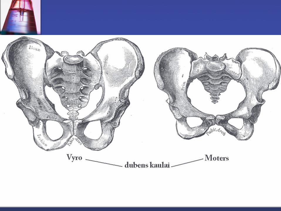
Differences between a Man’s & a Woman’s skeleton
Size: Male skeleton is generally larger Shape of pelvis: male pelvis is deep and
narrow, female pelvis is broad and shallow Size of pelvic inlet: female pelvis inlet is
generally wider, normally large enough for baby’s head to pass through it
Pubic angle: angle between pubic bones of female generally wider

Joint (Articulations)
Kinds of joints Synarthroses (no movement)-fibrous connective tissue
grows between articulating bones, for example, sutures of skull
Amphiarthroses (slight movement)-cartilage connects articulating bones; for example symphysis pubis
Diarthroses (free movement)-most joints belong to this class
Structures of freely movable joints-joint capsule and ligaments hold adjoining bones together but permit movement at joint

Joints con’t
Articular cartilage-covers joint ends of bones and absorbs joints
Synovial membrane-lines joint capsule and secretes lubricating fluid
Joint cavity-space between joint ends of bones

Types of freely movable joints
Ball-and-Socket Hinge Pivot Saddle Gliding Condyloid

Bone Remodeling & Repair
Remodeling: balance of bone deposit & removal, deposits occur at a greater rate when bone is injured
Controlled by Hormone used to maintain blood calcium In response to mechanical stress and gravity,
remodels so it is able to withstand the stresses
Bone Remodeling & Repair con’t
Repair Fractures are breaks in bones & are classified
by: Comminuted: bone fragments into 3 or more pieces (common with more
brittle bones) Compression: bone is crushed (common with more brittle bones) Spiral: Ragged break occurs when excessive twisting force is applied to
bone (common sports fracture) Epiphyseal: epiphysis separates from the diaphysis along the
epiphyseal plate (common where cartilage is dying) Depressed: Broken bone portion is pressed inward (common of skull) Greenstick: bone breaks incompletely (common in children whose
bones are more flexible)

Bone Remodeling & Repair con’t
Repair 4 stages Hematoma formation Fibrocartilaginous callus formation Bony callus formation Remodeling of the bony callus

Bone & Joint Problems
Bone Problems Osteomalacia: number of disorders in adults in
which the bone is inadequately mineralized Rickets: inadequate mineralization of bones in
children caused by insufficient calcium or vitamin D deficiency
Osteoporosis: group of disorders in which the rate of bone reabsorption exceeds the rate of formation


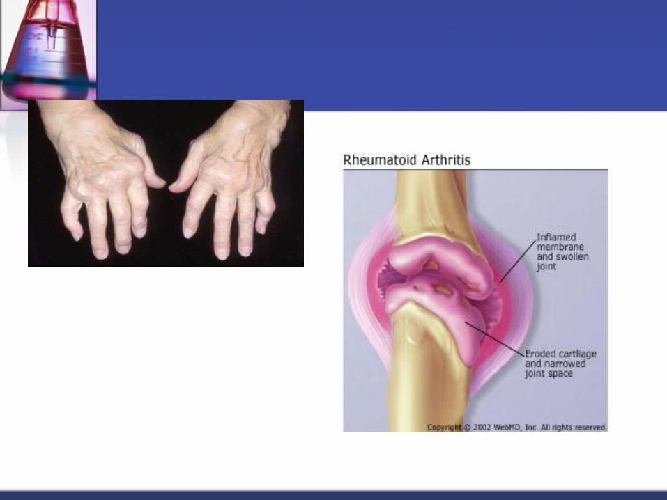
Bone & Joint Problems
Joint Problems Sprains Dislocations Bursitis: inflammation of bursa, caused by a
blow or friction Tendonitis: inflammation of the tendons, caused
by overuse Arthritis: inflammatory or degenerative diseases
that damage the joints, resulting in pain, stiffness, and swelling of joint


Spinal Curvatures
5 major divisions; cervical (7), thoracic (12), lumbar (5), fused of sacrum (5), fused of coccyx (4)
Increase resiliency & flexibility Cervical & lumbar curvatures are concave
posteriorly, and the thoracic & sacral curvatures are convex posteriorly

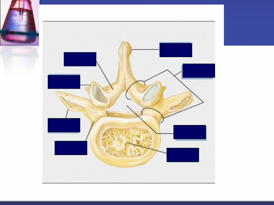
Intervertebral disc
Cushion like pads that act as shock absorbers and allow the spine to flex, extend, and bend laterally
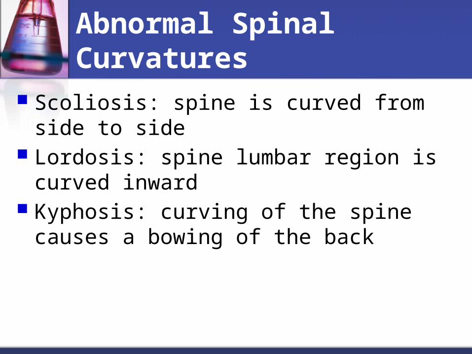
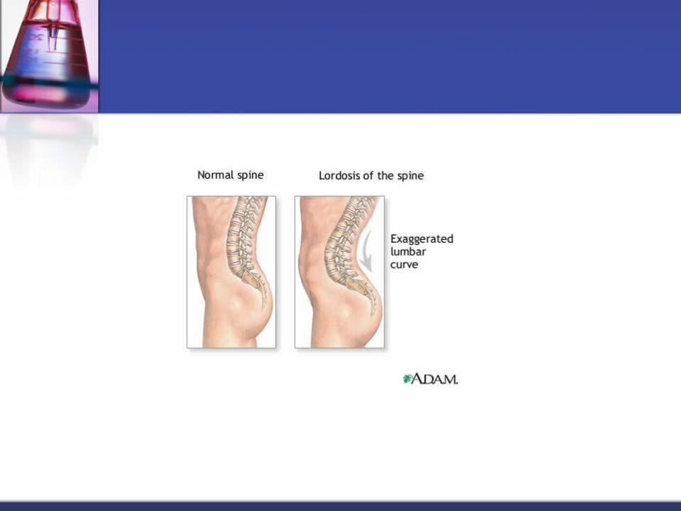
Abnormal Spinal Curvatures
Scoliosis: spine is curved from side to side Lordosis: spine lumbar region is curved
inward Kyphosis: curving of the spine causes a
bowing of the back