The Skeletal System An overview Includes: Bones – structure, protection and support Joints – for...
29
The Skeletal System An overview ncludes: ones – structure, protection and support oints – for flexibility and allow motion artilages and Ligaments – Hold bones together
-
Upload
marianna-chambers -
Category
Documents
-
view
219 -
download
1
Transcript of The Skeletal System An overview Includes: Bones – structure, protection and support Joints – for...

The Skeletal SystemAn overview
Includes:Bones – structure, protection and supportJoints – for flexibility and allow motionCartilages and Ligaments – Hold bones together

The Skeletal System . . . The Body’s Framework
in 2 parts
The human skeleton consists oftwo main divisions:
Axial skeleton – forms the uprightaxis of the body (74 bones + 6 tinybones of the middle ear)
Appendicular skeleton – forms theAppendages of the upper andLower body (126 bones)The limbs and pelvic girdle- - - - - - - - - - - - - - - - - - - - - - - - - - - -Total # = 206 bones in the human adult skeleton

Five General Functions of Bone
• Support – bones contribute to shape, alignment and positioning of the body parts
• Protection – bones act as hard “boxes” to protect the delicate structures they enclose
• Movement – bones with their joints constitute levers; muscle tissue attached to the bones contract, pulling on bones and producing movement
• Mineral storage – bones serve as a major reservoir for calcium, phosphorus and certain other minerals needed for essential healthy survival
• Hematopoiesis (blood cell formation) – a vital process that takes place in red bone marrow, myeloid tissue
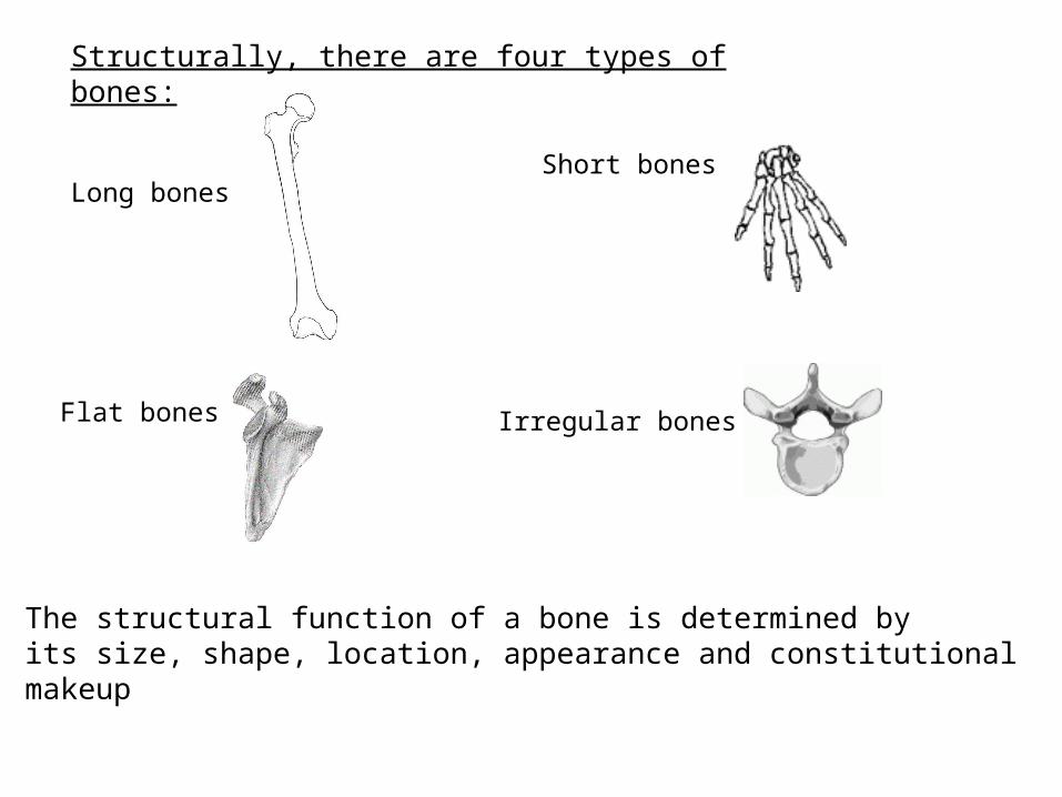
Structurally, there are four types of bones:
Long bones
Short bones
Flat bones Irregular bones
The structural function of a bone is determined byits size, shape, location, appearance and constitutionalmakeup

STRUCTURE OFTYPICAL LONG BONE
Prepare your Color Plateof the Femur, outlining thestructures that are seenhere

The periosteum is a dense, white fibrous membrane that covers bone; it
• attaches tendons firmly to bones
• contains blood vessels that send branches into bone
• is essential for bone cell survival and bone formation

Medullary cavity
The medullary (or marrow) cavity:
• tubelike, hollow space in diaphysis
• filled with yellow marrow

BONE GROWTH AND EARLY DEVELOPMENT
Parts of the skeletal system begin to form during the first few weeks oflife, and bony structures continue to develop and grow into adulthood.bones form by the replacement of existing connective tissues in one oftwo ways:
1. Intramembranous Bones• examples are the broad, flat bones of the skull . . . during their development, sheetlike masses of connective tissues appear at the sights of future bone
• Some of the connective tissues enlarge and differentiate to form osteoblasts
• Spongy bone then forms, growing outward in all directions building a bony matrix

2. Endochondral Bone
Most of the bones of the skeleton are of this type and are formed frommasses of hyaline cartilage with shapes similar to the future bony structure.
• Mesenchyme cells cluster together and transform into osteoblasts . . . Osteoblasts secrete boney matrix until cells are surrounded
• surrounded osteoblasts become osteocytes that reside in lacunae within the matrix . . .calcium and mineral salts are deposited rapidly
• the bone matrix develops into columns of bone called trabeculae that fuse together to form spongy bone
• red marrow forms in the spaces between the trabeculae
Double-click on the title of this page to observe a virtual ossification process

The skull of the neonate is not one sealed container, but a collection of plates, attached by flexible cartilage. . .
Areas where there is a considerable distance between the skullplates are called fontanelles. There are 4 main fontanelles; themost common of which is the anterior fontanelle called the“soft spot”.

There are 2 reasons for this arrangement of skull plates inthe fetus:
• the separations allow shifting and movement of these plates as the baby navigates the mother’s birth canal, allowing a shape that facilitates molding the head to a shape where the baby can deliver
• At birth the brain doesn’t stop growing. . . This arrangement of skull plates gives considerable slack so that the skull can expand outward as the brain grows

Later Bone Development
Throughout childhood, the epiphyses of developing long bones remains cartilaginous and continue to grow. . . As we reach adulthood, secondary ossification centers appear in the epiphyses, leaving an epiphyseal line where the two areas were once separated
• Young children often have a “greenstick” fracture in a bone rather than a complete break because the cartilage leaves the bone more resilient
• A break along the epiphyseal disk in a young child can result in an interference of growth in that bone

EPIPHYSEALLINES

BONE TISSUE STRUCTURE
• BONE TEXTURES: COMPACT AND SPONGY BONE
- The dense outer layer that appears smooth and solid to the naked eye is compact bone- The internal area of the bone is called spongy bone
(also called cancellous bone) . . . It appears as a honeycomb of small needle-like or flat pieces called trabeculae (“little beams”). In living bones, the open spaces between trabeculae are filled with red or yellow bone marrow.
trabeculae
Spaces containingred marrow
Compact bone


Endosteum
The endosteum is the thin epithelial tissue thatlines the medullary cavity

COMPOSITION OF BONE TISSUE – THE MOST DISTINCTIVE FORM OF CONNECTIVE TISSUE
Extracellular components are hard and calcified
• Apatite – highly specialized chemical crystals of calcium and phosphate contribute to bone hardness
• slender needle-like crystals are oriented to effectively resist stress and mechanical deformation
• magnesium and sodium are also found in bone

Organic matrix of bone:
• composite of collagenous fibers and an amorphous mixture of protein and polysaccharides (ground substance)• ground substance substance secreted by connective tissue cells• adds to overall strength and resilience of bones

Osteon/Haversian System

Cancellous (spongy or trabecular) bone:• characterized by open spaces partially filled by an assemblage of needlelike spicules called (trabeculae)• no osteons are present• bone cells are found within the trabeculae• nutrients are delivered to cells, and waste products removed, by diffusion through tiny canaliculi that extend to the surface of the very thin spicules• found in the epipyses of long bones• this type of bone greatly enhances the bone’s strength

Bone Marrow – A Production Factory

• Bone marrow is a specialized type of soft, diffuse connective tissue called myeloid tissue
• It serves as the site for production of blood cells
• It is found in the medullary cavities of long bones and in the spaces of spongy bone
During the lifetime of an individual, two types of marrow occur:
Red Marrow – its function is production of red blood cells; in an infant or young child, virtually all bones contain red marrow
Yellow Marrow – cells are saturated with fat, rendering them inactive in blood cell production; red marrow is replaced by yellow marrow as a person ages

Hip Replacement
Click on the picture to see video

In the adult, the main locations of red marrow are the ribs, iliac crests, bodies of the vertebrae, and the epiphyses of long bones
During times of decreased blood supply (prolonged anemia, blood loss, exposure to radiation or chemotherapy), yellow marrow can alter to become red marrow
Bone Marrow Transplant
In patients with leukemia, aplastic anemia,Hodgkin’s disease (lymphomas) and some immune deficiency diseases, stem cells in the bone marrow malfunction and produceImmature or defective blood cells.
Marrow transplant may help these patients.Their own marrow is destroyed by radiation or chemotherapy; they then receive an infusion of donated marrow. . . In successfultransplants, the marrow migrates to cavities of large bones and begins producingnormal blood cells

Bone Fractures
Fractures are breaks in bone and canbe divided into 3 general categories:
• Incomplete (often called “greenstick” fractures) – a bending of the bone; most often seen in children whose bones have not been completely ossified by calcium deposits
• Simple (nondisplaced, transverse) – bone is broken at a right angle to the bone’s axis and is “in line” with the corresponding part (may also be simple oblique fracture)
• Compound (open) – bone is usually broken obliquely and breaks through the skin

SCOLIOSISKYPHOSIS
LORDOSIS

Bone MarkingsBumps, holes, ridges or grooves for muscle
attachment and blood vessel and nerves to pass through.
2 Categories of Markings
Projections or Process are raised. Depressions or Cavities are not raised. Know these….
Angle body condyle epicondyle
Crest tuberosity suture foramen Tubercle
Fossa head line margin trochanter
Meatusprocess notch sinus spine Look up the meanings for these markings in your book.

Synovial Joints (Diarthroses)•Freely movable•Most numerous of the body’s joints•Examples:
Uniaxial Biaxial Hinge joint (elbow) Saddle joint thumb joint)
Pivot joint (atlas & axis) Condyloid (ellipsoidal) joint (wrist)
Multiaxial Ball and socket (shoulder and hip) Gliding (between vertebrae; tarsals/carpals)