The prognostic role of Bcl-2, Ki67, c-MYC and p53 in ... · PDF fileStreptavidine–HRP...
7
Rom J Morphol Embryol 2017, 58(3):837–843 ISSN (print) 1220–0522 ISSN (online) 2066–8279 ORIGINAL PAPER The prognostic role of Bcl-2, Ki67, c-MYC and p53 in diffuse large B-cell lymphoma ANA-MARIA PĂTRAŞCU 1) , IONELA ROTARU 2) , LAVINIA OLAR 1) , ŞTEFAN PĂTRAŞCU 3) , MIRELA-CORINA GHILUŞI 4) , SIMONA-DANIELA NEAMŢU 2) , JANINA GEORGIANA NACEA 5) , ADRIAN GLUHOVSCHI 6) 1) PhD Student, Department of Morphology, University of Medicine and Pharmacy of Craiova, Romania 2) Department of Hematology, Faculty of Medicine, University of Medicine and Pharmacy of Craiova, Romania 3) Department of Surgery, University of Medicine and Pharmacy of Craiova, Romania 4) Department of Pathology, Emergency County Hospital, Craiova, Romania 5) PhD Student, Department of Obstetrics and Gynecology, University of Medicine and Pharmacy of Craiova, Romania 6) Department of Obstetrics and Gynecology, “Victor Babeş” University of Medicine and Pharmacy, Timişoara, Romania Abstract Diffuse large B-cell lymphoma (DLBCL) is a heterogeneous group of lymphoid malignancies, which counts for more than a third of non- Hodgkin’s lymphoma cases. The aim of the current study is to evaluate the prognostic role of several immunohistochemical (IHC) markers involved in the pathological process of DLBCL. This is a retrospective analysis of the 97 de novo DLBCL patients admitted between January 2007 and December 2016 in the Department of Hematology, “Filantropia” Municipal Hospital, Craiova, Romania. The expression of Bcl-2, Ki67, c-MYC and p53 was assessed by immunohistochemistry. A significant level of association was observed between high prognostic index values and Bcl-2, Ki67, c-MYC and p53 positive cases. Moreover, overall survival and disease-free survival were higher in patients with negative expression for these markers. Bcl-2, Ki67, c-MYC and p53 could make important diagnostic and therapeutic targets; therefore, their routine assessment should be mandatory. Keywords: diffuse large B-cell lymphoma, immunohistochemistry, Bcl-2, Ki67, c-MYC, p53. Introduction Diffuse large B-cell lymphoma (DLBCL) is one of the most frequent lymphoid neoplasia, accounting for 30–40% of all cases of non-Hodgkin’s lymphomas, and displaying an aggressive course, a variable and unpredictable evolution that resides in its clinical and morphological heterogeneity [1–4]. The subtype of lymphoma is determined by the B- cell maturation stage and by the genetic abnormalities that occur during cell differentiation and maturation [5]. Although gene expression profiling has the highest accuracy identifying the “cell of origin” for certain lymphoma subtypes in DLBCL, it still has numerous limitations (various cellular regulating mechanisms, financial constraints, etc.). Therefore, a multitude of surface markers distinguishing germinal center B-cell like (GCB) DLBCL and activated B-cell like (ABC) DLBCL, that are consistent with gene expression profiling, have been tested. This is possible either by using the CD10, multiple myeloma oncogene-1 (MUM-1) and Bcl-6 bio- markers (the Hans algorithm) in 80% of cases, or using GCTE1, CD10, Bcl-6, MUM-1 and FOXP1 biomarkers (the Tally algorithm) in 90% of cases [6–8]. Furthermore, the detection of certain cell markers expression has become a fundamental component both in establishing an accurate prognosis, and in the development of an optimal therapeutic algorithm. This study aims to assess the prognostic value of four immunohistochemical (IHC) markers, other than those used in Hans protocol, by investigating the correlation between their expression and International Prognostic Index (IPI) score, as well as by comparing two important survival endpoints (disease-free survival and overall survival). Materials and Methods Case selection A retrospective study was conducted on all de novo diffuse large B-cell lymphomas, diagnosed and treated in the Department of Hematology, “Filantropia” Municipal Hospital, Craiova, Romania, between January 2007 and December 2016, which revealed 128 newly discovered cases of DLBCL. In case of IPI scoring system, a progressive scale from 0 to 5 was calculated by allocating one point for each individual parameter: clinical stage III/IV, age ≥60, the Eastern Cooperative Oncology Group (ECOG) score ≥2, extranodal lesions >1, and increased lactate dehydrogenase serum levels. Cases were included in two risk categories: low-risk group for patients with IPI 0–2, and high-risk group for patients with IPI>2. The following cases were excluded from the analysis: (a) lack of histological and immunohistochemical confir- mation of DLBCL; (b) unavailable histological blocks necessary for further pathology sample examinations; R J M E Romanian Journal of Morphology & Embryology http://www.rjme.ro/
-
Upload
nguyenthien -
Category
Documents
-
view
216 -
download
1
Transcript of The prognostic role of Bcl-2, Ki67, c-MYC and p53 in ... · PDF fileStreptavidine–HRP...
Rom J Morphol Embryol 2017, 58(3):837–843
ISSN (print) 1220–0522 ISSN (online) 2066–8279
OORRIIGGIINNAALL PPAAPPEERR
The prognostic role of Bcl-2, Ki67, c-MYC and p53 in diffuse large B-cell lymphoma
ANA-MARIA PĂTRAŞCU1), IONELA ROTARU2), LAVINIA OLAR1), ŞTEFAN PĂTRAŞCU3), MIRELA-CORINA GHILUŞI4), SIMONA-DANIELA NEAMŢU2), JANINA GEORGIANA NACEA5), ADRIAN GLUHOVSCHI6)
1)PhD Student, Department of Morphology, University of Medicine and Pharmacy of Craiova, Romania 2)Department of Hematology, Faculty of Medicine, University of Medicine and Pharmacy of Craiova, Romania 3)Department of Surgery, University of Medicine and Pharmacy of Craiova, Romania 4)Department of Pathology, Emergency County Hospital, Craiova, Romania 5)PhD Student, Department of Obstetrics and Gynecology, University of Medicine and Pharmacy of Craiova, Romania 6)Department of Obstetrics and Gynecology, “Victor Babeş” University of Medicine and Pharmacy, Timişoara, Romania
Abstract Diffuse large B-cell lymphoma (DLBCL) is a heterogeneous group of lymphoid malignancies, which counts for more than a third of non-Hodgkin’s lymphoma cases. The aim of the current study is to evaluate the prognostic role of several immunohistochemical (IHC) markers involved in the pathological process of DLBCL. This is a retrospective analysis of the 97 de novo DLBCL patients admitted between January 2007 and December 2016 in the Department of Hematology, “Filantropia” Municipal Hospital, Craiova, Romania. The expression of Bcl-2, Ki67, c-MYC and p53 was assessed by immunohistochemistry. A significant level of association was observed between high prognostic index values and Bcl-2, Ki67, c-MYC and p53 positive cases. Moreover, overall survival and disease-free survival were higher in patients with negative expression for these markers. Bcl-2, Ki67, c-MYC and p53 could make important diagnostic and therapeutic targets; therefore, their routine assessment should be mandatory.
Keywords: diffuse large B-cell lymphoma, immunohistochemistry, Bcl-2, Ki67, c-MYC, p53.
Introduction
Diffuse large B-cell lymphoma (DLBCL) is one of the most frequent lymphoid neoplasia, accounting for 30–40% of all cases of non-Hodgkin’s lymphomas, and displaying an aggressive course, a variable and unpredictable evolution that resides in its clinical and morphological heterogeneity [1–4].
The subtype of lymphoma is determined by the B-cell maturation stage and by the genetic abnormalities that occur during cell differentiation and maturation [5]. Although gene expression profiling has the highest accuracy identifying the “cell of origin” for certain lymphoma subtypes in DLBCL, it still has numerous limitations (various cellular regulating mechanisms, financial constraints, etc.). Therefore, a multitude of surface markers distinguishing germinal center B-cell like (GCB) DLBCL and activated B-cell like (ABC) DLBCL, that are consistent with gene expression profiling, have been tested. This is possible either by using the CD10, multiple myeloma oncogene-1 (MUM-1) and Bcl-6 bio-markers (the Hans algorithm) in 80% of cases, or using GCTE1, CD10, Bcl-6, MUM-1 and FOXP1 biomarkers (the Tally algorithm) in 90% of cases [6–8].
Furthermore, the detection of certain cell markers expression has become a fundamental component both in establishing an accurate prognosis, and in the development of an optimal therapeutic algorithm.
This study aims to assess the prognostic value of four immunohistochemical (IHC) markers, other than those used in Hans protocol, by investigating the correlation between their expression and International Prognostic Index (IPI) score, as well as by comparing two important survival endpoints (disease-free survival and overall survival).
Materials and Methods
Case selection
A retrospective study was conducted on all de novo diffuse large B-cell lymphomas, diagnosed and treated in the Department of Hematology, “Filantropia” Municipal Hospital, Craiova, Romania, between January 2007 and December 2016, which revealed 128 newly discovered cases of DLBCL.
In case of IPI scoring system, a progressive scale from 0 to 5 was calculated by allocating one point for each individual parameter: clinical stage III/IV, age ≥60, the Eastern Cooperative Oncology Group (ECOG) score ≥2, extranodal lesions >1, and increased lactate dehydrogenase serum levels. Cases were included in two risk categories: low-risk group for patients with IPI 0–2, and high-risk group for patients with IPI>2.
The following cases were excluded from the analysis: (a) lack of histological and immunohistochemical confir-mation of DLBCL; (b) unavailable histological blocks necessary for further pathology sample examinations;
R J M ERomanian Journal of
Morphology & Embryologyhttp://www.rjme.ro/

Ana-Maria Pătraşcu et al.
838
(c) human immunodeficiency virus (HIV) infection detected; (d) primary mediastinal or central nervous system B-cell lymphomas; (e) lack of specific therapy and follow-up. The diagnosis of DLBCL respected the criteria of the 2008 World Health Organization (WHO) classification. Cases with unrepresentative or insufficient paraffin material were excluded. When the exclusion criteria were applied, 31 cases were omitted.
Ethical approval for this study was acquired from the University of Medicine and Pharmacy of Craiova Ethical Research Committee.
Histology and immunohistochemistry study
For the current study, we analyzed 97 formalin-fixed, paraffin-embedded tissue samples of the DLBCL patients admitted and treated in the Department of Hematology, “Filantropia” Municipal Hospital, Craiova.
The specimens were surgically or endoscopically retrieved for diagnostic confirmation and were preserved in the Department of Pathology, Emergency County Hospital, Craiova.
The paraffin-embedded tissue blocks were cut at 4 μm sections on a HM350 Microm rotary microtome (Thermo Fisher Scientific, Walldorf, Germany) with laminar water bath section transfer system (STS™, Thermo Fisher Scientific). For histological analysis, Hematoxylin–Eosin (HE) and Goldner–Szekely (GS) trichrome stainings were subsequently performed.
For the immunohistochemical assessment, the histo-logical sections were collected on poly-L-lysine covered
slides that were dried in a thermostat, at 370C, for 24 hours to enhance the cell attachment to the plates. The sections subsequently underwent dewaxing, rehydration, antigen retrieval by boiling the histological slides in pH 6 sodium citrate buffer solution for 21 minutes (3 minutes cycles) in microwave oven. The slides were then rinsed in tap water and washed in distilled-deionized water for 15 minutes. The endogenous peroxidase was blocked by immersing the slides in 3% hydrogen peroxide for 30 minutes, at room temperature, followed by rinsing in distilled water for 10 minutes, washing in 1% phosphate-buffered saline (PBS) for 5 minutes and blocking of non-specific sites with non-fat dry milk for 30 minutes. Next, the slides were incubated with primary antibodies for 18 hours, at 40C. The following day, the sections were incubated for 30 minutes, at room temperature, with the secondary biotinylated antibody and washed in 1% PBS (three cycles of 5 minutes each). Streptavidine–HRP (Horseradish peroxidase) was added for 30 minutes, at room tempe-rature, followed by washing in 1% PBS. The signal was detected by using 3,3’-Diaminobenzidine (DAB) (Dako, Agilent Pathology Solutions, Santa Clara, California, USA), and the reaction was blocked in 1% PBS. The next steps consisted of Mayer’s Hematoxylin counterstaining, alcohol dehydration and clearing with xylene. The coverslips were then mounted on DPX (Fluka, Honeywell Fluka™, Morris Plains, New Jersey, USA).
A brief description of the markers used in this study can be checked in Table 1.
Table 1 – Markers characteristics for IHC analysis of DLBCL patients
Antibody Code Clone Antigen retrieval Specificity Dilution Manufacturer
Anti-Bcl-2 M0887 124 EDTA Germinal center B-cells 1:50 Dako
Anti-Ki67 M7240 MIB-1 EDTA Proliferation factor 1:50 Dako
Anti-c-MYC ab32072 Y69 Sodium citrate buffer pH 6 Prognostic factor 1:250 Abcam
Anti-p53 M7001 DO-7 Sodium citrate buffer pH 6 TP53 mutations 1:50 Dako
IHC: Immunohistochemical; DLBCL: Diffuse large B-cell lymphoma; EDTA: Ethylenediaminetetraacetic acid.
The presence of different cellular staining patterns
(such as positive membrane, cytoplasmic or nuclear staining) was assessed in every slide. Each case was examined by 4×, 10× and 40× objectives (Nikon Eclipse 55i).
Potential disagreement was solved by an expert consensus committee, which included two expert pathologists of the Department of Histopathology and Immunohistochemistry.
Statistical analysis
Data were investigated by descriptive statistics and logistic regression analysis. Five-year overall survival (OS) was defined as the interval in months from the initiation of therapy until death from any cause. All surviving patients or those who were drop out of the study at the end of the five-year follow-up were censored. Disease-free survival (DFS), as acknowledged by NCI Dictionary of Cancer Terms (https://www.cancer.gov/ publications/dictionaries/cancer-terms), was defined as the time elapsed between primary treatment completion and any disease recurrence. Kaplan–Meier method was used to estimate the overall survival and disease-free
survival probability, and survival curves were compared using both the Log Rank (Mantel–Cox) and Gehan–Wilcoxon regression methods. P-value less than 0.05 threshold was considered statistically significant. The statistical analysis was performed using GraphPad Prism 7.0 (GraphPad Software Inc., San Diego, USA).
Results
Subgroup correlations according to IHC pattern
The histological and IHC studies were performed on 97 tissue samples (85 cases of lymph node involvement, three cases of gastric lymphoma, two right colon tumors, one testicular tumor, three cutaneous “leg type” lymphoma, one maxillary tumor and two thoracic lymphomas). The specimens were taken from patients with a mean age of 56 years, undergoing diagnostic or radical oncological surgery in the Departments of Urology, Thoracic Surgery, General Surgery, and Maxillofacial Surgery, and treated in the Department of Hematology, “Filantropia” Municipal Hospital, Craiova.
Histopathological examination using HE and GS
The prognostic role of Bcl-2, Ki67, c-MYC and p53 in diffuse large B-cell lymphoma
839
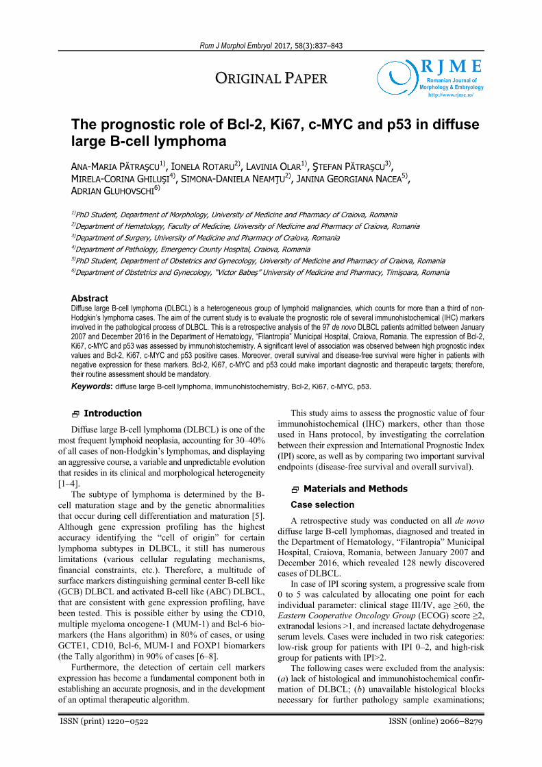
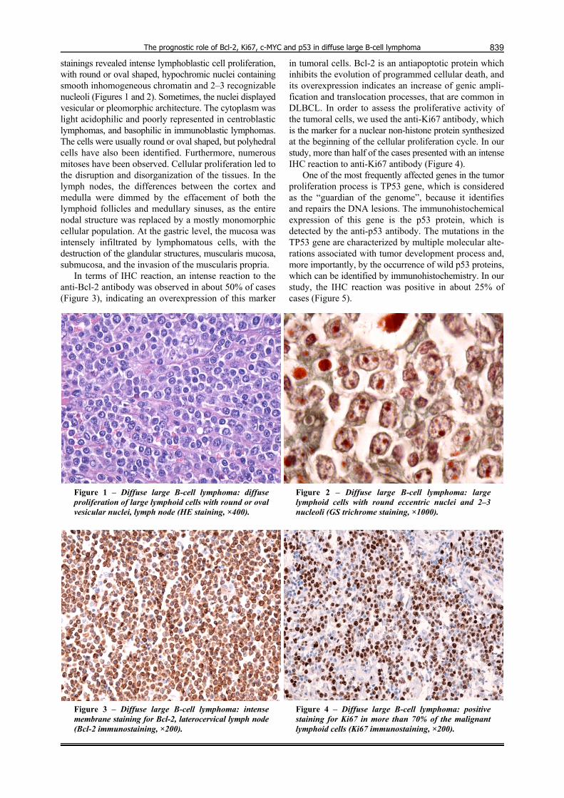
stainings revealed intense lymphoblastic cell proliferation, with round or oval shaped, hypochromic nuclei containing smooth inhomogeneous chromatin and 2–3 recognizable nucleoli (Figures 1 and 2). Sometimes, the nuclei displayed vesicular or pleomorphic architecture. The cytoplasm was light acidophilic and poorly represented in centroblastic lymphomas, and basophilic in immunoblastic lymphomas. The cells were usually round or oval shaped, but polyhedral cells have also been identified. Furthermore, numerous mitoses have been observed. Cellular proliferation led to the disruption and disorganization of the tissues. In the lymph nodes, the differences between the cortex and medulla were dimmed by the effacement of both the lymphoid follicles and medullary sinuses, as the entire nodal structure was replaced by a mostly monomorphic cellular population. At the gastric level, the mucosa was intensely infiltrated by lymphomatous cells, with the destruction of the glandular structures, muscularis mucosa, submucosa, and the invasion of the muscularis propria.
In terms of IHC reaction, an intense reaction to the anti-Bcl-2 antibody was observed in about 50% of cases (Figure 3), indicating an overexpression of this marker
in tumoral cells. Bcl-2 is an antiapoptotic protein which inhibits the evolution of programmed cellular death, and its overexpression indicates an increase of genic ampli-fication and translocation processes, that are common in DLBCL. In order to assess the proliferative activity of the tumoral cells, we used the anti-Ki67 antibody, which is the marker for a nuclear non-histone protein synthesized at the beginning of the cellular proliferation cycle. In our study, more than half of the cases presented with an intense IHC reaction to anti-Ki67 antibody (Figure 4).
One of the most frequently affected genes in the tumor proliferation process is TP53 gene, which is considered as the “guardian of the genome”, because it identifies and repairs the DNA lesions. The immunohistochemical expression of this gene is the p53 protein, which is detected by the anti-p53 antibody. The mutations in the TP53 gene are characterized by multiple molecular alte-rations associated with tumor development process and, more importantly, by the occurrence of wild p53 proteins, which can be identified by immunohistochemistry. In our study, the IHC reaction was positive in about 25% of cases (Figure 5).
Figure 1 – Diffuse large B-cell lymphoma: diffuse proliferation of large lymphoid cells with round or oval vesicular nuclei, lymph node (HE staining, ×400).
Figure 2 – Diffuse large B-cell lymphoma: large lymphoid cells with round eccentric nuclei and 2–3 nucleoli (GS trichrome staining, ×1000).
Figure 3 – Diffuse large B-cell lymphoma: intense membrane staining for Bcl-2, laterocervical lymph node (Bcl-2 immunostaining, ×200).
Figure 4 – Diffuse large B-cell lymphoma: positive staining for Ki67 in more than 70% of the malignant lymphoid cells (Ki67 immunostaining, ×200).

Ana-Maria Pătraşcu et al.
840
Figure 5 – Diffuse large B-cell lymphoma: positive staining for p53 in more than 30% of the malignant lymphoid cells (p53 immunostaining, ×200).
c-MYC nuclear phosphoprotein is a transcription factor encoded by c-myc gene, which is involved in cellular growth, differentiation and apoptosis, making it one of the key molecules in tumorigenesis. Almost 32% of cases had a positive expression for c-MYC.
Table 2 indicates the immunophenotypic features of the 97 patients with de novo DLBCL, according to their cell-of-origin distribution. Forty-four (45.36%) cases were GCB DLBCL and 53 (54.64%) cases were non-GCB lymphomas. Bcl-2 displayed similar distribution in both subgroups (positivity in 41.67% of cases for GCB and in 58.33% of non-GCB patients, p>0.05). Likewise, no correlations could be observed for p53 expression. However, a strong correlation between c-MYC+ expression and non-Bcl subtype was observed (p<0.005, Odds ratio – OR=4.374), with similar results found in Ki67 expression (p<0.001, OR=7.382).
When the IPI distribution across the group was inves-tigated, significant correlations were observed between high prognostic index values (>2) and positive expression for all four markers (Table 3).
Survival analysis according to IHC features
The median survival for the full cohort was 26 months.
However, for a more rigorous survival analysis, of the 97 newly diagnosed cases with DLBCL, we selected only the patients subjected to standard R-CHOP (Rituximab, Cyclophosphamide, Doxorubicin, Vincristine, Prednisone) treatment.
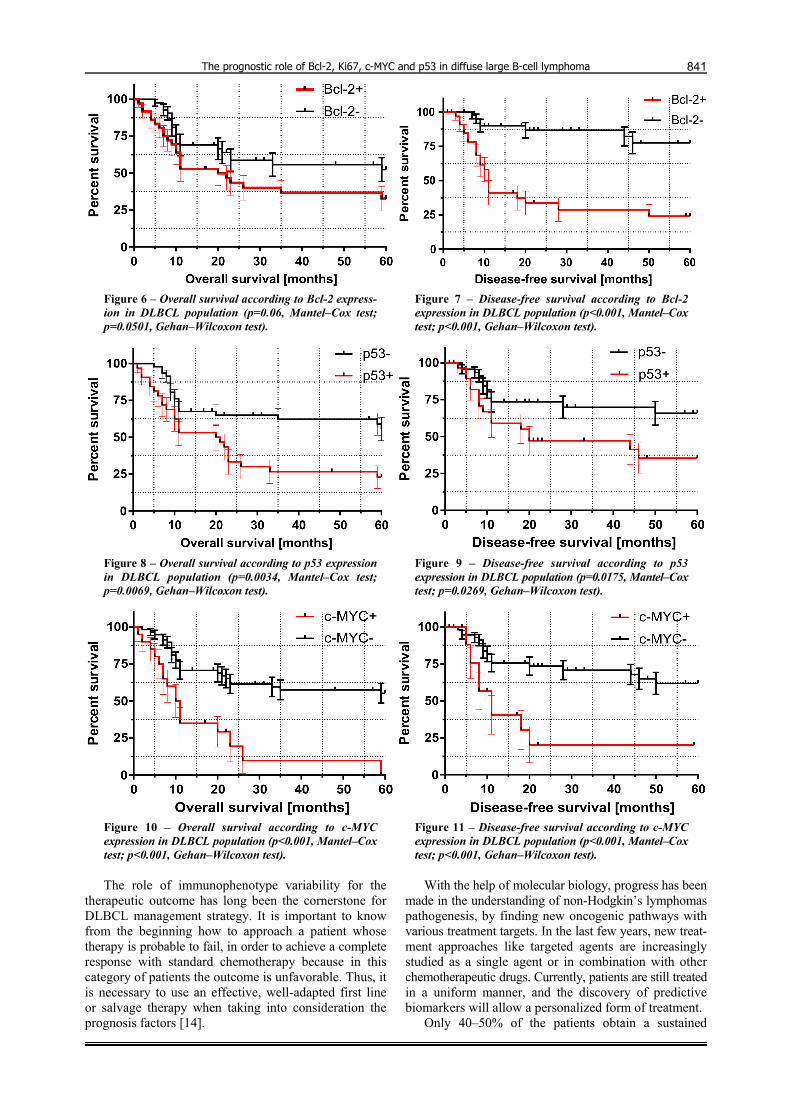
Kaplan–Meier analysis of the 78 patients that followed R-CHOP regimen revealed a tendency toward decreased five-year overall survival (32.6% vs. 52.4%, p<0.001) and disease-free survival (23.9% vs. 77.5%, p<0.001) in patients with positive Bcl-2 expression than for those cases with reduced or negative expression (Figures 6 and 7). Similarly, statistically significant differences were also observed when p53 expression was analyzed. Overall survival in p53 negative group was 55.6%, while for p53 expressing cases the five years survival was 22.8% (p=0.0034) (Figure 8). Furthermore, significant differences were also observed in disease-free survival distribution, with p53 negative patients exhibiting higher DFS pro-portions (65.7% vs. 35.3%, p=0.0175) (Figure 9). Another protein expression that negatively correlated with worse survival outcomes was c-MYC. The overall survival in c-MYC+ group was significantly lower (9.72%) than in c-MYC- group (55.2%, p<0.001), with a similar tendency being observed in case of disease-free survival (20.2% vs. 61.8%, p<0.001) (Figures 10 and 11).
Discussion
DLBCL is one of the most frequently encountered types of aggressive lymphoma, so sustained efforts have been made to find the relevant features involved in its clinical behavior and therapeutic response [9]. A long list of clinical, demographic, topographical, and histological factors have been investigated, but most of the results were insubstantial [10, 11]. DLBCL can affect patients of all ages, with a wide range of clinical manifestations. However, mean age at presentation is 64 years, and the disease has a slightly higher incidence in male patients. DLBCL can involve any organ, including the central nervous system, but 50% of cases are diagnosed in the advanced stages (stages III and IV Ann Arbor) [12, 13].
Table 2 – Immunophenotypic features of DLBCL patients according to the cell-of-origin subtype
Bcl-2 Ki67 c-MYC p53
– + – + – + – +
n (%) 49 (50.5) 48 (49.5) 40 (41.23) 57 (58.77) 66 (68.04) 31 (31.96) 72 (74.22) 25 (25.78)
GCB 24 20 29 15 37 7 37 7
Non-GCB 25 28 11 42 29 24 35 18
P-value (Fisher) 0.5426 <0.001 0.0023 0.0616
OR (95% CI) 1.344 (0.5847–2.888) 7.382 (3.011–17.72) 4.374 (1.643–10.86) 2.718 (1.058–6.841)
DLBCL: Diffuse large B-cell lymphoma; GCB: Germinal center B-cell like; OR: Odds ratio; CI: Confidence interval.
Table 3 – Association between the IPI and the immunohistochemical profile of DLBCL patients
Bcl-2 Ki67 c-MYC p53
– + – + – + – +
0–2 30 18 29 19 42 6 35 13 IPI
3–5 19 30 11 38 24 25 17 32
P-value (Fisher) 0.0257 0.0002 <0.0001 0.0002
OR (95% CI) 2.632 (1.131–5.995) 5.273 (2.238–12.98) 7.292 (2.56–20.16) 5.068 (2.037–11.72)
IPI: International Prognostic Index; DLBCL: Diffuse large B-cell lymphoma; OR: Odds ratio; CI: Confidence interval.
The prognostic role of Bcl-2, Ki67, c-MYC and p53 in diffuse large B-cell lymphoma
841
Figure 6 – Overall survival according to Bcl-2 express-ion in DLBCL population (p=0.06, Mantel–Cox test; p=0.0501, Gehan–Wilcoxon test).
Figure 7 – Disease-free survival according to Bcl-2 expression in DLBCL population (p<0.001, Mantel–Cox test; p<0.001, Gehan–Wilcoxon test).
Figure 8 – Overall survival according to p53 expression in DLBCL population (p=0.0034, Mantel–Cox test; p=0.0069, Gehan–Wilcoxon test).
Figure 9 – Disease-free survival according to p53 expression in DLBCL population (p=0.0175, Mantel–Cox test; p=0.0269, Gehan–Wilcoxon test).
Figure 10 – Overall survival according to c-MYC expression in DLBCL population (p<0.001, Mantel–Cox test; p<0.001, Gehan–Wilcoxon test).
Figure 11 – Disease-free survival according to c-MYC expression in DLBCL population (p<0.001, Mantel–Cox test; p<0.001, Gehan–Wilcoxon test).
The role of immunophenotype variability for the therapeutic outcome has long been the cornerstone for DLBCL management strategy. It is important to know from the beginning how to approach a patient whose therapy is probable to fail, in order to achieve a complete response with standard chemotherapy because in this category of patients the outcome is unfavorable. Thus, it is necessary to use an effective, well-adapted first line or salvage therapy when taking into consideration the prognosis factors [14].
With the help of molecular biology, progress has been made in the understanding of non-Hodgkin’s lymphomas pathogenesis, by finding new oncogenic pathways with various treatment targets. In the last few years, new treat-ment approaches like targeted agents are increasingly studied as a single agent or in combination with other chemotherapeutic drugs. Currently, patients are still treated in a uniform manner, and the discovery of predictive biomarkers will allow a personalized form of treatment.
Only 40–50% of the patients obtain a sustained

Ana-Maria Pătraşcu et al.
842
response with classical R-CHOP, therefore it is important to establish from the moment of diagnosis if it is necessary to apply a more aggressive or even an experimental treat-ment. The process through which this can be achieved is by independently assessing the morphological, IHC and genetic profile of every patient who will be subsequently selected for a more individualized treatment.
For the current analysis, the morphological classi-fication of cases was performed in compliance with the 2008 WHO classification. We considered the following morphological subtypes:
▪ immunoblastic – when immunoblasts accounted for 90% or more of the tumor cells;
▪ centroblastic – when centroblasts represented 90% or more of the neoplasic cells; and
▪ anaplastic subtype – roughly characterized by round cells with bizarre, pleomorphic nuclei.
The Hans protocol was used for DLBCL subdivision into GCB and non-GCB type. Positive expression for CD10 or co-expression of CD10 and Bcl-6 allowed the distribution of cases to the GCB group, whereas negative expression of CD10 with positive Bcl-6 dictated for the evaluation of MUM-1. The negative expression of MUM-1 confirmed the inclusion in the GCB group.
The role of this study was to determine how several molecular markers (Bcl-2, Ki67, c-MYC and p53) not included in Hans algorithm may influence the survival endpoints and thus correlate with the prognosis. In order to have unequivocal and reproducible results, single cutoff values were attributed to each of these parameters. We assessed each case as positive if 30% or more of the tumor cells were stained with an antibody, in accordance with the criteria recommended by Hans et al. [8]. We immunohistochemically analyzed the expression of Ki67 in tissue sections by considering a cut-off value of 70%. The cutoffs used for Bcl-2, c-MYC and p53 were 70%, 40% and 30%, respectively.
As a routinely determined marker necessary in Hans algorithm, Bcl-6, along with the other specific markers needed for cell-of-origin classification, such as CD10 and MUM-1, can serve as selective diagnostic and therapeutic target for GCB DLBCL. As there are several studies that already investigated this specific aspect [15, 16], more efforts should be made towards exploring new, insuffi-ciently investigated markers.
One notable example is Bcl-2, an important regulating protein that is heterogeneously expressed in both GCB and non-Bcl DLBCL, by distinct mechanisms. In GCB DLBCL, t(14,18) translocation is involved, unlike non-GCB DLBCL where gene amplification and transcriptional upregulation are responsible for its expression. GCB DLBCL expressing Bcl-2 present a low response to the classical R-CHOP treatment compared to the non-GCB DLBCL, therefore Bcl-2 inhibitors are also under inves-tigations [17, 18].
Up to 30% of DLBCL cases express c-MYC protein, which is involved in growth and cell proliferation. Expression of c-MYC seems to be uncorrelated with poor prognosis, instead co-expression of c-MYC and Bcl-2 appear to signal a low overall survival and disease-free survival [19]. Moreover, c-MYC–Bcl-2 co-expression seems to have a higher incidence in non-Bcl cases, and appear
to bear a higher importance to prognosis than the cell-of-origin classification in itself [20]. Based on these characteristics, patients with DLBCL should be routinely investigated for the expression of these two molecules with prognosis importance, so that different therapy could be considered in this category of patients.
P53, on the other hand, plays a controversial role as a prognostic marker, as the results of several studies investigating this aspect are discordant. There might be several explanations for this phenomenon, such as low cutoff values for IHC expression, small sample size, heterogeneous population or different treatment protocols [20, 21]. No such inconsistencies were noted in our group, as the sample size was adequate for proper statistical analysis and only a single, routinely used treatment regimen was considered (R-CHOP). Moreover, a high cutoff value for p53 (>30%) was used for more consistent results. Nevertheless, positive expression of p53 was correlated with worse results in terms of overall survival and disease-free survival, which is consistent with the results of other studies.
Ki67 is a nuclear marker that signalizes the lympho-proliferative activity of the non-Hodgkin’s lymphoma. Therefore, its expression is routinely marked as an index of proliferation, with potential prognostic value for various DLBCL subtypes. Despite numerous attempts to correlate its expression to other biological markers or to certain clinical and therapeutic outcomes, no definitive conclu-sions could be delivered [22–24]. Similar to the level of the IHC expression of the other three markers, our results for Ki67 are unequivocal, providing a good argument for their routine use as useful additions to the diagnostic protocol and treatment algorithm of DLBCL.
However, our study has several limitations, due to the reduced number of patients and its retrospective design, thus the need for the initiation of larger, prospective randomized controlled trials in order to have a definitive and clear view of the role these markers play in the clinical outcome of DLBCL patients.
Conclusions
The results of our study suggest that the positive expression of Bcl-2, Ki67, c-MYC and p53 positively correlates with low prognostic index and poor survival rate, thus making them valuable diagnostic and therapeutic targets, hence the need for their routine evaluation as part of a new IHC algorithm for DLBCL.
Conflict of interests The authors have no conflict of interests to disclose.
Author contribution Ana-Maria Pătraşcu and Ionela Rotaru had equal
contribution to this study thus share main authorship.
References [1] Swerdlow SH, Campo E, Pileri SA, Harris NL, Stein H,
Siebert R, Advani R, Ghielmini M, Salles GA, Zelenetz AD, Jaffe ES. The 2016 revision of the World Health Organization classification of lymphoid neoplasms. Blood, 2016, 127(20): 2375–2390.
[2] Jaffe ES, Harris NL, Stein H, Isaacson PG. Classification of lymphoid neoplasms: the microscope as a tool for disease discovery. Blood, 2008, 112(12):4384–4399.
The prognostic role of Bcl-2, Ki67, c-MYC and p53 in diffuse large B-cell lymphoma
843
[3] Pileri SA, Agostinelli C, Sabattini E, Bacci F, Sagramoso C, Pileri A Jr, Falini B, Piccaluga PP. Lymphoma classification: the quiet after the storm. Semin Diagn Pathol, 2011, 28(2): 113–123.
[4] Song CG, Huang JJ, Li YJ, Xia Y, Wang Y, Bi XW, Jiang WQ, Huang HQ, Lin TY, Li ZM. Epstein–Barr virus-positive diffuse large B-cell lymphoma in the elderly: a matched case-control analysis. PLoS One, 2015, 10(7):e0133973.
[5] Küppers R, Dalla-Favera R. Mechanisms of chromosomal translocations in B cell lymphomas. Oncogene, 2001, 20(40): 5580–5594.
[6] Schneider C, Pasqualucci L, Dalla-Favera R. Molecular patho-genesis of diffuse large B-cell lymphoma. Semin Diagn Pathol, 2011, 28(2):167–177.
[7] Reber R, Banz Y, Garamvölgyi E, Perren A, Novak U. Determination of the molecular subtypes of diffuse large B-cell lymphomas using immunohistochemistry: a case series from the Inselspital, Bern, and a critical appraisal of this determination in Switzerland. Swiss Med Wkly, 2013, 143: w13748.
[8] Hans CP, Weisenburger DD, Greiner TC, Gascoyne RD, Delabie J, Ott G, Müller-Hermelink HK, Campo E, Braziel RM, Jaffe ES, Pan Z, Farinha P, Smith LM, Falini B, Banham AH, Rosenwald A, Staudt LM, Connors JM, Armitage JO, Chan WC. Confirmation of the molecular classification of diffuse large B-cell lymphoma by immunohistochemistry using a tissue microarray. Blood, 2004, 103(1):275–282.
[9] Picleanu AM, Novelli S, Monter A, Garcia-Cadenas I, Caballero AC, Martino R, Esquirol A, Briones J, Sierra J. Allogenic hematopoietic stem cell transplantation for non-Hodgkin’s lymphoma: a retrospective analysis of 77 cases. Ann Hematol, 2017, 96(5):787–796.
[10] Rotaru I, Găman GD, Stănescu C, Găman AM. Evaluation of parameters with potential prognosis impact in patients with primary gastric diffuse large B-cell lymphoma (PG-DLBCL). Rom J Morphol Embryol, 2014, 55(1):15–21.
[11] Saygin C, Jia X, Hill B, Dean R, Pohlman B, Smith MR, Jagadeesh D. Impact of comorbidities on outcomes of elderly patients with diffuse large B-cell lymphoma. Am J Hematol, 2017, 92(10):989–996.
[12] Moskowitz C. Diffuse large B cell lymphoma: how can we cure more patients in 2012? Best Pract Res Clin Haematol, 2012, 25(1):41–47.
[13] Shah HJ, Keraliya AR, Jagannathan JP, Tirumani SH, Lele VR, DiPiro PJ. Diffuse large B-cell lymphoma in the era of precision oncology: how imaging is helpful. Korean J Radiol, 2017, 18(1):54–70.
[14] Sehn LH, Gascoyne RD. Diffuse large B-cell lymphoma: optimizing outcome in the context of clinical and biologic heterogeneity. Blood, 2015, 125(1):22–32.
[15] Cerchietti LC, Ghetu AF, Zhu X, Da Silva GF, Zhong S, Matthews M, Bunting KL, Polo JM, Farès C, Arrowsmith CH, Yang SN, Garcia M, Coop A, Mackerell AD Jr, Privé GG, Melnick A. A small-molecule inhibitor of BCL6 kills DLBCL cells in vitro and in vivo. Cancer Cell, 2010, 17(4):400–411.
[16] Lu TX, Miao Y, Wu JZ, Gong QX, Liang JH, Wang Z, Wang L, Fan L, Hua D, Chen YY, Xu W, Zhang ZH, Li JY. The distinct clinical features and prognosis of the CD10+MUM1+ and CD10–Bcl6–MUM1– diffuse large B-cell lymphoma. Sci Rep, 2016, 6:20465.
[17] Cang S, Iragavarapu C, Savooji J, Song Y, Liu D. ABT-199 (venetoclax) and BCL-2 inhibitors in clinical development. J Hematol Oncol, 2015, 8:129.
[18] Llanos M, Alvarez-Argüelles H, Alemán R, Oramas J, Diaz-Flores L, Batista N. Prognostic significance of Ki-67 nuclear proliferative antigen, bcl-2 protein, and p53 expression in follicular and diffuse large B-cell lymphoma. Med Oncol, 2001, 18(1):15–22.
[19] Johnson NA, Slack GW, Savage KJ, Connors JM, Ben-Neriah S, Rogic S, Scott DW, Tan KL, Steidl C, Sehn LH, Chan WC, Iqbal J, Meyer PN, Lenz G, Wright G, Rimsza LM, Valentino C, Brunhoeber P, Grogan TM, Braziel RM, Cook JR, Tubbs RR, Weisenburger DD, Campo E, Rosenwald A, Ott G, Delabie J, Holcroft C, Jaffe ES, Staudt LM, Gascoyne RD. Concurrent expression of MYC and BCL2 in diffuse large B-cell lymphoma treated with rituximab plus cyclophosphamide, doxorubicin, vincristine, and prednisone. J Clin Oncol, 2012, 30(28):3452–3459.
[20] Hu S, Xu-Monette ZY, Tzankov A, Green T, Wu L, Balasubramanyam A, Liu WM, Visco C, Li Y, Miranda RN, Montes-Moreno S, Dybkaer K, Chiu A, Orazi A, Zu Y, Bhagat G, Richards KL, Hsi ED, Choi WW, Zhao X, van Krieken JH, Huang Q, Huh J, Ai W, Ponzoni M, Ferreri AJ, Zhou F, Slack GW, Gascoyne RD, Tu M, Variakojis D, Chen W, Go RS, Piris MA, Møller MB, Medeiros LJ, Young KH. MYC/ BCL2 protein co-expression contributes to the inferior survival of activated B-cell subtype of diffuse large B-cell lymphoma and demonstrates high-risk gene expression signatures: a report from The International DLBCL Rituximab–CHOP Consortium Program. Blood, 2013, 121(20):4021–4031; quiz 4250.
[21] Xie Y, Bulbul MA, Ji L, Inouye CM, Groshen SG, Tulpule A, O’Malley DP, Wang E, Siddiqi IN. P53 expression is a strong marker of inferior survival in de novo diffuse large B-cell lymphoma and may have enhanced negative effect with MYC coexpression: a single institutional clinicopathologic study. Am J Clin Pathol, 2014, 141(4):593–604.
[22] Li ZM, Huang JJ, Xia Y, Zhu YJ, Zhao W, Wei WX, Jiang WQ, Lin TY, Huang HQ, Guan ZZ. High Ki-67 expression in diffuse large B-cell lymphoma patients with non-germinal center sub-type indicates limited survival benefit from R-CHOP therapy. Eur J Haematol, 2012, 88(6):510–517.
[23] Sanchez E, Chacon I, Plaza MM, Muñoz E, Cruz MA, Martinez B, Lopez L, Martinez-Montero JC, Orradre JL, Saez AI, Garcia JF, Piris MA. Clinical outcome in diffuse large B-cell lymphoma is dependent on the relationship between different cell-cycle regulator proteins. J Clin Oncol, 1998, 16(5):1931–1939.
[24] He X, Chen Z, Fu T, Jin X, Yu T, Liang Y, Zhao X, Huang L. Ki-67 is a valuable prognostic predictor of lymphoma but its utility varies in lymphoma subtypes: evidence from a systematic meta-analysis. BMC Cancer, 2014, 14:153.
Corresponding author Ştefan Pătraşcu, Teaching Assistant, MD, PhD, Department of Surgery, University of Medicine and Pharmacy of Craiova, 2 Petru Rareş Street, 200349 Craiova, Romania; Phone +40724–216 642, e-mail: [email protected] Received: March 18, 2017
Accepted: November 9, 2017