2b p53 p15 Ki67 immunoexpression in OSCC.pdf
5
Rom J Morphol Embryol 2012, 53(1):89–93 ISSN (print) 1220–0522 ISSN (on-line) 2066-8279 ORIGINAL PAPER P53, p16 and Ki67 immunoexpression in oral squamous carcinomas L. P. DRAGOMIR 1,2) , CRISTIANA SIMIONESCU 2) , CL. MĂRGĂRITESCU 2) , A. STEPAN 2) , IULIANA MANUELA DRAGOMIR 3) , M. R. POPESCU 1) 1) Department of Occlusology Dental Prosthetics 2) Department of Pathology 3) Department of Public Health University of Medicine and Pharmacy of Craiova Abstract In this study, we have analyzed clinically, histopathologically and immunohistochemically a total of 34 cases of oral squamous carcinoma in 11 of the cases being identified adjacent epithelial dysplastic lesions. Carcinomas were diagnosed in patients aged 40–60 years, males, with chronic exposure to tobacco and/or alcohol, being located especially on the lips. Well-differentiated carcinomas have been predominant (52.9%) in stage I/II tumoral (88.3%). Immunoexpression analysis of p53, p16 and Ki67 did not reveal statistically significant differences between the expression of markers and clinical or histopathological parameters, except Ki67 whose increased expression was associated to the decrease of the degree of tumoral differentiation and with high degree dysplasia. The positivity index and the intensity of reaction were increased at the level of dysplasic epithelium for p16 and at the level of tumoral invasion front for the p53 and Ki67. The study highlights the value of the immunostain for p16 in identifying dysplasic lesions and predictive importance of p53 and Ki67 markers in identifying the aggressive forms of oral carcinomas. Keywords: squamous oral carcinoma, dysplastic epithelium, p53, p16, Ki67. Introduction Squamous carcinomas represent about 3% of human cancers and over 90% of malignant tumors with oral location, being diagnosed worldwide each year over 350 000 new cases [1–3]. In lesions appearance are incriminated various risk factors and chronic exposure to tobacco and alcohol are the most important [4, 5]. Epidemiological data indicates a predominance of squamous oral carcinomas of the lip and lingual localization in men over 40 years [2, 6]. Oral carcinogenesis is a multistage process, pre- cancerous lesions, invasive and metastases being often present simultaneously [7, 8]. Browsing of cell cycle and proliferation of malignant tumoral cells involves loss of control mechanisms that ensure normal tissular functioning. Studies that have investigated the expression of some proteins involved in these biomolecular mechanisms demonstrate the permanent concern for the identification of biomarkers with oral squamous carcinomas predictive potential. P53 and p16 are cell cycle control proteins, which provide the “arrest” and apoptosis of cancer cells and stops their proliferation by maintaining in a hypophosphorilate status of retino- blastoma protein, while Ki67 is present in the nucleus located in the division [9–11] Currently, there is no consensus on the timing of occurrence and prognostic significance of aberrant expression of these proteins [12–15]. The purpose of this study was to analyze p53, p16, Ki67 immunoexpression in oral carcinomas and adjacent dysplastic epithelium, given their role in regulating cellular cycle and proliferation. Materials and Methods We conducted a retrospective study that included a number of 34 oral squamous carcinomas diagnosed during 2010–2011 in the Laboratory of Pathological Anatomy of the Emergency County Hospital, Craiova. The biological material was represented by pieces of tumoral resection, which after fixation in buffered neutral formalin 10%, were processed for paraffin embedding and Hematoxylin–Eosin staining. Histopathological classification of lesions was done according to the criteria proposed by the working group from WHO for oral cavity tumors [2]. Immunohistochemical analysis was performed on serial sections, as indicated on LSAB™ + Technical Kit/HRP (DAKO, code K0679) for p53 and Ki67, and of CINtec ® Histology Kit (DAKO, code K5334) for determination of p16. Development of reactions was performed with DAB (diaminobenzidine tetrahydro- chloride). The antibodies used, clone, dilution and antigenic retrieval are shown in Table 1. Table 1 – Panel of used antibodies Antibody Clone Dilution Antigenic Recovery P53 DO-7 1:50 Tris-EDTA, pH 9 P16 E6H4 Ready to use Epitope Retrieval Solution / Kit Ki67 MIB-1 1:100 Tris-EDTA, pH 9 R J M E Romanian Journal of Morphology & Embryology http://www.rjme.ro/
-
Upload
ria-mayanti -
Category
Documents
-
view
236 -
download
1
Transcript of 2b p53 p15 Ki67 immunoexpression in OSCC.pdf

Rom J Morphol Embryol 2012, 53(1):89–93
ISSN (print) 1220–0522 ISSN (on-line) 2066-8279
OORRIIGGIINNAALL PPAAPPEERR
P53, p16 and Ki67 immunoexpression in oral squamous carcinomas
L. P. DRAGOMIR1,2), CRISTIANA SIMIONESCU2), CL. MĂRGĂRITESCU2), A. STEPAN2), IULIANA MANUELA DRAGOMIR3), M. R. POPESCU1)
1)Department of Occlusology Dental Prosthetics 2)Department of Pathology
3)Department of Public Health University of Medicine and Pharmacy of Craiova
Abstract In this study, we have analyzed clinically, histopathologically and immunohistochemically a total of 34 cases of oral squamous carcinoma in 11 of the cases being identified adjacent epithelial dysplastic lesions. Carcinomas were diagnosed in patients aged 40–60 years, males, with chronic exposure to tobacco and/or alcohol, being located especially on the lips. Well-differentiated carcinomas have been predominant (52.9%) in stage I/II tumoral (88.3%). Immunoexpression analysis of p53, p16 and Ki67 did not reveal statistically significant differences between the expression of markers and clinical or histopathological parameters, except Ki67 whose increased expression was associated to the decrease of the degree of tumoral differentiation and with high degree dysplasia. The positivity index and the intensity of reaction were increased at the level of dysplasic epithelium for p16 and at the level of tumoral invasion front for the p53 and Ki67. The study highlights the value of the immunostain for p16 in identifying dysplasic lesions and predictive importance of p53 and Ki67 markers in identifying the aggressive forms of oral carcinomas. Keywords: squamous oral carcinoma, dysplastic epithelium, p53, p16, Ki67.
Introduction
Squamous carcinomas represent about 3% of human cancers and over 90% of malignant tumors with oral location, being diagnosed worldwide each year over 350 000 new cases [1–3]. In lesions appearance are incriminated various risk factors and chronic exposure to tobacco and alcohol are the most important [4, 5]. Epidemiological data indicates a predominance of squamous oral carcinomas of the lip and lingual localization in men over 40 years [2, 6].
Oral carcinogenesis is a multistage process, pre-cancerous lesions, invasive and metastases being often present simultaneously [7, 8]. Browsing of cell cycle and proliferation of malignant tumoral cells involves loss of control mechanisms that ensure normal tissular functioning. Studies that have investigated the expression of some proteins involved in these biomolecular mechanisms demonstrate the permanent concern for the identification of biomarkers with oral squamous carcinomas predictive potential. P53 and p16 are cell cycle control proteins, which provide the “arrest” and apoptosis of cancer cells and stops their proliferation by maintaining in a hypophosphorilate status of retino-blastoma protein, while Ki67 is present in the nucleus located in the division [9–11] Currently, there is no consensus on the timing of occurrence and prognostic significance of aberrant expression of these proteins [12–15].
The purpose of this study was to analyze p53, p16, Ki67 immunoexpression in oral carcinomas and adjacent
dysplastic epithelium, given their role in regulating cellular cycle and proliferation.
Materials and Methods
We conducted a retrospective study that included a number of 34 oral squamous carcinomas diagnosed during 2010–2011 in the Laboratory of Pathological Anatomy of the Emergency County Hospital, Craiova.
The biological material was represented by pieces of tumoral resection, which after fixation in buffered neutral formalin 10%, were processed for paraffin embedding and Hematoxylin–Eosin staining.
Histopathological classification of lesions was done according to the criteria proposed by the working group from WHO for oral cavity tumors [2].
Immunohistochemical analysis was performed on serial sections, as indicated on LSAB™ + Technical Kit/HRP (DAKO, code K0679) for p53 and Ki67, and of CINtec® Histology Kit (DAKO, code K5334) for determination of p16. Development of reactions was performed with DAB (diaminobenzidine tetrahydro-chloride). The antibodies used, clone, dilution and antigenic retrieval are shown in Table 1.
Table 1 – Panel of used antibodies Antibody Clone Dilution Antigenic Recovery
P53 DO-7 1:50 Tris-EDTA, pH 9
P16 E6H4 Ready to use Epitope Retrieval Solution / Kit
Ki67 MIB-1 1:100 Tris-EDTA, pH 9
R J M ERomanian Journal of
Morphology & Embryologyhttp://www.rjme.ro/

L. P. Dragomir et al.
90
In order to validate the reactions external negative controls were used, by omitting the primary antibody. Semiquantitative evaluation of immunohistochemical marking was performed in accordance with previous studies [16, 17]. In this respect, an index of positivity (IP) was calculated which represented the percentage of labeled cells in 1000 counted cells in the 40× microscope field, the values being allocated in one of the following categories: <10%, 10–50%, >50%. The intensity of the stain was estimated as being intense, moderate or weak. The quantification of reactions was performed independently by two people who did not have data about the study.
Statistical analysis has used the Pearson index, ANOVA and chi-square test performed by using the automatic software SPSS16.
Image acquisition was performed with Nikon Eclipse E600 microscope equipped with Lucia software 5.
Results
The performed study included 34 oral squamous carcinomas, most of the lesions being diagnosed in patients aged 20–40 years (61.8%), males (85.3%), smokers and/or alcohol consumers (85 3%) and located mainly on the lips (61.8%) (Table 2).
Table 2 – Clinical, histopathological and immuno-histochemical parameters of oral squamous carcinomas Clinico-morphological parameters No. of cases %
<40 4 11.840–60 21 61.8Age [years] >60 9 26.4
Female 5 14.7Sex
Male 29 85.3Smoking 11 32.4Alcohol 4 11.8
Family history 1 2.9 Risk factors
Associations 18 52.9Lip 21 61.8
Tongue 11 32.4Localization Soft palate 2 5.8
Well-differentiated 18 52.9Moderately
differentiated 12 35.3Degree of differentiation
Poorly differentiated 4 11.8T1 17 50 T2 13 38.3T3 3 8.8 T4 1 2.9 N 0 0
The study of tumoral progression
M 0 0 P53 28 82.3P16 22 64.7Immunohistochemistry Ki67 34 100
Histopathological analysis indicated that prevailed the well-differentiated carcinomas (52.9%) found in T1/T2 (88.3%) category without satellite adenopathies or distant metastases. In 11 cases (32.4%) were identified adjacent to tumors dysplastic epithelial lesions of high
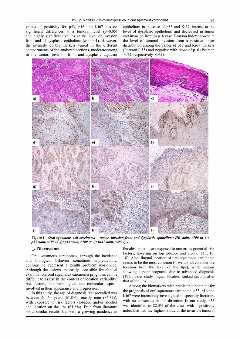
degree (three cases, 8.8%) or low degree (eight cases, 23.5%) (Figure 1c). Subsequently, we analyzed the IP values of p53, p16 and Ki67 in the tumor, invasion tumoral front and the dysplastic epithelium (Table 3).
Table 3 – Medium index of positivity for p53, p16 and Ki67 in the investigated areas
Medium IP Tumor Invasion front Dysplasic epithelium
P53 31.3% 61.8% 23.8% P16 35.1% 27.6% 45.4% Ki67 36% 62.3% 30.1%
P53 immunostain was identified at a nuclear level in 28 of the 34 analyzed squamous oral carcinomas (82.3%). The reaction was observed in the tumor and at the level of adjacent dysplasic epithelium, being present especially in basal and parabasal cells, as well as in rare cells of superjacent layers (Figure 1d-f). The stain was also present at the level of some normal ductal and glandular structures, as well as at the level of endothelial cells (IP<10%). At a tumoral level, the positivity index IP was of 31.3%, the stain being strong in the peripheric cells from tumoral isles. Also, the immunoreaction was intense at the level of the invasion front, were IP was of 61.8%. In dysplastic epithelium, the value was 23.8% and moderate reaction intensity.
The immunoexpression p16 was present at nuclear and cytoplasmic level in 22 of the 34 analyzed cases (64.7%). The reaction was found in tumor and at the level of adjacent dysplastic epithelium, labeled cells being located mainly basal and parabasal and sometimes located in the entire epithelium (Figure 1g-i). The reaction was identified and at the level of some normal elements such as fibroblasts, glandular acinus, muscle fibers, ductal epithelium and some endothelium (IP<10%). P16 marker had a weak intensity both at the level of the investigated tumors as well as at the invasion front, IP being of 35.1%, respectively 27.6%. At the level of dysplastic adjacent epithelium the reaction was intense/ moderate, with 45.4% IP with some values that exceeded 50% (Figure 1).
We have not identified significant statistical differences in what p53, respectively p16 stain is concerned and age, sex, risk factors, degree of tumoral differentiation, degree of dysplasia and tumoral stage (p>0.05, chi-square test). Ki67 nuclear marker was identified at a nuclear level in all analyzed cases, being identified at a tumoral level and in basal and parabasal cells of dysplasic epithelium (Figure 1j-l). Inside the tumor, the marking was present especially in the periphery of tumoral isles in the case of well-differentiated carcinomas. The immunoreaction was not identified at the level of normal elements. Positivity index had at a tumoral level medium values of 36% and at the level of the invasion front of 62.3%, the reactions presenting an increased intensity. At the level of dysplasic epithelium, the average percentage of labelled cells was of 30.1% the reaction being moderate/strong.
In case of Ki67, the immunoexpression was correlated to the degree of tumoral differentiation (p<0.05, chi-square). The ANOVA test of comparing the index
P53, p16 and Ki67 immunoexpression in oral squamous carcinomas
91
values of positivity for p53, p16 and Ki67 has no significant differences at a tumoral level (p>0.05) and highly significant values at the level of invasion front and of dysplasic epithelium (p<0.001). However, the intensity of the markers varied in the different compartments of the analyzed sections, moderate/strong in the tumor, invasion front and dysplasic adjacent
epithelium in the case of p53 and Ki67, intense at the level of dysplasic epithelium and decreased in tumor and invasion front in p16 case. Pearson index showed at the level of tumoral invasion front a positive linear distribution among the values of p53 and Ki67 markers (Pearson 0.55) and negative with those of p16 (Pearson -0.72, respectively -0.65).
Figure 1 – Oral squamous cell carcinoma – tumor, invasion front and dysplastic epithelium: HE stain, ×100 (a–c); p53 stain, ×100 (d–f); p16 stain, ×100 (g–i); Ki67 stain, ×200 (j–l).
Discussion
Oral squamous carcinomas, through the incidence and biological behavior sometimes unpredictable, continue to represent a health problem worldwide. Although the lesions are easily accessible for clinical examination, oral squamous carcinoma prognosis can be difficult to assess in the context of location variability, risk factors, histopathological and molecular aspects involved in their appearance and progression.
In this study, the age of diagnosis that prevailed was between 40–60 years (61.8%), mostly men (85.3%), with exposure to risk factors (tobacco and/or alcohol and location on the lips (61.8%). Data from literature show similar results, but with a growing incidence in
females, patients are exposed to numerous potential risk factors, hovering on top tobacco and alcohol [13, 16, 18]. Also, lingual location of oral squamous carcinoma seems to be the most common (if we do not consider the location from the level of the lips), while lesions showing a poor prognosis due to advanced diagnosis [19]. In our study lingual location ranked second after that of the lips.
Among the biomarkers with predictable potential for the prognosis of oral squamous carcinoma, p53, p16 and Ki67 were intensively investigated in specialty literature with no consensus in this direction. In our study, p53 was identified in 82.5% of the cases with a positivity index that had the highest value at the invasion tumoral

L. P. Dragomir et al.
92
front, without any correlations to the study of the tumoral degree.
The wild form of p53 plays an important role in arresting the cells with damaged DNA that pass from G1 phase to S phase of cellular cycle and the inducing of their apoptosis, the alteration of the expression being documented in various localizations of malignant cells [18, 20]. The positivity percentage for p53 in squamous oral carcinomas varies in different studies from 0–100%, being reported correlations of this one to the severity of the injury [18, 21, 22].
Some authors have shown its correlation with the degree of differentiation, while others have refuted this association [12, 13, 18, 23, 24]. In the study by Kato K et al., in 2008, the authors identified the increased expression of the protein in the invasion front, which was correlated to a decreased survival [13]. In the study by Carlos de Vicente J et al., in 2004, which included a number of 287 oral squamous carcinomas, found out that a weak or negative expression of the protein indicates a good prognosis and a higher survival period [18].
P16 represents a negative regulator of cellular cycle that ensures the control of the cellular passage from phase G1 to phase S. Mitogenic stimuli, as well as the growth factors determine the activation of cyclin D, that binds and activates the cyclin-dependent kinases 4 and 6, resulting in retinoblastoma protein phosphorylation, following by the release of a transcription factor that ensures cell proliferation [25].
In our study, the p16 immunostain was identified in 64.7% of the cases, the positivity index and the intensity of the reaction having increased values at the level of dysplastic adjacent epithelium and reduced in tumor and their invasion front.
Existing studies up to the present moment indicate controversial results regarding the p16 immunoexpression and the relation with histopathological prognostic factors [12, 16, 25]. In 2002, Yuen PW et al. have investigated the p16 expression in carcinomas of the head and neck and showed that there is a correlation only between the decreased marking of protein and T category of tumoral stage [16]. Also, some authors indicate that at the level of the invasion front of poorly differentiated tumors p16 immunoexpression is low or absent [15]. Other studies consider that p16 expression is not correlated to the tumoral grade and stage, without potential for assessing the aggressiveness of carcinomas [12, 14].
In our study, p53 and p16 showed no statistically significant differences taking into consideration the degree of dysplasia, but compared to the stains from the tumoral level, p16 proved to be a specific marker for dysplasic epithelium (IP 45.7%, strong intensity) and p53 for invasive oral squamous carcinomas (IP 61.8%, strong). In a study conducted in 2009, by Bilde A et al., it found in the uninvaded tumor resection margins p16 and p53 expression in 25%, respectively 12.5% cases, with location at the level of basal and parabasal cells and the author interprets as being premalignant modifications. In the same study, the analyzed markers showed expression in all layers of dysplastic epithelium
adjacent to oral squamous carcinoma in 80% and 40% of cases. The loss of p16 expression is considered in this study an early event of oral carcinogenesis [26]. Also, the suprabasal expression of p53 is considered by some authors as being a risk factor for subsequent occurrence of epithelial dysplasia and oral squamous cell carcinoma [26, 27]. In a study conducted in 2009 by Sousa FA et al., on 24 cases of dysplasia of various degrees, it does not find the differences in p53 expression, unlike the study conducted in 2008 on 58 oral biopsies by Angiero F et al. who have observed an increase in p53 and Ki67 expression with the degree of dysplasia, p16 presenting values similar to the immunoexpression for different degrees of dysplasia. The authors conclude that p53 and p16 are potentially predictive for the progression of dysplasic lesions by an invasive carcinoma [28, 29].
In our study, Ki67 immunostain was identified in all cases and was correlated to the degree of dysplasia and degree of differentiation of oral squamous carcinomas and presenting high values and intensity at the tumoral invasion front (IP 62.3%). The results are in accordance to data in the literature, Ki67 proving to be useful in assessing tumor aggressiveness [17, 23, 30].
Although studies from specialty literature analyze groups of patients different in number and presenting clinical and histo-molecular signs, the obtained results are more homogenous as compared to other locations of carcinomas, thing that can argument the practical utility of this panel of antibodies in these lesions. In addition, clinical features number and histo-molecular results on p53 expression, p16 and Ki67 are more homogeneous than other locations of carcinomas, which may argue the practical utility of this panel of antibodies in these lesions. Thus, the investigation of p16 is useful in identifying dysplasic lesions, and the decrease of its immunoexpression is constituted in a predictive factor of neoplasic transformation of these lesions while the evaluation of markers expression p53 and Ki67 has a prognostic value allowing the identification of aggressive forms, highly proliferative by oral carcinomas.
Conclusions
The performed study demonstrates the specificity of the p16 immunostain at the dysplastic lesions of oral mucosa, while p53 and Ki67 markers seem to have prognostic value especially for the invasive carcinomas.
References [1] Cooper JS, Porter K, Mallin K, Hoffman HT, Weber RS,
Ang KK, Gay EG, Langer CJ, National Cancer Database report on cancer of the head and neck: 10-year update, Head Neck, 2009, 31(6):748–758.
[2] Barnes L, Eveson JW, Reichart P, Sidransky D (eds), Pathology and genetics of head and neck tumours, World Health Organization Classification of Tumors, IARC Press, Lyon, 2005.
[3] Parkin DM, Whelan SL, Ferlay J, Teppo L, Thomas DB (eds), Cancer incidence in five continents, vol. VIII, IARC Press, Lyon, 2003.
[4] Petti S, Scully C, Determinants of oral cancer at the national level: just a question of smoking and alcohol drinking prevalence? Odontology, 2010, 98(2):144–152.
P53, p16 and Ki67 immunoexpression in oral squamous carcinomas
93[5] Meurman JH, Infectious and dietary risk factors of oral
cancer, Oral Oncol, 2010, 46(6):411–413. [6] ***, Cancer facts & figures 2010, American Cancer Society,
Atlanta, 2010. [7] Stoll C, Baretton G, Löhrs U, The influence of p53 and
associated factors on the outcome of patients with oral squamous cell carcinoma, Virchows Arch, 1998, 433(5):427–433.
[8] Simionescu C, Mărgăritescu C, Surpăţeanu M, Mogoantă L, Zăvoi R, Ciurea R, Surlin P, Stepan A, The study of E-cadherine and CD44 immunoexpression in oral squamous cell carcinoma, Rom J Morphol Embryol, 2008, 49(2):189–193.
[9] Hartwell L, Defects in a cell cycle checkpoint may be responsible for the genomic instability of cancer cells, Cell, 1992, 71(4):543–546.
[10] Stone S, Jiang P, Dayananth P, Tavtigian SV, Katcher H, Parry D, Peters G, Kamb A, Complex structure and regulation of the P16 (MTS1) locus, Cancer Res, 1995, 55(14):2988–2994.
[11] Endl E, Gerdes J, The Ki-67 protein: fascinating forms and an unknown function, Exp Cell Res, 2000, 257(2):231–237.
[12] Abrahao AC, Bonelli BV, Nunes FD, Dias EP, Cabral MG, Immunohistochemical expression of p53, p16 and hTERT in oral squamous cell carcinoma and potentially malignant disorders, Braz Oral Res, 2011, 25(1):34–41.
[13] Kato K, Kawashiri S, Tanaka A, Noguchi N, Nakaya H, Hase T, Yamamoto E, Predictive value of measuring p53 labeling index at the invasive front of oral squamous cell carcinomas, Pathol Oncol Res, 2008, 14(1):57–61.
[14] Bradley KT, Budnick SD, Logani S, Immunohistochemical detection of p16INK4a in dysplastic lesions of the oral cavity, Mod Pathol, 2006, 19(10):1310–1316.
[15] Natarajan E, Omobono JD 2nd, Jones JC, Rheinwald JG, Co-expression of p16INK4A and laminin 5 by keratinocytes: a wound-healing response coupling hypermotility with growth arrest that goes awry during epithelial neoplastic progression, J Investig Dermatol Symp Proc, 2005, 10(2):72–85.
[16] Yuen PW, Man M, Lam KY, Kwong YL, Clinicopathological significance of p16 gene expression in the surgical treatment of head and neck squamous cell carcinomas, J Clin Pathol, 2002, 55(1):58–60.
[17] Raju B, Mehrotra R, Oijordsbakken G, Al-Sharabi AK, Vasstrand EN, Ibrahim SO, Expression of p53, cyclin D1 and Ki-67 in pre-malignant and malignant oral lesions: association with clinicopathological parameters, Anticancer Res, 2005, 25(6C):4699–4706.
[18] Carlos de Vicente J, Junquera Gutiérrez LM, Zapatero AH, Fresno Forcelledo MF, Hernández-Vallejo G, López Arranz JS, Prognostic significance of p53 expression in oral squamous cell carcinoma without neck node metastases, Head Neck, 2004, 26(1):22–30.
[19] Yuen AP, Lam KY, Chan AC, Wei WI, Lam LK, Ho WK, Ho CM, Clinicopathological analysis of elective neck dissection for N0 neck of early oral tongue carcinoma, Am J Surg, 1999, 177(1):90–92.
[20] Yonish-Rouach E, Resnitzky D, Lotem J, Sachs L, Kimchi A, Oren M, Wild-type p53 induces apoptosis of myeloid leukaemic cells that is inhibited by interleukin-6, Nature, 1991, 352(6333):345–347.
[21] Girod SC, Krämer C, Knüfermann R, Krueger GR, p53 expression in the carcinogenesis in the oral mucosa, J Cell Biochem, 1994, 56(4):444–448.
[22] Regezi JA, Zarbo RJ, Regev E, Pisanty S, Silverman S, Gazit D, p53 protein expression in sequential biopsies of oral dysplasias and in situ carcinomas, J Oral Pathol Med, 1995, 24(1):18–22.
[23] Kannan S, Chandran GJ, Pillai KR, Mathew B, Sujathan K, Nalinakumary KR, Nair MK, Expression of p53 in leukoplakia and squamous cell carcinoma of the oral mucosa: correlation with expression of Ki67, Clin Mol Pathol, 1996, 49(3):M170–M175.
[24] Nishioka H, Hiasa Y, Hayashi I, Kitahori Y, Konishi N, Sugimura M, Immunohistochemical detection of p53 oncoprotein in human oral squamous cell carcinomas and leukoplakias: comparison with proliferating cell nuclear antigen staining and correlation with clinicopathological findings, Oncology, 1993, 50(6):426–429.
[25] Nilsson K, Svensson S, Landberg G, Retinoblastoma protein function and p16INK4a expression in actinic keratosis, squamous cell carcinoma in situ and invasive squamous cell carcinoma of the skin and links between p16INK4a expression and infiltrative behavior, Mod Pathol, 2004, 17(12):1464–1474.
[26] Bilde A, von Buchwald C, Dabelsteen E, Therkildsen MH, Dabelsteen S, Molecular markers in the surgical margin of oral carcinomas, J Oral Pathol Med, 2009, 38(1):72–78.
[27] Cruz IB, Snijders PJ, Meijer CJ, Braakhuis BJ, Snow GB, Walboomers JM, van der Waal I, p53 expression above the basal cell layer in oral mucosa is an early event of malignant transformation and has predictive value for developing oral squamous cell carcinoma, J Pathol, 1998, 184(4):360–368.
[28] Sousa FA, Paradella TC, Carvalho YR, Rosa LE, Immuno-histochemical expression of PCNA, p53, bax and bcl-2 in oral lichen planus and epithelial dysplasia, J Oral Sci, 2009, 51(1):117–121.
[29] Angiero F, Berenzi A, Benetti A, Rossi E, Del Sordo R, Sidoni A, Stefani M, Dessy E, Expression of p16, p53 and Ki-67 proteins in the progression of epithelial dysplasia of the oral cavity, Anticancer Res, 2008, 28(5A):2535–2539.
[30] Motta Rda R, Zettler CG, Cambruzzi E, Jotz GP, Berni RB, Ki-67 and p53 correlation prognostic value in squamous cell carcinomas of the oral cavity and tongue, Braz J Otorhinolaryngol, 2009, 75(4):544–549.
Corresponding author Cristiana Simionescu, Professor, MD, PhD, Department of Pathology, University of Medicine and Pharmacy of Craiova, 66, 1 May Avenue, 200628 Craiova, Romania; Phone/Fax +40251–599 228, e-mail: csimionescu2004@ yahoo.com Received: November 12th, 2011
Accepted: January 25th, 2012