THE GASTROINTESTINAL SYSTEMdspace.zsmu.edu.ua/bitstream/123456789/4310/1/5 5 16 THE...tongue,...
132
1 MINISTRY OF HEALTH SERVICE OF UKRAINE ZAPOROZHYE STATE MEDICAL UNIVERSITY THE DEPARTMENT OF NORMAL PHYSIOLOGY THE GASTROINTESTINAL SYSTEM Methodical manual for 2 d -year students of international faculty, discipline "normal physiology" Zaporozhye, 2016
Transcript of THE GASTROINTESTINAL SYSTEMdspace.zsmu.edu.ua/bitstream/123456789/4310/1/5 5 16 THE...tongue,...

1
MINISTRY OF HEALTH SERVICE OF UKRAINE
ZAPOROZHYE STATE MEDICAL UNIVERSITY
THE DEPARTMENT OF NORMAL PHYSIOLOGY
THE GASTROINTESTINAL SYSTEM
M e t h o d i c a l m a n u a l f o r 2d
- y e a r s t u d e n t s o f
i n t e r n a t i o n a l f a c u l t y , d i s c i p l i n e " n o r m a l p h y s i o l o g y "
Zaporozhye, 2016

2
UDC 612.3(07)=111
BBC 28.91я73G24
Затверджено
ЦМР ЗДМУ: протокол № 5 від 02/06/ 2016 р.
Authors: Voteva W.E., Sukhomlinova I.E., Tichonovskaya M.A., Prosorova
T.M.
Reviewers:
Kamyshny Alexandr M. – head of the microbiology, virology and
immunology department, PhD, professor.
Gancheva Olga V. - professor of the pathophysiology department, PhD,
professor.
The gastrointestinal system: methodical manual for 2d-year students of
international faculty, discipline "normal physiology"/ W.E. Voteva, I.E.
Sukhomlinova, M.A. Tichonovskaya [et al.]. – Zaporozhye: [ZSMU], 2016. – 132 c.
Methodical recommendations compiled in accordance with the program of "normal physiology".
Guidelines are intended to help students prepare for practical classes and learn the material. Can
be used for training of 2th
-years students of international faculty, discipline "normal physiology".

3
CONTENTS
Theme actuality, study purposes, concrete purposes of the module………….. 5
Digestive function…………………………………………………………….. 6
General principles of anatomical organization of the digestive system…….. 7
Digestion in the mouth………………………………………………………. 10
Stomach: structure and function……………………………………………… 17
Gastric juice…………………………………………………………………... 20
Phases of gastric secretion………………………………………………….. 24
Digestion in the small intestine. Role of pancreas………………………….. 27
D Digestive functions of pancreatic juice. Digestion of Proteins……………… 30
Digestion of Lipids…………………………………………………………... 32
Digestion of Carbohydrates………………………………………………… 33
Regulation of Pancreatic Secretion………………………………………….. 35
Functions of liver and biliary tree…………………………………………… 39
Bile……………………………………………………………………………. 42
Secretions of the Small Intestine……………………………………………. 48
Secretions of the Large Intestine……………………………………………… 51
Digestion and Absorption in the Gastrointestinal Tract. Digestion and
absorption of carbohydrates…………………………………………………..
52
Digestion and absorption of lipids…………………………………………… 56
Digestion and absorption of proteins……………………………………….. 62
Water and Mineral Absorption………………………………………………. 64
General Principles of Gastrointestinal Motility…………………………….. 66
Neural Control of Gastrointestinal Function—Enteric Nervous System…… 70
Gastrointestinal Reflexes……………………………………………………. 73
Propulsion and Mixing of Food in the Alimentary Tract. Swallowing
(deglutition)……………………………………………………………………
74
Movements of stomach…………………………………………………. 77
Movements of small intestine……………………………………………….. 81

4
Movements of large intestine……………………………………………….. 85
Defecation……………………………………………………………………. 86
Control questions……………………………………………………………. 88
Task for initial independent training Gastrointestinal function. Digestion and
absorption of substances. Digestion in the Mouth………………………….
89
Рractical skills. Topic: Gastrointestinal function. Digestion and absorption of
substances. Digestion in the Mouth…………………………………………..
95
Task for initial independent training. Topic: Digestive functions of Stomach
and Pancreas………………………………………………………………….
99
Рractical skills. Topic: Digestion functions of Stomach and Pancreas……… 105
Task for initial independent training. Topic: Digestion functions of Liver,
Small Intestine and Colon…………………………………………………….
108
Рractical skills. Topic: Digestion functions of Liver, Small Intestine and
Colon………………………………………………………………………….
114
Tasks for final control……………………………………………………….. 117
Recommended literature……………………………………………………… 132

5
Theme actuality. The gastrointestinal tract is a continuous tube that
stretches from the mouth to the anus. Its primary function is to serve as a portal
whereby nutrients and water can be absorbed into the body. Residues of the meal
that cannot be absorbed, along with cellular debris and lipid-soluble metabolic end
products that are excreted in the bile rather than the urine, are expelled from the
body. All of these functions are tightly regulated in concert with the ingestion of
meals. Diseases in the digestive systems, also called gastrointestinal diseases, have
become quite common in the modern day world. The health of a body depends on
the health of the digestive system and any barrier in its functioning will ultimately
affect the body. Future doctors should know about the functional peculiarities of
the digestive system in order to diagnose various disorders and use appropriate
methods of treatment.
Study purposes: to know the functions of the digestive system and
principles of their regulation.
Concrete purposes of the module:
A student must know:
the functional significance of the gastrointestinal system, and in particular,
its roles in nutrient assimilation, excretion, and immunity.
the structure of the gastrointestinal tract, the glands that drain into it, and its
subdivision into functional segments.
the major gastrointestinal secretions, their components, and the stimuli that
regulate their production.
the major hormones, other peptides, and key neurotransmitters of the
gastrointestinal system.
the special features of the enteric nervous system and the splanchnic
circulation.
differences between mechanical and chemical digestion.
the general neural and chemical controls over digestive function.

6
Digestive Function
The digestive system is the organ system that processes food, extracts
nutrients from it, and eliminates the residue. It does this in four stages:
1. ingestion, the selective intake of food;
2. digestion, the mechanical and chemical breakdown of food into a form
usable by the body;
3. absorption, the uptake of nutrient molecules into the epithelial cells of
the digestive tract and then into the blood or lymph; and finally
4. defecation, the elimination of undigested residue.
The digestion stage itself has two facets, mechanical and chemical.
Mechanical digestion is the physical breakdown of food into smaller particles. It
is achieved by the cutting and grinding action of the teeth and the churning
contractions of the stomach and small intestine. Mechanical digestion exposes
more food surface to the action of digestive enzymes. Chemical digestion is a
series of hydrolysis reactions that break dietary macromolecules into their
monomers (residues): polysaccharides into monosaccharides, proteins into amino
acids, fats into glycerol and fatty acids, and nucleic acids into nucleotides. It is
carried out by digestive enzymes produced by the salivary glands, stomach,
pancreas, and small intestine. Some nutrients are already present in usable form in
the ingested food and are absorbed without being digested: vitamins, free amino
acids, minerals, cholesterol, and water.
Digestion involves the processes of motility, secretion, and membrane
transport. Motility refers to the muscular contractions that break up food, propel it
through the canal, mix it with digestive enzymes, and eliminate the waste.
Secretion releases enzymes, hormones, and other products that carry out or
regulate digestion. Membrane transport includes all the mechanisms such as active
transport and facilitated diffusion that absorb nutrients and transfer them to the
blood and lymph.

7
General principles of anatomical organization of the digestive system
The digestive system has two anatomical subdivisions, the digestive tract
and the accessory organs (fig. 1). The digestive tract is a tube extending from
mouth to anus, measuring about 9 m (30 ft) long in the cadaver. It is also known as
the alimentary canal. It includes the oral cavity, pharynx, esophagus, stomach,
small intestine, and large intestine. Part of this, the stomach and intestines,
constitute the gastrointestinal (GI) tract. The accessory organs are the teeth,
tongue, salivary glands, liver, gallbladder, and pancreas.
The digestive tract is open to the environment at both ends. Most of the
material in it has not entered any body tissues and is considered to be external to
the body until it is absorbed by epithelial cells of the alimentary canal. In the strict
sense, defecated food residue was never in the body.
Fig.1. The digestive system

8
Most of the digestive tract follows the basic structural plan shown in figure
2, with a wall composed of the following tissue layers, in order from the inner to
the outer surface:
Fig.2. Tissue Layers of the Digestive Tract
Mucosa
Epithelium
Lamina propria
Muscularis mucosae
Submucosa
Muscularis externa
Inner circular layer
Outer longitudinal layer
Serosa
Areolar tissue
Mesothelium
Slight variations on this theme are found in different regions of the tract.

9
The mucosa, lining the lumen, consists of an inner epithelium, a loose
connective tissue layer called the lamina propria, and a thin layer of smooth
muscle called the muscularis mucosae. The epithelium is simple columnar in
most of the digestive tract, but of the nonkeratinized stratified squamous type from
the oral cavity through the esophagus and in the lower anal canal, where the tract is
subject to more abrasion.
The submucosa is a thicker layer of loose connective tissue containing
blood vessels, lymphatic vessels, a nerve plexus, and in some places, glands that
secrete lubricating mucus into the lumen.
The muscularis externa consists of usually two layers of smooth muscle
near the outer surface. Cells of the inner layer encircle the tract while those of the
outer layer run longitudinally.
The serosa is composed of a thin layer of areolar tissue topped by a simple
squamous mesothelium. The serosa begins in the lower 3 to 4 cm of the esophagus
and ends with the sigmoid colon. The oral cavity, pharynx, most of the esophagus,
and the rectum are surrounded by a fibrous connective tissue layer called the
adventitia.
The esophagus, stomach, and intestines have a nervous network called the
enteric nervous system, which regulates digestive tract motility, secretion, and
blood flow. Two nerve networks make up this system: the submucosal (Meissner)
plexus in the submucosa and the myenteric (Auerbach) plexus between the two
layers of the muscularis externa.
10
Digestion in the mouth
Mouth is the first part of the alimentary tract, where digestion of food starts.
The mouth is otherwise known as oral cavity or buccal cavity. It is formed by
cheeks, lips and palate. It encloses the teeth and tongue. It opens outside anteriorly
through the lips and posteriorly through fauces.
The functions of mouth are the ingestion of food materials, chewing and
mixing the food with saliva, appreciation of the taste of food and transfer of food
(bolus) to the esophagus by swallowing. Digestive juice present in the mouth is
saliva. It is secreted by the salivary glands.
First of all food must be broken down into smaller particles for adequate
action of different enzymes. It is achieved by the cutting and grinding action of the
teeth during the process of mastication.
Mastication (Chewing)
The teeth are admirably designed for chewing, the anterior teeth (incisors)
providing a strong cutting action and the posterior teeth (molars), a grinding action.
All the jaw muscles working together can close the teeth with a force as great as 55
pounds on the incisors and 200 pounds on the molars.
Most of the muscles of chewing are innervated by the motor branch of the
fifth cranial nerve, and the chewing process is controlled by nuclei in the brain
stem. Stimulation of specific reticular areas in the brain stem taste centers will
cause rhythmical chewing movements. Also, stimulation of areas in the
hypothalamus, amygdala, and even the cerebral cortex near the sensory areas for
taste and smell can often cause chewing.
Much of the chewing process is caused by a chewing reflex, which may be
explained as follows: the presence of a bolus of food in the mouth at first initiates
reflex inhibition of the muscles of mastication, which allows the lower jaw to drop.
The drop in turn initiates a stretch reflex of the jaw muscles that leads to rebound
contraction. This automatically raises the jaw to cause closure of the teeth, but it
also compresses the bolus again against the linings of the mouth, which inhibits the

11
jaw muscles once again, allowing the jaw to drop and rebound another time; this is
repeated again and again.
Chewing is important for digestion of all foods, but especially important for
most fruits and raw vegetables because these have indigestible cellulose
membranes around their nutrient portions that must be broken before the food can
be digested. Also, chewing aids the digestion of food for still another simple
reason: digestive enzymes act only on the surfaces of food particles; therefore, the
rate of digestion is absolutely dependent on the total surface area exposed to the
digestive secretions. In addition, grinding the food to a very fine particulate
consistency prevents excoriation of the gastrointestinal tract and increases the ease
with which food is emptied from the stomach into the small intestine, then into all
succeeding segments of the gut.
Saliva and salivary glands
Saliva moistens the mouth, digests a little starch and fat, cleanses the teeth,
inhibits bacterial growth, dissolves molecules so they can stimulate the taste buds,
and moistens food and binds particles together to aid in swallowing. It is a
hypotonic solution of 97.0% to 99.5% water and the following solutes:
• salivary amylase, an enzyme that begins starch digestion in the mouth;
• lingual lipase, an enzyme that is activated by stomach acid and digests fat
after the food is swallowed;
• mucus, which binds and lubricates the food mass and aids in swallowing;
• lysozyme, an enzyme that kills bacteria;
• immunoglobulin A (IgA), an antibody that inhibits bacterial growth;
• electrolytes, including sodium, potassium, chloride, phosphate, and
bicarbonate ions.
Saliva has a pH of 6.8 to 7.0. Daily secretion of saliva normally ranges
between 800 and 1500 milliliters, saliva contains two major types of protein
secretion: a serous secretion that contains ptyalin (an α-amylase), which is an
12
enzyme for digesting starches, and mucus secretion that contains mucin for
lubricating and for surface protective purposes.
There are two kinds of salivary glands, intrinsic and extrinsic. The intrinsic
salivary glands are an indefinite number of small glands dispersed amid the other
oral tissues. They include lingual glands in the tongue, labial glands on the inside
of the lips, and buccal glands on the inside of the cheeks. They secrete relatively
small amounts of saliva at a fairly constant rate whether we are eating or not. This
saliva contains lingual lipase and lysozyme and serves to moisten the mouth and
inhibit bacterial growth.
The extrinsic salivary glands are three pairs of larger, more discrete organs
located outside of the oral mucosa; they communicate with the oral cavity by way
of ducts:
1. The parotid gland is located just beneath the skin anterior to the earlobe.
Its duct passes superficially over the masseter, pierces the buccinator, and opens
into the mouth opposite the second upper molar tooth.
2. The submandibular gland is located halfway along the body of the
mandible, medial to its margin, just deep to the mylohyoid muscle. Its duct empties
into the mouth at a papilla on the side of the lingual frenulum, near the lower
central incisors.
3. The sublingual gland is located in the floor of the mouth. It has multiple
ducts that empty into the mouth posterior to the papilla of the submandibular duct.
The parotid glands secrete almost entirely the serous type of secretion, while
the submandibular and sublingual glands secrete both serous secretion and mucus.
The buccal glands secrete only mucus.
Salivary glands are formed by the cells arranged in small groups around a
central globular cavity called acinus or alveolus. The central cavity or lumen is
continuous with the lumen of the duct. The fine duct draining each acinus is called
the intercalated ducts. Many of the intercalated ducts join together to form
intralobular ducts. Two or more intralobular ducts join to form interlobular ducts,
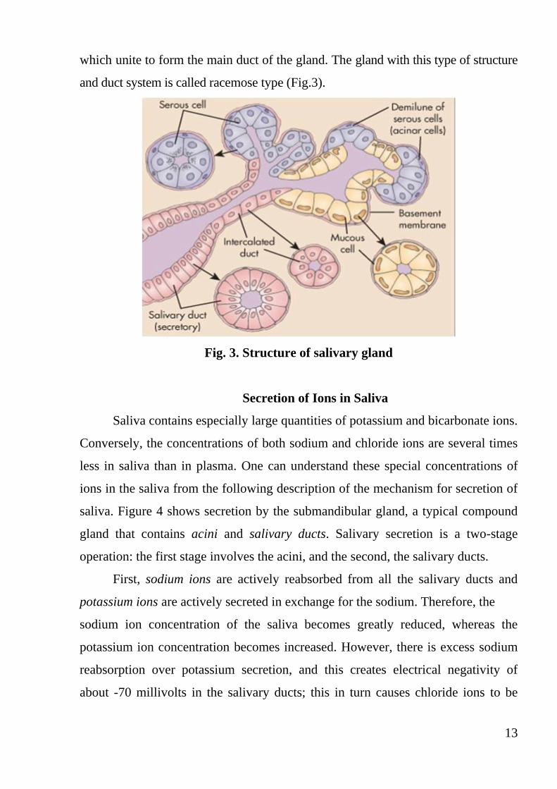
13
which unite to form the main duct of the gland. The gland with this type of structure
and duct system is called racemose type (Fig.3).
Fig. 3. Structure of salivary gland
Secretion of Ions in Saliva
Saliva contains especially large quantities of potassium and bicarbonate ions.
Conversely, the concentrations of both sodium and chloride ions are several times
less in saliva than in plasma. One can understand these special concentrations of
ions in the saliva from the following description of the mechanism for secretion of
saliva. Figure 4 shows secretion by the submandibular gland, a typical compound
gland that contains acini and salivary ducts. Salivary secretion is a two-stage
operation: the first stage involves the acini, and the second, the salivary ducts.
First, sodium ions are actively reabsorbed from all the salivary ducts and
potassium ions are actively secreted in exchange for the sodium. Therefore, the
sodium ion concentration of the saliva becomes greatly reduced, whereas the
potassium ion concentration becomes increased. However, there is excess sodium
reabsorption over potassium secretion, and this creates electrical negativity of
about -70 millivolts in the salivary ducts; this in turn causes chloride ions to be
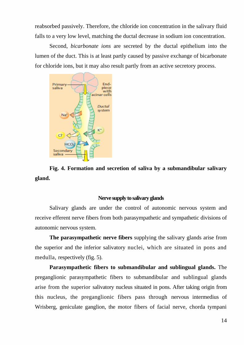
14
reabsorbed passively. Therefore, the chloride ion concentration in the salivary fluid
falls to a very low level, matching the ductal decrease in sodium ion concentration.
Second, bicarbonate ions are secreted by the ductal epithelium into the
lumen of the duct. This is at least partly caused by passive exchange of bicarbonate
for chloride ions, but it may also result partly from an active secretory process.
Fig. 4. Formation and secretion of saliva by a submandibular salivary
gland.
Nerve supply to salivary glands
Salivary glands are under the control of autonomic nervous system and
receive efferent nerve fibers from both parasympathetic and sympathetic divisions of
autonomic nervous system.
The parasympathetic nerve fibers supplying the salivary glands arise from
the superior and the inferior salivatory nuclei, which are situated in pons and
medulla, respectively (fig. 5).
Parasympathetic fibers to submandibular and sublingual glands. The
preganglionic parasympathetic fibers to submandibular and sublingual glands
arise from the superior salivatory nucleus situated in pons. After taking origin from
this nucleus, the preganglionic fibers pass through nervous intermedius of
Wrisberg, geniculate ganglion, the motor fibers of facial nerve, chorda tympani

15
branch of facial nerve and lingual branch of trigeminal nerve and finally reach the
submaxillary ganglion. The postganglionic fibers arising from this ganglion supply
the submaxillary and sublingual glands.
Parasympathetic fibers to parotid gland. The preganglionic fibers to
parotid gland arise from inferior salivatory nucleus situated in the upper part of
medulla oblongata. From here, the fibers pass through the tympanic branch of
glossopharyngeal nerve, tympanic plexus and lesser petrosal nerve and end in otic
ganglion. The postganglionic fibers from otic ganglion reach the parotid gland by
passing through the auriculotemporal branch in mandibular division of trigeminal
nerve.
Sympathetic fibers. The preganglionic sympathetic preganglionic fibers to
salivary glands arise from the lateral horns of first and second thoracic segments of
spinal cord. The fibers leave the cord through the anterior nerve roots and end in
superior cervical sympathetic ganglion. The postganglionic fibers from this
ganglion are distributed to the salivary glands along the nerve plexus around the
arteries supplying the glands.
Function of Parasympathetic Fibers. When the parasympathetic fibers of
salivary glands are stimulated, profuse and watery saliva is secreted. The amount of
organic constituents is less. The parasympathetic fibers activate the acinar cells and
dilate the blood vessels of salivary glands. The neurotransmitter is acetylcholine.
Function of Sympathetic Fibers. The stimulation of sympathetic fibers
causes less secretion of saliva, which is thick and rich in mucus. This is because,
these fibers activate the acinar cells and cause vasoconstriction by secreting
noradrenaline.

16
Fig. 5. Parasympathetic nervous regulation of salivary secretion.

17
Stomach: structure and function
The major function of the stomach is storage, but it also absorbs water-
soluble and lipid-soluble substances (e.g., alcohol and some drugs). An important
function of the stomach is to prepare the chyme for digestion in the small intestine.
Chyme is the semi-fluid material produced by the gastric digestion of food. Chyme
results partly from the conversion of large solid particles into smaller particles via
the combined peristaltic movements of the stomach and contraction of the pyloric
sphincter.
The stomach is divided into four regions: (1) The cardiac region (cardia) is
a small area immediately inside the cardiac orifice. (2) The fundic region (fundus)
is the domeshaped portion superior to the esophageal attachment. (3) The body
(corpus) makes up the greatest part of the stomach inferior to the cardiac orifice.
(4) The pyloric region is a slightly narrower pouch at the inferior end; it is
subdivided into a funnel-like antrum and a narrower pyloric canal. The latter
terminates at the pylorus, a narrow passage into the duodenum. The pylorus is
surrounded by a thick ring of smooth muscle, the pyloric (gastroduodenal)
sphincter, which regulates the passage of chyme into the duodenum (fig. 6).
Fig.6. Parts of the stomach and gastric glands

18
Structure of the stomach wall
The wall of the stomach has four layers:
1. Outer serous layer: This is formed by peritoneum which covers the stomach
except at the lesser and greater curvatures, where omenta are attached.
2. Muscular coat: This consists of three layers of smooth muscle fibers namely, inner
oblique, middle circular and outer longitudinal layers. Auerbach's plexus is situated
between the longitudinal and circular muscle fibers.
3. Submucus layer: This is formed by areolar tissue. Blood vessels, lymph vessels
and Meissner's nerve plexus are present in this layer.
4. Inner mucus layer: This is lined by mucus secreting columnar epithelial cells.
The gastric glands are situated in this layer. Under resting conditions, the
mucosa of the stomach is thrown into many folds. These folds are called rugae.
However, the rugae disappear when the stomach is distended after meals.
Throughout the inner surface of stomach, small depressions are seen. These are
called gastric pits. The glands of the stomach open into these pits. The
inner surface of mucus layer is covered by 2 mm thick mucus.
Classification of glands of the stomach
The glands of the stomach or gastric glands are of three types depending upon
the situation: fundic glands: these are situated in body and fundus of stomach. These
glands are also called main gastric glands or oxyntic glands; pyloric glands: these
are present in the pyloric part of the stomach; cardiac glands: these glands are
situated in the cardiac region of the stomach.
Structure and Functions of Fundic Glands. The fundic glands are long
and tubular glands, which secrete hydrochloric acid, pepsinogen, mucin and intrinsic
factor of Castle. These tubular glands have body, neck and isthmus. Many glands
open into a common gastric pit, which in turn opens on the surface of gastric
mucosa. The fundic glands are considered as the typical glands. The different cells
of these glands are (fig. 7):
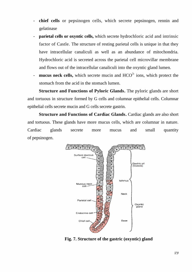
19
- chief cells or pepsinogen cells, which secrete pepsinogen, rennin and
gelatinase
- parietal cells or oxyntic cells, which secrete hydrochloric acid and intrinsic
factor of Castle. The structure of resting parietal cells is unique in that they
have intracellular canaliculi as well as an abundance of mitochondria.
Hydrochloric acid is secreted across the parietal cell microvillar membrane
and flows out of the intracellular canaliculi into the oxyntic gland lumen.
- mucus neck cells, which secrete mucin and HCO3-
ions, which protect the
stomach from the acid in the stomach lumen.
Structure and Functions of Pyloric Glands. The pyloric glands are short
and tortuous in structure formed by G cells and columnar epithelial cells. Columnar
epithelial cells secrete mucin and G cells secrete gastrin.
Structure and Functions of Cardiac Glands. Cardiac glands are also short
and tortuous. These glands have more mucus cells, which are columnar in nature.
Cardiac glands secrete more mucus and small quantity
of pepsinogen.
Fig. 7. Structure of the gastric (oxyntic) gland

20
Gastric juice
Gastric juice is the mixture of secretions from different glands of the
stomach.
Properties of gastric juice:
Volume: 1200 to 1500 ml/day.
Reaction: Gastric juice is highly acidic with pH of 0.9 to 1.2. The acidity of
gastric juice is due the hydrochloric acid.
Specific gravity: 1.002 to 1.004
Composition of gastric juice: gastric juice contains 99.5% of water and
0.5% solids. The solids are organic and inorganic substances. The organic
substances present in gastric juice are the enzymes, mucus and intrinsic factor.
Gastric Enzymes
1.Pepsin: this is the major protein splitting (proteolytic) enzyme in the gastric
juice. The precursor of pepsin is pepsinogen.
2.Rennin: it is a milk curdling enzyme. It is not present in man.
3.Gastric lipase: gastric lipase is a weak lipid splitting (lipolytic) enzyme.
4.Other gastric enzymes: the other enzymes of gastric juice are the gelatinase
and urase.
Gastric Mucus
Gastric mucus is secreted by neck cells of the gastric glands and surface
mucus cells in fundus, body and other parts of stomach. It is like a flexible gel covering
the gastric mucus membrane. Mucus is a glycoprotein.
Intrinsic Factor
This is necessary for absorption of the extrinsic factor.
Functions of gastric juice:
1. Digestive function. The gastric juice mainly acts on proteins. The
enzymes of the gastric juice acting on protein digestion are pepsin and rennin.
Gastric juice also contains some other enzymes like gastric lipase, gelatinase and
urase. Pepsin is the major proteolytic enzyme in gastric juice. It is secreted as
pepsinogen, which is inactive. Pepsinogen is formed in zymogen granules in the

21
cytoplasm of chief cells. Pepsinogen is converted into pepsin by some catalytic
enzyme in acid medium at a pH below 6. The acid medium is provided by the
hydrochloric acid secreted from parietal cells. HCl removes some of the amino acids
from pepsinogen and coverts it to pepsin. Pepsin catalyzes the production of more
pepsin (autocatalytic effect), as well as partially digesting dietary protein (fig. 8).
Fig. 8. The Production and Action of Pepsin.
Gastric Lipase is a weak lipolytic enzyme when compared to pancreatic
lipase. Gastric lipase is inactive at a pH below 2.5 and it becomes active only
when the pH is between 4 and 5. The products of lipid digestion by gastric lipase are
fatty acids and glycerols.
Gelatinase acts on gelatin, urase acts on urea and produces ammonia.
2. Hemopoietic function: the intrinsic factor present in gastric juice plays
an important role in erythropoiesis. This is necessary for absorption of the
extrinsic factor (vitamin B12) from gastrointestinal tract into the blood. Absence of
intrinsic factor in gastric juice causes deficiency of vitamin B12. And, the deficiency of
vitamin B12 leads to pernicious anemia.
3. Protective function. The mucus lubricates the gastric mucosa and
protects it from irritation or mechanical injury by virtue of its high viscosity. It
also prevents the digestive action of pepsin on the wall of the stomach particularly
gastric mucosa. Because of alkaline nature and its acid combining power, mucus
protects the gastric mucosa from hydrochloric acid of gastric juice.

22
4. Function of hydrochloric acid:
- It activates pepsinogen into pepsin
- It has bacteriolytic action. It kills some of the bacteria entering the
stomach along with food substances
- It causes acidity of the chyme. When the acid chyme leaves stomach and
enters intestine, the acidity of the chyme causes release of hormones-secretin and
cholecystokinin. These hormones, in turn stimulate the release of other digestive
juices into intestine
- It provides acid medium for the action of enzymes.
Mechanism of secretion of gastric juice
Secretion of pepsinogen: pepsinogen is synthesized from amino acids. The
synthesis occurs in ribosomes attached to endoplasmic reticulum in chief cells.
The pepsinogen molecules are packed into zymogen granules by Golgi apparatus.
When zymogen granule is secreted into stomach from chief cells, the granule is
dissolved and pepsinogen is released into gastric juice. Pepsinogen is activated into
pepsin by hydrochloric acid.
The mechanism of HCl production is depicted in fig. 9. An H+/K
+-
ATPase in the apical (luminal) cell membrane of the parietal cell actively pumps
H+ out of the cell in exchange for K
+ entering the cell. The H
+/K
+-ATPase is
inhibited by omeprazole. Although the secreted H+ is often depicted as being
derived from carbonic acid (see Fig. 9), the source of H+ is probably mostly from
the dissociation of H2O. Carbonic acid (H2CO3) is formed from carbon dioxide
(CO2) and H2O in a reaction catalyzed by carbonic anhydrase. The CO2 is provided
by metabolic sources inside the cell and from the blood. The H+/K
+-ATPase
recycles K+ ions back into the cell in exchange for H
+ ions. The basolateral cell
membrane has an electroneutral Cl-/HCO
3- exchanger that balances the entry of Cl
-
into the cell with an equal amount of HCO3-
entering the bloodstream. The Cl-
inside the cell then leaks into the lumen through Cl- channels, down an
electrochemical gradient. Consequently, HCl is secreted into the lumen. The

23
osmotic gradient created by the HCl concentration in the gland lumen drives water
passively into the lumen, thereby, maintaining the isoosmolality of the gastric
secretion.
Fig.9. The mechanism of HCl production

24
Phases of gastric secretion
Secretion of gastric juice occurs when the food is taken in the mouth. Neural
and hormonal mechanisms are involved in gastric secretion, which occurs in three
phases (Fig. 10): cephalic phase, gastric phase and intestinal phase. In human beings,
a fourth phase called interdigestive phase exists, i.e. secretion of small amount of
gastric juice in between meals. Thus, the gastric secretion tends to be continuous.
1. Cephalic Phase
This is solely under nervous control. While taking food, the secretion of
gastric juice starts even before food enters the stomach. The impulses are sent from
head and so this phase is called cephalic phase. The gastric juice secreted in this
phase is called appetite juice. This occurs as conditioned and unconditioned reflex. In
both, pepsinogen and hydrochloric acid are secreted.
Unconditioned Reflex: this causes gastric secretion when food is placed in
the mouth. Afferent impulses arise from taste buds and other receptors in the mouth
and reach the appetite center in amygdala and hypothalamus. From here, the
efferent impulses pass through dorsal nucleus of vagus and vagal fibers to the wall of
the stomach. The gastric secretion occurs by the release of acetylcholine.
Conditioned Reflex: in this, the sight, smell, hearing or thought of food causes
gastric secretion. The impulses arising from cerebral cortex reach stomach via
vagus.
2. Gastric Phase
This phase is under both nervous and hormonal control. When the food enters
the stomach, secretion of gastric juice increases which is rich in pepsinogen
and hydrochloric acid mechanisms involved in this phase of gastric secretion are:
-Local myenteric reflex - nervous mechanism
-Vagovagal reflex - nervous mechanism and
-Gastrin - hormonal mechanism
The stimuli, which activate these mechanisms, are the distention of
stomach, mechanical stimulation of gastric mucosa by food and, the chemical
stimulation by the contents of food.

25
Local Myenteric Reflex: when food enters the stomach, the food particles
stimulate the local nerve plexus in the wall of the stomach. These nerves, in turn
activate the glands of the stomach, and, a large quantity of gastric juice is secreted.
Vagovagal Reflex: presence of food in stomach stimulates the sensory nerve
endings. Now, the sensory impulses pass to the brainstem via sensory fibers of vagus.
The efferent impulses pass through the motor fibers of vagus back to stomach and
cause secretion. As both afferent and efferent impulses pass through vagus, this is
called vagovagal reflex.
Gastrin Mechanism: gastrin is one of the gastrointestinal hormones. It is
secreted by the G cells present in pyloric glands of stomach. Small amount is also
secreted in mucosa of upper small intestine. Gastrin is a polypeptide containing
G14, G17 or G34 amino acids. Gastrin is released when food enters stomach. The
mechanism involved may be the local nervous reflex or vagovagal reflex. The
neurotransmitter called gastrin releasing peptide is released at vagal nerve ending.
This acts on the G cells and causes release of the hormone.
Actions of Gastrin
- It stimulates the secretion of pepsinogen and hydrochloric acid by the
gastric glands
- It increases the motility of stomach
- It promotes growth of gastric mucosa
- It causes secretion of pancreatic juice, which is rich in enzymes
- It also stimulates the production of hormones by pancreas.
3. Intestinal Phase. When the chyme enters the intestine from stomach,
initially the secretion of gastric juice is increased and later it is inhibited. This is due
to enterogastrone and some other hormonal substances like gastric inhibitory peptide
and vasoactive intestinal polypeptide secreted in small intestine.
- enterogastrone: is a gastrointestinal hormone secreted in the mucosa of
duodenum, when acid chyme enters from stomach. Enterogastrone inhibits gastric
secretion and also gastric motility. This is the major hormone involved in intestinal
phase.
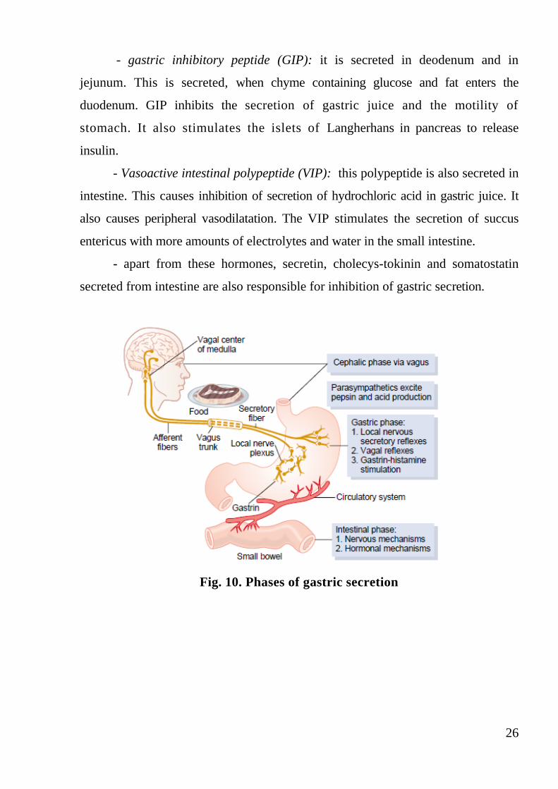
26
- gastric inhibitory peptide (GIP): it is secreted in deodenum and in
jejunum. This is secreted, when chyme containing glucose and fat enters the
duodenum. GIP inhibits the secretion of gastric juice and the motility of
stomach. It also stimulates the islets of Langherhans in pancreas to release
insulin.
- Vasoactive intestinal polypeptide (VIP): this polypeptide is also secreted in
intestine. This causes inhibition of secretion of hydrochloric acid in gastric juice. It
also causes peripheral vasodilatation. The VIP stimulates the secretion of succus
entericus with more amounts of electrolytes and water in the small intestine.
- apart from these hormones, secretin, cholecys-tokinin and somatostatin
secreted from intestine are also responsible for inhibition of gastric secretion.
Fig. 10. Phases of gastric secretion

27
Digestion in the small intestine. Role of pancreas
The small intestine receives not only chyme from the stomach but also
secretions from the liver and pancreas, which enter the digestive tract near the
junction of the stomach and small intestine. These secretions are so important to
the digestive processes of the small intestine that it is necessary to understand them
before continuing with intestinal physiology.
The human pancreas is located in close apposition to the duodenum. It
performs both endocrine and exocrine functions, but here we discuss only its
exocrine function. The exocrine pancreas is composed of numerous small, sac-like
dilatations called acini composed of a single layer of pyramidal acinar cells (Fig.
11). These cells are actively involved in the production of enzymes. Their
cytoplasm is filled with an elaborate system of ER and Golgi apparatus. Zymogen
granules are observed in the apical region of acinar cells. A few centroacinar cells
line the lumen of the acinus. In contrast to acinar cells, these cells lack an elaborate
ER and Golgi apparatus. Their major function seems to be modification of the
electrolyte composition of the pancreatic secretion.
Fig. 11. Structure of pancreatic acinus.
The acini empty their secretions into intercalated ducts, which join to form
intralobular and then interlobular ducts. The interlobular ducts empty into two
pancreatic ducts: a major duct, the duct of Wirsung, and a minor duct, the duct of

28
Santorini. The duct of Santorini enters the duodenum more proximally than the
duct of Wirsung. The pancreatic digestive enzymes are secreted by pancreatic
acini, and large volumes of sodium bicarbonate solution are secreted by the small
ductules and larger ducts leading from the acini. The combined product of enzymes
and sodium bicarbonate then flows through a long pancreatic duct of Wirsung,
which enters the duodenum usually together with the common bile duct. A ring of
smooth muscle, the sphincter of Oddi, surrounds the opening of these ducts in the
duodenum. The sphincter of Oddi not only regulates the flow of bile and pancreatic
juice into the duodenum but also prevents the reflux of intestinal contents into the
pancreatic ducts (fig. 12).
Fig.12. Anatomy of the pancreas
Properties and composition of pancreatic juice
Pancreatic juice is an alkaline with pH of 8 to 8.3, volume is 500 to 800
ml/day. Pancreatic juice contains 99.5% of water and 0.5% of solids. The solids are

29
the organic and inorganic substances. The organic substances are enzymes and other
organic substances.
Enzymes of Pancreatic Juice
Proteolytic Enzymes
1. Trypsin
2. Chymotrypsin
3. Carboxypeptidase A
4. Carboxypeptidase B
5. Nuclease
6. Elastase
7. Collagenase
Lipolytic Enzymes
1. Pancreatic lipase
2. Cholesterol ester hydrolase
3. Phospholipase A
4. Phospholipase B
Amylolytic Enzyme: pancreatic amylase
Inorganic Substances Present in Pancreatic Juice: sodium, calcium,
potassium, magnesium, bicarbonate, chloride, sulfate and phosphate. The
bicarbonate content is very high in pancreatic juice. It is about 110 to 150 mEq/ L
against the concentration of 24 mEq/L in plasma. This high concentration of
bicarbonate is responsible for the alkalinity of pancreatic juice.
Functions of pancreatic juice
Pancreatic juice has digestive functions and the neutralizing action.
30
Digestive functions of pancreatic juice
Pancreatic juice plays an important role in the digestion of proteins and lipids.
It also digests the carbohydrates.
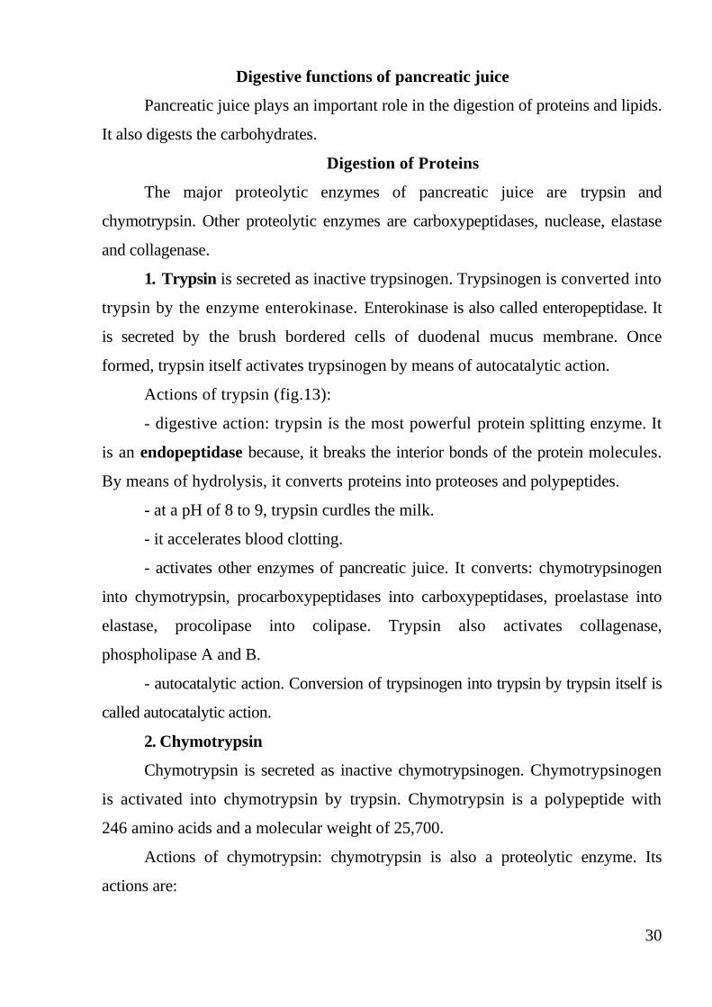
Digestion of Proteins
The major proteolytic enzymes of pancreatic juice are trypsin and
chymotrypsin. Other proteolytic enzymes are carboxypeptidases, nuclease, elastase
and collagenase.
1. Trypsin is secreted as inactive trypsinogen. Trypsinogen is converted into
trypsin by the enzyme enterokinase. Enterokinase is also called enteropeptidase. It
is secreted by the brush bordered cells of duodenal mucus membrane. Once
formed, trypsin itself activates trypsinogen by means of autocatalytic action.
Actions of trypsin (fig.13):
- digestive action: trypsin is the most powerful protein splitting enzyme. It
is an endopeptidase because, it breaks the interior bonds of the protein molecules.
By means of hydrolysis, it converts proteins into proteoses and polypeptides.
- at a pH of 8 to 9, trypsin curdles the milk.
- it accelerates blood clotting.
- activates other enzymes of pancreatic juice. It converts: chymotrypsinogen
into chymotrypsin, procarboxypeptidases into carboxypeptidases, proelastase into
elastase, procolipase into colipase. Trypsin also activates collagenase,
phospholipase A and B.
- autocatalytic action. Conversion of trypsinogen into trypsin by trypsin itself is
called autocatalytic action.
2. Chymotrypsin
Chymotrypsin is secreted as inactive chymotrypsinogen. Chymotrypsinogen
is activated into chymotrypsin by trypsin. Chymotrypsin is a polypeptide with
246 amino acids and a molecular weight of 25,700.
Actions of chymotrypsin: chymotrypsin is also a proteolytic enzyme. Its
actions are:

31
- digestive action on proteins: this is also an endopeptidase. It hydrolyses the
proteins into polypeptides.
- digestion of milk: digests casein faster than trypsin. The combination of
both enzymes causes more rapid digestion of milk. Chymotrypsin does not affect
blood clotting.
Fig. 13. Trypsin and its action
3. Carboxypeptidases
The two carboxypeptidases are carboxypeptidase A and carboxypeptidase
B. Procarboxypeptidase A is the precursor of carboxypeptidase A.
Procarboxypeptidase B is the precursor for carboxypeptidase B. The procar-
boxypeptidases are activated into carboxypeptidases by trypsin.
Actions of carboxypeptidases: they break the terminal bond of protein
molecules. Therefore, these enzymes are called exopeptidases. The exopeptidases
act on polypeptides and other proteins and convert them into amino acids.
Carboxypeptidase A splits the proteins into amino acids having aromatic or
aliphatic side chains. Carboxypeptidase B converts the proteins into amino acids
having basic side chains.
4. Nucleases

32
The nucleases present in pancreatic juice are ribonuclease and
deoxyribonuclease, which are responsible for the digestion of nucleic acids. These
enzymes convert the ribonucleic acid and deoxyribonucleic acid into
mononucleotides.
5. Elastase
Proelastase is activated into elastase by trypsin. Elastase is an important
enzyme, which digests the elastic fibers.
6. Collagenase
This enzyme is also activated by trypsin. Collagenase causes digestion of
collagen.
Digestion of Lipids
The lipolytic enzymes present in pancreatic juice are pancreatic lipase,
phospholipase A and phospholipase B.
1. Pancreatic Lipase
Pancreatic lipase is a powerful lipolytic enzyme. It hydrolyses the natural fats
such as triglycerides. It converts triglycerides into monoglycerides and fatty acids.
The activity of pancreatic lipase is accelerated in the presence of bile. The optimum
pH required for activity of this enzyme is 7 to 9. Digestion of fat by pancreatic lipase
requires two more factors namely bile salts and colipase. Bile salts are responsible
for the emulsification of fat prior to their digestion. Colipase is a coenzyme necessary
for the pancreatic lipase to hydrolyze the dietary lipids. Procolipase is activated
into colipase by trypsin.
2. Cholesterol Ester Hydrolase
Cholesterol ester hydrolase or cholesterol esterase hydrolyses cholesterol
ester into free cholesterol and fatty acid.
3. Phospholipase A
This is activated by trypsin. Phospholipase A acts on the phospholipids and
convert them into lysophospholipids. It converts lecithin into lysolecithin and
cephalin into lysocephalin.

33
4. Phospholipase B
Phospholipase B is also activated by trypsin. This enzyme acts on
lysophospholipids, i.e. lysolecithin and lysocephalin and converts them into
phosphoryl choline and free fatty acids.
Digestion of Carbohydrates
Pancreatic amylase is the only amylolytic enzyme present in pancreatic juice. Its
action on carbohydrates is like that of salivary amylase. It converts starch into
maltose.
Mechanism of pancreatic secretion
Secretion of pancreatic enzymes. The pancreatic enzymes are synthesized
in ribosomes, which are attached to the endoplasmic reticulum of acinar cells in
pancreas. The raw materials for the synthesis of pancreatic enzymes are the amino
acids. The amino acids are derived from the blood. These enzymes synthesized in the
ribosomes are packed into different zymogen granules by Golgi apparatus and
stored in cytoplasm. When stimulated, the acinar cells release the zymogen
granules into the pancreatic duct. From the granules, the enzymes are liberated into
intestine where these enzymes are activated.
Secretion of Bicarbonate Ions
Although the enzymes of the pancreatic juice are secreted entirely by the
acini of the pancreatic glands, the other two important components of pancreatic
juice, bicarbonate ions and water, are secreted mainly by the epithelial cells of the
ductules and ducts that lead from the acini. When the pancreas is stimulated to
secrete copious quantities of pancreatic juice, the bicarbonate ion concentration can
rise to as high as 145 mEq/L, a value about five times that of bicarbonate ions in
the plasma. This provides a large quantity of alkali in the pancreatic juice that
serves to neutralize the hydrochloric acid emptied into the duodenum from the
stomach.

34
The basic steps in the cellular mechanism for secreting sodium bicarbonate
solution into the pancreatic ductules and ducts are shown in Figure 14.They are the
following:
1. Carbon dioxide diffuses to the interior of the cell from the blood and,
under the influence of carbonic anhydrase, combines with water to form carbonic
acid (H2CO3). The carbonic acid in turn dissociates into bicarbonate ions and
hydrogen ions (HCO3-
and H+). Then the bicarbonate ions are actively transported
in association with sodium ions (Na+) through the luminal border of the cell into
the lumen of the duct.
2. The hydrogen ions formed by dissociation of carbonic acid inside the cell
are exchanged for sodium ions through the blood border of the cell by a secondary
active transport process. This supplies the sodium ions (Na+) that are transported
through the luminal border into the pancreatic duct lumen to provide electrical
neutrality for the secreted bicarbonate ions.
3. The overall movement of sodium and bicarbonate ions from the blood into
the duct lumen creates an osmotic pressure gradient that causes osmosis of water
also into the pancreatic duct, thus forming an almost completely isosmotic
bicarbonate solution.
Fig. 14. Secretion of isosmotic sodium bicarbonate solution by the pancreatic
ductules and ducts.

35
Neutralizing action of pancreatic juice. When acid chyme enters intestine
from stomach, immediately pancreatic juice with more bicarbonate is secreted and
released into intestine. Due to the presence of large quantity of bicarbonate ions, the
pancreatic juice is highly alkaline. Because of this, pancreatic juice neutralizes
acidity of chyme in the intestine. This is an important function, because, the acid
chyme can destroy the intestinal mucus membrane. Thus, the pancreatic juice
protects the intestine from destructive action of acid chyme.
Regulation of Pancreatic Secretion
Basic Stimuli That Cause Pancreatic Secretion. Three basic stimuli are
important in causing pancreatic secretion:
1. Acetylcholine, which is released from the parasympathetic vagus nerve
endings and from other cholinergic nerves in the enteric nervous system.
2. Cholecystokinin, which is secreted by the duodenal and upper jejunal
mucosa when food enters the small intestine.
3. Secretin, which is also secreted by the duodenal and jejunal mucosa when
highly acid food enters the small intestine.
The first two of these stimuli, acetylcholine and cholecystokinin, stimulate
the acinar cells of the pancreas, causing production of large quantities of pancreatic
digestive enzymes but relatively small quantities of water and electrolytes to go
with the enzymes. Without the water, most of the enzymes remain temporarily
stored in the acini and ducts until more fluid secretion comes along to wash them
into the duodenum. Secretin, in contrast to the first two basic stimuli, stimulates
secretion of large quantities of water solution of sodium bicarbonate by the
pancreatic ductal epithelium.
Phases of Pancreatic Secretion. Pancreatic secretion occurs in three phases,
the same as for gastric secretion: the cephalic phase, the gastric phase, and the
intestinal phase. Their characteristics are as follows.
Cephalic phase. During the cephalic phase of pancreatic secretion, the same
nervous signals from the brain that cause secretion in the stomach also cause
36
acetylcholine release by the vagal nerve endings in the pancreas. This causes
moderate amounts of enzymes to be secreted into the pancreatic acini, accounting
for about 20 per cent of the total secretion of pancreatic enzymes after a meal. But
little of the secretion flows immediately through the pancreatic ducts into the
intestine because only small amounts of water and electrolytes are secreted along
with the enzymes.
Gastric phase. When food enters stomach, the nervous mechanism may
continue. However, the important factor in this phase is the hormonal mechanism.
The hormone involved is gastrin. Gastrin is the gastrointestinal hormone secreted
from stomach when food enters the stomach. The gastrin is transported by blood, and
while reaching the pancreas, it causes release of the pancreatic juice. During the
gastric phase, the nervous stimulation of enzyme secretion continues, accounting
for another 5 to 10 per cent of pancreatic enzymes secreted after a meal. But,
again, only small amounts reach the duodenum because of continued lack of
significant fluid secretion.
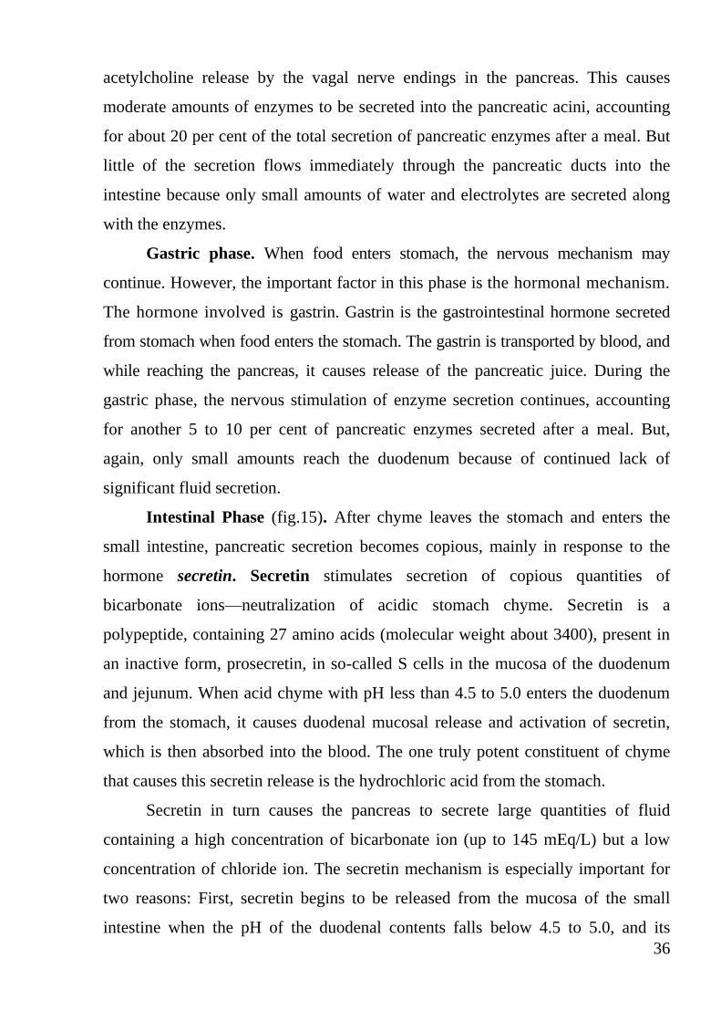
Intestinal Phase (fig.15). After chyme leaves the stomach and enters the
small intestine, pancreatic secretion becomes copious, mainly in response to the
hormone secretin. Secretin stimulates secretion of copious quantities of
bicarbonate ions—neutralization of acidic stomach chyme. Secretin is a
polypeptide, containing 27 amino acids (molecular weight about 3400), present in
an inactive form, prosecretin, in so-called S cells in the mucosa of the duodenum
and jejunum. When acid chyme with pH less than 4.5 to 5.0 enters the duodenum
from the stomach, it causes duodenal mucosal release and activation of secretin,
which is then absorbed into the blood. The one truly potent constituent of chyme
that causes this secretin release is the hydrochloric acid from the stomach.
Secretin in turn causes the pancreas to secrete large quantities of fluid
containing a high concentration of bicarbonate ion (up to 145 mEq/L) but a low
concentration of chloride ion. The secretin mechanism is especially important for
two reasons: First, secretin begins to be released from the mucosa of the small
intestine when the pH of the duodenal contents falls below 4.5 to 5.0, and its

37
release increases greatly as the pH falls to 3.0. This immediately causes copious
secretion of pancreatic juice containing abundant amounts of sodium bicarbonate.
The net result is then the following reaction in the duodenum:
HCl +NaHCO3→NaCl +H2CO3
Then the carbonic acid immediately dissociates into carbon dioxide and
water. The carbon dioxide is absorbed into the blood and expired through the
lungs, thus leaving a neutral solution of sodium chloride in the duodenum. In this
way, the acid contents emptied into the duodenum from the stomach become
neutralized, so that further peptic digestive activity by the gastric juices in the
duodenum is immediately blocked. Because the mucosa of the small intestine
cannot withstand the digestive action of acid gastric juice, this is an essential
protective mechanism to prevent development of duodenal ulcers.
Fig. 15. Regulation of pancreatic secretion
Bicarbonate ion secretion by the pancreas provides an appropriate pH for
action of the pancreatic digestive enzymes, which function optimally in a slightly
38
alkaline or neutral medium, at a pH of 7.0 to 8.0. Fortunately, the pH of the sodium
bicarbonate secretion averages 8.0.
Cholecystokinin—its contribution to control of digestive enzyme secretion
by the pancreas. The presence of food in the upper small intestine also causes a
second hormone, cholecystokinin, a polypeptide containing 33 amino acids, to be
released from yet another group of cells, the I cells, in the mucosa of the duodenum
and upper jejunum. This release of cholecystokinin results especially from the
presence of proteoses and peptones (products of partial protein digestion) and
long-chain fatty acids in the chyme coming from the stomach.
Cholecystokinin, like secretin, passes by way of the blood to the pancreas
but instead of causing sodium bicarbonate secretion causes mainly secretion of still
much more pancreatic digestive enzymes by the acinar cells. This effect is similar
to that caused by vagal stimulation but even more pronounced, accounting for 70
to 80 per cent of the total secretion of the pancreatic digestive enzymes after a
meal.

39
Functions of liver and biliary tree
Liver
Liver is both secretory and excretory organ. It is the largest gland in the body. It
weighs about 1.5 kg in man. It is located in the upper and right side of the abdominal
cavity immediately beneath diaphragm.
Liver is made up of liver cells called hepatocytes and a system of blood
vessels. Liver consists of many lobes. Each lobe consists of large number of lobules.
Each lobule is a honey comb like structure. The hepatocytes are arranged in
different plates. Each plate is one cell thick with a central vein. In between the cells,
are bile canaliculi. Each lobule is surrounded by portal triads. Each portal triad
consists of a branch of hepatic artery, a branch of portal vein and a tributary of bile
duct.
In between the plates, the sinusoids or blood spaces are present. The sinusoid
receives blood from a branch of portal vein and a branch of hepatic artery of the
portal triad. Sinusoids are lined by endothelial cells. Few macrophage cells
called Kupffer's cells are also found in between the endothelial cells.
Functions of liver:
1. metabolic function (it is the organ where maximum metabolic actions are
carried out);
2. storage function (many substances like glycogen, amino acids, iron, folic
acid and vitamins A, B12, and D are stored in liver);
3. synthetic function (liver produces glucose by gluconeogenesis. It
synthesizes all the plasma proteins. It synthesizes other proteins (except
immunoglobulins) such as clotting factors and complement factors, steroids,
hormone binding proteins, somatomedin and heparin);
4. secretion of bile
5. excretory function (liver excretes cholesterol, bile pigments, heavy
metals (like lead, arsenic and bismuth), toxins, bacteria like typhoid and virus
(like that of yellow fever).
6. heat production

40
7. hemopoietic function (in fetus the blood cells are produced in liver. It
stores vitamin B12 necessary for erythropoiesis and iron necessary for synthesis of
hemoglobin in red blood cells. Liver produces thrombopoietin that promotes
production of thrombocytes);
8. hemolytic function (the senile red blood cells after the life span of 120
days are destroyed by reticuloendothelial cells of the liver. The Kupffer's cells are the
reticuloendothelial cells in liver.
9. inactivation of hormones and drugs;
10. defensive and detoxification functions (the foreign bodies like
bacteria or antigens are swallowed and digested by reticuloendothelial cells of liver
by means of phagocytosis, liver cells are involved in removal of toxic property of
various harmful substances. The removal of toxic property of the harmful agent
is known as detoxification).
Physiologic Anatomy of Biliary Secretion
Bile is secreted in two stages by the liver:
(1) The initial portion is secreted by the principal functional cells of the
liver, the hepatocytes; this initial secretion contains large amounts of bile acids,
cholesterol, and other organic constituents. It is secreted into minute bile canaliculi
that originate between the hepatic cells
(2) Next, the bile flows in the canaliculi toward the interlobular septa, where
the canaliculi empty into terminal bile ducts and then into progressively larger
ducts, finally reaching the hepatic duct and common bile duct. From these the bile
either empties directly into the duodenum or is diverted for minutes up to several
hours through the cystic duct into the gallbladder, shown in Fig. 15.
In its course through the bile ducts, a second portion of liver secretion is
added to the initial bile. This additional secretion is a watery solution of sodium
and bicarbonate ions secreted by secretory epithelial cells that line the ductules and
ducts. This second secretion sometimes increases the total quantity of bile by as
much as an additional 100 per cent. The second secretion is stimulated especially

41
by secretin, which causes release of additional quantities of bicarbonate ions to
supplement the bicarbonate ions in pancreatic secretion (for neutralizing acid that
empties into the duodenum from the stomach).
Fig. 15. Bile is secreted by the liver, stored in the gallbladder and
ejected in the small intestine

42
Bile
Composition of Bile. By far the most abundant substances secreted in the
bile are bile salts, which account for about one half of the total solutes also in the
bile. Also secreted or excreted in large concentrations are bilirubin, cholesterol,
lecithin, and the usual electrolytes of plasma.
In the concentrating process in the gallbladder, water and large portions of
the electrolytes (except calcium ions) are reabsorbed by the gallbladder mucosa;
essentially all other constituents, especially the bile salts and the lipid substances
cholesterol and lecithin, are not reabsorbed and, therefore, become highly
concentrated in the gallbladder bile.
Bile serves two important functions:
First, bile plays an important role in fat digestion and absorption, not
because of any enzymes in the bile that cause fat digestion, but because bile acids
in the bile do two things: (1) they help to emulsify the large fat particles of the food
into many minute particles, the surface of which can then be attacked by lipase
enzymes secreted in pancreatic juice, and (2) they aid in absorption of the digested
fat end products through the intestinal mucosal membrane.
Second, bile serves as a means for excretion of several important waste
products from the blood. These include especially bilirubin, an end product of
hemoglobin destruction, and excesses of cholesterol.
Function of Bile Salts in Fat Digestion and Absorption
The liver cells synthesize about 6 grams of bile salts daily. The precursor of
the bile salts is cholesterol, which is either present in the diet or synthesized in the
liver cells during the course of fat metabolism. The cholesterol is first converted to
cholic acid or chenodeoxycholic acid in about equal quantities. These acids in
turn combine principally with glycine and to a lesser extent with taurine to form
glyco-and tauroconjugated bile acids. The salts of these acids, mainly sodium
salts, are then secreted in the bile.
The bile salts have two important actions in the intestinal tract:

43
1. They have a detergent action on the fat particles in the food. This
decreases the surface tension of the particles and allows agitation in the intestinal
tract to break the fat globules into minute sizes. This is called the emulsifying or
detergent function of bile salts.
2. Bile salts help in the absorption of (1) fatty acids, (2) monoglycerides, (3)
cholesterol, and (4) other lipids from the intestinal tract. They do this by forming
very small physical complexes with these lipids; the complexes are called micelles,
and they are semi-soluble in the chyme because of the electrical charges of the bile
salts. The intestinal lipids are “ferried” in this form to the intestinal mucosa, where
they are then absorbed into the blood. Without the presence of bile salts in the
intestinal tract, up to 40 per cent of the ingested fats are lost into the feces, and the
person often develops a metabolic deficit because of this nutrient loss.
3. Choliretic action: bile salts stimulate the secretion of bile from liver.
4. Cholagogue action: cholagogue is an agent, which increases the release of
bile from gallbladder into the intestine. Bile salts act as cholagogues indirectly by
stimulating the secretion of hormone CCK-PZ. This hormone causes contraction
of gallbladder and release of bile.
5. Laxative is an agent which induces defecation. Bile salts show this action
by stimulating peristaltic movements of the intestine.
6. Prevention of gallstone formation: Bile salts prevent the formation of
gallstone by keeping the cholesterol and lecithin in solution. In the absence of bile
salts, cholesterol precipitates along with lecithin and forms gallstone.
The Enterohepatic Circulation Recycles Bile Salts Between the Small
Intestine and the Liver
The enterohepatic circulation of bile salts is the recycling of bile salts
between the small intestine and the liver. The total amount of bile acids in the
body, primary or secondary, conjugated or free, at any time is defined as the total
bile acid pool. In healthy people, the bile acid pool ranges from 2 to 4g. The
enterohepatic circulation of bile acids in this pool is physiologically extremely
44
important. By cycling several times during a meal, a relatively small bile acid pool
can provide the body with sufficient amounts of bile salts to promote lipid
absorption. In a light eater, the bile acid pool may circulate 3 to 5 times a day; in a
heavy eater, it may circulate 14 to 16 times a day. The intestine is normally
extremely efficient in absorbing the bile salts by carriers located in the distal ileum.
Inflammation of the ileum can lead to their malabsorption and result in the loss of
large quantities of bile salts in the feces. Depending on the severity of illness,
malabsorption of fat may result.
Bile salts in the intestinal lumen are absorbed via four pathways (Fig. 16).
First, they are absorbed throughout the entire small intestine by passive diffusion,
but only a small fraction of the total amount of bile salts is absorbed in this
manner. Second, and most important, bile salts are absorbed in the terminal ileum
by an active carrier-mediated process, an extremely efficient process in which
usually less than 5% of the bile salts escape into the colon. Third, bacteria in the
terminal ileum and colon deconjugate the bile salts to form bile acids, which are
much more lipophilic than bile salts and, thus, can be absorbed passively. Fourth,
these same bacteria are responsible for transforming the primary bile acids to
secondary bile acids (deoxycholic and lithocholic acids) by dehydroxylation.
Deoxycholic acid may be absorbed, but lithocholic acid is poorly absorbed.
Although bile salt and bile acid absorption is extremely efficient, some salts
and acids are nonetheless lost with every cycle of the enterohepatic circulation.
About 500 mg of bile acids are lost daily. They are replenished by the synthesis of
new bile acids from cholesterol. The loss of bile acid in feces is, therefore, an
efficient way to excrete cholesterol.
Absorbed bile salts are transported in the portal blood bound to albumin or
high-density lipoproteins (HDLs). The uptake of bile salts by hepatocytes is
extremely efficient. In just one pass through the liver, more than 80% of the bile
salts in the portal blood is removed. Once taken up by hepatocytes, bile salts are
secreted into bile. The uptake of bile salts is a primary determinant of bile salt
secretion by the liver.

45
Fig. 16. The Enterohepatic Circulation of Bile Salts
The Liver Secretes Bile Pigments
The major pigment present in bile is the orange compound bilirubin, an end-
product of hemoglobin degradation in the monocyte-macrophage system in the
spleen, bone marrow, and liver (Fig. 17). Hemoglobin is first converted to
biliverdin with the release of iron and globin. Biliverdin is then converted into
bilirubin, which is transported in blood bound to albumin. The liver removes
bilirubin from the circulation rapidly and conjugates it with glucuronic acid. The
glucuronide is secreted into the bile canaliculi through an active carrier-mediated
process.
In the small intestine, bilirubin glucuronide is poorly absorbed. In the colon,
however, bacteria deconjugate it, and part of the bilirubin released is converted to
the highly soluble, colorless compound called urobilinogen. Urobilinogen can be
oxidized in the intestine to stercobilin or absorbed by the small intestine. It is
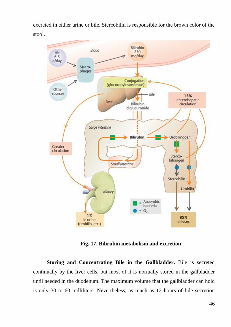
46
excreted in either urine or bile. Stercobilin is responsible for the brown color of the
stool.
Fig. 17. Bilirubin metabolism and excretion
Storing and Concentrating Bile in the Gallbladder. Bile is secreted
continually by the liver cells, but most of it is normally stored in the gallbladder
until needed in the duodenum. The maximum volume that the gallbladder can hold
is only 30 to 60 milliliters. Nevertheless, as much as 12 hours of bile secretion

47
(usually about 450 milliliters) can be stored in the gallbladder because water,
sodium, chloride, and most other small electrolytes are continually absorbed
through the gallbladder mucosa (fig. 18-1), concentrating the remaining bile
constituents that contain the bile salts, cholesterol, lecithin, and bilirubin.
Most of this gallbladder absorption is caused by active transport of sodium
through the gallbladder epithelium, and this is followed by secondary absorption of
chloride ions, water, and most other diffusible constituents. Bile is normally
concentrated in this way about 5-fold, but it can be concentrated up to a maximum
of 20-fold.
Gallbladder contraction is triggered by CCK, which binds to CCKA
receptors, and the neuronal plexus of the gallbladder wall, which is innervated by
preganglionic parasympathetic fibers of the vagus nerve (fig. 18-2). Calcitonin
gene-related peptide CGRP and substance P released by sensory fibers appear to
stimulate the gallbladder musculature indirectly by increasing acetylcholine
release. The sympathetic nervous system inhibits gallbladder contractions via α2
adrenoreceptors located on cholinergic fiber terminals. As cholagogues, fatty acids
and products of protein digestion as well as egg yolk and MgSO4 effectively
stimulate CCK secretion.
Fig. 18. Regulation of gallbladder emptying

48
Secretions of the Small Intestine
Secretion of Mucus by Brunner’s Glands in the Duodenum
An extensive array of compound mucous glands, called Brunner’s glands, is
located in the wall of the first few centimeters of the duodenum, mainly between
the pylorus of the stomach and the papilla of Vater where pancreatic secretion and
bile empty into the duodenum. These glands secrete large amounts of alkaline
mucus in response to (1) tactile or irritating stimuli on the duodenal mucosa; (2)
vagal stimulation, which causes increased Brunner’s glands secretion concurrently
with increase in stomach secretion; and (3) gastrointestinal hormones, especially
secretin.
The function of the mucus secreted by Brunner’s glands is to protect the
duodenal wall from digestion by the highly acid gastric juice emptying from the
stomach. In addition, the mucus contains a large excess of bicarbonate ions, which
add to the bicarbonate ions from pancreatic secretion and liver bile in neutralizing
the hydrochloric acid entering the duodenum from the stomach.
Brunner’s glands are inhibited by sympathetic stimulation; therefore, such
stimulation in very excitable persons is likely to leave the duodenal bulb
unprotected and is perhaps one of the factors that cause this area of the
gastrointestinal tract to be the site of peptic ulcers in about 50 per cent of ulcer
patients.
Secretion of Intestinal Digestive Juices by the Crypts of Lieberkühn
Located over the entire surface of the small intestineare small pits called
crypts of Lieberkühn, one of which is illustrated in figure 19. These crypts lie
between the intestinal villi. The surfaces of both the crypts and the villi are covered
by an epithelium composed of two types of cells: (1) a moderate number of goblet
cells, which secrete mucus that lubricates and protects the intestinal surfaces, and
(2) a large number of enterocytes, which, in the crypts, secrete large quantities of
water and electrolytes and, over the surfaces of adjacent villi, reabsorb the water
and electrolytes along with end products of digestion.

49
Fig. 19. A crypt of Lieberkühn
The intestinal secretions are formed by the enterocytes of the crypts at a rate
of about 1800 ml/day. These secretions are almost pure extracellular fluid and have
a slightly alkaline pH in the range of 7.5 to 8.0. The secretions also are rapidly
reabsorbed by the villi. This flow of fluid from the crypts into the villi supplies a
watery vehicle for absorption of substances from chyme when it comes in contact
with the villi. Thus, the primary function of the small intestine is to absorb
nutrients and their digestive products into the blood.
Mechanism of Secretion of the Watery Fluid. The exact mechanism that
controls the marked secretion of watery fluid by the crypts of Lieberkühn is not
known. It is believed to involve at least two active secretory processes: (1) active
secretion of chloride ions into the crypts and (2) active secretion of bicarbonate
ions. The secretion of both of these ions causes electrical drag as well of positively
charged sodium ions through the membrane and into the secreted fluid. Finally, all
these ions together cause osmotic movement of water.
Digestive Enzymes in the Small Intestinal Secretion. When secretions of
the small intestine are collected without cellular debris, they have almost no
enzymes. The enterocytes of the mucosa, especially those that cover the villi, do
contain digestive enzymes that digest specific food substances while they are being

50
absorbed through the epithelium. These enzymes are the following: (1) several
peptidases for splitting small peptides into amino acids, (2) four enzymes—
sucrase, maltase, isomaltase, and lactase—for splitting disaccharides into
monosaccharides, and (3) small amounts of intestinal lipase for splitting neutral
fats into glycerol and fatty acids.
The epithelial cells deep in the crypts of Lieberkühn continually undergo
mitosis, and new cells migrate along the basement membrane upward out of the
crypts toward the tips of the villi, thus continually replacing the villus epithelium
and also forming new digestive enzymes. As the villus cells age, they are finally
shed into the intestinal secretions. The life cycle of an intestinal epithelial cell is
about 5 days. This rapid growth of new cells also allows rapid repair of
excoriations that occur in the mucosa.
Regulation of Small Intestine Secretion—Local Stimuli
By far the most important means for regulating small intestine secretion are
local enteric nervous reflexes, especially reflexes initiated by tactile or irritative
stimuli from the chyme in the intestines.

51
Secretions of the Large Intestine
Mucus Secretion. The mucosa of the large intestine, like that of the small
intestine, has many crypts of Lieberkühn; however, unlike the small intestine, there
are no villi. The epithelial cells contain almost no enzymes. Instead, they consist
mainly of mucous cells that secrete only mucus. The great preponderance of
secretion in the large intestine is mucus. This mucus contains moderate amounts of
bicarbonate ions secreted by a few non–mucus-secreting epithelial cells. The rate
of secretion of mucus is regulated principally by direct, tactile stimulation of the
epithelial cells lining the large intestine and by local nervous reflexes to the
mucous cells in the crypts of Lieberkühn.
Stimulation of the pelvic nerves from the spinal cord, which carry
parasympathetic innervation to the distal one half to two thirds of the large
intestine, also can cause marked increase in mucus secretion. This occurs along
with increase in peristaltic motility of the colon.
During extreme parasympathetic stimulation, often caused by emotional
disturbances, so much mucus can occasionally be secreted into the large intestine
that the person has a bowel movement of ropy mucus as often as every 30 minutes;
this mucus often contains little or no fecal material.
Mucus in the large intestine protects the intestinal wall against excoriation,
but in addition, it provides an adherent medium for holding fecal matter together.
Furthermore, it protects the intestinal wall from the great amount of bacterial
activity that takes place inside the feces, and, finally, the mucus plus the alkalinity
of the secretion (pH of 8.0 caused by large amounts of sodium bicarbonate)
provides a barrier to keep acids formed in the feces from attacking the intestinal
wall.

52
Digestion and Absorption in the Gastrointestinal Tract
Digestion and absorption of carbohydrates
The digestion and absorption of dietary carbohydrates takes place in the
small intestine. These are extremely efficient processes, in that essentially all of the
carbohydrates consumed are absorbed. Carbohydrates are an extremely important
component of food intake, since they constitute about 45 to 50% of the typical
Europian diet and provide the greatest and least expensive source of energy.
Carbohydrates must be digested to monosaccharides before absorption.
The Diet Contains Both Digestible and Nondigestible Carbohydrates.
Humans can digest most carbohydrates; those we cannot digest constitute the
dietary fiber that forms roughage. Carbohydrate is present in food as
monosaccharides, disaccharides, oligosaccharides, and polysaccharides. The
monosaccharides are mainly hexoses (six-carbon sugars), and glucose is by far
the most abundant of these. Glucose is obtained directly from the diet or from the
digestion of disaccharides, oligosaccharides, or polysaccharides. The next most
common monosaccharides are galactose, fructose, and sorbitol. Galactose is
present in the diet only as milk lactose, a disaccharide composed of galactose and
glucose. Fructose is present in abundance in fruit and honey and is usually present
as disaccharides or polysaccharides. Sorbitol is derived from glucose and is almost
as sweet as glucose, but sorbitol is absorbed much more slowly and, thus,
maintains a high blood sugar level for a longer period when similar amounts are
ingested. It has been used as a weight-reduction aid to delay the onset of hunger
sensations.
The major disaccharides in the diet are sucrose, lactose, and maltose.
Sucrose, present in sugar cane and honey, is composed of glucose and fructose.
Lactose, the main sugar in milk, is composed of galactose and glucose. Maltose is
composed of two glucose units.
The digestible polysaccharides are starch, dextrins, and glycogen. Starch,
by far the most abundant carbohydrate in the human diet, is made of amylose and
amylopectin. Amylose is composed of a straight chain of glucose units;

53
amylopectin is composed of branched glucose units. Dextrins, formed from heating
(e.g., toasting bread) or the action of the enzyme amylase, are intermediate
products of starch digestion. Glycogen is a highly branched polysaccharide that
stores carbohydrates in the body. Normally, about 300 to 400 g of glycogen is
stored in the liver and muscle, with more stored in muscle than in the liver. Muscle
glycogen is used exclusively by muscle, and liver glycogen is used to provide
blood glucose during fasting.
Dietary fiber is made of polysaccharides that are usually poorly digested by
the enzymes in the small intestine. They have an extremely important
physiological function in that they provide the “bulk” that facilitates intestinal
motility and function. Many vegetables and fruits are rich in fibers, and their
frequent ingestion greatly decreases intestinal transit time.
Carbohydrates Are Digested in Different Parts of the GI Tract
The digestion of carbohydrates starts when food is mixed with saliva during
chewing. The enzyme salivary amylase acts on the α-1,4-glycosidic linkage of
amylose and amylopectin of polysaccharides to release the disaccharide maltose
and oligosaccharides maltotriose and α-limit dextrins (Fig. 20). Because salivary
amylase works best at neutral pH, its digestive action terminates rapidly after the
bolus mixes with acid in the stomach. However, if the food is thoroughly mixed
with amylase during chewing, a substantial amount of complex carbohydrates is
digested before this point. Pancreatic amylase continues the digestion of the
remaining carbohydrates. However, the chyme must first be neutralized by
pancreatic secretions, since pancreatic amylase works best at neutral pH. The
products of pancreatic amylase digestion of polysaccharides are also maltose,
maltotriose, and α-limit dextrins.
The digestion products of starch and glycogen, together with disaccharides
(sucrose and lactose), are further digested by enzymes located at the brush border
membrane of the enterocytes. The final products are glucose, fructose, and
galactose.

54
Fig. 20. The digestion products of starch after exposure
to salivary or pancreatic α-amylase.
Role of enterocytes in carbohydrate absorption and metabolism
Monosaccharides are absorbed by enterocytes either actively or by
facilitated transport. Glucose and galactose are absorbed via secondary active
transport by a symporter that transports two Na+ ions for every molecule of
monosaccharide (Fig. 21). The movement of Na+
into the cell, down concentration
and electrical gradients, effects the uphill movement of glucose into the cell. The
low intracellular Na+ concentration is maintained by the basolateral membrane
Na+/K
+-ATPase. Sugars accumulate in the cell at a higher concentration than in
plasma and leave the cell by Na+-independent facilitated transport or passive
diffusion through the basolateral cell membrane. Glucose and galactose share a
common transporter at the brush border membrane of enterocytes and, thus,
compete with each other during absorption.
Fructose is taken up by facilitated transport. Although facilitated transport is
carrier-mediated, it is not an active process. Fructose absorption is much slower
than glucose and galactose absorption and is not Na+-dependent.

55
The sugars absorbed by enterocytes are transported by the portal blood to the
liver where they are converted to glycogen or remain in the blood. After a meal,
the level of blood glucose rises rapidly, usually peaking at 30 to 60 minutes. The
concentration of glucose can be as high as 150 mg/dL.
Fig. 21. The enterocyte Na+-dependent carrier system for glucose and
galactose.
Dietary Fiber Plays an Important Role in GI Motility
Dietary fiber includes indigestible carbohydrates and carbohydrate- like
components mainly found in fruits and vegetables. The most common are
cellulose, hemicellulose, pectins, and gums. Cellulose and hemicellulose are
insoluble in water and are poorly digested by humans, thus, providing the
bulkiness of stool.
Dietary fiber imparts bulk to the bolus and, therefore, greatly shortens transit
time. It has been proposed that dietary fiber reduces the incidence of colon cancer
by shortening GI transit time, which, in turn, reduces the formation of carcinogenic
bile acids (e.g., lithocholic acid). Because dietary fiber also binds bile acids, which
are formed from cholesterol, fiber consumption can result in a lowering of blood
cholesterol by promoting excretion.

56
Digestion and absorption of lipids
Lipids are a concentrated form of energy. Lipids are also essential for
normal body functions, as they form part of cellular membranes and are precursors
of bile acids, steroid hormones, prostaglandins, and leukotrienes. The human body
is capable of synthesizing most of the lipids it requires with the exception of the
essential fatty acids linoleic acid and arachidonic acid. Both of these acids belong
to the family of omega-6 fatty acids. Recently, researchers have provided
convincing evidence that eicosapentaenoic acid and docosahexaenoic acid are
also essential for the normal development of vision in newborns. Both of these
acids are omega-3 fatty acids and are abundant in seafood and algae.
Lipids are comprised of several classes of compounds that are insoluble in
water but soluble in organic solvents. By far the most abundant dietary lipids are
triglycerides. They consist of a glycerol backbone esterified in the three positions
with fatty acids. More than 90% of the daily dietary lipid intake is in the form of
triglycerides.
The other lipids in the human diet are cholesterol and phospholipids.
Cholesterol is a sterol derived exclusively from animal fat. Humans also ingest a
small amount of plant sterols, notably β-sitosterol and campesterol. The
phospholipid molecule is similar to a triglyceride with fatty acids occupying the
first and second positions of the glycerol backbone (Fig. 27.23B). However, the
third position of the glycerol backbone is occupied by a phosphate group coupled
to a nitrogenous base (e.g., choline or ethanolamine), for which each type of
phospholipid molecule is named.
Bile serves as an endogenous source of cholesterol and phospholipids. Bile
contributes about 12 g/day of phospholipid to the intestinal lumen, most in the
form of phosphatidylcholine, whereas dietary sources contribute 2 to 3 g/day.
Another important endogenous source of lipid is desquamated intestinal villus
epithelial cells.

57
Role of Different Lipases in Lipid Hydrolysis
Lipid digestion mainly occurs in the lumen of the small intestine. Humans
secrete an overabundance of pancreatic lipase. Depending on the substrate being
digested, pancreatic lipase has an optimal pH of 7 to 8.0, allowing it to work well
in the intestinal lumen after the acidic contents from the stomach have been
neutralized by pancreatic HCO3-
secretion. Pancreatic lipase hydrolyzes the
triglyceride molecule to a 2-monoglyceride and two fatty acids (Fig. 22). It works
on the triglyceride molecule at the oil-water interface; thus, the rate of lipolysis
depends on the surface area of the interface. The products from the partial di-
gestion of dietary triglyceride by gastric lipase and the churning action of the
stomach produce a suspension of oil droplets (an emulsion) that help increase the
area of the oil-water interface. Pancreatic juice also contains the peptide colipase,
which is necessary for the normal digestion of fat by pancreatic lipase. Colipase
binds lipase at a molar ratio of 1:1, thereby allowing the lipase to bind to the oil-
water interface where lipolysis takes place. Colipase also counteracts the inhibition
of lipolysis by bile salt, which, despite its importance in intestinal fat absorption,
prevents the attachment of pancreatic lipase to the oil-water interface.
Fig. 22. The digestion of dietary lipids by pancreatic lipase
Phospholipase A2 is the major pancreatic enzyme for digesting
phospholipids, forming lysophospholipids and fatty acids. For instance,
phosphatidylcholine (lecithin) is hydrolyzed to form lysophosphatidylcholine
(lysolecithin) and fatty acid.
58
Dietary cholesterol is presented as a free sterol or as a sterol ester
(cholesterol ester). The hydrolysis of cholesterol ester is catalyzed by the
pancreatic enzyme cholesterol esterase. The digestion of cholesterol ester is
important because cholesterol can be absorbed only as the free sterol.
Bile Salt Plays an Important Role in Lipid Absorption
A layer of poorly stirred fluid called the unstirred water layer coats the
surface of the intestinal villi (Fig. 23A). The unstirred water layer reduces the
absorption of lipid digestion products because they are poorly soluble in water.
They are rendered water-soluble by micellar solubilization by bile salts in the
small intestinal lumen. This mechanism greatly enhances the concentration of these
products in the unstirred water layer (Fig. 23B). The lipid digestion products are
then absorbed by enterocytes, mainly by passive diffusion. Fatty acid and
monoglyceride molecules are taken up individually. Similar mechanisms seem to
operate for cholesterol and lysolecithin.
Bile salts are derived from cholesterol, but they are different from
cholesterol in that they are water-soluble. They are essentially detergents—
molecules that possess both hydrophilic and hydrophobic properties. Because bile
salts are polar molecules, they penetrate cell membranes poorly. This is significant
because it ensures their minimal absorption by the jejunum where most fat
absorption takes place. At or above a certain concentration of bile salts, the
critical micellar concentration, they aggregate to form micelles; the
concentration of luminal bile salts is usually well above the critical micellar
concentration. When bile salts alone are present in the micelle, it is called a simple
micelle. Simple micelles incorporate the lipid digestion products— monoglyceride
and fatty acids—to form mixed micelles. This renders the lipid digestion products
water-soluble by incorporation into mixed micelles. Mixed micelles diffuse across
the unstirred water layer and deliver lipid digestion products to the enterocytes for
absorption.

59
Fig. 23. Formation of micelles with the help of bile acids
Enterocytes Process Absorbed Lipid to Form Lipoproteins
After entering the enterocytes, the fatty acids and monoglycerides migrate to
the smooth ER. In the smooth ER, monoglycerides and fatty acids are rapidly
reconstituted to form triglycerides (Fig. 24). Fatty acids are first activated to form
acyl-CoA, which is then used to esterify monoglyceride to form diglyceride, which

60
is transformed into triglyceride. The lysolecithin absorbed by the enterocytes can
be reesterified in the smooth ER to form lecithin.
Cholesterol can be transported out of the enterocytes as free cholesterol or as
esterified cholesterol. The enzyme responsible for the esterification of cholesterol
to form cholesterol ester is acyl-CoA cholesterol acyltransferase (ACAT).
The reassembled triglycerides, lecithin, cholesterol, and cholesterol esters
are then packaged into lipoproteins and exported from the enterocytes. The
intestine produces two major classes of lipoproteins: chylomicrons and very low
density lipoproteins (VLDLs). Both are triglyceriderich lipoproteins with densities
less than 1.006 g/mL. Chylomicrons are made exclusively by the small intestine,
and their primary function is to transport the large amount of dietary fat absorbed
by the small intestine from the enterocytes to the lymph. Chylomicrons are large,
spherical lipoproteins with diameters of 80 to 500 nm. They contain less protein
and phospholipid than VLDLs and are, therefore, less dense than VLDLs. VLDLs
are made continuously by the small intestine during both fasting and feeding,
although the liver contributes significantly more VLDLs to the circulation.
Fig. 24. The intracellular metabolism of absorbed lipid digestion products to
form chylomicrons

61
Apoproteins—apo A-I, apo A-IV, and apo B—are among the major
proteins associated with the production of chylomicrons and VLDLs. Apo B is the
only protein that seems to be necessary for the normal formation of intestinal
chylomicrons and VLDLs. This protein is made in the small intestine. It has a
molecular weight of 250,000 and it is extremely hydrophobic.
Newly synthesized lipoproteins in the smooth ER are transferred to the
Golgi apparatus, where they are packaged in vesicles. Chylomicrons and VLDLs
are released into the intercellular space by exocytosis (Fig. 24). From there, they
are transferred to the central lacteals (the beginnings of lymphatic vessels) by a
process that is not well understood. Experimental evidence seems to indicate that
the transfer probably occurs mostly by diffusion. Intestinal lipid absorption is
associated with a marked increase in lymph flow called the lymphagogic effect of
fat feeding. This increase in lymph flow plays an important role in the transfer of
lipoproteins from the intercellular spaces to the central lacteal.
Fatty acids can also travel in the blood bound to albumin. While the most of
the long-chain fatty acids are transported from the small intestine as triglycerides
packaged in chylomicrons and VLDLs, some are transported in the portal blood
bound to serum albumin. Most of the medium-chain (8 to 12 carbons) and all of
the short-chain fatty acids are transported by the hepatic portal route.
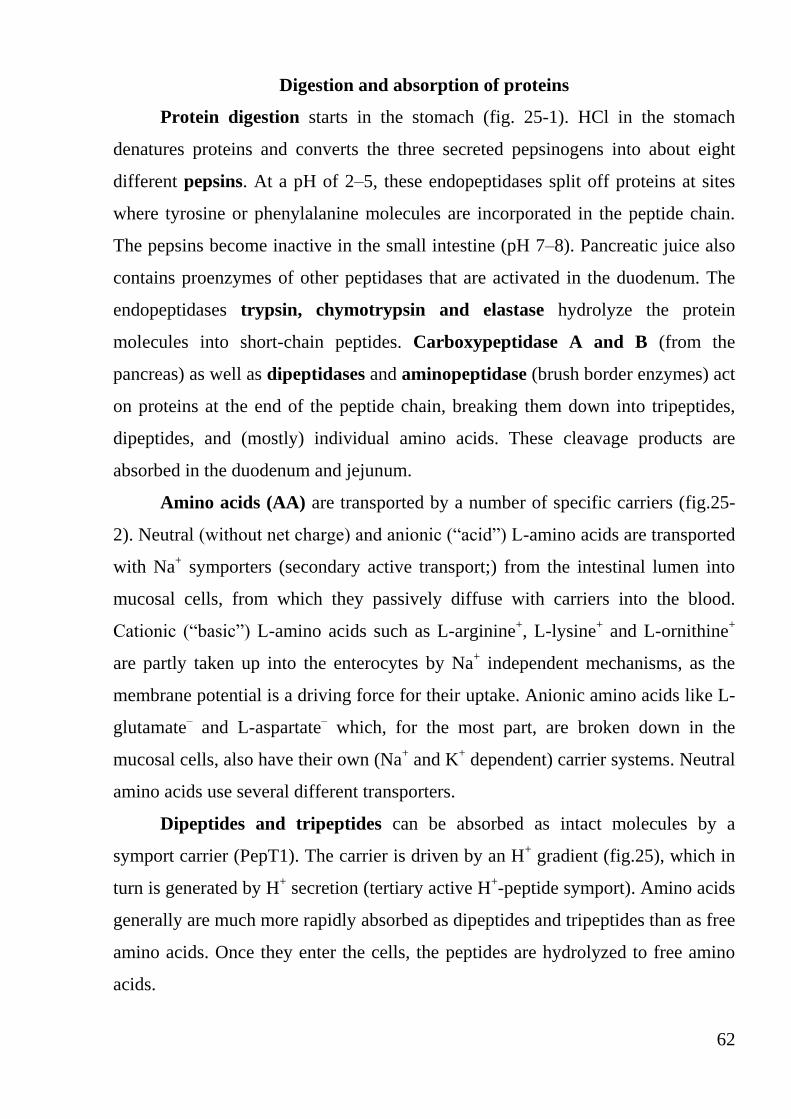
62
Digestion and absorption of proteins
Protein digestion starts in the stomach (fig. 25-1). HCl in the stomach
denatures proteins and converts the three secreted pepsinogens into about eight
different pepsins. At a pH of 2–5, these endopeptidases split off proteins at sites
where tyrosine or phenylalanine molecules are incorporated in the peptide chain.
The pepsins become inactive in the small intestine (pH 7–8). Pancreatic juice also
contains proenzymes of other peptidases that are activated in the duodenum. The
endopeptidases trypsin, chymotrypsin and elastase hydrolyze the protein
molecules into short-chain peptides. Carboxypeptidase A and B (from the
pancreas) as well as dipeptidases and aminopeptidase (brush border enzymes) act
on proteins at the end of the peptide chain, breaking them down into tripeptides,
dipeptides, and (mostly) individual amino acids. These cleavage products are
absorbed in the duodenum and jejunum.
Amino acids (AA) are transported by a number of specific carriers (fig.25-
2). Neutral (without net charge) and anionic (“acid”) L-amino acids are transported
with Na+ symporters (secondary active transport;) from the intestinal lumen into
mucosal cells, from which they passively diffuse with carriers into the blood.
Cationic (“basic”) L-amino acids such as L-arginine+, L-lysine
+ and L-ornithine
+
are partly taken up into the enterocytes by Na+ independent mechanisms, as the
membrane potential is a driving force for their uptake. Anionic amino acids like L-
glutamate– and L-aspartate
– which, for the most part, are broken down in the
mucosal cells, also have their own (Na+ and K
+ dependent) carrier systems. Neutral
amino acids use several different transporters.
Dipeptides and tripeptides can be absorbed as intact molecules by a
symport carrier (PepT1). The carrier is driven by an H+ gradient (fig.25), which in
turn is generated by H+ secretion (tertiary active H
+-peptide symport). Amino acids
generally are much more rapidly absorbed as dipeptides and tripeptides than as free
amino acids. Once they enter the cells, the peptides are hydrolyzed to free amino
acids.

63
Fig. 25. Protein digestion and absorption of amino acids

64
Water and Mineral Absorption
GI absorption of water occurs mainly in the jejunum and ileum, with
smaller quantities being absorbed by the colon (fig. 26). Water is driven through
the intestinal epithelium by osmosis. When solutes (Na+, Cl
–, etc.) are absorbed in
the intestine, water follows (fig. 27). Conversely, the secretion of substances into
the lumen or the ingestion of non-absorbable substances leads to water fluxes into
the lumen. Poorly absorbable substances therefore act as laxatives (e.g. sulfate,
sorbitol, polyethylene glycol).
Fig. 26. Water and electrolyte absorption in the gut
Water absorption is mainly driven by the absorption of Na+, Cl
– and
organic compounds. The luminal concentration of Na+ and Cl
– steadily decreases
from the duodenum to the colon. Na+ is absorbed by various mechanisms, and the
Na+-K+-ATPase on the basolateral cell membrane is the primary driving
mechanism for all of them (Fig. 27, 28).
Symport of Na+ and organic substances. Na
+ passively influxes into cells
of the duodenum and jejunum via symporter carriers, which actively cotransport
glucose, amino acids, phosphates and other compounds (secondary active
transport; fig.28-1). Since this is an electrogenic transport mechanism, a lumen-
negative transepithelial potential that drives Cl– out of the lumen forms (fig. 28-2).
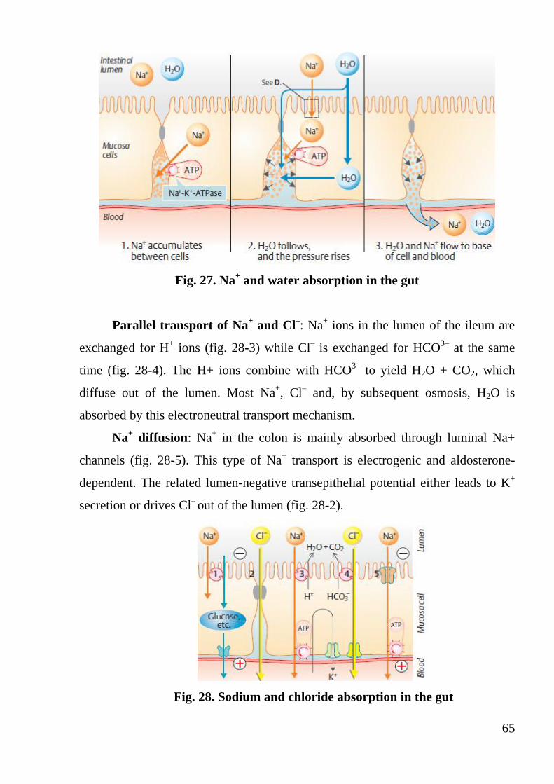
65
Fig. 27. Na+ and water absorption in the gut
Parallel transport of Na+ and Cl
–: Na
+ ions in the lumen of the ileum are
exchanged for H+ ions (fig. 28-3) while Cl
– is exchanged for HCO
3– at the same
time (fig. 28-4). The H+ ions combine with HCO3–
to yield H2O + CO2, which
diffuse out of the lumen. Most Na+, Cl
– and, by subsequent osmosis, H2O is
absorbed by this electroneutral transport mechanism.
Na+ diffusion: Na
+ in the colon is mainly absorbed through luminal Na+
channels (fig. 28-5). This type of Na+ transport is electrogenic and aldosterone-
dependent. The related lumen-negative transepithelial potential either leads to K+
secretion or drives Cl– out of the lumen (fig. 28-2).
Fig. 28. Sodium and chloride absorption in the gut

66
General Principles of Gastrointestinal Motility
Electrical Activity of Gastrointestinal Smooth Muscle
The individual smooth muscle fibers in the gastrointestinal tract are 200 to
500 micrometers in length and 2 to 10 micrometers in diameter, and they are
arranged in bundles of as many as 1000 parallel fibers. In the longitudinal muscle
layer, the bundles extend longitudinally down the intestinal tract; in the circular
muscle layer, they extend around the gut.
Within each bundle, the muscle fibers are electrically connected with one
another through large numbers of gap junctions that allow low-resistance
movement of ions from one muscle cell to the next. Therefore, electrical signals
that initiate muscle contractions can travel readily from one fiber to the next within
each bundle but more rapidly along the length of the bundle than sideways.
Each bundle of smooth muscle fibers is partly separated from the next by
loose connective tissue, but the muscle bundles fuse with one another at many
points, so that in reality each muscle layer represents a branching latticework of
smooth muscle bundles. Therefore, each muscle layer functions as a syncytium;
that is, when an action potential is elicited anywhere within the muscle mass, it
generally travels in all directions in the muscle. The distance that it travels depends
after only a few millimeters and at other times it travels many centimeters or even
the entire length and breadth of the intestinal tract.
Also, a few connections exist between the longitudinal and circular muscle
layers, so that excitation of one of these layers often excites the other as well.
The smooth muscle of the gastrointestinal tract is excited by almost
continual slow, intrinsic electrical activity along the membranes of the muscle
fibers. This activity has two basic types of electrical waves: (1) slow waves and (2)
spikes (fig. 29). In addition, the voltage of the resting membrane potential of the
gastrointestinal smooth muscle can be made to change to different levels, and this
too can have important effects in controlling motor activity of the gastrointestinal
tract.

67
Slow Waves. Most gastrointestinal contractions occur rhythmically, and this
rhythm is determined mainly by the frequency of so-called “slow waves” of
smooth muscle membrane potential. These waves are not action potentials. Instead,
they are slow, undulating changes in the resting membrane potential. Their
intensity usually varies between 5 and 15 millivolts, and their frequency ranges in
different parts of the human gastrointestinal tract from 3 to 12 per minute: about 3
in the body of the stomach, as much as 12 in the duodenum, and about 8 or 9 in the
terminal ileum. Therefore, the rhythm of contraction of the body of the stomach
usually is about 3 per minute, of the duodenum about 12 per minute, and of the
ileum 8 to 9 per minute.
Fig. 29. Types of electrical activity in intestinal smooth muscle
The precise cause of the slow waves is not completely understood, although
they appear to be caused by complex interactions among the smooth muscle cells
and specialized cells, called the interstitial cells of Cajal, that are believed to act
as electrical pacemakers for smooth muscle cells. These interstitial cells form a
network with each other and are interposed between the smooth muscle layers,
with synaptic-like contacts to smooth muscle cells. The interstitial cells of Cajal
undergo cyclic changes in membrane potential due to unique ion channels that

68
periodically open and produce inward (pacemaker) currents that may generate slow
wave activity.
The slow waves usually do not by themselves cause muscle contraction in
most parts of the gastrointestinal tract, except perhaps in the stomach. Instead, they
mainly excite the appearance of intermittent spike potentials, and the spike
potentials in turn actually excite the muscle contraction.
Spike Potentials. The spike potentials are true action potentials. They occur
automatically when the resting membrane potential of the gastrointestinal smooth
muscle becomes more positive than about -40 millivolts (the normal resting
membrane potential in the smooth muscle fibers of the gut is between -50 and -60
millivolts).Thus, each time the peaks of the slow waves temporarily become more
positive than -40 millivolts, spike potentials appear on these peaks. The higher the
slow wave potential rises, the greater the frequency of the spike potentials, usually
ranging between 1 and 10 spikes per second. The spike potentials last 10 to 40
times as long in gastrointestinal muscle as the action potentials in large nerve
fibers, each gastrointestinal spike lasting as long as 10 to 20 milliseconds.
Another important difference between the action potentials of the
gastrointestinal smooth muscle and those of nerve fibers is the manner in which
they are generated. In nerve fibers, the action potentials are caused almost entirely
by rapid entry of sodium ions through sodium channels to the interior of the fibers.
In gastrointestinal smooth muscle fibers, the channels responsible for the
action potentials are somewhat different; they allow especially large numbers of
calcium ions to enter along with smaller numbers of sodium ions and therefore are
called calcium-sodium channels. These channels are much slower to open and
close than are the rapid sodium channels of large nerve fibers. The slowness of
opening and closing of the calcium-sodium channels accounts for the long duration
of the action potentials. Also, the movement of large amounts of calcium ions to
the interior of the muscle fiber during the action potential plays a special role in
causing the intestinal muscle fibers to contract.

69
Changes in Voltage of the Resting Membrane Potential. In addition to the
slow waves and spike potentials, the baseline voltage level of the smooth muscle
resting membrane potential also can change. Under normal conditions, the resting
membrane potential averages about -56 millivolts, but multiple factors can change
this level. When the potential becomes less negative, which is called
depolarization of the membrane, the muscle fibers become more excitable. When
the potential becomes more negative, which is called hyperpolarization, the fibers
become less excitable.
Factors that depolarize the membrane—that is, make it more
excitable—are 1) stretching of the muscle, 2) stimulation by acetylcholine, 3)
stimulation by parasympathetic nerves that secrete acetylcholine at their endings,
and 4) stimulation by several specific gastrointestinal hormones.
Important factors that make the membrane potential more negative—
that is, hyperpolarize the membrane and make the muscle fibers less excitable—
are 1) the effect of norepinephrine or epinephrine on the fiber membrane and 2)
stimulation of the sympathetic nerves that secrete mainly norepinephrine at their
endings.
Calcium Ions and Muscle Contraction. Smooth muscle contraction occurs
in response to entry of calcium ions into the muscle fiber. Calcium ions, acting
through a calmodulin control mechanism, activate the myosin filaments in the
fiber, causing attractive forces to develop between the myosin filaments and the
actin filaments, thereby causing the muscle to contract.
The slow waves do not cause calcium ions to enter the smooth muscle fiber
(only sodium ions).Therefore, the slow waves by themselves usually cause no
muscle contraction. Instead, it is during the spike potentials, generated at the peaks
of the slow waves, that significant quantities of calcium ions do enter the fibers and
cause most of the contraction.

70
Neural Control of Gastrointestinal Function—Enteric Nervous System
The gastrointestinal tract has a nervous system all its own called the enteric
nervous system. It lies entirely in the wall of the gut, beginning in the esophagus
and extending all the way to the anus. The number of neurons in this enteric
system is about 100 million, almost exactly equal to the number in the entire spinal
cord. This highly developed enteric nervous system is especially important in
controlling gastrointestinal movements and secretion.
The enteric nervous system is composed mainly of two plexuses (fig. 30): 1)
an outer plexus lying between the longitudinal and circular muscle layers, called
the myenteric plexus or Auerbach’s plexus, and 2) an inner plexus, called the
submucosal plexus or Meissner’s plexus, that lies in the submucosa.
Fig. 30. Neural control of the gut wall
The myenteric plexus controls mainly the gastrointestinal movements, and
the submucosal plexus controls mainly gastrointestinal secretion and local blood
flow.
The extrinsic sympathetic and parasympathetic fibers connect to both the
myenteric and submucosal plexuses. Although the enteric nervous system can

71
function on its own, independently of these extrinsic nerves, stimulation by the
parasympathetic and sympathetic systems can greatly enhance or inhibit
gastrointestinal functions.
Sensory nerve endings that originate in the gastrointestinal epithelium or gut
wall and send afferent fibers to both plexuses of the enteric system, as well as 1) to
the prevertebral ganglia of the sympathetic nervous system, 2) to the spinal cord,
and 3) in the vagus nerves all the way to the brain stem. These sensory nerves can
elicit local reflexes within the gut wall itself and still other reflexes that are relayed
to the gut from either the prevertebral ganglia or the basal regions of the brain.
Differences Between the Myenteric and Submucosal Plexuses. The
myenteric plexus consists mostly of a linear chain of many interconnecting
neurons that extends the entire length of the gastrointestinal tract.
Because the myenteric plexus extends all the way along the intestinal wall
and because it lies between the longitudinal and circular layers of intestinal smooth
muscle, it is concerned mainly with controlling muscle activity along the length of
the gut. When this plexus is stimulated, its principal effects are 1) increased tonic
contraction, or “tone,” of the gut wall, 2) increased intensity of the rhythmical
contractions, 3) slightly increased rate of the rhythm of contraction, and 4)
increased velocity of conduction of excitatory waves along the gut wall, causing
more rapid movement of the gut peristaltic waves.
The myenteric plexus should not be considered entirely excitatory because
some of its neurons are inhibitory; their fiber endings secrete an inhibitory
transmitter, possibly vasoactive intestinal polypeptide or some other inhibitory
peptide. The resulting inhibitory signals are especially useful for inhibiting some of
the intestinal sphincter muscles that impede movement of food along successive
segments of the gastrointestinal tract, such as the pyloric sphincter, which controls
emptying of the stomach into the duodenum, and the sphincter of the ileocecal
valve, which controls emptying from the small intestine into the cecum.
The submucosal plexus, in contrast to the myenteric plexus, is mainly
concerned with controlling function within the inner wall of each minute segment

72
of the intestine. For instance, many sensory signals originate from the
gastrointestinal epithelium and are then integrated in the submucosal plexus to help
control local intestinal secretion, local absorption, and local contraction of the
submucosal muscle that causes various degrees of infolding of the gastrointestinal
mucosa.
Types of Neurotransmitters Secreted by Enteric Neurons. In an attempt
to understand better the multiple functions of the gastrointestinal enteric nervous
system, research workers the world over have identified a dozen or more different
neurotransmitter substances that are released by the nerve endings of different
types of enteric neurons. Two of them with which we are already familiar are
acetylcholine and norepinephrine. Others are adenosine triphosphate, serotonin,
dopamine, cholecystokinin, substance P, vasoactive intestinal polypeptide,
somatostatin, leu-enkephalin, metenkephalin, and bombesin.
Acetylcholine most often excites gastrointestinal activity. Norepinephrine
almost always inhibits gastrointestinal activity. This is also true of epinephrine,
which reaches the gastrointestinal tract mainly by way of the blood after it is
secreted by the adrenal medullae into the circulation. The other aforementioned
transmitter substances are a mixture of excitatory and inhibitory agents.

73
Gastrointestinal Reflexes
The anatomical arrangement of the enteric nervous system and its
connections with the sympathetic and parasympathetic systems support three types
of gastrointestinal reflexes that are essential to gastrointestinal control. They are
the following:
1. Reflexes that are integrated entirely within the gut wall enteric
nervous system. These include reflexes that control much gastrointestinal
secretion, peristalsis, mixing contractions, local inhibitory effects, and so forth.
2. Reflexes from the gut to the prevertebral sympathetic ganglia and
then back to the gastrointestinal tract. These reflexes transmit signals long
distances to other areas of the gastrointestinal tract, such as signals from the
stomach to cause evacuation of the colon (the gastrocolic reflex), signals from the
colon and small intestine to inhibit stomach motility and stomach secretion (the
enterogastric reflexes), and reflexes from the colon to inhibit emptying of ileal
contents into the colon (the colonoileal reflex).
3. Reflexes from the gut to the spinal cord or brain stem and then back
to the gastrointestinal tract. These include especially 1) reflexes from the
stomach and duodenum to the brain stem and back to the stomach—by way of the
vagus nerves—to control gastric motor and secretory activity;
2) pain reflexes that cause general inhibition of the entire gastrointestinal
tract;
3) defecation reflexes that travel from the colon and rectum to the spinal
cord and back again to produce the powerful colonic, rectal, and abdominal
contractions required for defecation (the defecation reflexes).

74
Propulsion and Mixing of Food in the Alimentary Tract
Swallowing (deglutition)
1. Oral stage or first stage.
This stage occurs after mastication. During mastication, food is rolled into a
bolus. The passage of bolus through oral cavity into the pharynx occurs in this stage.
And, this is a voluntary stage.
First, this bolus is placed over posterodorsal surface of the tongue. This is
called the preparatory position. Now, the front part of tongue is retracted and
depressed. The posterior part of tongue is elevated and retracted against hard palate.
This makes the bolus to move back into the pharynx. The forceful contraction of
tongue against the palate produces a positive pressure in the posterior part of oral
cavity. This pressure in the oral cavity also pushes the food into pharynx to some
extent (fig. 31-1).
2. Pharyngeal stage or second stage
In the pharyngeal or the second stage of deglutition, the bolus is pushed from
pharynx into the esophagus. It is an involuntary stage. The pharynx is a common
passage for food and air. It divides into larynx and esophagus. Larynx lies anteriorly
and continues as respiratory passage. Esophagus lies behind the larynx and
continues as gastrointestinal tract. During this stage of swallowing, the bolus from
the pharynx can enter into four paths: 1) back into mouth; 2) upwards into
nasopharynx; 3) forwards into larynx and 4) downwards into esophagus.
The various movements are coordinated so that, bolus enters only into the
esophagus. The entrance of bolus through other paths is prevented as follows:
1. Return of bolus back into the mouth is prevented by: the position of tongue
against the soft palate (roof of the mouth) and the high intraoral pressure developed
by the movement of tongue.
2. The movement of bolus into the nasopharynx from pharynx is prevented
by elevation of soft palate along with its extension called uvula (fig. 31-2).
3. The movement of bolus into the larynx is prevented by the following
means (fig. 31-3):

75
- approximation of the vocal cords
- forward and upward movement of larynx
- the backward movements of epiglottis to seal the opening of the larynx.
Another important factor, which prevents entry of food into respiratory tract
during deglutition, is the temporary arrest of breathing. This occurs during the
second stage of swallowing. The temporary arrest of breathing is called the apnea.
And, apnea during deglutition is called the deglutition apnea or swallowing apnea.
Fig. 31. Mechanism of swallowing.
3. Entrance of bolus into esophagus.
Since the other three paths are closed for the bolus, it has to pass only
through the esophagus. This occurs by the combined effects of various factors given
below: the upward movement of the larynx stretches the opening of the esophagus,
simultaneously, the upper 3 to 4 cm of esophagus relaxes. This part of the esophagus
is formed by the cricopharyngeal muscle and it is called upper esophageal
sphincter or pharyngoesophageal sphincter (fig. 31-4). At the same time, the
peristaltic contractions start in the pharynx due to the contraction of pharyngeal
muscles. Elevation of larynx also lifts the glottis away from the food passage.
All the above-mentioned factors act together so that, the bolus moves easily
into the esophagus. The whole process takes place within 1 to 2 seconds. And this
process is purely involuntary.
3. Esophageal stage or third stage
This is also an involuntary stage. The function of esophagus is to transport
the food from the pharynx to the stomach. The movements of esophagus are

76
specifically organized for this function and the movements are called peristaltic
waves. Peristalsis means a wave of contraction followed by the wave of relaxation of
muscle fibers of Gl tract, which travel in aboral direction (away from mouth). By
this type of movement, the contents are propelled down along the Gl tract.
When bolus reaches the esophagus, the peristaltic waves are initiated,
which propel the bolus into the stomach (fig. 31-5). Usually, two types of peristaltic
contractions are produced in esophagus.
Primary Peristaltic Contractions. When bolus reaches the upper part of
esophagus, the peristalsis starts. This is known as primary peristalsis. After origin,
the peristaltic contractions pass down through the rest of the esophagus propelling
the food towards stomach.
The pressure developed during the primary peristaltic contractions is important
to propel the bolus. Initially, the pressure becomes negative in the upper part of
esophagus. This is due to the stretching of the closed esophagus by the elevation of
larynx. But, immediately the pressure becomes positive and increases up to 10 to 15
cm of H2O.
Secondary Peristaltic Contractions. The secondary peristaltic contractions
arise in esophagus locally due to the distention of upper esophagus by the bolus.
After origin, these contractions pass down like the primary contractions, producing a
positive pressure. If the primary peristaltic contractions are unable to propel the
bolus into the stomach, the secondary peristaltic contractions appear and push the
bolus into stomach.
Role of Lower Esophageal Sphincter. The distal 2 to 5 cm of esophagus
acts like a sphincter. And, it is called lower esophageal sphincter. When bolus enters
this part of the esophagus, this sphincter relaxes so that the contents enter the
stomach. Later, the sphincter contracts. The relaxation and contraction of sphincter
occur in sequence with the arrival of peristaltic contractions of esophagus.

77
Movements of stomach
1. Hunger contractions
The activities of smooth muscles of stomach are increased during gastric
digestion (when stomach is filled with food) and when the stomach is empty. The
movements of empty stomach are related to the sensations of hunger. So, these
movements are called the hunger contractions.
Hunger contractions are the peristaltic waves superimposed over the
contractions of gastric smooth muscle as a whole. This type of peristaltic waves is
different from the digestive peristaltic contractions. The digestive peristaltic
contractions usually occur in body and pyloric parts of the stomach. But, the
peristaltic contractions of empty stomach involve the entire stomach. The
hunger contractions are of three types:
- Type I Hunger Contractions. The type I hunger contractions are the first
contractions to appear in the empty stomach when the tone of the stomach is low.
The type I contractions are single contractions and each one lasts for 20 seconds.
The interval between the contractions is about 3 to 4 seconds. The tonus of the
stomach is not increased between the contractions. The pressure produced by these
contractions is about 5 cm of water.
- Type II Hunger Contractions. Type II hunger contractions occur if the food
intake is postponed even after the appearance of the type I contractions. Type II
hunger contractions occur when the tonus of stomach is stronger. These
contractions also last for 20 seconds. The pause between the contractions is
absent. The pressure produced by these contractions is 10-15 cm of water.
- Type III Hunger Contractions. Type III hunger contractions appear when
the hunger becomes severe. Otherwise called incomplete tetanus, these contractions
are rare in man as the intake of food usually occurs before the appearance of these
contractions. Type III contractions last for 1 to 5 minutes. The pressure rise is 10 to
20 cm of water. And the tonus is increased to a great extent.

78
When the stomach is empty, the type I contractions occur first followed by
type II. If food intake is still postponed, then type 111 contractions appear and, as
soon as food is consumed, hunger contractions disappear.
2. Receptive relaxation
Relaxation of the upper portion of the stomach when bolus enters the stomach
from esophagus is called receptive relaxation. It involves the fundus and upper part
of the body of stomach. Its significance is to accommodate the food without much
increase in pressure inside the stomach.
3. Peristalsis of stomach
When the stomach contains food, the peristaltic contraction or peristaltic wave
occurs regularly with a frequency of 3/minute. It starts from the lower part of the
body of stomach.
The contraction first appears as a slight indentation on the greater and lesser
curvatures and travels towards pylorus. The contraction becomes deeper while
traveling. And finally, it ends with the contraction of pyloric sphincter. Some of the
waves disappear before reaching the sphincter. Each peristaltic wave takes about
one minute to travel from the point of origin to the point of ending.
These contractions are called digestive peristalsis because of their
involvement in mixing of food with gastric juice, grinding of food particles and the
digestive activities.
Filling of stomach. While taking food, the food arranges itself in the stomach
in different layers. The first eaten food is placed against the greater curvature in
the fundus and body of the stomach. The successive layers of food particles lie nearer
the lesser curvature until the last portion of food eaten lies near the upper end of
lesser curvature adjacent to cardiac sphincter.
The liquid remains near the lesser curvature and flows towards the pyloric end
of the stomach along a V shaped groove. This groove is formed by the smooth muscle
and it is called magenstrasse. But, if a large quantity of fluid is taken, it flows
around the entire food mass and is distributed over the interior part of stomach
between wall of the stomach and food mass.

79
Emptying of stomach
The emptying of stomach contents into the intestine occurs due to the peristaltic
waves arising in pyloric part of the stomach and simultaneous relaxation of pyloric
sphincter. Various factors influence the emptying of stomach.
1. Volume of Gastric Content. For any type of meal, gastric emptying is
directly proportional to the volume. If the content of stomach is more, a large
amount is emptied into the intestine.
2. Consistency of Gastric Content. Emptying of the stomach is proportional to
the consistency of the contents. Liquids, particularly the inert liquids (which do not
stimulate the stomach) leave the stomach rapidly. Water is emptied into intestine as
soon as it is swallowed. But, the solids are moved out of stomach only after being
converted into fluid or semifluid. Normally, the undigested solid particles are not
easily emptied.
3. Chemical Composition. The chemical composition of the food also
plays an important role in the emptying of the stomach. The gastric content with
more carbohydrates leaves the stomach more rapidly than the content with proteins.
The proteins leave the stomach more rapidly than the fat. Thus, the fatty food
remains in stomach for a longer period.
4. pH of the Gastric Content. The strong acid content leaves the stomach slowly.
The emptying of such content is accelerated by neutralizing the acid. This has an important
function of protecting the duodenal mucosa from the acidity.
5. Osmolar Concentration of Gastric Content. The gastric content, which is
isotonic to blood, leaves the stomach rapidly than the hypotonic or hypertonic content.
Regulation of gastric emptying
The emptying of stomach stops mainly due to the inhibition of gastric motility.
The inhibition of motility of stomach is caused by both nervous and hormonal
factors. The nervous factor, which regulates the emptying of stomach, is the
enterogastric reflex.

80
Enterogastric Reflex. When the chyme enters the intestine, the gastric
muscle is inhibited and the motility stops leading to stoppage of emptying. This is
called enterogastric reflex. This reflex involves the vagus nerves.
Hormonal Factors. The major hormone, which controls the gastric emptying,
is enterogastrone. When an acid chyme enters the duodenum, the hormone
enterogastrone is released from the duodenal mucus membrane. When this
hormone enters the stomach through blood it inhibits the motility of stomach. The
other hormones, which inhibit the gastric motility and emptying, are VIP, GIP,
secretin and cholecystokinin.

81
Movements of small intestine
The movements of small intestine are essential for mixing the chyme with
digestive juices, propulsion of food and absorption. Four types of movements
occur in small intestine.
1) Mixing movements: segmentation movements and pendular movements
2) Propulsive movements: peristaltic movements and peristaltic rush
3) Peristalsis in fasting - Migrating motor complex
4) Movements of villi.
Mixing movements. The mixing movements of small intestine are
responsible for proper mixing of chyme with digestive juices like pancreatic
juice, bile and intestinal juice. The mixing movements of small intestine are
segmentation contractions and pendular movements.
Segmentation Contractions. The segmentation contractions are the
common type of movements of small intestine, which occur regularly or
irregularly but in a rhythmic fashion. So, these movements are also called
rhythmic segmentation contractions. The contractions occur at spaced intervals
along a section of intestine. The segment of the intestine involved in each
contraction is about 1 to 5 cm long. The segments of intestine in between the
contracted segments are relaxed. The length of the relaxed segments is same as
that of the contracted segments. All the segments (both contracted and relaxed
segments) give a ring like appearance resembling the chain of sausages. After
some time, the contracted segments are relaxed and the relaxed segments are
contracted (fig. 32). Therefore, the segmentation contractions chop the chyme
many times. This helps in mixing of chyme with digestive juices.
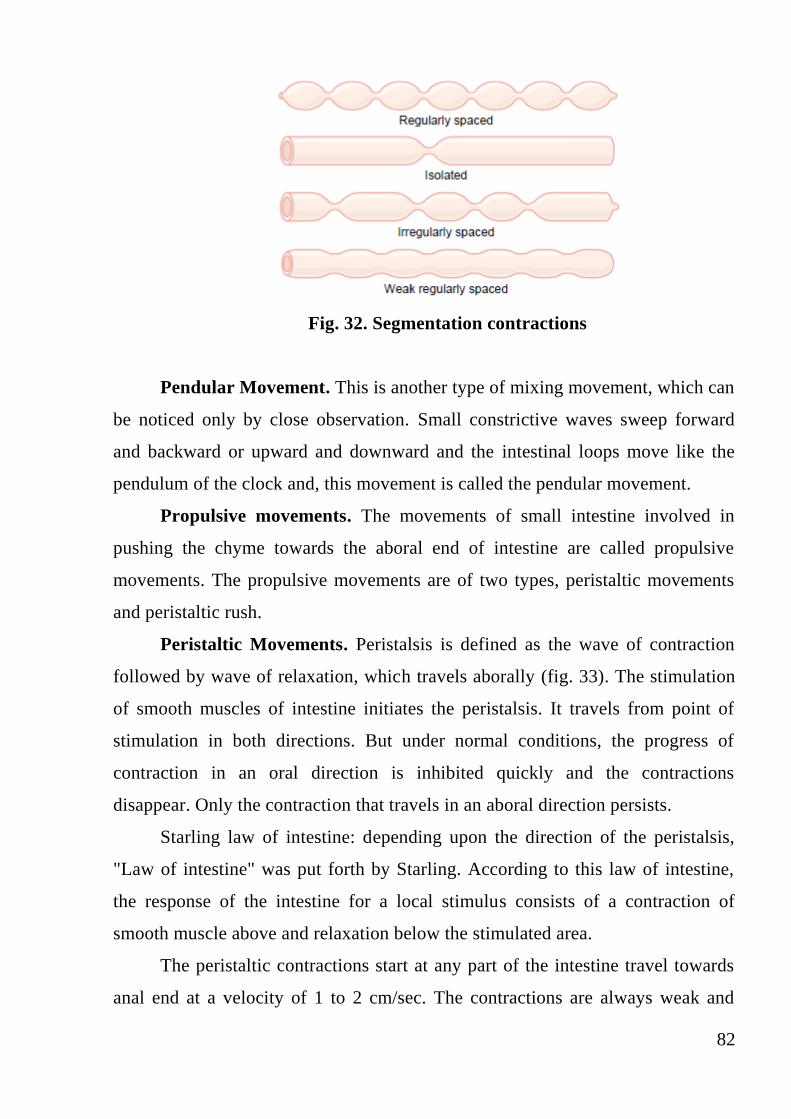
82
Fig. 32. Segmentation contractions
Pendular Movement. This is another type of mixing movement, which can
be noticed only by close observation. Small constrictive waves sweep forward
and backward or upward and downward and the intestinal loops move like the
pendulum of the clock and, this movement is called the pendular movement.
Propulsive movements. The movements of small intestine involved in
pushing the chyme towards the aboral end of intestine are called propulsive
movements. The propulsive movements are of two types, peristaltic movements
and peristaltic rush.
Peristaltic Movements. Peristalsis is defined as the wave of contraction
followed by wave of relaxation, which travels aborally (fig. 33). The stimulation
of smooth muscles of intestine initiates the peristalsis. It travels from point of
stimulation in both directions. But under normal conditions, the progress of
contraction in an oral direction is inhibited quickly and the contractions
disappear. Only the contraction that travels in an aboral direction persists.
Starling law of intestine: depending upon the direction of the peristalsis,
"Law of intestine" was put forth by Starling. According to this law of intestine,
the response of the intestine for a local stimulus consists of a contraction of
smooth muscle above and relaxation below the stimulated area.
The peristaltic contractions start at any part of the intestine travel towards
anal end at a velocity of 1 to 2 cm/sec. The contractions are always weak and

83
usually disappear after traveling for about few cm distance. Because of this, the
average movement of chyme through small intestine is very slow, and the
average velocity of movement of the chyme is < 1 cm/sec. Thus, the chyme
requires several hours to travel from duodenum to the end of small intestine.
The peristaltic waves in small intestine are increased very much
immediately after a meal. This is caused by gastroenteric reflex. The
gastroenteric reflex is initiated by the distention of stomach. And, the impulses
are conducted through myenteric plexus from stomach along the wall of the
intestine.
Fig. 33. Peristalsis
Peristaltic Rush. Sometimes, the small intestine shows a powerful peri-
staltic contraction. This is caused by excessive irritation of intestinal mucus
membrane or extreme distention of the intestine. This type of powerful
contraction begins in duodenum and passes through entire length of small
intestine and finally reaches the ileocecal valve within few minutes. This is called
peristaltic rush or rush waves. The peristaltic rush sweeps the contents of
intestine into colon. Thus, it relieves the small intestine off either irritants or
excessive distention.
Peristalsis in fasting-migrating motor complex. This is a type of
peristaltic contraction, which occurs in stomach and small intestine during the
long periods of fasting or several hours after the meals. It is different from the
regular peristalsis because, a large portion of stomach or intestine is involved in
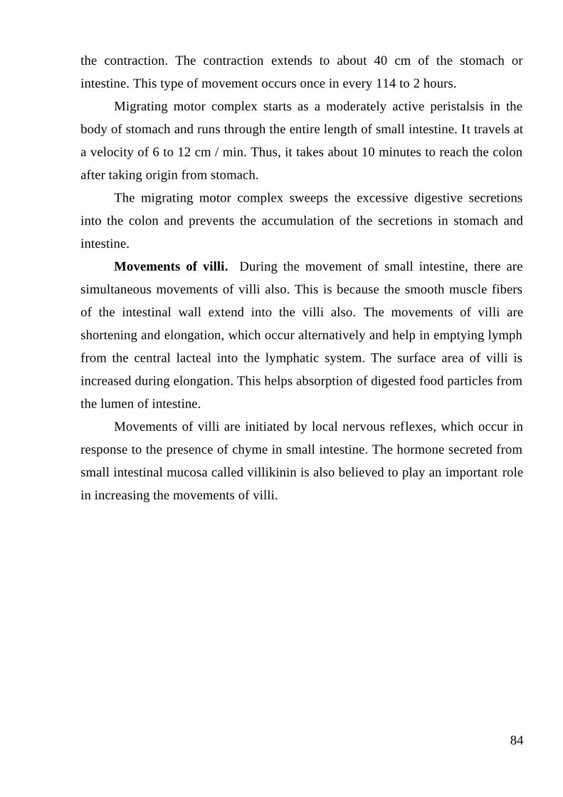
84
the contraction. The contraction extends to about 40 cm of the stomach or
intestine. This type of movement occurs once in every 114 to 2 hours.
Migrating motor complex starts as a moderately active peristalsis in the
body of stomach and runs through the entire length of small intestine. It travels at
a velocity of 6 to 12 cm / min. Thus, it takes about 10 minutes to reach the colon
after taking origin from stomach.
The migrating motor complex sweeps the excessive digestive secretions
into the colon and prevents the accumulation of the secretions in stomach and
intestine.
Movements of villi. During the movement of small intestine, there are
simultaneous movements of villi also. This is because the smooth muscle fibers
of the intestinal wall extend into the villi also. The movements of villi are
shortening and elongation, which occur alternatively and help in emptying lymph
from the central lacteal into the lymphatic system. The surface area of villi is
increased during elongation. This helps absorption of digested food particles from
the lumen of intestine.
Movements of villi are initiated by local nervous reflexes, which occur in
response to the presence of chyme in small intestine. The hormone secreted from
small intestinal mucosa called villikinin is also believed to play an important role
in increasing the movements of villi.
85
Movements of large intestine
The movements of large intestine are mostly sluggish. Still, these
movements are important for mixing, propulsive and absorptive functions. Two
types of movements occur in large intestine.
1. Mixing movements—Segmentation contractions
2. Propulsive movements—Mass peristalsis.
Segmentation contractions. Large circular constrictions, which appear in
the colon, are called mixing segmentation contractions. The length of the portion
of colon involved in each contraction is nearly about 2.5 cm. The contractions
occur at regular distance in colon.
Mass peristalsis. Mass peristalsis or mass movement propels the feces
from colon towards anus. Usually, this movement is developed only a few times
every day. The duration of the mass movement is about 10 minutes in the
morning before or after breakfast. This is because of the neurogenic factors like
gastrocolic reflex and parasympathetic stimulation.
86
Defecation
Voiding of feces is known as defecation. Feces is formed in the large
intestine and stored in sigmoid colon. By the influence of an appropriate
stimulus, it is expelled out through the anus. This is prevented by tonic
constriction of anal sphincters in the absence of the stimulus.
Defecation reflex. The mass movement drives the feces into sigmoid or
pelvic colon. In the sigmoid colon the feces is stored. The desire for defecation
occurs when some feces enters rectum due to the mass movement. Usually, the
desire for defecation is elicited by an increase in the intrarectal pressure to about
20 to 25 cm H2O.
The usual stimulus is intake of liquid like coffee or tea or water. But it
differs from person to person.
The act of defecation is preceded by voluntary efforts like assuming an
appropriate posture, voluntary relaxation of external sphincter and the
compression of abdominal contents by voluntary contraction of abdominal
muscles.
Usually, the rectum is empty. During the development of mass movement,
the feces is pushed into rectum and the defecation reflex is initiated. The
processes of defecation involves the contraction of rectum and relaxation of
internal and external anal sphincters.
The internal anal sphincter is made up of smooth muscle and it is
innervated by parasympathetic nerve fibers via pelvic nerve. The external anal
sphincter is composed of skeletal muscle and it is controlled by somatic nerve
fibers, which pass through pudendal nerve. The pudendal nerve always keeps the
external sphincter constricted and the sphincter can relax only when the pudendal
nerve is inhibited.
Usually, defecation occurs by the gastrocolic reflex mediated by intrinsic
nerves of gastrointestinal tract. In this, the distention of stomach by food causes
contraction of rectum followed by desire for defecation. However, this reflex
causes only a weak contraction of rectum. The strong contraction of rectum and

87
the relaxation of anal sphincters occur due to the reflex mediated by
parasympathetic nerves and the reflex center is in the sacral segment of spinal
cord (fig. 34).
When rectum is distended due to the entry of feces by mass movement,
sensory nerve endings are stimulated and the impulses are transmitted via afferent
fibers of pelvic nerve to sacral segments (center) of spinal cord. Spinal cord, in
turn sends motor impulses to the descending colon, sigmoid colon and rectum via
efferent nerve fibers of pelvic nerve. The efferent impulses cause strong
contraction of descending colon, sigmoid colon and rectum and relaxation of
internal sphincter. Simultaneously, voluntary relaxation of external sphincter
occurs. This is due to the inhibition of pudendal nerve by impulses arising from
cerebral cortex.
Fig. 34. Defecation reflex.

88
CONTROL QUESTIONS:
1. What are the different types of salivary glands? Describe the composition, functions
and regulation of secretion of saliva.
2. Explain the composition and functions of gastric juice. And, give an account of
hormonal regulation of gastric secretion.
3. Describe the different phases of gastric secretion with experimental evidences.
4. Explain the composition, functions and regulation of secretion of pancreatic juice.
5. Describe the composition, functions and regulation of secretion of bile. Enumerate
the differences between the liver bile and gallbladder bile. Add a
note on enterohepatic circulation.
6. Explain the mechanisms of absorption in GI tract.
7. Write an essay on gastric motility. What are the factors influencing gastric
emptying?
8. Describe in detail the gastrointestinal movements.

89
Task for initial independent training
Topic: Gastrointestinal function. Digestion and absorption of substances.
Digestion in the Mouth
1. During a sharp experiment an animal's chorda tympani was electrically irritated.
How does it influence the secretion of the parotid salivary gland?
A. Saliva is not secreted.
B. Little liquid saliva secreted.
C. Much liquid saliva secreted.
D. Little viscid saliva secreted.
E. Much viscid saliva secreted.
2. What kind of enzymes does Alimentary tract use for digestion of starch in the
mouth?
А. Lingual lipase
B. Chymotrypsins
C. ά-amylase
D. Lactase
E. Pepsins
3. A peripheral part of sympathetic fibers which innervate the sublingual salivary
gland of an experimental animal is irritated. How does it influence the secretion of
the sublingual salivary gland?
A. Much viscid saliva secreted.
B. Little liquid saliva secreted.
C. Saliva is not secreted.
D. Much liquid saliva secreted.
E. Little viscid saliva secreted.
4. A patient has got a chronic neuritis of the trigeminus. Which of the digestive
processes is considerably broken?

90
A. Mastication.
B. Salivation.
C. Formation of taste feeling.
D. Swallowing.
E. Formation of saliva.
5. Saliva decreases gingivitis and caries because it contains immunoglobulin
A and
A. lysozyme
B. mucus
C. salivary amylase
D. water
E. A and C
6. The salivary gland that produces primarily mucus is the … salivary gland.
А. parotid
B. labial
C. submandibular.
D. A and B
E. sublingual
7. The salivary gland which is mixed but produces more serous than mucous
secretions and whose duct opens inferiorly in the floor of the oral cavity next to the
frenulum of the tongue is the … salivary gland.
А. submandibular
B. sublingual
C. palatine
D. A and B
E. parotid

91
8. A surgeon makes an incision in the jejunum starting at the serosal surface and
ending in the lumen. What is the sequential order of bisected structures as the
scalpel passes through the intestinal wall?
A. Circular muscle → longitudinal muscle → submucous plexus
B. Longitudinal muscle → myenteric plexus → circular muscle
C. Myenteric plexus → circular muscle → longitudinal muscle
D. Network of interstitial cells of Cajal → longitudinal muscle → circular muscle
E. Longitudinal muscle → network of interstitial cells of Cajal → submucous
plexus
9. A mouse with a new genetic mutation is discovered not to have electrical slow
waves in the small intestine. What cell type is most likely affected by the
mutation?
A. Enteric neurons
B. Inhibitory motor neurons
C. Enterochromaffin cells
D. Interstitial cells of Cajal
E. Enteroendocrine cells
10. A 10-cm segment of small intestine is removed surgically and placed in a 370C
physiological solution containing tetrodotoxin. A stimulus at one end of the
segment evokes an action potential and an accompanying contraction that travels to
the opposite end of the segment. This finding is best explained by
A. Electrical slow waves
B. Varicose motor nerve fibers
C. Interstitial cells of Cajal
D. Functional electrical syncytial properties
E. Release of neurotransmitters
92
11. Lactase is a brush border enzyme involved in the digestion of lactose. The
digestion product or products of lactose are
A. Glucose
B. Glucose and galactose
C. Glucose and fructose
D. Galactose and fructose
E. Fructose
12. Maltase hydrolyzes maltose to form
A. Glucose
B. Glucose and galactose
C. Glucose and fructose
D. Galactose and fructose
E. Galactose
13. Which sugar is taken up by enterocytes by facilitated diffusion?
A. Glucose
B. Galactose
C. Fructose
D. Xylose
E. Sucrose
14. Dietary triglyceride is a major source of nutrient for the human body. It is
digested mostly in the intestinal lumen by pancreatic lipase to release
A. Lysophosphatidylcholine and fatty acids
B. Glycerol and fatty acids
C. Diglyceride and fatty acids
D. 2-Monoglyceride and fatty acids
E. Lysophosphatidylcholine and diglyceride

93
15. After a meal of pizza, dietary lipid is absorbed by the small intestine and
transported in the lymph mainly as:
A. VLDLs
B. Free fatty acids bound to albumin
C. Chylomicrons
D. LDLs
E. HDLs
16. The submucosal plexus, in contrast to the myenteric plexus, is mainly
concerned with
A. Local intestinal secretion
B. Local absorption
C. Local contraction of the submucosal muscle
D. Increased tonic contraction, or “tone,” of the gut wall
E. True answers are A, B and C
17. The myenteric plexus, is mainly concerned with:
A. Increased tonic contraction, or “tone,” of the gut wall
B. Increased intensity of the rhythmical contractions
C. Slightly increased rate of the rhythm of contraction
D. Increased velocity of conduction of excitatory waves along the gut wall
E. All answers are correct
18. The extrinsic sympathetic and parasympathetic fibers connect to both the
myenteric and submucosal plexuses. Stimulation by the parasympathetic system
can greatly enhance gastrointestinal functions because neurotransmitter released at
the nerve endings is:
A. Norepinephrine
B. Acetylcholine
C. VIP

94
D. substance P
E. Villikinin
19. The extrinsic sympathetic and parasympathetic fibers connect to both the
myenteric and submucosal plexuses. Stimulation by the sympathetic system can
greatly inhibit gastrointestinal functions because neurotransmitter released at the
nerve endings is:
A. Norepinephrine
B. Acetylcholine
C. VIP
D. substance P
E. Villikinin
20. Absorption of glucose in the small intestine occurs with the help of:
A. Primary-active transport
B. Simple diffusion
C. Secondary-active transport
D. Osmosis
E. Facilitated diffusion
ANSWERS
1 C 6 E 11 B 16 E
2 C 7 A 12 A 17 E
3 E 8 B 13 C 18 B
4 A 9 D 14 D 19 A
5 A 10 D 15 C 20 C

95
PRACTICAL SKILLS
Topic: Gastrointestinal function. Digestion and absorption of substances.
Digestion in the Mouth
TASK1. Draw and note at this figure the Mouth, the Salivary glands, the
Esophagus, the liver, all parts of the intestine, the Anus. Describe their
function.
TASK 2. Draw and note at this figure the Myenteric plexus, the Submucosal
plexus, Epithelium. Explain and describe their function. To prevertebral ganglia,
Spinal cord and brai stem Sympathetic Parasypathetic
(mainly postganglionic) ( preganglionic)
TASK 3. Draw and note at figure the Capillary, the Basement membrane, the
Endoplasmic reticulum, the Goldgy apparatus, the Nerve fiber, the

96
Mitochondria, the Ribosomes and the Zymogen granules. Explain their
function
Typical glandular cell.
The TASK4. Draw this figure and write an explanation about the formation
and secretion of saliva by a salivary gland.
The TASK5. Studying of reaction on Mucin.
During 1-2 minutes rinse a mouth of distilled water of 20 ml. Repeat manipulation
3 times. Filter the collected saliva through a filtering paper. Add to 2 ml of a saliva

97
some drops of an acetic acid. After that Мucin drops out as a white sediment. And
the saliva loses viscosity.
RESEARCH PROBLEMS
1. Describe this research and write a conclusion on value of saliva’s Mucin.
The TASK6. Studying of saliva’s pH
Eat one sweet before performance of work. Then, collect 2 ml of a saliva in a test
tube. Place a strip of a display paper in a test tube with the help of a tweezers. Take
out a strip and immediately compare the received colouring to a scale.
RESEARCH PROBLEMS
1.Describe this research and write a conclusion by the received results.
The TASK8. Write the information about normal transport of substances by
the intestine and location of maximum absorption or secretion in this table
Small intestine
Absorption of Upper Mid Lower Colon
Sugars
Amino acids
Water-soluble
and fast- soluble
vitamins
Betaine,sarcosine
Antibodies in
newborns
Pyrimidines
Long-chain fatty
acid absorption
and conversion
to triglyceride
Bile salts
Vitamin B12
Na+
K+
Ca+
Fe+
Cl+

98
2-
SO4
TASK 7. Write the information in the table
source enzyme activator substrate Catalytic
function or products
Salivary ά-amylase
Lingual lipase
Pepsins
Gastric lipase
Trypsin
Chymotrypsins
Elastase
Carboxypeptidase
Colipase
Pancreatic lipase
Bile salt –acid lipase
Cholesteryl ester hydrolase
Pancreatic ά-amylase
Ribonuclease
Deoxyribonuclease
Phospholipase A2
Enteropeptidase
aminopeptidases
Carboxypeptidases
Endopeptidases
Dipeptidases
Maltase
Lactase
Sucrase
Ά-Dextinase
Trehalase
Nuclease and related enzyms
Various peptidases

99
Task for initial independent training
Topic: Digestive functions of Stomach and Pancreas
1. 150 gr of meat broth was introduced into the gastric cavity of an experimental
clog through a probe. The content of which of the following substances will be
quickly increased in the dog's blood?
A. Vasointerstinal polypeptide.
B. Somatostatin.
C. Insulin.
D. Neurotensin.
E. Gastrin.
2. Deficit of what enzyme is the most often the reason for incomplete digestion of
fats in the digestive tract and the increase of the neutral fat quantity in excrements?
A. Hepatic lipase.
B. Gastric lipase.
C. Pancreatic lipase.
D. Intestinal lipase.
E. Enterokinase.
3. During a patient's examination the decrease of the motor and evacuatory
functions of the stomach was determined. The deficit of which of the following
substances can it be connected with?
A. Secretin.
B. Gastrin.
C. Adenosine.
D. Somatostatin.
E. Gastric-inhibiting peptide.

100
4. In the process of aging of a human being there is a decrease of the synthesis and
secretion of the pancreatic juice, a decrease of trypsin content in it. To the violation
of the splitting of what substances does it lead first of all?
A. Polysaccharides.
B. Phospholipids.
C. Proteins.
D. Nucleic acids.
E. Lipids.
5. A patient's duodenum is ablated. The decrease of the secretion of what hormone
will it cause?
A. Gastrin.
B. Cholecystokinin and secretin.
C. Histamine.
D. Gastrin and histamine.
E. Neurotensin.
6. A lean solution of hydrochloric acid was introduced into an experimental dog's
duodenum through a probe. The secretion increase of what hormone will it cause?
A. Gastrin.
B. Secretin.
C. Histamin.
D. Cholecystokinin.
E. Neurotensin.
7. Which of the following processes of a hungry man who sees tasty food will be
activated first of all?
A. Secretion of gastric juice.
B. Secretion of intestinal juice.
C. Colon motility.

101
D. Contraction of Oddi's sphincter.
E. Small intestine motor activity.
8. A person has got considerable violation in digestion of proteins, fats and
carbonhydrates. Reduced secretion of what digestive juice is the result of it?
A. Saliva.
B. Pancreatic.
C. Gastric.
D. Bile.
E. Intestinal.
9. A part of a patient's pancreas was ablated. What products is it necessary for him
to limit in his food ration?
A. Fruit.
B. Boiled vegetables.
C. Dairy produce.
D. Vegetables rich in proteins (beans, soy).
E. Fat meat, beef tea.
10. How many ml of Gastric juice does healthy Stomach produce a day?
А. 500- 600 ml
B. 700- 900 ml
C. 900- 1000 ml
D. 1200- 1500 ml
E. 2000- 2500 ml
11. What inactive enzymes does Pancreas produce for Proteins digestion?
А. lipase
B. Chymotrypsins
C. ά-amylase

102
D. Trypsinogens
E. Pepsins
12. Absence of intrinsic factor in gastric juice causes deficiency of …….
А. Lipase
B. Chymotrypsins
C. vitamin B12
D. Cholesteryl ester hydrolase
E. Pepsins
13. Which one of the following cells in the gastric glands produce pepsinogen?
A. endocrine cells
B. mucous neck cells
C. parietal or oxyntic cells
D. all of the above
E. chief or zymogenic cells
14. What substance can inhibit hydrochloric acid secretion?
А. Gastrin
B. Enterogastrone and adrenaline
C. Histamine
D. acetylcholine
E. adrenaline
15. What inactive enzymes does stomach produce for proteins digestion?
А. lipase
B. Chymotrypsins
C. ά-amylase
D. Pepsinogens
E. Pepsins

103
16. The parietal cells in the gastric glands of the pyloric region produce … that
binds with Vitamin … to make it more readily absorbed in the ileum.
A. hydrochloric acid; B12
B. mucus; D
C. pepsinogen; D
D. intrinsic factor; B12
E. mucus; C
17. What substance can stimulate hydrochloric acid secretion?
А. Gastrin, Histamine
B. Enterogastrone
C. Histamine
D. adrenaline
E. Sympathetic stimulation
18. The major food digested by gastric secretions is
A. fat
B. starch
C. nucleic acids
D. nucleic acids and fat
E. protein
19. What substances stimulate the secretion of pancreatic juice during gastric
phase?
А. Gastrin
B. Enterogastrone
C. Secretin
D. Cholecystokinin
E. Pepsin

104
20. Secretin, which is produced in duodenum, acts on the pancreas, causing
production of pancreatic juice with large amount of:
A. enzymes
B. bicarbonate ions
C. magnesium ions
D. gastrin
E. pepsin
ANSWERS
1 E 8 B 15 D
2 C 9 E 16 D
3 B 10 D 17 A
4 C 11 D 18 E
5 B 12 C 19 A
6 B 13 E 20 B
7 A 14 B

105
PRACTICAL SKILLS
Topic: Digestion functions of Stomach and Pancreas
TASK1. Put information about the Stomach functions in this table.
# Stomach functions your explanation
1. Storage function
2. Mechanical function
3. Digestive function
4. Protective function
5. Hemopoetic function
6. Excretory function
TASK2. Two years ago Bob developed cancer and a doctor removed his
Pylorus. What happened with Stomach secretory function after that? Write
an explanation.
TASK3. Put information about the role of gastric enzymes in digestion of
various types of food.
Food types Final products of
digestion
Gastric amylase
Gastric gelatinase
Pepsin
Gastric lipase
Rennin
TASK4. Analysis of Basal Acid Output (BAO) and Maximal Acid Output
(MAO) in Gastric juice.
The specimen is collected over a 2½ hour period. The first 60 minutes,
collected in 15-minute intervals is called the Basal Acid Output (BAO). The
amount of gastric juice collected from a normal patient will range from 30 mLs to

106
80 mLs. After chemical stimulation with pentagastrin, histalog, or histamine, the
following continuous 60 minutes of 15-minute interval collections is called the
Maximal Acid Output (MAO). These four consecutive 15-minute samples are used
for the MAO value. Normal values for basal and maximal acid output are as
follows:
BAO mMol/hr MAO
mMol/hr
Typical ratio of
BAO to MAO
man 0-10 7-
48
~20%
women 0-6 5-30 ~20%
Gastric ulcer >2 1-20 20%-40%
Gastric cancer >2 0-20 ~20%
Pernicious anemia 0 0 0
TASK5. Definition the debit of hydrochloric acid in gastric juice by a
nomogram.
Nomogram

107
Mark volume of gastric juice (ml) in the right branch of this curve. Then
mark acidity of this one (T/unit) in the left branch. Connect these two points by a
ruler. The point of intersection between the ruler and vertical line of Nomogramm
is quantity of hydrochloric acid (ml) .
The amount of hydrochloric acid in gastric juice collected from a normal
patient will range from 40 to 150 mg (during the first 60 minutes of gastric
secretion) and from 40 to 220 mg (during the second 60 minutes of one).
The patient’s second portion of gastric juice (90ml) contains about 60
T/unit of acidity. How many mg of hydrochloric acid are there in his
specimen? Explain your result and write an explanation.
TASK 6. Write your explanations for these clinical situations.
a). The patient has got gastric juice hyper secretion. Why can’t he eat a fried
meat?
b).The acidity of patient’s gastric juice was increased. Can he eat meat-broth?
TASK7. Draw this figure and write your explanation for this clinical situation.
The person’s Secretin level was increased. How the Pancreatic juice pH will
change in this case?
108
Task for initial independent training
Topic: Digestion functions of Liver, Small Intestine and Colon.
1. How many ml of bile does liver produce in a healthy person per day?
А. 500- 600 ml
B. 100- 400 ml
C. 800- 1200 ml
D. 1500- 1600 ml
E. 2000- 2500 ml
2.The small intestine contains fingerlike projections called ____________, which
function is to
A. villi; secret mucus
B. rugae; allow for expansion
C. Crypts of Lieberkuhn; produce mucus, digestive enzymes and hormones
D. villi; increase surface area
E. A and B
3. What substance can increase the release of bile from gallbladder into the
intestine?
А. Acetylcholine
B. Gastrin
C. Bile salts
D. Histamine
E. Cholecystokinin
4.Each day about 9 liters of water enter the gastrointestinal tract. By the time the
contents leave the large intestine as feces, about ______% of water is reabsorbed.
A. 1
B. 6-7

109
C. 25
D. 92
E. 99
5. Which proteolytic enzymes does Small intestine produce?
А. Gastrin
B. Enterogastrone
C. Aminopeptidase
D. Maltase
E Cholecystokinin
6. Arrange these sphincters in correct order according the food passage through
them:
1. external anal sphincter
2. ileocecal sphincter
3. lower esophageal (cardiac) sphincter
4. pyloric sphincter
A. 1,3,2,4
B. 2,3,4,1
C. 2,1,3,4
D. 3,4,2,1
E. 4,2,1,3
7. Absence of enterokinase in intestinal juice causes deficiency of …….
А. Lipase
B. Trypsin
C. vitamin B12
D. Cholesteryl ester hydrolase
E. Amylase
110
8. The mucosal lining of the large intestine contains predominantly
A. absorptive cells
B. endocrine cells
C. goblet cells
D. granular cells
E. parietal cells
9. What substances can’t Large intestine absorb?
А. Water and Electrolytes
B. Lipids
C. Alcohol
D. Proteins
E. Lipids and Proteins
10. Gastrin secretion is stimulated by
A. duodenal pH greater than 3.
B. secretin
C. cholecystokinin
D. gastric inhibitory polypeptide
E. all of these
11. How many ml of succus entericus does small intestine produce in a healthy
person per day?
А. 500- 600 ml
B. 100- 400 ml
C. 800- 1200 ml
D. 1500- 1600 ml
E. 1800ml
12. Cells in duodenum monitor chyme coming from the stomach that has a pH of

111
1. What would you expect from the duodenal cells to do?
A. release gastric inhibitory polypeptide and cholecystokinin, which inhibits
gastric secretions
B. initiate the enterogastric reflex, which increases gastric secretions
C. release secretin, which inhibits gastric secretions
D. release enterogastrone, which inhibits gastric secretions
E. C and D
13. What substance can stimulate the bile secretion from liver?
А. Acetylcholine and Bile salts
B. Gastrin
C. Bile salts
D. Histamine
E. Cholecystokinin
14. Which one of the following hormones is released by both the stomach and
small intestine?
A. gastrin and secretin
B. secretin
C. cholecystokinin
D. gastric inhibitory polypeptide
E. gastrin
15. Which enzymes does small intestine produce for sugar digestion?
А. Gastrin
B. Enterogastrone
C. Lactase
D. Maltase and Lactase
E. Cholecystokinin

112
16. Which one of the following parts of the GI tract has the following
characteristics: simple columnar epithelium, muscularis mucosa, Meissner's
plexus, two layers of smooth muscle in the tunica muscularis and Peyer's patches
of lymph nodules?
A. duodenum
B. jejunum
C. ileum
D. colon
E. rectum
17. Absence of intrinsic factor in the small intestine causes deficiency of …….
А. Lipase
B. Trypsin
C. vitamin B12
D. Cholesteryl ester hydrolase
E. Amylase
18. All of these occur in the large intestine EXCEPT:
A. large numbers of bacteria utilize undigested food
B. fatty acids are absorbed
C. vitamin K is produced
D. sodium and water are absorbed
E. mucus is produced
19. What substances does large intestine absorb?
А. Water, Alcohol , Electrolytes
B. Lipids
C. Alcohol
D. Proteins
E. Electrolytes
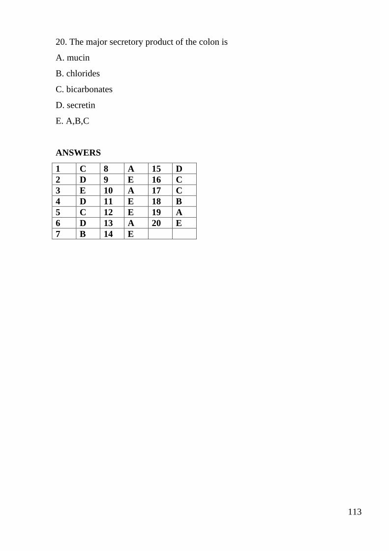
113
20. The major secretory product of the colon is
A. mucin
B. chlorides
C. bicarbonates
D. secretin
E. A,B,C
ANSWERS
1 C 8 A 15 D
2 D 9 E 16 C
3 E 10 A 17 C
4 D 11 E 18 B
5 C 12 E 19 A
6 D 13 A 20 E
7 B 14 E

114
PRACTICAL SKILLS
Topic: Digestion functions of Liver, Small Intestine and Colon.
TASK1. Put information about the functions of Liver in this table.
# Liver functions your explanation
1. Metabolic function.
2. Storage function
3. Synthetic function
4. Secretion of Bile
5. Excretory function
6. Heat production
7. Hemopoetic function
8. Hemolytic function
9. Inactivation of Hormones and Drugs
10. Defensive and detoxification functions
TASK2. Put information about the Bile functions in this table.
# Bile functions your explanation
1. Digestive function.
2. Absorptive function
3. Excretory function
4. Laxative action
5. Antiseptic action
6. Choleretic action
7. Maintenance of pH in gastrointestinal tract
8. Prevention of Gallstone formation
9. Lubrication function
10. Cholagogue action
TASK3. Study the bile action to fat filtration. Take two test tubes with funnels.
Put filters paper into the funnels. Moisten the first filter with bile but
the second one with water. Fill about 1 ml of oil into the each tube. Oil is faster to
filter off through the bile layer then through moist paper. Why? Describe this
experiment. Write an explanation
TASK 4. Draw this figure. Write an explanation about the regulation of Liver
secretion and gallbladder emptying.

115
TASK5. Two years ago Den had a gallstone and a doctor removed his
gallbladder. What happened with digestion function after that? Write an
explanation.
TASK6. Put information about the Intestine enzymes role for digestive in this
table.
Enzymes type Food types Final products
of digestion
Proteolytic enzymes : such as
aminopeptidase, dipeptidase and tripeptidase
Amylolytic enzymes: such as lactase,
sucrase, maltase, dextrinase and trehalase
Lipolytic enzymes: such as intestinal lipase
TASK7. Put information about the regulation of succus entericus secretion in
this table.
type of regulation mechanism effect
1. Nervous regulation :
a). Stimulation of parasympathetic
nerves
b). Stimulation of sympathetic nerves
c). The local nervous reflexes
2. Hormonal regulation:
a). cholecysto-kinin
b). secretin
c). enterocrinin
TASK 8. Study the parietal digestion in rat’s Intestine.

116
Put about a ml of physiological solution and 0, 5 ml of starch slurry in two test
tubes. Add a piece of rat’s crushed intestine into the first of the two. Put these test
tubes in a thermostat (at 37 ° С) for 20 minutes. Then add about a drop of iodine
solution in both ones. Estimate the Amylase activity by color change. Describe
this experiment. Write an explanation.
TASK9.The person’s secretin level was decrease. How can it change the
succus entericus secretion?
TASK10. Put information about the Colon functions in this table.
# Large intestine functions your explanation
1. Digestive function.
2. Absorptive function
3. Formation of feces
4. Excretory function
5. Secretory function
7. Synthetic functions
TASK 11. Draw this figure. Write an explanation about the afferent and
efferent pathways of the parasympathetic mechanism for the defecation
reflex.
External anal sphincter Internal anal sphincter

117
Tasks for final control
1. Part of the stomach was removed in the patient. What diet should he follow?
A. small meals 6-8 times a day.
B. Major portions 2 times a day.
C. Normal food 3 times a day
D. Eat at night
E. Eat lunch one time a day.
2. In terms of experience "sham feeding" we can learn secretion of:
A. salivary glands
B. stomach in all phases of its secretion
C. stomach in cephalic phase
D. stomach in gastric phase
E. stomach in the intestinal phase
3. The doctor advised the patient, with increased acidity of gastric juice to eat
boiled, not grilled meat. Because grilled meat contains substances that stimulate
gastric secretion. What is the name of these substances?
A. Fat.
B. Hydrochloric acid.
C. Carbohydrates.
D. Extractives
E. Gastrin.
4. Peripheral sympathetic fibers supplying the parotid gland were stimulated during
experiment in an animal. What happened with the secretion of saliva?
A. Saliva was not produced.
B. A little amount of liquid saliva was produced.
C. A little amount of thick saliva was produced.
D. A large amount of liquid saliva was produced.

118
E. A large amount of thick saliva was produced.
5. What hormonal stimulants of gastric juice in gastric phase of secretion are
released?
A. kinins and prostaglandins
B. Histamine and acetylcholine.
C. Enterogastrone and secretin.
D. secretin, CCK-PZ.
E. Histamine and gastrin.
6. Insert the missing words: primary protease in gastric juice is ... which is
produced in ...
A. Gastrin, active form
B. Pepsin, inactive form
C. Pepsin, active form
D. Gastrin, inactive form
E. trypsin, inactive form
7. Which of the following substances are natural endogenous stimulators of gastric
secretion. Choose the most correct answer:
A. histamine, gastrin, secretin
B. histamine, gastrin, acetylcholine
C. Histamine, hydrochloric acid
D. enterokinase, gastrin, hydrochloric acid
E. Gastrin, hydrochloric acid, secretin
8. The nerve is irritated, which leads to the secretion of large amount of liquid
saliva by the parotid gland. What nerve is irritated?
A. N. trigeminus
B. N. sympathicus

119
C. N. facialis
D. N. vagus
E. N. Glossopharyngeus
9. How does the secretory function of stomach change under the stimulation of H2
(histamine) receptors?
A. secretion of hydrochloric acid and pepsin decreases
B. pepsin secretion increases
C. secretion of hydrochloric acid decreases and pepsin - increases
D. secretion of mucus and pepsin increases
E. secretion of hydrochloric acid increases
10. 150 ml of meat broth was introduced in the stomach of a dog. Concentration of
what substance rapidly increases in the blood of animal?
A. Insulin
B. Somatostatin
C. Gastrin
D. Neurotensine
E. VIP
11. The sympathetic nerves to the gastrointestinal tract
A. Reach the wall of the tract in the form of preganglionic fibres.
B. Have preganglionic fibres that liberate a cholinergic transmitter
C. have postganglionic fibres which liberate predominantly NANC (non-
adrenergic, non-cholinergic) transmitters.
D. have postganglionic fibres which synapse directly with the muscular fibres of
the muscularis externa.
E. have postganglionic fibres which liberate transmitters than generally depolarize
the smooth muscle fibres.
120
12. The gastrointestinal electrical control activity (ECA)
A. originates in the myenteric plexus.
B. is dependent on extracellular Ca++
C. is abolished by atropine.
D. is detectable in the circular but not in the longitudinal muscle fibres.
E. is detectable both during the digestive period and the interdigestive period.
13. The bursts of spike potentials that constitute the gastrointestinal electrical
response activity (ERA) are initiative of contractions which
A. have an amplitude that is related to the number of spikes per burst.
B. have an amplitude that is characteristically lower following sympathetic
denervation of the tract.
C. have a maximal frequency that is limited by the number of spikes per burst.
D. have a higher maximal frequency in the longitudinal muscle fibres that in the
circular muscle fibres at corresponding points along the tract.
E. have a higher maximal frequency in the ileum than in the jejunum.
14. "Deglutition Apnea"
A. gives rise to odynophagia.
B. normally precedes every primary esophageal peristaltic wave.
C. may be induced by intraluminal distension of the esophagus.
D. is seen only in individuals who have abnormal patterns of swallowing.
E. results from activation of inhibitory nerve fibres to the respiratory muscles.
15. With respect to the resting tone of the lower esophageal sphineter (LES), all of
the following are correct EXCEPT:
A. It diminishes significantly following section of its vagal innervation.
B. It diminishes significantly following the administration of progesterone.

121
C. It does not diminish significantly following the administration of ganglionic
blocking agents.
D. It increases significantly following the administration of very large
(pharmacological) doses of gastrin.
E. It is normally in the range of 10-30 mmHg above intragastric pressure.
16. Before fat molecules are eventually placed into micelles in the lumen of the
gastrointestinal (GI) tract, they are digested by pancreatic lipase to form:
A. Glycerol
B. Monoglycerides
C. Free fatty acids
D. A and B
E. A and C
17. The chemical digestion of this class of compounds begins in the small intestine.
A. Carbohydrates
B. Proteins
C. Lipids
D. Nucleic acids
E. Starch
18. Which of the following is true about the enteric nervous system?
A. It is capable of responding independently to stimuli within the
gastrointestinal tract.
B. Its function is regulated by the autonomic nervous system.
C. It is sometimes referred to as the "little brain".
D. It uses serotonin as its main neurotransmitter.
E. All of the above.
122
19. All of the following are secreted by the pancreas into the duodenal region of
the small intestine EXCEPT:
A. Bicarbonate
B. Amylase
C. Nuclease
D. Peptidase
E. Intrinsic factor
20. The interstitial cells of Cajal are responsible for:
A. Regulating the osmolality of the kidney interstitial fluid.
B. Secreting secretin into the interstitial fluid of the small intestine.
C. Setting the contractile rhythm of the smooth muscle of the segments of the
gastrointestinal (GI) tract.
D. Secreting cholecystokinin into the bloodstream.
E. Secreting the antidiuretic hormone into the bloodstream.
21. Stomach parietal cells are responsible for secreting:
A. Hydrochloric acid (HCl)
B. Pepsinogen
C. Intrinsic factor
D. Bicarbonate (HCO3−)
E. A and C.
22. After ingestion of a meal, the pH of gastric fluid is about:
A. 1,5
B. 3,5
C. 5,5
D. 7,5
E. 9,5

123
23. Which one of the following is consistent with absorption of glucose molecules
across the intestinal epithelium?
A. Glucose enters the intestinal epithelial cell through glucose channels in the
apical membrane and leaves the cell through the basal membrane by facilitated
diffusion.
B. Glucose enters through the apical membrane by facilitated diffusion and leaves
through the basal membrane by facilitated diffusion also.
C. Glucose enters the cell through the apical membrane by co-transport with
Na+ and leaves the cell through the basal membrane by facilitated diffusion.
D. Glucose enters the cell through the apical membrane by facilitated diffusion and
leaves the cell through the basal membrane by co-transport with Na+.
E. None of the above.
24. Most of the chemical digestion in the gastrointestinal (GI) tract occurs in the:
A. Mouth
B. Stomach
C. Small intestine
D. Large intestine
E. Colon
25. Most of the water and electrolytes absorbed across the walls of the
gastrointestinal (GI) tract is absorbed in this region:
A. Mouth
B. Stomach
C. Small intestine
D. Large intestine
E. Colon
26. Fat digestion begins in this region of the gastrointestinal (GI) tract.
A. Mouth
124
B. Stomach
C. Duodenum of the Small intestine
D. Jejunum of the Small intestine
E. Large intestine
27. Which one of the following cells in the gastric glands produce pepsinogen?
A. endocrine cells
B. mucous neck cells
C. parietal or oxyntic cells
D. all of the above cells
E. chief or zymogenic cells
28. What substance is able to inhibit hydrochloric acid secretion?
A. Gastrin
B. Enterogastrone and adrenaline
C. Histamine
D. acetylcholine
E. Adrenaline
29. How many ml of pancreatic juice does healthy pancreas produce per day?
A. 500- 800 ml
B. 60- 80 ml
C. 900- 1000 ml
D. 1200- 1500 ml
E. 2000- 2500 ml
30. What kind of substances are produced by accessory cells of stomach?
A. mucoid secretion
B. enzymes of the gastric juice
C. hydrochloric acid

125
D. mucoid secretion and hydrochloric acid
E. All of the above
31. This substance is an important maturation factor during erythropoiesis and
factor of Castle is necessary for the absorption of this one.
A. vitamin B12
B. hydrochloric acid
C. Vitamin B6
D. Vitamin D
E. Pepsinogen
32. This substance causes denaturation and swelling of proteins and in that way
facilitates their breakdown by enzymes
A. vitamin B12
B. hydrochloric acid
C. Vitamin B6
D. Vitamin D
E. All of the above
33. Each day about 9 liters of water enter the gastrointestinal tract. By the time the
contents leave the large intestine as feces, about ______% of water is reabsorbed.
A. 1%
B. 6-7%
C. 25%
D. 92%
E. 99%
34. Pancreatic juice collected from the pancreatic duct has no effect on proteins
because it contains inactive forms of enzymes.
A. Trypsinogen

126
B. Chymotrypsinogen
C. Trypsinogen and Chymotrypsinogen
D. Pepsinogen
E. Pepsin
35. What factors can arrest the action of pepsin in the duodenum?
A. alkaline content
B. hydrogen ions
C. water
D.acid
E. all of the above
36. What factors can arrest the action of pepsin in the duodenum?
A. bile
B. hydrogen ions
C. water
D. acid
E. all of the above
37. Pancreatic secretion begins …to…minutes after a meal
A. 4,5
B. 5,6
C. 6,7
D. 7,8
E. 2, 3
38. Pancreatic secretion after a meal lasts for … to …hours
A. 1,2
B. 3,4
C. 5,6

127
D. 6,14
E. 1,7
39. Substances, which increase the secretion of bile from liver, are known as …….
A. Cholemimetic
B. Choleretics
C. Cholagogues
D. Cholelitics
E. all of the above
40. What substances are Choleretic?
A. Acetylcholine
B. Secretin and bile salts
C. CCK-PZ
D. acid chyme in intestine
E. all of the above
41. This substance increases the release of bile from gallbladder into the intestine.
A. Cholemimetic
B. Choleretics
C. Cholagogues
D. Cholelitics
E. all not correct
42. Which of the following is not a macronutrient?
A. Vitamin C
B. Protein
C. Carbohydrates
D. Fats
E. all of the above
128
43. Water is an important component of the diet because
A. it helps stabilize body temperature
B. it is required for many chemical reactions
C. it helps maintain normal concentrations of wastes and nutrients in the body
D. A and B
E. All of the above
44. What ions can block sugar influx into the epithelial cells?
A. a high concentration of Na+
B. a low concentration of sodium
C. a high concentration of potassium
D. a low concentration of potassium
E. a low concentration of iron
45. The esophageal phase of swallowing is caused by the
A. peristaltic reflex
B. coughing reflex
C. swallowing center
D. enteric reflex
E. all answers are incorrect
46. What can happen with salivation after stimulation of the sympathetic nerve?
A. secretion of small amounts of saliva and a high concentration of organic
material
B. secretion of small amounts of saliva
C. secretion of watery saliva
D. a low concentration of organic material
E. a high concentration of organic material

129
47. Some nerve fibers of Auerbach's plexus accelerate the movements by secreting
the excitatory neurotransmitter substances like ………
A. Acetylcholine
B. serotonin
C. substance P
D. Acetylcholine, serotonin, substance P
E. Noradrenalin
48. These nerve fibers cause constriction of blood vessels of gastrointestinal tract.
A. Auerbach's plexus
B. Giza plexus
C. Meissner's plexus
D. Salary plexus
E. Auerbach's plexus and Salary plexus
49. These nerve fibers inhibit the movements of gastrointestinal tract
A. Sympathetic nerve fibers
B. Auerbach's plexus
C. Giza plexus
D. Meissner's plexus
E. Salary plexus
50. These nerve fibers decrease the secretions of gastrointestinal tract
A. Sympathetic nerve fibers
B. Auerbach's plexus
C. Giza plexus
D. Meissner's plexus
E. Salary plexus

130
51. These nerve fibers accelerate the secretory activities of the glands in the
gastrointestinal tract.
A. Sympathetic nerve fibers
B. Auerbach's plexus
C. parasympathetic nerve fibers
D. Meissner's plexus
E. Salary plexus
52. These nerve fibers increase the motility of gastrointestinal tract
A. Sympathetic nerve fibers
B. parasympathetic nerve fibers
C. Auerbach's plexus
D. Meissner's plexus
E. Salary plexus
53. This neurotransmitter is responsible for the activation of the motility of
gastrointestinal tract and is secreted by the ……..fibers
A. Acetylcholine, parasympathetic nerve
B. Acetylcholine, Auerbach's plexus
C. Noradrenalin, Auerbach's plexus
D. Noradrenalin, sympathetic nerve
E. Serotonin, Meissner's plexus
54. This neurotransmitter is responsible for the inhibition of the movements of
gastrointestinal tract and decrease the secretions of gastrointestinal tract by
secreting the neurotransmitter ……..
A. Acetylcholine, parasympathetic nerve
B. Acetylcholine, Auerbach's plexus
C. Noradrenalin, Auerbach's plexus
D. Noradrenalin, sympathetic nerve

131
E. Serotonin, Meissner's plexus
55. This substance contributes to the curdling of milk, i. e. to the conversion of
caseinogen into casein by the pepsins and rennin.
A. vitamin B12
B. hydrochloric acid
C. Vitamin B6
D. Vitamin D
E. trypsin
ANSWERS
1A 9E 17D 25C 33E 41C 50A
2C 10C 18E 26A 34C 42A 51C
3D 11B 19E 27E 35A 44B 52B
4C 12E 20C 28B 36A 45D 53A
5E 13A 21E 29A 37E 46A 54D
6B 14B 22A 30A 38D 47D 55B
7B 15A 23C 31A 39B 48C
8E 16C 24C 32B 40E 49A

132
Recommended literature
Basic:
1. A.C. Guyton. Textbook of Medical Physiology: Philadelphia, 2011, 1116 p.
2. K. Sembulingam. Essentials of Medical Physiology. Jaypee, 2013, 895 p.
3. Sujit K. Chaudhuri. Concise Medical Physiology.- New Central Book Agency,
2013. – 752 p.
4. Human physiology V.F. Hanong, Lvov: BaK, 2009. – 784 p.
Additional literature:
1. Nowicki P.T., Granger D.N. Gastrointestinal blood flow. In: Textbook of
Gastroenterology. Yamada T, ed. 5th edition. pp. 540–556, 2008. Wiley-
Blackwell.
2. Silberg D.G. and Wu G.D. Development of the alimentary tract, liver and
pancreas. In: Gastrointestinal Cancers, edited by Rustgi AK; Saunders, 2003,
Chapter 7, pp. 105–119.
3. Flock M.H. Stomach and duodenum. In: Netter's Gastroenterology, edited by
Flock MH; Icon Learning Systems, 2005, Chapter 2, pp. 106–15.
4. Drozdowski L.A. Ontogeny, growth and development of the small intestine:
understanding pediatric gastroenterology / L.A. Drozdowski, T. Clandinin,
A.B. Thomson // World J. Gastroenterol. – 2010. – Vol. 16, № 7. – Р. 787–799.
5. Zabielski R. Control of development of gastrointestinal system in neonates / R.
Zabielski, M.M. Godlewski, P. Guilloteau // J. Physiol. Pharmacol. – 2008. – Vol.
59. – P. 35–54.
6. Dockry G. Gastrin: old hormone, new functions / G. Dockry, R. Dimaline, A.
Varro // Pflugers. Arch. – 2005. – Vol. 449, № 4. – Р. 344–355.
7. Сhu S. Gastric secretion / S.Chu, M.L. Schubert // Curr. Opin. Gastroenterol. –
2012. – Vol. 28, № 6. – Р. 587–593.