Nerve cells Muscle cells Epithelial cells Bone cell Fat cells.
Upload
linda-lorraine-robertsonCategory
view
215download
1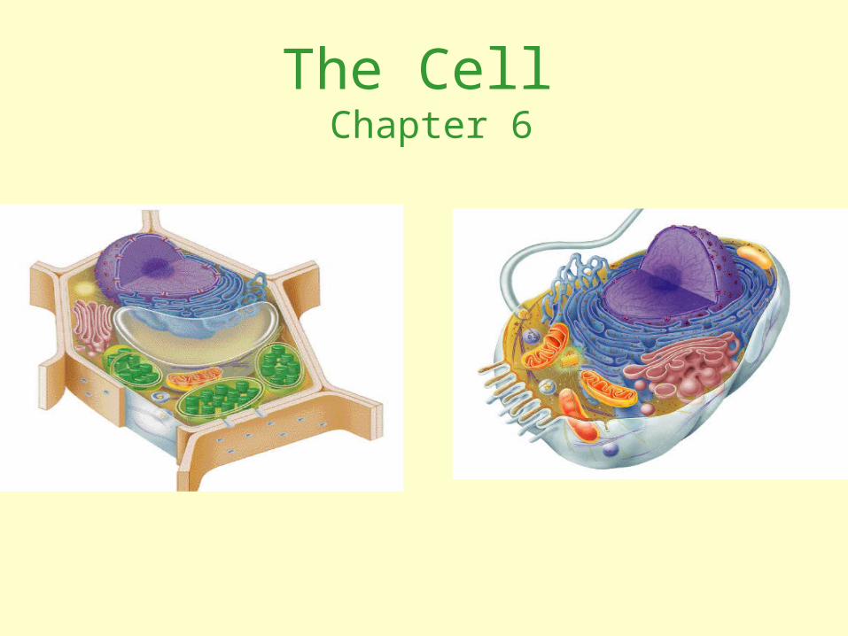
The Cell Chapter 6
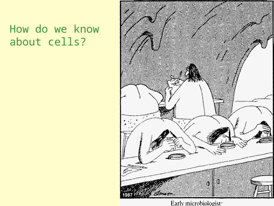
How do we know about cells?

1. Microscopes: windows to the world of the cell
• The discovery and early study of cells progressed with the invention and improvement of microscopes in the 17th century.
• In a light microscope (LM) visible light passes through the specimen and then through glass lenses

• Microscopes vary in magnification and resolving power.
Resolving power is a measure of image clarity.
• It is the minimum distance two points can be separated by and still be viewed as two separate points.
Robert Hooke 1665

• The minimum resolution of a light microscope is about 2 microns, the size of a small bacterium
• Light microscopes can magnify effectively to about 1,000 times the size of the actual specimen.

• Techniques developed in the 20th century have enhanced contrast and enabled cell components to be labeled so that they stand out.

To resolve cell organelles we use an electron microscope (EM), which focuses a beam of electrons through the specimen or onto its surface.
• Electron microscopes have finer resolution than light microscopes

• Transmission electron microscopes (TEMs) are used mainly to study the internal ultrastructure of cells.
• A TEM aims an electron beam through a thin section of the specimen.
Cucumber cotyledon

• Scanning electron microscopes (SEMs) are useful for studying surface structures.
• The image is focused on a screen
• Three dimensional
• The SEM has great depth of field, resulting in an image that seems three-dimensional.
Rabbit trachea cells (SEM)

• Electron microscopes reveal organelles, but they can only be used on dead cells.
• Light microscopes do not have as high a resolution, but they can be used to study live cells.

• Cell fractionation separates the major organelles of the cells so that their individual functions can be studied.
2. Cell biologists can isolate organelles to study their functions and separate chemical components

• This process is driven by an ultracentrifuge, a machine that can spin at up to 130,000 revolutions per minute and apply forces more than 1 million times gravity (1,000,000 g).

• Microcentrifuge is standard equipment in biotechnology labs activities.

Equipment used to study cells at the genetic and protein level.
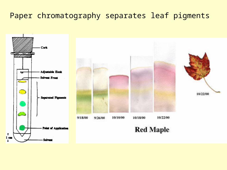
Paper chromatography separates leaf pigments

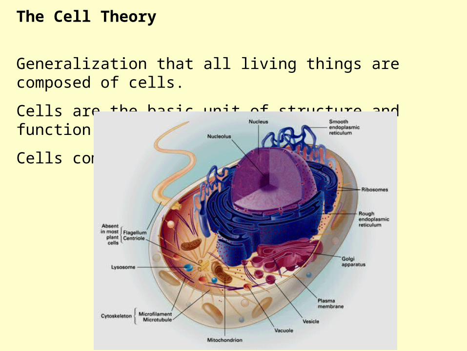
The Cell Theory
Understanding the cellular nature of life followed the development of tools and techniques:In 1665, Robert Hooke observed "compartments" in a thin slice of cork (oak bark) using a light microscope. Used the term “Cell.”
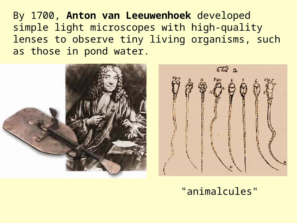
By 1700, Anton van Leeuwenhoek developed simple light microscopes with high-quality lenses to observe tiny living organisms, such as those in pond water.
"animalcules"
The Cell Theory
Generalization that all living things are composed of cells.
Cells are the basic unit of structure and function in living things
Cells come from pre-existing cells

• All cells are surrounded by a plasma membrane.
• All cells contain chromosomes which have genes in the form of DNA.
• All cells also have ribosomes
3. Two Major Classes of Cells: Prokaryotic and Eukaryotic
Prokaryotic cell movie

•Prokaryotic and eukaryotic cells differ in the location of chromosomes.•Eukaryotic cell chromosomes are in a nucleus.•In a prokaryotic cell, the DNA is concentrated in the nucleoid without a membrane separating it from the rest of the cell.
Eukaryotic cell movie

The prokaryotic cell is much simpler in structure, lacking a nucleus and the other membrane-enclosed organelles of the eukaryotic cell.


• What limits cell size?
• As a cell increases in size its volume increases faster than its surface area.
• Smaller objects have a greater ratio of surface area to volume.
• Square/Cube Law
• What cell organelle is critical in maintaining this ratio?

• The plasma membrane functions as a selective barrier that allows passage of oxygen, nutrients, and wastes for the whole volume of the cell.

• The volume of cytoplasm determines the need for this exchange.
• Rates of chemical exchange may be inadequate to maintain a cell with a very large cytoplasm.
• The need for a surface sufficiently large to accommodate the volume explains the microscopic size of most cells.
• Larger organisms do not generally have larger cells than smaller organisms - simply more cells.

• A eukaryotic cell has extensive and elaborate internal membranes, which partition the cell into compartments.
• Many enzymes are built into membranes.
• Membranes provide different local environments for specific metabolic functions.
• Each type of membrane has a unique combination of lipids and proteins for its specific functions.
4. Internal membranes compartmentalize the functions of a eukaryotic cell


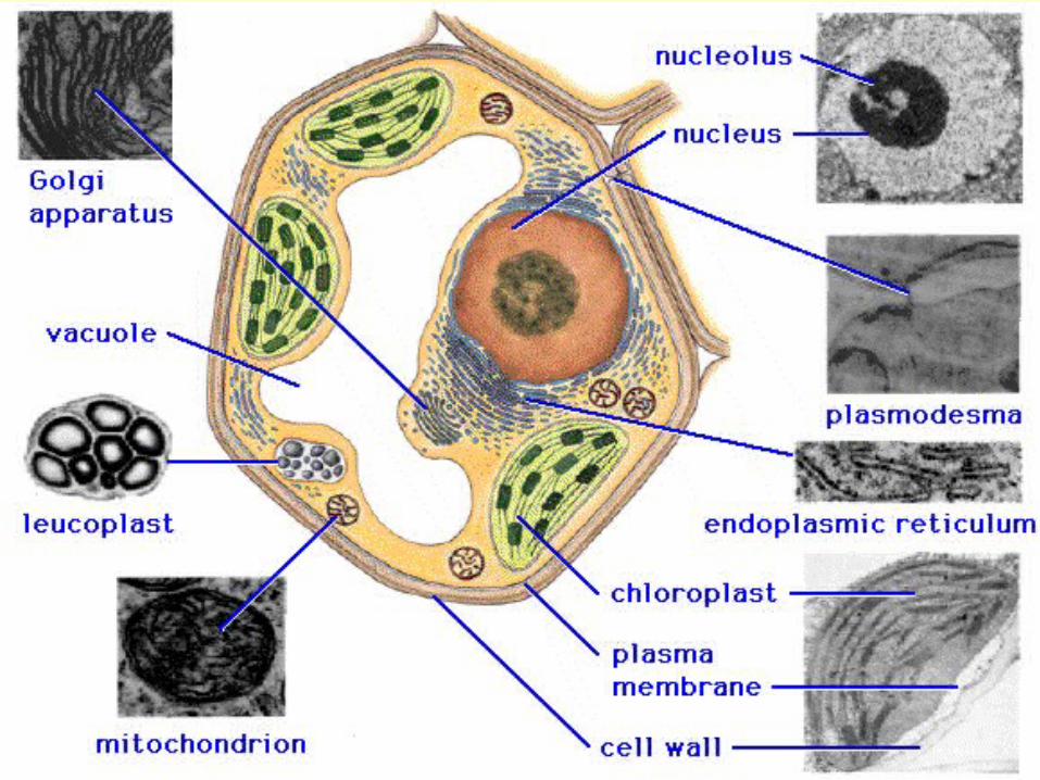

• The nucleus contains most of the genes in a eukaryotic cell.
• Some genes are located in mitochondria and chloroplasts.
• The nucleus is separated from the cytoplasm by a double membrane.
• Pores allows large macromolecules and particles to pass through.
5. The nucleus contains a eukaryotic cell’s genetic library

• The nuclear side of the envelope is lined by a network of filaments that maintain the shape of the nucleus.

• Within the nucleus, the DNA and associated proteins are organized into chromatin.
• In a normal cell they appear as a diffuse mass.
• When the cell prepares to divide, the chromatin fibers coil up to be seen as separate structures, chromosomes.
• What is special about chromosome numbers?

• In the nucleus is the nucleolus.
• In the nucleolus, ribosomal RNA is synthesized and assembled with proteins to form ribosomal subunits.
• The subunits pass from the nuclear pores to the cytoplasm where they combine to form ribosomes.
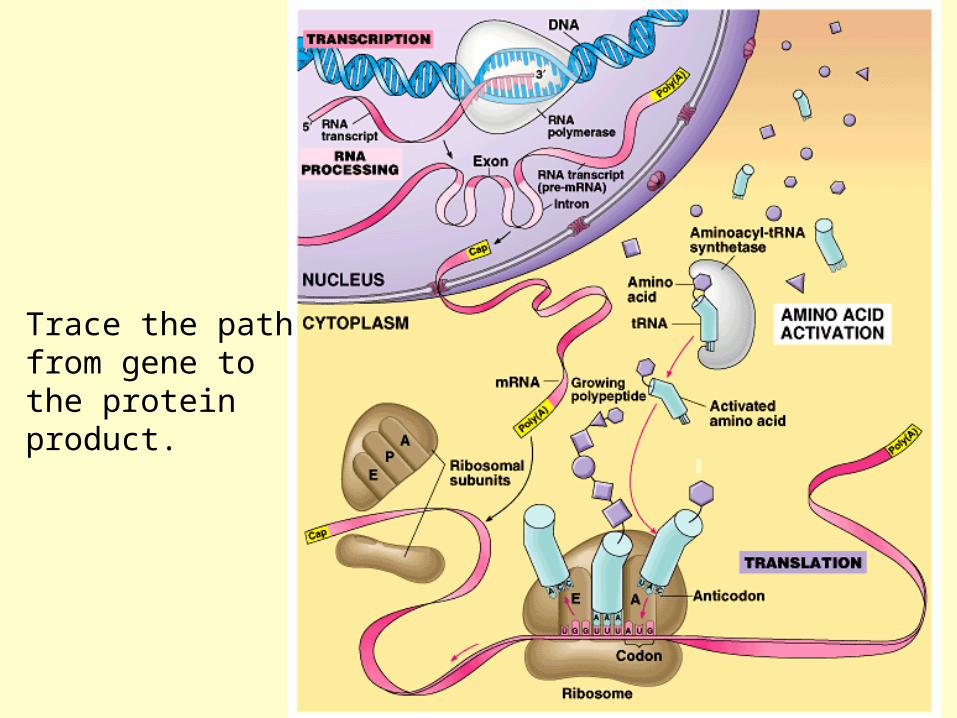
Trace the path from gene to the protein product.

• Ribosomes contain rRNA and protein.
• A ribosome is composed of two subunits that combine to carry out protein synthesis.
6. Ribosomes build a cell’s proteins

• What is implied if a cell type has large numbers of ribosomes and prominent nuclei. (e.g., pancreas)
• Free ribosomes, are suspended in the cytoplasm and synthesize proteins that function within the cytoplasm.
• Bound ribosomes, are attached to the outside of the endoplasmic reticulum.

The Endomembrane System
•Many internal membranes in a eukaryotic cell are part of the endomembrane system.
•The endomembrane system includes the nuclear envelope, endoplasmic reticulum, Golgi apparatus, lysosomes, vacuoles, and the plasma membrane.
What is the adaptive value of this system?

• The endoplasmic reticulum (ER) accounts for half the membranes in a eukaryotic cell.
• The ER includes membranous tubules and internal, fluid-filled spaces, the cisternae.
7. The endoplasmic reticulum manufactures membranes and modifies proteins

• There are two regions of ER that differ in structure and function.
• Smooth ER looks smooth because it lacks ribosomes.
• Rough ER looks rough because ribosomes (bound ribosomes) are attached to the outside, including the outside of the nuclear envelope.

• Smooth ER is rich in enzymes and plays a role in a variety of metabolic processes.
• Enzymes of smooth ER synthesize lipids, including oils, phospholipids, and steroids.
• The smooth ER helps catalyze conversion of glucose from stored glycogen in the liver.
• Smooth ER of the liver help detoxify drugs and poisons. (proliferation of smooth ER increases tolerance to the target and other drugs)

• Rough ER is especially abundant in those cells that secrete proteins.
• As a polypeptide is synthesized by the ribosome, it is threaded into the cisternal space through a pore formed by a protein in the ER membrane.
• The protein is modified in the ER
• These secretory proteins are packaged in transport vesicles that carry them to their next stage.

• Many transport vesicles from the ER travel to the Golgi apparatus for modification of their contents.
• The Golgi is a center of manufacturing, warehousing, sorting, and shipping.
• Which cells would have extensive Golgi apparatus?
8. The Golgi apparatus finishes, sorts, and ships cell products
DR. CAMILLO GOLGI(1843-1926)

• The Golgi apparatus consists of flattened membranous sacs - cisternae - looking like a stack of pita bread.
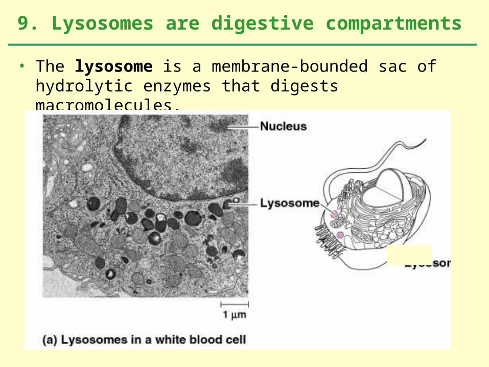
• The lysosome is a membrane-bounded sac of hydrolytic enzymes that digests macromolecules.
9. Lysosomes are digestive compartments

• Lysosomal enzymes can hydrolyze proteins, fats, polysaccharides, and nucleic acids.
• These enzymes work best at pH 5.
• What is the value of this compartmentalization?

• The lysosomal enzymes and membrane are synthesized by rough ER and then transferred to the Golgi.
• At least some lysosomes bud from the trans face of the Golgi.

• Lysosomes can fuse with food vacuoles, formed when a food item is brought into the cell by phagocytosis.
• Lysosomes can also fuse with another organelle or part of the cytosol.
• This recycling,or autophagy,renews the cell.
Lysosome Movie
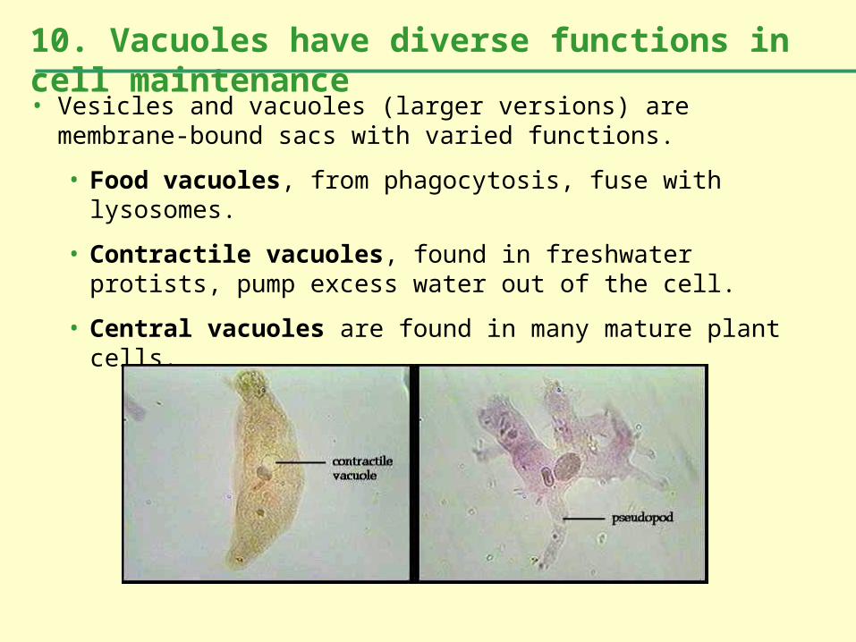
• Vesicles and vacuoles (larger versions) are membrane-bound sacs with varied functions.
• Food vacuoles, from phagocytosis, fuse with lysosomes.
• Contractile vacuoles, found in freshwater protists, pump excess water out of the cell.
• Central vacuoles are found in many mature plant cells.
10. Vacuoles have diverse functions in cell maintenance


• What is the adaptive role of the endomembrane system?
• The endomembrane system plays a key role in the synthesis (and hydrolysis) of macromolecules in the cell.
• The various components modify macromolecules for their various functions.
• Mitochondria and chloroplasts are the organelles that convert energy to forms that cells can use for work.
• Mitochondria are the sites of cellular respiration, generating ATP from the breakdown of sugars, fats, and other fuels in the presence of oxygen.
• Chloroplasts, found in plants and eukaryotic algae, are the sites of photosynthesis.
• They convert solar energy to chemical energy and synthesize new organic compounds from CO2 and H2O.
11. Mitochondria and chloroplasts are the main energy transformers of cells

• Mitochondria and chloroplasts are not part of the endomembrane system.
• Their proteins come primarily from free ribosomes in the cytosol and a few from their own ribosomes.
• Both organelles have small quantities of DNA that direct the synthesis of the polypeptides produced by these internal ribosomes.
• Mitochondria and chloroplasts grow and reproduce as semi-independent organelles.

• Mitochondria have a smooth outer membrane and a highly folded inner membrane, the cristae.
• This creates a fluid-filled space between them.
• The cristae present ample surface area for the enzymes that synthesize ATP.
• The inner membrane encloses the mitochondrial matrix, a fluid-filled space with DNA, ribosomes, and enzymes.


• The chloroplast is one of several members of a generalized class of plant structures called plastids.
• The chloroplast produces sugar via photosynthesis.
• Chloroplasts gain their color from high levels of the green pigment chlorophyll.
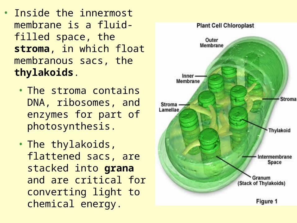
• Inside the innermost membrane is a fluid-filled space, the stroma, in which float membranous sacs, the thylakoids.
• The stroma contains DNA, ribosomes, and enzymes for part of photosynthesis.
• The thylakoids, flattened sacs, are stacked into grana and are critical for converting light to chemical energy.

• Like mitochondria, chloroplasts are dynamic structures.
• Their shape is plastic and they can reproduce themselves by pinching in two.
• Mitochondria and chloroplasts are mobile and move around the cell along tracks in the cytoskeleton.

12. Providing structural support to the cell, the cytoskeleton also functions in cell motility and regulation• The cytoskeleton is a network of fibers that provide mechanical support and maintains shape of the cell.
• The cytoskeleton provides anchorage for many organelles, enzymes, and organizes cell structures and activities.

• The cytoskeleton also plays a major role in cell motility.
• The cytoskeleton interacts with motor proteins.
• In cilia and flagella motor proteins pull components of the cytoskeleton past each other.
• This is also true in muscle cells.

• Motor molecules also carry vesicles or organelles to various destinations along “monorails’ provided by the cytoskeleton.
• Interactions of motor proteins and the cytoskeleton circulate materials within a cell by cytoplasmic streaming.

•There are three main types of fibers in the cytoskeleton: microtubules, microfilaments, and intermediate filaments.
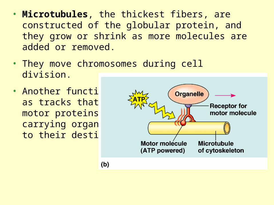
• Microtubules, the thickest fibers, are constructed of the globular protein, and they grow or shrink as more molecules are added or removed.
• They move chromosomes during cell division.
• Another function is as tracks that guide motor proteins carrying organelles to their destination.

•In many cells, microtubules grow out from a centrosome near the nucleus.•In animal cells, the centrosome has a pair of centrioles, each withnine triplets of microtubules arranged in a ring.•During cell division the centrioles replicate.
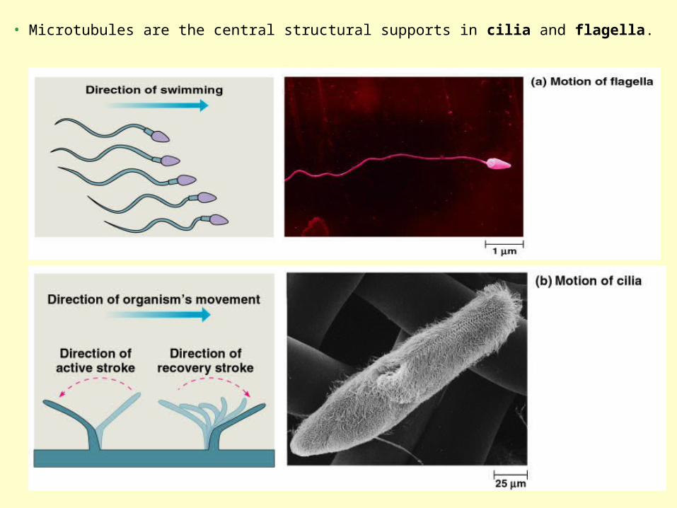
• Microtubules are the central structural supports in cilia and flagella.

• In spite of their differences, both cilia and flagella have the same ultrastructure.
• Microtubules arranged in the “9 + 2” pattern.

• The bending of cilia and flagella is driven by the arms of a motor protein, dynein.
• Addition to dynein of a phosphate group from ATP and its removal causes changes in the protein.
• Dynein arms alternately grab, move, and release the outer microtubules.
• Protein cross-links limit sliding and the force is expressed as bending.
Cilia and Flagella Movie

• Microfilaments, the thinnest class of the cytoskeletal fibers, are solid rods of the globular protein actin.
• With other proteins, they form a three-dimensional network just inside the plasma membrane. The shape of the microvilli in this intestinal cell are supported by microfilaments, anchored to a network of intermediate filaments.

• In muscle cells, thousands of actin filaments are arranged parallel to one another.
• Thicker filaments composed of a motor protein, myosin, interdigitate with the thinner actin fibers.
• Myosin molecules walk along the actin filament, pulling stacks of actin fibers together and shortening the cell.

• In other cells, these actin-myosin clusters still cause localized contraction.
• A contracting belt of microfilaments divides the cytoplasm of animal cells during cell division.
• Localized contraction also drives amoeboid movement.

• In plant cells (and others), actin-myosin interactions and sol-gel transformations drive cytoplasmic streaming.
• This creates a circular flow of cytoplasm in the cell.
• This speeds the distribution of materials within the cell.

• Intermediate filaments are specialized for bearing tension.
• Intermediate filaments are built of proteins called keratins.
• Intermediate filaments are more permanent fixtures of the cytoskeleton than are the other two classes.
• They reinforce cell shape and fix organelle location.

• The cell wall, found in prokaryotes, fungi, and some protists, has multiple functions.
• In plants, the cell wall protects the cell, maintains its shape, and prevents excessive uptake of water.
• The thickness and chemical composition of cell walls differs from species to species and among cell types.
13. Plant cells are encased by cell walls

• Consists of microfibrils of cellulose embedded in a matrix of proteins and other polysaccharides.
• A mature cell wall consists of a primary cell wall, a middle lamella with sticky polysaccharides that holds cell together, and layers of secondary cell wall.

• Lacking cell walls, animals cells have an elaborate extracellular matrix (ECM).
14. Animal cells have an extracellular matrix functions in support, adhesion, movement, and regulation

• Neighboring cells in tissues, organs, or organ systems often adhere, interact, and communicate through direct physical contact.
• Plant cells are perforated with plasmodesmata, channels allowing cysotol to pass between cells.
15. Intercellular junctions help cells transport and communicate

MEMBRANE STUCTURE AND FUNCTION

• A membrane is a collage of different proteins embedded in the fluid matrix of the lipid bilayer.
1. Membranes are mosaics of structure and function
2. Membrane Structure
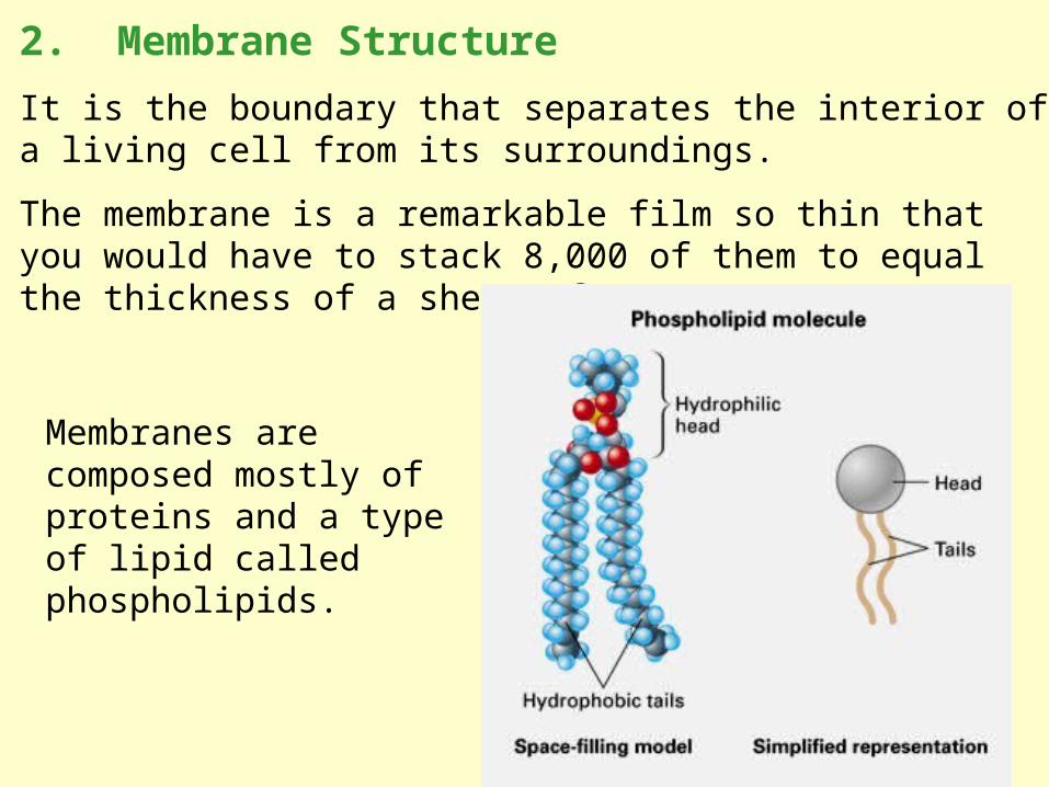
It is the boundary that separates the interior of a living cell from its surroundings.
The membrane is a remarkable film so thin that you would have to stack 8,000 of them to equal the thickness of a sheet of paper.
Membranes are composed mostly of proteins and a type of lipid called phospholipids.
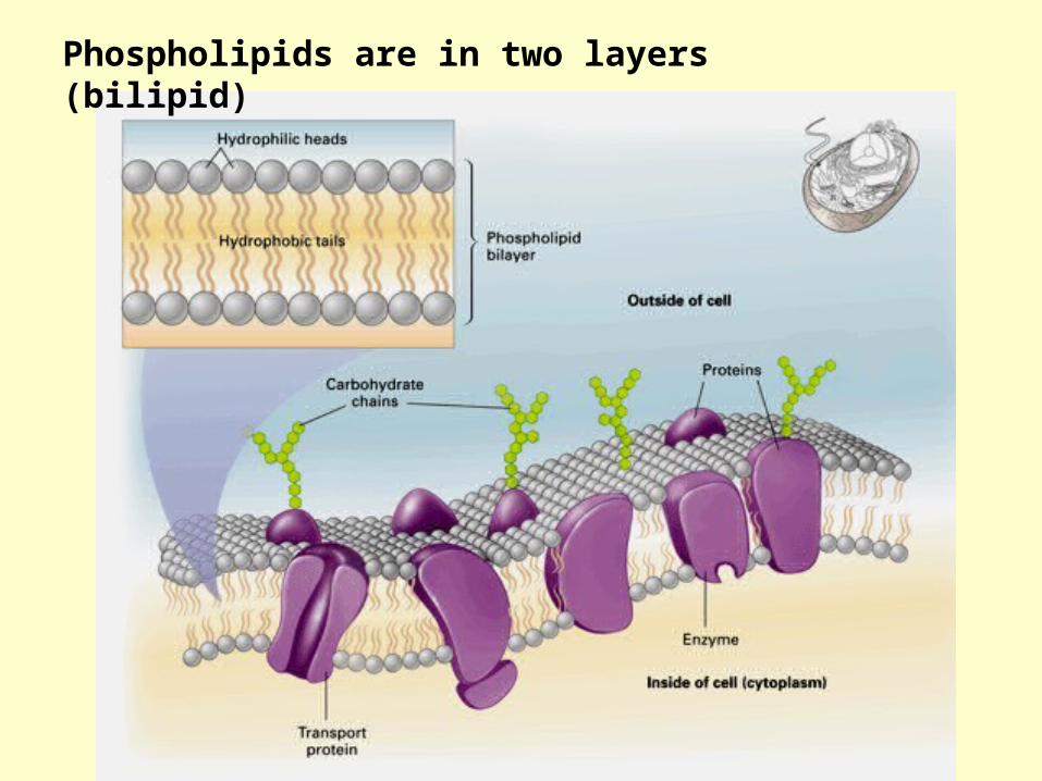
Phospholipids are in two layers (bilipid)

• Membrane molecules are held in place by relatively weak hydrophobic interactions.
• Most of the lipids and some proteins can drift laterally in the plane of the membrane, but rarely flip-flop from one layer to the other.
3. Membranes are fluid
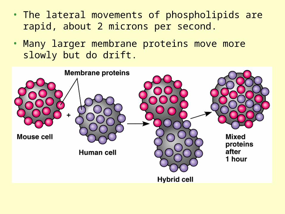
• The lateral movements of phospholipids are rapid, about 2 microns per second.
• Many larger membrane proteins move more slowly but do drift.

Proteins are important to membrane functions

Cell membranes have many functions beyond serving as a boundary!
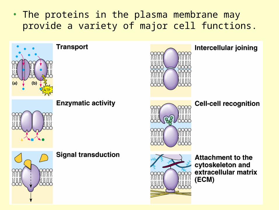
• The proteins in the plasma membrane may provide a variety of major cell functions.

• The proteins determine most of the membrane’s specific functions.
• Surface of the protein often connect to the other membrane proteins.
• Integral proteins penetrate and may span the hydrophobic core of the lipid bilayer.
How do you think the amino acids differ in the integral proteins?
Membrane Structure Movie

Membrane carbohydrates are important for cell-cell recognition
• Membrane carbohydrates are usually branched saccharides with fewer than 15 sugar units.
• They may be covalently bonded either to lipids or proteins.
The saccharides on the membrane may be unique and serve for cell recognition.
Human blood groups (A, B, AB, and O) differ in the external carbohydrates on red blood cells.

• A steady traffic of small molecules and ions moves across the plasma membrane in both directions.
• Sugars, amino acids, and other nutrients enter a cell and metabolic waste products leave.
• The cell absorbs oxygen and excretes carbon dioxide.
• It also regulates concentrations of inorganic ions, like Na+, K+, Ca2+, and Cl-, by shuttling them across the membrane.
• However, substances do not move across the barrier indiscriminately; membranes are selectively permeable.
• What determines whether materials pass through membranes?
4. A membrane’s molecular organization results in selective permeability

• Permeability of a molecule depends on the interaction of that molecule with the hydrophobic core of the membrane.
• Hydrophobic molecules, like hydrocarbons, CO2, and O2 can and cross easily.
• Ions and polar molecules pass through with difficulty.
•This includes small molecules, like water, and larger critical molecules, like glucose and other sugars.
•Ions, whether atoms or molecules, and their surrounding shell of water also have difficulties penetrating the hydrophobic core.
• Specific ions and polar molecules can cross the lipid bilayer by passing through transport proteins that span the membrane.
• Each transport protein is specific as to the substances that it will translocate (move).

• Diffusion is the tendency of molecules of any substance to spread out in the available space
• Diffusion is driven by energy (thermal motion or heat) of molecules.
• Movements of individual molecules are random.
• However, movement of a population of molecules may be directional.
5. Passive transport is diffusion across a membrane

• A substance will diffuse from where it is more concentrated to where it is less concentrated, down its concentration gradient.
• Each substance diffuses down its own concentration gradient, independent of the concentration gradients of other substances.
• The concentration gradient represents potential energy and drives diffusion.
Diffusion Movie

• Differences in the relative concentration of dissolved materials in two solutions can lead to the movement of ions from one to the other.
• The solution with the higher concentration of solutes is hypertonic.
• The solution with the lower concentration of solutes is hypotonic.
• These are comparative terms.
•The hypertonic solution has a lower water concentration than the hypotonic solution.
• Solutions with equal solute concentrations are isotonic.
6. Osmosis is the passive transport of water

• Water molecules will move from the hypotonic solution to the hypertonic solution.
• This diffusion of water across a selectively permeable membrane is a special case of passive transport called osmosis.
• Osmosis continues until the solutions are isotonic.

7. Cell survival depends on balancing water

• Organisms without rigid walls have osmotic problems in either a hypertonic or hypotonic environment and must have adaptations for osmoregulation to maintain their internal environment.
• Paramecium, a freshwater protist, is hypertonic when compared to the pond water in which it lives.
• So, even with a less permeable membrane water still continually enters the Paramecium cell.
• Paramecium have a specialized organelle, the contractile vacuole, that functions as a bilge pump to force water out of the cell.

• A cell with a cell wall in a hypotonic solution will swell until the elastic wall opposes further uptake.
• At this point the cell is turgid, a healthy state for most plant cells.
• Turgid cells contribute to the mechanical support of the plant.
Tonicity movie

• In a hypertonic solution, the plant cell loses water, and the plasma membrane pulls away from the wall.
• This plasmolysis is usually lethal.

• The passive movement of molecules down its concentration gradient via a transport protein is called facilitated diffusion.
8. Specific proteins facilitate passive transport of water and selected solutes

Transport proteins provide corridors for specific molecule or ion to cross the membrane.
• These channel proteins allow fast transport.
• For example, water channel proteins, aquaprorins, facilitate massive amounts of diffusion.
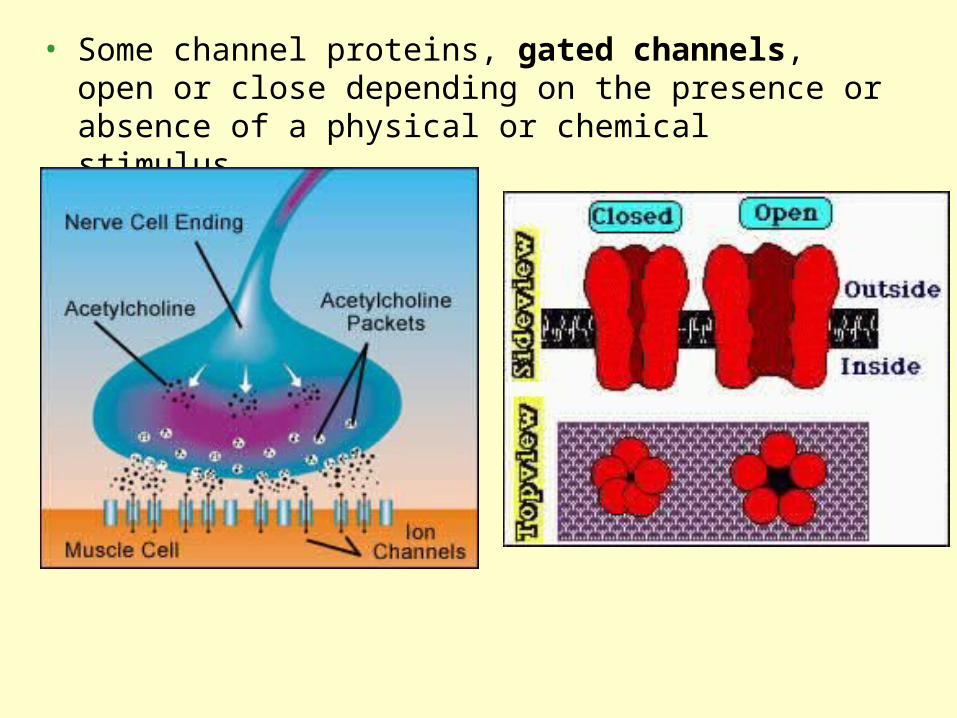
• Some channel proteins, gated channels, open or close depending on the presence or absence of a physical or chemical stimulus.

• Some transport proteins actually translocate the solute across the membrane as the protein changes shape.
• These shape changes could be triggered by the binding and release of the transported molecule.

• Active transport requires the cell to use its own metabolic energy.
• Active transport is performed by specific proteins embedded in the membranes.
• ATP supplies the energy for most active transport
9. Active transport is the pumping of molecules against their gradients

The sodium-potassium pump actively maintains the gradient of sodium (Na+) and potassium ions (K+) across the membrane.

Both diffusion and facilitated diffusion are forms of passive transport of molecules down their concentration gradient, while active transport requires an investment of energy to move molecules against their concentration gradient.

• Large molecules, such as polysaccharides and proteins, cross the membrane via vesicles.
• During exocytosis, a transport vesicle budded from the Golgi apparatus is moved by the cytoskeleton to the plasma membrane.
• When the two membranes come in contact, the bilayers fuse and spill the contents to the outside.
12. Exocytosis and endocytosis transport large molecules

• During endocytosis, a cell brings in macromolecules and particulate matter by forming new vesicles from the plasma membrane.
• Three types of endocytosis: phagocytosis, pinocytosis, and receptor-mediated endocytosis

• In phagocytosis, the cell engulfs a particle by extending pseudopodia around it and packaging it in a large vacuole.
• The contents of the vacuole are digested when the vacuole fuses with a lysosome.

Electron Micrograph of a Macrophage Phagocytosis of E. coli

In pinocytosis, “cellular drinking,” a cell creates a vesicle around a droplet of extracellular fluid.This is a non-specific process.
Pinocytosis smooth muscle (Guinea pig).