
THE ARABIDOPSIS GENE GRASSY, IS REQUIRED FOR AUXIN TRANSPORT
88
University of Lethbridge Research Repository OPUS http://opus.uleth.ca Theses Arts and Science, Faculty of 2008 The arabidopsis gene Grassy, is required for auxin transport and patterning of leaf vein, shoot and root Pahari, Shankar Lethbridge, Alta. : University of Lethbridge, Faculty of Arts and Science, 2008 http://hdl.handle.net/10133/670 Downloaded from University of Lethbridge Research Repository, OPUS
Transcript of THE ARABIDOPSIS GENE GRASSY, IS REQUIRED FOR AUXIN TRANSPORT
University of Lethbridge Research Repository
OPUS http://opus.uleth.ca
Theses Arts and Science, Faculty of
2008
The arabidopsis gene Grassy, is
required for auxin transport and
patterning of leaf vein, shoot and root
Pahari, Shankar
Lethbridge, Alta. : University of Lethbridge, Faculty of Arts and Science, 2008
http://hdl.handle.net/10133/670
Downloaded from University of Lethbridge Research Repository, OPUS
i
THE ARABIDOPSIS GENE GRASSY, IS REQUIRED FOR AUXIN TRANSPORT AND PATTERNING OF LEAF VEIN, SHOOT AND ROOT
SHANKAR PAHARI M.Sc. Tribhuvan University, Nepal, 2004
A Thesis Submitted to the School of Graduate Studies
of the University of Lethbridge in Partial Fulfilment of the
Requirements for the Degree
MASTER OF SCIENCE
Biological Sciences University of Lethbridge
LETHBRIDGE, ALBERTA, CANADA
© Shankar Pahari, 2008

iii
I dedicate this thesis to my beloved wife Sushila and daughter Sadikshya
iv
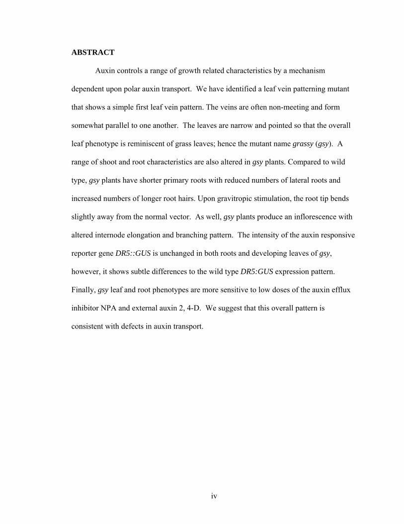
ABSTRACT
Auxin controls a range of growth related characteristics by a mechanism
dependent upon polar auxin transport. We have identified a leaf vein patterning mutant
that shows a simple first leaf vein pattern. The veins are often non-meeting and form
somewhat parallel to one another. The leaves are narrow and pointed so that the overall
leaf phenotype is reminiscent of grass leaves; hence the mutant name grassy (gsy). A
range of shoot and root characteristics are also altered in gsy plants. Compared to wild
type, gsy plants have shorter primary roots with reduced numbers of lateral roots and
increased numbers of longer root hairs. Upon gravitropic stimulation, the root tip bends
slightly away from the normal vector. As well, gsy plants produce an inflorescence with
altered internode elongation and branching pattern. The intensity of the auxin responsive
reporter gene DR5::GUS is unchanged in both roots and developing leaves of gsy,
however, it shows subtle differences to the wild type DR5:GUS expression pattern.
Finally, gsy leaf and root phenotypes are more sensitive to low doses of the auxin efflux
inhibitor NPA and external auxin 2, 4-D. We suggest that this overall pattern is
consistent with defects in auxin transport.
v
ACKNOWLEDGEMENTS
I would like to wholeheartedly thank my supervisor, Dr. Elizabeth Schultz,
Associate Professor of Biology, for her consistent guidance, support and encouragement
throughout my program. In these two years I learnt a lot from her distinguished expertise
and I am very grateful to be a part of her research team. I wish to acknowledge my
supervisory committee members, Dr. Igor Kovalchuk, Associate Professor of Biology
and Dr. Marc Roussel, Professor of Chemistry, for their valuable comments, suggestions
and insightful discussions.
I am indebted to Dr. John Bain, Professor of Biology for helping me with
restriction enzymes. I am thankful to the members of Schultz’s lab, Kamran, Mike and
Hongwei, for their cooperation, help and support. I would also like to thank James
Meservy for his technical help in designing primers.
I wish to express my sincere gratitude to my beloved wife Sushila Adhikari Pahari
for her persistent love, care and patience. This work would not have been possible
without her tolerance and the sacrifice that she made in staying back home thousands of
miles away taking care of our daughter, Sadikshya Pahari. They have been patiently
awaiting my achievements without a word of reproach or complaints for my long absence
from home. I hereby dedicate this thesis to them. Last but not the least I am very grateful
to my parents, sisters and brothers for their blessings and inspirations, which has always
helped me, achieve my goals in life.

vi
TABLE OF CONTENTS PAGE Approval/signature Page ii Dedication iii Abstract iv Acknowledgements v Table of Contents vi List of Tables viii List of Figures ix List of Abbreviations x Introduction 1 Materials and Methods 14
Plant Materials 14
Growth Conditions 14
Mutant Isolation 15
Mapping of gsy 15 First leaf Analysis of gsy 16
Plant shoot morphology 17 Root assays 17
Generation of Double Mutants 18
Histochemical staining for Gus 19
Microscopy and Imaging and Statistics 20 Results 21 Mutant isolation and Mapping of GSY 21
vii
gsy has simplified and non-meeting first leaf phenotype 22 DR5:GUS expression is intact in leaves 24 gsy shoot morphology 26 gsy root phenotype 27
DR5::GUS expression in root 28
Effect of auxin transport inhibition in leaves and roots 29 Exogenous auxin and root phenotypes 31 Double mutants 32 Discussions 37 gsy shows no reduction in auxin response 37
gsy is defective in auxin transport 39
Shoot transport 40 Root transport 41 Leaf transport 44
Conclusion 46
References 47

viii
LIST OF TABLES PAGE Table 1. Primer markers developed and used in mapping gsy 58 Table 2. List of candidate genes and their functions 57 Table 3. Cotyledon vascular pattern characters for various genotypes at 14 DAG 59 Table 4: First leaf vascular pattern characters of various genotypes at
21DAG 60 Table 5: Appearance of leaf vein characteristics in WT and gsy 61 Table 6: Analysis of various developmental characteristics in gsy as
compared to WT 62 Table 7: Shoot and root branching in various genotypes 63 Table 8: Primary root growth of seedlings exposed to 2,4-D 64
ix
LIST OF FIGURES PAGE Figure 1. Banding pattern for molecular markers used in mapping 65 Figure 2. Vascular pattern of cleared 21 DAG first leaves and 14 DAG Cotyledons 66 Figure 3. Vascular pattern and FKD::GUS expression in developing first leaves
of wild type and gsy 67 Figure 4. DR5::GUS expression in developing first leaves
of wild type and gsy 68 Figure 5. Whole plant phenotype of various genotypes at 35 DAG 69 Figure 6. Root gravitropic assay for wild type and gsy 70 Figure 7. DR5::GUS expression in 5 DAG roots of WT and gsy 71 Figure 8: Root hairs of 8 DAG wild type and gsy gsy exposed to
NPA treatment 72 Figure 9. DR5::GUS expression in roots of 6 DAG wild type and gsy
seedlings exposed to NPA treatment 73
Figure 10.. Vascular pattern and DR5::GUS expression of 10 DAG wild type and gsy exposed to NPA treatment 74
Figure 11. Roots of 9 DAG seedlings of wild type and gsy exposed to
2,4-D treatment 75
Figure 12. Roots of various genotypes 76
x
LIST OF ABBREVIATIONS Genes and Protein Nomenclature GSY GRASSY wild type gene gsy GRASSY mutant allele GSY GRASSY protein product Genes ATHB8 ARABIDOPSIS THALIANA HOMEOBOX 8 ATMDR1 ARABIDOPSIS THALIANA MULTIDRUG RESISTANT1 ATPGP19 ARABIDOPSIS THALIANA P-GLYCOPROTEINS19 AUX1 AUXIN INSENSITIVE1 AXR1 AUXIN RESISTANT1 AXR2 AUXIN RESISTANT2 AXR6 AUXIN RESISTANT6 BDL BODENLOS CVP2 COTYLEDON VASCULAR PATTERN DR5 DIRECT REPEAT FKD1 FORKED1 GN/EMB30 GNOM GUS uidA HVE HEMIVENATA LOP1 LOPPED1 MAX1 MORE AXILLARY BRANCHING
xi
MP MONOPTEROS MYA2 MYASIN 2 PID PINOID PIN1 PIN FORMED RMS RAMOSUS RTY ROOTY SFC SCARFACE VAN3 VASCULAR NETWORK3 Chemicals 2,4-D 2,4-Dichlorophenoxyacetic acid EMS Ethyl methane sulfonate NAA 1-naphthyl-acetic acid IAA Indole-3-acetic acid NaPO4 sodium phosphate NPA 1-N-naphthylphthalamic acid X-gluc 5-bromo-4-chloro-3-indolylglucuronide EDTA ethylenediaminetetraacetic acid Terms ABC ATP-binding cassette transporters AT arabidopsis thaliana ADP Adenosine diphosphate ARF Auxin Response Factor CAPS Cleaved Amplified Polymorphic Sequence
xii
Col Columbia ecotype CUL1 Cullin protein DAG Days After Germination dCAPS derived Cleaved amplified polymorphic sequence GTP/GDP Exchange Proteins- two forms of the guanosine nucleotide, guanosine
triphosphate and guanosine diphosphate. NMS Non meeting secondaries NMT Non meeting tertiaries PAT Polar auxin transport PCR Polymerase Chain Reaction SSLP Simple sequence length polymorphism SNP Single Nucleotide Polymorphism TAIR The Arabidopsis Information Resource GM Ground Meristem
1
INTRODUCTION
Auxin is a unique signaling molecule controlling many plant development
processes (Friml and Palme, 2002, Palme and Galweiler, 1999). This signal is perceived
by cells and rapidly transduced into a wide variety of responses in growth and
development including tropism (Rosen et al., 1999, Friml et al., 2002), patterning of early
embryos (Jurgens, 2001, Friml et al., 2003), root patterning and elongation (Blilou et al.,
2005), lateral root initiation (Casimiro et al., 2001), the positioning and expansion of
leaves and flowers (Berleth and Sachs, 2001, Benkova et al., 2003, Reinhardt et al.,
2003), and vascular differentiation (Berleth et al., 2000, Aloni, 2003). Polar auxin
transport represents a special delivery system used by the plant to mobilize auxin from its
site of synthesis in the shoots to basal sink tissues such as roots (Bennett et al., 1996).
The control of auxin distribution is sufficiently precise to mediate differential cell
behavior even within a small group of cells (Berleth and Mattsson, 2000). Multi-level
feedback loops between the signal transduction network and the auxin transport network
provide self-stabilizing patterns that remain sensitive to the external environment and to
the developmental progression of the plant (Leyser, 2006).
Auxin enters the plant cell both by diffusion and through the facilitating action of
an auxin influx carrier thought to be encoded by AUX1. Auxin cannot diffuse out of the
plant cell and thus exits only through an efflux carrier apparatus that may involve the
activity of at least two polypeptides, members of the PIN-FORMED (PIN) family (Palme
and Galweiler), and members of the multidrug resistance P-glycoprotein (MDR/PGP)
subfamily of ABC transporters (Muday and DeLong, 2001). Conditional gain-of-function
alleles and quantitative measurements of auxin accumulation in Arabidopsis and tobacco-
2
cultured cells revealed that the action of PINs in auxin efflux is specific to auxin and
sensitive to auxin transport inhibition (Petrasek et al., 2006). This suggests that PINs have
direct involvement in catalyzing cellular auxin efflux. Both the AUX1 and PIN proteins
show an asymmetric localization in the plasma membrane and the asymmetric
localization of these membrane proteins underpins the characteristic polarity of auxin
transport (Blilou et al., 2005).
Asymmetric PIN localization is dependent upon the actin-dependent cycling of
vesicles through a mechanism similar to mammalian insulin-inducible GLUT4 glucose
transporter system (Muday and Murphy, 2002). Sterols are required for correct docking
to take place at the plasma membrane (Geldner et al., 2004). Plants mutant for PINOID
(PID) also show similar phenotypes to pin1 because of mislocalization of PIN proteins.
PID encodes a Serine Thr protein kinase and is known to control localization of PIN in
an auxin dependent manner (Christensen et al., 2000). Loss-of-function and over
expression of the PID gene has induced basal and apical placement of PIN proteins,
respectively (Friml et al., 2004). Polar PIN localization and thus auxin efflux activity
mediated by vesicle trafficking depends on the presence of functional GNOM
(GN/EMB30, an exchange factor for adenosyl ribosylation factor GTPase-Guanosine
exchange factor, ARF-GEF) which is functionally homologous to a yeast protein required
for vesicle cycling and is sensitive to the fungal metabolite brefeldin A, (BFA). The
membrane trafficking inhibitor BFA inhibits GN/EMB30 activity and results in PIN1
accumulation in endosomes (Muday and Murphy 2002).
In addition to the PIN and AUX1 proteins, members of the multidrug resistance P-
glycoprotein (MDR/PGP) subfamily of ABC transporters have been shown to function in
3
the transport of auxin in both monocots and dicots. A link to auxin transport was
suggested when hypocotyls of AtPGP1 over expression transformants were found to
elongate under dim light in a very similar manner to wild-type seedlings treated with low
concentrations of auxin, and AtPGP anti-sense lines exhibited reduced elongation
resembling seedlings treated with auxin transport inhibitors like 1-N-naphthylphthalamic
acid (NPA) (Sidler et al., 1998). Subsequently, interruption of the gene encoding
AtPGP19/AtMDR1, an auxin-inducible close homologue of AtPGP1, was found to result
in partial dwarfism and reduced polar auxin transport (PAT) in hypocotyls and
inflorescences (Noh et al., 2001).
It has been suggested that PIN–PGP pairings provide specificity and directionality
to polar auxin transport (Luschnig, 2002). Comparative analysis of developmental
phenotypes of pin and pgp mutants suggests that both PINs and PGPs function as
transporters or transport activators in some way, with PIN proteins providing a basal
transport vector and PGPs providing increased cellular loading and unloading (Geisler
and Murphy, 2006). Of the 21 genes encoding the PGP subfamily of ABC transporter
super-family only three Arabidopsis PGPs (PGP1, PGP19, and PGP4), all of which have
been shown to bind to the auxin efflux inhibitor 1-N-naphthylphthalamic acid (NPA) with
high affinity, have been biochemically characterized (Geisler and Murphy, 2006). PGP1
and PGP19 showed auxin efflux activities in plant, yeast, and animal cells (Geisler et al.,
2005, Petrasek et al., 2006, Blakeslee et al., 2007). Co-expression of PGP4 and PIN2
resulted in enhanced uptake while PGP4 co-expression with PIN1 reversed PGP4 uptake
transport suggesting that specific tissues regulate auxin transport in animal systems
(Blackslee et al., 2007). While this study suggests that PGP4 acts as an auxin influx
4
protein, a recent study by Cho et al., 2007 in two independent plant cell systems, the
Arabidopsis root hair cells and tobacco cells, showed that Arabidopsis PGP4 displays
auxin efflux transporter properties.
Once auxin is transported to a cell, auxin response in a cell is mediated through
the ubiquitin degradation pathway. The pathway involves ubiquitin-mediated proteolysis
of a set of transcription factors, Aux/IAA proteins (Leyser, 2001). Auxin Response
Factors (ARFs) bind to Auxin Response Elements (AREs) and either activate or repress
transcription. The activity of ARF is modulated through heterodimerization with
AUX/IAA proteins that act as transcriptional repressors. Increased auxin causes
phosphorylation of Aux/IAAs marking them for proteolysis, and releasing the ARFs to
alter transcription of target genes. As the Aux/IAA gene promoters contain AREs that are
themselves responsive to auxin induced ARF activity, the AUX/IAA-ARF proteins are
components of a potential auto-feedback loop that may be important in responses to
auxin.
Numerous experiments suggest that auxins, such as indole-3-acetic acid (IAA),
can contribute to vascularization (Berleth et al., 2000a). Local application of auxin
induces the formation of vascular strands (Sachs 1981) and high levels have been
detected in pre-procambial cells (Mattsson et al., 2003). This alone is enough to predict
that auxin acts as a positional cue for vascular development. Yet the specific mechanism
by which auxin forms complex and unique venation patterns in leaves is poorly
understood.
In Arabidopsis, leaf vein patterning is a progressive and hierarchical process.
Ground cells are progressively recruited into vascular cell fate, which eventually form
5
veins in a spatially regulated manner. Vascular tissues are derived from elongated
precursor cells called procambium, which form from the undifferentiated ground
meristem (GM) cell population and later differentiate into vascular elements (xylem and
phloem) (Scarpella et al., 2004). As the leaf primordium forms, parallel formation of the
midvein is in continuation with the central axis of the plant and connects with the
vascular tissue of the stem. As the lamina expands, the secondary veins diverge from the
midvein and tertiary veins from secondary veins. The secondary veins connect to one
another or to higher order veins at the distal margins to form closed areoles (Turner and
Sieburth, 2002, Steynen and Schultz, 2003).
Of the several hypotheses proposed to explain vein pattern formation, Sachs
(1981) proposed a mechanism, the auxin signal flow canalization hypothesis, that
produces the continuous strand formation and likely the final vascular pattern. The auxin
canalization hypothesis states that a positive feedback mechanism causes auxin-
transporting cells to become more efficient in auxin flux (both influx and efflux) resulting
in stable ‘auxin canals’. The increased conductivity of these cells would not only lead to
their vascular differentiation (caused by the high levels of auxin), but would also deplete
neighboring cells of auxin preventing them from taking on a vascular cell fate (Sachs,
1981).
Auxin biosynthesis occurs in both aerial portions of the plant and in roots. In early
developmental stages, cotyledon-derived auxin has been suggested to be exported
acropetally into the shoot apical meristem and the first leaf primordium (Cnops et al.,
2006). Basipetal transport into this new primordium results in the formation of the
midvein (Mattson et al., 1999. Sieburth, 1999, Aloni, 2001, Avsian-Kretchmer et al.,
6
2002). Slightly later in development as lateral growth occurs, the primorium is transferred
from a sink to a source of auxin, first at the leaf tip and then at the margins (hydathodes).
This coincides with formation of the secondary veins, starting apically from the midvein
(Avsian-Kretchmer et al., 2002). At a later stage, free auxin is also produced at low levels
in the leaf lamina, inducing the tertiary and quaternary veins (Aloni et al., 2003). Recent
studies have uncovered that auxin is also synthesized to some extent in roots, with the
most prominent auxin source located in the meristematic zone of primary root tip and
developing lateral roots (Ljung et al., 2005). Thus, the auxin required for root
development could come from shoot and/or root source.
A vast majority of studies support the model that polar auxin transport plays a
central role in vascular pattern formation (Nelson and Dangler 1997, Berleth et al., 2000,
Aloni 2001, Scarpella et al., 2006). The PIN1 protein is the earliest detected pre-
procambial marker in incipient vein cells and its subcellular localization in the cells
contribute to procambium formation for all vein classes (Scarpella et al., 2006). The PIN
polarity gives directionality to the auxin flow and the auxin flow determines the selection
of pre-procambial cells, consistent with canalization concept. Analysis of vascular
development in plants in which auxin transport is defective either because of mutation in
PIN1 or because the plant was treated with a chemical transport inhibitor (Mattsson et al.,
1999, Sieburth et al., 1999, Mattsson et al., 2003) support the canalization hypothesis.
Plants mutant for PIN show increased leaf marginal venation (Mattsson et al., 1999). The
effect of auxin transport inhibition (Mattsson et al., 2003) indicates a role for auxin
signals in restricting vascular differentiation to narrow zones, promoting vascular
continuity, and specifying the venation pattern in leaves. Compared to control plants, first
7
leaves of treated plants exhibited increased vascularization at the margin near the distal
tip and increased vasculature in the petiole and midvein region due to an increase in
number and size of vascular bundles. As suggested by DR5::GUS expression in the
leaves, vascular differentiation occurs at sites of maximum auxin response and proper
positioning of these auxin response maxima requires polar auxin transport (Mattsson et
al., 2003).
The dynamics of PIN::GFP localization have clarified how auxin transport defines
vein pattern (Scarpella et al., 2006). As suggested by PIN::GFP expression, at the earliest
stage, PIN1 is expressed asymmetrically towards the apex in the marginal epidermal cells
of leaf primordia and creates a convergence point at the apex which then directs the
midvein basipetally into the internal cells (Scarpella et al., 2006). In generating loops of
secondary veins, a convergence point is established at the leaf margin, and PIN is then
expressed in underlying cells asymmetrically at the ends facing the midvein. The auxin
then moves from the marginal source to the sink (midvein) connecting a linear file of
cells (pre-procambium) to the existing strand. If this linearity is arrested a freely ending
veinlet is formed (Scarpella et al., 2006). Generally higher order veins are free ending in
wild type, however mutants that affect PIN localization such as cvp2-1, fkd1, sfc40
(Carland and Nelson, 2004, Deyholos et al., 2000, Hou et al., manuscript in preparation)
exhibit more than normal frequency of non-meeting veins in secondary as well as higher
order veins.
Given the importance of auxin in vascular development, auxin response mutants
should also have profound effects on the development of vasculature. The auxin response
gene AXR1 contributes to auxin response by targeting AUX/IAA proteins for degradation
8
through the ubiquitin pathway (Berleth et al., 2000a). AXR1 has been shown to be
expressed in the vasculature of leaves and all other organs based on in-situ hybridization,
AXR1::GUS expression and immunolocalization (del Pozo et al., 2002). axr1-3 has
significantly fewer aeroles and branch points compared to wild type (Steynen and
Schultz, 2003). Impaired auxin response due to mutation in MONOPTEROS (MP)
(Przemeck et al., 1996) is characterized by severe defects in the vascular system, embryo
axis formation and consequent seedling lethality. MP is member of ARF family that
functions to activate expression of auxin inducible genes important in producing an auxin
response (Hardtke and Berleth, 1998). The mp partial loss of function mutants show a
reduction in vasculature in the cotyledons, leaves and other organs. Complete loss of MP
function results in loss of roots and shoot, and decreased vasculature in the cotyledons.
Shoot branches arise from axillary shoot meristems, located in the axils of a leaf
and their outgrowth depends in part on leaf-derived auxin transported basipetally through
the shoot (Ward and Leyser 2004). Both auxin and cytokinin have a major influence on
bud outgrowth. Basipetally transported auxin inhibits bud outgrowth (Leyser, 2006),
whereas cytokinins travel acropetally and promote bud outgrowth (Ongaro and Leyser,
2008). The highly branched shoot phenotype of mature axr1 mutant plants has been taken
as genetic evidence for a role of auxin in the control of shoot branching (Stirnberg et al.,
1999). With respect to the regulation of lateral bud outgrowth, several gene products
(MAX1-MAX4) have been identified that act in the synthesis, perception and
transduction of an unknown branching inhibiting molecule (McSteen and Leyser, 2005).
Sorefan et al. (2003) showed that mutations in MAX4 gene of Arabidopsis result in
increased branching and auxin resistant bud outgrowth, and that MAX4 acts downstream
9
of auxin to produce a mobile branch-inhibiting signal. A similar role has been proposed
for the pea gene, RMS1 (Beveridge et al., 2000 in Sorefan et al., 2003).
Auxin also contributes to the patterning of the primary root (Friml et al., 2004)
and the formation of organs such as the lateral roots and root hairs (Benkova et al., 2003).
Considerable progress has been made in both proving the essential role of auxin in root
tip patterning, and the mechanism behind this process. The apical tip is the meristem or
zone of cell division. The next zone proximal to the meristem is the zone of elongation
where cell division ceases and there is rapid cell growth by elongation. Then comes the
zone of differentiation or specialization, where cells assume their final fate (Dolan et al.,
1993). The auxin level in root involves three basic mechanisms: a transport-regulated
auxin gradient in the root meristem (Sabatini et al., 1999), local auxin biosynthesis and
catabolism contributing to the auxin concentration profile (Ljung et al., 2002), and SFC
TIR1 mediated proteolysis regulating auxin response in different root hair zones (Estelle,
2004). In roots, there are two polarities of auxin movement that are linked to different
physiological processes. Auxin entering the root from the shoot is acropetally transported
through the central tissues of the root toward the tip, where it is presumably combined
with apically produced auxin (Ljung et al., 2005), redistributed toward the flanks, and
then transported basipetally through the lateral root cap and epidermis (Swarup and
Bennett, 2003). Both of these polarities of IAA movement have been detected and linked
to specific physiological processes (Reed et al., 1998, Rashotte et al., 2000).
Inhibition of polar auxin transport and/or improper auxin flux affects the distal
auxin maximum, which correlates with the pattern formation, orientation and extent of
cell division (Sabatini et al., 1999). The gravitropic bending of root has been suggested
10
to be mediated by redistribution of auxin from the normal polar transport stream to lateral
transport across the root (Muday and Delong, 2001). Auxin is redistributed in response to
gravity so that it accumulates along the lower side of the root tip (Young et al., 1990).
This redistribution of auxin by PIN3 in the root tip is suggested to be important for
gravitropism because removal of the root tip abolishes the response (Blancaflor et al.,
1998, Friml et al., 2002). Both mutants resistant to auxin, axr1 and auxin transport
mutants (eir, aux1-7 and pin3) are agravitropic supporting the involvement of auxin in
gravitropism. (Lusching et al., 1998, Lincoln et al., 1990, Marchant et al., 1999, Friml et
al., 2002). The finding that NPA arrests root elongation suggests that auxin transport is
also necessary for root elongation (Muday, 2001). The role of another phytohormone
gibberellin has also been associated with root elongation and it has been suggested that
auxin controls the growth of roots by modulating cellular responses to gibberellin (Fu and
Harberd, 2003).
Lateral roots (LR) initiation is also controlled by auxin. LR initiate from internal
cells of the pericycle in the late cell elongation/early cell differentiation zone, in
pericycle cells that are partially to fully differentiated. Later in the development of the
root, these cells can undergo a defined program of oriented cell divisions and expansion
to form a lateral root primordium (Malamy and Benfey, 1997, Dubrovsky et al., 2001,
Casimiro et al., 2003). Extensive proliferation of adventitious and lateral roots develop in
plants with elevated auxin content like rty. The RTY/SUR1 gene encodes a protein that is
similar to Tyr aminotransferases and that is possibly implicated in auxin synthesis
(Golparaj et al., 1996). The RTY gene product has a critical role in regulating auxin
concentrations and thereby facilitating normal plant growth and development. The mutant

11
phenotype is due to accumulation of increased free IAA (Boerjan et al., 1995, King et al.,
1995). Conversely mutants with reduced sensitivity to auxin such as axr-1 and axr-2
produce fewer LR than wild type (Estelle and Somerville, 1987). The axr1-3 mutant
produces about 70% fewer LRs compared to wild type and no lateral roots when in
combination with another auxin resistant mutant, axr4 (Hobbie and Estelle, 1995). The
more extreme axr1-12 produces no lateral roots by inhibiting initiation, as indicated by a
lack of pericycle CYCB1:GUS expression (del Pozo et al., 2002). Incorporating the
AXR1:GUS reporter in wild-type plants revealed that AXR1 is expressed in all actively
dividing cells of the root and shoot, including presumptive LR primordium pericycle cells
and LR primordium (del Pozo et al., 2002).
Along with auxin levels, auxin transport has also been implicated in lateral root
emergence and development. Both auxin influx and auxin efflux mutants have been
shown to influence lateral root initiation, providing some insight into how the localized
auxin maximum is created. In the loss of function auxin influx mutant aux1, about half of
the wild type number of LRs are produced (Marchant, et al., 2002). The AUX1 protein is
important in shoot derived auxin (acropetal) pulse which has been suggested to promote
the emergence of lateral root primordia (Bhalerao et al., 2002). Casimiro et al., 2001,
from their study on the shoot meristemless 1 mutant have shown that basipetal and
acropetal polar auxin transport activities are required during the initiation and emergence
phases respectively. A direct involvement of AtPGP4 in auxin transport processes
controlling lateral roots was suggested since AtPGP4 loss of function enhanced lateral
root initiation (Santelia et al., 2005). Root hairs are an excellent model system to study
hormonal signals in cell developmental processes. They are easily accessible, single-
12
celled, long tubular extensions of root epidermal cells formed in the differentiating zone
of the root (Ringli et al., 2005). The trichoblasts (hair-forming cells) and atrichoblasts
(non-hair cells) are arranged in alternating files along the root surface so hairs are
produced in a simple and invariant striped pattern (Dolan et al., 1994, Dolan and Costa,
2001). Genetic and physiological studies implicate auxin and ethylene in root hair
development (Pitts et al., 1998). Several mutants defective in auxin transport show
alterations in root hair growth that are most likely due to either a defect in auxin supply to
the root hair cell or a loss of auxin transporting activity in the hair cell (Cho et al., 2007),
which in either case changes intracellular auxin levels. The root hair model for cellular
auxin transport relies on the simple fact that auxin transporter-mediated changes in
intracellular auxin levels can influence root hair elongation: shorter root hairs by
enhanced auxin efflux (or reduced influx) activity, and longer root hairs by enhanced
auxin influx (or reduced efflux) activity (Cho et al., 2007). Overexpression of PIN3 (the
auxin efflux transporter) or PINOID (the positive regulator of PINs) in the root hair cell
greatly reduced root hair length (Lee and Cho, 2006). PGP4, was also strongly expressed
in the root hair cells and plants mutants for PGP4 showed enhanced root hair elongation
suggesting that PGP4 catalyzes auxin efflux in the root hair cells. Further, when PGP4
was overexpressed, it inhibited root hair elongation (Cho et al., 2007). Root hair–specific
overexpression of PGP1 and PGP19, members of the same family of ABC transporters
(Geisler et al., 2005, Petrasek et al., 2006) also decreased root hair elongation, further
supporting the role of auxin transport in root hair elongation.
In this thesis, I describe the Arabidopsis mutant grassy (gsy) which, my analysis
indicate, has reduced vein complexity and vein meeting, and increased growth of lateral
13
buds, decreased internode elongation, reduction in lateral root and root elongation, as
well as increased root hair growth. The spectrum of phenotypic defects is consistent with
defects to auxin and here I propose that GSY is likely a component of the global
mechanism that controls auxin transport.
14
MATERIALS AND METHODS
Plant Materials
Arabidopsis thaliana, Columbia ecotype, was used as a wild type control in all
experiments. Lines of Arabidopsis previously treated with ethyl methanesulfonate (EMS)
were purchased from Lehle Seed (Round Rock, TX). Glabrous (gl1-1), axr1-3 seed of
Columbia ecotype (Col) was generously provided by George Haughn (Department of
Botany, University of British Columbia, Vancouver, BC), pin1-1, mpG92 by Thomas
Berleth (University of Toronto, Toronto, ON), cvp2 by Francine Carland and Timothy
Nelson (Department of Molecular, Cellular, and Developmental Biology, Yale
University, New Haven, CT) and DR5::GUS by Jane Murfett (University of Missouri,
Columbia, MO). FKD1::GUS seeds were produced in the lab (Hou et al., manuscript
under preparation). All other seed material (aux1-7, eir1/pin2 and rty) was obtained from
the Arabidopsis Biological Resource Center (Columbus, Ohio).
Growth Conditions
Seed were planted either on a mixture of ¾ Flora Compo compost (The
Professional Gardener Co Ltd., Calgary AB) and ¼ vermiculite (Coaldale nurseries,
Coaldale, AB) in 100 cm2 pots or on Petri dishes containing Arabidopsis thaliana (AT)
growth medium (Ruegger et. al., 1998). Pots were covered with saran wrap and both pots
and dishes were incubated at 4°C in the dark for 3 days, after which they were transferred
to growth chambers (Percival Scientific, Perry, IA) with 24 hours of light at an intensity
of approx. 130 μmol s-1 m-2 from Sylvania Cool White, Grow Lux, and incandescent
bulbs (Osram Sylvania Inc, Danvers, MA). Chambers were set at 21°C and 60% relative
15
humidity. The day of transfer to the growth chambers was considered to be the day of
germination or 0 Days After Germination (DAG). Saran wrap was removed at 7 DAG
and plants were maintained under constant growth conditions.
Mutant Isolation
Approximately 6000 EMS (Kim et al., 2005) mutagenized M2 generation seeds in
Columbia background (Lehle seed, Round Rock, TX) were sown at a density of about 30
seeds per pot and screened for aberrations in venation patterning at 14 DAG for
cotyledons and 21 DAG for first rosette leaves. Putative vascular pattern mutants were
grown to maturity and M3 seed was harvested and subsequently re-screened. The gsy
mutant represents one of several mutants identified. The gsy mutant line was backcrossed
to Columbia four times prior to all analyses.
Mapping of gsy
Mapping was carried out through the use of ecotypic specific markers visible
through PCR (SSLPs) or a combination of PCR and restriction endonucleases (CAPS) as
described in Lukowitz et al. (2000). gsy was crossed into the Landsberg (Ler)
background, and DNA was extracted (after Dellaporta et al., 1983) from the leaves of F2
plants showing the gsy mutant phenotype to confirm homozygosity for gsy allele. F3 seed
was collected from each plant. DNA from 30 F2 gsy plants was first used to map GSY to
chromosome 1 using chromosome specific primer sets. SSLPs and SNPs (for CAPS)
between Col and Ler in the flanking regions of chromosome 1 (Table 1) were identified
through the Cereon polymorphism database (http://www.arabidopsis.org/Cereon/) (Jander
16
et al., 2002). The web-based programs Primer3 (http://frodo.wi.mit.edu/) (Rozen &
Skaletsky, 2000), Blastdigester (http://bbc.botany.utoronto.ca/ntools/cgi-
bin/ntools_blast_digester.cgi) (Ilic et al., 2005), and dCAPS finder 2.0
(http://helix.wustl.edu/dcaps/dcaps.html) (Neff et al., 2002) were used in locating or
designing primers around each polymorphism. Generally large SSLP’s were used first,
then SNPs (CAPS and dCAPS) were used in regions where SSLP’s could not be found.
380 gsy F2 plants were tested for segregation and PCR was done using standard
conditions (Bell and Ecker, 1994). PCR products were resolved by gel electrophoresis
using a 4% agarose gel at 100 or 150 V.
Ethidium bromide was purchased from Sigma Chemical Co (St. Louis, MO);
dNTP’s were purchased from Invitrogen (Burlington, ON); primers were synthesized by
Integrated DNA Technologies (Coralville, IA); Taq DNA Polymerase was purchased
from New England Biolabs (NEB, Ipswich, MA); Restriction enzymes were obtained
through NEB. Agarose was purchased from Bioshop Canada Inc. (Burlington ON).
First leaf analysis of gsy
To analyze and compare the first leaves of all genotypes, seeds were sown at a
density of 20 seeds per pot. First leaves (21 DAG) of all genotypes were mounted in
cytoseal or cleared overnight in 70% ethanol (V/V) and transferred to chloral hydrate
(Sigma):water:glycerol (8:2:1 v/v/v, after Koizumi et al., 2000). For developmental series
analysis, first leaves from wild type (Col ecotype) and gsy seedlings grown on AT
medium were removed at 3, 4, 5, 6, and 7 DAG and treated as above in ethanol and
chloral hydrate.
17
To examine the effects of NPA on leaf vascular development, gsy and wild type
seeds were sown on AT dishes and AT dishes supplemented with 0μM, 5μM, 10μM,
30μM, 100μM NPA (Chem Service, West Chester, PA) concentrations. These chemicals
were added to the autoclaved medium. The leaf phenotype was examined at 10 DAG.
Plant shoot morphology
To analyze plant shoot morphology, gsy and Col seed were sown on soil at a
density of 25 seeds per pot. Final density was maintained at 12 healthy plants per pot
from two leaved stage. Plants were scored for days to bolting of 50% of plants, leaves at
bolting and at 35 DAG for number of rosette leaves, rosette and cauline branches and
internode length.
Root assays
To study the effects of the synthetic auxin 2,4-Dichlorophenoxyacetic acid (2,4-
D) on root length and lateral root formation, seeds were sown on non-supplemented AT
medium and the seedlings were transferred to supplemented media (medium containing
either 1.0x10⁻⁹, 1.0x10⁻⁸ or 1.0x10⁻⁷ M 2,4-D). Root growth was measured from the
position of the root tip at transfer to the position 4 days later (Steynen and Schultz, 2003).
To analyze the gravitropic response of roots, seedlings were grown vertically on petri
dishes containing AT medium for 5 days. Plates were then rotated 90° and the gravitropic
response was measured after 72 hours.
18
Generation of Double Mutants
Double mutants were generated between gsy and mpG92, pin1-1, axr2, axr1-3,
eir1, cvp2-1, rty1, and aux1-7. All double mutant populations were screened in the F2
generation and ratios were analyzed using the chi-square goodness of fit statistic. Seeds
from the F2 plants that were homozygous for gsy were harvested. When the double
mutant plants were fertile, double mutants segregating in the F3 were allowed to self
fertilize and F4 plants were characterized. If double mutants were sterile (pin1, rty, mp),
analysis was done in the segregating F3 population. In generating double mutant lines
with eir1, and aux1-7, lack of gravitropism was used to confirm the presence of the other
mutation in the double mutant. In case of axr1-3, resistance to 1X10⁻⁷M 2,4-D was used
in AT medium to isolate and confirm the double mutant.
All the double mutants, except for gsy mp, were analysed for 21DAG first leaf,
root hairs, shoot and root branching as well as root gravitropic assays. Leaves were
scored for total number of secondary veins and number of non meeting secondaries
(NMS), total number of tertiaries and number of non meeting tertiaries (NMT), total
number of quaternary veins, number of areoles and number of vascular islands. Since gsy
mp failed to produce proper leaves, shoot and root; only 14-day old cotyledons could be
characterized. Cotyledons were scored for number of secondary veins and number of
areoles.
In this paper, the midvein (primary vein) is considered to be the linear vascular
strand approximately along the midline, secondaries are considered to be those vascular
strands connected to the midvein, tertiaries are veins connected to secondaries (but not
the midvein), and quaternaries are veins connected to tertiaries (but not the midvein or
19
secondaries). These vascular strands were identified based on differentiated xylem.
Areoles (area of leaf completely enclosed by veins), vascular islands (fragments of
discontiguous vasculature), as well as free-ending secondary and tertiary veins (veins
connected at one end but disconnected at the other end were scored.
Histochemical staining for GUS
The gsy mutation was introduced into DR5::GUS (Ulmasov et al., 1997)
transgenic plants by out crossing of gsy plants with homozygous DR5::GUS plants in the
Col-0 background. Plants expressing gsy phenotype in the F2 were allowed to set seeds.
Individual F3 populations that segregated for DR5::GUS were allowed to self fertilize,
and a homozygous F4 generation was used for characterization. A similar procedure was
used for generating a gsy FKD1::GUS line (Hou et al., manuscript under preparation)
Seed from the gsy DR5::GUS, and Col DR5::GUS lines were planted on AT
plates with or without chemical treatments. Seedlings were stained for 6 hours. GUS
staining were performed after Kang and Dengler (2002). Seedlings were removed from
the medium, kept under chilled condition of acetone for 20 minutes and washed twice
with 50μM sodium phosphate buffer pH 7 wash solution. The seedling were stained with
GUS staining buffer [5-bromo-4-chloro-3-indolyl glucuronide (X-gluc), Rose Scientific,
Edmonton, AB] followed by vacuum filtration for 10 minutes and then left for 6 hours
(unless otherwise stated) before the GUS buffer was removed and decolourized with four
rinses of 70% ethanol and finally cleared with chloral hydrate.

20
Microscopy, Imaging and Statistics
A Leica MZ8 dissecting light microscope (Leica Microsystems, Wetzlar,
Germany) was used for analysis of mature cotyledons, leaves and flowers. Seedlings
were dissected by hand using 23 gauge needles (Becton Dickinson, Oakville ON) and
were mounted on slides with 50% glycerol. Analysis of leaf developmental stages,
FKD1::GUS and DR5::GUS expression in leaves, and auxin transport inhibitor treated
leaves was performed using an Eclipse E600 compound light microscope (Nikon,
Mississauga, ON). Tissues were photographed using a Nikon Coolpix 990 camera
(Nikon, Mississauga, ON) and analyzed using Adobe Photoshop 5.0 (Adobe Systems
Inc., San Jose, CA) and NIH Image (http://rsb.info.nih.gov/nih-image/). Measurements
were recorded in Microsoft (Redmond, WA) Excel for subsequent determination of
averages, standard errors, and P values by F-test and Student's t tests. Data sets, which
had significantly different variances (p<0.05), as determined by the F-test, were analyzed
using two tailed T-test assuming unequal variances, otherwise two tailed T-test assuming
equal variances was used.
21
RESULTS
We have identified a leaf vein-patterning mutant generated through EMS
mutagenesis of Arabidopsis thaliana seeds (Columbia ecotype, Col-0). In addition to a
simplified and non-meeting vein pattern, the plants mutant for GRASSY (GSY) show
distinct morphological characteristics that include defects to root and shoot morphology
suggesting GSY has a global role in plant development.
Mutant isolation and Mapping of GSY
An M2 population of Arabidopsis (gl1-1, Col background) previously treated with
ethyl methanesulphonate (EMS) was screened for vein pattern defects using Columbia
ecotype as a control for phenotypic comparisons. From this initial screen, a leaf vein
patterning mutant (isolation family 32-14-1) showing simple first leaf vein pattern with
reduced numbers of secondary, tertiary and quaternary veins was identified. Furthermore,
secondaries as well as higher order veins are often non-meeting and form somewhat
parallel to one another. Finally the leaves are narrow and pointed so that the overall leaf
phenotype is reminiscent of grass leaves; hence we named the mutant grassy abbreviated
hereafter as gsy.
Polymorphism such as simple sequence length polymorphism (SSLP) and
Cleaved Amplified Polymorphic Sequences (CAPS) have been identified and compiled
for two Arabidopsis ecotypes, Columbia (Col) and Landsberg erecta (Ler) in Monsanto
(http://www.arabidopsis.org/browse/Cereon/help.jsp). In order to map the GSY gene, gsy
(Columbia background) mutant was crossed to Landsberg erecta (Ler) and the
22
segregating F2 population was used for molecular mapping (Bell and Ecker, 1994).
Several new CAPS and SSLP markers in chromosome 1 were developed from the TAIR
database (Table 1). From a total of 760 chromosomes (380 F2 gsy samples) GSY was
mapped (Figure 1) to a 135 KB region flanked by 1-42-C and the nga392 marker on
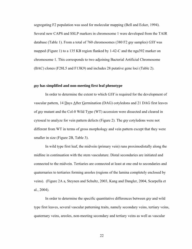
chromosome 1. This corresponds to two adjoining Bacterial Artificial Chromosome
(BAC) clones (F28L5 and F13K9) and includes 28 putative gene loci (Table 2).
gsy has simplified and non-meeting first leaf phenotype
In order to determine the extent to which GSY is required for the development of
vascular pattern, 14 Days After Germination (DAG) cotyledons and 21 DAG first leaves
of gsy mutant and the Col-0 Wild Type (WT) accession were dissected and cleared in
cytoseal to analyze for vein pattern defects (Figure 2). The gsy cotyledons were not
different from WT in terms of gross morphology and vein pattern except that they were
smaller in size (Figure 2B, Table 3).
In wild type first leaf, the midvein (primary vein) runs proximodistally along the
midline in continuation with the stem vasculature. Distal secondaries are initiated and
connected to the midvein. Tertiaries are connected at least at one end to secondaries and
quaternaries to tertiaries forming areoles (regions of the lamina completely enclosed by
veins). (Figure 2A a, Steynen and Schultz, 2003, Kang and Dangler, 2004, Scarpella et
al., 2004).
In order to determine the specific quantitative differences between gsy and wild
type first leaves, several vascular patterning traits, namely secondary veins, tertiary veins,
quaternary veins, areoles, non-meeting secondary and tertiary veins as well as vascular
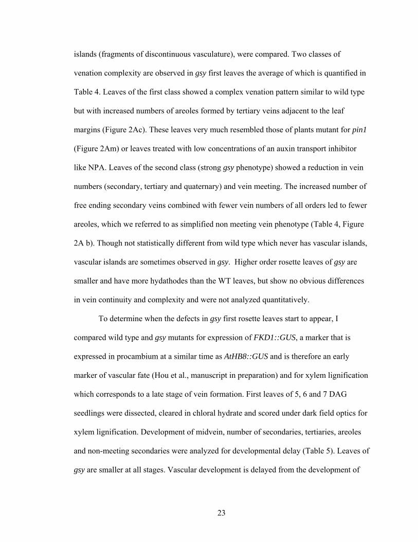
23
islands (fragments of discontinuous vasculature), were compared. Two classes of
venation complexity are observed in gsy first leaves the average of which is quantified in
Table 4. Leaves of the first class showed a complex venation pattern similar to wild type
but with increased numbers of areoles formed by tertiary veins adjacent to the leaf
margins (Figure 2Ac). These leaves very much resembled those of plants mutant for pin1
(Figure 2Am) or leaves treated with low concentrations of an auxin transport inhibitor
like NPA. Leaves of the second class (strong gsy phenotype) showed a reduction in vein
numbers (secondary, tertiary and quaternary) and vein meeting. The increased number of
free ending secondary veins combined with fewer vein numbers of all orders led to fewer
areoles, which we referred to as simplified non meeting vein phenotype (Table 4, Figure
2A b). Though not statistically different from wild type which never has vascular islands,
vascular islands are sometimes observed in gsy. Higher order rosette leaves of gsy are
smaller and have more hydathodes than the WT leaves, but show no obvious differences
in vein continuity and complexity and were not analyzed quantitatively.
To determine when the defects in gsy first rosette leaves start to appear, I
compared wild type and gsy mutants for expression of FKD1::GUS, a marker that is
expressed in procambium at a similar time as AtHB8::GUS and is therefore an early
marker of vascular fate (Hou et al., manuscript in preparation) and for xylem lignification
which corresponds to a late stage of vein formation. First leaves of 5, 6 and 7 DAG
seedlings were dissected, cleared in chloral hydrate and scored under dark field optics for
xylem lignification. Development of midvein, number of secondaries, tertiaries, areoles
and non-meeting secondaries were analyzed for developmental delay (Table 5). Leaves of
gsy are smaller at all stages. Vascular development is delayed from the development of
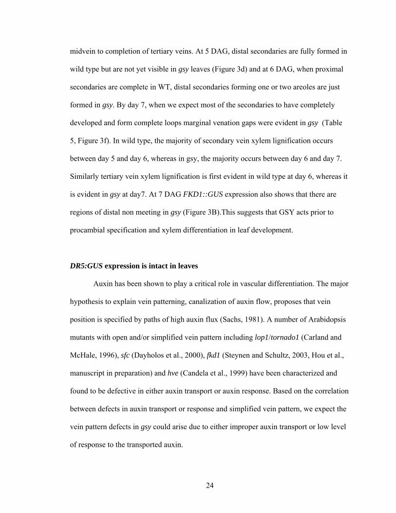
24
midvein to completion of tertiary veins. At 5 DAG, distal secondaries are fully formed in
wild type but are not yet visible in gsy leaves (Figure 3d) and at 6 DAG, when proximal
secondaries are complete in WT, distal secondaries forming one or two areoles are just
formed in gsy. By day 7, when we expect most of the secondaries to have completely
developed and form complete loops marginal venation gaps were evident in gsy (Table
5, Figure 3f). In wild type, the majority of secondary vein xylem lignification occurs
between day 5 and day 6, whereas in gsy, the majority occurs between day 6 and day 7.
Similarly tertiary vein xylem lignification is first evident in wild type at day 6, whereas it
is evident in gsy at day7. At 7 DAG FKD1::GUS expression also shows that there are
regions of distal non meeting in gsy (Figure 3B).This suggests that GSY acts prior to
procambial specification and xylem differentiation in leaf development.
DR5:GUS expression is intact in leaves
Auxin has been shown to play a critical role in vascular differentiation. The major
hypothesis to explain vein patterning, canalization of auxin flow, proposes that vein
position is specified by paths of high auxin flux (Sachs, 1981). A number of Arabidopsis
mutants with open and/or simplified vein pattern including lop1/tornado1 (Carland and
McHale, 1996), sfc (Dayholos et al., 2000), fkd1 (Steynen and Schultz, 2003, Hou et al.,
manuscript in preparation) and hve (Candela et al., 1999) have been characterized and
found to be defective in either auxin transport or auxin response. Based on the correlation
between defects in auxin transport or response and simplified vein pattern, we expect the
vein pattern defects in gsy could arise due to either improper auxin transport or low level
of response to the transported auxin.
25
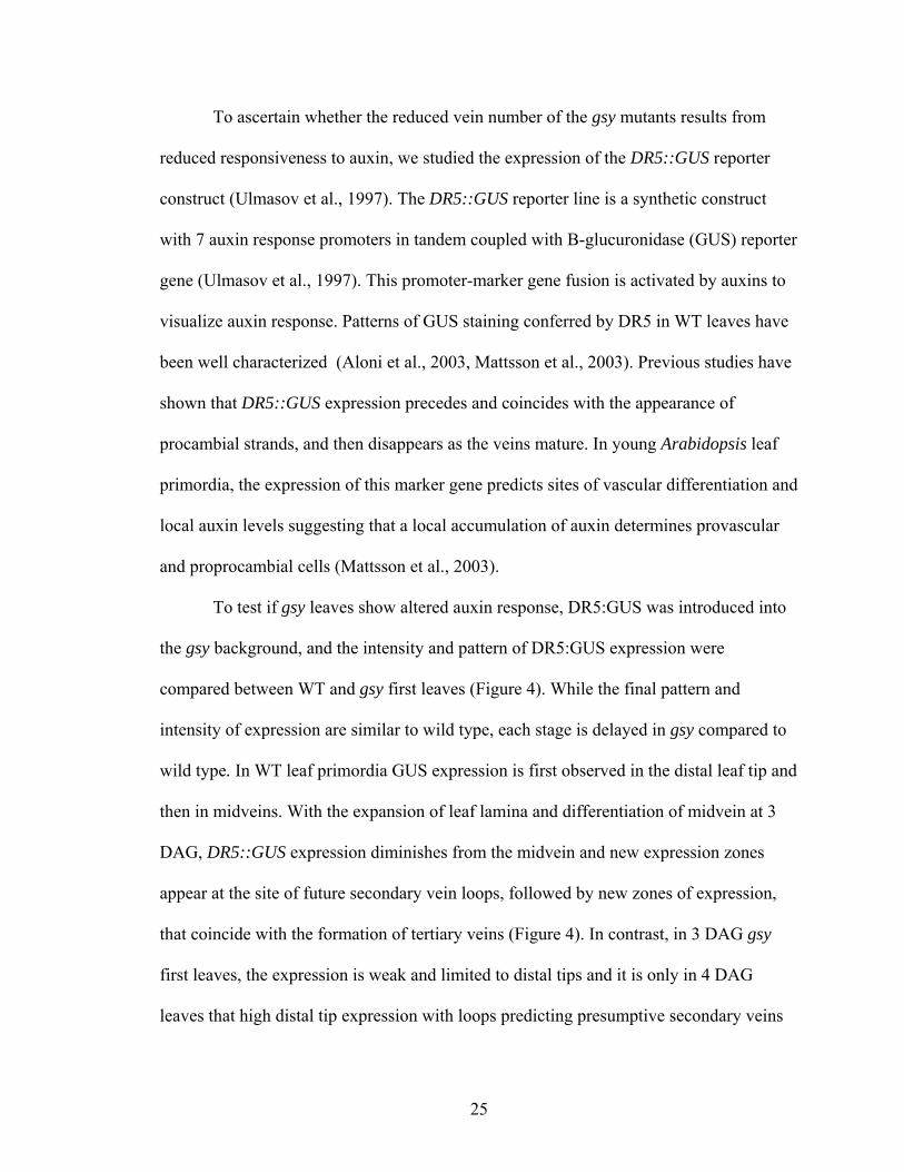
To ascertain whether the reduced vein number of the gsy mutants results from
reduced responsiveness to auxin, we studied the expression of the DR5::GUS reporter
construct (Ulmasov et al., 1997). The DR5::GUS reporter line is a synthetic construct
with 7 auxin response promoters in tandem coupled with B-glucuronidase (GUS) reporter
gene (Ulmasov et al., 1997). This promoter-marker gene fusion is activated by auxins to
visualize auxin response. Patterns of GUS staining conferred by DR5 in WT leaves have
been well characterized (Aloni et al., 2003, Mattsson et al., 2003). Previous studies have
shown that DR5::GUS expression precedes and coincides with the appearance of
procambial strands, and then disappears as the veins mature. In young Arabidopsis leaf
primordia, the expression of this marker gene predicts sites of vascular differentiation and
local auxin levels suggesting that a local accumulation of auxin determines provascular
and proprocambial cells (Mattsson et al., 2003).
To test if gsy leaves show altered auxin response, DR5:GUS was introduced into
the gsy background, and the intensity and pattern of DR5:GUS expression were
compared between WT and gsy first leaves (Figure 4). While the final pattern and
intensity of expression are similar to wild type, each stage is delayed in gsy compared to
wild type. In WT leaf primordia GUS expression is first observed in the distal leaf tip and
then in midveins. With the expansion of leaf lamina and differentiation of midvein at 3
DAG, DR5::GUS expression diminishes from the midvein and new expression zones
appear at the site of future secondary vein loops, followed by new zones of expression,
that coincide with the formation of tertiary veins (Figure 4). In contrast, in 3 DAG gsy
first leaves, the expression is weak and limited to distal tips and it is only in 4 DAG
leaves that high distal tip expression with loops predicting presumptive secondary veins
26
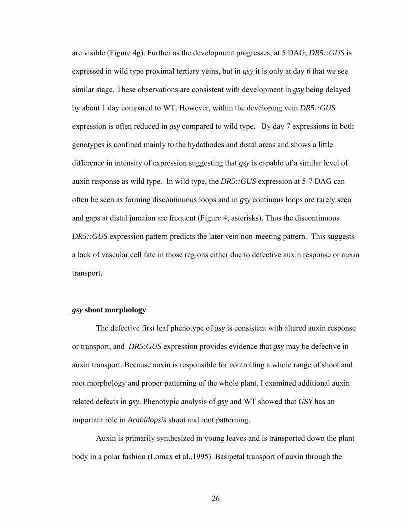
are visible (Figure 4g). Further as the development progresses, at 5 DAG, DR5::GUS is
expressed in wild type proximal tertiary veins, but in gsy it is only at day 6 that we see
similar stage. These observations are consistent with development in gsy being delayed
by about 1 day compared to WT. However, within the developing vein DR5::GUS
expression is often reduced in gsy compared to wild type. By day 7 expressions in both
genotypes is confined mainly to the hydathodes and distal areas and shows a little
difference in intensity of expression suggesting that gsy is capable of a similar level of
auxin response as wild type. In wild type, the DR5::GUS expression at 5-7 DAG can
often be seen as forming discontinuous loops and in gsy continous loops are rarely seen
and gaps at distal junction are frequent (Figure 4, asterisks). Thus the discontinuous
DR5::GUS expression pattern predicts the later vein non-meeting pattern. This suggests
a lack of vascular cell fate in those regions either due to defective auxin response or auxin
transport.
gsy shoot morphology
The defective first leaf phenotype of gsy is consistent with altered auxin response
or transport, and DR5:GUS expression provides evidence that gsy may be defective in
auxin transport. Because auxin is responsible for controlling a whole range of shoot and
root morphology and proper patterning of the whole plant, I examined additional auxin
related defects in gsy. Phenotypic analysis of gsy and WT showed that GSY has an
important role in Arabidopsis shoot and root patterning.
Auxin is primarily synthesized in young leaves and is transported down the plant
body in a polar fashion (Lomax et al.,1995). Basipetal transport of auxin through the
27
plant body from its site of synthesis in young leaves inhibits shoot branching by
inhibiting elongation of bud growth (Leyser, 2003). The axr1 mutation does not affect the
timing of axillary meristem formation; however, subsequent lateral shoot development
proceeds more rapidly in axr1 plants (Stirnberg et al., 1999). As well, auxin response is
important in regulating flowering time and inflorescence height. Mutation in ARF2, a
transcriptional repressor of the auxin response, results in late flowering and elongated
inflorescence (Okusima et al., 2005). gsy mutants have defects in plant growth at a
variety of developmental stages. Compared to wild type, gsy plants are dwarf and have
fewer rosette and cauline leaves (Table 6, Figure 5). Less pronounced elongation in
internodes of primary inflorescences, along with an enhanced initiation of axillary shoot
in the rosette (Table 7, Figure 5b) confers a bushy and dwarf-like appearance to the adult
mutant plant. The bushy appearance is intensified upon reiteration of bud outgrowth in
secondary and tertiary inflorescence. gsy mutants bolt, flower and produce mature seed
earlier than wild type. They also have smaller siliques that shatter more easily than wild
type.
gsy root phenotype
Auxin is a key regulator of primary root elongation and gravitropism, lateral root
development and root hair development (Laskowski et al., 1995, Estelle, 1996, Casimiro
et al., 2001). While basipetal movement of IAA from the root tip back has been linked to
root elongation and gravity response (Rashotte et al., 2000), basipetal and acropetal polar
auxin transport activities are required during the initiation and emergence phases of
lateral root development respectively (Casimiro et al., 2001). The primary root in gsy was
shorter and the growth rate was also significantly reduced compared to the wild type
28
(Table 8, Figure 6). We also see a slight defect in root gravitropism where 6 DAG gsy
roots, when grown vertically on AT media, showed a slight deviation from the normal
vector (Figure 6). Plants mutant for GSY also showed significantly reduced number of
lateral roots (Table 7). All of these changes suggest that basipetal transport may also be
defective in gsy.
If gsy is defective in auxin transport then we might expect to see defects in root
hair phenotype. Root hair elongation can be used as a biological marker to study the
activity of auxin transporters (Cho et al., 2007). Several mutants defective in auxin
transport show enhanced root hair length (Pitts., 1998, Rahman et al., 2002) that is most
likely due to either a defect in auxin supply to the root hair cell or a loss of auxin-
transporting activity in the hair cell (Cho et al., 2007). The gsy mutant root has longer root
hairs than the wild-type root (Figure 7A ). Enhancement of root hair elongation following
the loss of GSY might imply that GSY catalyzes auxin efflux in root hair cells, resulting in
increased auxin retention inside the hair cell and the stimulation of root hair elongation.
DR5::GUS expression in roots.
Inhibition of polar auxin transport and/or improper auxin flux affects the distal
auxin maximum, which correlates with the pattern formation, orientation and extent of
cell division (Sabatini et al., 1999). The role of the GSY in auxin transport was examined
by analysis of the expression of DR5:GUS in the roots of wild type and the gsy mutant.
Wild type plants displayed the highest GUS activity in the quiescent center, columella
initials and mature columella root cap (Figure 8a,b, Sabatini et al., 1999). The GUS
staining pattern in gsy (Figure 8 e,f,g) shows a range of variation in expression including
stronger response in the regions including columella, root meristem and quiescent center
29
(e), similar response but asymmetric in epidermal cells (f) and weaker expression (g). As
well, gsy often shows ectopic DR5::GUS expression in the stele, lateral root cap and
epidermis. The ectopic and asymmetric expression pattern, which is common in gsy, was
never observed in the wild type plants in identical conditions of growth media and
staining duration. This suggests that GSY is required for correct localization of the DR5
peak in the root tip. This mislocalization could also be linked to defects in root hair and
lateral root suggesting that proper root patterning and cell division require proper auxin
localization. Together with increased root hair length, reduction in lateral roots and slight
defects of gravitropism, the ectopic auxin response in root epidermis is consistent with
gsy being defective in basipetal auxin transport.
Effect of auxin transport inhibition in leaves and roots
To further investigate the relationship between GSY function and polar auxin
transport in vein pattern formation, root patterning and overall morphology of the plant, I
treated gsy and WT plants with the auxin transport inhibitor, N-1-naphthylphthalamic
acid (NPA). Treating root tissues directly with NPA arrests lateral root development by
blocking the first transverse division(s) of xylem pole pericycle cells. NPA appears to
exert its developmental effects by causing IAA to accumulate in the root apex while
reducing levels in basal tissues critical for lateral root initiation (Casimiro et al., 2001). In
gsy the primary and lateral root growth was arrested at 1μM NPA compared to 10μM
NPA in WT. Similarly, lack of gravitropic response and proliferation of root hairs was
evident in gsy at lower concentration (5μM) while in WT it was observed at 10μM
(Figure 7B). Finally root tip bulging was also evident in gsy at a lower concentration of
30
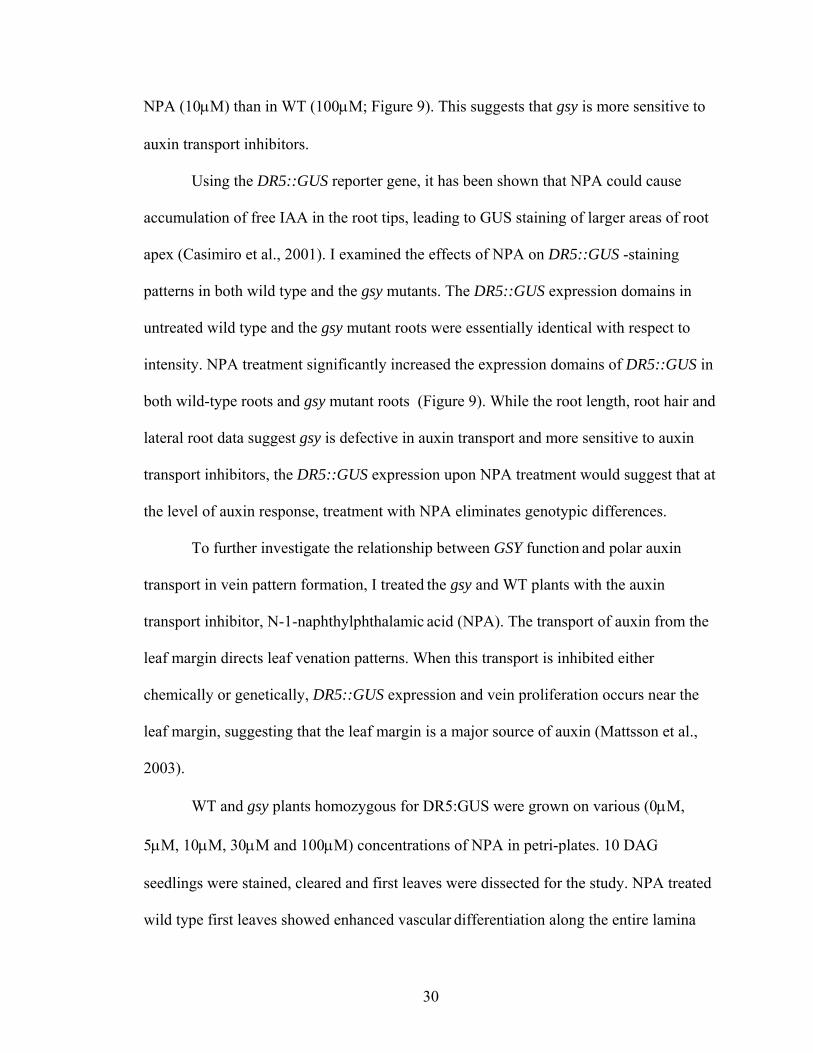
NPA (10μM) than in WT (100μM; Figure 9). This suggests that gsy is more sensitive to
auxin transport inhibitors.
Using the DR5::GUS reporter gene, it has been shown that NPA could cause
accumulation of free IAA in the root tips, leading to GUS staining of larger areas of root
apex (Casimiro et al., 2001). I examined the effects of NPA on DR5::GUS -staining
patterns in both wild type and the gsy mutants. The DR5::GUS expression domains in
untreated wild type and the gsy mutant roots were essentially identical with respect to
intensity. NPA treatment significantly increased the expression domains of DR5::GUS in
both wild-type roots and gsy mutant roots (Figure 9). While the root length, root hair and
lateral root data suggest gsy is defective in auxin transport and more sensitive to auxin
transport inhibitors, the DR5::GUS expression upon NPA treatment would suggest that at
the level of auxin response, treatment with NPA eliminates genotypic differences.
To further investigate the relationship between GSY function and polar auxin
transport in vein pattern formation, I treated the gsy and WT plants with the auxin
transport inhibitor, N-1-naphthylphthalamic acid (NPA). The transport of auxin from the
leaf margin directs leaf venation patterns. When this transport is inhibited either
chemically or genetically, DR5::GUS expression and vein proliferation occurs near the
leaf margin, suggesting that the leaf margin is a major source of auxin (Mattsson et al.,
2003).
WT and gsy plants homozygous for DR5:GUS were grown on various (0μM,
5μM, 10μM, 30μM and 100μM) concentrations of NPA in petri-plates. 10 DAG
seedlings were stained, cleared and first leaves were dissected for the study. NPA treated
wild type first leaves showed enhanced vascular differentiation along the entire lamina
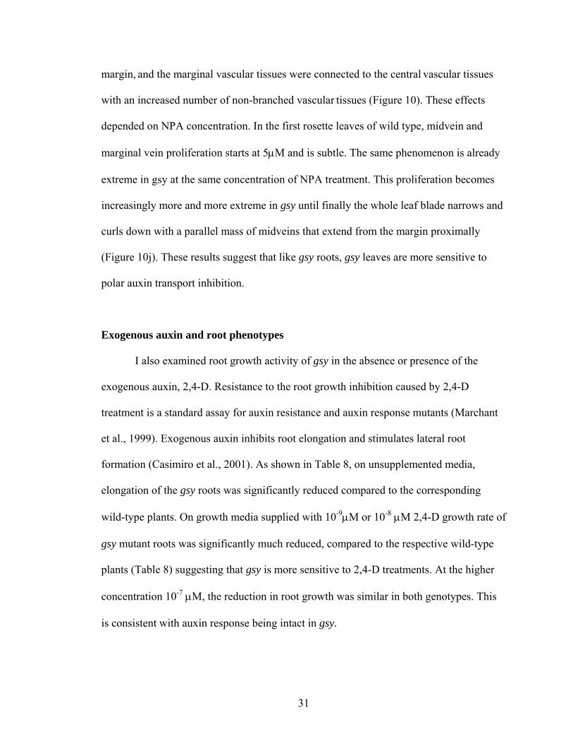
31
margin, and the marginal vascular tissues were connected to the central vascular tissues
with an increased number of non-branched vascular tissues (Figure 10). These effects
depended on NPA concentration. In the first rosette leaves of wild type, midvein and
marginal vein proliferation starts at 5μM and is subtle. The same phenomenon is already
extreme in gsy at the same concentration of NPA treatment. This proliferation becomes
increasingly more and more extreme in gsy until finally the whole leaf blade narrows and
curls down with a parallel mass of midveins that extend from the margin proximally
(Figure 10j). These results suggest that like gsy roots, gsy leaves are more sensitive to
polar auxin transport inhibition.
Exogenous auxin and root phenotypes
I also examined root growth activity of gsy in the absence or presence of the
exogenous auxin, 2,4-D. Resistance to the root growth inhibition caused by 2,4-D
treatment is a standard assay for auxin resistance and auxin response mutants (Marchant
et al., 1999). Exogenous auxin inhibits root elongation and stimulates lateral root
formation (Casimiro et al., 2001). As shown in Table 8, on unsupplemented media,
elongation of the gsy roots was significantly reduced compared to the corresponding
wild-type plants. On growth media supplied with 10-9μM or 10-8 μM 2,4-D growth rate of
gsy mutant roots was significantly much reduced, compared to the respective wild-type
plants (Table 8) suggesting that gsy is more sensitive to 2,4-D treatments. At the higher
concentration 10-7 μM, the reduction in root growth was similar in both genotypes. This
is consistent with auxin response being intact in gsy.
32
To examine whether GSY is required for auxin-mediated lateral-root formation, I
examined the lateral-root formation in the presence or absence of exogenous auxin (Table
8). When grown vertically on unsupplemented media, gsy mutants produced fewer lateral
roots than did wild type control plants (Table 7). When 5DAG seedlings were transferred
to media supplemented with 10-8 M 2,4-D and grown for 4 days, gsy was sensitive to the
auxin treatment and developed more lateral roots compared to wild type and at 10-7 M,
the gsy mutant still produced the same frequency of lateral roots as wild type. Like the
primary root growth assay, this suggests that gsy is more sensitive than wild type to low
levels of 2,4-D and at higher level the auxin is in sufficient excess that genotypic
differences are eliminated.
Double mutants
To investigate the functional relationship of GSY with genes required for auxin
transport (PIN, AUX, EIR), perception (AXR1, MP) and synthesis (RTY) as well as a gene
specifically required for vascular development (CVP2), I obtained the corresponding
double mutants and quantitatively analyzed their venation patterns for secondaries,
tertiaries, quarternaries, areoles, non meeting veins and vascular islands (Table 4), lateral
root and shoot branching (Table 7) and root hair morphology (Figure 12).
gsy and auxin transport mutants
To examine possible roles of GSY in auxin transport we analyzed the double
mutants of gsy together with pin1, eir1, aux1. The auxin efflux carrier PIN1 has been
shown to be expressed at the earliest stages of leaf primordium development (Reinhardt
et al., 2003 Scarpella et al., 2006). Like plants in which auxin transport has been inhibited
chemically, plants homozygous for the loss-of-function allele, pin1-1, are defective in
33
polar auxin transport displaying phenotypes including increased marginal venation
(Mattsson et al., 1999, Galweiler et al., 1998) and sometimes fused leaves. The overall
leaf vein pattern of pin1-1 was not significantly different from wild type except that they
had fewer non-meeting tertiaries. EIR/PIN2 is a root specific efflux protein with a less
significant role in leaves. The roots are agravitropic and have a reduced sensitivity to
ethylene (Lusching et al., 1998). No specific leaf vascular phenotype has been reported
for the mutant and PIN2 transcripts were not fount in 5 DAG leaves (Scarpella et al.,
2006) but in this present study I found that the first leaf of eir is simpler with fewer
tertiary and quaternary veins and fewer areoles. AUX1 encodes a membrane protein that
is believed to be a component of the auxin influx carrier (Bennet et al., 1996, Merchant et
al., 1999, Swarup et al., 2001). AUX1 regulates root gravitropism by facilitating auxin
uptake within the root apical tissues (Marchant et al., 1999) so we expect the mutation in
AUX1 to impair auxin influx carrier activity. Although no previous leaf vein defect has
been reported (Steynen and Schultz, 2003) my analysis shows that like eir1, it is simpler
with fewer veins and areoles.
Compared with gsy, the gsy pin1-1 double mutant first leaves were significantly
more complex than gsy with increased numbers of areoles, secondary, tertiary and
quaternary veins as well as fewer non meeting veins (Table 4), suggesting that pin1 is
epistatic to gsy in leaves. The pin1 mutants has wild type rosette leaves but a single pin-
formed inflorescences with no cauline leaves, secondary inflorescence, flowers or
siliques due to lack of auxin transported to the shoot apex from the young organs (Figure
5e). I also analyzed the effect of pin1 on gsy shoot branching which suggest that pin1
shoot phenotype is suppressed by gsy. The lack of organs in pin1 was rescued in gsy
34
background, both axillary shoots and partial flowers develop in gsy pin1. The
suppression of pin1 by gsy in shoot apex suggest that gsy may work in opposition to
pin1, perhaps by causing auxin accumulation in the shoot apex.
Like the gsy pin1 double mutant, the first leaf of gsy eir and gsy aux1-7 double
mutants is more complex than gsy single mutant suggesting that gsy is suppressed (Table
4, Figure 2A). Other characteristics of gsy are also supressed in other transport mutant
backgrounds (Table 7). In gsy we have fewer lateral roots and higher number of rosettes
branches whereas, in the double mutants with eir1-1 and aux1-7 the number of rosette
branches is decreased and lateral roots increased (Table 7). This suppression of the gsy
first leaf phenotype by eir1, aux1 or pin1 suggest either that gsy and the auxin
transporters have opposing function or that the gsy phenotype results from over
expression of these genes.
gsy and auxin response (mp, axr1) and auxin synthesis (rty) mutant
AUXIN RESISTANT1 (AXR1), encodes a subunit of the RUB1-activating enzyme
that promotes the modification of CUL1 with RUB1 of the ubiquitin pathway (Lincoln et
al., 1990; Leyser et al., 1993, del Pozo et al., 2002) and loss of AXR1 function causes
auxin insensitivity and a bushy inflorescence, the latter due to a decrease in the inhibition
of apical dominance mediated by auxin (Estelle and Somerville, 1987; Lincoln et al.,
1990, Stirnberg et al., 1999). The axr1 homozygous single mutant has a very simple first
leaf venation with a higher number of non-meeting veins secondaries and lower numbers
of secondary, tertiary, quaternary veins and areoles (Table 4, Figure 2A). The double
mutant is more extreme than either of gsy or axr1 with higher number of non-meeting
veins secondaries, fewer secondaries, tertiaries, no quartenaries and very few areoles.
35
One could interpret the phenotype as either additive or synergistic suggesting that two
genes function either in independent pathways or overlapping and partially redundant
pathways. With respect to the shoot branching and root hairs, axr1 appears to be epistatic
to gsy, with the double mutant having higher number of cauline branches and sparsely
distributed root hairs characteristic of axr1 single mutant (Table 7, Figure 5).
The MP gene encodes an auxin response factor IAA24/ARF5 that mediates auxin
signaling. Mutations in the MP gene interfere with the initiation of body axis in the early
embryo and with the formation of vascular strands at all stages (Hardtke and Berleth
1998). Phenotypic characterizations have shown that loss-of-function allele mpG92
mutants have no roots, highly reduced hypocotyls, and cotyledons with highly reduced
vein pattern (Berleth and Jurgens, 1993). To examine the genetic interaction between the
gsy and mp G92 mutations, I generated gsy mp G92 double mutants. Since no other organs
except cotyledons were produced, I compared the cotyledons of the corresponding single
and double mutant (Table 3). The cotyledons of mp G92 mutants contained a midvein that
rarely extends the full length and less often a secondary vein arising from the midvein
that fails to join the midvein in the proximal region, thus no areoles (Figure 2B). The
double mutant phenotype was essentially the same as mp G92 single mutant in that it
lacked hypocotyls, root and any leaves. The cotyledon vein formation was also similar to
mp G92 except that sometimes two secondaries originated from the midvein but again
failed to form an areoles. The overall data suggest that that mp is epistatic to gsy. (Table
3).
To investigate further the role of GSY in auxin-regulated leaf root architecture,
double mutants were made by crossing the gsy mutant with the rooty (rty) mutant. rty is
36
is characterized by high endogenous levels of auxin and the production of supernumerary
lateral roots and adventitious roots on the hypocotyl (King et al., 1995, Boerjan et al.,
1995, Figure 12m). The gsy rty double mutants produced the same root phenotype as the
rty mutant (Figure 12n). Again, like the strong adventitious root mutant rty (Boerjan et
al., 1995, King et al., 1995), gsy rty double mutant was also sterile. Finally, the first leaf
of rty gsy showed drastic simplification in its vein pattern very similar to rty and often
resembling that of the cotyledon (Figure 2Aj,o). This overall result suggests that gsy is
sensitive to auxin. rty is epistatic to gsy and the simple vein pattern of gsy cannot be
ameliorated simply by increasing auxin levels.
gsy and cvp2
CVP2 encodes an inositol polyphosphate 5’ phosphatase and the mutants produce
first leaves and cotyledons with non-meeting veins veins and vascular islands (Table 4,
Figure 2Ad, Carland and Nelson, 2004). The gsy cvp2 double mutant was additive, with
similar low complexity as either of the single mutants together with non-meeting veins
and vascular islands associated with the cvp2 single mutant. cvp 2 seems to suppress gsy
in lateral roots and rosette branches but gsy is epistatic to cvp2 in cauline branches.
37
DISCUSSION
It is now widely accepted that auxin has a universal role in plant development.
Molecular genetic research of auxin transport and auxin signaling has yielded great
insight into a wide variety of patterning processes from apical/basal polarities in young
embryos to tissue patterning. We have identified a leaf vein patterning mutant called
grassy (gsy) that shows a simple first leaf vein pattern with reduced numbers of
secondary, tertiary and quaternary veins. Furthermore, the vascular development is
delayed in gsy by about one day as suggested by temporal expression of DR5::GUS and
xylem differentiation, implying that gsy acts prior to auxin response in leaf development.
At each stage, gsy leaf primordia are smaller than wild type, suggesting that all aspects of
leaf development are delayed. Moreover, the veins are often non-meeting and form
somewhat parallel to one another and the leaves are narrow and pointed. Along with
alterations to leaf phenotype, gsy shoots show increased growth of axillary buds and
shortened internodes, gsy roots are shortened, show slight defects to gravitropism, have
decreased formation of lateral roots, and the root hairs are elongated.
The spectrum of defects suggests that GSY has a global role in plant development
likely involving auxin response or transport. The recessive nature of gsy indicates that it
likely represents a loss-of-function mutation. I hypothesize that these auxin related
phenotypic traits are the consequence of altered auxin transport.
gsy shows no reduction in auxin response
While many of the gsy phenotypic characteristics are consistent with defects to
either auxin response or auxin transport, my analysis suggests that gsy is not defective in
38
auxin response. Resistance to the root growth inhibition caused by 2,4-D treatment is a
standard assay for auxin resistance in auxin response mutants (Marchant et al., 1999).
Roots of seedlings homozygous for axr1 are resistant to auxin inhibition of growth
(Estelle and Somerville, 1987). My results show that inhibition of gsy root elongation is
not resistant but rather more sensitive to 2,4-D as compared to wild type. Lateral root
formation is induced by either exogenous or endogenous shoot derived auxin and
exogenous auxin results in increased formation of lateral roots in wild type (Bholero et
al., 2002, Casimiro et al., 2003). Exogenous auxin restored lateral root production in gsy.
Like root elongation, lateral root development is more sensitive to 2,4-D. As compared to
wild type, induction of root hair elongation in gsy mutants by 2,4-D is greater than that in
wild type. Moreover, the additive nature of the gsy axr1 double mutant suggests that GSY
and AXR1 act in independent pathway. These findings suggest that gsy mutants are not
resistant to auxin inhibition of growth. Further support for this conclusion comes from
gsy rty double mutant where rty, an auxin over-synthesizing mutant (King et al., 2005),
further simplifies the gsy simple leaf vein pattern, reduces the overall length of primary
root, increases root hair elongation and rescues lateral root production in gsy. Together
these results suggest that response to both endogenous and exogenous auxin is intact in
gsy.
Further indication that the auxin response is intact in gsy plants is the normal level
of DR5::GUS expression seen in gsy. In DR5::GUS construct, b-glucuronidase is
controlled by synthetic auxin response element and, when assayed histochemically,
provides an indication of auxin activity in the tissues (Ulmasov et al., 1997). My analysis
of developing first leaf and root indicates that the gsy mutation disrupts the temporal and
39
spatial arrangement of the auxin response cells but not the intensity of expression. The
combination of normal intensity and change to patterning of auxin response suggests that
while auxin the response is intact, transport may be defective.
gsy is defective in auxin transport
If the phenotypic characteristics of gsy including simplified leaf vein pattern,
reduced root growth, defective gravitropism and reduced number of lateral roots result
from defects to auxin transport, we might expect that these characteristics would be very
sensitive to further chemical inhibition of auxin transport by NPA.
As my results show, the first leaf vein pattern in gsy is in fact more sensitive to
auxin transport inhibition treatments. Similar results have been found in the van3 mutant
where the effect of an auxin transport inhibitor and of the VAN3 mutation are additive in
the formation of the venation pattern in Arabidopsis leaf (Koizumi et al., 2005).
Moreover reduced root growth, enhanced root hair elongation, loss of gravitropism and
loss of capacity to produce lateral roots in gsy mutants occur at relatively lower
concentrations of NPA compared to wild type, suggesting that GSY is involved in auxin
transport. I expected that gsy would show similar sensitivity to auxin transport mutants
such as pin1, aux1 and pin2/eir1. Surprisingly, all auxin transport mutants tested either
suppress or are suppressed by the gsy mutation. This suggests either that the transport
activity of GSY acts in opposition to that of these genes, or that the defective auxin
transport in gsy results from the over expression of these genes.
40
Shoot transport
A range of shoot phenotypic characters defective in gsy have previously been
found to be due to defective auxin transport. gsy plants bolt and flower early with fewer
rosette leaves, have increased rosette branching (axillary bud growth) pattern and produce
an inflorescence with reduced internode elongation. Similar phenotypes may result from
defects to auxin transport for example, plants mutant for MY 2, MAX1 or MAX2 (Holweg
and Nick, 2004, Stirnberg et al., 2002).
The increased rosette branching in gsy mutants suggests that axillary buds are
released from apical dominance more frequently indicating that GSY negatively regulates
axillary growth in Arabidopsis. Auxin is required for shoot elongation (Lomax et al.,
1995) and apically derived auxin inhibits shoot branching by inhibiting the activity of
axillary buds (Leyser, 2003). The increased lateral bud outgrowth and altered internode
elongation of the gsy shoot is consistent with defects to basipetal auxin transport.
The branching of gsy is suppressed in either eir1 or aux1 backgrounds. One
explanation is that since EIR1 and AUX act in roots, loss of either activity might lead to
more auxin in the shoot due to lack of a functional sink. The increased auxin would then
prevent lateral outgrowth in the double mutant. The lack of axillary meristem in pin1 is
proposed to result from lack of auxin within the shoot apical meristem. The formation of
axillary meristems in the gsy pin1 double mutant is most consistent with gsy showing
reduced basipetal transport, which would then cause auxin to accumulate in the apex.
41
Root transport
A coordinated action of PIN proteins determines the stable pattern of auxin
accumulation in the root. Since much of the auxin in the root is shoot derived, changes to
shoot transport are expected to affect root transport. Two of the shoot derived auxin
fluxes, polar auxin transport and phloem transport, function to mobilize shoot-derived
auxin to the root apex. After reaching the root apex, auxin forms a gradient that is centred
over the root apical meristem. Mutant studies have revealed that five PIN proteins (PIN1,
PIN2, PIN3, PIN4 and PIN7) collectively fine tune the root apical auxin gradient (Blilou
et al., 2005). The coordinated action of PIN1, PIN4 and PIN7 mediate acropetal auxin
transport through the central vasculature towards the tip, PIN3 exports auxin from the
meristem and finally PIN2/EIR1 transports auxin basipetally back up the sides through
the epidermis (Friml, 2003, Rashotte et al., 2000). This basipetal flow of auxin has been
shown to be an important component regulating cell expansion in the elongation zone.
The auxin maximum at the root tip is the result of the steady state balance between the
arrival and departure of auxin. A further auxin flux in the root apex, termed auxin reflux,
involving the recycling of auxin from epidermal to stele tissues and back to the root apex,
has been proposed (Blilou et al., 2005). This suggests that the auxin maximum in the root
tip is supplied, not only by an acropetal flow of auxin from further up the root, but also
by auxin being constantly recycled back into the maximum. Multiple pin mutant
combinations disrupt the meristematic and cell expansion zone organization (Blilou et al.,
2005) suggesting that PIN auxin efflux facilitators might be functionally important for
this auxin reflux pathway.
42
The spectrum of gsy root phenotypic characteristics is most consistent with GSY
being required for basipetal auxin transport and possibly having a role in reflux looping
of the auxin. Compared to wild type, gsy plants have changes to DR5::GUS expression
pattern, shorter and slightly agravitropic primary root, reduced numbers of lateral roots
and increased numbers of longer root hairs.
I expected that the reduced basipetal auxin transport in gsy shoot would result in
greatly reduced DR5::GUS expression in root. In fact four different patterns and levels
of DR5::GUS expression are observed in the gsy root tip. Consistent with my prediction,
some roots show weaker DR5::GUS expression (Figure 8g) likely due to defective auxin
transport from the shoot. Other roots show a similar or higher level of expression as in
wild type (Figure 8f). This can be explained either by compensatory synthesis of auxin at
the root tip or by increased reflux from the basipetal stream. In many gsy roots, ectopic
and asymmetric localization in the epidermal region and lateral root (Figure 8 e,f)
suggests defective basipetal transport of auxin and ectopic expression at the stele (Figure
8 e,g) likely by auxin backing up in the stele, also due to defective auxin transport.
The reduced number of lateral roots in gsy mutants could either be due to reduced
auxin delivery from the gsy shoot or altered auxin transport within the root itself. It is
widely accepted that impairing auxin transport affects lateral roots (Reed et al., 1998) and
recent studies suggest that tip-produced, basipetally transported auxin is required for the
initiation of lateral root primordia, while acropetally transported auxin from the shoot is
required for its subsequent growth (Casimiro et al., 2001, Bhalerao, et al., 2002). Table 7
shows a reduced number of emerged lateral roots in gsy; I observed a similar reduction in
lateral root primordium as indicated by DR5::GUS maximum suggesting that lateral root
43
initiation is defective. A decrease in lateral root initiation is consistent with defective
basipetal transport of auxin in gsy.
Auxin is a well defined positive regulator of root hair elongation. PIN2 is
expressed in a non-overlapping pattern in the lateral root cap and older epidermis and
cortex cells with apical (upper) polarity in the epidermis and predominantly basal polarity
in the cortex (Müller et al., 1998, Friml et al., 2003). It has been shown that defects to
auxin efflux carriers PIN2 or AtPGP4 (Cho et al., 2007) within epidermal cells can
increase auxin in the hair and hence induce root hair elongation. The elongated root hairs
seen in gsy are consistent with GSY being an auxin transporter expressed in epidermal
cells.
The agravitropic root behaviour is often observed in mutants defective in auxin
transport (Marchant et al., 1999, Muller et al., 1998) or signalling (Hobbie and Estelle,
1995, Nagpal et al., 2000). This bending depends on differential cell elongation resulting
from differential lateral and basipetal auxin transport from the root tip back to the
elongation zone (Friml, 2003, Rashotte et al., 2000). The lateral auxin gradient is
transported via the lateral root cap to expanding epidermal cells through the combined
activities of AUX1 and PIN2. The basipetal stream is thought to be the mechanism that
delivers auxin asymmetrically from the tip after reorientation, so impairments in it might
be expected to impair curvature development. Mutations in PIN2, AUX1 (Chen et al.,
1998; Muller et al., 1998) or auxin transport inhibitors such as naphthylphthalamic acid
(NPA) impair basipetal auxin transport and gravitropism (Muday, 2001). Consistent with
the asymmetric DR5::GUS expression in gsy epidermis the primary root of gsy was
44
slightly deviated from the vertical axis compared to wild type (Figure 6b) further
supporting the role of GSY in basipetal auxin transport.
My results overall suggest defects to basipetal transport in gsy roots. While GSY
may directly affect basipetal transport, an alternative suggested by the asymmetric and
sometimes overexpression of DR5::GUS is that the change in basipetal transport is the
result of root tip compensation, either by synthesis or by reflux, of reduced auxin
transported from the shoot.
In either case, we might now predict that GSY participates in the standard fountain
model of auxin flow in roots (Swarup and Bennett, 2003) to include reflux loops that
cause auxin to recirculate from epidermal and cortical cells back into the stele, where it
rejoins the acropetal stream (Blilou et al., 2005). Thus GSY plays a role in forming and
maintaining apical–basal auxin gradients that are essential for establishing polarity and
functions in overall root auxin transport.
Leaf transport
The gsy mutant first leaf vascular pattern is distinct from wildtype with a
reduction in number of veins and areoles and an increase in non-meeting veins. However
the normal cotyledon pattern of the gsy mutant suggests that GSY acts organ specifically
in vein pattern formation. The non-meeting veins leaf phenotype is consistent with
altered auxin transport mutants such as fkd1, sfc40 (Steynen and Schultz, 2003, Deyholos
et al., 2000). The first observable defect is the delayed leaf expansion and delayed
expression of DR5::GUS at the distal tip. Auxin is essential for cell growth affecting
both cell division and cellular expansion and it is likely that the delay in establishment of
45
DR5::GUS maxima at the distal tip also delays expansion. Since distal tip expression in
wild type is proposed to arise through PIN directed auxin transport within the epidermis
(Scarpella et al., 2006), it could be that GSY is required for PIN localization to bring
about this initial transport or that GSY it works together with PIN1, like PGP (Blakeslee
et al., 2007), to bring specificity and directionality to polar auxin transport. In later
developmental stages, there is a continued delay in the expression of the DR5::GUS
together with lack of expression of DR5::GUS in areas of distal junction. It is likely that
the lack in auxin response leads to subsequent lack of vein meeting. One explanation for
the lack of DR5::GUS expression at the junctions is that altered auxin transport results in
insufficient auxin in those regions and since PIN1 is the key regulator of efflux in
procambial cells (Scarpella et al., 2006), it is possible that as in the epidermis, GSY
influences PIN activity. Consistent with this idea is the observation that the pin1
phenotype is epistatic to gsy.
Auxin induces the differentiation of procambial cells/procambial initials from
parenchymal cells (Scarpella et al., 2004). One explanation for the distal non-meeting
veins in gsy might be that decreased transport causes reduced auxin response at distal
junctions and hence a decrease in the number of procambial initials that are differentiated
from parenchymal cells. The increased distance between each procambial initial in the
gsy leaves may prevent them from connecting to one another, thus forming a
discontinuous vascular network. Another explanation would be that the reduced transport
results in delayed establishment of auxin response, and during this delay, mesophyll cells
differentiate and interrupt the network formation of procambial cells (Scarpella et al.,
2004).
46
The idea that GSY reduces auxin transport while consistent with much of my
data seems inconsistent with the mutual suppression of gsy and the auxin transport
mutants eir1 and aux1-7. Instead the suppression suggests either that the genes act in
opposition to GSY in transporting auxin or that these genes are overexpressed in absence
of GSY. The first idea seems unlikely, since it implies that both influx and efflux proteins
act in opposition to GSY. The second possibility suggests an alternative hypothesis that
the absence of GSY is compensated for by increased expression of other transporters.
According to this scenario, the simplified gsy phenotype would result from increased
auxin transport which would increase auxin drainage from the ground meristem cells and
increase the spacing between adjacent vascular strands. The increased transport could be
relieved either by mutating auxin transport related genes (EIR1, AUX1-7). In contrast the
more complex gsy phenotype which is similar to pin1 results when compensation for the
decreased auxin transport caused by GSY loss does not result in increased transport.
Conclusions.
The overall shoot, root and leaf phenotype in gsy suggests that GSY is involved in
auxin transport in Arabidopsis. In all tissues, it seems likely that the primary defect is a
reduction in auxin transport. Some aspects of the root phenotype, as well as the simplified
vein pattern seen in some leaves suggest that the primary reduction in auxin transport
may sometimes be compensated for by increased auxin synthesis or reflux and that
increased transport results from over compensation.
47
REFERENCES Aloni R, Schwalm K, Langhans M, Ullrich CI. 2003. Gradual shifts in sites of free-auxin
production during leaf- primordium development and their role in vascular differentiation and leaf morphogenesis in Arabidopsis. Planta. 216: 841–853.
Aloni R. 2001. Foliar and axial aspects of vascular differentiation: Hypotheses and
evidence. J. Plant Growth Reg. 20: 22-34. Avsian-Kretchmer O, Cheng J-C, Chen L, Moctezuma E, Sung ZR. 2002. Indole acetic
acid distribution coincides with vascular differentiation pattern during Arabidopsis thaliana leaf ontogeny. Plant Physiol. 130: 199–209.
Bell CJ, Ecker JR. 1994. Assignment of 30 Microsatellite loci to the linkage map of
Arabidopsis. Genomics. 19: 137-144. Benková E, Michniewicz M, Sauer M, Teichmann T, Seifertová D, Jürgens G, Friml J.
2003. Local, effux-dependent auxin gradients as a common module for plant organ formation. Cell. 115: 591–602.
Bennett MJ, Marchant A, Green HG, May ST, Ward SP, Millner PA, Walker AR, Schulz
B, Feldmann KA. 1996. Arabidopsis AUX1 gene: a permease-like regulator of root gravitropism. Science 16: 273(5277):948-50.
Berleth T, Jürgens G. 1993. The role of the MONOPTEROS gene in organising the basal
body region of the Arabidopsis embryo. Development 118: 575–587. Berleth T, Mattsson J, Hardtke CS. 2000. Vascular continuity and auxin signals. Trends
in Plant Sciences. 5: 387–393. Berleth T, Mattsson J, Hardtke CS. 2000. Vascular continuity, cell axialization, and
auxin. Plant Growth Regulation. 32: 173-85. Berleth T, Mattsson J. 2000. Vascular development: tracing signals along veins. Current
Opinion in Plant Biology. 3 : 406-411. Berleth T, Sachs T. 2001. Plant morphogenesis: Long-distance coordination and local
patterning. Curr. Opin. Plant Biol. 4: 57–62. Bhalerao RP, Eklöf J, Ljung K, Marchant A, Bennett M, Sandberg G. 2002 Shoot-
derived auxin is essential for early lateral root emergence in Arabidopsis seedlings. Plant J. 29: 325–332.
Blakeslee JJ, Bandyopadhyay A, Lee OR, Mravec J, Titapiwatanakun B, Sauer M,
Makam S N, Cheng Y, Bouchard R, Adamec J. 2007. Interactions among PIN-
48
FORMED and P-Glycoprotein Auxin Transporters in Arabidopsis. Plant Cell. 19: 131-147.
Blancaflor EB, Fasano JM, Gilroy S. 1998. Mapping the functional roles of cap cells in
the response of Arabidopsis primary roots to gravity. Plant Physiol. 116: 213-222. Blilou I, Xu J, Wildwater M, Willemsen V, Paponov I, Friml J, Heidstra R, Aida M,
Palme K, Scheres B. 2005. The PIN auxin efflux facilitator network controls growth and patterning in Arabidopsis roots. Nature. 433: 39– 44.
Boerjan W, Cervera MT, Delarue M, Beeckman T, Dewitte W, Bellini C, Caboche M,
Van Onckelen H, Van Montagu M, Inze D. 1995. Superroot, a recessive mutation in Arabidopsis, confers auxin overproduction. Plant Cell 7: 1405–1419.
Candela H, Martínez-Laborda A, Micol JL. 1999. Venation pattern formation in
Arabidopsis thaliana vegetative leaves. Dev Biol 205: 205-216. Carland FM, McHale NA. 1996. LOP1: a gene involved in auxin transport and vascular
patterning in Arabidopsis. Development. 122:1811 –1819. Carland FM, Nelson T. 2004. Cotyledon vascular pattern2-mediated inositol (1,4,5)
triphosphate signal transduction is essential for closed venation patterns of Arabidopsis foliar organs. Plant Cell 16: 1263–1275.
Casimiro I, Beeckman T, Graham N, Bhalerao R, Zhang H, Casero P, Sandberg G,
Bennett M. 2003. Dissecting Arabidopsis lateral root development. Trends Plant Sci. 8: 165-171.
Casimiro I, Marchant A, Bhalerao RP, Beeckman T, Dhooge S, Swarup R, Graham N,
Inzé D, Sandberg G, Casero PJ, Bennett MJ. 2001. Auxin transport promotes Arabidopsis lateral root initiation. Plant Cell. 13: 843–852.
Chen R, Hilson P, Sedbrook J, Rosen E, Caspar T and Masson PH (1998) The
Arabidopsis thaliana AGRAVITROPIC 1 gene encodes a component of the polar-auxin-transport efflux carrier. Proc Natl Acad Sci USA, 95, 15112–15117.
Cho M, Lee OR, Ganguly A, Cho HT. 2007. Auxin-signaling: Short and long. J. Plant
Biol. 50: 79–89. Christensen SK, Dagenais N, Chory J, Weigel D. 2000. Regulation of auxin response by
the protein kinase PINOID. Cell. 100: 469–478. Cnops G, Neyt P, Raes J, Petrarulo M, Nelissen H, Malenica N, Luschnig C, Tietz O,
Ditengou F, Palme K. 2006. The TORNADO1 and TORNADO2 genes function in several patterning processes during early leaf development in Arabidopsis thaliana. Plant Cell. 18: 852 -866.
49
del Pozo JC, Dharmasiri S, Hellmann H, Walker L, Gray WM, Estelle M. 2002. AXR1-
ECR1- dependent conjugation of RUB1 to the Arabidopsis Cullin AtCUL1 is required for auxin response. Plant Cell. 14: 421-33.
Dellaporta S, J Wood, JB Hicks. 1983. A plant DNA minipreparation: version II. Plant Mol. Biol. Rept. 1: 19-21. Deyholos MK, Cordner G, Beebe D, Sieburth LE. 2000. The SCARFACE gene is
required for cotyledon and leaf vein patterning. Development 127: 3205–3213. Dolan L, Costa S. 2001. Evolution and genetics of root hair stripes in the root epidermis.
Journal of Experimental Botany. 52: 413-418. Dolan L, Duckett CM, Grierson C, Linstead P, Schneider K, Lawson E, Dean C, Poethig
S, Roberts K. 1994. Clonal relationships and cell patterning in the root epidermis of Arabidopsis. Development. 120: 2465-2474.
Dolan L, Janmaat K, Willemsen V, Linstead P, Poethig S, Roberts K, Scheres B. 1993. Cellular organisation of the Arabidopsis root. Development. 119: 71-84.
Dubrovsky JG, Rost TL, Colón-Carmona A, Doerner P. 2001. Early primordium
morphogenesis during lateral root initiation in Arabidopsis thaliana. Planta 214(1): 30-36.
Estelle M. 2004. Auxin signaling and regulated protein degradation. Trends Plant Sci. 9:
302–308. Estelle MA, Somerville C. 1987. Auxin-resistant mutants of Arabidopsis thaliana with
an altered morphology. Mol Gen Genet. 206: 200–206. Friml J, Palme K. 2002. Polar auxin transport – old questions and new concepts? Plant
Molecular Biology. 49: 273–284. Friml J, Vieten A, Sauer M, Weijers D, Schwarz H, Hamann T, Offringa R, Jurgens G
2003. Efflux–dependent auxin gradients establish the apical-basal axis of
Arabidopsis. Nature. 426:147– 153. Friml J, Wisniewska J, Benkova E, Mendgen K, Palme K. 2002. Lateral relocation of
auxin efflux regulator PIN3 mediates tropism in Arabidopsis. Nature 415: 806–809.
Friml J, Yang X, Michniewicz M, Weijers D, Quint A, Tietz O, Benjamins R, Ljung K,
Sandberg G, Hooykaas P, Palme K and Offringa R. 2004. A PINOID-dependent
50
binary switch in apicalbasal PIN polar targeting directs auxin efflux. Science 306, 862–865.
Friml, J. 2003. Auxin transport – shaping the plant. Curr. Opin. Plant Biol. 6: 7-12 Fu X, Harberd NP. 2003. Auxin promotes Arabidopsis root growth by modulating
gibberellin response. Nature 421: 740-743.
Gälweiler L, Guan C, Muller A, Wisman E K, Yephremov A, Palme K. 1998. Regulation of polar auxin transport by AtPIN1 in Arabidopsis vascular tissue. Science. 282: 2226 -2230.
Geisler M, Blakeslee JJ, Bouchard R, Lee OR, Vincenzetti V, Bandyopadhyay A,
Titapiwatanakun B, Peer WA, Bailly A, Richards EL, Ejendal KF, Smith AP, Baroux C, Grossniklaus U, Müller A, Hrycyna CA, Dudler R, Murphy AS, Martinoia E. 2005. Cellular efflux of auxin catalyzed by the Arabidopsis MDR/PGP transporter AtPGP1. Plant J. 44: 179–194.
Geisler M, Murphy AS. 2006. The ABC of auxin transport: The role of P-glycoproteins
in plant development. FEBS Lett. 580: 1094–1102. Geldner N, Richter S, Vieten A, Marquardt S, Torres-Ruiz RA, Mayer U, Jürgens G.
2004. Partial loss-of-function alleles reveal a role for GNOM in auxin transport-related post-embryonic development of Arabidopsis. Development 131: 389–400.
Golparaj M, Tseng TS, Olszewski N. 1996. The Rooty gene of Arabidopsis encodes a
protein with highest similarity to aminotransferases. Plant Physiol. 111(suppl.):114.
Hamann T, Benkova E, Bäurle I, Kientz M, Jürgens G. 2002. The Arabidopsis
BODENLOS gene encodes an auxin response protein inhibiting MONOPTEROS-mediated embryo patterning. Genes and Development. 16: 1610–1615.
Hardtke CS, Berleth T. 1998. The Arabidopsis gene MONOPTEROS encodes a
transcription factor mediating embryo axis formation and vascular development. EMBO Journal 17: 1405–1411.
Hobbie L, Estelle M. 1995. The axr4 auxin-resistant mutants of Arabidopsis thaliana
define a gene important for root gravitropism and lateral root initiation. Plant J. 7: 211–220.
Holweg C, Nick P. 2004. Arabidopsis myosin XI mutant is defective in organelle
movement and polar auxin transport. Proc Natl Acad Sci USA 101: 10488–10493. Ilic K, Berleth T, Provart NJ, 2004. BlastDigester - a web-based program for efficient
CAPS marker design. Trends in Genetics 20: 280-283.
51
Jander, G., Norris, S.R., Rounsley, S.D., Bush, D.F., Levin, I.M., Last, R.L. 2002.
Arabidopsis map-based cloning in the post-genome era. Plant Physiol. 129, 440–450.
Jürgens G. 2001. Apical–basal pattern formation in Arabidopsis embryogenesis. EMBO
J. 20: 3609–3616 Kang J, Dengler N. 2004. Vein pattern development in adult leaves of Arabidopsis
thaliana. International Journal of Plant Sciences. 165: 231–242. Kang, J. and Dengler, N. 2002. Cell cycling frequency and expression of the homeobox
gene ATHB-8 during leaf vein development in Arabidopsis. Planta. 216: 212-219. Kim YS, Karen Y, Schumaker S, Jian-Kang Zhu JK. 2005. EMS Mutagenesis of
Arabidopsis in Methods in Molecular Biology, vol. 323: Arabidopsis Protocols, Second Edition. Ed. J. Salinas and J. J. Sanchez-Serrano © Humana Press Inc., Totowa, NJ
King JJ, Stimart DP, Fisher RH, Bleecker AB. 1995. A mutation altering auxin
homeostasis and plant morphology in Arabidopsis. Plant Cell 7: 2023-2037. Koizumi K, Naramoto S, Sawa S, Yahara N, Ueda T, Nakano A, Sugiyama M, Fukuda
H. 2005. VAN3 ARF-GAP-mediated vesicle transport is involved in leaf vacular network formation. Development 132, 1699–1711.
Koizumi K, Sugiyama M, Hiroo F. 2000. A series of novel mutants of Arabidopsis thaliana that are defective in the formation of continuous vascular network: calling the auxin signal flow canalization hypothesis into question. Development. 127: 3197-3204. Laskowski MJ, Williams ME, Nusbaum C, Sussex IM. 1995. Formation of lateral root
meristems is a two-stage process. Development 121: 3303–3310. Lee SH, Cho HT. 2006. PINOID positively regulates auxin efflux in Arabidopsis root
hair cells and tobacco cells. Plant Cell. 18: 1604–1616. Leyser HMO, Lincoln CA, Timpte C. 1993. Arabidopsis auxin-resistance gene AXR1
encodes a protein related to ubiquitin-activating enzyme E1. Nature. 364: 161-164.
Leyser O. 2001. Auxin signalling: The beginning, the middle and the end. Curr Opin
Plant Biol 4: 382–386. Leyser O. 2003. Regulation of shoot branching by auxin, Trends Plant Sci. 11: 541–545.
52
Leyser O. 2006. Dynamic integration of auxin transport and signaling. Curr. Biol. 16: R424–R433.
Lincoln C, Britton JH, Estelle M. 1990. Growth and development of the axr1 mutants of
Arabidopsis. Plant Cell. 2: 1071-1080. Ljung K, Hull AK, Celenza J, Yamada M, Estelle M, Normanly J, Sandberg G. 2005.
Sites and regulation of auxin biosynthesis in Arabidopsis roots. Plant Cell. 17, 1090-1104.
Ljung K, Hull AK, Kowalczyk M, Marchant A, Celenza J, Cohen JD, Sandberg G. 2002.
Biosynthesis, conjugation, catabolism and homeostasis of indole-3-acetic acid in Arabidopsis thaliana. Plant Mol. Biol. 50: 309–332.
Lomax TL, Muday GK, Rubery PH. 1995. Auxin transport. In Plant Hormones, PJ
Davies, ed Dordrecht, The Netherlands: Kluwer Academic Publishers, pp. 509–530.
Lukowitz W, Gillmor CS, Scheible WR. 2000. Positional cloning in Arabidopsis: why it feels good to have a genome initiative working for you. Plant Physiol. 123: 795–805. Luschnig C, Gaxiola RA, Grisafi P, Fink GR. 1998. EIR1, a root-specific protein
involved in auxin transport, is required for gravitropism in Arabidopsis thaliana. Genes Dev. 12: 2175-87.
Luschnig C. 2002. Auxin transport: ABC proteins join the club, Trends Plant Sci. 7. 329–
332. Malamy JE Benfey PN. 1997. A rapid system for assessing transgene expression in roots
of Arabidopsis. Plant Journal. 12, 957-963. Marchant A, Bhalerao R, Casimiro I, Eklof J, Casero PJ, Bennett M, Sandberg G. 2002.
AUX1 promotes lateral root formation by facilitating indole-3-acetic acid distribution between sink and source tissues in the Arabidopsis seedling. Plant Cell. 14: 589-97.
Marchant A, Kargul J, MayST, Muller P, Delbarre A, Perrot-Rechenmann C, Bennett
MJ. 1999. AUX1 regulates root gravitropism in Arabidopsis by facilitating auxin uptake within root apical tissues. EMBO J. 18: 2066–2073
Mattsson J, Ckurshumova W, Berleth T. 2003. Auxin signaling in Arabidopsis leaf
vascular development. Plant Physiology. 131: 1327–1339. Mattsson J, Sung ZR, Berleth T. 1999. Responses of plant vascular systems to auxin
transport inhibition. Development 126: 2979–2991.
53
McSteen P, Leyser O. 2005. Shoot branching. Annu Rev Plant Biol 56: 353–374 Muday GK, DeLong A. 2001. Polar auxin transport: controlling where and how much.
Trends in Plant Sciences. 6: 535–542. Muday GK, Murphy AS. 2002. An emerging model of auxin transport regulation. Plant
Cell. 14: 293–299. Muday GK. 2001. Auxins and tropisms. Journal of Plant Growth Regulation. 20: 226-
243.
Müller A, Guan C, Gälweiler L, Tanzler P, Huijser P, Marchant A, Parry G, Bennett M, Wisman E, Palme K. 1998. AtPIN2 defines a locus of Arabidopsis for root gravitropism control. EMBO Journal 17: 6903– 6911.
Nagpal P, Walker LM, Young JC, Sonawala A, Timpte C, Estelle M, Reed JW. 2000. AXR2 encodes a member of the Aux/IAA protein family. Plant. Physiol. 123: 563-574.
Neff MM, Turk E, Kalishman M. 2002. Web-based primer design for single nucleotide polymorphism analysis. Trends in Genetics. 18: 613-615. Nelson T, Dengler N. 1997. Leaf vascular pattern formation. Plant Cell 9: 1121–1135.
Noh B, Murphy AS, Spalding EP. 2001. Multidrug resistance-like genes of Arabidopsis required for auxin transport and auxin-mediated development. Plant Cell. 13: 2441–2454.
Okushima Y, Mitina I, Quach HL, Theologis A. 2005. AUXIN RESPONSE FACTOR 2
(ARF2): a pleiotropic developmental regulator. The Plant Journal. 43: 29–46. Palme K, Gälweiler G. 1999. PIN-pointing the molecular basis of Auxin transport.
Current Opinion in Plant Biology 2: 375–381. Petrasek J, Mravec J, Bouchard R, Blakeslee JJ, Abas M,Seifertova D. 2006. PIN
proteins perform a rate-limiting function in cellular auxin efflux. Science 312, 914−918.
Pitts RJ, Cernac A, Estelle M. 1998. Auxin and ethylene promote root hair elongation in
Arabidopsis. Plant J. 16:553-560. Rahman A, Hosakawa S, Oono Y, Amakawa T, Goto N, Tsurumi S. 2002. Auxin and
ethylene response interactions during Arabidopsis root hair development dissected by auxin influx modulators. Plant Physiology 130, 1908–1917.
54
Przemeck, G. K. H., Mattsson, J., Hardtke, C. S., Sung, Z. R. and Berleth, T.1996.
Studies on the role of the Arabidopsis gene MONOPTEROS in vascular development and plant cell axialization. Planta 200, 229-237.
Rashotte AM, Brady SR, Reed RC, Ante SJ, Muday GK. 2000. Basipetal Auxin
Transport Is Required for Gravitropism in Roots of Arabidopsis thaliana. Plant Physiol. 122: 481-490.
Reed RC, Brady SR, Muday GK. 1998. Inhibition of auxin movement from the shoot into
the root inhibits lateral root development in Arabidopsis. Plant Physiol 118: 1369–1378.
Reinhardt D, Pesce ER, Stieger P, Mandel T, Baltensperger K, Bennett M, Traas J, Friml
J, Kuhlemeier C. 2003. Regulation of phyllotaxis by polar auxin transport. Nature 426: 255-60.
Ringli C, Baumberger N, Keller B. 2005. The Arabidopsis Root Hair Mutants der2-der9
Are Affected at Different Stages of Root Hair Development. Plant Cell Physiol. 46(7): 1046-1053
Rosen E, Chen R, Masson PH. 1999. Root gravitropism: a complex response to a simple
stimulus? Trends in Plant Science 4: 407–412 Rozen S, Skaletsky H. 2000. Primer3 on the WWW for general users and for biologist
programmers. In Krawetz S, Misener S (eds) Bioinformatics Methods and Protocols: Methods in Molecular Biology. Humana Press, Totowa, NJ, pp 365-386.
Ruegger M, Dewey E, Gray WM, Hobbie L, Turner J, Estelle M. 1998. The TIR1 protein
of Arabidopsis functions in auxin response and is related to human SKP2 and yeast grr1p. Genes Dev. 12: 198–207
Sabatini S, Beis D, Wolkenfelt H, Murfett J, Guilfoyle T, Malamy J, Benfey P, Leyser O,
Bechtold O, Weisbeek P, Scheres B. 1999 An Auxin-Dependent Distal Organizer of Pattern and Polarity in the Arabidopsis Root. Cell. 99, 463-472.
Sachs T. 1981. The control of patterned differentiation of vascular tissues. Adv. Bot. Res.
9: 151–262. Santelia D, Vincenzetti V, Azzarello E, Bovet L, FukaoY, Düchtig P, Mancuso S,
Martinoia E, Geisler M 2005. MDR-like ABC transporter AtPGP4 is involved in auxin-mediated lateral root and root hair development. FEBS Lett. 579: 5399–5406.
55
Scarpella E, Francis P, Berleth T. 2004. Stage-specific markers define early steps of procambium development in Arabidopsis leaves and correlate termination of vein formation with mesophyll differentiation. Development. 131: 3445–3455.
Scarpella E, Francis P, Berleth T. 2004. Stage-specific markers define early steps of
procambium development in Arabidopsis leaves and correlate termination of vein formation with mesophyll differentiation. Developmen.t 131: 3445–3455
Scarpella E, Marcos D, Friml J, Berleth T 2006. Control of leaf vascular patterning by
polar auxin transport. Genes & Dev. 20:1015-1027. Sidler M, Hassa P, Hasan S, Ringli C, Dudler R. 1998. Involvement of an ABC
transporter in a developmental pathway regulating hypocotyl cell elongation in the light. Plant Cell. 10: 1623–1636.
Sieburth LE. 1999. Auxin transport is required for leaf vein pattern in Arabidopsis. Plant
Physiol. 121, 1179–1190. Sorefan K, Booker J, Haurogne K, Goussot M, Bainbridge K, Foo E, Chatfield S, Ward
S, Beveridge C, Rameau C 2003. MAX4 and RMS1 are orthologous dioxygenase-like genes that regulate shoot branching ion Arabidopsis and pea. Genes Dev 17: 1469-1474
Steinmann T, Geldner N, Grebe M, Mangold S, Jackson CL, Paris S, Gälweiler L, Palme
K, Jürgens G. 1999. Coordinated polar localization of auxin efflux carrier PIN1 by GNOM ARF GEF. Science 286:316–318.
Steynen QJ, Schultz EA. 2003. The FORKED genes are essential for distal vein meeting
in Arabidopsis. Development 130: 4695–4708.
Stirnberg P, van De Sande K, Leyser HM. 2002. MAX1 and MAX2 control shoot lateral branching in Arabidopsis. Development. 129: 1131–1141.
Stirnberg P, Chatfield SP, Leyser HMO. 1999. AXR1 acts after lateral bud formation to
inhibit lateral bud growth in Arabidopsis. Plant Physiol. 121, 839 –847. Swarup R, Bennett M. 2003. Auxin transport: The fountain of life in plants? Dev. Cell. 5:
824–826.
Swarup R, Friml J, Marchant A, Ljung K, Sandberg G, Palme K, Bennett M. 2001. Localization of the auxin permease AUX1 suggests two functionally distinct hormone transport pathways operate in the Arabidopsis root apex. Genes Dev. 15, 2648-2653.
Terasaka K, Blakeslee JJ, Titapiwatanakun B, Peer WA, Bandyopadhyay A, Makam SN,
Lee OR, Richards EL, Murphy AS, Sato F, Yazaki K. 2005. PGP4, an ATP
56
binding cassette P-glycoprotein, catalyzes auxin transport in Arabidopsis thaliana roots. Plant Cell. 17: 2922–2939.
Turner S, Sieburth LE. 2002. Vascular Patterning. The Arabidopsis Book, eds. C.R.
Somerville and E.M. Meyerowitz, American Society of Plant Biologists, Rockville, MD, doi/10.1199/tab.0073
Ulmasov T, Murfett J, Hagen G, Guilfoyle TJ. 1997. Aux/IAA proteins repress
expression of reporter genes containing natural and highly active synthetic auxin response elements. Plant Cell 9: 1963–1971.
Ward SP, Leyser O. 2004. Shoot branching. Curr Opin Plant Biol 7: 73–78 Young LM, Evans ML, and Hertel R. 1990. Correlations between gravitropic curvature
and auxin movement across gravistimulated roots of Zea mays. Plant Physiol. 92: 792–796.
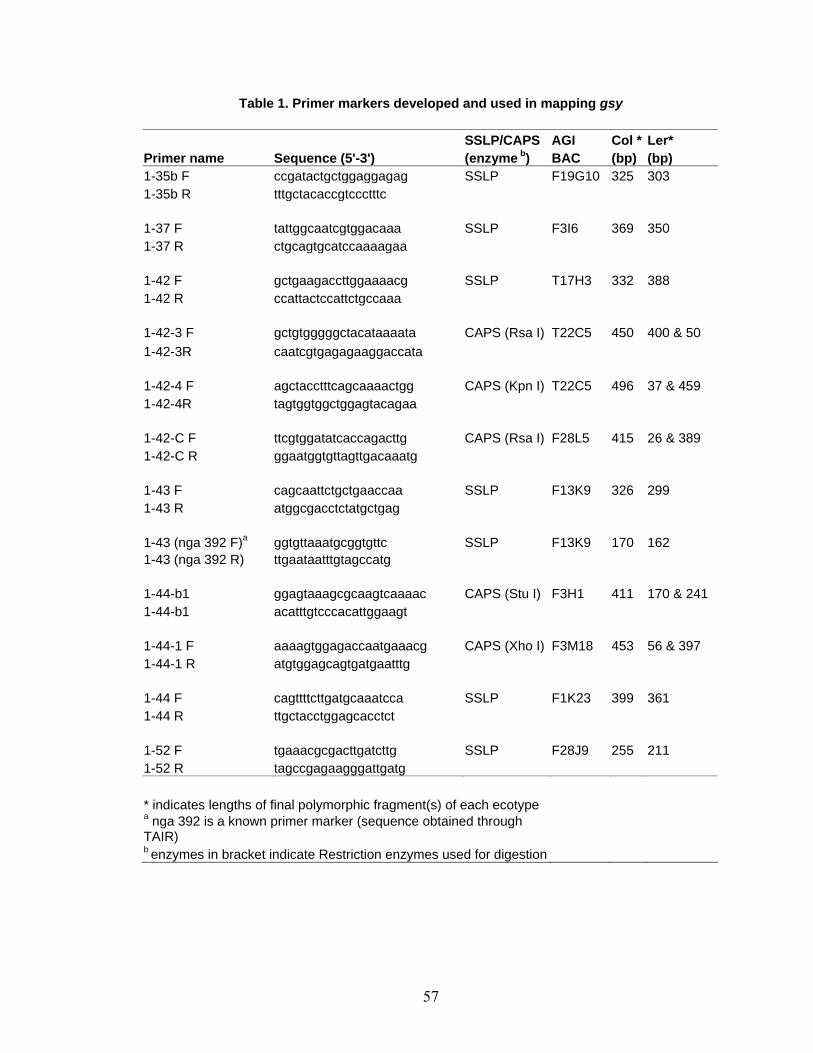
57
Table 1. Primer markers developed and used in mapping gsy SSLP/CAPS AGI Col * Ler* Primer name Sequence (5'-3') (enzyme b) BAC (bp) (bp) 1-35b F ccgatactgctggaggagag SSLP F19G10 325 303 1-35b R tttgctacaccgtccctttc 1-37 F tattggcaatcgtggacaaa SSLP F3I6 369 350 1-37 R ctgcagtgcatccaaaagaa 1-42 F gctgaagaccttggaaaacg SSLP T17H3 332 388 1-42 R ccattactccattctgccaaa 1-42-3 F gctgtgggggctacataaaata CAPS (Rsa I) T22C5 450 400 & 50 1-42-3R caatcgtgagagaaggaccata 1-42-4 F agctacctttcagcaaaactgg CAPS (Kpn I) T22C5 496 37 & 459 1-42-4R tagtggtggctggagtacagaa 1-42-C F ttcgtggatatcaccagacttg CAPS (Rsa I) F28L5 415 26 & 389 1-42-C R ggaatggtgttagttgacaaatg 1-43 F cagcaattctgctgaaccaa SSLP F13K9 326 299 1-43 R atggcgacctctatgctgag 1-43 (nga 392 F)a ggtgttaaatgcggtgttc SSLP F13K9 170 162 1-43 (nga 392 R) ttgaataatttgtagccatg 1-44-b1 ggagtaaagcgcaagtcaaaac CAPS (Stu I) F3H1 411 170 & 241 1-44-b1 acatttgtcccacattggaagt 1-44-1 F aaaagtggagaccaatgaaacg CAPS (Xho I) F3M18 453 56 & 397 1-44-1 R atgtggagcagtgatgaatttg 1-44 F cagttttcttgatgcaaatcca SSLP F1K23 399 361 1-44 R ttgctacctggagcacctct 1-52 F tgaaacgcgacttgatcttg SSLP F28J9 255 211 1-52 R tagccgagaagggattgatg * indicates lengths of final polymorphic fragment(s) of each ecotype a nga 392 is a known primer marker (sequence obtained through TAIR) b enzymes in bracket indicate Restriction enzymes used for digestion
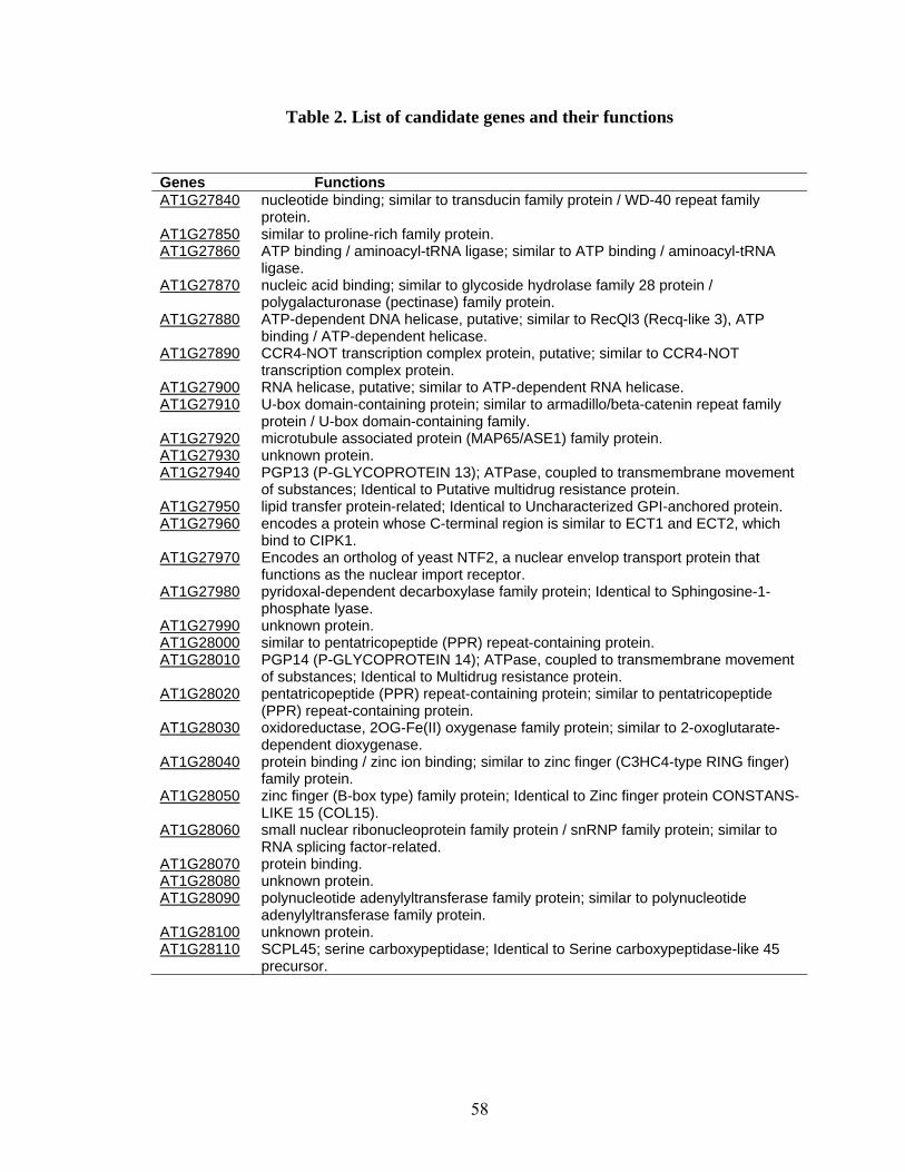
58
Table 2. List of candidate genes and their functions
Genes Functions AT1G27840 nucleotide binding; similar to transducin family protein / WD-40 repeat family
protein. AT1G27850 similar to proline-rich family protein. AT1G27860 ATP binding / aminoacyl-tRNA ligase; similar to ATP binding / aminoacyl-tRNA
ligase. AT1G27870 nucleic acid binding; similar to glycoside hydrolase family 28 protein /
polygalacturonase (pectinase) family protein. AT1G27880 ATP-dependent DNA helicase, putative; similar to RecQl3 (Recq-like 3), ATP
binding / ATP-dependent helicase. AT1G27890 CCR4-NOT transcription complex protein, putative; similar to CCR4-NOT
transcription complex protein. AT1G27900 RNA helicase, putative; similar to ATP-dependent RNA helicase. AT1G27910 U-box domain-containing protein; similar to armadillo/beta-catenin repeat family
protein / U-box domain-containing family. AT1G27920 microtubule associated protein (MAP65/ASE1) family protein. AT1G27930 unknown protein. AT1G27940 PGP13 (P-GLYCOPROTEIN 13); ATPase, coupled to transmembrane movement
of substances; Identical to Putative multidrug resistance protein. AT1G27950 lipid transfer protein-related; Identical to Uncharacterized GPI-anchored protein. AT1G27960 encodes a protein whose C-terminal region is similar to ECT1 and ECT2, which
bind to CIPK1. AT1G27970 Encodes an ortholog of yeast NTF2, a nuclear envelop transport protein that
functions as the nuclear import receptor. AT1G27980 pyridoxal-dependent decarboxylase family protein; Identical to Sphingosine-1-
phosphate lyase. AT1G27990 unknown protein. AT1G28000 similar to pentatricopeptide (PPR) repeat-containing protein. AT1G28010 PGP14 (P-GLYCOPROTEIN 14); ATPase, coupled to transmembrane movement
of substances; Identical to Multidrug resistance protein. AT1G28020 pentatricopeptide (PPR) repeat-containing protein; similar to pentatricopeptide
(PPR) repeat-containing protein. AT1G28030 oxidoreductase, 2OG-Fe(II) oxygenase family protein; similar to 2-oxoglutarate-
dependent dioxygenase. AT1G28040 protein binding / zinc ion binding; similar to zinc finger (C3HC4-type RING finger)
family protein. AT1G28050 zinc finger (B-box type) family protein; Identical to Zinc finger protein CONSTANS-
LIKE 15 (COL15). AT1G28060 small nuclear ribonucleoprotein family protein / snRNP family protein; similar to
RNA splicing factor-related. AT1G28070 protein binding. AT1G28080 unknown protein. AT1G28090 polynucleotide adenylyltransferase family protein; similar to polynucleotide
adenylyltransferase family protein. AT1G28100 unknown protein. AT1G28110 SCPL45; serine carboxypeptidase; Identical to Serine carboxypeptidase-like 45
precursor.
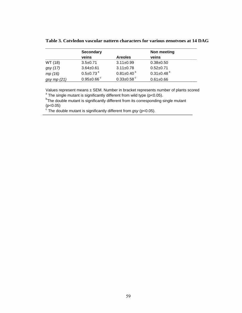
59
Values represent means ± SEM. Number in bracket represents number of plants scored a The single mutant is significantly different from wild type (p<0.05). bThe double mutant is significantly different from its corresponding single mutant (p<0.05) c The double mutant is significantly different from gsy (p<0.05).
Table 3. Cotyledon vascular pattern characters for various genotypes at 14 DAG
Secondary Non meeting veins Areoles veins WT (18) 3.5±0.71 3.11±0.99 0.38±0.50 gsy (17) 3.64±0.61 3.11±0.78 0.52±0.71 mp (16) 0.5±0.73 a 0.81±0.40 a 0.31±0.48 a gsy mp (21) 0.95±0.66 c 0.33±0.58 c 0.61±0.66

60
Table 4: First leaf vascular pattern characters of various genotypes at 21DAG
Secondary Tertiary Quaternary Free ending Free ending Vascular Genotype Veins veins veins Areoles secondaries (%) tertiaries (%) Islands WT (25) 8.53±0.83 22.53±5.47 4.4±1.18 23.6±5.13 0.40±0.51 (4.69) 7.06±3.84 (31.33) 0 gsy (25) 7.56±1.33 a 12.76±7.02 a 1.44±1.22 a 14.8±6.27 a 2.00±1.32 (26.45) a 4.56±2.81 (35.73) a 0.12±0.33 axr1-3 (25) 5.04±1.10 a 6.20±2.59 a 0.16±0.37 a 5.36±2.41 a 1.32±1.11 (26.19) a 4.24±1.76 (68.38) a 0.12±0.33 gsy axr1-3 (24) 4.87±0.94 cd 2.85±1.83 bcd 0 c d 1.35±1.39 bcd 3.78±0.97 (77.94) bcd 2.57±1.78 (90) bcd 0.07±0.26 cvp2-1 (22) 8.41±1.74 18.20±5.61 a 1.95±0.90 a 7.41±1.69 a 3.16±1.52 (37.57) a 15±5.21 (82.41) a 5.00±2.08 a gsy cvp2-1 (21) 8.61±1.28 c 15.85±4.31 d 1.57±0.87 d 9.52±3.14 bcd 3.42±1.50 (39.72) cd 10.85±3.48 (86.45) bcd 4.42±1.98 cd eir1-1 (22) 8.18±1.13 13.81±4.14 a 2.09±1.01 a 16.00±3.72 a 0.32±0.47 (3.91) 3.90±2.15 (28.24) a 0 gsy eir1-1 (18) 8.83±1.04 c 17.11±4.53 bcd 2.66±1.49 cd 20.11±4.99 bcd 0.83±0.71 (9.39) bcd 3.88±1.40 (22.67)d 0 aux1-7 (23) 7.47±0.99 a 10.43±2.84 a 1.34±0.88 a 13.04±3.52 a 0.52±0.79 (6.96) 4.21±2.04 (40.36) a 0 gsy aux1-7 (21) 8.42±1.07bc 16.80±4.24 bcd 2.47±0.86 bd 18.90±4.32 bcd 0.47±0.60 (5.58) c 4.19±1.54 (24.94) d 0 rty1 (14) 4.57±072 a 0.67±0.71 a 0 a 4.64±0.97 a 0.35±0.48 (7.81) 0.21±0.41 (33.33) a 0 gsy rty1 (12) 4.58±0.76 cd 0.50±0.65 cd 0 cd 4.66±1.11 cd 0.08±0.27 (1.82) cd 0.33±0.47 (66.66) cd 0 pin1-1 (18) 9.00±1.61 21.27±6.05 2.36±1.4 a 23.27±5.93 0.45±0.68 (5.05) 4.81±2.35 (22.64) a 0 gsy pin1 (20) 9.5±1.28 cd 24.5±5.04c 2.90±2.7 29.4±6.05 bcd 0.30±0.45 (3.16) c 4.00±2.00 (16.33) d 0
Values represent means ± SD. Number in bracket represents number of plants scored. a The single mutant is significantly different from wild type (p<0.05). bThe double mutant is significantly different from its corresponding single mutant (p<0.05). c The double mutant is significantly different from gsy (p<0.05). d The double mutant is significantly different from the wild type
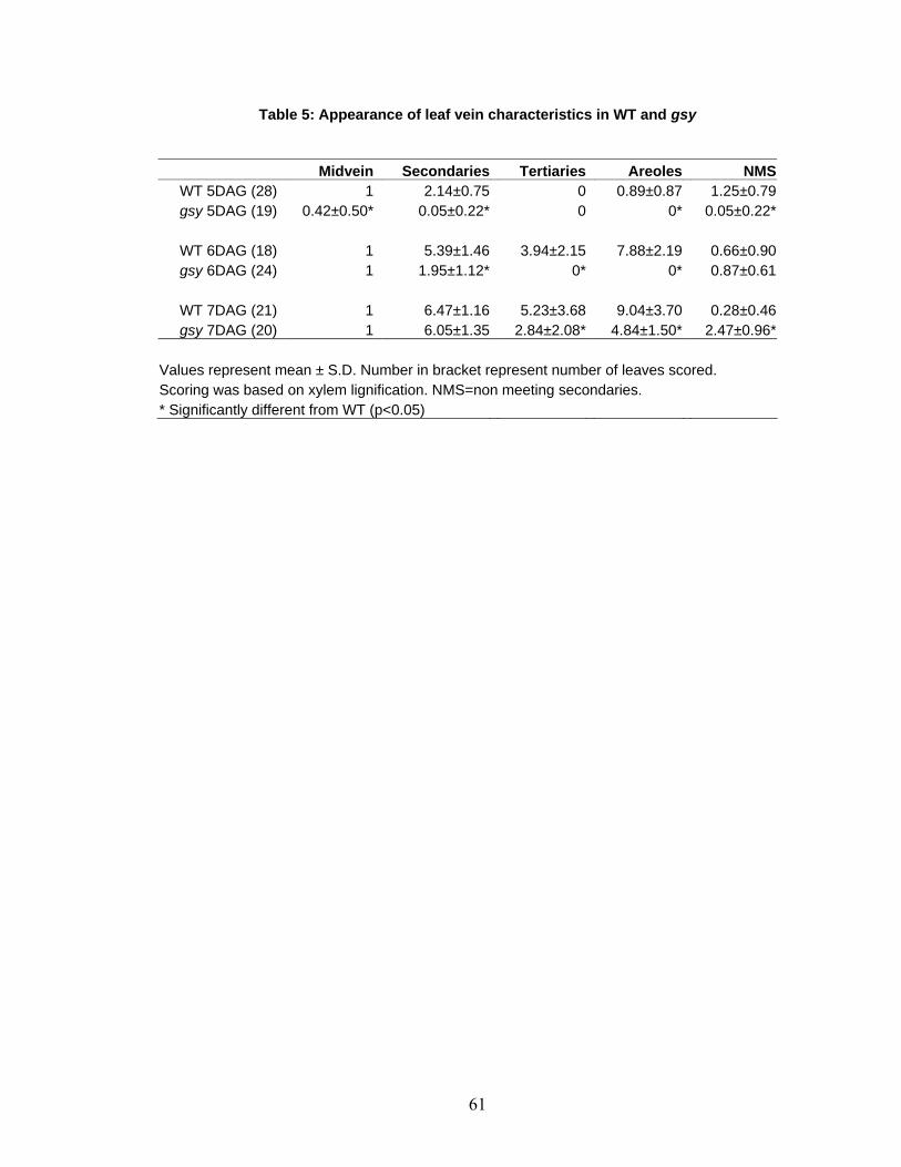
61
Table 5: Appearance of leaf vein characteristics in WT and gsy
Midvein Secondaries Tertiaries Areoles NMSWT 5DAG (28) 1 2.14±0.75 0 0.89±0.87 1.25±0.79gsy 5DAG (19) 0.42±0.50* 0.05±0.22* 0 0* 0.05±0.22*
WT 6DAG (18) 1 5.39±1.46 3.94±2.15 7.88±2.19 0.66±0.90gsy 6DAG (24) 1 1.95±1.12* 0* 0* 0.87±0.61
WT 7DAG (21) 1 6.47±1.16 5.23±3.68 9.04±3.70 0.28±0.46gsy 7DAG (20) 1 6.05±1.35 2.84±2.08* 4.84±1.50* 2.47±0.96*
Values represent mean ± S.D. Number in bracket represent number of leaves scored. Scoring was based on xylem lignification. NMS=non meeting secondaries. * Significantly different from WT (p<0.05)
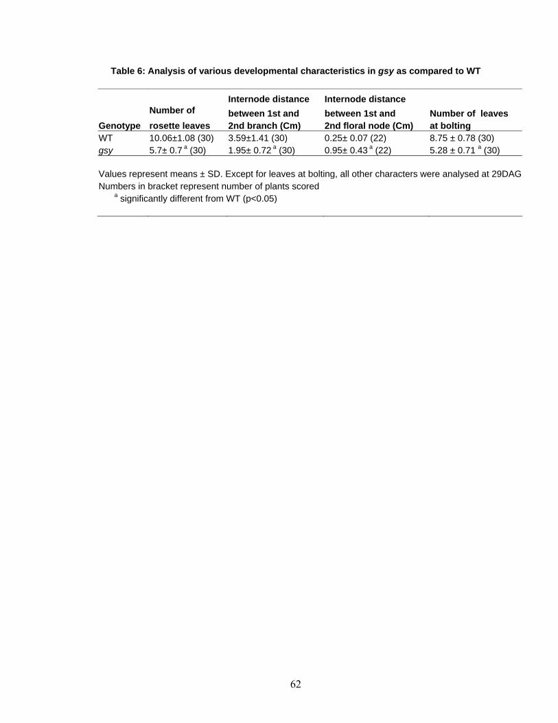
62
Table 6: Analysis of various developmental characteristics in gsy as compared to WT
Internode distance Internode distance Number of between 1st and between 1st and Number of leaves
Genotype rosette leaves 2nd branch (Cm) 2nd floral node (Cm) at bolting WT 10.06±1.08 (30) 3.59±1.41 (30) 0.25± 0.07 (22) 8.75 ± 0.78 (30) gsy 5.7± 0.7 a (30) 1.95± 0.72 a (30) 0.95± 0.43 a (22) 5.28 ± 0.71 a (30) Values represent means ± SD. Except for leaves at bolting, all other characters were analysed at 29DAGNumbers in bracket represent number of plants scored
a significantly different from WT (p<0.05)
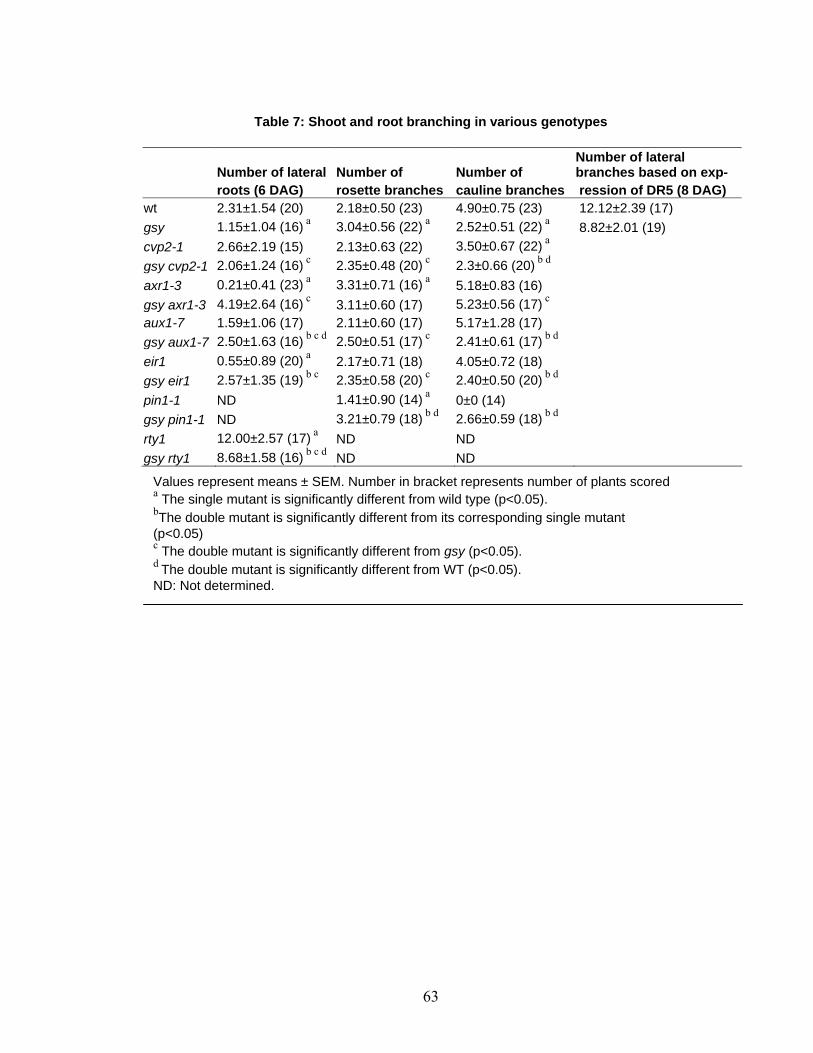
63
Table 7: Shoot and root branching in various genotypes
Number of lateral Number of Number of Number of lateral branches based on exp-
roots (6 DAG) rosette branches cauline branches ression of DR5 (8 DAG) wt 2.31±1.54 (20) 2.18±0.50 (23) 4.90±0.75 (23) 12.12±2.39 (17) gsy 1.15±1.04 (16) a 3.04±0.56 (22) a 2.52±0.51 (22) a 8.82±2.01 (19) cvp2-1 2.66±2.19 (15) 2.13±0.63 (22) 3.50±0.67 (22) a gsy cvp2-1 2.06±1.24 (16) c 2.35±0.48 (20) c 2.3±0.66 (20) b d axr1-3 0.21±0.41 (23) a 3.31±0.71 (16) a 5.18±0.83 (16) gsy axr1-3 4.19±2.64 (16) c 3.11±0.60 (17) 5.23±0.56 (17) c aux1-7 1.59±1.06 (17) 2.11±0.60 (17) 5.17±1.28 (17) gsy aux1-7 2.50±1.63 (16) b c d 2.50±0.51 (17) c 2.41±0.61 (17) b d eir1 0.55±0.89 (20) a 2.17±0.71 (18) 4.05±0.72 (18) gsy eir1 2.57±1.35 (19) b c 2.35±0.58 (20) c 2.40±0.50 (20) b d pin1-1 ND 1.41±0.90 (14) a 0±0 (14) gsy pin1-1 ND 3.21±0.79 (18) b d 2.66±0.59 (18) b d rty1 12.00±2.57 (17) a ND ND gsy rty1 8.68±1.58 (16) b c d ND ND
Values represent means ± SEM. Number in bracket represents number of plants scored a The single mutant is significantly different from wild type (p<0.05). bThe double mutant is significantly different from its corresponding single mutant (p<0.05) c The double mutant is significantly different from gsy (p<0.05). d The double mutant is significantly different from WT (p<0.05). ND: Not determined.
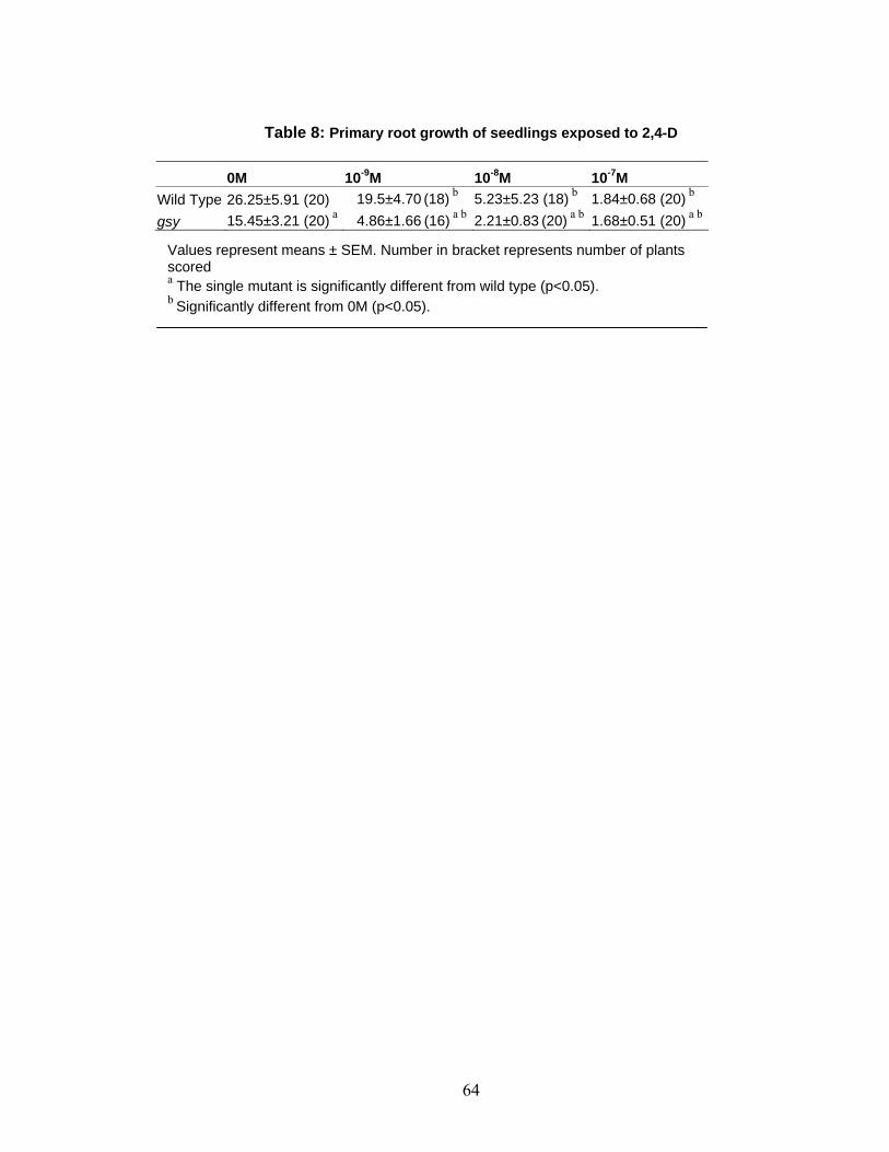
64
Table 8: Primary root growth of seedlings exposed to 2,4-D
0M 10-9M 10-8M 10-7M Wild Type 26.25±5.91 (20) 19.5±4.70 (18) b 5.23±5.23 (18) b 1.84±0.68 (20) b gsy 15.45±3.21 (20) a 4.86±1.66 (16) a b 2.21±0.83 (20) a b 1.68±0.51 (20) a b
Values represent means ± SEM. Number in bracket represents number of plants scored a The single mutant is significantly different from wild type (p<0.05). b Significantly different from 0M (p<0.05).
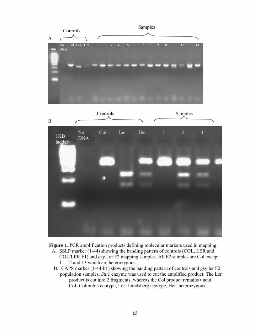
65
Std.
B
Figure 1. PCR amplification products defining molecular markers used in mapping. A. SSLP marker (1-44) showing the banding pattern of controls (COL, LER and
COL/LER F1) and gsy Ler F2 mapping samples. All F2 samples are Col except 11, 12 and 13 which are heterozygous.
B. CAPS marker (1-44-b1) showing the banding pattern of controls and gsy ler F2 population samples. Stu1 enzyme was used to cut the amplified product. The Ler
product is cut into 2 fragments, whereas the Col product remains uncut. Col- Columbia ecotype, Ler- Landsberg ecotype, Het- heterozygous
Aladder
No Col Ler Het 1 2 3 4 5 6 7 8 9 10 11 12 13 14 DNA
ControlsSamples
No Col Ler Het 1 2 3 DNA 1KB
ladder
Controls Samples
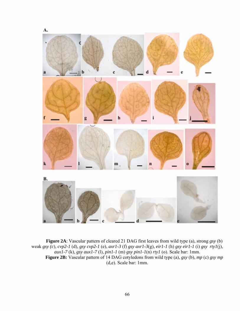
66
Figure 2A: Vascular pattern of cleared 21 DAG first leaves from wild type (a), strong gsy (b) weak gsy (c), cvp2-1 (d), gsy cvp2-1 (e), axr1-3 (f) gsy axr1-3(g), eir1-1 (h) gsy eir1-1 (i) gsy rty1(j),
aux1-7 (k), gsy aux1-7 (l), pin1-1 (m) gsy pin1-1(n) rty1 (o). Scale bar: 1mm. Figure 2B: Vascular pattern of 14 DAG cotyledons from wild type (a), gsy (b), mp (c) gsy mp
(d,e). Scale bar: 1mm.
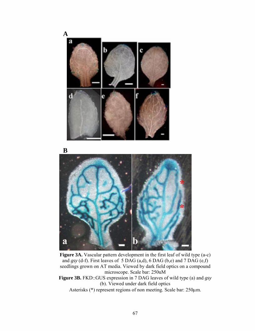
67
Figure 3A. Vascular pattern development in the first leaf of wild type (a-c) and gsy (d-f). First leaves of 5 DAG (a,d), 6 DAG (b,e) and 7 DAG (e,f)
seedlings grown on AT media. Viewed by dark field optics on a compound microscope. Scale bar: 250uM
Figure 3B. FKD::GUS expression in 7 DAG leaves of wild type (a) and gsy (b). Viewed under dark field optics
Asterisks (*) represent regions of non meeting. Scale bar: 250μm.
A
B
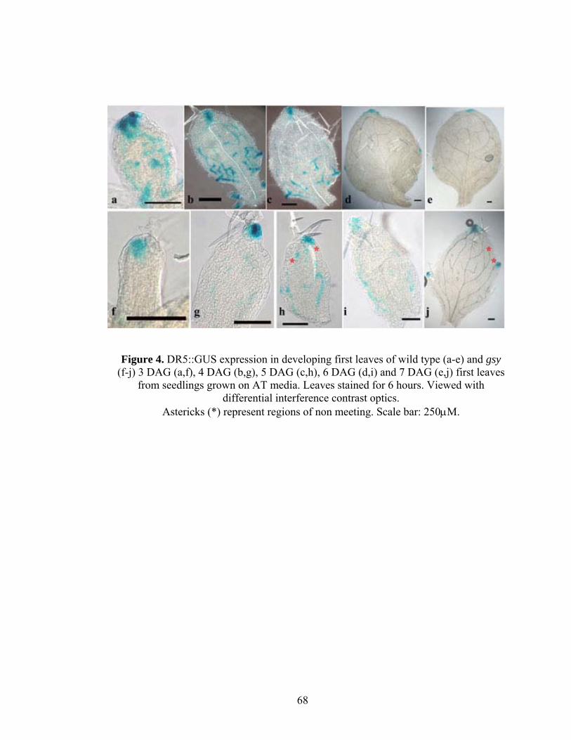
68
Figure 4. DR5::GUS expression in developing first leaves of wild type (a-e) and gsy (f-j) 3 DAG (a,f), 4 DAG (b,g), 5 DAG (c,h), 6 DAG (d,i) and 7 DAG (e,j) first leaves
from seedlings grown on AT media. Leaves stained for 6 hours. Viewed with differential interference contrast optics.
Astericks (*) represent regions of non meeting. Scale bar: 250μM.
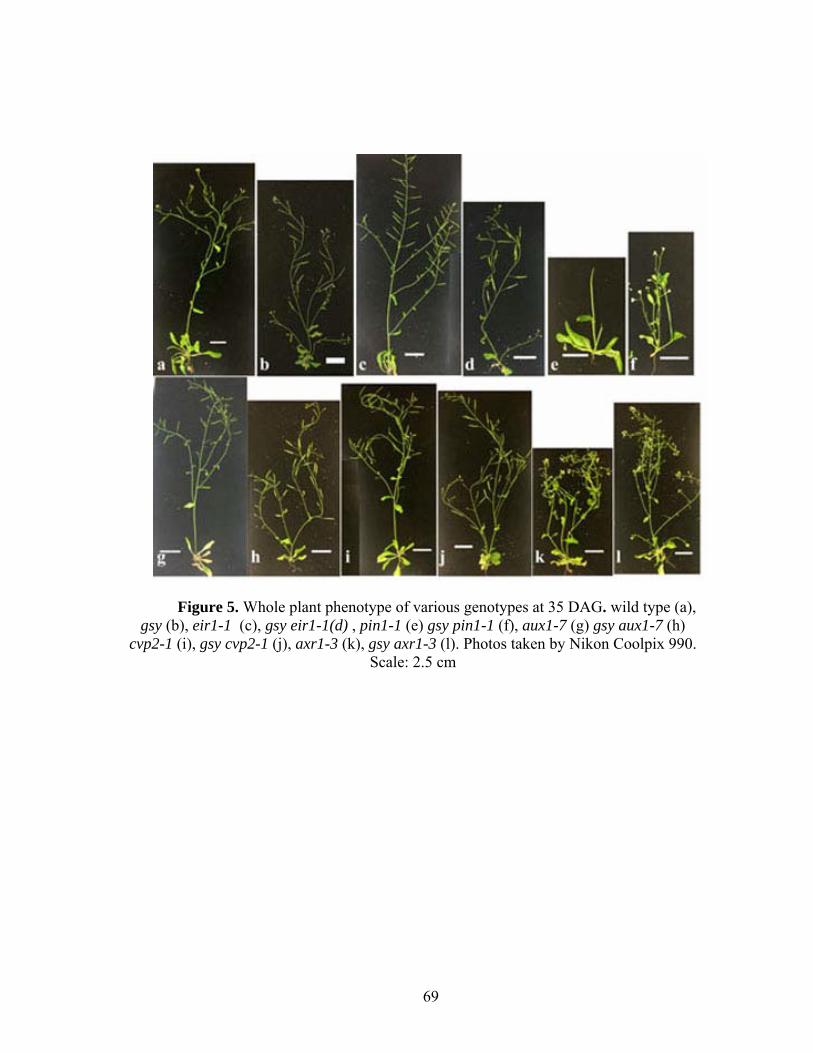
69
Figure 5. Whole plant phenotype of various genotypes at 35 DAG. wild type (a), gsy (b), eir1-1 (c), gsy eir1-1(d) , pin1-1 (e) gsy pin1-1 (f), aux1-7 (g) gsy aux1-7 (h)
cvp2-1 (i), gsy cvp2-1 (j), axr1-3 (k), gsy axr1-3 (l). Photos taken by Nikon Coolpix 990. Scale: 2.5 cm
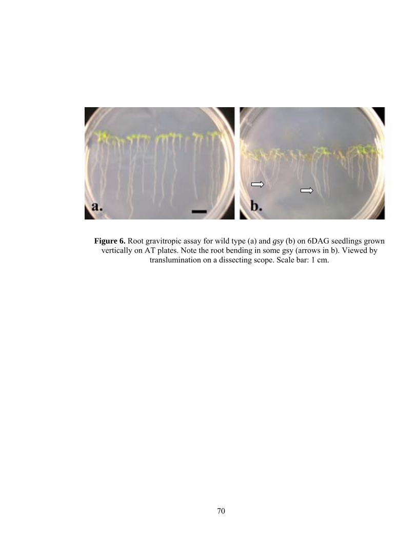
70
Figure 6. Root gravitropic assay for wild type (a) and gsy (b) on 6DAG seedlings grown vertically on AT plates. Note the root bending in some gsy (arrows in b). Viewed by
translumination on a dissecting scope. Scale bar: 1 cm.
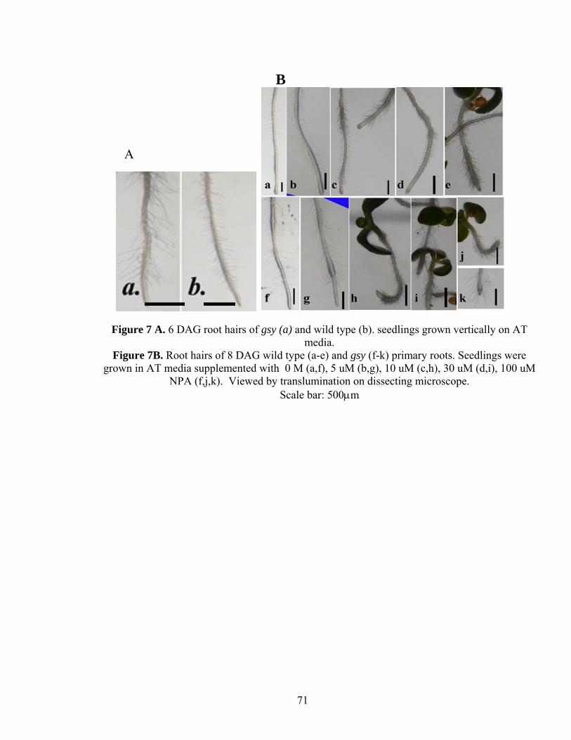
71
A
B
Figure 7 A. 6 DAG root hairs of gsy (a) and wild type (b). seedlings grown vertically on AT media.
Figure 7B. Root hairs of 8 DAG wild type (a-e) and gsy (f-k) primary roots. Seedlings were grown in AT media supplemented with 0 M (a,f), 5 uM (b,g), 10 uM (c,h), 30 uM (d,i), 100 uM
NPA (f,j,k). Viewed by translumination on dissecting microscope. Scale bar: 500μm

72
Figure 8. DR5::GUS expression in 5 DAG roots of WT (a-d) and gsy (e-h) grown on unsupplemented AT media (a, b, e, f, g) and AT media
supplemented with 10-7 M 2,4-D (c,d,h). Roots were stained for 4 hours. Viewed by phase contrast optics. Scale: 100μm
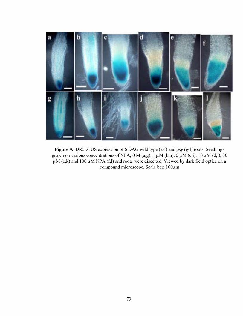
73
Figure 9. DR5::GUS expression of 6 DAG wild type (a-f) and gsy (g-l) roots. Seedlings grown on various concentrations of NPA, 0 M (a,g), 1 μM (b,h), 5 μM (c,i), 10 μM (d,j), 30 μM (e,k) and 100 μM NPA (f,l) and roots were disectted, Viewed by dark field optics on a
compound microscope. Scale bar: 100μm
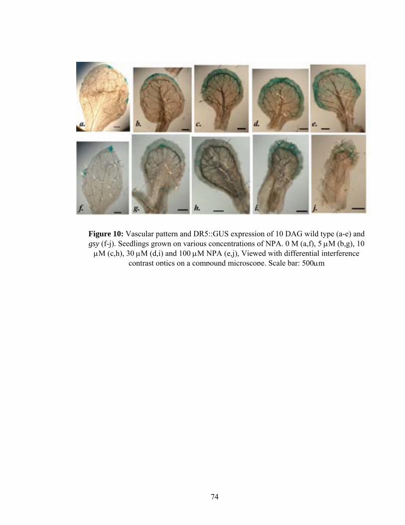
74
Figure 10: Vascular pattern and DR5::GUS expression of 10 DAG wild type (a-e) and gsy (f-j). Seedlings grown on various concentrations of NPA. 0 M (a,f), 5 μM (b,g), 10
μM (c,h), 30 μM (d,i) and 100 μM NPA (e,j), Viewed with differential interference contrast optics on a compound microscope. Scale bar: 500μm

75
Figure 11. Roots of 9 DAG seedlings of wild type (a-d) and gsy (e-h) treated with 0M 2,4-D (a,e) 10-9 M 2,4-D, 10-8 M 2,4-D and 10-7 M 2,4-D. Seedlings were grown in unsupplemented AT media for five days and transferred to AT
media supplemented with 2,4-D. Viewed by translumination on dissecting microscope. Scale bar: 500μm
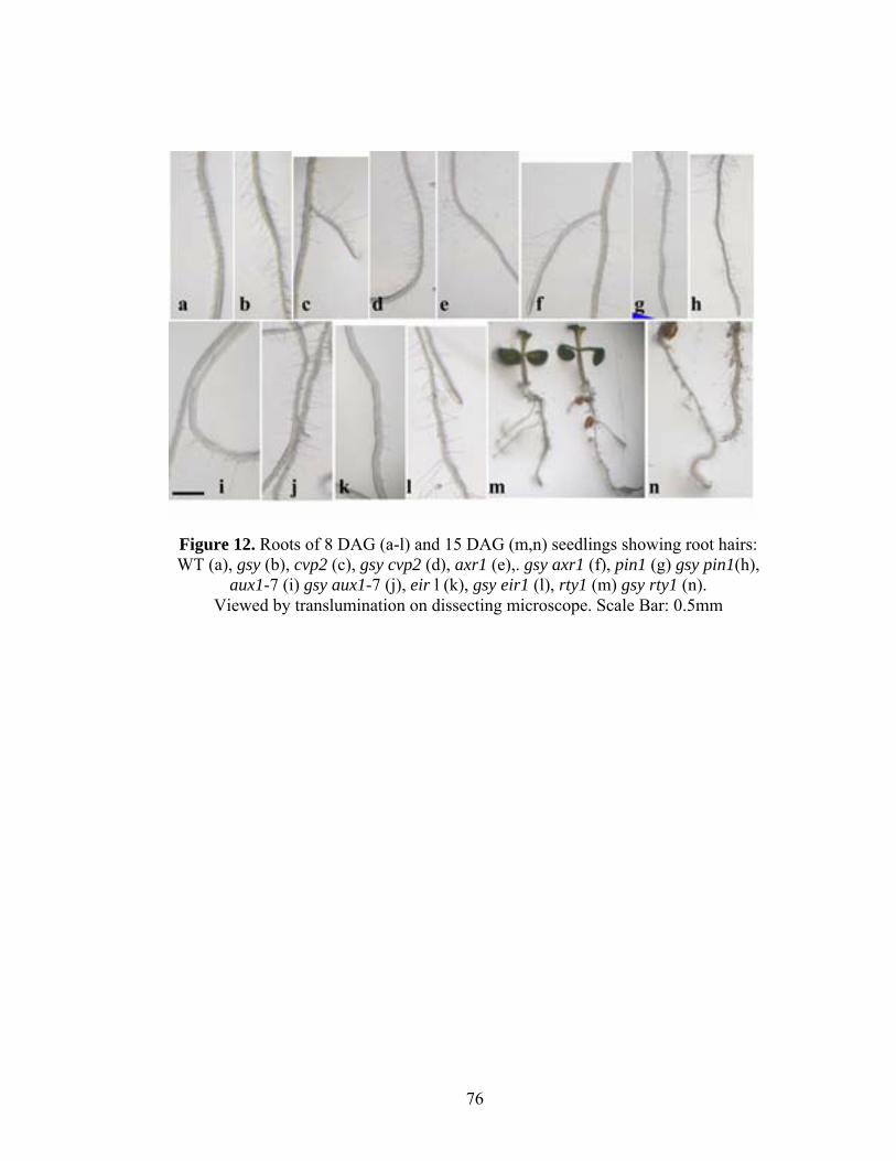
76
Figure 12. Roots of 8 DAG (a-l) and 15 DAG (m,n) seedlings showing root hairs: WT (a), gsy (b), cvp2 (c), gsy cvp2 (d), axr1 (e),. gsy axr1 (f), pin1 (g) gsy pin1(h),
aux1-7 (i) gsy aux1-7 (j), eir l (k), gsy eir1 (l), rty1 (m) gsy rty1 (n). Viewed by translumination on dissecting microscope. Scale Bar: 0.5mm


![Role of the Arabidopsis PIN6 Auxin Transporter in Auxin ......central stele and basipetally through the epidermis and outer cortex near the root tip [4,5]. Polar cell-to-cell auxin](https://static.fdocuments.net/doc/165x107/611cd25da6ea2f70970f71db/role-of-the-arabidopsis-pin6-auxin-transporter-in-auxin-central-stele-and.jpg)

