Protein Levels of Several Arabidopsis Auxin Response ...
20
International Journal of Molecular Sciences Article Protein Levels of Several Arabidopsis Auxin Response Factors Are Regulated by Multiple Factors and ABA Promotes ARF6 Protein Ubiquitination Keke Li 1,2, † , Sheng Wang 2, † , Hong Wu 1, * and Hong Wang 2, * 1 State Key Laboratory for Conservation and Utilization of Subtropical Agro-Bioresouces, College of Life Sciences, South China Agricultural University, Guangzhou 510642, China; [email protected] 2 Department of Biochemistry, Microbiology and Immunology, University of Saskatchewan, Saskatoon, SK S7N 5E5, Canada; [email protected] * Correspondence: [email protected] (H.W.); [email protected](H.W.) † These authors contributed equally to this work. Received: 25 October 2020; Accepted: 8 December 2020; Published: 11 December 2020 Abstract: The auxin response factor (ARF) transcription factors are a key component in auxin signaling and play diverse functions in plant growth, development, and stress response. ARFs are regulated at the transcript level and posttranslationally by protein modifications. However, relatively little is known regarding the control of ARF protein levels. We expressed five different ARFs with an HA (hemagglutinin) tag and observed that their protein levels under the same promoter varied considerably. Interestingly, their protein levels were affected by several hormonal and environmental conditions, but not by the auxin treatment. ABA (abscisic acid) as well as 4 ◦ C and salt treatments decreased the levels of HA-ARF5, HA-ARF6, and HA-ARF10, but not that of HA-ARF19, while 37 ◦ C treatment increased the levels of the four HA-ARFs, suggesting that the ARF protein levels are regulated by multiple factors. Furthermore, MG132 inhibited the reduction of HA-ARF6 level by ABA and 4 ◦ C treatments, suggesting that these treatments decrease HA-ARF6 level through 26S proteasome-mediated protein degradation. It was also found that ABA treatment drastically increased HA-ARF6 ubiquitination, without strongly affecting the ubiquitination profile of the total proteins. Together, these results reveal another layer of control on ARFs, which could serve to integrate multiple hormonal and environmental signals into the ARF-regulated gene expression. Keywords: abscisic acid; Arabidopsis; auxin response factor; ARF6; protein level; protein ubiquitination; temperature stress 1. Introduction The auxin signaling cascade from auxin perception to activation of auxin-inducible genes has several core components including an SCF (SKP1–Cullin1–F-box) E3 complex, Aux/IAA and ARF (Auxin Response Factor) transcription factors [1]. The binding of auxin to the receptor from the TIR1/AFB (Transport Inhibitor Response and Auxin F-Box protein) family of F-box proteins stabilizes the interaction between the SCF TIR1/AFB E3 complex and transcription repressor Aux/IAA proteins, triggering the ubiquitination and degradation of Aux/IAAs. The degradation of Aux/IAA proteins frees ARFs which can bind to the auxin response elements in the promoters of auxin inducible genes and activate their expression [1,2]. Thus, auxin functions essentially through ARFs. In land plants, ARF genes have evolved into three conserved A, B, and C classes or subfamilies [3]. The Arabidopsis genome encodes 23 ARF genes, but ARF23 is a truncated pseudogene [4,5]. Based on the reporter activation assays in transfected protoplasts, ARFs in Class A are considered as transcriptional Int. J. Mol. Sci. 2020, 21, 9437; doi:10.3390/ijms21249437 www.mdpi.com/journal/ijms
Transcript of Protein Levels of Several Arabidopsis Auxin Response ...
International Journal of
Molecular Sciences
Article
Protein Levels of Several Arabidopsis AuxinResponse Factors Are Regulated by Multiple Factorsand ABA Promotes ARF6 Protein Ubiquitination
Keke Li 1,2,†, Sheng Wang 2,†, Hong Wu 1,* and Hong Wang 2,*1 State Key Laboratory for Conservation and Utilization of Subtropical Agro-Bioresouces,
College of Life Sciences, South China Agricultural University, Guangzhou 510642, China;[email protected]
2 Department of Biochemistry, Microbiology and Immunology, University of Saskatchewan,Saskatoon, SK S7N 5E5, Canada; [email protected]
* Correspondence: [email protected] (H.W.); [email protected] (H.W.)† These authors contributed equally to this work.
Received: 25 October 2020; Accepted: 8 December 2020; Published: 11 December 2020�����������������
Abstract: The auxin response factor (ARF) transcription factors are a key component in auxin signalingand play diverse functions in plant growth, development, and stress response. ARFs are regulatedat the transcript level and posttranslationally by protein modifications. However, relatively littleis known regarding the control of ARF protein levels. We expressed five different ARFs with anHA (hemagglutinin) tag and observed that their protein levels under the same promoter variedconsiderably. Interestingly, their protein levels were affected by several hormonal and environmentalconditions, but not by the auxin treatment. ABA (abscisic acid) as well as 4 ◦C and salt treatmentsdecreased the levels of HA-ARF5, HA-ARF6, and HA-ARF10, but not that of HA-ARF19, while 37 ◦Ctreatment increased the levels of the four HA-ARFs, suggesting that the ARF protein levels areregulated by multiple factors. Furthermore, MG132 inhibited the reduction of HA-ARF6 level byABA and 4 ◦C treatments, suggesting that these treatments decrease HA-ARF6 level through 26Sproteasome-mediated protein degradation. It was also found that ABA treatment drastically increasedHA-ARF6 ubiquitination, without strongly affecting the ubiquitination profile of the total proteins.Together, these results reveal another layer of control on ARFs, which could serve to integrate multiplehormonal and environmental signals into the ARF-regulated gene expression.
Keywords: abscisic acid; Arabidopsis; auxin response factor; ARF6; protein level; protein ubiquitination;temperature stress
1. Introduction
The auxin signaling cascade from auxin perception to activation of auxin-inducible genes hasseveral core components including an SCF (SKP1–Cullin1–F-box) E3 complex, Aux/IAA and ARF(Auxin Response Factor) transcription factors [1]. The binding of auxin to the receptor from theTIR1/AFB (Transport Inhibitor Response and Auxin F-Box protein) family of F-box proteins stabilizesthe interaction between the SCFTIR1/AFB E3 complex and transcription repressor Aux/IAA proteins,triggering the ubiquitination and degradation of Aux/IAAs. The degradation of Aux/IAA proteinsfrees ARFs which can bind to the auxin response elements in the promoters of auxin inducible genesand activate their expression [1,2]. Thus, auxin functions essentially through ARFs.
In land plants, ARF genes have evolved into three conserved A, B, and C classes or subfamilies [3].The Arabidopsis genome encodes 23 ARF genes, but ARF23 is a truncated pseudogene [4,5]. Based on thereporter activation assays in transfected protoplasts, ARFs in Class A are considered as transcriptional
Int. J. Mol. Sci. 2020, 21, 9437; doi:10.3390/ijms21249437 www.mdpi.com/journal/ijms
Int. J. Mol. Sci. 2020, 21, 9437 2 of 20
activators and ARFs in Class B as repressors [6,7]. Class C-ARFs are also considered as transcriptionalrepressors based mainly on their lack of a glutamine-rich middle region which is important fortranscriptional activation [8]. In Arabidopsis, five ARFs (ARF5, 6, 7, 8, and 19) are considered astranscription activators while the others as repressors in Class B and C [4,9]. However, evidence forthe majority of the repressor ARFs to function as transcription repressors remained to be obtained [5,9].Since much of the understanding on ARFs has been gained using Arabidopsis as a model, when wedescribe a specific ARF here it usually refers to the Arabidopsis gene or protein unless a plant speciesis specified otherwise.
Most ARFs possess three functional domains, an N-terminal DNA binding domain (DBD),a middle region (MR), and a C-terminal PB1 domain [2]. The conserved DBD at the N-terminusis responsible for binding the auxin responsive element [6,10]. The transactivator ARFs in Class Acontain a glutamine-rich middle region important for transcriptional activation, which is missing inClass C-ARFs [7,8]. The PB1 domain (formerly referred to as domain III/IV) mediates homo- andhetero-oligomerization with Aux/IAAs and ARFs [11–13].
The interactions of ARFs with the Aux/IAA proteins are an important aspect of auxin signalingand its regulation. Since one ARF can interact with multiple Aux/IAAs and vice versa, there is acomplex web of interactions among different ARFs and different Aux/IAAs [14]. Results from the yeasttwo-hybrid studies have shown that the activator ARFs (ARF5, 6, 7, 8, and 19) have more interactionswith Aux/IAA proteins compared with the repressor-type ARFs [15,16]. Since the levels of proteinexpression for various ARFs and Aux/IAAs may differ between the yeast system and plants, and alsothe tissue specificity is missing in the yeast system, it remains to be seen to what extent the interactionsobserved in the yeast system reflect the ARF-Aux/IAA interactions in plants. In addition, ARFs caninteract with other transcriptional factors. For instance, ARF4 interacts with the transcription factorKAN4 that has an important role in ovule development [17] and ARF8 interacts with a transcriptionfactor named BPEp to limit petal growth [18]. Further, ARF6 is found to interact synergisticallywith the light/temperature-regulated transcription factor PIF4 and the brassinosteroid-signalingtranscription factor BZR1 to regulate large numbers of target genes, while the gibberellin-inactivatedrepressor RGA blocks their DNA-binding activities [19], revealing more complex interaction amongthe transcription factors.
Most of the information regarding the functions of ARF genes has been obtained using arfmutants [4,5,20]. Due to the functional redundancy among the closely related ARF genes, the majorityof single mutants for 18 Arabidopsis ARF genes did not show obvious phenotypical changes [21],although single mutants of ARF2, ARF3, ARF5, ARF7, and ARF8 displayed phenotypes revealing thefunctions in several aspects of plant growth and development [22–25]. For instance, ARF5 functions inthe establishment of vascular and body patterns in embryonic and post-embryonic development [24].In the arf7/nph4-1 mutant, the expression of auxin-responsive reporter genes was much reduced intransfected mesophyll protoplasts which could only be partially restored by other ARFs, suggestingthat ARF7 has a major role in regulating a subset of genes in mesophyll cells [26]. Results fromvarious studies using double and multiple mutants helped to identify many different functions of ARFsparticularly for the Class-A ARFs (ARF5 to 8 and ARF19) [5,20]. For instance, ARF6 and ARF8 havebeen found to function in adventitious root formation [27], hypocotyl elongation (ARF6, 7, and 8) [28]and flower organ development and maturation [29,30], while ARF7 and ARF19 are involved in theregulation of phosphate starvation response [31]. In addition, ARF1 and ARF2 of Class B have arole in regulating senescence and floral organ abscission [32]. ARF2 also functions together with atranscription factor (PLT1) and an auxin transporter PIN1 to control ABA-mediated root meristemactivity [33]. Furthermore, ARF10 of Class C is found to be important for root cap formation [34],in vitro callus initiation and shoot regeneration [35,36]. These results demonstrate that ARFs playimportant and diverse functions in plants.
It is well known that the ARF genes are transcriptionally regulated and different ARFs havedifferent expression levels and patterns in different tissues [4,37]. In one study, the expression of all
Int. J. Mol. Sci. 2020, 21, 9437 3 of 20
23 ARFs in embryogenesis and primary root meristem was characterized using the promoter: GFPreporter approach [38]. The results revealed complex cell-specific patterns and that different ARFshad overlapping expression in different cells. Another systematic analysis using in situ hybridizationrevealed that 13 Arabidopsis ARFs were expressed in the shoot apex, showing differences in the patternand level [15]. One commonality among the expressed ARFs is that they tended to be expressedat higher levels in the periphery of the shoot meristem. In addition, ARF transcript levels are alsoregulated by other mechanisms such as alternative transcript splicing and microRNAs [5,37].
The functions of ARFs are also regulated by posttranslational protein modifications. ARF2 canbe phosphorylated by a brassinosteroid (BR)-regulated kinase BIN2, leading to the loss of ARF2DNA binding and repression activities [39]. ARF2 is also phosphorylated under low potassiumstress [40]. ARF2 normally represses the expression of a potassium transporter gene HAK5, and thephosphorylation abolishes ARF2 binding to HAK5 promoter and relieves the repression. In addition,BIN2 has been reported to phosphorylate ARF7 and ARF19, and in contrast to reduce activity of ARF2,ARF7, and ARF19 phosphorylation results in the enhanced transactivation activity, which is attributedto reduced ARF7 and ARF19 interactions with the Aux/IAA repressors [41]. On the other hand, ARF7 ismodified with the small ubiquitin-like modifier protein and the sumoylation inhibits the interactionwith Aux/IAA3 and affects root branching [42].
In terms of the control of protein stability and degradation, there are only a few reports onthe control of ARF1 and ARF2 protein levels. Arabidopsis ARF1 is fast degraded in plants and thedegradation is inhibited by MG132, a 26S proteasome inhibitor [43]. In addition, ARF1 degradation isnot affected in a CUL1 mutant background and by auxin treatment, suggesting that ARF1 is degradedby the 26S proteasome, but the degradation is through a CUL1-independent pathway [43]. It has beenreported that ARF2 protein expression driven by the native promoter is decreased by ethylene, which isinhibited by MG132 [22]. On the other hand, ARF2 protein abundance is enhanced by gibberellin (GA)treatment [44]. Recently, ARF6, ARF8, and ARF17 were also shown to be unstable and their levels wereenhanced by MG132 [45]. Apart from these studies, there has been little information on the protein-levelcontrol of other ARFs and further regarding how different hormonal and environmental conditionsaffect ARF protein levels. In this study, we investigated the protein expression of several ARFs andobserved that their levels are affected by several different conditions. We further demonstrated thatABA treatment promotes the ubiquitination and degradation of ARF6.
2. Results
2.1. Analysis of Five ARF Protein Expression in Transgenic Arabidopsis Plants
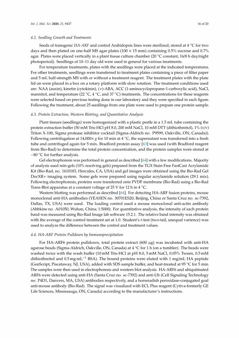
To study protein expression, we prepared the plant expression constructs of several ARFs taggedwith the HA (hemagglutinin) epitope. We selected three activator ARFs from Class A, ARF5 (At1g19850),ARF6 (At1g30330), and ARF19 (At1g19220), due to the importance of the activator ARFs in activatingauxin-regulated genes, as well as ARF1 (At1g59750) from Class B and ARF10 (At2g28350) fromClass C [9] for the study. We used the widely used 35S promoter which is not known to be significantlyregulated by auxin or any other hormones, so that we could separate the protein control from thetranscriptional control by the native ARF promoters. First, Arabidopsis primary transformants ofeach construct were analyzed for HA-ARF protein expression by western blotting. Each proteinsample was prepared from a pool of independent Arabidopsis transformants (about 25 in each plate)to represent the average level of the HA-ARF protein for these transformants. With the expectedmolecular size of the 2× HA tag (3.0 kD) included, the theoretical sizes of HA-ARF1, HA-ARF5,HA-ARF6, HA-ARF10, and HA-ARF19 are 76.7, 102.6, 106.3, 79.7, and 123.6 kD, respectively. As shownin Figure 1, no HA-specific band was detected in the wild type and transgenic control of a GFP (greenfluorescence protein) line. A full-length HA-ARF protein band was detected in HA-ARF5, HA-ARF6,and HA-ARF19 transformants, while only a strong partial HA protein band was detected in ARF1transformants and no band was detected in ARF10 transformants. Also, it was noted that the two
Int. J. Mol. Sci. 2020, 21, 9437 4 of 20
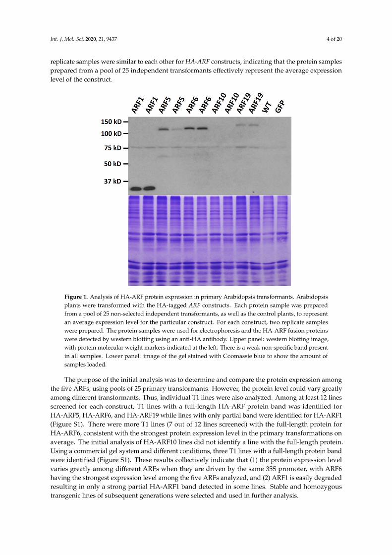
replicate samples were similar to each other for HA-ARF constructs, indicating that the protein samplesprepared from a pool of 25 independent transformants effectively represent the average expressionlevel of the construct.Int. J. Mol. Sci. 2020, 21, x FOR PEER REVIEW 4 of 19
Figure 1. Analysis of HA-ARF protein expression in primary Arabidopsis transformants. Arabidopsis plants were transformed with the HA-tagged ARF constructs. Each protein sample was prepared from a pool of 25 non-selected independent transformants, as well as the control plants, to represent an average expression level for the particular construct. For each construct, two replicate samples were prepared. The protein samples were used for electrophoresis and the HA-ARF fusion proteins were detected by western blotting using an anti-HA antibody. Upper panel: western blotting image, with protein molecular weight markers indicated at the left. There is a weak non-specific band present in all samples. Lower panel: image of the gel stained with Coomassie blue to show the amount of samples loaded.
The purpose of the initial analysis was to determine and compare the protein expression among the five ARFs, using pools of 25 primary transformants. However, the protein level could vary greatly among different transformants. Thus, individual T1 lines were also analyzed. Among at least 12 lines screened for each construct, T1 lines with a full-length HA-ARF protein band was identified for HA-ARF5, HA-ARF6, and HA-ARF19 while lines with only partial band were identified for HA-ARF1 (Figure S1). There were more T1 lines (7 out of 12 lines screened) with the full-length protein for HA-ARF6, consistent with the strongest protein expression level in the primary transformations on average. The initial analysis of HA-ARF10 lines did not identify a line with the full-length protein. Using a commercial gel system and different conditions, three T1 lines with a full-length protein band were identified (Figure S1). These results collectively indicate that (1) the protein expression level varies greatly among different ARFs when they are driven by the same 35S promoter, with ARF6 having the strongest expression level among the five ARFs analyzed, and (2) ARF1 is easily degraded resulting in only a strong partial HA-ARF1 band detected in some lines. Stable and homozygous transgenic lines of subsequent generations were selected and used in further analysis.
2.2. Effects of Hormonal and Environmental Treatments on ARF Protein Expression
To determine the influence of plant hormones and environmental conditions on ARF protein expression, various hormonal and stress treatments were applied to Arabidopsis seedlings of HA-ARF transgenic lines. After 8 or 12 h of treatment, protein samples were prepared from treated seedlings and used in western blotting analysis. For HA-ARF6, ABA (abscisic acid) and 4 °C
Figure 1. Analysis of HA-ARF protein expression in primary Arabidopsis transformants. Arabidopsisplants were transformed with the HA-tagged ARF constructs. Each protein sample was preparedfrom a pool of 25 non-selected independent transformants, as well as the control plants, to representan average expression level for the particular construct. For each construct, two replicate sampleswere prepared. The protein samples were used for electrophoresis and the HA-ARF fusion proteinswere detected by western blotting using an anti-HA antibody. Upper panel: western blotting image,with protein molecular weight markers indicated at the left. There is a weak non-specific band presentin all samples. Lower panel: image of the gel stained with Coomassie blue to show the amount ofsamples loaded.
The purpose of the initial analysis was to determine and compare the protein expression amongthe five ARFs, using pools of 25 primary transformants. However, the protein level could vary greatlyamong different transformants. Thus, individual T1 lines were also analyzed. Among at least 12 linesscreened for each construct, T1 lines with a full-length HA-ARF protein band was identified forHA-ARF5, HA-ARF6, and HA-ARF19 while lines with only partial band were identified for HA-ARF1(Figure S1). There were more T1 lines (7 out of 12 lines screened) with the full-length protein forHA-ARF6, consistent with the strongest protein expression level in the primary transformations onaverage. The initial analysis of HA-ARF10 lines did not identify a line with the full-length protein.Using a commercial gel system and different conditions, three T1 lines with a full-length protein bandwere identified (Figure S1). These results collectively indicate that (1) the protein expression levelvaries greatly among different ARFs when they are driven by the same 35S promoter, with ARF6having the strongest expression level among the five ARFs analyzed, and (2) ARF1 is easily degradedresulting in only a strong partial HA-ARF1 band detected in some lines. Stable and homozygoustransgenic lines of subsequent generations were selected and used in further analysis.
Int. J. Mol. Sci. 2020, 21, 9437 5 of 20
2.2. Effects of Hormonal and Environmental Treatments on ARF Protein Expression
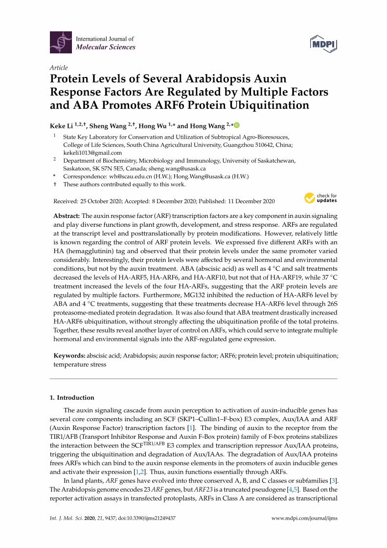
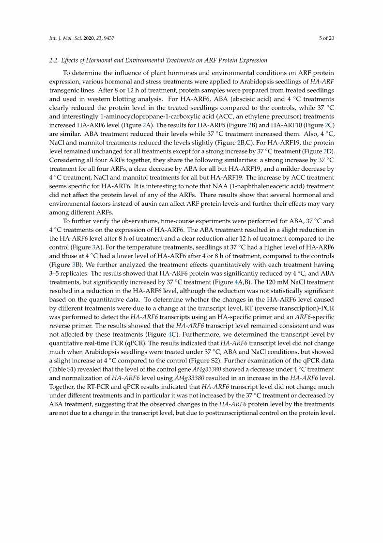
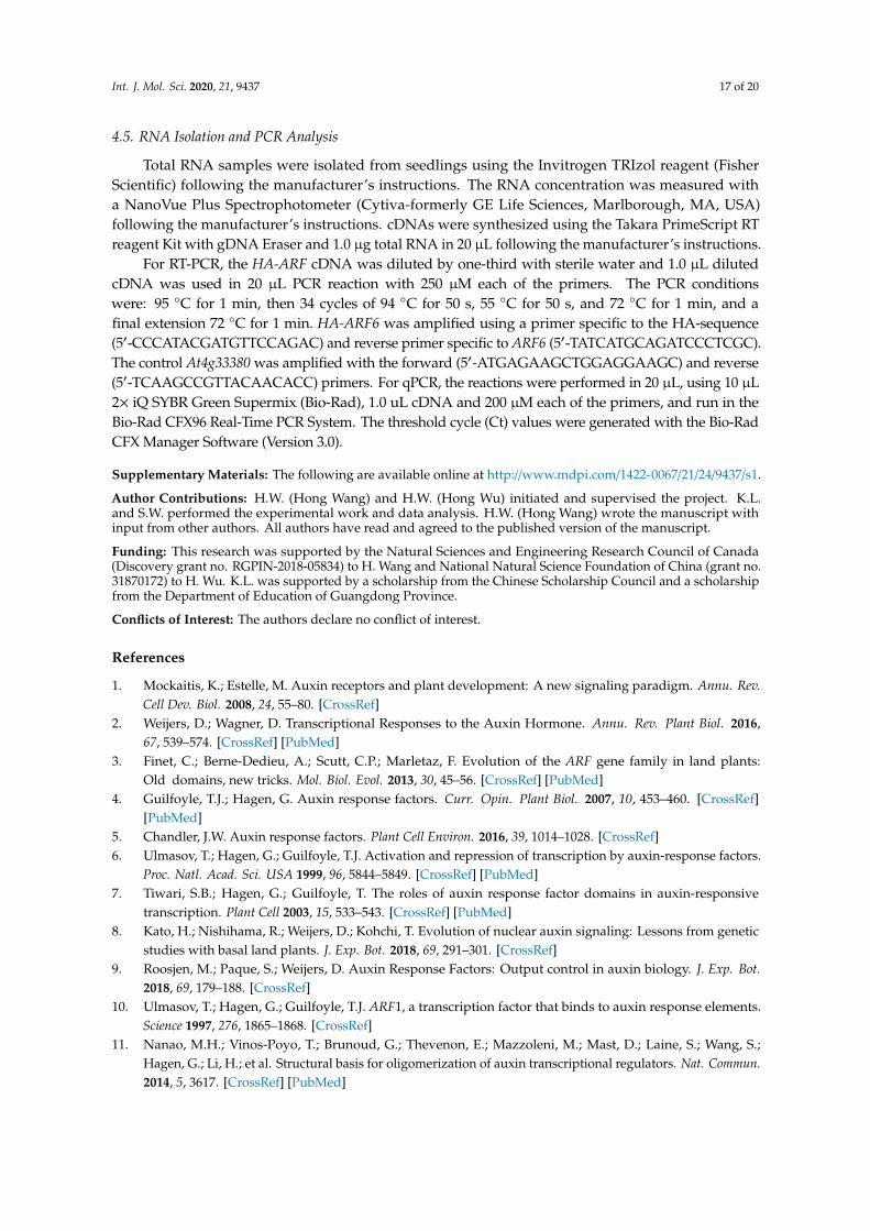
To determine the influence of plant hormones and environmental conditions on ARF proteinexpression, various hormonal and stress treatments were applied to Arabidopsis seedlings of HA-ARFtransgenic lines. After 8 or 12 h of treatment, protein samples were prepared from treated seedlingsand used in western blotting analysis. For HA-ARF6, ABA (abscisic acid) and 4 ◦C treatmentsclearly reduced the protein level in the treated seedlings compared to the controls, while 37 ◦Cand interestingly 1-aminocyclopropane-1-carboxylic acid (ACC, an ethylene precursor) treatmentsincreased HA-ARF6 level (Figure 2A). The results for HA-ARF5 (Figure 2B) and HA-ARF10 (Figure 2C)are similar. ABA treatment reduced their levels while 37 ◦C treatment increased them. Also, 4 ◦C,NaCl and mannitol treatments reduced the levels slightly (Figure 2B,C). For HA-ARF19, the proteinlevel remained unchanged for all treatments except for a strong increase by 37 ◦C treatment (Figure 2D).Considering all four ARFs together, they share the following similarities: a strong increase by 37 ◦Ctreatment for all four ARFs, a clear decrease by ABA for all but HA-ARF19, and a milder decrease by4 ◦C treatment, NaCl and mannitol treatments for all but HA-ARF19. The increase by ACC treatmentseems specific for HA-ARF6. It is interesting to note that NAA (1-naphthaleneacetic acid) treatmentdid not affect the protein level of any of the ARFs. There results show that several hormonal andenvironmental factors instead of auxin can affect ARF protein levels and further their effects may varyamong different ARFs.
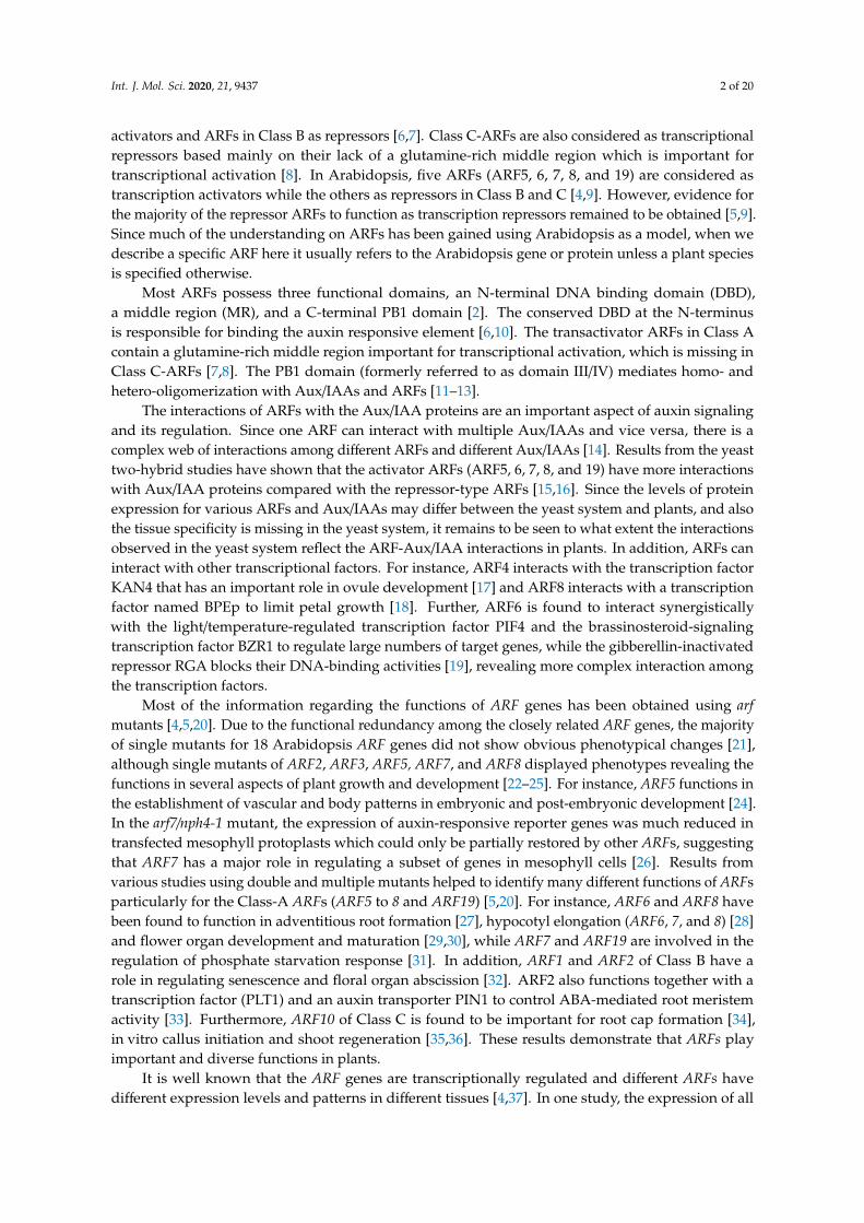
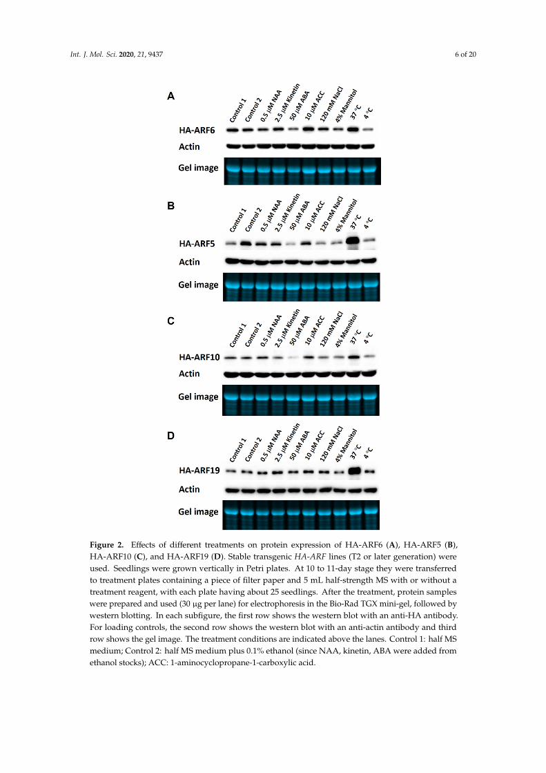
To further verify the observations, time-course experiments were performed for ABA, 37 ◦C and4 ◦C treatments on the expression of HA-ARF6. The ABA treatment resulted in a slight reduction inthe HA-ARF6 level after 8 h of treatment and a clear reduction after 12 h of treatment compared to thecontrol (Figure 3A). For the temperature treatments, seedlings at 37 ◦C had a higher level of HA-ARF6and those at 4 ◦C had a lower level of HA-ARF6 after 4 or 8 h of treatment, compared to the controls(Figure 3B). We further analyzed the treatment effects quantitatively with each treatment having3–5 replicates. The results showed that HA-ARF6 protein was significantly reduced by 4 ◦C, and ABAtreatments, but significantly increased by 37 ◦C treatment (Figure 4A,B). The 120 mM NaCl treatmentresulted in a reduction in the HA-ARF6 level, although the reduction was not statistically significantbased on the quantitative data. To determine whether the changes in the HA-ARF6 level causedby different treatments were due to a change at the transcript level, RT (reverse transcription)-PCRwas performed to detect the HA-ARF6 transcripts using an HA-specific primer and an ARF6-specificreverse primer. The results showed that the HA-ARF6 transcript level remained consistent and wasnot affected by these treatments (Figure 4C). Furthermore, we determined the transcript level byquantitative real-time PCR (qPCR). The results indicated that HA-ARF6 transcript level did not changemuch when Arabidopsis seedlings were treated under 37 ◦C, ABA and NaCl conditions, but showeda slight increase at 4 ◦C compared to the control (Figure S2). Further examination of the qPCR data(Table S1) revealed that the level of the control gene At4g33380 showed a decrease under 4 ◦C treatmentand normalization of HA-ARF6 level using At4g33380 resulted in an increase in the HA-ARF6 level.Together, the RT-PCR and qPCR results indicated that HA-ARF6 transcript level did not change muchunder different treatments and in particular it was not increased by the 37 ◦C treatment or decreased byABA treatment, suggesting that the observed changes in the HA-ARF6 protein level by the treatmentsare not due to a change in the transcript level, but due to posttranscriptional control on the protein level.
Int. J. Mol. Sci. 2020, 21, 9437 6 of 20
Int. J. Mol. Sci. 2020, 21, x FOR PEER REVIEW 5 of 19
treatments clearly reduced the protein level in the treated seedlings compared to the controls, while 37 °C and interestingly 1-aminocyclopropane-1-carboxylic acid (ACC, an ethylene precursor) treatments increased HA-ARF6 level (Figure 2A). The results for HA-ARF5 (Figure 2B) and HA-ARF10 (Figure 2C) are similar. ABA treatment reduced their levels while 37 °C treatment increased them. Also, 4 °C, NaCl and mannitol treatments reduced the levels slightly (Figure 2B,C). For HA-ARF19, the protein level remained unchanged for all treatments except for a strong increase by 37 °C treatment (Figure 2D). Considering all four ARFs together, they share the following similarities: a strong increase by 37 °C treatment for all four ARFs, a clear decrease by ABA for all but HA-ARF19, and a milder decrease by 4 °C treatment, NaCl and mannitol treatments for all but HA-ARF19. The increase by ACC treatment seems specific for HA-ARF6. It is interesting to note that NAA (1-naphthaleneacetic acid) treatment did not affect the protein level of any of the ARFs. There results show that several hormonal and environmental factors instead of auxin can affect ARF protein levels and further their effects may vary among different ARFs.
Figure 2. Effects of different treatments on protein expression of HA-ARF6 (A), HA-ARF5 (B), HA-ARF10 (C), and HA-ARF19 (D). Stable transgenic HA-ARF lines (T2 or later generation) were used. Figure 2. Effects of different treatments on protein expression of HA-ARF6 (A), HA-ARF5 (B),HA-ARF10 (C), and HA-ARF19 (D). Stable transgenic HA-ARF lines (T2 or later generation) wereused. Seedlings were grown vertically in Petri plates. At 10 to 11-day stage they were transferredto treatment plates containing a piece of filter paper and 5 mL half-strength MS with or without atreatment reagent, with each plate having about 25 seedlings. After the treatment, protein sampleswere prepared and used (30 µg per lane) for electrophoresis in the Bio-Rad TGX mini-gel, followed bywestern blotting. In each subfigure, the first row shows the western blot with an anti-HA antibody.For loading controls, the second row shows the western blot with an anti-actin antibody and thirdrow shows the gel image. The treatment conditions are indicated above the lanes. Control 1: half MSmedium; Control 2: half MS medium plus 0.1% ethanol (since NAA, kinetin, ABA were added fromethanol stocks); ACC: 1-aminocyclopropane-1-carboxylic acid.
Int. J. Mol. Sci. 2020, 21, 9437 7 of 20
Int. J. Mol. Sci. 2020, 21, x FOR PEER REVIEW 7 of 19
Figure 3. Time-course analysis of ABA and temperature treatments on HA-ARF6 protein levels. Transgenic HA-ARF6 seedlings were grown vertically in Petri plates. For ABA treatments, seedlings (10 to 11-day old) were transferred to treatment plates containing a piece of filter paper and 5 mL half-strength MS with or without a treatment reagent, with each plate having about 25 seedlings. For temperature treatments, seedlings grown on one-half MS plates were treated directly at the temperatures with the control at 22 °C. After the treatments, protein samples were used for electrophoresis (30 μg per lane) in the Bio-Rad TGX mini-gel, followed by western analysis. In each subfigure, the first row shows the western blot with an anti-HA antibody, the second row shows the western blot with an anti-actin antibody and the third row shows the gel image. The treatment conditions are indicated directly above the lanes. (A) ABA treatments. Seedlings were treated in control (one-half MS medium plus 0.1% ethanol) and 50 μM (+)-ABA for the indicated hours. Each time point had two replicate samples. (B) Temperature treatments. Seedlings were treated for the indicated hours with the control at 22 °C.
Figure 3. Time-course analysis of ABA and temperature treatments on HA-ARF6 protein levels.Transgenic HA-ARF6 seedlings were grown vertically in Petri plates. For ABA treatments, seedlings(10 to 11-day old) were transferred to treatment plates containing a piece of filter paper and 5 mLhalf-strength MS with or without a treatment reagent, with each plate having about 25 seedlings.For temperature treatments, seedlings grown on one-half MS plates were treated directly at thetemperatures with the control at 22 ◦C. After the treatments, protein samples were used forelectrophoresis (30 µg per lane) in the Bio-Rad TGX mini-gel, followed by western analysis. In eachsubfigure, the first row shows the western blot with an anti-HA antibody, the second row showsthe western blot with an anti-actin antibody and the third row shows the gel image. The treatmentconditions are indicated directly above the lanes. (A) ABA treatments. Seedlings were treated in control(one-half MS medium plus 0.1% ethanol) and 50 µM (+)-ABA for the indicated hours. Each time pointhad two replicate samples. (B) Temperature treatments. Seedlings were treated for the indicated hourswith the control at 22 ◦C.
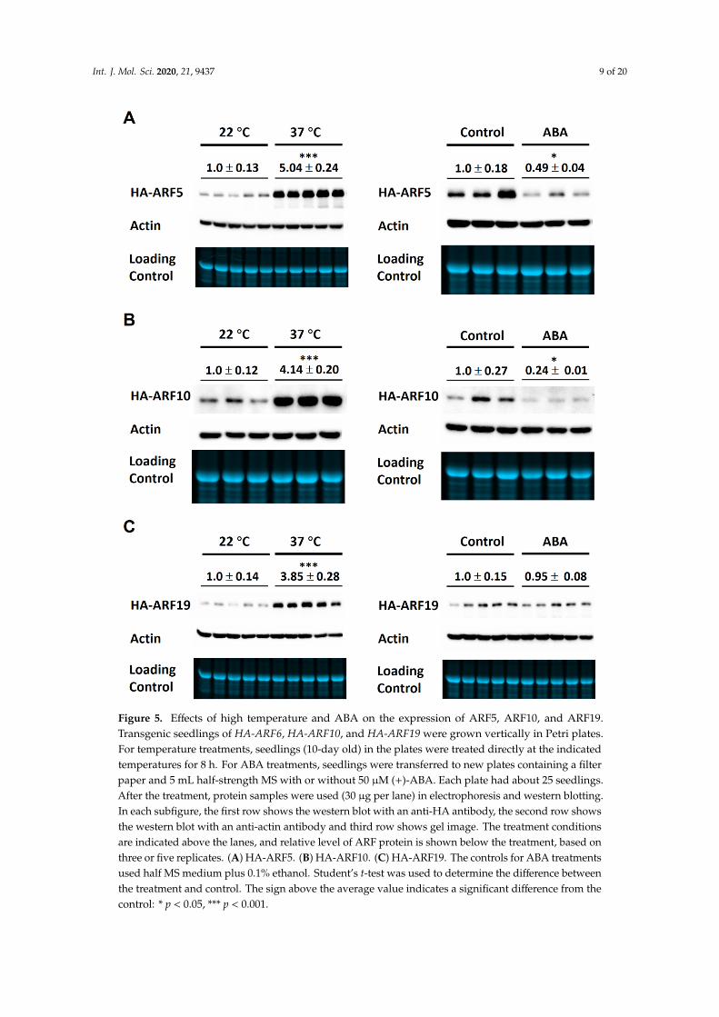
We then examined the effects of ABA and 37 ◦C treatments on the protein levels of HA-ARF5,HA-ARF10, and HA-ARF19. As shown in Figure 5, 37 ◦C treatment of 8 h strongly increased the levelsof HA-ARF5, HA-ARF10, and HA-ARF19 by similar 3–4 folds. The 50 µM ABA treatment decreasedthe levels of HA-ARF5 and HA-ARF10, but not that of HA-ARF19 (Figure 5). Statistical analysisrevealed that the changes by ABA treatments were significant for three of the four ARFs except forHA-ARF19. Taken together, those results show clearly that the 37 ◦C treatment strongly increases theARF protein levels, while ABA treatment reduces the levels of HA-ARF5, HA-ARF6, and HA-ARF10,but not HA-ARF19, consistent with earlier observations (Figure 2).
Int. J. Mol. Sci. 2020, 21, 9437 8 of 20Int. J. Mol. Sci. 2020, 21, x FOR PEER REVIEW 8 of 19
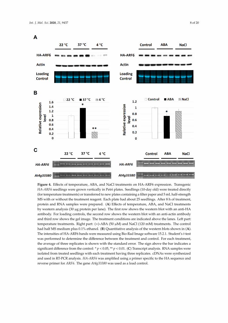
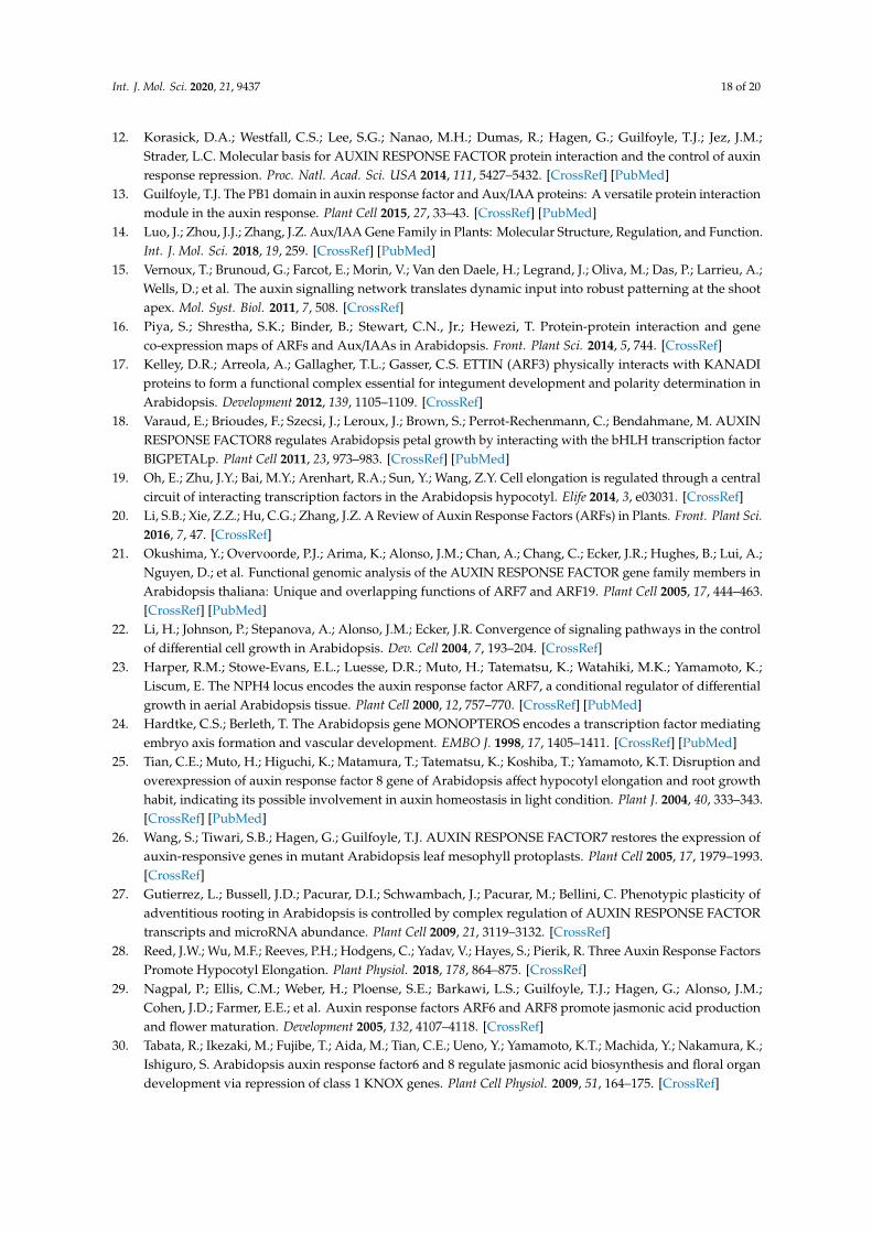
Figure 4. Effects of temperature, ABA, and NaCl treatments on HA-ARF6 expression. Transgenic HA-ARF6 seedlings were grown vertically in Petri plates. Seedlings (10-day old) were treated directly (for temperature treatments) or transferred to new plates containing a filter paper and 5 mL half-strength MS with or without the treatment reagent. Each plate had about 25 seedlings. After 8 h of treatment, protein and RNA samples were prepared. (A) Effects of temperature, ABA, and NaCl treatments by western analysis (30 μg protein per lane). The first row shows the western blot with an anti-HA antibody. For loading controls, the second row shows the western blot with an anti-actin antibody and third row shows the gel image. The treatment conditions are indicated above the lanes. Left part: temperature treatments. Right part: (+)-ABA (50 μM) and NaCl (120 mM) treatments. The control had half MS medium plus 0.1% ethanol. (B) Quantitative analysis of the western blots shown in (A). The intensities of HA-ARF6 bands were measured using Bio-Rad Image software 15.2.1. Student’s t-test was performed to determine the difference between the treatment and control. For each treatment, the average of three replicates is shown with the standard error. The sign above the bar indicates a significant difference from the control: * p < 0.05, ** p < 0.01. (C) Transcript analysis. RNA samples were isolated from treated seedlings with each treatment having three replicates. cDNAs were synthesized and used in RT-PCR analysis. HA-ARF6 was amplified using a primer specific to the HA sequence and reverse primer for ARF6. The gene At4g33380 was used as a load control.
Figure 4. Effects of temperature, ABA, and NaCl treatments on HA-ARF6 expression. TransgenicHA-ARF6 seedlings were grown vertically in Petri plates. Seedlings (10-day old) were treated directly(for temperature treatments) or transferred to new plates containing a filter paper and 5 mL half-strengthMS with or without the treatment reagent. Each plate had about 25 seedlings. After 8 h of treatment,protein and RNA samples were prepared. (A) Effects of temperature, ABA, and NaCl treatmentsby western analysis (30 µg protein per lane). The first row shows the western blot with an anti-HAantibody. For loading controls, the second row shows the western blot with an anti-actin antibodyand third row shows the gel image. The treatment conditions are indicated above the lanes. Left part:temperature treatments. Right part: (+)-ABA (50 µM) and NaCl (120 mM) treatments. The controlhad half MS medium plus 0.1% ethanol. (B) Quantitative analysis of the western blots shown in (A).The intensities of HA-ARF6 bands were measured using Bio-Rad Image software 15.2.1. Student’s t-testwas performed to determine the difference between the treatment and control. For each treatment,the average of three replicates is shown with the standard error. The sign above the bar indicates asignificant difference from the control: * p < 0.05, ** p < 0.01. (C) Transcript analysis. RNA samples wereisolated from treated seedlings with each treatment having three replicates. cDNAs were synthesizedand used in RT-PCR analysis. HA-ARF6 was amplified using a primer specific to the HA sequence andreverse primer for ARF6. The gene At4g33380 was used as a load control.
Int. J. Mol. Sci. 2020, 21, 9437 9 of 20
Int. J. Mol. Sci. 2020, 21, x FOR PEER REVIEW 9 of 19
Figure 5. Effects of high temperature and ABA on the expression of ARF5, ARF10, and ARF19. Transgenic seedlings of HA-ARF6, HA-ARF10, and HA-ARF19 were grown vertically in Petri plates. For temperature treatments, seedlings (10-day old) in the plates were treated directly at the indicated temperatures for 8 h. For ABA treatments, seedlings were transferred to new plates containing a filter paper and 5 mL half-strength MS with or without 50 μM (+)-ABA. Each plate had about 25 seedlings. After the treatment, protein samples were used (30 μg per lane) in electrophoresis and western blotting. In each subfigure, the first row shows the western blot with an anti-HA antibody, the second row shows the western blot with an anti-actin antibody and third row shows gel image. The treatment conditions are indicated above the lanes, and relative level of ARF protein is shown below the treatment, based on three or five replicates. (A) HA-ARF5. (B) HA-ARF10. (C) HA-ARF19. The controls for ABA treatments used half MS medium plus 0.1% ethanol. Student’s t-test was used to determine the difference between the treatment and control. The sign above the average value indicates a significant difference from the control: * p < 0.05, *** p < 0.001.
Figure 5. Effects of high temperature and ABA on the expression of ARF5, ARF10, and ARF19.Transgenic seedlings of HA-ARF6, HA-ARF10, and HA-ARF19 were grown vertically in Petri plates.For temperature treatments, seedlings (10-day old) in the plates were treated directly at the indicatedtemperatures for 8 h. For ABA treatments, seedlings were transferred to new plates containing a filterpaper and 5 mL half-strength MS with or without 50 µM (+)-ABA. Each plate had about 25 seedlings.After the treatment, protein samples were used (30 µg per lane) in electrophoresis and western blotting.In each subfigure, the first row shows the western blot with an anti-HA antibody, the second row showsthe western blot with an anti-actin antibody and third row shows gel image. The treatment conditionsare indicated above the lanes, and relative level of ARF protein is shown below the treatment, based onthree or five replicates. (A) HA-ARF5. (B) HA-ARF10. (C) HA-ARF19. The controls for ABA treatmentsused half MS medium plus 0.1% ethanol. Student’s t-test was used to determine the difference betweenthe treatment and control. The sign above the average value indicates a significant difference from thecontrol: * p < 0.05, *** p < 0.001.
Int. J. Mol. Sci. 2020, 21, 9437 10 of 20
2.3. Effects of ABA and Temperature Treatments on the Degradation and Ubiquitination of HA-ARF6
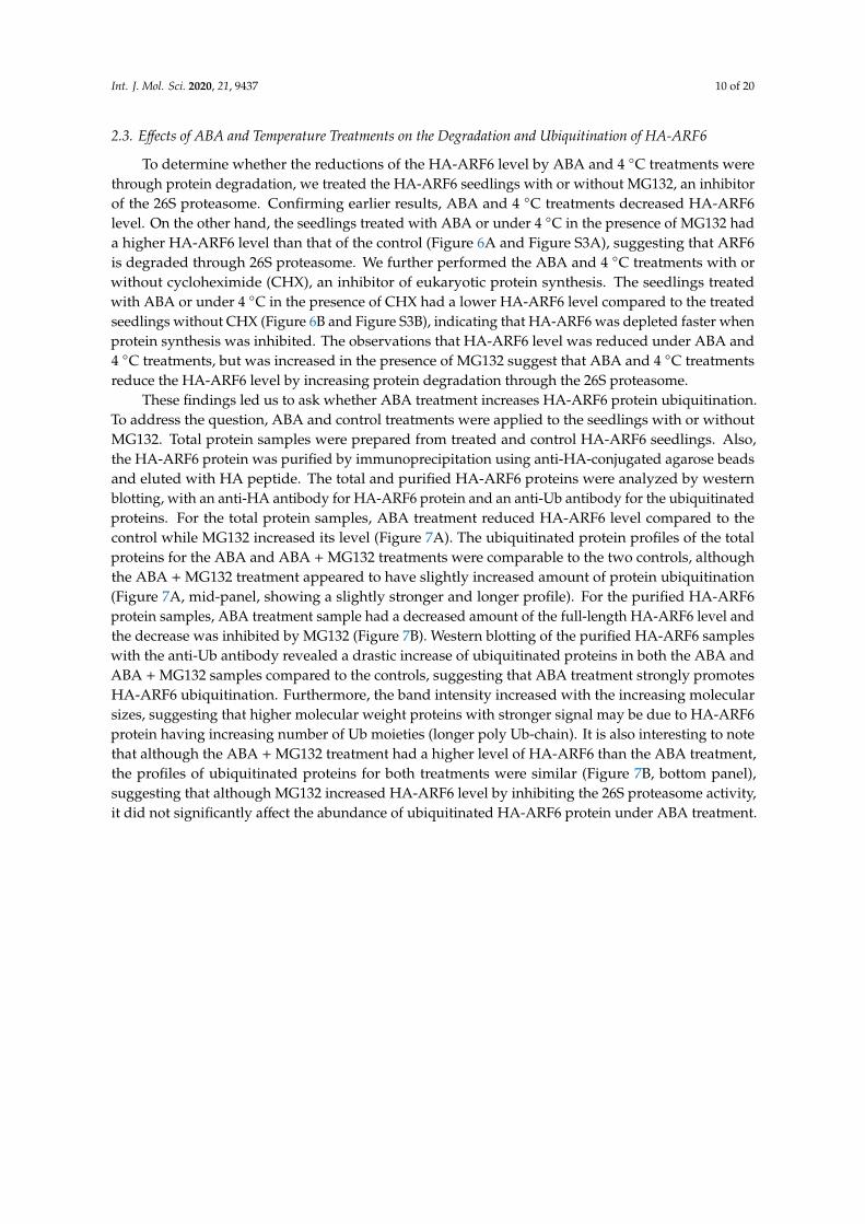
To determine whether the reductions of the HA-ARF6 level by ABA and 4 ◦C treatments werethrough protein degradation, we treated the HA-ARF6 seedlings with or without MG132, an inhibitorof the 26S proteasome. Confirming earlier results, ABA and 4 ◦C treatments decreased HA-ARF6level. On the other hand, the seedlings treated with ABA or under 4 ◦C in the presence of MG132 hada higher HA-ARF6 level than that of the control (Figure 6A and Figure S3A), suggesting that ARF6is degraded through 26S proteasome. We further performed the ABA and 4 ◦C treatments with orwithout cycloheximide (CHX), an inhibitor of eukaryotic protein synthesis. The seedlings treatedwith ABA or under 4 ◦C in the presence of CHX had a lower HA-ARF6 level compared to the treatedseedlings without CHX (Figure 6B and Figure S3B), indicating that HA-ARF6 was depleted faster whenprotein synthesis was inhibited. The observations that HA-ARF6 level was reduced under ABA and4 ◦C treatments, but was increased in the presence of MG132 suggest that ABA and 4 ◦C treatmentsreduce the HA-ARF6 level by increasing protein degradation through the 26S proteasome.
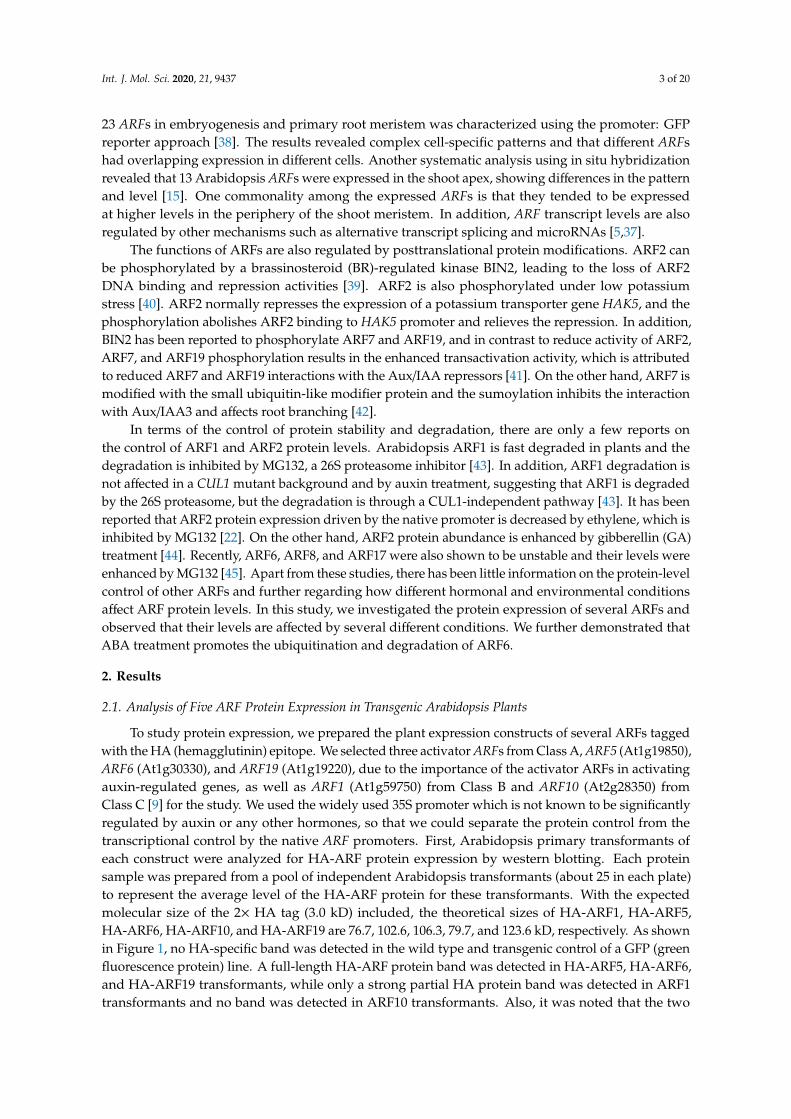
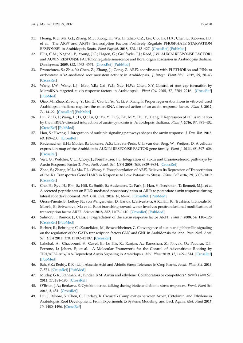
These findings led us to ask whether ABA treatment increases HA-ARF6 protein ubiquitination.To address the question, ABA and control treatments were applied to the seedlings with or withoutMG132. Total protein samples were prepared from treated and control HA-ARF6 seedlings. Also,the HA-ARF6 protein was purified by immunoprecipitation using anti-HA-conjugated agarose beadsand eluted with HA peptide. The total and purified HA-ARF6 proteins were analyzed by westernblotting, with an anti-HA antibody for HA-ARF6 protein and an anti-Ub antibody for the ubiquitinatedproteins. For the total protein samples, ABA treatment reduced HA-ARF6 level compared to thecontrol while MG132 increased its level (Figure 7A). The ubiquitinated protein profiles of the totalproteins for the ABA and ABA + MG132 treatments were comparable to the two controls, althoughthe ABA + MG132 treatment appeared to have slightly increased amount of protein ubiquitination(Figure 7A, mid-panel, showing a slightly stronger and longer profile). For the purified HA-ARF6protein samples, ABA treatment sample had a decreased amount of the full-length HA-ARF6 level andthe decrease was inhibited by MG132 (Figure 7B). Western blotting of the purified HA-ARF6 sampleswith the anti-Ub antibody revealed a drastic increase of ubiquitinated proteins in both the ABA andABA + MG132 samples compared to the controls, suggesting that ABA treatment strongly promotesHA-ARF6 ubiquitination. Furthermore, the band intensity increased with the increasing molecularsizes, suggesting that higher molecular weight proteins with stronger signal may be due to HA-ARF6protein having increasing number of Ub moieties (longer poly Ub-chain). It is also interesting to notethat although the ABA + MG132 treatment had a higher level of HA-ARF6 than the ABA treatment,the profiles of ubiquitinated proteins for both treatments were similar (Figure 7B, bottom panel),suggesting that although MG132 increased HA-ARF6 level by inhibiting the 26S proteasome activity,it did not significantly affect the abundance of ubiquitinated HA-ARF6 protein under ABA treatment.
Int. J. Mol. Sci. 2020, 21, 9437 11 of 20
Int. J. Mol. Sci. 2020, 21, x FOR PEER REVIEW 10 of 19
2.3. Effects of ABA and Temperature Treatments on the Degradation and Ubiquitination of HA-ARF6
To determine whether the reductions of the HA-ARF6 level by ABA and 4 °C treatments were through protein degradation, we treated the HA-ARF6 seedlings with or without MG132, an inhibitor of the 26S proteasome. Confirming earlier results, ABA and 4 °C treatments decreased HA-ARF6 level. On the other hand, the seedlings treated with ABA or under 4 °C in the presence of MG132 had a higher HA-ARF6 level than that of the control (Figure 6A and Figure S3A), suggesting that ARF6 is degraded through 26S proteasome. We further performed the ABA and 4 °C treatments with or without cycloheximide (CHX), an inhibitor of eukaryotic protein synthesis. The seedlings treated with ABA or under 4 °C in the presence of CHX had a lower HA-ARF6 level compared to the treated seedlings without CHX (Figure 6B and Figure S3B), indicating that HA-ARF6 was depleted faster when protein synthesis was inhibited. The observations that HA-ARF6 level was reduced under ABA and 4 °C treatments, but was increased in the presence of MG132 suggest that ABA and 4 °C treatments reduce the HA-ARF6 level by increasing protein degradation through the 26S proteasome.
Figure 6. Effects of ABA and 4 °C treatments on HA-ARF6 level in the presence of MG132 or cycloheximide (CHX). Transgenic HA-ARF6 seedlings were grown vertically in Petri plates. Seedlings (10-day old) were transferred to new plates containing a filter paper and 5 mL half-strength MS with or without the treatment reagent. Each plate had about 25 seedlings. After 4 or 8 h of treatment, proteins were extracted and used in electrophoresis (30 μg protein per lane) in Bio-Rad TGX mini-gels and western blotting. In each subfigure, the first row shows the western blot with an anti-HA antibody. For loading controls, the second row shows the western blot with an anti-actin antibody and third row shows the gel image. The treatment conditions are indicated above the lanes. (A) ABA and 4 °C treatments with or without MG132. Control 1: normal half MS medium; Control 2: half MS medium plus 0.1% ethanol and 0.1% DMSO (ABA and MG132 stocks were prepared in ethanol and DMSO, respectively); ABA: 50 μM; MG132: 25 μM. (B) ABA and 4 °C treatments with or without CHX. Control: half MS medium plus 0.1% ethanol and 0.1% DMSO; ABA: 50 μM; CHX: 200 μM.
Figure 6. Effects of ABA and 4 ◦C treatments on HA-ARF6 level in the presence of MG132 orcycloheximide (CHX). Transgenic HA-ARF6 seedlings were grown vertically in Petri plates. Seedlings(10-day old) were transferred to new plates containing a filter paper and 5 mL half-strength MS with orwithout the treatment reagent. Each plate had about 25 seedlings. After 4 or 8 h of treatment, proteinswere extracted and used in electrophoresis (30 µg protein per lane) in Bio-Rad TGX mini-gels andwestern blotting. In each subfigure, the first row shows the western blot with an anti-HA antibody.For loading controls, the second row shows the western blot with an anti-actin antibody and thirdrow shows the gel image. The treatment conditions are indicated above the lanes. (A) ABA and 4 ◦Ctreatments with or without MG132. Control 1: normal half MS medium; Control 2: half MS mediumplus 0.1% ethanol and 0.1% DMSO (ABA and MG132 stocks were prepared in ethanol and DMSO,respectively); ABA: 50 µM; MG132: 25 µM. (B) ABA and 4 ◦C treatments with or without CHX. Control:half MS medium plus 0.1% ethanol and 0.1% DMSO; ABA: 50 µM; CHX: 200 µM.
Int. J. Mol. Sci. 2020, 21, 9437 12 of 20
Int. J. Mol. Sci. 2020, 21, x FOR PEER REVIEW 11 of 19
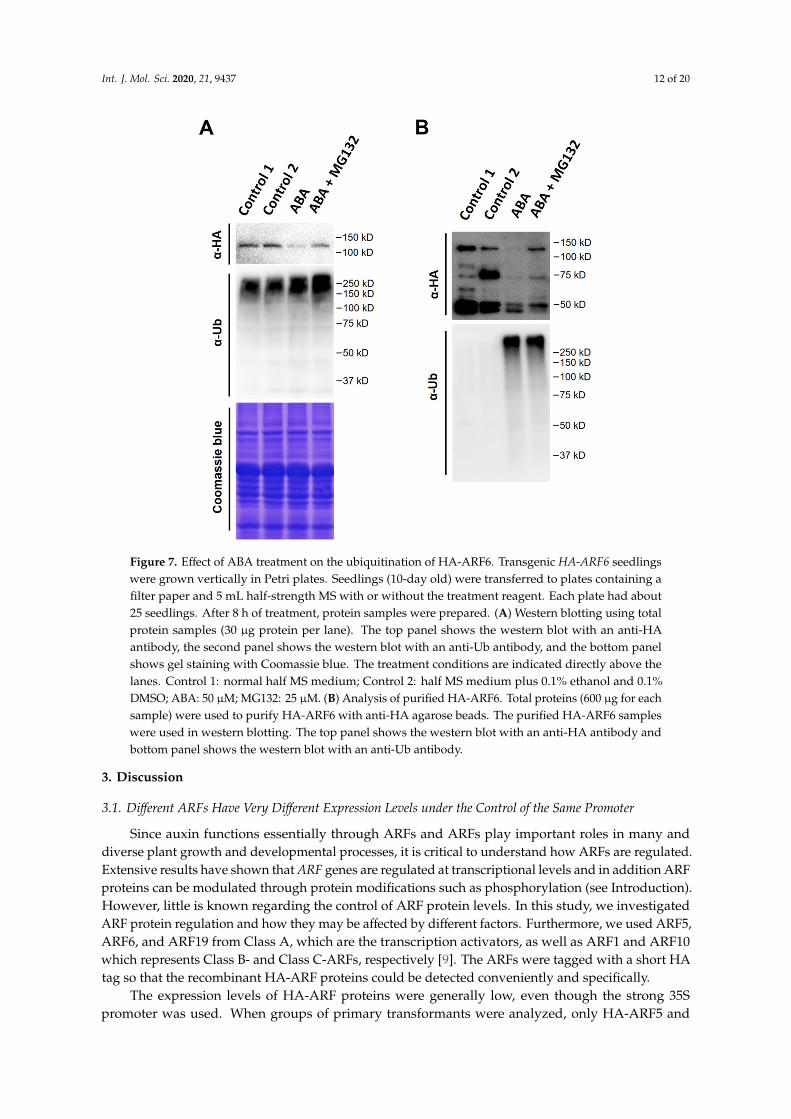
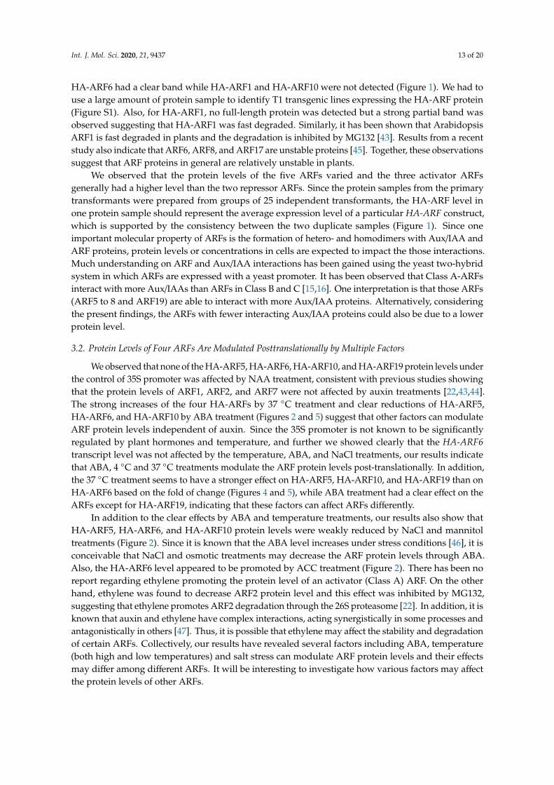
These findings led us to ask whether ABA treatment increases HA-ARF6 protein ubiquitination. To address the question, ABA and control treatments were applied to the seedlings with or without MG132. Total protein samples were prepared from treated and control HA-ARF6 seedlings. Also, the HA-ARF6 protein was purified by immunoprecipitation using anti-HA-conjugated agarose beads and eluted with HA peptide. The total and purified HA-ARF6 proteins were analyzed by western blotting, with an anti-HA antibody for HA-ARF6 protein and an anti-Ub antibody for the ubiquitinated proteins. For the total protein samples, ABA treatment reduced HA-ARF6 level compared to the control while MG132 increased its level (Figure 7A). The ubiquitinated protein profiles of the total proteins for the ABA and ABA + MG132 treatments were comparable to the two controls, although the ABA + MG132 treatment appeared to have slightly increased amount of protein ubiquitination (Figure 7A, mid-panel, showing a slightly stronger and longer profile). For the purified HA-ARF6 protein samples, ABA treatment sample had a decreased amount of the full-length HA-ARF6 level and the decrease was inhibited by MG132 (Figure 7B). Western blotting of the purified HA-ARF6 samples with the anti-Ub antibody revealed a drastic increase of ubiquitinated proteins in both the ABA and ABA+MG132 samples compared to the controls, suggesting that ABA treatment strongly promotes HA-ARF6 ubiquitination. Furthermore, the band intensity increased with the increasing molecular sizes, suggesting that higher molecular weight proteins with stronger signal may be due to HA-ARF6 protein having increasing number of Ub moieties (longer poly Ub-chain). It is also interesting to note that although the ABA + MG132 treatment had a higher level of HA-ARF6 than the ABA treatment, the profiles of ubiquitinated proteins for both treatments were similar (Figure 7B, bottom panel), suggesting that although MG132 increased HA-ARF6 level by inhibiting the 26S proteasome activity, it did not significantly affect the abundance of ubiquitinated HA-ARF6 protein under ABA treatment.
Figure 7. Effect of ABA treatment on the ubiquitination of HA-ARF6. Transgenic HA-ARF6 seedlings were grown vertically in Petri plates. Seedlings (10-day old) were transferred to plates containing a filter paper and 5 mL half-strength MS with or without the treatment reagent. Each plate had about 25 seedlings. After 8 h of treatment, protein samples were prepared. (A) Western blotting using total
Figure 7. Effect of ABA treatment on the ubiquitination of HA-ARF6. Transgenic HA-ARF6 seedlingswere grown vertically in Petri plates. Seedlings (10-day old) were transferred to plates containing afilter paper and 5 mL half-strength MS with or without the treatment reagent. Each plate had about25 seedlings. After 8 h of treatment, protein samples were prepared. (A) Western blotting using totalprotein samples (30 µg protein per lane). The top panel shows the western blot with an anti-HAantibody, the second panel shows the western blot with an anti-Ub antibody, and the bottom panelshows gel staining with Coomassie blue. The treatment conditions are indicated directly above thelanes. Control 1: normal half MS medium; Control 2: half MS medium plus 0.1% ethanol and 0.1%DMSO; ABA: 50 µM; MG132: 25 µM. (B) Analysis of purified HA-ARF6. Total proteins (600 µg for eachsample) were used to purify HA-ARF6 with anti-HA agarose beads. The purified HA-ARF6 sampleswere used in western blotting. The top panel shows the western blot with an anti-HA antibody andbottom panel shows the western blot with an anti-Ub antibody.
3. Discussion
3.1. Different ARFs Have Very Different Expression Levels under the Control of the Same Promoter
Since auxin functions essentially through ARFs and ARFs play important roles in many anddiverse plant growth and developmental processes, it is critical to understand how ARFs are regulated.Extensive results have shown that ARF genes are regulated at transcriptional levels and in addition ARFproteins can be modulated through protein modifications such as phosphorylation (see Introduction).However, little is known regarding the control of ARF protein levels. In this study, we investigatedARF protein regulation and how they may be affected by different factors. Furthermore, we used ARF5,ARF6, and ARF19 from Class A, which are the transcription activators, as well as ARF1 and ARF10which represents Class B- and Class C-ARFs, respectively [9]. The ARFs were tagged with a short HAtag so that the recombinant HA-ARF proteins could be detected conveniently and specifically.
The expression levels of HA-ARF proteins were generally low, even though the strong 35Spromoter was used. When groups of primary transformants were analyzed, only HA-ARF5 and
Int. J. Mol. Sci. 2020, 21, 9437 13 of 20
HA-ARF6 had a clear band while HA-ARF1 and HA-ARF10 were not detected (Figure 1). We had touse a large amount of protein sample to identify T1 transgenic lines expressing the HA-ARF protein(Figure S1). Also, for HA-ARF1, no full-length protein was detected but a strong partial band wasobserved suggesting that HA-ARF1 was fast degraded. Similarly, it has been shown that ArabidopsisARF1 is fast degraded in plants and the degradation is inhibited by MG132 [43]. Results from a recentstudy also indicate that ARF6, ARF8, and ARF17 are unstable proteins [45]. Together, these observationssuggest that ARF proteins in general are relatively unstable in plants.
We observed that the protein levels of the five ARFs varied and the three activator ARFsgenerally had a higher level than the two repressor ARFs. Since the protein samples from the primarytransformants were prepared from groups of 25 independent transformants, the HA-ARF level inone protein sample should represent the average expression level of a particular HA-ARF construct,which is supported by the consistency between the two duplicate samples (Figure 1). Since oneimportant molecular property of ARFs is the formation of hetero- and homodimers with Aux/IAA andARF proteins, protein levels or concentrations in cells are expected to impact the those interactions.Much understanding on ARF and Aux/IAA interactions has been gained using the yeast two-hybridsystem in which ARFs are expressed with a yeast promoter. It has been observed that Class A-ARFsinteract with more Aux/IAAs than ARFs in Class B and C [15,16]. One interpretation is that those ARFs(ARF5 to 8 and ARF19) are able to interact with more Aux/IAA proteins. Alternatively, consideringthe present findings, the ARFs with fewer interacting Aux/IAA proteins could also be due to a lowerprotein level.
3.2. Protein Levels of Four ARFs Are Modulated Posttranslationally by Multiple Factors
We observed that none of the HA-ARF5, HA-ARF6, HA-ARF10, and HA-ARF19 protein levels underthe control of 35S promoter was affected by NAA treatment, consistent with previous studies showingthat the protein levels of ARF1, ARF2, and ARF7 were not affected by auxin treatments [22,43,44].The strong increases of the four HA-ARFs by 37 ◦C treatment and clear reductions of HA-ARF5,HA-ARF6, and HA-ARF10 by ABA treatment (Figures 2 and 5) suggest that other factors can modulateARF protein levels independent of auxin. Since the 35S promoter is not known to be significantlyregulated by plant hormones and temperature, and further we showed clearly that the HA-ARF6transcript level was not affected by the temperature, ABA, and NaCl treatments, our results indicatethat ABA, 4 ◦C and 37 ◦C treatments modulate the ARF protein levels post-translationally. In addition,the 37 ◦C treatment seems to have a stronger effect on HA-ARF5, HA-ARF10, and HA-ARF19 than onHA-ARF6 based on the fold of change (Figures 4 and 5), while ABA treatment had a clear effect on theARFs except for HA-ARF19, indicating that these factors can affect ARFs differently.
In addition to the clear effects by ABA and temperature treatments, our results also show thatHA-ARF5, HA-ARF6, and HA-ARF10 protein levels were weakly reduced by NaCl and mannitoltreatments (Figure 2). Since it is known that the ABA level increases under stress conditions [46], it isconceivable that NaCl and osmotic treatments may decrease the ARF protein levels through ABA.Also, the HA-ARF6 level appeared to be promoted by ACC treatment (Figure 2). There has been noreport regarding ethylene promoting the protein level of an activator (Class A) ARF. On the otherhand, ethylene was found to decrease ARF2 protein level and this effect was inhibited by MG132,suggesting that ethylene promotes ARF2 degradation through the 26S proteasome [22]. In addition, it isknown that auxin and ethylene have complex interactions, acting synergistically in some processes andantagonistically in others [47]. Thus, it is possible that ethylene may affect the stability and degradationof certain ARFs. Collectively, our results have revealed several factors including ABA, temperature(both high and low temperatures) and salt stress can modulate ARF protein levels and their effectsmay differ among different ARFs. It will be interesting to investigate how various factors may affectthe protein levels of other ARFs.
Int. J. Mol. Sci. 2020, 21, 9437 14 of 20
3.3. ABA Treatment Promotes the Ubiquitination and Degradation of ARF6
There are complex cross-talks among different plant hormones, which are important for plantdevelopment and stress response [47–49]. It is thus critical to understand the specific mechanisms thatunderlie hormone cross-talks. There is some strong genetic evidence on the auxin and ABA interactions.From auxin to ABA direction, it has been shown that the arf10 arf16 double mutant had decreased seeddormancy and lower transcript level of ABI3, an important component in ABA signaling, suggestingthat ARF10 and ARF16 may enhance ABA-mediated seed dormancy through the activation of ABI3 [50].Conversely, ABI3 represses a microRNA (MIR160B) that targets ARF10 and ARF16 [51]. Furthermore,Arabidopsis ARF2 may affect seed size and drought tolerance through regulating ABA signaling [52].In addition, transgenic expression of a sweet potato IbARF5 in Arabidopsis and downregulationof a tomato SlARF4 have been found to increase ABA content in the plants [53,54]. From ABA toauxin direction, an ABI3-like factor from the bean is known to bind an auxin-inducible promoter [55].Additionally, Arabidopsis ARF2 expression is induced by ABA treatment and the ARF2 mutantsshowed enhanced ABA sensitivity in seed germination and primary root growth [56]. Results fromthose studies indicate that auxin and ABA interact mostly through gene expression and transcriptregulation. Our findings that ABA promotes the degradation of several Arabidopsis ARFs and stronglyinduces the ubiquitination of HA-ARF6. These results indicate that ABA can regulate ARF proteinlevel by promoting their ubiquitination, thus providing a novel link for the ABA and auxin cross-talk.
Also, it is known that cold stress can change auxin transporters and gradient, thus affectingplant growth [57]. Our observation that the 4 ◦C treatment promotes ARF6 degradation providesanother possible mechanism by which cold stress can affect plant growth. Furthermore, the 37 ◦Ctreatment strongly increased the levels of all four ARFs, while ABA did not have an effect on ARF19.Such differences imply that 37 ◦C treatment affects ARFs through a different mechanism from thatof ABA. It is interesting to note that NAA treatment did not affect the level of any of the four ARFsanalyzed, suggesting that ABA and temperature treatments affect ARF protein levels likely throughauxin-independent pathways. It will be important to identify the specific E2 and E3 componentsresponsible for ARF6 ubiquitination and degradation induced by ABA treatment, and investigate themechanism by which 37 ◦C treatment increases ARF protein levels.
3.4. ARF Protein-Level Control Provides Another Layer of Regulation for ARFs to Integrate Multiple Hormonaland Environmental Signals
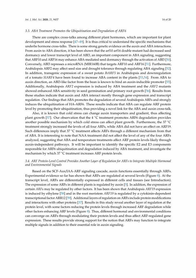
Based on the SCF-Aux/IAA-ARF signaling cascade, auxin functions essentially through ARFs.Experimental evidence so far has shown that ARFs are regulated at several levels (Figure 8). At thetranscript level, ARFs are differentially expressed in different cells and tissues (see Introduction).The expression of some ARFs in different plants is regulated by auxin [5]. In addition, the expression ofcertain ARFs may be regulated by other factors. It has been shown that Arabidopsis ARF19 expressionis induced by ethylene [58] and in the root meristem ARF19 is regulated by a cytokinin-dependenttranscriptional factor ARR12 [59]. Additional layers of regulation on ARFs include protein modificationsand interactions with other proteins [37]. Results in this study reveal another layer of regulation at theprotein level, with some factors reducing the protein levels through increased ARF degradation whileother factors enhancing ARF levels (Figure 8). Thus, different hormonal and environmental conditionscan converge on ARFs through modulating their protein levels and thus affect ARF-regulated geneexpression. These results provide strong support for the notion that ARFs may function to integratemultiple signals in addition to their essential role in auxin signaling.
Int. J. Mol. Sci. 2020, 21, 9437 15 of 20
Int. J. Mol. Sci. 2020, 21, x FOR PEER REVIEW 14 of 19
3.4. ARF Protein-Level Control Provides Another Layer of Regulation for ARFs to Integrate Multiple Hormonal and Environmental Signals
Based on the SCF-Aux/IAA-ARF signaling cascade, auxin functions essentially through ARFs. Experimental evidence so far has shown that ARFs are regulated at several levels (Figure 8). At the transcript level, ARFs are differentially expressed in different cells and tissues (see Introduction). The expression of some ARFs in different plants is regulated by auxin [5]. In addition, the expression of certain ARFs may be regulated by other factors. It has been shown that Arabidopsis ARF19 expression is induced by ethylene [58] and in the root meristem ARF19 is regulated by a cytokinin-dependent transcriptional factor ARR12 [59]. Additional layers of regulation on ARFs include protein modifications and interactions with other proteins [37]. Results in this study reveal another layer of regulation at the protein level, with some factors reducing the protein levels through increased ARF degradation while other factors enhancing ARF levels (Figure 8). Thus, different hormonal and environmental conditions can converge on ARFs through modulating their protein levels and thus affect ARF-regulated gene expression. These results provide strong support for the notion that ARFs may function to integrate multiple signals in addition to their essential role in auxin signaling.
Figure 8. A schematic diagram to show different types of regulation on ARFs, which can integrate multiple signals into the auxin signaling cascade. In auxin signaling, the binding of auxin stabilizes the interaction between the SCFTIR1/AFB E3 complex, resulting in the ubiquitination and degradation of Aux/IAA proteins, which frees ARFs. The ARFs function to activate or suppress ARF-regulated genes. ARFs can be regulated at different levels. Results from this study reveal particularly that several factors can regulate ARF protein levels, likely through auxin-independent mechanisms. ABA, low temperature, and salt stress reduce the levels of certain ARFs while 37 °C temperature increases their levels. Furthermore, ABA promotes ARF6 ubiquitination.
4. Materials and Methods
4.1. Plant Growth, Construct Preparation, and Plant Transformation
For normal plant growth and seed harvest, Arabidopsis thaliana ecotype ‘‘Columbia-0′’ WT and transgenic lines were grown in a growth room or chamber (20 °C constant, 16/8 h day/night photoperiod with a fluence rate of 90–120 μmoles/m2/min).
For preparing HA-ARF expression constructs, the ARF coding regions were amplified by PCR from Arabidopsis cDNA using Pfu DNA polymerase and gene-specific primers listed in Table S2.
Figure 8. A schematic diagram to show different types of regulation on ARFs, which can integratemultiple signals into the auxin signaling cascade. In auxin signaling, the binding of auxin stabilizes theinteraction between the SCFTIR1/AFB E3 complex, resulting in the ubiquitination and degradation ofAux/IAA proteins, which frees ARFs. The ARFs function to activate or suppress ARF-regulated genes.ARFs can be regulated at different levels. Results from this study reveal particularly that several factorscan regulate ARF protein levels, likely through auxin-independent mechanisms. ABA, low temperature,and salt stress reduce the levels of certain ARFs while 37 ◦C temperature increases their levels.Furthermore, ABA promotes ARF6 ubiquitination.
4. Materials and Methods
4.1. Plant Growth, Construct Preparation, and Plant Transformation
For normal plant growth and seed harvest, Arabidopsis thaliana ecotype “Columbia-0” WT andtransgenic lines were grown in a growth room or chamber (20 ◦C constant, 16/8 h day/night photoperiodwith a fluence rate of 90–120 µmoles/m2/min).
For preparing HA-ARF expression constructs, the ARF coding regions were amplified by PCRfrom Arabidopsis cDNA using Pfu DNA polymerase and gene-specific primers listed in Table S2.The DNA fragments were purified and digested with restriction enzymes, before they were clonedinto a vector (based on pBI121) containing a 2×HA-tag sequence at the N-terminus [60] to make theHA-ARF fusion. The HA-ARF fragments were then cloned into a plant expression vector modifiedfrom pCambia1300 (http://www.cambia.org/daisy/cambia/585.html) behind the CaMV (cauliflowermosaic virus) 35S promoter.
The constructs were used to transform WT Arabidopsis plants, using the infiltration method [61]with modifications [62]. Seeds from Agrobacterium-infiltrated plants were plated on agar platescontaining hygromycin (30 mg/L). Hygromycin-resistant transformants were transferred to soil andgenotyped using a primer specific to the HA sequence (CCCATACGATGTTCCAGAC) and a primerspecific to the ARF gene (Table S2). Transgenic lines expressing the HA-ARF protein were furtherconfirmed by western blotting (see below). At least two independent lines were analyzed initially.For most of the analysis, homozygous stable transgenic lines of T2 or later generations were used.
Int. J. Mol. Sci. 2020, 21, 9437 16 of 20
4.2. Seedling Growth and Treatments
Seeds of transgenic HA-ARF and control Arabidopsis lines were sterilized, stored at 4 ◦C for twodays and then plated on one-half MS agar plates (100 × 15 mm) containing 0.5% sucrose and 0.7%agar. Plates were placed vertically in a plant tissue culture chamber (20 ◦C constant, 16/8 h day/nightphotoperiod). Seedlings of 10–11 day old were used in general for various treatments.
For temperature treatments, plates with the seedlings were placed at the indicated temperatures.For other treatments, seedlings were transferred to treatment plates containing a piece of filter paperand 5 mL half-strength MS with or without a treatment reagent. The treatment plates with the platelid on were placed in a box on a rotary platform with slow rotation. The treatment conditions usedare: NAA (auxin), kinetin (cytokinin), (+)-ABA, ACC (1-aminocyclopropane-1-carboxylic acid), NaCl,mannitol, and temperature (22 ◦C, 4 ◦C, and 37 ◦C) treatments. The concentrations for these reagentswere selected based on previous testing done in our laboratory and they were specified in each figure.Following the treatment, about 25 seedlings from one plate were used to prepare one protein sample.
4.3. Protein Extraction, Western Blotting, and Quantitative Analysis
Plant tissues (seedlings) were homogenized with a plastic pestle in a 1.5 mL tube containing theprotein extraction buffer (50 mM Tris-HCl pH 8.0, 200 mM NaCl, 10 mM DTT (dithiothreitol), 1% (v/v)Triton X-100, Sigma protease inhibitor cocktail (Sigma-Aldrich no. P9599, Oakville, ON, Canada)).Following centrifugation at 14,000× g for 10 min at 4 ◦C, the supernatant was transferred into a freshtube and centrifuged again for 5 min. Bradford protein assay [63] was used (with Bradford reagentfrom Bio-Rad) to determine the total protein concentration, and the protein samples were stored at−80 ◦C for further analysis.
Gel electrophoresis was performed in general as described [64] with a few modifications. Majorityof analysis used min-gels (10% resolving gels) prepared from the TGX Stain-Free FastCast AcrylamideKit (Bio-Rad, no. 1610183, Hercules, CA, USA) and gel images were obtained using the Bio-Rad GelDocXR+ imaging system. Some gels were prepared using regular acrylamide solution (29:1 mix).Following electrophoresis, proteins were transferred onto PVDF membrane (Bio-Rad) using a Bio-RadTrans-Blot apparatus at a constant voltage of 25 V for 12 h in 4 ◦C.
Western blotting was performed as described [64]. For detecting HA-ARF fusion proteins, mousemonoclonal anti-HA antibodies (YEASEN no. 30701ES20, Beijing, China or Santa Cruz no. sc-7392,Dallas, TX, USA) were used. The loading control used a mouse monoclonal anti-actin antibody(Abbkine no. A01050, Wuhan, China; 1:5000). For quantitative analysis, the intensity of each proteinband was measured using Bio-Rad Image lab software 15.2.1. The relative band intensity was obtainedwith the average of the control treatment set at 1.0. Student’s t-test (two-tail, unequal varience) wasused to analyze the difference between the control and treatment values.
4.4. HA-ARF Protein Pulldown by Immunoprecipitation
For HA-ARF6 protein pulldown, total protein extract (600 µg) was incubated with anti-HAagarose beads (Sigma-Aldrich, Oakville, ON, Canada) at 4 °C for 1 h (on a tumbler). The beads werewashed twice with the wash buffer (10 mM Tris-HCl at pH 8.0, 5 mM NaCl, 0.05% Tween, 0.5 mMdithiothreitol and 0.5 mg mL−1 BSA). The bound proteins were eluted with 1 mg/mL HA peptide(GenScript, Piscataway, NJ, USA), added with SDS sample buffer, and heat-treated at 95 °C for 5 min.The samples were then used in electrophoresis and western blot analysis. HA-ARF6 and ubiquitinatedARF6 were detected using anti-HA (Santa Cruz no. sc-7392) and anti-Ub (Cell Signaling Technologyno. P4D1, Danvers, MA, USA) antibodies respectively, and a horseradish peroxidase-conjugated goalanti-mouse antibody (Bio-Rad). The signal was visualized with ECL Plus reagent (Cytiva-formerly GELife Sciences, Mississauga, ON, Canada) according to the manufacturer’s instructions.
Int. J. Mol. Sci. 2020, 21, 9437 17 of 20
4.5. RNA Isolation and PCR Analysis
Total RNA samples were isolated from seedlings using the Invitrogen TRIzol reagent (FisherScientific) following the manufacturer’s instructions. The RNA concentration was measured witha NanoVue Plus Spectrophotometer (Cytiva-formerly GE Life Sciences, Marlborough, MA, USA)following the manufacturer’s instructions. cDNAs were synthesized using the Takara PrimeScript RTreagent Kit with gDNA Eraser and 1.0 µg total RNA in 20 µL following the manufacturer’s instructions.
For RT-PCR, the HA-ARF cDNA was diluted by one-third with sterile water and 1.0 µL dilutedcDNA was used in 20 µL PCR reaction with 250 µM each of the primers. The PCR conditionswere: 95 ◦C for 1 min, then 34 cycles of 94 ◦C for 50 s, 55 ◦C for 50 s, and 72 ◦C for 1 min, and afinal extension 72 ◦C for 1 min. HA-ARF6 was amplified using a primer specific to the HA-sequence(5′-CCCATACGATGTTCCAGAC) and reverse primer specific to ARF6 (5′-TATCATGCAGATCCCTCGC).The control At4g33380 was amplified with the forward (5′-ATGAGAAGCTGGAGGAAGC) and reverse(5′-TCAAGCCGTTACAACACC) primers. For qPCR, the reactions were performed in 20 µL, using 10 µL2× iQ SYBR Green Supermix (Bio-Rad), 1.0 uL cDNA and 200 µM each of the primers, and run in theBio-Rad CFX96 Real-Time PCR System. The threshold cycle (Ct) values were generated with the Bio-RadCFX Manager Software (Version 3.0).
Supplementary Materials: The following are available online at http://www.mdpi.com/1422-0067/21/24/9437/s1.
Author Contributions: H.W. (Hong Wang) and H.W. (Hong Wu) initiated and supervised the project. K.L.and S.W. performed the experimental work and data analysis. H.W. (Hong Wang) wrote the manuscript withinput from other authors. All authors have read and agreed to the published version of the manuscript.
Funding: This research was supported by the Natural Sciences and Engineering Research Council of Canada(Discovery grant no. RGPIN-2018-05834) to H. Wang and National Natural Science Foundation of China (grant no.31870172) to H. Wu. K.L. was supported by a scholarship from the Chinese Scholarship Council and a scholarshipfrom the Department of Education of Guangdong Province.
Conflicts of Interest: The authors declare no conflict of interest.
References
1. Mockaitis, K.; Estelle, M. Auxin receptors and plant development: A new signaling paradigm. Annu. Rev.Cell Dev. Biol. 2008, 24, 55–80. [CrossRef]
2. Weijers, D.; Wagner, D. Transcriptional Responses to the Auxin Hormone. Annu. Rev. Plant Biol. 2016,67, 539–574. [CrossRef] [PubMed]
3. Finet, C.; Berne-Dedieu, A.; Scutt, C.P.; Marletaz, F. Evolution of the ARF gene family in land plants:Old domains, new tricks. Mol. Biol. Evol. 2013, 30, 45–56. [CrossRef] [PubMed]
4. Guilfoyle, T.J.; Hagen, G. Auxin response factors. Curr. Opin. Plant Biol. 2007, 10, 453–460. [CrossRef][PubMed]
5. Chandler, J.W. Auxin response factors. Plant Cell Environ. 2016, 39, 1014–1028. [CrossRef]6. Ulmasov, T.; Hagen, G.; Guilfoyle, T.J. Activation and repression of transcription by auxin-response factors.
Proc. Natl. Acad. Sci. USA 1999, 96, 5844–5849. [CrossRef] [PubMed]7. Tiwari, S.B.; Hagen, G.; Guilfoyle, T. The roles of auxin response factor domains in auxin-responsive
transcription. Plant Cell 2003, 15, 533–543. [CrossRef] [PubMed]8. Kato, H.; Nishihama, R.; Weijers, D.; Kohchi, T. Evolution of nuclear auxin signaling: Lessons from genetic
studies with basal land plants. J. Exp. Bot. 2018, 69, 291–301. [CrossRef]9. Roosjen, M.; Paque, S.; Weijers, D. Auxin Response Factors: Output control in auxin biology. J. Exp. Bot.
2018, 69, 179–188. [CrossRef]10. Ulmasov, T.; Hagen, G.; Guilfoyle, T.J. ARF1, a transcription factor that binds to auxin response elements.
Science 1997, 276, 1865–1868. [CrossRef]11. Nanao, M.H.; Vinos-Poyo, T.; Brunoud, G.; Thevenon, E.; Mazzoleni, M.; Mast, D.; Laine, S.; Wang, S.;
Hagen, G.; Li, H.; et al. Structural basis for oligomerization of auxin transcriptional regulators. Nat. Commun.2014, 5, 3617. [CrossRef] [PubMed]
Int. J. Mol. Sci. 2020, 21, 9437 18 of 20
12. Korasick, D.A.; Westfall, C.S.; Lee, S.G.; Nanao, M.H.; Dumas, R.; Hagen, G.; Guilfoyle, T.J.; Jez, J.M.;Strader, L.C. Molecular basis for AUXIN RESPONSE FACTOR protein interaction and the control of auxinresponse repression. Proc. Natl. Acad. Sci. USA 2014, 111, 5427–5432. [CrossRef] [PubMed]
13. Guilfoyle, T.J. The PB1 domain in auxin response factor and Aux/IAA proteins: A versatile protein interactionmodule in the auxin response. Plant Cell 2015, 27, 33–43. [CrossRef] [PubMed]
14. Luo, J.; Zhou, J.J.; Zhang, J.Z. Aux/IAA Gene Family in Plants: Molecular Structure, Regulation, and Function.Int. J. Mol. Sci. 2018, 19, 259. [CrossRef] [PubMed]
15. Vernoux, T.; Brunoud, G.; Farcot, E.; Morin, V.; Van den Daele, H.; Legrand, J.; Oliva, M.; Das, P.; Larrieu, A.;Wells, D.; et al. The auxin signalling network translates dynamic input into robust patterning at the shootapex. Mol. Syst. Biol. 2011, 7, 508. [CrossRef]
16. Piya, S.; Shrestha, S.K.; Binder, B.; Stewart, C.N., Jr.; Hewezi, T. Protein-protein interaction and geneco-expression maps of ARFs and Aux/IAAs in Arabidopsis. Front. Plant Sci. 2014, 5, 744. [CrossRef]
17. Kelley, D.R.; Arreola, A.; Gallagher, T.L.; Gasser, C.S. ETTIN (ARF3) physically interacts with KANADIproteins to form a functional complex essential for integument development and polarity determination inArabidopsis. Development 2012, 139, 1105–1109. [CrossRef]
18. Varaud, E.; Brioudes, F.; Szecsi, J.; Leroux, J.; Brown, S.; Perrot-Rechenmann, C.; Bendahmane, M. AUXINRESPONSE FACTOR8 regulates Arabidopsis petal growth by interacting with the bHLH transcription factorBIGPETALp. Plant Cell 2011, 23, 973–983. [CrossRef] [PubMed]
19. Oh, E.; Zhu, J.Y.; Bai, M.Y.; Arenhart, R.A.; Sun, Y.; Wang, Z.Y. Cell elongation is regulated through a centralcircuit of interacting transcription factors in the Arabidopsis hypocotyl. Elife 2014, 3, e03031. [CrossRef]
20. Li, S.B.; Xie, Z.Z.; Hu, C.G.; Zhang, J.Z. A Review of Auxin Response Factors (ARFs) in Plants. Front. Plant Sci.2016, 7, 47. [CrossRef]
21. Okushima, Y.; Overvoorde, P.J.; Arima, K.; Alonso, J.M.; Chan, A.; Chang, C.; Ecker, J.R.; Hughes, B.; Lui, A.;Nguyen, D.; et al. Functional genomic analysis of the AUXIN RESPONSE FACTOR gene family members inArabidopsis thaliana: Unique and overlapping functions of ARF7 and ARF19. Plant Cell 2005, 17, 444–463.[CrossRef] [PubMed]
22. Li, H.; Johnson, P.; Stepanova, A.; Alonso, J.M.; Ecker, J.R. Convergence of signaling pathways in the controlof differential cell growth in Arabidopsis. Dev. Cell 2004, 7, 193–204. [CrossRef]
23. Harper, R.M.; Stowe-Evans, E.L.; Luesse, D.R.; Muto, H.; Tatematsu, K.; Watahiki, M.K.; Yamamoto, K.;Liscum, E. The NPH4 locus encodes the auxin response factor ARF7, a conditional regulator of differentialgrowth in aerial Arabidopsis tissue. Plant Cell 2000, 12, 757–770. [CrossRef] [PubMed]
24. Hardtke, C.S.; Berleth, T. The Arabidopsis gene MONOPTEROS encodes a transcription factor mediatingembryo axis formation and vascular development. EMBO J. 1998, 17, 1405–1411. [CrossRef] [PubMed]
25. Tian, C.E.; Muto, H.; Higuchi, K.; Matamura, T.; Tatematsu, K.; Koshiba, T.; Yamamoto, K.T. Disruption andoverexpression of auxin response factor 8 gene of Arabidopsis affect hypocotyl elongation and root growthhabit, indicating its possible involvement in auxin homeostasis in light condition. Plant J. 2004, 40, 333–343.[CrossRef] [PubMed]
26. Wang, S.; Tiwari, S.B.; Hagen, G.; Guilfoyle, T.J. AUXIN RESPONSE FACTOR7 restores the expression ofauxin-responsive genes in mutant Arabidopsis leaf mesophyll protoplasts. Plant Cell 2005, 17, 1979–1993.[CrossRef]
27. Gutierrez, L.; Bussell, J.D.; Pacurar, D.I.; Schwambach, J.; Pacurar, M.; Bellini, C. Phenotypic plasticity ofadventitious rooting in Arabidopsis is controlled by complex regulation of AUXIN RESPONSE FACTORtranscripts and microRNA abundance. Plant Cell 2009, 21, 3119–3132. [CrossRef]
28. Reed, J.W.; Wu, M.F.; Reeves, P.H.; Hodgens, C.; Yadav, V.; Hayes, S.; Pierik, R. Three Auxin Response FactorsPromote Hypocotyl Elongation. Plant Physiol. 2018, 178, 864–875. [CrossRef]
29. Nagpal, P.; Ellis, C.M.; Weber, H.; Ploense, S.E.; Barkawi, L.S.; Guilfoyle, T.J.; Hagen, G.; Alonso, J.M.;Cohen, J.D.; Farmer, E.E.; et al. Auxin response factors ARF6 and ARF8 promote jasmonic acid productionand flower maturation. Development 2005, 132, 4107–4118. [CrossRef]
30. Tabata, R.; Ikezaki, M.; Fujibe, T.; Aida, M.; Tian, C.E.; Ueno, Y.; Yamamoto, K.T.; Machida, Y.; Nakamura, K.;Ishiguro, S. Arabidopsis auxin response factor6 and 8 regulate jasmonic acid biosynthesis and floral organdevelopment via repression of class 1 KNOX genes. Plant Cell Physiol. 2009, 51, 164–175. [CrossRef]
Int. J. Mol. Sci. 2020, 21, 9437 19 of 20
31. Huang, K.L.; Ma, G.J.; Zhang, M.L.; Xiong, H.; Wu, H.; Zhao, C.Z.; Liu, C.S.; Jia, H.X.; Chen, L.; Kjorven, J.O.;et al. The ARF7 and ARF19 Transcription Factors Positively Regulate PHOSPHATE STARVATIONRESPONSE1 in Arabidopsis Roots. Plant Physiol. 2018, 178, 413–427. [CrossRef] [PubMed]
32. Ellis, C.M.; Nagpal, P.; Young, J.C.; Hagen, G.; Guilfoyle, T.J.; Reed, J.W. AUXIN RESPONSE FACTOR1and AUXIN RESPONSE FACTOR2 regulate senescence and floral organ abscission in Arabidopsis thaliana.Development 2005, 132, 4563–4574. [CrossRef] [PubMed]
33. Promchuea, S.; Zhu, Y.; Chen, Z.; Zhang, J.; Gong, Z. ARF2 coordinates with PLETHORAs and PINs toorchestrate ABA-mediated root meristem activity in Arabidopsis. J. Integr. Plant Biol. 2017, 59, 30–43.[CrossRef]
34. Wang, J.W.; Wang, L.J.; Mao, Y.B.; Cai, W.J.; Xue, H.W.; Chen, X.Y. Control of root cap formation byMicroRNA-targeted auxin response factors in Arabidopsis. Plant Cell 2005, 17, 2204–2216. [CrossRef][PubMed]
35. Qiao, M.; Zhao, Z.; Song, Y.; Liu, Z.; Cao, L.; Yu, Y.; Li, S.; Xiang, F. Proper regeneration from in vitro culturedArabidopsis thaliana requires the microRNA-directed action of an auxin response factor. Plant J. 2012,71, 14–22. [CrossRef] [PubMed]
36. Liu, Z.; Li, J.; Wang, L.; Li, Q.; Lu, Q.; Yu, Y.; Li, S.; Bai, M.Y.; Hu, Y.; Xiang, F. Repression of callus initiationby the miRNA-directed interaction of auxin-cytokinin in Arabidopsis thaliana. Plant J. 2016, 87, 391–402.[CrossRef] [PubMed]
37. Han, S.; Hwang, I. Integration of multiple signaling pathways shapes the auxin response. J. Exp. Bot. 2018,69, 189–200. [CrossRef]
38. Rademacher, E.H.; Moller, B.; Lokerse, A.S.; Llavata-Peris, C.I.; van den Berg, W.; Weijers, D. A cellularexpression map of the Arabidopsis AUXIN RESPONSE FACTOR gene family. Plant J. 2011, 68, 597–606.[CrossRef]
39. Vert, G.; Walcher, C.L.; Chory, J.; Nemhauser, J.L. Integration of auxin and brassinosteroid pathways byAuxin Response Factor 2. Proc. Natl. Acad. Sci. USA 2008, 105, 9829–9834. [CrossRef]
40. Zhao, S.; Zhang, M.L.; Ma, T.L.; Wang, Y. Phosphorylation of ARF2 Relieves Its Repression of Transcriptionof the K+ Transporter Gene HAK5 in Response to Low Potassium Stress. Plant Cell 2016, 28, 3005–3019.[CrossRef]
41. Cho, H.; Ryu, H.; Rho, S.; Hill, K.; Smith, S.; Audenaert, D.; Park, J.; Han, S.; Beeckman, T.; Bennett, M.J.; et al.A secreted peptide acts on BIN2-mediated phosphorylation of ARFs to potentiate auxin response duringlateral root development. Nat. Cell. Biol. 2014, 16, 66–76. [CrossRef] [PubMed]
42. Orosa-Puente, B.; Leftley, N.; von Wangenheim, D.; Banda, J.; Srivastava, A.K.; Hill, K.; Truskina, J.; Bhosale, R.;Morris, E.; Srivastava, M.; et al. Root branching toward water involves posttranslational modification oftranscription factor ARF7. Science 2018, 362, 1407–1410. [CrossRef] [PubMed]
43. Salmon, J.; Ramos, J.; Callis, J. Degradation of the auxin response factor ARF1. Plant J. 2008, 54, 118–128.[CrossRef] [PubMed]
44. Richter, R.; Behringer, C.; Zourelidou, M.; Schwechheimer, C. Convergence of auxin and gibberellin signalingon the regulation of the GATA transcription factors GNC and GNL in Arabidopsis thaliana. Proc. Natl. Acad.Sci. USA 2013, 110, 13192–13197. [CrossRef]
45. Lakehal, A.; Chaabouni, S.; Cavel, E.; Le Hir, R.; Ranjan, A.; Raneshan, Z.; Novak, O.; Pacurar, D.I.;Perrone, I.; Jobert, F.; et al. A Molecular Framework for the Control of Adventitious Rooting byTIR1/AFB2-Aux/IAA-Dependent Auxin Signaling in Arabidopsis. Mol. Plant 2019, 12, 1499–1514. [CrossRef][PubMed]
46. Sah, S.K.; Reddy, K.R.; Li, J. Abscisic Acid and Abiotic Stress Tolerance in Crop Plants. Front. Plant Sci. 2016,7, 571. [CrossRef] [PubMed]
47. Muday, G.K.; Rahman, A.; Binder, B.M. Auxin and ethylene: Collaborators or competitors? Trends Plant Sci.2012, 17, 181–195. [CrossRef]
48. O’Brien, J.A.; Benkova, E. Cytokinin cross-talking during biotic and abiotic stress responses. Front. Plant Sci.2013, 4, 451. [CrossRef]
49. Liu, J.; Moore, S.; Chen, C.; Lindsey, K. Crosstalk Complexities between Auxin, Cytokinin, and Ethylene inArabidopsis Root Development: From Experiments to Systems Modeling, and Back Again. Mol. Plant 2017,10, 1480–1496. [CrossRef]
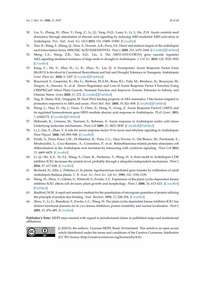
Int. J. Mol. Sci. 2020, 21, 9437 20 of 20
50. Liu, X.; Zhang, H.; Zhao, Y.; Feng, Z.; Li, Q.; Yang, H.Q.; Luan, S.; Li, J.; He, Z.H. Auxin controls seeddormancy through stimulation of abscisic acid signaling by inducing ARF-mediated ABI3 activation inArabidopsis. Proc. Natl. Acad. Sci. USA 2013, 110, 15485–15490. [CrossRef]
51. Tian, R.; Wang, F.; Zheng, Q.; Niza, V.; Downie, A.B.; Perry, S.E. Direct and indirect targets of the arabidopsisseed transcription factor ABSCISIC ACID INSENSITIVE3. Plant J. 2020, 103, 1679–1694. [CrossRef] [PubMed]
52. Meng, L.S.; Wang, Z.B.; Yao, S.Q.; Liu, A. The ARF2-ANT-COR15A gene cascade regulatesABA-signaling-mediated resistance of large seeds to drought in Arabidopsis. J. Cell Sci. 2015, 128, 3922–3932.[CrossRef]
53. Kang, C.; He, S.; Zhai, H.; Li, R.; Zhao, N.; Liu, Q. A Sweetpotato Auxin Response Factor Gene(IbARF5) Is Involved in Carotenoid Biosynthesis and Salt and Drought Tolerance in Transgenic Arabidopsis.Front. Plant Sci. 2018, 9, 1307. [CrossRef] [PubMed]
54. Bouzroud, S.; Gasparini, K.; Hu, G.; Barbosa, M.A.M.; Rosa, B.L.; Fahr, M.; Bendaou, N.; Bouzayen, M.;Zsogon, A.; Smouni, A.; et al. Down Regulation and Loss of Auxin Response Factor 4 Function UsingCRISPR/Cas9 Alters Plant Growth, Stomatal Function and Improves Tomato Tolerance to Salinity andOsmotic Stress. Genes 2020, 11, 272. [CrossRef] [PubMed]
55. Nag, R.; Maity, M.K.; Dasgupta, M. Dual DNA binding property of ABA insensitive 3 like factors targeted topromoters responsive to ABA and auxin. Plant Mol. Biol. 2005, 59, 821–838. [CrossRef] [PubMed]
56. Wang, L.; Hua, D.; He, J.; Duan, Y.; Chen, Z.; Hong, X.; Gong, Z. Auxin Response Factor2 (ARF2) andits regulated homeodomain gene HB33 mediate abscisic acid response in Arabidopsis. PLoS Genet. 2011,7, e1002172. [CrossRef] [PubMed]
57. Shibasaki, K.; Uemura, M.; Tsurumi, S.; Rahman, A. Auxin response in Arabidopsis under cold stress:Underlying molecular mechanisms. Plant Cell 2009, 21, 3823–3838. [CrossRef] [PubMed]
58. Li, J.; Dai, X.; Zhao, Y. A role for auxin response factor 19 in auxin and ethylene signaling in Arabidopsis.Plant Physiol. 2006, 140, 899–908. [CrossRef]
59. Perilli, S.; Perez-Perez, J.M.; Di Mambro, R.; Peris, C.L.; Diaz-Trivino, S.; Del Bianco, M.; Pierdonati, E.;Moubayidin, L.; Cruz-Ramirez, A.; Costantino, P.; et al. Retinoblastoma-related protein stimulates celldifferentiation in the Arabidopsis root meristem by interacting with cytokinin signaling. Plant Cell 2013,25, 4469–4478. [CrossRef]
60. Li, Q.; Shi, X.Z.; Ye, S.J.; Wang, S.; Chan, R.; Harkness, T.; Wang, H. A short motif in Arabidopsis CDKinhibitor ICK1 decreases the protein level, probably through a ubiquitin-independent mechanism. Plant J.2016, 87, 617–628. [CrossRef]
61. Bechtold, N.; Ellis, J.; Pelletier, G. In planta Agrobacterium mediated gene transfer by infiltration of adultArabidopsis thaliana plants. C. R. Acad. Sci. Paris Sci. Life Sci. 1993, 316, 1194–1199.
62. Wang, H.; Zhou, Y.; Gilmer, S.; Whitwill, S.; Fowke, L.C. Expression of the plant cyclin-dependent kinaseinhibitor ICK1 affects cell division, plant growth and morphology. Plant J. 2000, 24, 613–623. [CrossRef][PubMed]
63. Bradford, M.M. A rapid and sensitive method for the quantitation of microgram quantities of protein utilizingthe principle of protein-dye binding. Anal. Biochem. 1976, 72, 248–254. [CrossRef]
64. Zhou, Y.; Li, G.; Brandizzi, F.; Fowke, L.C.; Wang, H. The plant cyclin-dependent kinase inhibitor ICK1 hasdistinct functional domains for in vivo kinase inhibition, protein instability and nuclear localization. Plant J.2003, 35, 476–489. [CrossRef]
Publisher’s Note: MDPI stays neutral with regard to jurisdictional claims in published maps and institutionalaffiliations.
© 2020 by the authors. Licensee MDPI, Basel, Switzerland. This article is an open accessarticle distributed under the terms and conditions of the Creative Commons Attribution(CC BY) license (http://creativecommons.org/licenses/by/4.0/).


![Role of the Arabidopsis PIN6 Auxin Transporter in Auxin ......central stele and basipetally through the epidermis and outer cortex near the root tip [4,5]. Polar cell-to-cell auxin](https://static.fdocuments.net/doc/165x107/611cd25da6ea2f70970f71db/role-of-the-arabidopsis-pin6-auxin-transporter-in-auxin-central-stele-and.jpg)