Synthesis and characterization of superparamagnetic iron ...618655/FULLTEXT02.pdf ·...
50
Synthesis and characterization of superparamagnetic iron oxide nanoparticles coated with silica Aleksandr Marinin Master Thesis Stockholm 2012 Division of Functional Materials School of Information and Communication Technology Royal Institute of Technology
Transcript of Synthesis and characterization of superparamagnetic iron ...618655/FULLTEXT02.pdf ·...

Synthesis and characterization of superparamagnetic iron oxide nanoparticles coated with silica
Aleksandr Marinin
Master Thesis
Stockholm 2012
Division of Functional Materials
School of Information and Communication Technology
Royal Institute of Technology

2
Abstract
Multifunctional superparamagnetic iron oxide nanoparticles (SPIONs) coated with silica are a
promising research field for lots of biomedical applications. The scope of this work is a
preparation of SPIONs and coating them with silica to form core-shell structured nanoparticles
for nanomedicine applications.
SPIONs were synthesized by two chemical methods – co-precipitation and thermal
decomposition of organic iron precursor. Prepared nanoparticles were carefully characterized –
average size, size distribution, morphology, crystallinity, colloidal stability and magnetic
properties were studied. After comparing SPIONs synthetized by two routes the most suitable
method for biomedical applicable nanoparticles preparation is determined. The nanomedicine
requires nanoparticles of the highest quality.
The next step was coating SPIONs with silica shell. For this purpose inverse microemulsion
method was chosen. TEOS was used as a silica precursor. Mean size, size distribution, magnetic
properties, structure of silica shell were studied.
Keywords : magnetite, co-precipitation, thermal decomposition, silica, inverse microemulsion, TEOS, biomedical applications, core-shell.

3
Table of Contents
1 Introduction ............................................................................................................................ 5
1.1 Objectives .......................................................................................................................... 6
1.2 Outline .............................................................................................................................. 7
1.3 Magnetic nanoparticles ...................................................................................................... 8
1.3.1 Magnetic properties ................................................................................................... 8
1.3.2 Iron oxides ............................................................................................................... 13
1.3.3 Synthesis of SPIONs ................................................................................................ 14
1.3.3.1 Co-precipitation synthesis method ................................................................. 14
1.3.3.2 Thermal decomposition of iron precursor synthesis method ........................... 16
1.4 Core-shell magnetic nanoparticles ................................................................................... 17
1.4.1 Silica shell properties .............................................................................................. 18
1.4.2 Silica coating by inverse microemulsion method ..................................................... 18
1.5 Multifunctional core-shell nanoparticles .......................................................................... 20
1.6 Use of core-shell nanoparticles in medicine ..................................................................... 21
1.6.1 MRI contrast agent.................................................................................................. 21
1.6.2 Drug delivery systems ............................................................................................. 24
1.6.3 Hyperthermia .......................................................................................................... 25
2 Experimental ........................................................................................................................ 27
2.1 Synthesis of SPIONs ....................................................................................................... 27
2.1.1 Co-precipitation method ......................................................................................... 27
2.1.2 Thermal decomposition method .............................................................................. 27
2.2 Synthesis of SiO2@Fe3O4 ................................................................................................ 28
2.3 Characterization .............................................................................................................. 29
2.3.1 Transmission electron microscopy .......................................................................... 30

4
2.3.2 Dynamic light scattering ......................................................................................... 31
2.3.3 Zeta-potential analysis ............................................................................................ 33
2.3.4 Magnetic characterization ....................................................................................... 34
2.3.5 Inductively coupled plasma – atomic emission spectroscopy ................................... 35
3 Results and discussions ........................................................................................................ 37
3.1 Morphology, structure and size characterization .............................................................. 37
3.1.1 Transmission electron microscopy .......................................................................... 37
3.1.2 Dynamic light scattering ......................................................................................... 42
3.2 Magnetic characterization ................................................................................................ 44
3.2 Colloidal stability characterization ................................................................................... 45
4 Conclusions ........................................................................................................................... 47
References................................................................................................................................ 48

5
1 Introduction
Nanotechnologies are fast growing area of researches in all over the world. The reason of
serious attention is a wide spectrum of possibilities to improve the quality of life. The key is
new significant physical, chemical, biological properties of nanosize constructed materials –
nanoparticles, nanotubes, nanowires, extremely thin films, nanocomposites etc. High surface to
volume ratio allow to functionalize nanoparticles with different ligands, coatings and other
useful tools for lots of biomedical applications. Nowadays medicine has great future
opportunities based on use of engineered nanomaterials.
Nanomedicine is a serious field of researches, promising not only improvement of the
existing applications, but new highly effective methods for diagnostics and therapy. Nanoscale
findings can make diagnostics more accurate and forward-looking; provide treatments with
minimum side effects. High efficiency MRI contrast agents, quantum dot based imaging
methods, accurate thermosensitive drug delivery systems, hyperthermia tumour treatment are
only few fast developing nanomedicine directions. Significant advance takes place in cellular
bioapplications – engineered nanotools give an opportunity to operate with cell organellas, DNA
and modify the cellular structure1.
Main discussion of this thesis is multifunctional core-shell nanocomposits for biomedical in
vivo applications. Magnetic core makes the position of these composites controllable in human
organism media; inert shell provides non-toxicity, stability and polyfunctionality, being a
suitable platform for variety of ligands2.
The great potential of nanotechnologies makes them the most perspective field of scientific
interest. In the nearest future nanodevices will help us in any aspect of our lives. One atom can
change the world.

6
1.1 Objectives
In this thesis superparamagnetic iron oxide nanoparticles (SPIONs) coated with silica will be
observed. Synthesis, characterization and further works on functionalization for biomedical
purposes are main topics of discussion. There are several certain requirements, which must be
strictly followed in order to gain maximum efficiency and no side effects for human organism.
These requirements are :
1) superparamagnetic properties;
2) non-toxicity and non-reactivity to a human organism;
3) colloidal stability in an organism inner media.
In this work two methods of SPIONs synthesis will be evaluated and compared – co-
precipitation and thermal decomposition. Differences between produced particles will be fixed
and analysed. Their magnetical properties, size and shape will be carefully charaterized. The
main criterias in giving labor to one of methods are :
1) ability to control the overal size of produced nanoparticles in order to provide
superparamagnetic properties;
2) relative value of size distribution of synthetised SPIONs – the narrower it is, the higher is
homogeneity of induced magnetic field and colloidal stability in organism inner media;
3) ability to manage morphology, spherical shape of SPIONs is the most preferrable.
After synthesis of SPIONs, which more correspond to listed above criterias, they will be
coated with silica using inverse microemulsion method, forming core-shell nanocomposites.
Silica core makes SPIONs inert to human organism and colloidly stable at certain pH of
organism environment (excluding acidic pH of alimentary tract) and is a suitable platform for
further functionalization. Varying of reaction conditions results in different thickness of silica
layer, so it can be finely tuned. Dependence of magnetization values from silica shell thickness
will be studied.
Colloidly stable core-shell structured nanocomposites with narrow size distribution of
magnetite core and silica shell with high magnetization values and ready for further
functionalization are expected as a result of this work.

7
1.2 Outline
The aim of this thesis is to develop multifunctional Fe3O4@SiO2 nanocomposites for
biomedical applications, such as MRI T2 contrast agents, hyperthermia cancer treatment,
accurate drug delivery systems etc. Biomedical applications require highest quality
nanomaterials.
Chapter 1.1 tells about targets of this work, describing conditions and steps of realization.
Chapter 1.3 briefly describes an idea of magnetic nanoparticles in medicine : introduces in
magnetic properties of materials and especially iron oxides (chapters 1.3.1 and 1.3.2), reviews
two SPIONs synthesis methods in chapter 1.3.3.
Chapter 1.4 evaluates core-shell structured magnetic nanoparticles. Silica shell properties
(chapter 1.4.1) and coating method (chapter 1.4.2) are discussed.
Chapter 1.5 describes further functionalization opportunities of core-shell nanoparticles.
Chapter 1.6 gives an overview on biomedical applications based on multifunctional core-
shell SPIONs.
Chapters 2.1 – 2.2 tell about experimental part – procedures, preparation steps of CSNPs.
Chapter 2.3 describes characterization methodology of produced nanomaterials.
Chapter 3 contains results and discussions about morphology, crystallinity and size
distribution (chapter 3.1), magnetic properties (chapter 3.2), colloidal stability (chapter 3.3) of
nanomaterials for the each step of experimental section.

8
1.3 Magnetic nanoparticles
Magnetic properties of nanomaterials are powerful manipulation and detection tools which
are studied for a long time. Since magnetic fields are not harmful to a human organism (but this
question is still opened for high strength magnetic fields) magnetic nanoparticles can be used for
biomedical in vivo and, of course, in vitro applications. The latest researches tell about high
potential of magnetic nanoparticles in environmental remediation applications, such as removal
of heavy metals from waste waters36. Magnetic properties depend on a size, shape, structure,
crystallinity, synthesis method and chemistry of materials. The most widely investigated
magnetic nanomaterials are iron, cobalt and nickel compounds and alloys37.
1.3.1 Magnetic properties
By a material reaction to a magnetic field, all substances are categorised into several groups :
- diamagnetics,
- paramagnetics,
- ferromagnetics,
- superparamagnetics.
First some background on magnetic properties of materials. Magnetization M of a sample is a
sum of magnetic moments m of all atoms in the sample per unit volume V :
𝑴𝑴 = 𝒎𝒎𝑽𝑽
(1)
There are 2 points which describe response of a material to applied magnetic field :
susceptibility and permeability. Susceptibility χ is a dimensionless proportionality constant that
shows the magnetization level M of a substance under an influence of an external magnetic field
H :
𝑴𝑴 = χH (2)
The permeability μ is the magnetic induction B change with the applied magnetic field H :
𝐵𝐵 = μH (3)

9
In other words, permeability shows how conductive a material is to a magnetic field. The
higher permeability, the lower is a resistance of a material to magnetic field. In SI system,
permeability is measured in henries per meter (H/m). μ0 is the permeability of vacuum, or
magnetic constant, and is equal to 4π×10−7 H/m. Relative permeability μr is a dependence of
material permeability to vacuum permeability :
(4)
Diamagnetics. Under an influence of an external magnetic field diamagnetics produce weak
magnetic field in the direction opposite to applied field. After removal of an external field
attracted spins relax to their initial position. Relative permeability μr ≤ 1, susceptibility χ =
-10-6...-10-3 (negative due to opposite direction of induced magnetic field).
Figure 1.2 Diamagnetic material under external magnetic field
Diamagnetics` magnetic behavior is not significant (exception – superconductors). Examples
of diamagnetics are water, wood, most of organic compounds, copper, silver (systems without
any unpaired electrons).
Paramagnetics. Under an influence of an external magnetic field paramagnetics possess
weak magnetic field in the direction of applied one. Like in diamagnetics, after removing of
external magnetic field attracted spins relax and material shows zero net magnetization. Relative
permeability μr ≥1, susceptibility χ = 10-3...10-5 .

10
Figure 1.3 Paramagnetic material under external magnetic field
Paramagnetics` response to applied magnetic field is weak, similarly to diamagnetics.
Examples of paramagnetics are aluminium, oxygen, magnesium, lithium (pure paramagnets are
systems with unpaired non-interacting spins).
Ferromagnetics. Materials with significant magnetic properties. All its` magnetic moments
can align in one direction, creating high magnetic moment of the sytem, even without any
external magnetic field (this phenomena is called spontaneous magnetization and may occur only
below critical temperature, called Curie point, Neel point for antiferromagnetics). Above critical
temperature ferromagnetic (and ferrimagnetic) properties disappear and paramagnetic ones
appear, magnetic moments are totally disordered. Relative permeability μr >> 1, susceptibility χ
>> 1.
Figure 1.4 Ferromagnetic material under external magnetic field
Ferromagnetics have multidomain structure. All moments in one domain are aligned in one
direction. These clusters are separated by domain walls. Its` location depends on crystal

11
structure, crystal defects. After removing of an external magnetic field, ferromagnetics` net
magnetization do not disappear – this phenomena is called the remanence magnetization.
When we apply an external magnetic field on ferromagnetic material, such hysteresis curve is
obtained (figure 1.5). Function extrems Ms and Ms’ are saturation magnetization. Hysteresis
means a process depends on direction of it`s distribution (history). Every applied field H value
has two magnetization M values (except saturation points). Remanence magnetization (points Mr
and Mr`) and coercivity (points Hc and Hc`) take place.
Examples are iron, nickel, cobalt, its` oxides and alloys, gadolinium, therbium.
Figure 1.5 Magnetization curve of ferromagnetic material
There are also ferrimagnetics and antiferromagnetics. In ferrimagnetic material there are two
antiparallel fractions of magnetic moments with different strength. Example - manganese,
magnetite, maghemite. In antiferromagnetics moment arrangement is the same, but moments are
equal in strength, so overall magnetic moment is zero. Examples – hematite, chromium.
Figure 1.6 Alignment of magnetic moments in a) ferromagnetics, b) ferrimagnetics, c)
antiferromagnetics
12
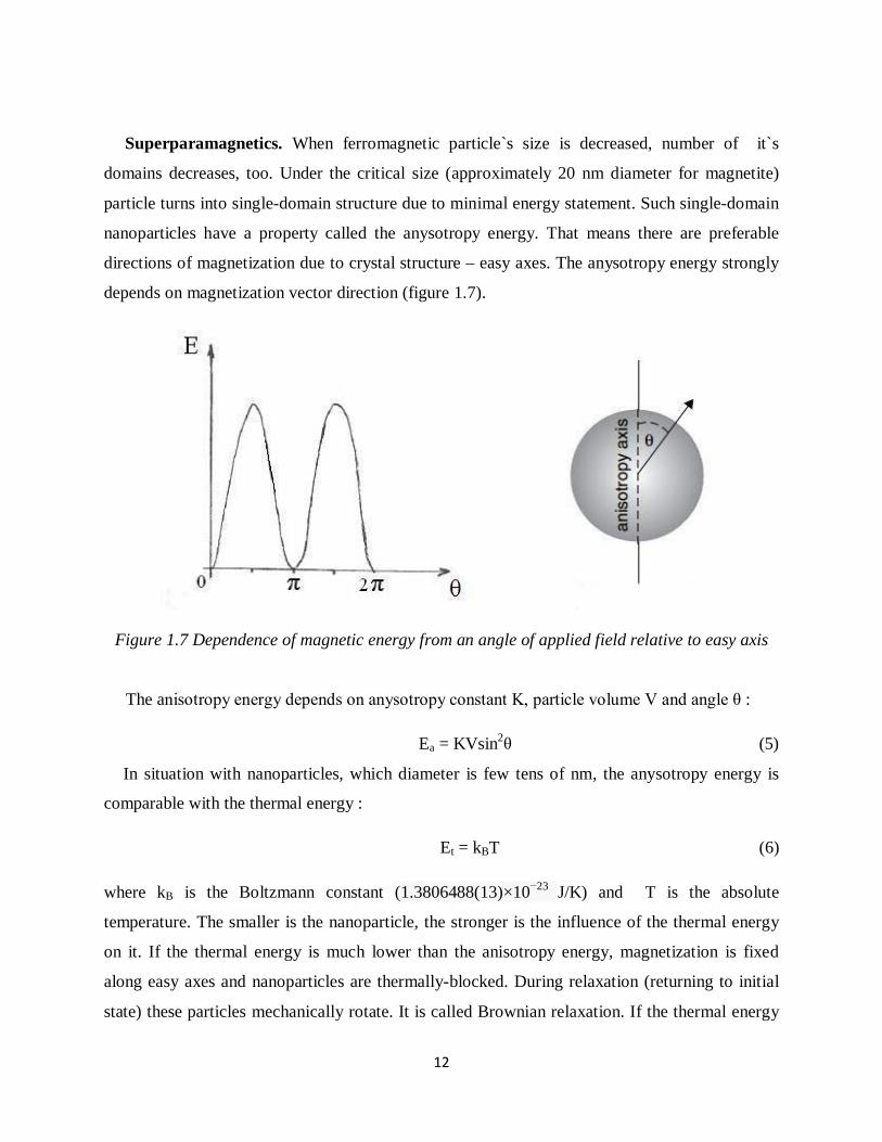
Superparamagnetics. When ferromagnetic particle`s size is decreased, number of it`s
domains decreases, too. Under the critical size (approximately 20 nm diameter for magnetite)
particle turns into single-domain structure due to minimal energy statement. Such single-domain
nanoparticles have a property called the anysotropy energy. That means there are preferable
directions of magnetization due to crystal structure – easy axes. The anysotropy energy strongly
depends on magnetization vector direction (figure 1.7).
Figure 1.7 Dependence of magnetic energy from an angle of applied field relative to easy axis
The anisotropy energy depends on anysotropy constant K, particle volume V and angle θ :
Ea = KVsin2θ (5)
In situation with nanoparticles, which diameter is few tens of nm, the anysotropy energy is
comparable with the thermal energy :
Et = kBT (6)
where kB is the Boltzmann constant (1.3806488(13)×10−23 J/K) and T is the absolute
temperature. The smaller is the nanoparticle, the stronger is the influence of the thermal energy
on it. If the thermal energy is much lower than the anisotropy energy, magnetization is fixed
along easy axes and nanoparticles are thermally-blocked. During relaxation (returning to initial
state) these particles mechanically rotate. It is called Brownian relaxation. If the thermal energy

13
is much higher than the anysotropy energy, magnetization frees from easy axes and can orient
easily, without mechanical rotation. System now behaves like a paramagnetic, without any
remanent magnetization and coercivity, but susceptibility and saturation magnetization are high.
This phenomena is named superparamagnetism. Relaxation mechanism of this system is called
Neel relaxation4.
Figure 1.8 Comparing of a) superparamagnetic magnetization curve and b) ferromagnetic
magnetization curve
Superparamagnetic, not ferromagnetic behavior of nanoparticles is a strict necessity for in
vivo applications. Nanoparticles must be colloidly stable. Due to the remanence magnetization of
ferromagnetic nanoparticles agglomerates occur and can stuck blood vessels – this harms to
patient`s organism and can cause death.
1.3.2 Iron oxides Mostly used materials for the superparamagnetic core are iron oxides : magnetite Fe3O4 and
maghemite γ-Fe2O3. Wustit FeO doesn`t show any significant magnetic properties. Both
magnetite and maghemite have cubic spinel structure.
Magnetite. Contains divalent and trivalent Fe ions, IUPAC name is iron (II,III) oxide,
chemical formula Fe3O4 or FeO·Fe2O3. Black or grayish black color mineral. Saturation

14
magnetization of bulk material at 250 C is 90-92 emu/g. Structural formula [Fe3+] Td [Fe3+,Fe2+]
Oh O2-4, which means there is a tetrahedral magnetic sublattice, containing Fe3+ ions, and an
octahedral sublattice, containing Fe3+ and Fe2+ ions. Spins from these two sublattices are
antiparallel, so magnetite net magnetization occurs due to Fe2+ ions from octahedral sublattice5.
Magnetite is sensitive to oxidation – oxygen transforms it to maghemite by oxidizing of Fe2+
ions.
Maghemite. Is formed by oxidation of magnetite. Contains only trivalent Fe ions, IUPAC
name is iron (III) oxide, chemical formula γ-Fe2O3. Brown color mineral. Saturation
magnetization of bulk material at 250 C is approximately 80 emu/g. Crystal structure is similar to
magnetite, but with vacancies in octahedral sites due to oxidation of divalent iron ions; so
maghemite is ferrimagnetic, too.6
In this work magnetite is preferrable due to it`s higher saturation magnetization values.
1.3.3 Synthesis of SPIONs
There are lots of SPIONs synthesis methods : water-in-oil microemulsion, polyol, gas
deposition, co-precipitation, sol-gel, pyrolisis, thermal decomposition of organic iron precursor,
hydrothermal and others. Every method possess specific performance procedure and conditions,
and of course nanoparticles of different properties ( shape, average size, size distribution,
cristallinity, magnetic properties, dispersibility, etc ). In this work two methods with high
product quality/synthesis difficulty ratio will be compared. These routes are co-precipitation and
thermal decomposition.
1.3.3.1 Co-precipitation method
The co-precipitation method is considered as the simplest chemical way to synthesize iron
oxide nanoparticles. Performed first time by Massart more than 30 years ago7, this method
gained broad variety of modifications, but it`s main chemistry didn`t change :
Fe2+ + 2Fe3+ 8OH- → Fe3O4 + 4H2O (7)
The reaction is not direct, a formation of some iron complexes takes place :
(Fe3+ (H2O)6)3+ → FeOOH + 3H+ +4H2O (8)

15
Fe2+ +2OH- → Fe(OH)2 (9)
2FeOOH + Fe(OH)2 → Fe3O4 + 2H2O (10)
Fe2+ and Fe3+ ions in molar ratio of 1:2 precipitate into magnetite in a basic solution (pH from 8
to 14). Due to magnetite is sensitive to oxidation, an inert gas purging is needed during the
reaction to prevent magnetite transformation to maghemite. The main advantage of this synthesis
method is amount of produced nanoparticles per one batch – up to 40 grams8. But co-
precipitation synthesis method possess a weak control of the nanoparticle size distribution due to
only kinetic factors influence nucleation and growth of the crystals. When the concentration of
precursors reaches the supersaturation, the nucleation occurs for a short moment of time –
“seeds” of synthetising nanoparticles are forming; then slow process of crystal growth via
diffusion of precursors on the surface of “seeds” takes place. The key to produce nanoparticles
with narrow size distribution is to separate nucleation from growth9. The nucleation time must be
very short to form highly monodispersed nanoparticles – final nanoparticle amount is formed at
this moment and won`t change during crystal growth. The LaMer diagram represents the
generation of uniform nanoparticles through the nucleation and growth processes.
Figure 1.9 LaMer digram : I – prenucleation, II – nucleation, III - growth
Nanoparticle size, morphology, magnetic properties can be controlled by varying of solution pH,
ionic strength, temperature, reaction time, type of salts (ferrous and ferric chlorides are most
popular). For example, addition of 1 M NaCl increases ionic strength of reaction media and
reduces the mean size of the nanoparticles for 1.5 nm. However, as it was said previously, the
co-precipitation suffers from a broad size distribution of produced nanoparticles – distribution is

16
about 30%. This occurs because of lack of an influence on the nucleation and growth; these two
processes are not separated properly.
1.5.3 Thermal decomposition of iron organic precursor method
Highly monodispersed nanoparticles can be synthesised via thermal decomposition of
organometallic precursors method. Some organic iron compounds (hydroxylamineferron
[Fe(Cup)3], iron pentacarbonyl [Fe(CO)5], ferric acetylacetonate [Fe(acac)3], iron oleate
[Fe(oleate)3]12 are decomposed at high temperature inside the non-polar boiling solvent with a
presence of the capping agent (it is worth to say that most of these precursors are toxic and not
friendly to environment). Narrow size distribution (σ<10%), high crystallinity and shape control
are the attributes of this route10. The precursor is heated up to the boiling point of boiling solvent
with a constant heating rate and kept at this temperature for the desired time. Narrow size
distribution is obtained due to feature of the nucleation and growth mechanisms during
decomposition – these processes occur at different temperatures and can be well-separated. The
nucleation starts at approximately 2000 - 2300 and the growth at 2600 – 2900. The nanoparticles
are coated with capping ligand (fatty acids, hexadecylamine)13 – it is not only the size adjustment
instrument, but also colloidal stabilizer. Prepared by this technique nanoparticles are
hydrophobic (not soluble in water) and can be storaged in hexane, cyclohexane, toluene or other
non-polar solvent. There are several routes of transferring hydrophobic nanoparticles to an
aqueous media, for example, CTAB method14, Pluronic F127 method, etc.
There are several ways how to control a size and shape of nanoparticles. The size can be
tuned by 3 factors : temperature of the decomposition reaction (depends on the boiling solvent),
precursor/capping agent ratio, duration of the reaction after reaching the boiling point. Varying
of these parameters is a suitable instrument for researches. Morphology of the nanoparticles is
mostly influenced by the heating rate and precursor/boiling solvent volumetric ratio. Many
different conditions of the experiment were investigated : changing of the boiling solvent (di-n-
hexyl ether (boiling point 2280 C), hexadecene (bp 2740 C), dioctyl ether (bp 2940 C), octadecene
(bp 3170 C), and eicosene (bp 3300 C)); different amounts of capping agent – oleic acid11. Here is
the graph showing an average size dependence from the boiling point and the capping
agent/precursor molar ratio11 :

17
Figure 1.10 Dependence of the average nanoparticles diameter from ligand/precursor molar ratio in di-n-octyl ether (blue curve) or from solvent boiling point (red curve)11.
This method gives high quality nanoparticles, however amount is quite low per one batch.
One of the most significant difficulties of this route is to establish constant heating rate,
especially in the range where the nucleation and growth occurs. „Green” synthesis with non-
toxic and environment friendly precursors (ferric chloride and sodium oleate, for example) is
preferrable15.
1.4 Core-shell magnetic nanoparticles
The pristine SPIONs have several limitations in case of biomedical applications : a reactivity
of iron oxides with blood, an aggregation at physiological blood pH (7.35 – 7.45). These
problems are solved by coating iron oxides with organic (polymers like dextran, polyethylene
glycol, polyvinyl alcohol, chitosan) or inorganic (gold, silver, silica, apatite) materials38. In this
work silica coating will be developed.

18
1.4.1 Silica properties
Silica, or silicon dioxide (chemical formula SiO2), is wide distributed in the Earth crust in a
form of sand or quartz. Chemical properties of silica provide active interactions between it`s
surface and H+ OH- ions at physiological pH making surface charged and electrostatic repulsion
mechanism working. This phenomena prevents aggregation of SPIONs in a human blood,
establishes colloidal stability. Also, silica is an inert and non-toxic material with high potential
for functionalization with different ligands assigned to a wide range of biomedical applications.
These properties make silica a convenient material for iron oxide shielding and functionalization.
1.4.2 Silica coating by inverse microemulsion method
Inverse microemulsion method is good for both hydrophilic and hydrophobic nanoparticles :
silica precursor (TEOS) transfers organic-soluble nanoparticles to water-soluble condition. This
CSNP preparation route possess highly tunable silica layer thickness and narrow size distribution
of it. Inverse, or water-in-oil, microemulsion means that a water phase covered with a surfactant
and co-surfactant is dispersed in a non-polar media. Formed water droplets (inverse micelles) are
constrained nanoreactors – this is the key to narrow size distribution of prepared
nanocomposites17.
Figure 1.11 The structure of water-in-oil microemulsion27

19
By changing ratio of surfactant, co-surfactant, water phase and organic phase the size of
inverse micelles is adjusted. Also concentration of the pristine nanoparticles and silica precursor
is significant – if amount of nanoparticles is too big, multicore nanocomposites will form. If
reverse – some part of the produced nanoparticles will be without any magnetic core.
Figure 1.12 Schematic view of w/o microemulsion silica coating method 17
Basic pH is the driving force for depositing of silica – OH- ions attack silicon via hydrolysis
which results in appearance of oxygen-silicon-oxygen site. The higher the pH of media, the
faster is the reaction. Condensation can be slowed down chemically – by adjusting pH to
approximately 1-2, or physically – by shock freezing in liquid nitrogen19. Deposited silica is not
crystalline, but amorphous.
No strong necking of nanoparticles is observed for this route, comparing with results from
Stöber18 method. One of the difficulties of w/o microemulsion silica coating method is removing
of surfactant and co-surfactant from the surface of prepared nanocomposites. High rpm
centrifuging is performed to separate phases, several careful washings with acetone or ethanol
are needed, significant amount of nanoparticles is lost during these processes. Due to this fact

20
and special concentrations of components for thermodynamically stable microemulsion system
an amount of synthesised core-shell nanoparticles is low-yelded.
1.5 Multifunctional core-shell nanoparticles
Silica layer is a great platform for binding additional functionalizations. Combined ligands on
silica surface allow to optimize multifunctional core-shell nanoparticles for certain purpose and
to process several biomedical applications siltumaneously.
Figure 1.1 Core-shell nanoparticle with different functions.
Engineered by this scheme nanocomposites pretend to become an ultimate solution in
imaging and treating of tumors. Silica coating provides high biocompatibility, non-reactivity,
colloidal stability, polyfunctionality – integrated quantum dots can work as an extra

21
visualization agents, targeting ligands provide accurate detecting of desired cells and high
accumulation in unhealthy tissues, high-controllable drug release mechanisms triggered by pH or
temperature. Magnetite core with superparamagnetic behavior shows no coercivity and
remanence magnetization, high saturation magnetization (but less than bulk material one) allows
to have great control on particle position and detection, so it is a promising contrast agent for
MRI3 and drug delivery system element.
1.6 Use of core-shell nanoparticles in medicine
1.6.1 MRI contrast agent
Magnetic resonance imaging (MRI) is a powerful tool for detailed visualization of body
internal structure. It gives an opportunity to visualize soft tissues, to detect physiological and
chemical changes in organism. MRI is a diagnostic technique based on interactions between
protons of human body and powerful magnetic field. Due to water percentage of our body is
approximately 80%, hydrogen nucleus (protons) with unpaired spins work as a great instrument
influenced by external magnetic field. Spins precess along an axis of exposed magnetic field
with the frequency, called precessional frequency, or Larmor frequency :
ω0 = γ B0 (11)
where ω0 is the recessional frequency, γ is the gyromagnetic ratio (ratio of the magnetic moment
to the angular moment of definite system, in our case – proton), B0 is a magnetic flux density.
When perpendicular to magnetic field precessional frequency (in radiofrequency range) impuls is
applied, magnetic resonance phenomena occur – protons absorb energy and transfer from stable
initial state to unstable excited state. After removal of Larmor frequency pulse excited spins
realign to equilibrium state parallel with B0 and radiate absorbed before energy. This phenomena
is named spin relaxation. Due to protons from different tissues possess different relaxation
values, there are differences between signals which are used to construct the image of organism`s
anatomy. Proton signals are registered and interpreted via a mathematical algoritm to a graphical
view3. The main advantages of the MRI is an opportunity to obtain high resolution soft tissue
visualizations and non-invasivity (comparing with rentgen CT).

22
Figure 1.13 Main parts of MRI scanner
Figure 1.13 simply represents a construction of MRI scanner : main coil (superconductor with
field strength from 0.5 to 3 T) which provides precession of protons; radiofrequency exciting
coils and gradient coils, which detects relaxation energy of protons.
Spin relaxation consists of 2 processes :
1) Longitudinal relaxation or T1-recovery. The z component of the proton magnetization is
reduced to zero at the excited state. During relaxation it exponentially recovers to it`s
initial value. T1-recovery has a time constant T1 – this is the time at which z component
has recovered to 63% of it`s equilibrium energy. During T1-recovery radiated energy is
absorbed by nearby structures (spin-to-latticerelaxation)20.
Figure 1.14 T1-recovery 20

23
2) Transverse relaxation or T2-decay. The xy plane component has a maximum
magnetization energy at excited state. During relaxation it loses coherence – dephasing of
magnetization vector takes place, and transverse component exponentially decreases till
zero. T2 time constant represents the time, when transversal component has lost 63% of
it`s excited state energy. During T2-decay radiated energy is absorbed by nearby spins
(spin-to-spin relaxation)20.
Figure 1.15 T2-decay 20
By changing sequences of exciting radiofrequency impulses, image can be formed mainly
from T1 relaxation or T2 relaxation signals. T1-weighted scanning shows fat brighter but water
darker and is called positive; T2-weighted scanning shows reverse – fat darker and water
brighter, so called negative. T1 sequence is more efficient for brain imaging, T2 for spinal cord
diagnostics.
Figure 1.16 T1 (A) AND T2 (B) weighted images of human brain 28

24
How do contrast agents improve MRI visualization? Due to they have their own
magnetization, T1 and T2 relaxation times of water protons are modified. This enhances a
contrast in a specific area where contrast agent is located. MRI signal intensity depends on
relaxation rates (r1,2) of tissue, which are back-proportional to T1 and T2 relaxation times (r1 =
1/T1 and r2 = 1/T2). Contrast agent is effective, if it significantly changes only one (transverse or
longitudinal) relaxation rate. r2/r1 ratio of contrast agent determines if it modifies mainly
transverse relaxation rate r2 (r2/r1 >1) or longitudinal relaxation rate r1 (r2/r1 <1). So contrast
agents are classified as T1 (positive) agents and T2 (negative) agents. Commercial T1 agents are
paramagnetic gadolinium or manganese complexes (DotaremTM, ProhanceTM, TeslascanTM), T2
agents are SPION based (ResovistTM, ResovistTM, Clariscan™)21. Nowadays commercial
contrast agents are satisfying main requirements like stability, safety, biodistribution, tolerance,
etc22; but efficiency and quality are poor so actual researches in this field are promising the new
generation of high efficiency contrast agents.
1.6.3 Drug delivery systems
Engineering of high controllable drug nanocarriers able to transport and release very accurate
amounts of therapeuticals is one of the most promising nanomedicine research fields. First
mentioned in science fictions of early XX century, it is now developing with giant steps.
An efficiency of drug delivery systems depends on 3 main parameters – neutrality to immune
system, an ability to transport drug carriers directly into unhealthy tissue with a high precision,
an ability to regulate drug release rate to provide an optimal concentration of medicaments –
higher than minimal therapeutical level and lower than toxic dose. Citotoxicity and material
studies proved that an inert material of nanoparticle coating is obligatoriness as well as
hydrodynamic diameter less than 200 nm29. Coating with polymers like poly(ethylene glycol)
(PEG) and poly(ethylene oxide) (PEO) minimizes an uptake by phagocytes34.
Delivery control instruments are passive/active targeting, magnetic targeting and combining
of these methods. Active targeting ligands are specific molecules (antibodies, for example)
which can recognize and bind to complementary molecules on the surface of pathogenic cells.
Passive targeting is based on enhanced permeability and retention effect35 – drug nanocarriers
concentrate in tumor through it`s abnormal defective vascular system.

25
There are several mechanisms of drug release : diffusional; temperature, pH, magnetic field,
electric field dependent; ultrasound; electromagnetic radiation30. Silica coated SPIONs are of
high interest in this area due to biocompatibility and porous structure of the shell which can be
functionalized according to all needs of high efficiency drug delivery. The porous silica structure
possess high surface to volume ratio and silanol groups convenient for attaching drug carrying
mechanisms. One of the most intensively developed is thermosensitive PNIPAAm (poly(N-
isopropylacrylamide)) drug loading/release mechanism. PNIPAAm undergoes phase transition
from wide hydrophylic state to shrunken dehydrated hydrophobic state above the lower critical
solubility temperature (LCST) of 32˚-33˚ C – this feature allows to use it as a precise carrying
ligand for hydrophylic drugs. Water soluted drugs are leaving the polymer during a phase
transfer process30.
Figure 1.17 Phase transfer of PNIPAAm
The phase transfer required temperature is gained by radiating with EM waves at resonant
frequences to induce photothermal conversion or by applying thermal energy from heaters.
Outstanding results in targeted cancer chemotherapy and other pharmaceutical treatments are
expected from these researches.
1.6.3 Hyperthermia
Novel cancer treatment methods are intensively researched by lots of scientific fields,
nanotechnologies are not exception – several treatment ideas are being investigated :

26
photodynamic therapy, photothermal therapy, oscillating magnetic field induced hyperthermia.
We will discuss the last method as the simplest and relatively less risky.
Concept is very simple : magnetic core-shell nanoparticles are concentrated in tumor tissues
and under applied changing magnetic field of resonant frequency CSNPs absorb magnetic energy
and convert it to thermal energy. As a result an overheat of nanoparticles` concentration area
causes a necrosis of tumor cells which are more sensitive to high temperature than normal cells31.
Cancer cells are eliminated at the temperature higher than 43̊ C which is not so fatal for healthy
cells32. The heat temperature depends on properties of magnetic material, external magnetic field
strength and frequency, concentration of SPIONs in tissues, biological factors. In this biomedical
application multifunctionality of core-shell SPIONs plays a very significant role – addition of
temperature-triggered drug carriers and active targeting ligands makes it an effective instrument
in cancer treatment. Functionalized in a such way, CSNPs can act as the contrast agent for tumor
imaging and combined therapy tool siltumaneously – merging of precisely dosated
chemotherapy and hyperthermia has shown good results33. Hyperthermia is also combined with
radiotherapy33.
This biomedical application needs SPIONs of the highest quality and many clinical tests due
to risks of harming normal cells, especially in treatments of the brain tumors.

27
2 Experimental
2.1 Synthesis of SPIONs
2.1.1 Co-precipitation method
Two SPION synthesis routes were performed – co-precipitation method and thermal
decomposition method. As iron precursors for co-precipitation ferrous and ferric chlorides (both
are hydrates) in a molar ratio 1:2 were chosen. Two solutions were prepared – ammonia
hydroxide solution as a precursor of OH- ions and iron chlorides solution as an Fe2,3+ ions
precursor. The basic solution was heated to 70o C and kept constant under continuous
mechanical stirring and nitrogen purging to hit O2 out of solution to prevent oxidation of
magnetite. The iron salt solution was prepared under N2 purging with an addition of small
amount of HCl to fasten a dissolution of the chlorides siltumaneously with heating of the basic
solution. When the desired 70o C temperature is gained, iron precursor solution was added to the
ammonia solution and reaction started immediately – a color of media became black and a bit
brownish. The nucleation occured during this moment. The growth process was allowed to
proceed for 45 minutes, then magnetite nanoparticles were decanted with a permanent magnet
and washed with deionized water several times to remove unreacted material and impurities.
Particles were dispersed in TMAOH solution and sonicated to achieve colloidal stability, then
kept at 4o C for further use.
2.1.2 Thermal decomposition method
The thermal decomposition preparation of magnetite consisted of two steps : synthesis of iron
oleate and high temperature decomposition of it. This route is relatively environment-friendly –
sodium oleate and ferric chloride are the precursors for iron pleate complex. Iron (III) chloride
was dispersed in water, sodium oleate in hexane, then solutions were mixed with an addition of
ethanol and heated to 65 o
under continuous mechanical stirring. According to the reaction
stoichiometry molar ratio between ferric chloride and sodium oleate was 1:3. Reaction was
allowed to reflux for 4 hours, then a liquid with 2 well-separated phases was washed with the

28
deionized water several times in a separation funnel in order to remove NaCl. After removal of
water phase brownish-black organic phase was sent to rotation evaporator to extract hexane.
Ready iron oleate is brownish-reddish-black viscous substance. The next step is the thermal
decomposition of it in high boiling temperature non-polar solvent. Dioctyl ether was chosen due
to appropriate size of expected nanoparticles and low difficulty of removing it from the synthesis
product.
Iron oleate was dissolved in dioctyl ether with an addition of capping agent – oleic acid.
Precursor and capping agent molar ratio was 2:1. The solvent was heated to 290 o
C at the
constant heating rate of 4 o
C under vigorous stirring and kept at this temperature for 1.5 hours.
SPIONs were collected by centrifugation, then washed by redispersing in hexane and
precipitating after addition of ethanol followed by centrifugation. The washing was repeated
several times to remove impurities and low quality nanoparticles. Ready SPIONs were dispersed
in hexane with an addition of oleic acid and stored at 4o C for further use.
2.2 Preparation of SiO2@Fe3O4
Water-in-oil microemulsion method was chosen due to a narrow size distribution of prepared
nanocomposites, very low amount of fused together nanocomposites and an ability to coat
hydrophobic nanoparticles via ligand exchange mechanism.
First thermodinamically stable microemulsion system Triton X100/hexanol/water +
NH3/cyclohexane was formed. To gain high realibility and productivity, optimized molar ratio of
components was used : water/Triton X100 = 6.3, Triton X100/hexanol = 0.5, TEOS/water =
0.008 23. Volume ratio of water phase (water + Triton X100 + hexanol) to organic phase
(cyclohexane + Fe3O4) was 1:1.5. SPION concentration was chosen 0.12 mg per 1 ml of
cyclohexane – this provides high amount of single-core nanoparticles in a batch.
The basic pH water phase was mixed with a mechanical stirring, then SPIONs dispersed in
cyclohexane were added. After 45 minutes (approximate microemulsion formation time) TEOS
was added dropwise. After 3 hours the condensation was stopped by adjusting the pH to 2 with
nitric acid and shock freezed with liquid N2 19. This separated unreacted TEOS. High rpm
centrifugation was performed to separate phases for 3 times with freezing before every cycle of
centrifugation, then ethanol was added to remove surfactant and co-surfactant from the

29
nanoparticles. Ethanol washing was performed for 3 times, nanocomposites were collected and
dispersed in water for further use.
On the scheme below phase transfer mechanism is revealed23 :
Figure 2.1 Phase transfer and silica coating mechanism23
2.3 Characterization
The most important part of experiments is characterization – it gives an opportunity to analyze
results of experiments and to choose the next step to achieve expected results. Different
parameters need different characterization techniques, an efficiency of analisys strongly depends

30
on how suitable was the chosen characterization technique for certain parameter. Well-composed
characterization plan increases efficiency of researches by economy of time, materials and other
resources. Now some background on analisys techniques used in this work.
2.3.1 Transmission electron microscopy
Transmission electron microscopy (TEM) is a powerful tool in exploring a shape, crystallinity,
mean size and size distribution of pristine and silica coated SPIONs. Nanoparticles were
investigated with a JEOL 2100 FEG-TEM using 200 kV electron acceleration voltage.
When electron gun emitted accelerated electrons collide with sample, two groups of
interactions take place : elastic (electron is reflected with the same energy as before an
interaction) and inelastic (initial energy is decreased – transmitted electrons, Auger electrons,
secondary electrons, X-ray, etc). 3-stage electron lense system guarantee wide range possibilities
of focusing an electron beam. High energy of electrons provides low diffraction effect so
resolution is high enough to explore crystallinity of nanoparticles. TEM detects the differences
between initial and final energies of electrons transmitted through an ultrathin sample and
constructs the image of the relief according to the density differencies.
Figure 2.2 Scheme of TEM optic system

31
Samples were prepared by pouring several drops of diluted sonicated SPION dispersion on a
carbon coated 200 mesh copper grids and letting them dry during a night. Images were obtained
by Gatan DigitalMicrograph 3.11.2 for GMS 1.6.2 with Orius SC1000 2 imaging software.
Nanoparticles` sizes were measured (at least 50 measurements from different areas per 1
sample) and mean size with size distribution were calculated using the statistical software. High
resolution TEM (HRTEM) was performed to investigate crystallinity of magnetite cores.
2.3.2 Dynamic light scattering
Dynamic light scattering (DLS) is the technique for hydrodynamic nanoparticle size
explorations. When a nanoparticle is surrounded by media, it`s surface (with different ligands or
without them) has a variety of interactions with solvent molecules and ions and this is strongly
influencing a behavior of a nanoparticle. These surface/media interactions form an additional
layer of a nanoparticle and the whole diameter of this system is determined as the hydrodynamic
size of a nanoparticle.
Figure 2.3 Comparing of a) TEM defined diameter b) DLS defined hydrodynamic diameter 24
At the temperature higher than absolute zero all particles suspended in liquid or gas possess the
Brownian motion – random movement of particles due to interactions with another particles and
solvent molecules.

32
As a rule, larger particles are moving slower than smaller ones. DLS is measuring the velocity of
the nanoparticle Brownian motion and calculating a hydrodynamic diameter using Stokes-
Einstein equation :
(11)
where D is the hydrodynamic diameter, d - translational diffusion coefficient (calculated from
the Brownian motion), kB - Boltzmann`s constant, T – absolute temperature, η – viscosity.
Simple measurement setup is represented on figure 2.4 :
Figure 2.4 DLS instrumental setup
A laser beam is interacting with particles in suspension, scattered beam intensity is registered by
detector and sent to correlator for further mathematical processing. Recorded intensity
fluctuations (caused by a motion of particles) allow to calculate translational diffusion coefficient
and then the hydrodynamic size.
Measurements were performed with Beckman CoulterTM DelsaNano C partycle analyzer. 400
measurements for every sample were processed to obtain more reliable statistics. However, for
nanoparticles dispersed in non-polar solvents (like hexane, cyclohexane, chloroform, toluene)
there were problems in obtaining reliable data, so another device was used - Malvern
Instruments Zetasizer Nano SZ.

33
2.3.3 Zeta-potential analysis
Zeta-potential measurements were performed to study colloidal stability of prepared
nanoparticles. Colloidal stability is a very important requirement for nanoparticles used in
biomedical applications due to aggregates can cause serious harm to patient organism. There are
2 colloidal dispersion stabilization mechanisms – steric repulsion and electrostatic repulsion.
Steric repulsion is obtained by coating nanoparticles with specific ligands which prevent close
contacts between nanoparticles (example – fatty acids). Electrostatic repulsion occurs due to
surface charge of nanoparticles – the higher is it`s magnitude, the higher stability of dispersion.
The surface charge depends on material properties (acidic or basic strengths of the surface
groups) and pH of the solvent – dissociated H+ and OH- ions form this charge. This phenomena
takes place only in polar solvents25. Zeta potential is the charge of nanoparticle`s ion slipping
plane, which is moving together with a nanoparticle. Figure 2.5 describes organization of ions
around a nanoparticle :
Figure 2.5 Ionic concentration and charge distribution around a nanoparticle
34
Zeta potential cannot be measured directly – it is calculated from the velocity of nanoparticles
during electrophoresis using Henry equation :
(12)
where UE is an electrophoretic mobility, z – zeta potential, ζ – dielectric constant, η – viscosity,
f(ka) – Henry`s function. It is considered, that system is stable if the surface charge of
nanoparticles is higher than ±30 mV. A pH value which possess neutral 0 charge of nanoparticle
surface is called isoelectric point or zero charge point – nanoparticles sediment under gravity
force.
The same device as for DLS was used for zeta potential measurements (but with other
instrument and software configuration) - Beckman CoulterTM DelsaNano C partycle analyzer.
Dispersion is flowing through capillary cell, electrodes in opposite sides of the cell form an
electric potential to establish electrophoretic effect – movement of a charged particle relative to
the solvent. The flow cell is radiated by laser, fluctuations of it`s intensity determine a velocity of
charged nanoparticles – the higher charge, the faster are particles, the higher a frequency of
intensity fluctuations.
Measurements were performed only for water solutions of nanoparticles – hydrophobic
SPIONs were stabilized by oleic acid coating.
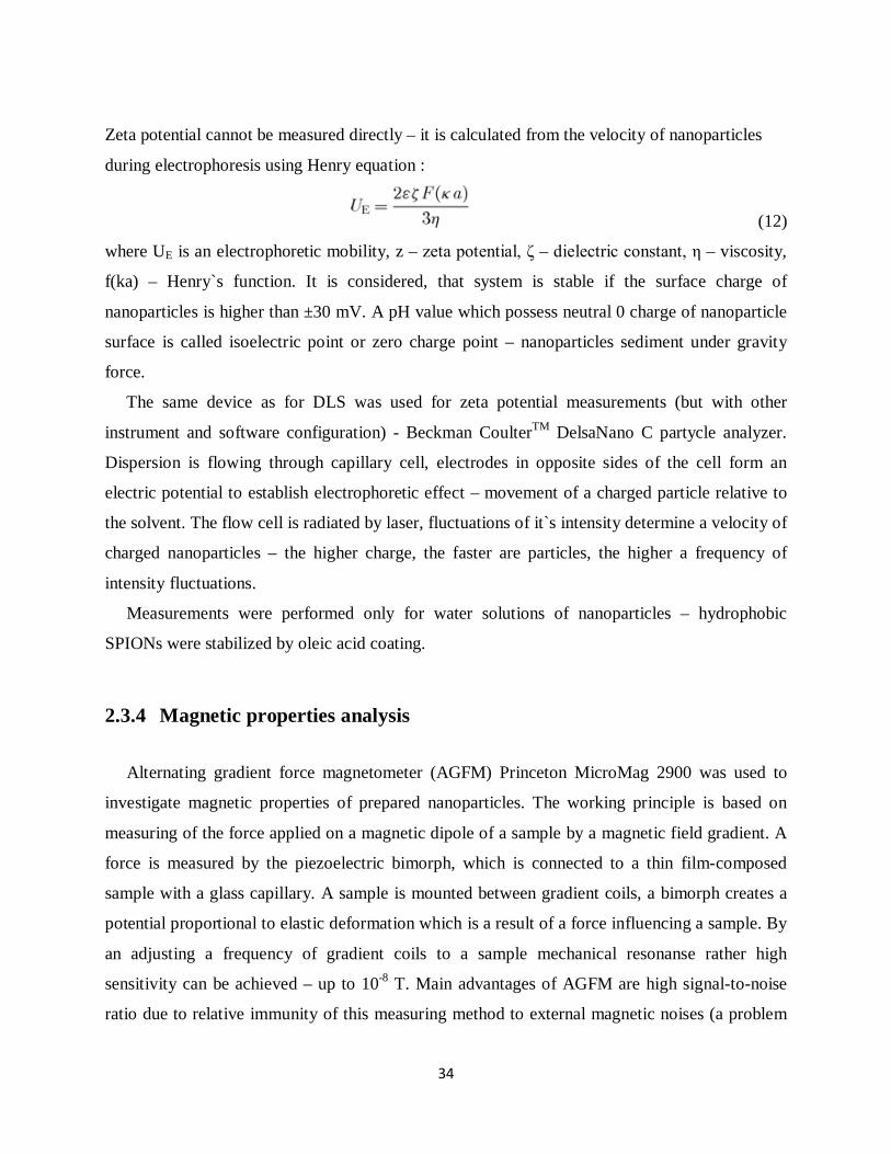
2.3.4 Magnetic properties analysis
Alternating gradient force magnetometer (AGFM) Princeton MicroMag 2900 was used to
investigate magnetic properties of prepared nanoparticles. The working principle is based on
measuring of the force applied on a magnetic dipole of a sample by a magnetic field gradient. A
force is measured by the piezoelectric bimorph, which is connected to a thin film-composed
sample with a glass capillary. A sample is mounted between gradient coils, a bimorph creates a
potential proportional to elastic deformation which is a result of a force influencing a sample. By
an adjusting a frequency of gradient coils to a sample mechanical resonanse rather high
sensitivity can be achieved – up to 10-8 T. Main advantages of AGFM are high signal-to-noise
ratio due to relative immunity of this measuring method to external magnetic noises (a problem

35
of SQUID detectors) and high measuring speed. Disadvantage of this device is a high calibration
difficulty due to a signal is proportional not only to a sample, but to a sample-bimorph system.
This problem can be partially solved by a compensation mechanism and a calibration coil near to
sample39.
Figure 2.6 Scheme of AGFM measuring part
Samples were prepared by drying nanoparticles containing suspensions and fixing resulted
nanopowders on 3x3 mm papers ( originally this method is used to investigate magnetic
properties of ultrathin films).
2.3.5 Inductively coupled plasma – atomic emission spectroscopy
It is worth to mention ICP-AES (inductively coupled plasma - atomic emission spectrometry)
metal ion concentration determination technique. The ICP-AES device can be divided into two

36
parts : an inductively coupled plasma torch and an optical spectrometer. The inductively coupled
argon plasma torch with the working temperature of 7000 K excites atoms and ions so they emit
an electromagnetic radiation of the spectrum, particular to different elements. The intensity of
this radiation shows a concentration of atoms in a sample. The working principle schematics are
shown on figure 2.7.
The measurements were processed by Thermo Fisher Scientific ICAP 6500 spectrometer.
Sample preparation consists of extracting the definite volume of analysed dispersion, mixing it
with a small amount of hydrochloric acid to dissolve metal compounds into ions and diluting
with deionized water till definite final volume. First two standart samples with known
concentration of definite metal ions are run to establish zero point for the sample with unknown
concentration. Only then an unknown concentration sample measurement is performed. The
results are shown in ppm – parts per milliom, or miligrams per liter. After mathematic
calculations the concentration in the undiluted storage dispersion is determined.
Figure 2.7 The working principle scheme of ICP-AES

37
3 Results and discussions
3.1 Morphology, structure and size characterizations
3.1.1 TEM
The main characterization technique for a morphology, structure and size of synthesized
nanoparticles was TEM. On the pictures below SPIONs synthesized by co-precipitation and
thermal decomposition methods are presented :
Figure 3.1 SPIONs synthesized by co-precipitation method (a,b) and thermal decomposition
method (c,d)
SPIONs` size measurements were processed using ImageJ software. The average size for co-
precipitation synthesized nanoparticles is 10.4 ± 3.6 nm (σ = 34.6%). The average size for
thermal decomposition prepared nanoparticles is 8.2 ± 0.8 nm (σ = 9.7%). As it can be seen from

38
the pictures, size and morphology are significantly different – narrow size distribution and
spherical shape for the thermal decomposition method, broad size distribution and random near-
spherical shapes for the co-precipitation method. Despite co-precipitation synthesized SPIONs
form aggregates on TEM images, they are stable and separated at certain pH range of a solvent.
A sonication device can be used to restore nanoparticles from aggregated state. Thermal
decomposition prepared SPIONs are coated with capping agent (oleic acid), that is why they are
well-separated and stable in non-polar solvents like n-hexane, toluene, chloroform (more about
colloidal stability in chapter 3.3).
It is worth to mention that shape and size are significantly dependent from a homogeneity of
a heating in the thermal decomposition method. Figure 3.2 shows nanoparticles obtained after
synthesis process with unhomogeneous distribution of heat in a reaction flask ( it wasn`t
completely covered with thermoisolation wool) :
Figure 3.2 Nanoparticles prepared with unhomogeneous heating conditions (thermal
decomposition method)

39
High resolution TEM was performed to investigate a cristallinity of synthesized SPIONs :
Figure 3.3 HRTEM of a) the thermal decomposition and b) the co-precipitation prepared
nanoparticles
No significant differences and defects are identified. For more detailed data on SPIONs`
structure X-ray diffraction (XRD) analisys is needed.
After observing TEM images of SPIONs synthesized by both methods, I decided to use
thermal decomposition prepared nanoparticles for coating with silica due to narrow size
distribution and regular spherical morphology. Also, this method doesn`t need a phase transfer
step – it performs during silica deposition reaction. Described before procedure23 of water-in-oil
microemulsion was chosen.

40
Figure 3.4 Core-shell structured nanoparticles
Core-shell nanoparticles with a diameter of 30.6 ± 4.9 nm (σ = 16.0% ) were synthesized.
Average silica shell thickness was calculated – 11.2 ± 1.7 nm (σ = 15.1% ). Prepared
nanocomposites are quite homogeneous in size, separated and single-cored. However, some
multi-core nanoparticles and fused together ones take place, but in a relatively small amount (
less than 1% ). Necking of the nanoparticles was the main problem – it could occur during
synthesis and during collecting and washing by centrifuging. Figure 3.5 represents a result of
„too careful” centrifuging :
Figure 3.5 Necked nanoparticles

41
HRTEM was performed to explore a structure of core-shell SPIONs:
Figure 3.5 High resolution TEM of silica coated magnetite
No cristallinity of silica shell is observed, it has an amorphous structure. However, previous
related to these nanoparticles work40 mention experiments on etching core-shell nanocomposites
with concentrated hydrochloric acid (I used a silica coating procedure from this work). As a
result, magnetite cores were dissolved and no significant changes of silica shells were noticed40.
This fact means, that there are pores in amorphous silica layer, so it can be efficiently used for
ligand functionalization.

42
3.1.2 DLS
Dynamic light scattering was processed to determine a hydrodynamic size of synthesized
nanoparticles – these values are even more significant than TEM data due to nanoparticles wil be
used as a dispersions in different solvents.
Hydrodynamic size dramatically differs from TEM identified size, especially for
nanoparticles functionalized with long-chained ligands.
Figure 3.6 Hydrodynamic size distribution histogram of a )co-precipitation and b) thermal
decomposition synthesized SPIONs
A mean hydrodynamic size value for co-precipitation synthesized SPIONs is 115.2 nm ±
18.1 nm (σ = 15.7 %). We can concude that significant amount of solvent binds to a surface of
nanoparticles, irregular shape of nanoparticles increases hydrodynamic diameter. Comparing
with TEM identified size distribution (σ = 34.6%) I can assume that an amount of solution
„attached” to a surface of nanoparticles depends on its` volume.
Unfortunately, Beckman CoulterTM DelsaNano C partycle analyzer was not able to process
reliable measurements for nanoparticles suspended in non-polar solvents (hexane, cyclohexane,
chloroform, toluene were tried). Malvern Instruments Zetasizer Nano SZ was used for this
purpose.
An average hydrodynamic size for thermal decomposition synthesized nanoparticles is 23.4 ±
2.4 nm (σ = 10.3%). It is very similar to calculated from TEM images one (σ = 9.7%). The mean

43
size significantly differs from dispersed in water SPIONs` size – it is almost 5 times smaller. The
reason is an oleic acid layer and features of it`s interactions with a solvent.
Figure 3.7 Hydrodynamic size distribution histogram of SiO2@Fe3O4
A mean hydrodynamic size value for silica coated SPIONs is 152.4 nm ± 25.2 nm (σ =
16.5 %). Nanoparticles with hydrodynamic size more than 200 nm were not included in average
size calculations – probably these are multicore nanocomposites. A hydrodynamic size
distribution value is very near to calculated from TEM value – 16.5% vs 16.0% respectively.
Probably, silica interacts with water in a different way than pristine magnetite. Also, measuring
technique should be checked for reliability. These assumptions should be carefully studied in
order to avoid measuring errors and predict a hydrodynamic behavior of nanoparticles.

44
3.1 Magnetic characterization
Three samples were examined on AGFM with an apllied field ranging from 1.4 T to -1.4 T.
All of them show superparamagnetic behavior with unsignificant coercivity field and remanence
magnetization.
Figure 3.8 Magnetization curves of SPIONs synthesized by co-precipitation, thermal
decomposition and silica coated thermal decomposition prepared SPIONs.
Co-precipitation Thermal decomposition
Silica coated
Saturation magnetization, emu/g 32,27 50,82 6,71
Remanence magnetization, emu/g 0,21 0,04 0,005
Coercivity field, T 0,0002 0,00007 0,00006
Table 3.1 Magnetic properties
Thermal decomposition prepared SPIONs show magnetic properties better than co-
precipitation method SPIONs (3 parameters were compared – saturation magnetization,
remanence magnetization and coercivity field). Silica coating dramatically influences magnetic

45
properties, especially saturation magnetization. Average silica layer thickness is 11.2 nm, it is
worth to decrease it in order to increase magnetization values.
3.3 Colloidal stability analysis
As it was mentioned before, colloidal stability of the nanoparticles plays very significant role
in introducing them to in vivo biomedical applications. Our goal is to provide stability of the
nanoparticles at certain pH value of human blood – 7.35 to 7.45. Nanoparticles with zeta-
potential from -35mV to +35mV are considered unstable. Pristine and silica coated magnetite
nanoparticles` zeta-potential was analysed ranging from pH 2 to pH 13. Measurements were
processed from 5 vertical positions of a capillar cell and average zeta-potential was calculated.
The peak potential normally is observed at 0.0 – the center of the capillar cell.
Figure 3.9 Zeta-potential distribution in a capillar cell.

46
Zeta-potential values for pristine magnetite and silica coated magnetite nanoparticles are represented on figure 3.10.
Figure 3.10 Pristine and silica coated SPIONs` zeta-potential depending from pH.
Comparing to uncoated magnetite nanoparticles, silica coated ones show stability region at physiological pH of 7.35 to 7.45 with zero potential point at pH around 2 ( and approximately 6.5 for bare SPIONs ). This proves a necessity of stabilization coating for SPIONs and an efficiency of silica for this purpose.

47
4 Conclusions
The aim of this work is to compare two synthesis methods of SPIONs, choose more
appropriate one for preparing SPIONs applicable in biomedicine and to coat chosen SPIONs
with silica layer.
Every synthesis method was run many times in order to understand it`s main properties and
features, aknowledge factors influencing experiments. Several characterization techniques were
used to carefully analyze prepared nanoparticles. After this step I can conclude, that the thermal
decomposition method synthesized SPIONs better fit to strict requirements of biomedical
applications, than co-precipitation synthesized ones. They possess narrower size distribution,
uniform spherical morphology, higher saturation magnetization and lower remanence
magnetization and coercivity field – so they do not form significant aggregates. However, the
synthesis process is more complicated and product low-yelded, comparing with co-precipitation.
Another disadvantage is that synthesized SPIONs are hydrophobic. But of course these negative
features are covered with high quality of nanoparticles, which is very crucial for biomedical
applications. The co-precipitation method allows to produce large amounts of hydrophilic
SPIONs per batch, but its` quality isn`t satisfying biomedicine requirements. Environmental
remediation is a very suitable and promising area for co-precipitation synthesized SPIONs.
Coating with silica is a strong obligatoriness due to SPIONs` reactivity with chemical
compounds of human organism and immune system, aggregation at phisiological pH and
hydrophobic surface of nanoparticles (in case for SPIONs prepared by thermal decomposition or
other organic solvent synthesis techniques). More of it, silica shell is a great platform for further
functionalization for several biomedical applications. Silica depositing was performed by water-
in-oil microemulsion method. After several attempts quite monodisperse separated core-shell
hydrophilic nanocomposites were synthesized. Zeta-potential analysis proved colloidal stability
of silica coated SPIONs at the pH of human blood.
Core-shell SPIONs synthesized during this work are ready for further functionalization and
biocompatibility studies.

48
References
(1) Caruthers, S.D.; Wickline, S.A.; Lanza, G.M., Nanotechnological applications in medicine,
Current opinion in Biotechnology, 2007, 18, 26-30.
(2) Vogt, C.M.; Toprak, M.; Muhammed, M.; Laurent, S., Engineered core-shell nanoparticles for
biomedical applications, Lic. Thesis, Royal Institute of Technology, ICT School, Stockholm,
2010, 1-9.
(3) Hashemi, R. H.; Bradley, W. G.; Lisanti, C. J., MRI: the Basics, Journal of Magnetic Resonance
Imaging, 1997, 7, 614-615.
(4) Bean, C. P.; Livingston, J. D., Superparamagnetism, Journal of Applied Physics, 1959, 30, 120-
129.
(5) Baker, R. R; Mather, J. G.; Kennaugh , J. H., Magnetic bones in human sinuses, Nature, 1983,
301, 79–80.
(6) Dronskowski, R.,The little maghemite story: A classic functional material, Adv. Funct. Mater.,
2001, 11, 27.
(7) Massart, R., Preparation of aqueous magnetic liquids in alkaline and acidic media, IEEE Trans.
Magn., 1981, 17, 1247.
(8) Cornell, R.M.; Schwertmann, U., Iron Oxides in the Laboratory : Preparation and
Characterization, Willey-Woch, ISBN 3-527-29669-7, Weinheim, Germany, 2000, 55-60.
(9) Boistelle, R; Astier, J.P., Crystallization Mechanisms in Solutions, Journal of Cryst. Growth
1988, 90, 14.
(10) Tartaj, P.; Morales, M.P.; Veintemillas-Verdaguer, S.; Gonzalez-Carreno, T.; Serna, C.J.
Synthesis, properties and biomedical applications of magnetic nanoparticles, Handbook of
Magnetic Materials, 2006, 9, 403.
(11) Demortiere, A.; Panissod, P.; Pichon, B. P.; Pourroy, G.; Guillon, D.; Donnio, B.; Begin-Colin,
S., Size-dependent properties of magnetic iron oxide nanocrystals, Nanoscale, 2011, 3, 226.
(12) Park, J.; An, K.; Hwang, Y.; Park, J.-G.; Noh, H.-J.; Kim, J.-Y.; Park, J.-H.; Hwang, N.-M.;
Hyeon, T., Nat. Mat,. 2004, 3, 891-895.
(13) Li, Y.; Afzaal, M.; O'Brien, P., The synthesis of amine-capped magnetic (Fe, Mn, Co, Ni) oxide
nanocrystals and their surface modification for aqueous dispersibility, Journal of Mater. Chem.,
2006, 16, 2175-2180.
(14) Lang, N.; Tuel, A., A Fast and Efficient Ion-Exchange Procedure to Remove Surfactant
Molecules from MCM-41 Materials, Chem. Mater., 2004, 16, 1961-1966.

49
(15) Chin, S. F.; Pang, S. C.; Tan, C. H., Green Synthesis of Magnetite Nanoparticles (via Thermal
Decomposition Method) with Controllable Size and Shape, J. Mater. Environ. Sci., 2011, 3,
299-302.
(16) Abarkan, I.; Doussineau, T.; Smaihi, M., Tailored microstructural properties of colloidal silica
nanoparticles via microemulsion preparation, Science Direct Polyhedron, 2006, 25, 1763–1770.
(17) Gupta, A. K.; Gupta, M., Synthesis and surface engineering of iron oxide nanoparticles for
biomedical applications, Biomaterials, 2005, 26, 3997–4001.
(18) Stöber, W.; Fink, A.; Bohn, E., Controlled growth of monodisperse silica spheres in the micron
size range, Journal of Colloid and Interface Science, 1968, 26, 62-69.
(19) Vogt, C. M.; Toprak, M.; Muhammed, M.; Laurent, S., Engineered core-shell nanoparticles for
biomedical applications, Lic. Thesis, Royal Institute of Technology, ICT School, Stockholm,
2010, 37-38.
(20) Ridgway, J. P., Cardiovascular Magnetic Resonance Physics for Clinicians : Part I, Journal of
Cardiovascular Magnetic Resonance, 2010, 12, 7-15.
(21) Ye, F.; Muhammed, M., Synthesis of Nanostructured and Hierarchical Materials for Bio-
apllications, Lic. Thesis, Royal Institute of Technology, ICT School, Stockholm, 2011, 11-12.
(22) Vogt, C. M.; Toprak, M.; Muhammed, M.; Laurent, S., Engineered core-shell nanoparticles
for biomedical applications, Lic. Thesis, Royal Institute of Technology, ICT School, Stockholm,
2010, 18-20.
(23) Vogt, C. M.; Toprak, M.; Muhammed, M.; Laurent, S., High quality and tuneable silica shell-
magnetic core nanoparticles, Journal of Nanoparticle Research, 2009, 12, 4-6.
(24) Alvarez, G. S.; Muhammed, M., Synthesis, characterisation and applications of iron oxide
nanoparticles, Doctoral Thesis, Royal Institute of Technology, ICT School, Stockholm, 2004,
27-28.
(25) Delgado, A. V.; Gonzalez-Caballero, F.; Hunter, R. J.; Koopal, L. K.; Lyklema, J.,
Measurement and Interpretation of Electrokinetic Phenomena, Pure Appl. Chem., 2005, 77,
1753–1850.
(26) Josephson, B.D., Possible new effects in superconductive tunneling, Phys. Lett., 1962, 1, 251–
253.
(27) Alonso, H. M. D.; Herrero, A. G.; Cánovas, L. A. R.;. Durán, A.; Fernández, M.; Díaz, O., New
Ecological Pigments in the Ca-Yb-S System, Journal Of Alloys and Compounds, 2001, 17, 297-
302.
(28) Dillon, W. P., Neuroimaging in Neurologic Disorders, Harrison's Principles of Internal
Medicine, 2008, 17-2, 2491-2497.

50
(29) Storm, G.; Belliot, S.O.; Daemen, T.; Lasic, D.D., Surface modification of nanoparticles to
oppose uptake by the mononuclear phagicyte system, Advanced Drug Delivery Review, 1995,
17, 31-48.
(30) Ye, F.; Muhammed, M., Synthesis of Nanostructured and Hierarchical Materials for Bio-
apllications, Lic. Thesis, Royal Institute of Technology, ICT School, Stockholm, 2011, 10; 16-
19; 31-34.
(31) Nielsen, O.S.; Horsman, M.; Overgaard, J., A future for hyperthermia in cancer treatment,
European Cancer Journal, 2001, 37, 1587-1589.
(32) Chan, D.F.C.; Kirpotin, D.B.; Bunn, J.R., Synthesis and evaluation of colloidal magnetic iron
oxides for the site specific radiofrequency-induced hyperthermia of cancer, Magnetic Materials
Journal, 1993, 122, 374-378.
(33) Sminia, P.; Hulshof, C .C. M., Hyperthermia and The Central Nervous System, Progress in
Brain Research, 1988, 115, 337-350.
(34) Harris, T. J.; Maltzahn, G.; Bhatia, S. N., edited by Amiji, M. A., Multifunctional Nanoparticles
for Cancer Therapy, Taylor & Francis Group, ISBN 978-0-8493-7194-3, Boca Raton, USA,
2007, 59-61.
(35) Duncan, R.; Sat, Y. N., Tumour targeting by enhanced permeability and retention (EPR)
effect, Annual Oncology, 1998, 2, 39.
(36) Koehler, F. M.; Fabian, M.; Rossier, M.; Waelle, M.; Athanassiou, E. K.; Limbach, L. K.;
Grass, R. N.; Günther, D.; Stark, W. J., Magnetic EDTA: Coupling heavy metal chelators to
metal nanomagnets for rapid removal of cadmium, lead and copper from contaminated
water, Chem. Commun., 2008, 32, 4862–4865.
(37) Ayyappan, S.; Mahadevan, S.; Chandramohan, P.; Srinivasan, M. P.; Raj, J. P.; Raj, b., Influence
of Co2 Ion Concentration on the Size, Magnetic Properties, and Purity of CoFe2O4 Spinel
Ferrite Nanoparticles, J. Phys. Chem., 2010, 114, 6334–6341.
(38) Laurent, S.; Forge, D.; Port, M.; Roch, A.; Robic, C.; Elst, L. V.; Muller, R., Magnetic Iron
Oxide Nanoparticles : Synthesis, Stabilization, Vectorization, Physicochemical
Characterizations and Biological Applications, Chemical Reviews, 2008, 108, 2073-2076.
(39) Edited by Czichos, H.; Saito, T.; Smith, L., Springer Handbook of Materials Measurement
Methods, Springer, ISBN 3-540-20785-6, Wurzburg, Germany, 2006, 525-526.
(40) Vogt, C. M.; Toprak, M.; Muhammed, M.; Laurent, S., Engineered core-shell nanoparticles for
biomedical applications, Lic. Thesis, Royal Institute of Technology, ICT School, Stockholm,
2010, 37.


![LHRH-functionalized superparamagnetic iron oxide ...mhaataja/PAPERS/meng_etal_msec2009… · cancer xenografts in-vivo. Shannon et al. [20] have also conducted multi-CRAZEDMRI experimentson](https://static.fdocuments.net/doc/165x107/603bcb847dad9d75c3338c36/lhrh-functionalized-superparamagnetic-iron-oxide-mhaatajapapersmengetalmsec2009.jpg)