Spring 1981 Gems & Gemology - GIA · PDF fileAN INTRODUCTION TO THE NEW GEMS & GEMOLOGY...
62
-, I.. - VOLUME XVll SPRING 1981 \ .: I
Transcript of Spring 1981 Gems & Gemology - GIA · PDF fileAN INTRODUCTION TO THE NEW GEMS & GEMOLOGY...

- , I . .
-
VOLUME XVll SPRING 1981 \ .: I

SPRING 1981 Volume 17 Number I
TABLE OF CONTENTS
EDITORIAL
FEATURE ARTICLES
NOTES AND NEW TECHNIQUES
REGULAR FEATURES
1 An Introduction to the New Gems & Gemology Richard T. Liddicoat, /r.
2 Zabargad: The Ancient Peridot Island in the Red Sea Edward Gu belin
9 Cubic Zirconia: An Update Kurt Nassau
20 A Simple Approach to Detecting Diamond Simulants !ill Hobbs
34 The Hidden Beauty of Amber: New Light on an Old Subject yohn I. Koivr~la
37 Artificially Induced Color in Amethyst-Citrine Quartz Kurt Nassau
40 Gem Trade Lab Notes 47 Gemological Abstracts 56 Gem News 59 Book Reviews
ABOUT THE COVER: The elemen~s oj gemology ore summed up in t h ~ s clossic pose: microscope, books, faceted stone (in this cnse, o 32.21 ct, oqriamorine), and nrriurol crystal (here, on oquamorine jrom Ajghonistan, courlesy of the Los Angeles County Museum of Noturol History, Los Angeles, CA). Cover design by Peter lohns~on, photogrophy by Michoel Havstad. Composition for Gems & Gemology is done by Printed Poge Graphics, Fullerton, CA. The color sepnrotions ore done by Effective Grophics, Compton, CA. Printing is by Woverly Press, Eoston, MD.
" 1981 Gemological Institute of America. All rights reserved.

EDITORIAL STAFF
Editor-in-Chief Richard T. Lidtlicoat, jr.
Associate Editor Peter C. Keller
Associate Editor D. Vincent manso on Contributing Editor John I. ICoivula
Managing Editor Alicc S. Keller 1660 S tcwar t St. Santn Monica, CA 90404 Telephone: (213) 829-299 1
Subscriptions Manager Margaret Orozco
Editor, Geni Trade Lab Notes Chuck Fryer
Editor, Geniologiwl Abstracts I lona Pvl. Dirlnrn
Editor, Book Reviews John I . I<oivula
Editor, G e m News Stephanie Dillon
PRODUCTION STAFF
Art Director Susan Kingsbury
Cover Drsigii Peter Johnston
Photographer Mikc Havstad
EDITORIAL REVIEW BOARD
Robert C r o ~ v n ~ n g s h i c l d N e w York, NY
Pete D u n n Woshir~gton, D C
Dennis Foltz Srrn to Monica, CA
Chuck Fryer Sonla Mollico, CA
C. S. t I u r l b ~ ~ t , Jr, Cambridge, MA
Anthony K. Kampf Los Angeles, CA
John ICoivula Sonta Mollico, CA
Lewis Kuhn N e w Yolk, NY
Sallie ~Morton Son lose, CA
Kurt Nassau Bernarclsville, N I
Glenn Nord Los A n ~ e l e s , CA
Kick Shaw Santo Monico, CA Iohn Sinltnnltas Son Diegn, CA
George Rossman Posadeno, CA
SUBSCRIPTIONS Subscr~p~ions in the U.S.A., ~ t s possessions, and in Canada and Mexlco are priced as follows: $16.50 for one year (4 issues), $45.00 for thrce years (12 issues). Subscriptions sent elsewhere are: $20.50 for one year, $55.00 for three years. Special annual subscription rates arc available for all students actively involved in a CIA program: $12.00 U.S.A., its possessions, Canada, and Mexico; $16.00 elsewhere. Your student number mtisr be listed at the time your subscription is enteretl. Single issues may be purchased for $5.00 in the U.S.A., $6.00 elsewhere. A limi~ed number of back issues of G&G arc also available for purchasc. Pleasc address all inquiries regarding subscriptions and the purcl~ase of slngle copies or back issues to the Subscriptions Manager.
NIANUSCRIPT SUBMISSIONS
COPYRIGHT A N D REPRINT PERMISSIONS
Gems d Gemology welcon~es thc submission of articles on all aspects of the ficld. For a copy of the Suggestions for Authors for preparing ~nanuscripts lor the journal, please contact thc Managing Editor. Lette~s on articles published in Gems d Gelnology and other relevant matters are also weIcome.
Abstracting is permitted with credit to the source. Libraries are perm~tted to photocopy beyond the Iinlits of U.S. copyright law for private use of patrons, Instructors are per~nitted to photocopy isolated articles for noncomn~ercial classroon~ use w~thout fee. For other copying, reprint, or rep~~hlicatioll permission, please contact the Managing Editor. Ge111s d Gentology is published quarterly by the Gemological Institute of America, a nonprofit educational organization for the jewelry industry, 1660 Stewart St., Santa Monica, CA 90404.
Any opinions expressed in signed articles are understood to be the vlews of the authors and not of the publishers.

AN INTRODUCTION TO THE NEW GEMS & GEMOLOGY
RICHARD T. LIDDICOAT, Jr. Editor-in-Chief
C ommencing with this, the first issue of the 17th volume of Gems d Gemology, the quarterly publication of the Gemological Institute of America has a new look, new size,
and a commitment to become the most comprehensive periodical in the field of gemology. It seems fitting that this dramatic change will be effected during the 50th anniversary cele- bration of the founding of GIA by Robert M. Shipley in 1931. Since its inception in 1934, Gems d Gemology traditionally has followed a 6" x 9 format and almost always in 32 pages. Over the years, color has been used only sparingly. During these years, however, the jewelry industry has expanded rapidly and become increas- ingly sophisticated and well-educated. GIA is acutely aware of the greater need and desire of this industry and others involved in the field to be well-informed about new materials, new techniques, and other developments in gemology. With the new Gems d Gemology, we have increased the size of the journal almost four-fold- to 8%" x 1 I", with a minimum of 64 pages per issue. Recognizing the integral role that color plays in dealing with gem materials, we have used color photographs throughout where pos- sible. Perhaps most important, to help ensure the accuracy and usefulness of the papers pub- lished we have called upon many of the prominent figures in the gemological community to serve on the journal's Editorial Review Board. All manuscripts submitted to Gems e?) Gem- ology now are reviewed by at least two members of the board in addition to the associate editors, Peter Keller and Vince Manson, and me. Only the meaningful articles will be published. Other editorial innovations have been made to increase the usefulness of this quarterly pub- lication. With news from Robert Crowningshield, Karin Hurwit, and Robert Kane, section editor Chuck Fryer has expanded and reorganized the Lab Notes column. GIA librarian Dona Dirlam, with the help of her large review board, has designed the Gemological Abstracts sec- tion to serve as the most comprehensive review available of articles relating to gemology. In addition to English publications, magazines in German, French, Spanish, Portuguese, and Jap- anese will be screened and abstracted on a regular basis. John Koivula will continue to provide a similar service for books in the field, and Stephanie Dillon will strive to present the latest information on current happenings in the gem world. As the journal of the Gemological Institute of America, Gems d Gemology will cover not only topics directly related to gems, but also those concerning jewelry arts, including design and manufacturing, as well as subjects related to the management of jewelry enterprises. Authors seeking publication of papers on any subject matter in the fields outlined above are encouraged to submit manuscripts to Gems d Gemology. Letters on articles published in the journal and other topics important to the industry also are welcome. It is the intention of the staff of Gems d Gemology to provide gemologists and gem enthu- siasts everywhere with the latest developments and most comprehensive coverage in the field.
GEMS & GEMOLOGY Spring 1981 1

ZABARGAD: THE ANCIENT PERIDOT ISLAND IN THE RED SEA By Edward Giibelin
Reflecting on his 1980 visit to Zabargad, the author provides 11ti update on this relatively unknown island and the bcarrtiful peridots for which i t has gained fame. He con~plements his discussion of the geology of Zal?agad, the peridot occurrences, the mining techniques, and the characteristics of the gemstone wi th a look ut the al~cient history o f the island and the etymological cliangcs surrounding the stone's current name, peridot. Although somewhat l o w on the hardness scale, this n~agnl f icent gemstone has regained popufrrr appreciation, and the crystals from Zabargud are still arnolig the finest i n the world.
- -
ABOUT THE AUTHOR
Dr. Gubelin is a gernolog~st and honorary professor at the University of Stellenbosch, Sw~tzerland.
Acknowledgmenls: The author extends his genuine thanks to Professor Dr. Max Weibel of the lnstitute for Crystallography and Petrography of the Univers~ty of Zurich for his reading of the manuscript and hrs helpful commentary. Many appreciative thanks also to Dr. P. Bancroft of Fallbrook, Cal~fornia, who arranged the excursion and was an excellent companion.
"'~7 98 1 Gemological Institute of America
z abargad is the oldest and longest-lznown source of gem peridots (fig. 1) ; yet the island, just as
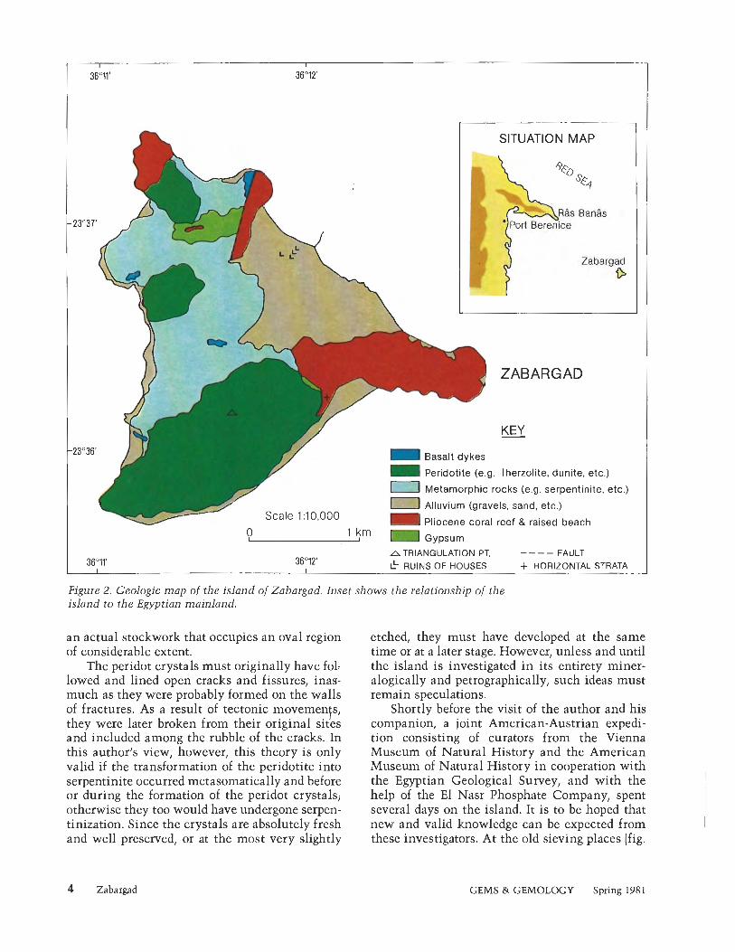
much as the gemstone i t hoarded, has slid repeatedly into oblivion, only to be rediscovered over and over again and forgotten once more. The author visited this tiny island i n the Red Sea i n March of 1980. Located about 60 miles southeast of the RBs Bands peninsula, at 23" 36' 16" N and 36" 11' 42" E (fig. 2)) it is situated 16 lzm north of the Tropic of Cancer. Zabargad is only 3.2 lzm long and 2.4 lzm wide, covering a n area of 4.5 lzm2. There is hardly any life on the island and no fresh water at all; one may justly describe it as a "desert island." In fact, apart from low-growing shrubs, several giant turtles, and a few birds such as wagtails, ospreys, and gulls, practically no flora or fauna exist on Zabargad. The highest ground is the so- called Peridot Hill (235 m above sea level), which to- gether with some smaller hills (135 m above sea level) forms the most impressive sight that the island offers the approaching seafarer (fig. 3). Despite the lack of vegeta- tion, the island is at i ts most beautiful when the yellow to dark brown toiles of the various roclzs and dump-heaps before the many pits brighten i n the light of the morning sun. The adventurer with any imagination at all cannot avoid letting the pageant of history unfold (see box).
REGIONAL GEOLOGY The occurrence of peridot on Zabargad is intimately related to the regional geology and the tectonic processes that on a larger scale were responsible for the formation of the Red Sea itself. As a n extension of the East African Rift Valley and part of the global rift system, the Red Sea is a geologically young feature that evolved i n the Ter- tiary period of geologic time, 65 to 13 million years ago.
The rocks seen on the island represent the results of magmatic activity with associated metamorphism of preexisting sediments and were all exposed through tec-
2 Zabargad GEMS 8: GEMOLOGY Spring 198 1

tonic uplift and erosion. Mafic igneous rocks (Badgley, 1965), which represent the bulk of the island, are of deep-seated origin. They are notable for their low silica content and consist primarily of lherzolites, characterized by abundant olivine, pyroxene, and amphibole. The metamorphic rocks, in turn, consist of serpentinites, granu- lites, schists, and slates. The alluvial sediments and an extensive gypsum deposit are of more recent origin.
The topography of the island reflects its tec- tonic history. The terrain is extremely irregular, the coastline consisting of fractured escarpment terraces. Numerous coral banlzs and reefs occur in the surrounding waters.
Zabargad
THE PERIDOT OCCURRENCES No detailed documentation of the peridot occur- rences on Zabargad has been made. An inter- esting review of available information is provided by Wilson (1 976).
It seems likely that peridots were once found on several parts of the island-in fact, almost everywhere the peridotites outcrop. The finest and largest gem crystals, i t is believed, occurred in such quantitites on the eastern slopes of Per- idot Hill that mining was worthwhile. Here they appear to have been recovered in vein-like areas of the serpentinized peridotite. The tiny veins run in all directions so that in places they form
GEMS h GEMOLOGY Spring 198 1 3
SITUATION MAP
bR4s Bands LPort Berenice
ZABARGAD r
Basalt dykes
Peridotite (e.g. Iherzolite, dunite, etc.)
Metamorphic rocks (e.g, serpentinite, etc.)
m Alluvium (gravels, sand, etc.) Scale 1 :10,000 - Pliocene coral reef & raised beach
0 , ' $m Gypsum
ATRIANGULATION PT. ---- FAULT 3 6 O 1 2 ' & RUINS OF HOUSES + HORIZONTAL STRATA
Figure 2. Geologic map of the island of Zabargad. Inset shows the relationship of the island to the Egyptian mainland.
an actual stockwork that occupies an oval region of considerable extent.
The peridot crystals must originally have fol- lowed and lined open cracks and fissures, inas- much as they were probably formed on the walls of fractures. As a result of tectonic movemenfs, they were later broken from their original sites and included among the rubble of the cracks. In this author's view, however, this theory is only valid if the transformation of the peridotite into serpentinite occurred metasomatically and before or during the formation of the peridot crystals; otherwise they too would have undergone serpen- tinization. Since the crystals are absolutely fresh and well preserved, or at the most very slightly
etched, they must have developed at the same time or at a later stage. However, unless and until the island is investigated in its entirety miner- alogically and petrographically, such ideas must remain speculations.
Shortly before the visit of the author and his companion, a joint American-Austrian expedi- tion consisting of curators from the Vienna Museum of Natural History and the American Museum of Natural History in cooperation with the Egyptian Geological Survey, and with the help of the El Nasr Phosphate Company, spent several days on the island. It is to be hoped that new and valid knowledge can be expected from these investigators. At the old sieving places (fig.
Zabargad GEMS & GEMOLOGY Spring 198 1

merly, however, one could find peridot crystals up to 10 cm long, although those 2-4 cm in length were much more abundant. The Geolog- ical Museum in London owns a splendid step-cut peridot of 146 ct., while the largest known cut peridot weighs 310 ct. and is exhibited at the Smithsonian Institution in Washington, DC. Both of these magnificent samples came originally from the island of Zabargad.
MINING The original mining methods consisted of prim- itive manual digging, whereby each individual vein w a ~ excavated (fig. 5). The miners worlzed
Figure 4. Sieving place in the foreground and dump-heaps in the background on the eastern slope of Peridot Hill.
4), in the prematurely abandoned sieve-heaps, and among the waste of the mines, the author and his companion found over a hundred fresh, transparent, well-preserved, in part broken but certainly cutable peridot crystals pale yellowish green to deep olive green in color. Conspicuous samples of the macroscopic paragenesis of these peridot crystals were green garnierite and fresh whitish to weathered grey cancrinite. The peridot Figure 5. tunnel dug into metamorphic crystals, pseudohexagonal after b(010) in form, rock to reach the deeper mafic rock (note the were between 5 and 15 mm along the a-axis. For- while bands of magnesite).
Zabargad GEMS & GEMOLOGY Spring 1981 5

FROM "TOPAZOS" TO PERIDOT: ZABARGAD AND ITS GEM SHARE A PLACE
IN HISTORY, L A N G U A G E , AND LORE
Topazios is the name used by Greelz author Alexander Polyhistor to refer to an island on which gemstones are found whose color "resembles that of fresh oil." The great naturalist of ancient times, Pliny the Elder (23-79 A.D.), mentions the island "Topazios" in his work Naturalis Historia. He refers specifically to Icing Juba I1 of Mauretania (25 B.C.-23 A.D.) who reportedly states in his writings that this legendary island in the Red Sea was first explored during the reign of Queen Berenice (340-279 B.C.]. Pliny also refers to another report that pirates driven by adverse winds landed on an island i n the Red Sea called Chytis or Cytis. Being in a famished condition, they sought herbs and roots in the ground and thereby fouild the first "topazos." Other names attributed to the island in ancient times include Island of Death (Nelzron) and Ophiodes ("snalze island," reported by Agatharchides of Knidos, 18 1- 146 B.C.). Not until the time of the Crusades did this mystery-bound scrap of land receive the name by which it is still mistalzenly lznown in the West, St. John's Island, and finally, Zabargad.
For Pliny the Elder, topazos was a jewel that mainly occurs in green but may also be yellow. Probably there is some confusion here with chrysolite, "chrysolithos," which was also lznown in ancient times (Ezelziel 10, 9). During the period, chrysos meant golden, that is, yellow. Not until much later did the word experience an etyn~ological change as the prefix chryso- entered common usage to describe a green stone (see chrysoberyl, chrysoprase, chr ysopal, chr ysocolla, chrysolite, etc. 1. When in the early 18th century the name topoz was finally affixed to the fluorine-bearing aluminum silicate that currently holds this title, a new name was needed for the green gemstone from Zabargad. In 1790, the sometime mineralogist A. G. Werner nained the mineral olivine because of its typical olive-green color. In the same year, he published a description of chrysolite in Bergmanns /ournu1 3, stating that it was a mineral in its own right. A few years later, M. H. Klaproth was able to prove that olivine and chrysolite belong to the same family of minerals. Although chrysolite was used by German and American ~nineralogists for over 100 years, this term has left general usage and is no longer accepted in English nomenclature for the gemstone. The English adopted Werner's name olivine, while the French gave preference to the new name peridot, which is derived from the Ara- bian word furidat, meaning gem. The latter is generally used today to refer to the gemstone, whereas the true name of the mineral is olivine (see Ball, 1950, Liischen, 1979, and Mitchel, 1979, for further information on the various names).
Archaeological excavations in Alexandria have apparently unearthed valuable peridots. Faceted samples, which could only have come from Zabargad, have also been discovered in ancient Greece. In all probability, the fabulous stone that once adorned King Ezelziel from Tyrus (about 586 B.C.; Ezelziel 28, 13) was a peridot from Zabargad. Where so much treasure and beauty were to be found, the authorities kept a wary eye. Diodorus Siculus writes in the first century before Christ: "The Egyptians Izept the island under constant watch, and anyone who tried to approach the treasure island without permission-let alone to attempt to land and steal the peridots-was threatened with death." Thus, this island in the Red Sea became one of the most closely guarded regions of the ancient world, and its treasure was held secret for centuries, virtually hidden from the Western world from biblical times until the onset of the baroque period in the 17th century.
6 Zabargad GEMS &k GEMOLOGY Spring 1981

Figure 6. Peridot crystal on 1 its original matrix, self-
collected b y the author. 1 The crystal is approxi- 1 mately 1.2 cm x 1.1 cm.
downwards until the vein, usually at a shallow depth, became sterile or contracted. However, the peridots were not found where they had crystal- lized. They were always loosely attached to the walls of the veinlets and could easily be removed from their position. Wilson (1976) noted that he knew of no peridot crystal that he could in good conscience claim was found directly grown on its mother-rock. However, the author of this present article was so lucky as to find a piece of matrix about the size of a child's fist with a fully grown, well-developed peridot crystal attached (fig. 6).
During the years before World War I, from about 1906 onwards, the island of Zabargad was generally lznown as the source of peridot, and mining rights were monopolized by the Khedive, the Turkish viceroy in Egypt. Within a four-year period, more than $2 million in peridot (present day value) was found and sent to France for cut- ting. The chief problem on the island then, as now, was the lack of fresh drinking water for the miners. The mining company overcame this dif- ficulty by erecting a large gasoline-powered water condenser (of which a few rusty parts .are still lying around today). In 1922 the Egyptian govern- ment gave the mining rights to the Red Sea Mining Company. In the years that followed, until the outbreak of World War 11, this company brought out a considerable amount of peridot. In 1958, the deposits were nationalized by Egyptian President G. A. Nasser.
DESCRIPTION A N D PROPERTIES Gem peridot lies compositionally between the end members forsterite (MgzSi04) and fayalite (Fe,SiO,) of the olivine isomorphous series (Mg, Fe),[SiO,], i n which the two divalent cations Mg and Fe can replace each other diadochally. The peridots from Zabargad contain 8%-10% FeO (FeO + MgO amount to approximately 64%; Wilson, 1976). The crystals are elongated along the a-axis so that the brachy pinacoid b(010), which is markedly striated vertically to the c-axis, forms the largest face. The gradations in color of this summer-green gemstone (the birth- stone for August), as well as the variations i n its other physical properties, are caused by the iron content (see Troeger, 1956, p. 54) and other com- ponents. The color tones of the finest qualities correspond with the DIN color chart 6164, page 24, colors 24:6:2;24:6:3 to 24:6:4 of the color norms Xc34.2; YC43.7; Z,10.2 or Xc23.0; Y,29.4; Zc6.9 or Xc15.2; Y"19.5, and Zc4.6. Refractive indices vary little, from 1.650 to 1.654 for na and between 1.686 and 1.690 for ny, with a constant value for np only a little below the middle of the two extreme readings. The high birefringence remains constant at 0.036, as does the density (3.34) which is only 0.01 above that of pure meth- ylene iodide, i n which liquid peridot remains sus- pended or sinks slowly. Pleochroism is weak but perceptible as pale green along cu, green along P ,
Zabargad GEMS & GEMOLOGY Spring 1981 7

and light green along y. The absorption spectrum reveals the iron content by means of three char- acteristically placed bands in the blue to blue- green region at 453, 473, and 493 nm.
Diagnostically important are the rounded plate-like and wafer-thin healing seams or residual drops, in the approximate center of which a black grain of chromite is situated. Their appearance may be likened to that of a water-lily leaf, and they are consequently termed "lily pads."
CONCLUSION Since mining on Zabargad has failed to remain lucrative, other sources have filled the demand for peridot. Relatively great numbers of large, at- tractive peridots from Kyaukpon, above Mogok in Burma are still reaching the market. Today's second largest supplier of cutable gem peridots is near San Carlos, in Arizona, followed up by a de- posit at Sondmore in Norway, which produces peridots that are slightly lighter in color and very brilliant when faceted. In addition, some small fragments of peridot are said to come from Hawaii.
Despite its somewhat low hardness of 6%-7 (lying just below the critical border of gemstone hardness, 7), this magnificent gemstone with its agreeable sparkle and its glimmer like damp moss in the evening sunlight, has today once more gained appreciation and popularity.
REFERENCES Agatharchides (150 A.D.) De Mare Erythmeo. Badgley PC (1965) Structr~rol and Tectonic Principles. Harper
& Row, New York. Ball SH (1950) A Roman Book on Precious Stones. Gemolog-
ical lnstitute of America, Los Angeles. Liischen H (1979) Die Namen der Sieine, 2nd ed. Ott Verlag
Thun, Munich. Mitchell RS (1979) Mineral Names, What Do They Mean! Van
Nostrand Keinhold Co., New York. Pliny GS (Elder) (70 B.C.) Naturalis Historlo, Vol. 37, Chap.
9 0 JL.
Siculus D (50 B.C.) Bibliothecae Historicae, Book 3, Chap. 39. Troeger WE (1956-67) Optische Besrimmung der gesteinsbil-
denden Minerale, 3rd. ed. E. Schweizerbart, Stuttgart. Wilson WE (1976) Saint John's Island, Egypt. Mineralogical
Record, Vol. 7, pp. 310-314. A German version of this article was published in the Ger- man magazine Lapis-A Monthly for Amuteurs of Minerols and Gemstones, Vol. 5, No. 10, pp. 19-26.
8 Zabargad GEMS 8: GEMOLOGY Spring 1981

CUBIC ZIRCONIA: AN UPDATE By Kurt Nassau
Soon after i t was first marketed in 1976, colorless cubic zirconia became the dominant diamond imitation, with current production of approximately 60 million carats per year. Although cubic zirconia was discovered as a natural mineral in 1937, crystals usable for faceting were first produced ill 1969 and it was not until a practical sltull-melting technique was developed in the USSR in 1972 that commercial production became feasible. This article reviews the sl<ull- melting technique used to produce cubic zirconio and examines the current status of this diamond simulant with regard to q ~ ~ a l i t y , production, ond market. The patent situation i s discussed, as well as prospects lor new diamond imitations and the recent surge of interest i n colored cubic zirconia. Also inclzlded is an Appendix summarizing the technical and gemological data for cubic zirconia and its distinguishing characteristics.
A BOUT THE AUTHOR
Dr. Nassau is a research scientist residing in Bernardsville, NJ.
"'1981 Gemological Institute of America
C ubic zirconia was discovered as a natural mineral in 1937, when two German mineralogists, von Staclz-
elberg and Chudoba (1937), were examining a highly metamict zircon given to them by B. W. Anderson. The zircon contained some tiny crystals which they identified by X-ray diffraction as the cubic form of zirconium oxide (or zirconia), a compound lznown as baddeleyite when in the monoclinic form. So little did von Staclzelberg and Chudoba think of this discovery that they did not even assign a name to the new mineral. As a result, it is lznown to this day by its scientific name, cubic zirconia, and the prefix synthetic, although proper, is not usually included.
This same material had already been used for many years as a ceramic composition for high-temperature in- dustrial and scientific purposes; because of an exception- ally high melting point, "stabilized zirconia" ceramics can be used at temperatures up to 2540°C (4604°F) and are very resistant to most chemical substances. Such stabi- lized zireonia typically consists of 96% ZrOs (zirconia) and 4% CaO (lime), although MgO (magnesia) or Y203 (yttria) also can be used in place of the CaO. Further dis- cussion of the early work on this material is given in Nassau (1977).
Whenever the powder or ceramic form of a substance has interesting properties, scientists usually attempt to grow large crystals; this permits a more precise study of a broader range of properties and often leads to additional uses. In the case of cubic zirconia, crystal growth proved most difficult in view of the material's exceptionally high melting point. Pure ZrOf melts at about 2750°C (4982"F), but at a somewhat lower temperature when stabilizers are added. Flux growth was tried (Nassau, 1977, 1980a), but i t produced either monoclinic baddeleyite or a tetrag- onal form. On heating, the former is transformed from monoclinic to tetragonal at about 1100°C (20!2"F), and from tetragonal to cubic at about 2000°C (3632°F). Un-
Cubic Zirconia GEMS & GEMOLOGY Spring 1981 9

Figure I . A large slzull-melting apparatus being operated by George Braml~all. Courtesy of the Ceres Corp., Walthnm, M A .
fortunately, these changes reverse on cooling and the starting material forms once again. The ad- dition of stabilizers such as CaO, Mg.0, or Y,O:, prevents these transformations on cooling: cubic zirconia then remains the stable form down to room temperature. Details of the different forms of zircoilia are given in the Appendix.
Crystals of cubic zirconia in a usable size were finally reported in 1969 by Roulin, Vitter, and DCportes of France. They used a difficult form
of melting the material without a crucible to ob- tain crystals more than 15 nlm (0.6 inch) long, of composition 87.5% Zr02, 12.5% Y203. Only a few properties of the crystals appear to have been measured at that time, however, and the potential remained unrecognized. A somewhat modified and greatly enlarged form of this apparatus was used by Alelzsandrov, Osilzo, Prolzliorov, and Ta- tarintsev at the Lebedev Physical Institute of the USSR Academy of Sciences in Moscow (1973).
10 Cubic Zirconia GEMS & GEMOLOGY

Their technique, called "slzull melting," is de- scribed in detail in the next section. Crystals one inch (2.5 cm) and larger in diameter can be grown with a modern embodiment of this arrangement (fig. 1) without excessive difficulty. The impetus for this work had been the hope-unfulfilled-of finding improved materials for laser or opto-elec- tronic uses, but measurements of the crystals showed a material that was hard, colorless, and with optical properties that approximate the bril- liance and fire of diamond better than any pre- vious diamond imitation.
With its introduction into the gem trade late in 1976, cubic zirconia as a diamond imitation immediately created a huge demand as well as considerable anxiety. As with the introduction of every diamond imitation before it (Nassau, 1980a), some jewelers who did not know how to recognize the new material were unable to pro- tect themselves. Nevertheless, examination tech- niques have now been adapted to readily distin- guish cubic zirconia from diamond, and new instruments have appeared to assist the gemolo- gist-jeweler in this task, as outlined in the Appendix.
As with YAG (yttrium aluminum garnet), which was overproduced and pealzed about 1972 (Nassau, 1980a), we are now seeing a rapid rise in the production of cubic zirconia accompanied by a rapid fall in the price. This supply-and-demand movement, together with the recent introduction of colored forms of cubic zirconia and the curious patent situation, is also discussed in the following sections.
THE SKULL-MELTING TECHNIQUE FOR GROWING CUBIC ZIRCONIA The technique of crystal growth by solidification from the melt in a container is one of the easiest and least costly ways of obtaining large crystals. When, however, a material has as high a melting point and is as reactive as cubic zirconia, no con- tainer can be found to hold the melt, and a "self- contained melt," "cold crucible," or "autocruci- ble" arrangement must be used. The technique described here was patented by Joseph F. Wenclzus and coworlzers (19771, now one of the major man- ufacturers of cubic zirconia as the Ceres Corpo- ration, Waltham, MA.
The apparatus shown in figures 2 and 3 con- sists of a cup-like arrangement made up of a circle of copper fingers, water-cooled by complicated in-
Figure 2. Schematic of one form of skull-melting apparatus; only some of the fingers are shown.
Figure 3. The split halves of a small slzull- melting apparatus.
Cubic Zirconia GEMS & GEMOLOGY Spring 1981 11
ternal plumbing. There are small gaps between the fingers. Surrounding this apparatus is a water- cooled copper coil activated by a radio-frequency generator, typically operated at a frequency of 4 MHz and at power levels as high as 100 1zW.
The small gaps between the metal fingers en- able the radio-frequency energy to pass into the cup, which is initially filled with zirconia powder mixed with the required stabilizer. The powder is an electrical insulator at room temperature; it will not begin to heat until after the addition of some pieces of zirconium metal, which are then rapidly heated by the radio-frequency field. As the zirconia adjacent to the zirconium metal heats up, it also begins to conduct electricity and melts in a matter of minutes. The zirconium metal reacts with the oxygen in the air to produce more zirconia.
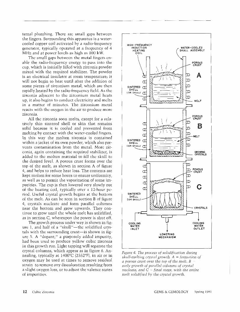
All the zirconia soon melts, except for a rela- tively thin sintered shell or slzin that remains solid because it is cooled and prevented from melting by contact with the water-cooled fingers. In this way the molten zirconia is contained within a jacket of its own powder, which also pre- vents contamination from the metal. More zir- conia, again containing the required stabilizer, is added to the molten material to fill the skull to the desired level. A porous crust forms over the top of the melt, as shown in section A of figure 4, and helps to reduce heat loss. The contents are kept molten for some hours to ensure uniformity, as well as to permit the vaporization of some im- purities. The cup is then lowered very slowly out of the heating coil, typically over a 12-hour pe- riod. Useful crystal growth begins at the bottom of the melt. As can be seen in section B of figure 4, crystals nucleate and form parallel columns near the bottom and grow upwards. They con- tinue to grow until the whole melt has solidified, as in section C, whereupon the power is shut off.
The growth process under way is shown in fig- ure 1, and half of a "skull"-the solidified crys- tals with the surrounding crust-is shown in fig- ure 5. A "dopant," a purposely added impurity, had been used to produce yellow cubic zirconia in this growth run. Light tapping will separate the crystal columns, which as in figure 6. An- Figure 4, The process o f solidification during nealing, typically at 1400°C (2552OFL in air or in skull-melting crystal growth. A = formation o/ oxygen may be used at times to remove residual a porozls crust over the top the melt , B =
strain to remove any discoloration resulting from early growth of parallel columns of crystal a slight oxygen loss, or to adjust the valence states nucleate, and Ci = final stage, with the entire of impurities. melt solidified h y the crystal growth.
HIGH- FREOUENCY WATER- COOLED
COOLING COOLING , "7 1 WATER , OUTLET
LOWER1 NO MECHANISM
12 Cubic Zirconia GEMS & GEMOLOGY Spring 1981

Figure 5. Half of a large skull of yellow cubic zirconia cryslals grown using the technique described in figure 4. Courtesy of the Ceres Corp., Waltham, M A .
At first, a slzull 3 inches (7.5 cm) in diameter was considered the state of the art for cubic zir- conia crystals, but today slzulls 12 inches (30 cm) in diameter and weighing 150 lbs. (68 kg), such as the one shown in part in figure 5, are produced. These yield about 100 lbs. (45.5 lzg or 227,000 car- ats) of usable rough. Individual crystals are well over one inch (2.5 cm) wide and several inches long; stones several hundred carats in weight have been faceted. The economy of large-scale produc- tion is significant, although the rather high cost of the high-purity starting material is not much affected by such a scale-up.
:igure 6. Columnar crystals of cubic zirconia me inch (2.5 cm) across.
THE CURRENT STATUS OF COLORLESS CUBIC ZIRCONIA Starting from a small amount in 1976, within just four years production has grown to an estimated 13 tons (12,000 lzg or 60 million carats] of cubic zirconia per year at the end of 1980. Correspond- ing to the figures for rough material, the annual production of faceted material is estimated at about 3 tons (2700 lzg or 13.5 million carats) per year. During this same time period, the wholesale price per faceted carat has fallen from an initial US $40 or more to US $4 or less, varying of course with the quality of the material and the faceting, as well as with size, shape, and quantity.
It is interesting to compare the production data for cubic zirconia with the data for synthet- ics previously used as diamond imitations, as shown in table 1. Annual production of cubic zir- conia has already well surpassed the YAG peak of 1972 which, it must be remembered, repre- sented overproduction, was more than the market could absorb, and resulted in the discontinuation of production by a number of manufacturers. A reasonable guess would be that the equivalent stage for cubic zirconia is close at hand. A factor that may prevent this from happening is the ap- parent opening up in recent months of the Euro- pean market, where previous diamond imitations never achieved significant popularity.
Many jewelry retailers at first showed a strong resistance to handling cubic zirconia, but expe- rience has now demonstrated that the simulant has had no significant effect on diamond sales, occupying instead a separate niche in the jew- eler's range of goods. The available data for 1977 permitted an estimate of the retail value of all diamond imitations sold in the United States of about $20 million, compared to about $2000 mil- lion for diamonds (Nassau, 1980a). Although by
. now the sales figure for diamond imitations has grown significantly, so has the figure for dia- monds, and the 1% ratio of diamond-imitation sales to diamond sales has probably increased only a little.
Major manufacturers of cubic zirconia in the U.S. include the Ceres Corporation of Waltham, MA (marketing through MSB Industries of New York City); ICT Corporation of Shelby, MI; Singh Industries of Randolph, NJ; the LambdalAirtron Division of Litton Systems of Morris Plains, NJ; and Commercial Crystal Laboratories of South Amboy, NJ. Outside the U.S. there is production
Cubic Zirconia GEMS & GEMOLOGY Spring 1981 13

TABLE 1. The historical sequence of synthetics used as diamond imitations.
Year of Peak annual initial production of rough
Synthetic use (in carats) Year of peak
Sapphire After 1905 ? ? Spinel About 1920 ? ? Rutile 1948 750,000 1955 Strontium titanate 1955 1,500,000 1968 YAG 1968 40,000,000" 1972" GGG 1975 Small 1976 Cubic zirconia 1976 60,000,000b 1980-1 981
aPrernature peak due to overproduction. bMay still be increasing.
in Moscow, USSR (marketed in the U.S. through Clayhill Resources of New York City); at V. Djkvahirdjian S.A. of Monthey, Switzerland [at first erroneously reported as using flux-growth [Nassau, 19761); as well as recent production in Taiwan and undoubtedly elsewhere. The rough material is faceted all over the world, but until recently almost all has been used in the U.S. De- spite the great improvement of cubic zirconia over YAG (in brilliance) and over strontium ti- tanate (in hardness), both of these other simulants continue to be manufactured and sold, albeit on a considerably smaller scale.
Claims are sometimes made that one manu- facturer's cubic zirconia is better than another's, for example, in respect to turning dark in normal use. Although some bad batches were undoubt- edly produced in the early days, currently there is no significant difference in the behavior of the high-grade material produced by the various man- ufacturers. Similarly, inclusions are virtually ab- sent in top-grade material.
Names used for marketing cubic zirconia include:
CZ Diamonique I11 Cerene Diamonite or Cubic Z Diamondite Cubic Zirconia Djevalite Cubic Zirconia I1 Fianite Cubic Zirconium Phianite or Cubic Zirconium Phyanite
Oxide or Dioxide Shelby Diamon-Z Singh Kohinoor Diamond-QU Zirconia Diamonair I1 Zirconium Diamonesque Zirconium Yttrium Diconia Oxide
Some of these names are registered trade- marks, others are not. It is important to note that, by themselves, zirconium and cubic zirconium are misnomers, since they would refer to zircon- ium metal! Also, some sellers are using the end- ing "-Z" with other terms, possibly to imply cubic zirconia when this may not, in fact, be the material at hand.
The stabilizer used in cubic zirconia is usually Y,03, although CaO has also been used. The phase diagrams indicate that up to 65 weight percent YzO3 or up to 14 weight percent CaO could be used in cubic zirconia. Too much stabilizer results in a softer and less brilliant product, how- ever, and amounts much smaller than those in- dicated in the phase diagrams are used for the commercial product, as described in the Appendix.
Now that large crystals of cubic zirconia are readily available at a relatively low cost, scien- tists are once again studying the material for pos- sible technological uses, thus coming full circle to the original purpose for growing the crystals!
Cubic hafnia-hafnium oxide (HfO,) stabi- lized with Y,03-has also been described in the cubic zirconia patents. This has about the same refractive index as cubic zirconia, but with an even higher specific gravity. Since hafnia is much more expensive than zirconia, cubic hafnia is not likely to be used as a diamond imitation.
THE CUBIC ZIRCOMA PATENTS In December 1972, the USSR group of V. I. Alelz- sandrov and coworkers applied for patents in the U.S. and other countries, the relevant claim being cubic zirconia stabilized with 10% to 30% ytt- rium oxide. British patent 1,373,888 was pub- lished on November 13, 1974, and German patent
14 Cubic Zirconia GEMS & GEMOLOGY Spring 1981

2,261,85 1 appeared on July 1 1, 1974. The U.S. pa- tent application was abandoned and then refiled in March 1977, was again abandoned and then re- filed in January 1978; it issued on May 8, 1979, as U.S. patent 4,153,469.
Meanwhile, additional patents had been ap- plied for in January 1975, leading to British patent 1,491,362, published on November 9, 1975, and German patent 2,501,800, published on July 22, 1976. The relevant claim in these patents was cu- bic zirconia stabilized with 5% to 10% yttrium oxide. Such a claim may also have been involved in one of the U.S. refilings; if so, i t was apparently not allowed. It should be noted that the items being patented are the chemical composition of cubic zirconia crystals, not the growth technique.
After the issuance of the U.S. patent in 1979, some retailers hesitated to purchase domestically produced material. At least one U.S. producer (the Ceres Corp.) guaranteed its customers that it would defend them in any suits that might arise from this patent. A suit based on this patent has, in fact, been filed; as of this writing i t is still un- decided. It should be noted that most cubic zir- conia produced in the U.S. is believed to be made with somewhat less than the 10% minimum claimed in the patent. Even if this argument were rejected, there is the fact that the patent contains no mention of the cubic zirconia stabilized with 12.5% Y,03 reported in 1969 (Roulin e t al.) and shown in large crystal form in a French film made at that time; this would seem to imply that a court test of the validity of the patent might well result in an invalidation.
COLORED CUBIC ZIRCONIA Aleksandrov and coworlzers gave details on the manufacture of colored cubic zirconia in their 1972 patent application. Until recently, however, most manufacturers have concentrated on lzeep- ing color out of their crystals, since even trace amounts of impurities such as iron can produce a yellow hue, undesirable in a diamond imitation. Some experimentation persisted, though, partic- ularly to find good ruby red and emerald green colors, neither of which seems to have been achieved thus far. Along the way, significant amounts of yellow and other colored material ac- cumulated. Some was faceted by amateurs for the fun of it and was found to be very attractive.
A major breakthrough in the market for col- ored cubic zirconia was apparent at the 1979 Tuc-
son (Arizona) Gem and Mineral Show, where all available colored cubic zirconia rough was quiclzly bought up by amateur as well as professional cut- ters. The attractiveness of the colors lies in their coupling with a high refractive index and disper- sion, and can be seen to some extent in figure 7. The optical constants of most colored gem- stones-ruby, sapphire, emerald, amethyst, ci- trine, topaz, etc.-have relatively low values, and their attractiveness resides predominantly in the color. With colored cubic zirconia, however, there is considerable brilliance, or "life," in addition to the color; this liveliness can be transmitted only poorly in even the best photograph.
A listing of the colors produced by specific do- pants (purposely added impurities) is given in table 2, and the dopant compounds used to pro- duce desired colors appear in table 3. The nature of the color can change with the concentration of the dopant as well as with its oxidation state, and combinations of dopants are used to obtain de- sired shades. Just some of the colors produced by one manufacturer are shown in figures 7 and 8. The manner in which allochromatic transition metal impurities produce colors is described else- where (Nassau, 1974-75, 1980a, and 1980b).
The most frequently used colors at present are (1) the amethyst to lavender to lilac range, (2) the yellow (canary) to orange to reddish-brown range, and (3) pinlz. Colored cubic zirconia produces fac- eted stones that are much inore "lively" than the analogous amethyst or purple sapphire for the first color group and citrine, padparadsha sap- phire, imperial topaz, or garnet for the second. Prices for colored rough are somewhat higher than for the colorless. At present, these colored stones seem to be used primarily for fad-type medium- priced fashion jewelry. The quantity of colored cubic zirconia produced is still quite small but is increasing rapidly and may soon represent a sig- nificant portion of the marlzet. Attractive intense greens, blues, and reds have not been produced thus far.
Were i t not for the relative rarity of colored diamonds and the fact that most people are not familiar with them, these colored cubic zirconias would provide excellent imitations for naturally occurring canary, pinlz, blue, and green diamond. Jewelers should indeed be aware that, with the correct shade and intensity of color, these cubic zirconias could easily be mistaken for colored diamonds.
Cubic Zirconia GEMS & GEMOLOGY Spring 1981 15

Figure 7. Faceted cubic zircoz~ias; the largest is 9
mm across. CourLesy of the Ceres Corp., Waltham, M A , and MSB Industries, New
York, NY.
TABLE 2. Colors produced by specific dopants added to cubic zirconia."
TABLE 3. Dopanls used in cubic zirconia to give desired colors.
Dopant Symbol Color Color range Dopants used
Cerium Chromium Cobalt Copper Erbium Europium Holmium l ron Manganese Neodymium Nickel Praseodymium Thulium Titanium Vanadium
Yellow-orange-red Olive Lilac Yellow Pink Pink Pink Yellow Brown-violet Lilac Yellow-brown Amber Green Yellow-brown Green
aTypically at the few tenths of a percent level; the color achieved may depend on the oxidation state of the dopant.
Yellow-orange-red CeO,, Ce203 Yellow-amber-brown CuO, Fe203, NiO, Pr203, TiO, Pink Er203, Eu203, Ho203 Green-olive Cr203, Tm203, V203 Lilac-violet C0203, MnOa, Nd203
PROSPECTS FOR THE FUTURE In chapter 1 and the epilogue of my book (Nassau, 1980a), there is an extensive discussion of the sources of new synthetics as well as of the way in which these sources have been changing over the years. Today, only the most difficult of the syntheses have not yet been achieved and, as crys- tal growth techniques have become ever more sophisticated, the search for new crystals has be- come more costly and time consuming.
16 Cubic Zirconia GEMS & GEMOLOGY Spring 1981

Figure 8. Colored cubic zirconia rough; the largest piece is 2.5 inches (6.5 cm) long. Courtesy of the Ceres Corp., Waltham, MA.
It is, therefore, hardly a coincidence that the most recent diamond imitations-YAG, GGG (gadolinium gallium garnet), and cubic zirconia- all originated as spin-offs from technological re- search. The use of YAG as a laser material is well lznown, GGG was developed as a substrate for magnetic bubble memories, and cubic zirconia was studied as a possible laser or opto-electronic material. Is it then lilzely that this source will produce another material that might provide a diamond imitation that is superior to cubic zir- conia?
I believe the answer to this question is yes, but a qualified yes. Although such a material will undoubtedly appear sooner or later, it must be re- alized that technological research for new mate- rials has slowed significantly. In part, this is because present needs have been largely filled and the areas left to be explored are becoming smaller; in part, it is because existing materials are being studied intensively so as to utilize their poten-
tials more fully. So it may not be all that soon that a potential successor to cubic zirconia will arrive.
Further qualifications arise from two factors. First, the optical constants of cubic zirconia are sufficiently close to diamond in a material of ad- equate hardness and wearability that a large im- provement cannot be expected. This was hardly true of any of the previous diamond imitations. Second, the cost involved in developing and mar- keting a new synthetic or imitation is never small and, given the existence of a highly satisfactory material in the marketplace, may not be justifi- able in terms of the potential returns. (One may note the difficulty synthetic flux-grown rubies have had in achieving market penetration in com- petition with the Verneuil-grown product; they recently came close to success by posing as nat- ural rubies with forged GIA certificates!) All in all, it would seem that cubic zirconia is lilzely to be with us for quite some time.
Cubic Zirconia GEMS & GEMOLOGY Spring 1981 17
APPENDIX: TECHNICAL AND GEMOLOGICAL DATA AND IDENTIFICATION
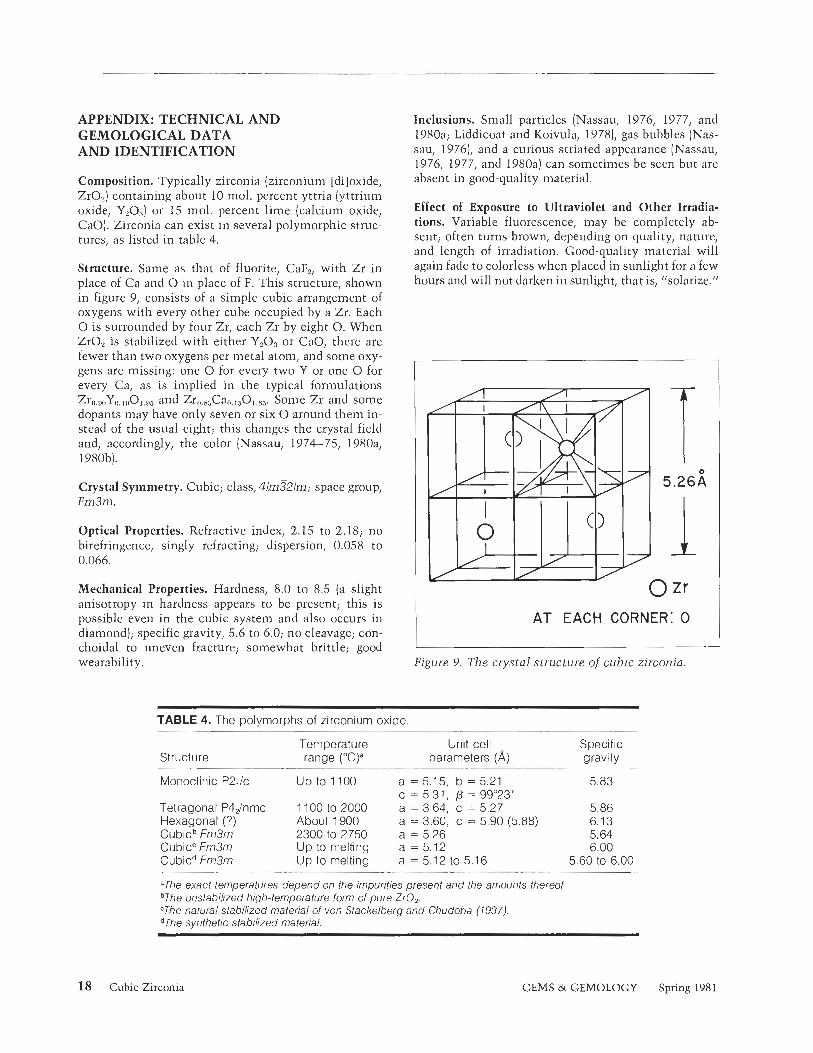
Composition. Typically zirconia (zirconium [diloxide, ZrO,] containing about 10 mol. percent yttria (yttrium oxide, Y,0,,] or 15 mol, percent lime (calcium oxide, CaO). Zirconia can exist in several polymorphic struc- tures, as listed in table 4.
Structure. Same as that of fluorite, CaF,, with Zr in place of Ca and 0 in place of F. This structure, shown in figure 9, consists of a simple cubic arrangement of oxygens with every other cube occupied by a Zr. Each 0 is surrounded by four Zr, each Zr by eight 0. When ZrO, is stabilized with either Y203 or CaO, there are fewer than two oxygens per metal atom, and some oxy- gens are missing: one 0 for every two Y or one 0 for every Ca, as is implied in the typical formulations Zr0.9,,Y0.1001.93 and Zro.8,Ca0.1501.8j. Sonle Zr and some dopants may have only seven or six 0 around them in- stead of the usual eight; this changes the crystal field and, accordingly, the color (Nassau, 1974-75, 1980a, 1980b).
Crystal Symmetry. Cubic; class, 41m321m; space group, Fm3m.
Optical Properties. Refractive index, 2.15 to 2.18; no birefringence, singly refracting; dispersion, 0.058 to 0.066.
Mechanical Properties. Hardness, 8.0 to 8.5 (a slight anisotropy in hardness appears to be present; this is possible even in the cubic system and also occurs in diamond); specific gravity, 5.6 to 6.0; no cleavage; con- choidal to uneven fracture; somewhat brittle; good wearability.
Inclusions. Small particles (Nassau, 1976, 1977, and 1980a; Liddicoat and Koivula, 19781, gas bubbles (Nas- sau, 19761, and a curious striated appearance (Nassau, 1976, 1977, and 1980a) can sometinles be seen but are absent in good-qualit y material.
Effect of Exposure to Ultraviolet and Other Irradia- tions. Variable fluorescence, may be completely ab- sent; often turns brown, depending on cluality, nature, and length of irradiation. Good-quality material will again fade to colorless when placed in sunlight for a few hours and will not darken in sunlight, that is, "solarize."
I A T EACH CORNER: 0 I Figure 9. The crystal structure of cubic zirconia.
TABLE 4. The polymorphs of zirconium oxide.
Structure Temperature Unit cell Specific range (°C)a parameters (A) gravity
Monoclinic P2,Ic Up to 1100 a = 5.15, b = 5.21 5.83 c = 5.31, P = 9g023'
Tetragonal P4dnrnc 1100 to 2000 a = 3.64, c = 5.27 5.86 Hexagonal (?) About 1900 a = 3.60, c = 5.90 (5.88) 6.13 Cubicb Fm3m 2300 to 2750 a = 5.26 5.64 Cubicc Fm3m Up to melting a = 5.12 6.00 Cubicd Fm3m Up to melling a = 5.12 to 5.16 5.60 to 6.00
"The exact lemperatures depend on the impurilies present and the amounts thereof. bThe unslabilized high-temperature form of pure ZrO,. "The natural stabilized material of von Stackelberg and Chudoba (7937). dThe synthetic stabilized material.
18 Cubic Zirconia GEMS W GEMOLOGY Spring 1981

Distinction from Diamond. Several articles in this journal have dealt with various aspects of this problem (Liddicoat and Koivula, 1978; Nassau, 1978-79a, 1978- 79b, 198 1; Nassau ancl Schonhorn, 1977-78; Shaw, 1978). The distinction is obvious to the trained, aware eye. In a loose stone, the high specific gravity is readily apparent. Flatness of faces and sharpness of edges are not foolproof criteria, and girdles apparently showing ~lilaturals" have been observed on cubic zircoilia (Lid- dicoat and Koivula, 1978; Nassau, 1978-79b). The uniquely high thermal conductivity of dianlond pro- vides an uilan~biguous identification of diamond when a well-compensated instrument such as the Ceres Dia- mond Probe (Nassau, 1978-79a) is used, but soille care is required with a simpler instrument such as the pocket-sized Ceres Czecl<mate. A colnprehensive test of such instruments, including the GEM Diamond Master is under way (Nassau, to be published). The breath test is a zero-cost version of the thermal con- ductivity tcst but recluircs conlpiirison stones (Nassau,
REFERENCES Aleksandrov VI, Osiko W, Prokhorov AM, Tatarintsev VM
(1973) Production of refractory single crystals and molten ceramics by a new method. Vestnik Aknd Nauk USSR, Vol. 12, pp. 29-39.
Kerr B (1979) Fashioning cubic zirconia. Gems el Gernology, Vol. 16, pp. 155-157.
Liddicoat RT Jr., Koivula JI (1978) Synthetic cubic stabilized zirconia. Gems d Gemology, Vol. 16, pp. 58-60.
Nassau K ( 1974-75) The origins of color in gems and minerals. Gems d Gemology, Vol. 14, pp. 354-361; Vol. 15, pp. 2- 1 I, 34-43.
Nassau K (1976) A new diamond imitation: cubic zirconia. G'erns d Gemology, Vol. 15, pp. 143-144.
Nassau K (1977) Cubic zil.conia, the latest diamond imitation and skull melting. Lr~piclary /ournal, Vol. 31, pp. 900-904, 922-926.
Nassau K ( 1978-793) A test of the Ceres diamond probe. Gems d Ge~nology, Vol. 16, pp. 98- 103.
Nassau K (1978-79b) Distinguishing diamond from cubic zir- conia: old and new tests for the identification of diamond. Gems 01 Gemology, Vol. 16, pp. 1 1 1-1 17.
1976-79b). Although there are some limitations, one of the many reflectivity meters should give a clear dis- tinction (Liddicoat and Koivula, 1978; Nassau, 1978- 79b). Finally, the wetting contact angle (Nassau and Schonhorn, 1978-79) provides a distinction when the GEM Diamond Pen or one of a number of other pens is used (see Shaw, 1978, for a discussioil of the GEM Diamond Pen, and Nassau, 1978-79b, for a discussion of that and other pens), but surface coatings may have to be removed. As always, the gemologist must guard against the possibility of a doublet.
Size, Weight, and Shape. Proportions a i d cuttiilg tech- niques have beell discussed (Kerr, 1979). A graph for the rapid conversion of size to weight and to the equivalent diamond weight has also been given (Nassau, 1979 and 1980a). It should be noted that cubic zirconia stones may be sold by size, by weight, or by equivalent dia- mond weight ( the last is not always so specified].
Nassau K (1979) The size and weight of diamond and diamond imitations. Gems d Gemology, Vol. 16, pp. 203-204.
Nassau K 11980a) Gems Made by Mr~n. Chilton Book Co., Radnor, PA.
Nassau K 11980bl The causes of color. Scientific American. Vol. 243, pp. 106-123.
Nassau K, Schonhorn S (1977-78) The contact angle of water on gems. Gems d Gemology, Vol. 15, pp. 354-360.
Roulin Y, Vitter G, Deportes CC (1969) New device for melt- ing without a crucible. Fusion of high melting oxides iri a multitubular furnace. Revue Internr~tionale des Hotltes Ten~perutures et des Refmctaires, Vol. 6, pp. 153- 157.
Shaw li (1978) The new diamond pen. Gems d Gernology, Vol. 16, pp. 92-95.
Stackelberg M von, Chudoba K (1937) Density and struc- ture of zircon, 11. Zeitschrift fu r Krista11ographie. Vol. 97, pp. 252-262.
Wenckus JF, Menashi WP, Castonguay A 11977) Cold crucible system. United States Potent 4,049,384, filed April 14, 1975, issued September 20, 1977.
Cubic Zirconia GEMS & GEMOLOGY Spring 198 1 19

A SIMPLE APPROACH TO DETECTING DIAMOND SIMULANTS By [ill Hob bs
T h i s article offers a step-by-step approach to the identification o f the mos t problematic diamond s im ulants. The optical and physical properties o f diamond are contrasted t o those of colorless strontium titanate, synthetic cubic zirconia, gadolinium gallium garnet, and yttrium aluminum garnet. I f the testing methods discrlssed i n this article are used, none o f the above materials should be mistaken for diamond.
ABOUT THE AUTHOR
Ms. Hobbs is a colored stonelgem identification instructor in the resident program of the Gemological lnstitute of America, Santa Monica, CA.
Acknowledgments: The photographs were taken by Michael R. Havstad and the line illustrations in property charts A and B were drawn by Peter Johnston, Gem Media, Gemological lnstitute of America, Santa Monica, CA.
Author's note: Strictly speaking, the word stone should be used to refer only to natural materials. For simplicity, the term is used here to refer to simulants as well as to diamond.
"1987 Gemological lnstitute of America
D iamond simulants need not frighten the gem iden- tifier. Quiclz methods of identification are available,
with many clues requiring only a trained eye. Although certain clues when used alone cannot establish the iden- tity of the stone, often two or three of these clues con- sidered together will lead to the identification of the gem material. Even if the clues available are insufficient for positive identification of the material at hand, other tests have been developed that at least prove whether or not the stone in question is a diamond.
The intent of this article is to provide a step-by-step approach to the identification of colorless diamond sim- ulants. The methods and techniques described are not new, but they have been arranged in such a way here to present a relatively simple, systematic approach to dia- mond identification. Basically, the approach involves the following:
Train your eyes to recognize as many visual char- acteristics of simulants as possible. A "trained eye" may spot a possible simulant without the use of laboratory equipment. Know and use basic tests to identify the type of diamond simulant. Even though your primary con- cern is probably not to name the simulant, it is helpful to be able to provide your customer with such information.
3. Stay abreast of the development of new instru- ments that identify loose or mounted diamonds. While in most cases such devices do not identify the type of simulant, they are very helpful at the repair or take-in counter, where employees may not have the training to recognize diamond simu- lants by their visual characteristics or with the basic tests.
20 Detecting Diamond Simulants GEMS & GEMOLOGY Spring 198 1

Of all the techniques available for diamond separations, perhaps the most important is the trained eye. If a machine fails, or if you cannot perform other verifying tests, your eyes may alert you to a possible simulant. The training requires a great deal of practice and continual updating, but visual examination can be performed any- where, anytime.
This article focuses on the identification of the four diamond simulants that cause the most confusion for the jeweler: (1) strontium titanate, (2) synthetic cubic zirconia [CZ), (3) gadolinium gallium garnet (GGG), and (4) yttrium alumiilum garnet (YAG). All of these stones share two prop- erties with diamond: each is over the limits of the conventional refractometer, and each is singly re- fractive in optic character. The properties of dia- mond and these four diamond simulants are listed in property chart A. The stones are listed in order from highest to lowest refractive index. The prop- erties of these materials are arranged from left to right in the order in which they are presented in this discussion.
Other significant materials that have been mistaken for diamond are listed in property chart B. They are: synthetic rutile, zircon, synthetic sapphirelsynthetic corundum, synthetic spinel, glass, and assembled stones. For convenience, these stones and their properties are arranged the same as chart A. All these materials are more easily identified than those i n chart A because they either have refractive indices within the range of the conventional refractometer or they are doubly refractive. Chart B is, then, supple- mental information. The following article as- sumes that the reader is familiar with the prop- erties of the materials listed on chart B, and does not have difficulty identifying them. For more information on these stones, refer to the newly revised CIA Diamond Assignment number 36.
VISUAL CHARACTERISTICS THAT SEPARATE DIAMOND FROM DIAMOND SIMULANTS As yet, no diamond simulant has been developed that fully duplicates the appearance of a diamond. A diamond has unique optical and physical prop- erties that make its appearance distinctive.
The four diamond simulants that cause the most confusion for the jeweler-CZ, GGG, stron- tium titanate, and YAG-have many visual traits that are unlike those of diamond. The different
visual characteristics reflect differences in the op- tical and physical properties of the various materials.
The "Read-Through" Effect. The visual trait that is often noticed first in round-brilliant-cut stones is the read-through effect. When the various dia- mond simulants are tilted or placed table-down on paper, each one "reads through" differently; that is, each simulant allows a different amount of light to be transmitted through the stone at different angles. This trait is related to the stone's refractive index, critical angle, and proportioning.
Fancy shapes (e.g., marquise, oval, pear) usu- ally read through to some extent regardless of the material involved (the proportioning of fancy cuts allows for greater light transmission). Properly cut in a round-brilliant shape, however, diamond and strontium titanate will generally not read through, while CZ, YAG, and GGG will. When the stones are placed in order of ascending re- fractive indices, the succession is as follows: YAG, GGG, CZ, strontium titanate, and diamond. YAG has the lowest refractive index and diamond the highest. (An easy way to remember the order is by associating the first letter of each of the stones with this phrase: You Go Crazy Staring at Dia- monds). If a diamond and the above four diamond simulants are cut to approximately the same size and proportion, the stones with the lowest re- fractive indices can be read through most easily.
If the stones are placed table-up in a tray, the read-through effect is apparent when the stones are tilted. In figure 1, this effect is seen where an orange paper has been placed in back of the stones to make the effect more visible. Note that the orange paper can easily be seen through YAG, but not through diamond. At different angles, the "read-throughJJ character of each stone varies. However, only when the tray is tilted to the angle where the stones almost spill will diamond trans- mit the orange of the paper. And, at that point, the amount of orange that can be seen through the diamond is still less than for any of the other materials. Again, the amount of orange seen in each of the five stones gradually increases in re- lation to the refractive index of each.
The read-through effect can be particularly helpful when examining a parcel of round-bril- liant-cut diamonds. Simply line the stones table- up, and then tilt the paper away from you. The lining or cotton inside the parcel paper should il-
Detecting Diamond Simulants GEMS & GEMOLOGY Spring 198 1 21

Slight
PROPERTY CHART A DIAMOND AND COLORLESS SIMULANTS
Refractive Read Color on Externall Internal Specific Reflection Typical Gemstone Index Througha Dispersion Pavilion Hardness Characteristics Gravity Pattern Fluorescenceb
Bearding, natural
3.52 El Weak to strong
None, if Orange or with trigons, blue (Iw and ~ iamor id properly cut 0.044 blue on a grainings, sw); may
few facets cleavage, fluoresce any included crystal color
Strontium None, if Spectral
titanate properly cut 0.190 colors, widespread
5.13 Generally ine.
Negative
5.95
None to weak-
Synthetic crystals, lined or moderate orange Orange
partially lined (Iw); none to weak- cubic 0.060 over most
of pavilion with zirconium moderate yellow, zirconia
oxide powder or greenish yellow (SW)
None to moderate
Gadolinium None, or
;.05
orange (Iw); orange Gas bubbles,
gallium moderate to 0.045 over most metallic platelets strong orange to
garnet of pavilion pinkish orange (sw)
None to Yttrium None, or moderate
aluminum blue over most of
garnet pavilion orange (sw)
'This technique IS meaningful only with round-brilliant-cut stones.
b l ~ = long-wave ultraviolet light; sw = short-wave ultraviolet light.
Moderate
Strong
Gas bubbles
Gas bubbles

PROPERTY CHART B COLORLESS DIAMOND SIMULANTS
Refractive Read Color on Externall Internal Specific Reflection Typical Gemstone Index Througha Dispersion Pavilion Hardness Characteristics Gravity Pattern Fluorescenceb
Synthetic rutile Various
(yellow 2.61 6-2.903' None 0.330 spectral 6-6% Gas bubbles 4.26 wlors body widespread
Generally ine.
color)
- ->
Blue over Zircon most of Liquid in two- (high 1.925-1.984' Moderate 0.038 pavilion; 7% phase Or 4.70 0 None to weak
fingerprint yellow (Iw & sw) property) may exhibit
orange pattern
Synthetic Blue over Gas bubbles, ps""
Sapphire 1.762-1.77OC Very strong 0.018 most of possibly in 4.00 :n None to weak
Synthetic pavilion thread-like bluish white (sw) corundum pattern
Synthetic Blue over ki Moderate to spinel 1.730 Very strong 0.020 most of 8 Gas bubbles 3.64 ' $7 , ii strong bluish
pavilion -+. .A white (sw) -, -'--
0.009- Gas bubbles,
0.098 concave facets, ' * --
mold marks, Variable wlors Glass 1.48-1.70 Extreme (usually Variable
about 5-6y2 flow lines, 4.50 orange-peel white overtones
0.037) effect
Separation Same to plane; gas
Variable; possibly bubbles near % on Variable different on crown and stones Variable cement, crazing Variable Variable Variable
crown and pavilion in cement; pavilion variable
inclusions
"This technique is meaningful only with round-brilliant-cut stones.
Vw = long-wave ultraviolet light; sw = short-wave ultrav~olet l~ght. cDoubly refractive.

?,' b d
A- - - -- -- - Figure I . Diamond and its
simulants will have different degrees of transparency, or read-
through effect, i f all are well- I- read through is. Also, the greater
the tilt, the clearer the read I through is. Order from left to
right (high to low R.I.): diamond,
rlgure 2. Zhe read-ttlrougn effect is seen on well-proportioned round brilliants when they are tilted on a light background, such as a stone clotl~. Order from right to left: diamond, strontium titanate, CZ, GGG, YAG.
luminate the effect. Also, the stones do not have to be specially aligned before the results are vis- ible. When a loose stone is placed in a stone cloth, the same results are evident. As you tilt or rock the stone inside the cloth, the degree of read through becomes evident (fig. 2).
The read-through effect can also be seen when the stones are placed table-down over a busi- ness card (fig. 3). With YAG, the letters of the business card are easily seen through the stone; the letters appear a bit magnified and slightly rounded, but they are still very readable. As the refractive index of the material increases, read through becomes progressively more difficult. Through GGG, the letters appear twisted; through CZ, they are almost indistinguishable. However,
even a small amount of read through may aid in identification because diamond and strontium ti- tanate do not read through in this position. (Syn- thetic rutile will also not read through, but it is easily identified on the basis of its other visual characteristics, as per property chart B).
In short, the read-through effect is a quick "indication" test, that is, a test that does not prove, but leads to, the identity of the material. It can be performed with simple aids such as a stone cloth, a business card, or parcel paper. The results are consistent provided that all the stones are well-proportioned round brilliants. YAG, GGG, and CZ will wash out or read through, while diamond and strontium titanate will not.
Figure 3. The read-through effect is also noted when round-brilliant-cut stones are laced table-down over a printed page. Order from left to i.ight: diamond, strontium titanate, CZ, GGG, YAG.
24 Detecting Diamond Simulants GEMS & GEMOLOGY Spring 1981

Figure 4. Spectral colors seen as a result of dispersion.
Magnified 10 X.
To obtain meaningful results, the tester should have comparison stones, or have sufficient expe- rience with diamond simulants to recognize the different degrees of read through of the various stones.
Dispersion and the Monochromatic Flash. If the stone is a properly cut round brilliant and has no read-through effect, it is most likely diamond or strontium titanate. At this point, the difference in the dispersion values of the two stones will easily separate them.
Dispersion is the ability of a substance to cause light to divide into its spectral colors. For exam- ple, when light is transmitted through a prism, a series of colors-red, orange, yellow, green, blue, indigo, and violet are seen, as illustrated in fig- ure 4. Angular pieces of glass that hang in a chan- delier often display dispersion. A cut stone has the same ability to break up light into these colors, although the distinctness of the colors that em- anate varies with the material.
Studies have determined the amount of dis- persion in each of the diamond simulants (see property chart A). How do the numerical values relate to the visual effect? The visual approxi- mation of dispersion takes more time to master than does read through; however, once mastered, i t can help to build a case for the identity of the material. The best way to judge dispersion is to reflect light off the surface of the stone and look closely at the crown and bezel facets.
With regard to the two materials that do not show read through, the dispersion of strontium titanate (0.190) is over four times that of diamond (0.044)-a difference not easily overlooked. Stron- tium titanate is literally "on fire," while diamond displays a moderate amount of spectral colors.
Of the three diamond simulants that do dis- play read through-YAG, GGG, and CZ-none has the dispersion of strontium titanate. YAG has the lowest dispersion value (0.028) and the spec- tral colors are hardly visible. In appearance it al- most resembles a low-lead-content glass; the lack of dispersion and extreme read through make YAG a lifeless simulant. GGG does not appear to have much dispersion. Its value (long thought to be 0.038, but recently verified as 0.045; Nassau, 1980, p. 148) is very close to that of diamond, yet the spectral colors do not seem as prominent. CZ, however, has a good deal more dispersion than diamond. The spectral colors are always evident, but the amount of dispersion is such that the stone is not always distinguishable from diamond (0.060 vs. 0.044) on this basis alone. In the event that there continues to be some uncertainty, the stone could be examined for a monochromatic color on its pavilion, as explained below.
On many of the gem simulants, one spectral color predominates on the back (pavilion) of the cut stone, creating an umbrella effect. The color is the result of dispersion, and the breadth of its circumference is the lzey to its usefulness as a visual aid. The monochromatic sheen can be seen
Detecting Diamond Simulants GEMS &; GEMOLOGY Spring 1981 25

Figure 5. Dispersion: orange commonly covers a large area on the pavilion o f CZ. Magnified l o x .
in reflected or transmitted light. The unaided eye might notice the color as light is reflected off the pavilion of the gemstone. Under a microscope, it is easily seen with dark-field illumination, that is, light directed at the gem from the side. Posi- tion the stone table-down, and gently tilt it in different directions.
Colorless cubic zirconia generally exhibits an orange flash over most of its pavilion surface (fig. 5). At times, the color seems restricted to one-half of the pavilion until the stone is gently rocked, in which case the orange usually extends over the other half as well. This orange effect is prevalent on most cuts of cubic zirconia. GGG may also exhibit an orange-flash effect, but gen- erally the orange is not as extensive.
YAG often shows a blue color on one-half of its pavilion surface (fig. 6). The results are not as consistent as with cubic zirconia, but they may still aid in identification. (The only other dia- mond simulants to exhibit the blue effect are within the limits of the refractometer, i.e., syn- thetic sapphire and synthetic spinel.)
Diamond usually does not display a mono- chromatic color over most of its pavilion surface. It may show blue or orange, but in most cases these colors extend only across one or two facets (fig. 7). Thus, the significance of the monochro- matic effect lies not only with the color present, but with the breadth of the color on the pavilion surface as well.
In the identification process thus far, the dia-
Figure 6. Dispersion: blue of ten covers a large area on the pavilion of YAG. Magnified l o x .
Figure 7. Dispersion: orange and blue extend over only a few facets of diamond. Magnified l o x .
mond simulants have been examined for read through and the effects of dispersion. These vis- ual traits can be seen in a glance. Just a quick look and turn of the stone can yield a great deal of in- formation to a trained observer. However, since these characteristics do not prove the identity of the material, a loupe is useful as we proceed to the next steps of identification.
Facet Junctions. When a stone is examined using a loupe, one of the first external characteristics worth noting is the sharpness of the facet junc-
26 Detecting Diamond Simulants GEMS & GEMOLOGY Spring 198 1

Figure 8. The facet junctions of diumond zlszlally appear sharp and well def ined because o f the stone's ex teme hardness. Magnified 30 X.
Figure 9. GGG often hus rounded facet jrlnctions becarlse i t has a hardness of only 6 Yz. Magnified 40 X .
tions. Because of diamond's extreme hardness (10 on the Moh's scale], its facet junctions are usually sharp and precise (fig. 81, whereas stones under 7 on the Moh's scale (e.g., GGG at 6'/2 and strontium titanate at 5-6) have a tendency to- ward rounded facet junctions (see figure 9). Dia- mond's facet junctions may become abraded over time, but they will not appear rounded.
Because of its relative softness, strontium ti- tanate usually has heavily abraded facet junctions as well. CZ and YAG, on the other hand, have hardness values over 8, so their facet junctions appear relatively well defined. Again, though, CZ can be separated from diamond by the orange-flash effect, and YAG1s laclzluster appear- ance malies it distinctive. The roundness or sharp- ness of facet junctions is best seen in reflected light.
Figure 10. The girdle o f a diamond o f t en has a m a t , waxy appearance. Magnified 40x .
Girdle Area. Next, a trained eye might glean infor- mation from the girdle area, provided that it is not faceted. The girdle of a diamond appears mat and waxy after bruting. As illustrated in figure 10, it is not dull, like ground glass, but has the shiny appearance of wax (Bruton, 1973, p. 341). The gir- dles of most diamond simulants are polished. However, if they are not, the girdle appears quite dull, similar to that of unpolished glass. The gir- dles of simulants may look frosty, but never waxy. The girdle surface has neither the fine-grained look of a carefully turned diamond nor the poorer, coarser look of a diamond subjected to rapid brut- ing. In addition, sometimes the girdle of a dia- mond is "bearded," that is, marlzed by hairline feathers that extend into the stone (fig. 11). The
A 1 ,4
Figure 11. The girdle of a diamond m a y s h o w bearcling, or hairline feathers that extend i n to the stone. Magnified 30x.
Detecting Diamond Simulants GEMS & GEMOLOGY Spring 1981 27

Figure 12. A natural, or part of the original crystal surface, is often found on or near the girdle of a diamond. Magnified 35 X.
feathers are actually tiny fractures caused by rapid bruting. If seen, bearding signifies a diamond.
A natural is a part of the original crystal sur- face and is often seen on or near the girdle. Its unique luster distinguishes it from the rest of an unpolished girdle (fig. 12). To the experienced eye, a natural, too, signifies a diamond.
Growth marlzings may confirm the process under which the crystal grew and verify that the stone is, in fact, a diamond. Several growth mark- ings are characteristic of diamond. Trigons, or tri- angular etch marlzs, may be seen on a natural, parallel to an octahedral face (fig. 13). Lines, or parallel grooves, are seen on those naturals that originate from a dodecahedra1 facej while square or rectangular depressions indicate a cube face.
Attempts have been made to duplicate these natural marlzings, but thus far the results have not been realistic. For example, attempts have been made to duplicate the appearance of trigons by marlzing triangles on the girdle of diamond simulants; instead of having the layered appear- ance of natural etch marlzs, however, the simu- lated marlzings appear to lie in one plane.
Inclusions. After the external characteristics of the material have been noted, the next step in identification is to examine the interior under magnification.
Figure 13. Trigons are growLh markings that are often found on a natural and, to the experienced observer, prove diamond. Magnified 40 X.
The chief reason for using a loupe is to find inclusions, which may prove whether or not the stone is man-made. With the emphasis on dia- mond certification today, most of us are acutely aware of the typical inclusions of diamond, such as cleavages, fractures, knots, and included crys- tals. While inclusions do not increase the value of a diamond, they can be extremely valuable in identifying one.
Most of the diamond simulants in the mar- ketplace today are flawless, since manufacturers have perfected the techniques of crystal growth. If inclusions are seen, they are generally tiny white or highly reflective dots composed of var- ious materials. In YAG, GGG, and strontium ti- tanate, the tiny dots are usually gas bubbles, in which case high magnification may be needed to resolve them. In cubic zirconia, the whitish in- clusions are small negative crystals that are lined, or partially lined, with a white powder that is probably unmelted zirconia oxide (Liddicoat and Koivula, 1978, p. 58). They may appear singly, or in rows. The only other significant inclusions found in diamond simulants are the triangular or hexagonal platelets seen in GGG.
Thus, if the material has inclusions typical only of natural stones, there is no doubt about the stone's identity. If tiny, round, high-relief inclu- sions are seen, the stone is probably man-made.
28 Detecting Diamond Simulants GEMS & GEMOLOGY Spring 1981

Figure 14. If visible, polishing marks on a diarnond travel in different directiol~s on c~djoining fdcets because of the slone's directional hordness. Magnified 20 X.
If the material has no inclusions, other tests can render the necessary proof.
Polishing Marks. When examining the stone for inclusions, polishing marlzs might be noted. Pol- ishing marlzs are a series of close, parallel lines on the surface of a facet.
Polishing marlzs are not always seen on dia- mond because its hardness allows for an excellent polish. If seen, the polishing marlzs will travel in different directions on adjoining facets (fig. 14). Because of diamond's directional hardness, facets are polished in the direction of the dodecahedra1 faces.
By contrast, the polishing marks on a diamond simulant will usually travel in approximately the same direction on adjoining facets. The uniform lines indicate a softer material and represent a mechanized process in which the concern of the producers is primarily mass production (fig. 15).
Grain Lines. Straight, parallel lines that might be confused with polishing lines may be the result of graining (fig. 16). Grain lines appear in the di- rection of twinning, or gliding, and are character- istic of diamond. The lines are usually seen on the surface, but they may also be internal. In ad- dition, graining may take on various shapes (Kane, 1980). Regardless of the form, graining proves the
Figure 15. Polishing marks on diamond simulants usually travel in approximately a he same direction on adjoining lncets. Magnified 2 0 x .
Figure 16. An experienced eye may see grain lines on diamond, which verifies the stone's natural origin. Magnified 35 X .
natural origin of the gem material, since only a natural gemstone could exhibit such markings.
BASIC TESTS TO IDENTIFY THE TYPE OF DIAMOND SIMULANT If the stone has no distinguishing visual charac- teristics and positive proof is still wanting, then several tests can be performed to verify the type
Detecting Diamond Simulants GEMS & GEMOLOGY Spring 198 1 29

of diamond simulant. The most important in- volve specific gravity determinations and special TABLE 1. Relationship of specific gravity to size immersion techniques. in diamond and cubic zirconia.
- Diameter
Specific Gravity Determinations. Specific gravity Carat weight is defined as the ratio of the density of one sub- Diamond Cubic Zirconia
stance to the density of another substance which 1 6.5 mm 5.5 mm is used as a standard when both densities are ob- tained by weighing the substances in air. The de- termination of specific gravity is extremely help- ful since diamond has a much lower value than any diamond simulant on property chart A. Spe- cific gravity is estimated by heft, or by the use of heavy liquids. Precise measurements are obtained on a hydrostatic balance.
Skilled gem identifiers can gauge the density of CZ and GGG in their hands, since GGG's den- sity is twice that of diamond and CZfs density is over one-and-one-half times greater than that of diamond. The diamond simulants that feel clos- est in density to diamond-YAG and possibly strontium titanate-can be distinguished on the basis of the visual characteristics described above.
Heavy liquids can also help approximate spe- cific gravity. Simply drop the stone in question in methylene iodide (density of 3.32) and note the rate at which the stone sinks. Diamond will sink much more slowly than any of its simulants. It appears to float to the bottom, while the diamond simulants plummet. If a known diamond is not available, the rate of sinking can be compared using a synthetic corundum (specific gravity of 4.00). All the diamond simulants in property chart A will sink faster than synthetic corundum. The use of methylene iodide will at least prove whether a stone is a diamond. A set of compari- son stones can be used to help identify the spe- cific diamond simulant.
An accurate specific-gravity measurement can be accomplished by a direct weighing method in which the stone is weighed first in air and then in water to determine the weight lost in water. The specific gravity is found by dividing the ma- terial's weight in air by the loss of weight in water. The stones can be weighed on a diamond balance that has been adapted for hydrostatic measurements. The results prove the identity of the material, although the measurements are not as accurate for stones under one carat.
Relationship of Specific Gravity to Size. The higher the specific gravity of a stone, the smaller
the stone is per carat, and vice versa. Thus, since diamond has a lower specific gravity than its sim- ulants, the diamond simulants will all be smaller, per carat, than diamond.
When the stones in question are round bril- liants, the relationship can be charted as shown in table 1. Two variables are needed for each stone: the diameter and the weight. When a dia- mond and a diamond simulant weigh the same, each will possess a different diameter; conversely, a diamond and a diamond simulant with the same diameter will not weigh the same. The figures may not work perfectly, but they will point to the identity of the material.
Immersion Techniques. B. W. Anderson has guided gemologists to a new technique for iden- tifying diamond simulants. Initially, he discov- ered that diamond simulants have different de- grees of visibility when placed table-down in im- mersion liquid. Stones with refractive indices greater than that of the liquid appear to have dark borders around the girdle and light facet edges, while stones with refractive indices lower than that of the liquid have light borders around the girdle and dark facet junctions. These differences in relief are best seen when the cut stones are immersed in 3.32 liquid table-down in a glass- bottomed cell that is strain-free. The cell should be slightly above its resting surface with light placed directly over the stones.
Interestingly, not only do the majority of the diamond simulants have dark borders when im- mersed in heavy liquid, but as their refractive in- dices increase, the width and darkness of the ring around the girdle also appear .to increase. Thus, as mentioned earlier, if the diamond simulants were aligned according to refractive index, YAG would have the lowest relief, since its refractive index is closest to that of the liquid, with GGG, CZ, strontium titanate, and diamond (in that or-
30 Detecting Diamond Simulants GEMS & GEMOLOGY Spring 1981

Figure 17. The higher the refractive index o f the stone, the more distinct the stone is when immersed i n methylene iodide and the darlzer and wider the shadow around the girdle is. Left t o right (high to l o w K.I.): diamond, strontium titanate, CZ, GGG, YAG.
der) demonstrating increasing levels of relief (see fig. 17).
Since relief in itself usually will not lead to the specific identity of the gem material, a new application of the Anderson principle has been developed to provide this information. The tech- nique is the same; that is, the stones are im- mersed in a glass cell and a light is placed over- head. However, the stones are viewed from the side instead of from directly overhead. Viewed a t a 45" angle from the resting surface, different stones will exhibit different reflection patterns (fig. 18). While viewing contact photographs of immersed stones, Anderson noted the differing reflections, but he did not specifically apply the principle to the identification of diamond simu- lants (Anderson, 1971, p. 35). Careful study has shown, however, that when two or more diamond simulants of the same cut are compared, each has a unique reflection pattern. Round brilliants with good proportions seein to provide the best results.
The reflection patterns of diamond and stron- tium titanate are similar in that they both exhibit
Fig1u.e 18. When light is placed over stones immersed i n methylene iodide, ench stone shows a different reflection pattern. Top row (lef t t o right): dinmond, strontium titr~nate. Bottom row: CZ, GGG, YAG.
bright rays that radiate from the middle of the stone and extend beyond the girdle outline. How- ever, the rays in strontium titanate show spectral colors as a result of the material's great dispersion and usually extend a bit farther than those of diamond.
CZ has a dark area round the girdle, but i t is not circularly defined. The middle of its pattern is bright like that of GGG, but contains distinc- tive sharp, star-like images that extend halfway into the dark area. The reflection pattern of GGG is doughnut-like, with a thick, dark girdle border and a bright interior in which the facet edges are light but somewhat rounded and blurry. The re- flection pattern of YAG is characterized by a com- paratively thin, dark girdle border and bright facet edges.
In most instances, the reflection pattern will identify the dialnond simulant. Since GGG and zircon have similar patterns, the optic character of the stone should be checked before i t is im- mersed. In addition, the patterns may be affected by the distance between the light source and the
Detecting Diamond Simulants GEMS & GEMOLOGY Spring 198 1 3 1

stone as well as by the distance between the cell and its resting place. If a penlight is used to create the pattern, the optimum distance between the light source and the stone is approximately three- quarters of an inch, and between the cell and its resting place is approximately one-quarter of an inch.
Lately, we have noticed that these reflection patterns can be revealed without immersing the stone. The ramifications of this technique are being studied to see if it can be applied to mounted goods.
Fluorescence. Fluorescence is useful only as a supplemental test in gem identification, since the fluorescence of the diamond simulants is not very constant. It may be helpful, though, to note whether the fluorescent reaction is stronger un- der long-wave or short-wave ultraviolet light. Diamond generally fluoresces stronger under long wave than under short wave, while most simu- lants fluoresce stronger under short wave.
The characteristic fluorescence of diamond is weak to strong blue under long- or short-wave ultraviolet light. However, it may be inert or flu- oresce a variety of other colors.
GGG's typical fluorescence is a moderate to strong orange (or pinkish orange) under short-wave ultraviolet light. The fluorescence is generally constant, although some stones have been inert.
The fluorescence of CZ is variable. When the material first appeared on the market, the typical fluorescence was orange under long-wave ul- traviolet light and yellow or greenish yellow un- der short-wave ultraviolet light. It has since been shown that CZ may be inert or may fluoresce a number of other colors.
YAG and strontium titanate are usually inert, although YAG has been known to fluoresce a weak orange. Because of its inconsistency, fluo- rescence should be used only in conjunction with other tests. Chemical impurities, foreign mate- rials, and strain may alter the typical fluorescence of a stone.
INSTRUMENTS THAT SEPARATE DIAMOND FROM DIAMOND SIMULANTS IN MOUNTED GOODS If the gemstone is mounted, or if a person has not been trained to use the above tests, instruments are available to separate diamond from its simulants. The results do not identify the simu-
lant, but they are very usefui at the repair or take- in counter to determine whether or not the stone in question is a simulant. The two most conclu- sive instruments are the GEM Diamond Pen (Gem Instruments Corp., Santa Monica, CA) and the diamond probe.
GEM Diamond Pen. The GEM Diamond Pen effectively separates diamond from the above sim- ulants on the basis of diamond's unique surface tension. Surface tension refers to the ease with which a loose diamond can be picked up by a damp finger; most simulants do not possess this property.
The effects of surface tension are illustrated by placing a small drop of liquid on the surfaces of a diamond and a diamond simulant simultane- ously. The liquid spreads on the surface of the diamond, while it beads on the surface of the sim- ulant. The GEM Diamond Pen uses a nondrying colored liquid to demonstrate the property quickly and easily.
TO ensure an accurate test, the stone's surface should be well polished and clean. Hold the pen perpendicular to the testing surface. In a quick even stroke, run the tip of the pen across the sur- face. In this position, the pen will allow only a metered amount of liquid to flow. A common mistake is to hold the instrument in the same position as a writing pen, in which case too much liquid is deposited on the surface.
Magnification may be needed to examine the drop of liquid. The reaction may be affected by abrasions or coatings on the surface, which can be removed by a polishing compound included in the GEM Diamond Pen kit. In addition, a dia- mond doublet would fool the pen unless surfaces on both the crown and pavilion were checlzed. However, the test can be performed on both loose and mounted goods and, if used properly, provides positive proof of diamond.
Diamond Probe. The diamond probe yields the fastest, most consistent results as to whether the material is diamond. Unlike the Diamond Pen, it is based on a bulk property that is not subject to miscalculations. The probe measures the thermal conductivity of diamond. Diamond's ability to conduct heat is greater than that of any known simulant, as evidenced by the fact that diamonds are colder to the touch than any simulant and become warm more quickly when worn. Simi-
32 Detecting Diamond Simulants GEMS & GEMOLOGY Spring 198 1

larly, moist breath evaporates much more quickly on a diamond.
A typical thermal conductivity unit consists of a small box with a pen-like probe attached to the unit by a cord. The only requirements for the use of the unit is that the stone be at room tem- perature and that the probe pen be held perpen- dicular to the testing surface. Position the pen, push lightly, and note the reaction. If the tip of the probe touches a diamond, the needle on the scale points to "diamond"; if it touches a dia- mond simulant, the needle points to the "simu- lant" area.
The diamond probe cannot distinguish among all diamond simulants because they do not all differ significantly in their ability to conduct heat. Some of the recently marketed probes have indi- cator marks for the important simulants; how- ever, YAG and GGG are generally too close to separate.
If the machine is working properly, it is im- possible to mistalze diamond. To ensure that the machine is operating properly, press the tip of the probe against one of the checlz areas, or compare reactions with known stones. Thin coatings on the stone's surface should not affect the readings. Only a diamond doublet can confuse the ma- chine. The results have been found to be accurate for both loose and mounted stones.
CONCLUSION There is no good reason to misidentify a diamond. Visual characteristics alone should reveal a sim-
ulant, and they may lead to the precise identity of the material at hand. If the visual character- istics are not satisfactory, an estimate of specific gravity coupled with the visual traits will often separate diamond from its imitators.
If a specific identification is needed, hydro- static measurements can provide the proof, or the B. W. Anderson reflection technique will identify simulants that are cut to certain proportions.
Last, the Diamond Pen and diamond probe conclusively identify a loose or mounted dia- mond. Even if the visual characteristics are ob- structed, and the stones cannot be weighed or immersed, these instruments will reassure the gem identifier. At present, no diamond simulant duplicates the optical and physical properties of diamond. Until synthetic diamond is manufac- tured for commercial use, if ever, diamond should pose no problem in identification.
REFERENCES Anderson BW (1971) Gem Testing, 8th ed. Butterworth & Co.,
London, England. Bruton E (1973) Diamonds, 2nd ed. Chilton Book Co., Radnor,
PA. Kane R (1980) The elusive naturc of graining in gem quality
diamonds. Gems e) Gemology, Vol. 16, pp. 294-314. Liddicoat RT jr. and Koivula jI (1978) Synthctic cubic stabi-
lized zirconia. Gems d Gemology, Vol. 16, p. 58. Nassau K (1980) The optical constants of G.G.G. ]ounial of
Gemmology, Vol. 17, p. 148.
Detecting Diamond Siinulants GEMS & GEMOLOGY Spring 1981 33

N O T E S A N D
N E W T E C H N I Q U E S
THE HIDDEN BEAUTY OF A M ~ E R : NEW LIGHT ON AN OLD SUBJECT By Iohn I. Koivula
While many aspects of he nature of amber and its organic ~nclusions have been reported in the literature, virtually nothing has been recorded about the inorganic-minerul, liquid, and gas-inclusioi~s "hidden" amid the better-documented l i fe forms. This article takes a microscopic look at the gas bubbles and crystal forms tllat also belong to amber's inner world. With the use of polarized light, the a~lthor also documents strain centers i n the amber, ~ncluding movements made millions o f years ago by animals trapped i n the resin.
T h e gemological literature is filled with a wealth of information on amber and its inclusions. Seem- ingly countless and exhaustive studies on the subject have been carried out by well-qualified, highly slzilled paleozoologsts, paleobotanists, and gemologists. Their Izeen observations have led to the identification of thousands of varieties of flora and fauna as inclusions in amber, and more will undoubtedly be discovered as the search contin- ues. Through the application of infrared spectro- photometry and other sophisticated instrumen- tation (Langenheim, 19641, the very origins of the
This journey into amber's hidden microscopic world begins with a simple spherical void, a bub- ble. Anyone who has studied amber under the microscope has seen bubbles suspended in its golden depths. But the microscopist's eye is usu- ally drawn away from any bubbles present to the more captivating inclusions of flora and fauna. In fact, if a piece of amber does not contain insects or other fossilized life forms, it is seldom given a second glance with the microscope. This sin- gular fixation on life forms has kept an intriguing portion of amber's inclusion world from the gem- ological community.
At first glance, except for minor differences in shape, the bubbles found in amber appear to be essentially the same. However, careful study un- der the microscope reveals that many have unique characteristics. Figure 1 shows a gas bubble dis- torted by the flow of the resin before it solidified. The bubble plays host to a transparent euhedral crystal of an unlznown solid, possibly quartz (Flamini et al., 1975). Figure 2 shows a group of white crystals that appear to adhere to the lower- front inner surface of this somewhat spherical bubble. The crystal group subsequently moved to the inner-back surface of the bubble, revealing it-
amber resin itself are now being studied. --
Consequently, when we scan the available lit- ABOUT THE AUTHOR
erature, it seems that there is very little that re- mains to be written on the subiect of amber, But, Mr. Koivula is a senior research fellow at the
Gemological Institute of America, Santa Monica, CA. to the close and careful observer, amid the often-
readers of paper have observed any noted inclusions of flowers and insects a world of seeming/y unuscla/ inclusions in amber, the author hidden beauty unfolds, a microscopic world never woald be delighted to hear of them.
before reported in the gemology literature. O1981 Gemological Institute of America
34 Notes and New Techniques GEMS & GEMOLOGY Spring 198 1

Figure 1. Distorted b y the flowing motion of i ts amber host, this bubble is unique in that it contains a well-formed crystal, possibly quartz. Magnified 65 X, dark-field illumination.
self as a mobile solid. The photomicrograph of a bubble in figure 3 illustrates a three-phase inclu- sion in amber. The solid phase consists of tiny brownish-red pollen grains. The largest of these appear to be attached to the inner surface of the bubble, while the smaller grains move around in- side the bubble chamber, propelled by heat-con- vection currents and water vapor. The liquid phase is a droplet of ancient rain or dew. It can be seen easily in various stages of condensation on the inner surface of the gas bubble, the third phase.
Figure 2. This gas bubble contains a group of whitish crystals that appears to be sticking to the bubble's lower front surfc~ce but revealed itself as a mobile solid. Magnified 40 X, oblique illumination.
It is an established fact that the physical bod- ies of all animal life are composed primarily of water. When insects or other fauna are trapped in amber, their internal organs deteriorate, leaving behind the fossilized exoslzeleton. Transparent, gem-quality amber is not porous, so any organic
~ i ~ u r e 3. The solid phase of this three-phase inclusion in amber consists o f microscopic pollen grains, some of which stick to the inner surface of the void while others fly around. The liquid phase, seen here i n various stages o f condensation on lhe inner surface of the gas bubble, is possibly a droplet o f ancienl rain or dew. Magnified 40 X, dark-field illumination.
body fluids trapped within the insect's remains are left behind in the exoslzeletal void. If the exo- slzeletal shell is thin enough to allow the passage of light, then a small mobile gas bubble often can be observed rolling about inside the fauna host. In figure 4, we see one of these mobile bubbles trapped in the abdomen of an ant. Occasionally, small fragments of the exoslzeleton will float with the bubble, creating a three-phase inclusion.
The application of polarized light to the study of amber reveals further heretofore hidden details. The spherical voids shown in the first photo in figure 5 house two-phase, liquid and gas, inclu- sions with a mobile gas phase in each. Under po- larized light, framed by bright interference colors, these inclusions show themselves as strain ten-
ters in the amber. A close-up of a strain-center
Notes and New Techniques GEMS & GEMOLOGY Spring 198 1 35

Figure 4. The internal organs of this ont in amber have deteriorated, and only the body fluids remain in the exoslzeleton. A bubble of abdominal gas can be seen floating about. Magnified 30x , dark-field illumination.
liquid-and-gas two-phase inclusion shows tiny liquid droplets that have condensed in the gas phase as a result of temperature-change and va- por-pressure alteration.
Figure 6. The death dance of three tiny bees. The final life movements of arthropods are often traced in amber, but only under polarized light do these final death throes become visible as vivid strain colors. Magnified IOX.
Figure 5. The bubbles in the top photomicrograph house two-phase, liquid and gas, inclusioi~s with mobile gas phases. Under polarized light, these inclusions show themselves to be strain centers in the amber. Magnified 35 X . Higher magnification reveals tiny liquid droplets that have condensed in the gas phase as a result of vapor-pressure alteration. Magnified 60x.
36 Notes and New Techniques
Polarized light may also disclose "hidden" de- tails in amber's fauna inclusions that often re- main invisible when other methods of illumina- tion, such as dark field, are used. If insects or other fauna are trapped alive by the resin, their final life movements-as they attempted to es- cape-are visible under polarized light in the form of vivid strain colors surrounding their bodies and legs. Figure 6 illustrates the death dance of three small bees.
A case of not seeing the forest for the trees. It is obvious that there is more to amber than has previously met the gemologist's eye. These newly recorded facts only add to the unique nature of amber as a gem material.
REFERENCES Flamini A, Graziani G, Grubessi 0 (1975) Inorganic inclusions
in amber. Archaeomstry, Vol. 1 , pp. 1 10- 1 12. Langenheim JH (1964) Present status of botanical studies of
amber. Harvard University Botanical Museum Leaflets, Vol. 2, No. 8, pp. 225-287.
GEMS & GEMOLOGY Spring 198 1
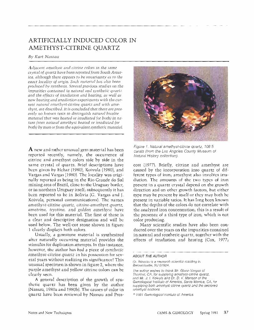
ARTIFICIALLY INDUCED COLOR IN AMETHYST-CITRINE QUARTZ B y Kurt Nassau
Adjacent amethyst and cltrine colors in the same crystal of quartz have been reported from So~rrh Arner- ica, although there appears to be uncertainty as to the exact locality of origin. Such material has also been prodr~ced by synthesis. Several previous studies on the imp~~ri t ies contained in natural and synthetic quartz and the effects of irrndiation and heating, as well as new heating and Irradiation experiments with the cur- rent natural amethyst-citrine quartz and with ame- thyst, are described. It is concluded that there are pres- ently no lznown tests to distinguish natural blcolor material that wns heated or irrudiated (or both) in nn- ture from natural amethyst heated or irradiated (or both) by man or from the ecluivaient synthetic material.
A new and rather unusual gem material has been reported recently, namely, the occurrence of citrine and amethyst colors side by side in the same crystal of quartz. Brief descriptions have been given by Hehar (1980), Koivula (1980), and Vargas and Vargas (1980). The locality was origi- nally reported as being in the Rio Grande do Sul mining area of Brazil, close to the Uruguay border, or in northern Uruguay itself; subsequently it has been reported to be in Bolivia (G. Vargas and J. Koivula, personal communications). The names amethyst-citrine quartz, citrine-amethyst quartz, ametrine, trystine, and golden amethyst have been used for this material. The first of these is a clear and descriptive designation and will be used below. The well-cut stone shown in figure 1 clearly displays both colors.
Usually, a gemstone material is synthesized after naturally occurring material provides the stimulus for duplication attempts. In this instance, however, the author has had a piece of synthetic amethyst-citrine quartz in his possession for sev- eral years without realizing its significance! This unusual specimen is shown in figure 2, where the purple amethyst and yellow citrine colors can be clearly seen.
A general description of the growth of syn- thetic quartz has been given by the author (Nassau, 1980a and 1980b). The causes of color in quartz have been reviewed by Nassau and Pres-
RE L."
Figure 1. Natural amethyst-citrine quartz, 108.5 carats (from the Los Angeles County Museum of Natural History collection).
cott (1977). Briefly, citrine and amethyst are caused by the incorporation into quartz of dif- ferent types of iron; amethyst also involves irra- diation. The amounts of the two types of iron present in a quartz crystal depend on the growth direction and on other growth factors, but either type may be present by itself or they may both be present i n variable ratios. It has long been lznown that the depths of the colors do not correlate with the analyzed iron concentration; this is a result of the presence of a third type of iron, which is not color producing.
Many scientific studies have also been con- ducted over the years on the impurities contained in natural and synthetic quartz, together with the effects of irradiation and heating (Cox, 1977;
ABOUT THE AUTHOR
Dr. Nassau is a research scienlist residing in Bernardsville, NJ 07924.
The author wishes to [hank Mr. Glenn Vargas of Thermal, CA, for supplying amelhyst-citrine quartz, and Mr. J. I. Koivula and Dr. D. V . Manson of the Gemological Institute of America, Sanla Monica, CA, for supplying both amethyst-citrine quartz and the sectored amethyst sections. :* 1987 Gemological Institute of America
Notes and New Techniques GEMS & GEMOLOGY Spring 1981 37

Figure 2. Synthetic amethyst-citrine quartz crystal, 3 cm long (author's collection).
Cohen and Hassan, 1974; Hassan, 1972; Cohen, 1975; Lehmann, 1975; and Nassau and Prescott, 1977 and 1978). These studies, as well as some current experiments, suggest that several possi- bilities exist as to the origin of any specific piece of amethyst-citrine quartz.
NATURAL AMETHYST-CITRINE QUARTZ In their reports, Hehar (1980), Vargas and Vargas (1980), and Koivula (1980) note six alternating regions, three each of amethyst and citrine, which clearly correspond to the major-rhombohedron growth regions of typical amethyst and the minor- rhombohedron growth regions of typical citrine. There are three significantly different ways in which such naturally occurring amethyst-citrine quartz could have been formed. First, it is possi- ble that the quartz simply grew under such con- ditions that the amethyst-forming iron was in- corporated under the major-rhombohedron faces, while only citrine-forming iron entered under the minor-rhombohedron faces (some may, of course, be present under the majors as well). Natural ir- radiation would then produce this end product.
There are, however, important alternative possibilities involving different mechanisms. In the second suggested mechanism, both ame- thyst-producing iron and citrine-producing iron are present in all sectors of the quartz, and the naturally occurring irradiation has produced an amethyst color in all sectors. According to the second scheme, the quartz next would have passed through a heating stage that bleached the minor-
rhombohedron amethyst to leave citrine, but which did not bleach the major-rhombohedron amethyst material. According to the third scheme, heating could have affected the material in such a way that a subsequent irradiation might have colored only some parts, thus producing the observed end product.
According to these alternative schemes, the important difference is not in the quantities of iron occupying the different sites, (although some such differences may be present], but in the behavior of the material with respect to heating. The determining factor is either the temperature at which the amethysts bleach (i.e., at which the color centers are destroyed) or the temperature of heat treatment after which the citrine can no longer be converted to amethyst. Such differences (e.g., in the bleaching temperatures) do occur in quartz (Nassau and Prescott, 1977), and sectored variation in properties has also been noted in conjunction with the pleochroic anisotropy in smoky quartz color centers (Nassau and Prescott, 19781 and in amethyst (Cox, 19771. There are two questions that arise should one of these schemes apply: (1) Was the heating or irradiation (or both) a naturally occurring event or was it performed by man! and (2) Is this property unique to the locality or will amethyst from other localities produce the same result if suitably treated!
HEATING AND IRRADIATION EXPERIMENTS Specimens of the natural amethyst-citrine cur- rently in the trade were made available to the au- thor by the very lzind cooperation of G. Vargas and by J. I. Koivula and D. V. Manson of the GIA.
Parts of two specimens were irradiated in a cobalt-60 gamma-ray cell to a dose of at least 1,000,000 rads. As a result, portions of some of the amethyst and citrine regions acquired an ad- ditional weak smoky tint. Heating to 350°C re- moved this tint and restored the specimens to their original state.
Parts of two different specimens were next heated in air in a furnace for two-hour periods first at 250°C and then at successively higher temperatures at intervals of 50°C. The amethyst sectors became yellow after the two hours at the 400°C step; the citrine regions were not affected by this heat treatment. A subsequent irradiation followed by heating to 350°C to remove smolzi- ness restored the original amethyst-citrine colors.
38 Notes and New Techniques GEMS & GEMOLOGY Spring 1981

In order to obtain comparison data, experi- ments were also performed on two specimens of sectored amethyst from Minas Gerais, Brazil, which were also obtained from the Gemological Institute of America. These specimens exhibited alternating sectors of dark (major rhombohedron)
I and pale (minor rhombohedron) amethyst. Dupli- cation of the first irradiation experiment above gave essentially the same results: smolzy purple and pale smoky purple, with the smolzy compo- nent removed and the original state restored upon heating to 350°C.
Heating the sectored amethyst to successively higher temperatures, however, produced an unex- pected result: the dark purple sectors became somewhat lighter but the pale purple sectors turned yellow at 400°C. To the eye, this material now had exactly the same appearance as the amethyst-citrine. Subsequent irradiation and heating to 350°C restored the original all- amethyst state. This process corresponds to the second mechanism discussed above, but the end product is unlike the current amethyst-citrine, which did not convert to all-amethyst even after 20 megarads of cobalt-60 gamma rays.
Heating of these same sectored-amethyst sec- tions to the slightly higher temperature of 450°C bleached all the amethyst sectors to yellow. A subsequent irradiation followed by heating to 350°C reproduced not only the appearance of the current amethyst-citrine, but also its behavior with respect to heating and irradiation: it could no longer be returned to all-amethyst. A spec- imen of this treatment-produced amethyst-citrine (see figure 3) has been returned to the Gemolog- ical Institute of America for archival purposes.
Iron analyses were not performed because such analyses cannot distinguish among the three types of iron but only give the total iron present; this total has not provided useful information in previous studies.
DISCUSSION The above experiments show that the amethyst- citrine combination in quartz can be obtained by heat-treating sectored amethyst from Minas Ger- ais, Brazil, in either one of two ways: (1) a medium heating step produces the change, but this can be reversed by irradiation; (2) a more intense heating followed by irradiation and a second, gentle heat- ing also produces the change, but the product can no longer be returned to all-amethyst.
Figure 3 . Amethyst-citrine quartz produced by heating and irradiating sectored amethyst from Brazil; 12 mm across.
Since the sectored amethysts tested were merely randomly chosen, it is lilzely that much such material from a variety of sources can be treated to produce amethyst-citrine, including man-made quartz. There are no lznown tests to distinguish the origin of color of natural inaterial that was heatedlirradiated in nature from natural amethyst heatedlirradiated by man or from the equivalent synthetic material.
REFERENCES Cohen AJ (1975) On the color centers of iron in amethyst and
synthetic quartz: a reply. American Mineralogist, Vol. 60, pp. 338-339.
Cohen AJ, Hassan F (1974) Ferrous and ferric ions in synthetic a-quartz and natural amethyst. American Mineri7logist. Vol. 59, pp. 719-728.
Cox RT (1977) Optical absorption of the d" ion Fe" in pleo- chroic amethyst quartz, lournal of Physics C: Solid State Physics, Vol. 10, pp. 4631-4643.
Hassan F 11972) Amethyst. Mineralogical Record, Vol. 3, pp. 221-225.
Hehar WC (1980) The discovery of golden amethyst. Lapidary ]ournal, Vol. 34, pp. 582-583.
Koivula J1 (1980) Citrine-amethyst quartz. Gems eJ Gemol- ogy, Vol. 16, pp. 290-293.
Lehmann G (1975) On the color centers of iron in amethyst and synthetic quartz: a discussion. American Mineralogist, Vol. 60, pp. 335-337.
Nassau K (1980aJ Gems Made by Mon. Chilton Book Co., Radnor, PA.
Nassau K [1980b) The history and present status of the syn- thetic quartz family. Lapidary 1ournal. Vol. 34, pp. 718- 726 and 956-963.
Nassau, 1<, Prescott BE (1977) Smoky, blue, greenish yellow, and other irradiation-related colors in quartz. Mineralogi- cal Magazine, Vol. 41, pp. 301-3 12.
Nassau K, Prescott BE (1978) Growth-induced radiation-de- veloped pleochroic anisotropy in smoky quartz. Americnn Mineralogist, Vol. 63, pp. 230-238.
Vargas G, Vargas M (1980) A new quartz gem material. Lop- idory ]ournal, Vol. 34, pp. 1504- 1506.
Notes and New Techniques GEMS & GEMOLOGY Spring 1981 39

LAB NOTES EDITOR
Chuck Fryer GIA, Sanla Monica
CONTRIBUTING EDITORS Robert Crowningshield
Gem Trade Laboratory, New York Karin N . Hurwit
Gem Trade Laboratory, Santa Monica Robert E . Kane
Gem Trade Laboralory, Los Angeles
A NOTE FROM THE EDITOR
This i s the first Gem Trade Lab Notes column under [he n e w for- mat of Gems & Gemology. Al- though the general style of the for- mer colrrmns will be retained, a few improvements have been made, in- cluding the use of more color photos.
Previously, there were separate columns for the New York, Los An- geles, and Santa Monica branch labs. It will be m y job to combine the information supplied b y each branch into one column. The New York col.respondent will continue to be Bob Crowningshield, vice presi- dent of GIA and director of the col- ored stone section of the Gem Trade Lab. Bob Kane will keep the datu from Los Angeles coming in, while Karin N. Hurwit supplies the i tems of interest from S ~ n t n Monica.
Perhaps the /nost important long- range improvement is the arrange- ment o f the material in alpha- betical order, b y variety. Such an arrangement makes i t easier to pro- vide an annual index of the stones coming into the labs and aids the reader in his or her search for more information on particular stones.
Please note that all magnifica- tions given refer to the power of the microscope at which the photo- graph was taken, not necessarily the final magnification of the printed image.
"1981 Gemological Institute of America
AQUAMARINE
Trapiche Aquamarine? An unusual stone came into the New Yorlz lab for identification. Figure 1 illustrates the green-blue melon-cut drop that appeared at first to be merely a drilled aquamarine bead. In reality, this stone has been cut from a trapiche-like crystal with six separations [apparently carbona- ceous) and a clear central hexagonal area, as shown in figure 2. Although the stone displayed evidence of chromium in its absorption spec- trum, its color was unlike that of any trapiche emerald crystals we have seen. However, the chalced- ony-lilze translucency and the black iilclusions seen in figure 2 do resem- Figure 2. End view of the ble characteristics common to tra- melon-cut aquamarine in piche emerald. figure 1. Magnified l o x .
Figure 1 . Melon-cut aqua- marine, approximately 35 cis.
DIAMONDS
Cyclotron-Treated Diamonds Diamonds were first treated in a cy- clotron to produce color nearly 40 years ago. A color then seen some- what frequently in treated dia- monds, but seldom seen today, is a pleasing orange-brown. However, cyclotron treatment, which imparts a shallow penetration of color, is an expensive and troublesome pro- cess. For these reasons, other meth- ods of artificially inducing color in diamonds have been developed, including atomic-pile treatment. Atomic-pile treatment penetrates the diamond completely, resulting
40 Lab Notes GEMS & GEMOLOGY Spring 198 1

Figure 3. Cyclotron-treated diamond. Left = crown facets viewed through the pavilion, magnified 4 0 ~ . Center = culet area viewed through the table, magnified 30x. Right = culet area and brown graining, magnified 12 X.
in even color throughout the stone. This process has largely replaced cyclotron treatment, and we seldom see cyclotron-treated diamonds in the laboratory today.
Several months ago, however, a cyclotron-treated diamond was sub- mitted to the Los Angeles lab for determination of the origin of color. Since the color penetration was shallow, this particular diamond did not exhibit the usual absorption spectrum with the typical 5920 A line.
The fact that the color is shallow and confined to a layer near thesur- face malzes cyclotron treatment eas- ily detectable when the stone is ex- amined under magnification. As seen in the three sections of figure 3, this diamond displayed a surface layer of intense color paralleling the edges of facets on the crown and around the culet that is very typical of cyclo- tron-treatment. The first photo in figure 3 shows crown facets viewed from the pavilion. The area around the culet is seen in figure 3 center as viewed through the table. The fi- nal pose in figure 3 shows, at lower magnification, the treatment-layer zone at the culet, as well as brown graining.
It should be noted that natural- color diamonds frequently display brown graining. Light brown dia- monds with brown graining are sometimes treated to darken their color and thus improve their ap- pearance; the graining, of course, is still present.
A dark blue-green diamond ex- amined in both California and New
Yorlz appeared watery when viewed from the side, which suggested that it was surface treated. However, it did not show the umbrella around the culet or dark penetration zone around the table and star facets which are typical of such treatment. Finally, with immersion and later in air we were able to determine that the stone was treated on one side of the pavilion only, where a grid-like penetration zone appeared when the diamond was viewed through the upper girdle and bezel facets [see fig- ure 4). We have heard that in the early days of cyclotron treatment if a stone fell from its fire-resistant holder, operating procedures did not allow stopping the equipment to right it. It is rumored that just such an accident led to the discovery that overexposed dark green stones could be heated to turn them brown, orange, or yellow.
Figure 4. Grid-like color zoning in a cyclotron-treated diamond. Magnified 4 0 ~ .
Figure 5. Radiation stains on a natural of a near-colorless diamond (see arrows). Magnified 45 X.
Natural Radiation Stains Radiation stains on diamond natu- rals are often associated with green stones, but they can occur on dia- monds of any color. In the Fall 1979 issue of Gems d Gemology, we mentioned a G-color round brilliant with radiation stains on both of the two naturals. Since that time, we have seen several diamonds, not green, that had this feature: Figure 5 shows a near-colorless diamond with two such small brown stains on a natural. Another diamond, of M to N range in color, was also ob- served to have a stain on a natural. Directly across from the natural
Lab Notes GEMS & GEMOLOGY Spring 1981 41

Figure 6. liadiation stain i n a diamond natural. Magnified 50 x.
there was a stain remaining in the bruted girdle where the rough dia- mond slzin had been removed. The stain in the natural is intense in color and penetrates deep into the stone (see figure 6).
Inclusions The cover of the Spring 1952 issue of Gems d Genlology featured a windowed diamond crystal with a beautiful flower-like iilclusion that shows both petals and sepals. This year we were shown a similarly
I LL- Figure 7. Flower-like inclusion in a diamond. Magnified 15 X.
Figure 9. A uniquely glassy diamond natural. Magnifier1 35 X.
windowed crystal with a flower-lilze of this natural was very smooth and inclusion that has a brown tubule glass-like (see figure 9) , almost as if which looks ever so much lilze a i t had melted! There was absolutely stem or the stick for a pinwheel (see no evidence of any growth or etch figure 7). marlzs on the surface of the natural.
We are at a loss to explain the No one in the Gem Trade Lab re- unusual inclusions in the round- members ever before seeing a dia- brilliant cut diamond shown in fig- mond natural with this appearance. ure 8. On examination under the microscope, they appeared to be Notes on Diamond Cutting hollow tubules, some brown or filled We admire the cutter of the orange with a brown material. cube diamond seen recently in New
Yorlz (see figure 10). Gem-quality An Unusual Natural cube-shaped diamond crystals are
Figrrre 8. Irregular trrbe-like The unique natural on a dia~nond almost unlznown, so in retaining the inclusions i n ci' diamond. brought into the Santa Monica lab cube shape with very symmetrical Magnified 18 X. warrants a description. The surface beveling of the edges the cutter has
42 Lab Notes GEMS W GEMOLOGY Spring 1981

Figure 10. Cube-cut diamond, 1.13 cts.
captured the stone's beauty and rarity.
Rare outside museums or royal collections are Peruzzi-cut dia- monds. We were privileged to re- ceive for grading and photographing such a stone weighing more than 2.00 cts. (see figure 11). Presumably this style of cutting, detailed in fig- ure 12, was first employed by a Venetian cutter in the 17th century, and it led to the old-mine cut, the old-European cut, and finally the modern brilliant. The stone we graded had no culet, had a table per- centage of 46, and showed a depth percentage of 97.9.
11, or Not 11, Type I diamonds are opaque to short- wave ultraviolet (UV) light, while type I1 are transparent. Type 11, dia- monds are not electroconductive, but type 11, are. Normally we check for
I Peruzzi cut.
a type I1 diamond in the darltroom, using short-wave ultraviolet light and a piece of scheelite placed be- hind a porthole. Only UV light that has passed through the stone can affect the scheelite. If the scheelite glows, we test the stone further for electrocoi~ductivity to determine whether it is type 11,. Of course, there is no permanent record of these UV transparency tests. To obtain a permanent record, place the stones table down on the emulsion side of photographic paper and expose them for a very short time (two seconds)
Figure 13. Ultraviolet transparency of a type I marquise con~pared t o a type I1 pear-shaped diamond.
Figure I I . Top v i e w of a Peruzzi-cut diamond, 2.00 cts.
Figure 12. Diagram of t he Peruzzi cut. (Diagram courtesy o f Robert A . P. Gaul, Diamond Dictionary, 2nd ed., Gemological Institute of America, Santa Monica, C A , 1977.)
with the ultraviolet lamp perhaps a foot away. Figure 13 shows the transparency difference between a type I yellowish marquise and a type II,, pear-shaped diamond.
EMERALD
An emerald sent to Santa Monica for identification showed quite unusual stn~ctural characteristics. First, we were surprised to see a significant difference in transparency when the stone was viewed from different an- gles. As we loolzed at the stone through the table, the emerald was almost transparent; yet when we looked sideways at the pavilion fac- ets, the stone was oilly translucent, having the appearance of dyed green chalcedony. Magnification of this stone revealed a peculiar structure: straight, colored growth lines were crossed by lightly curved, wave-like bands (see figure 14). The stone was of natural origin, possibly cut from a trapiche-type crystal.
Lab Notes GEMS & GEMOLOGY Spring 1981 43

Figure 14. Unusual growth lines in'an emerald. Magnified 63 x.
The New York lab encountered a synthetic emerald, reportedly of Russian manufacture, with a refrac- tive index of 1.560-1.563 and a low specific gravity. The stone trans- mitted red on the spectroscope light but showed only weak long-wave ultraviolet fluorescence. The stone had fairly typical flux fingerprints and even contained a platinum crys- tal in the girdle. What was partic- ularly unusual was the presence of long needles, which we have always felt indicated natural origin. Nearly as disturbing was the discovery of a cavity filled with yellow liquid in this otherwise obviously synthetic emerald (see figure 15). Elsewhere in the stone, the "Venetian blind" banding that is typical of a synthetic was visible (fig. 16).
.-
MALACHITE
Figure 15. Yellow inclusion in a synthetic emerald. Magnified 25 X.
Figure 16. "Venetion blind" banding in synthetic emerald. Mognified l o x .
ably like a cleavage that astonished us (see figure 17). Although mala- chite has one perfect cleavage direc- tion, massive material usually does not show it. What had h a ~ ~ e n e d ? -. This particular piece of malachite apparently was formed by the pro- cess of sedimentation. When the carving was damaged, the separation
occurred between bedding planes in the stone and produced a flat, smooth break.
PEARLS, Cultured
The Sailta Monica lab had the op- portunity to examine a necltlace
hi. . \ -.\ -
A malachite carving of an oriental 1 lady holding a flower basket was sent to the laboratory for a damage re- port. Part of the flower basltet and the right hand of the lady had been Figure 17. Damaged malachite carving showing separation broken off, leaving a surface remark- between bedding planes; 2.5 cm x 7.5 cm x 10.5 cm high.
44 Lab Notes GEMS & GEMOLOGY Spring 198 1

consisting of bluish-gray cultured pearls with one "oddball" in the strand. The layered structure of shell was apparent in this single bead (see figure 18). Notice also the concen- tration of dye at the drill hole. It would seem that at one time this pearl was crushed or received a blow that caused the nacreous outer layer to fall off, exposing the nucleus.
A "faith-shaker" was a pair of earrings set with a bronzy-black, drop-shaped pearl and a similarly shaped white pearl. Both were cul- tured. However, the blaclt pearl fluoresced reddish brown-much stronger than any ltnown Tahiti blaclt cultured pearl we have seen. The history of the earrings pre- cluded the possibility that the black pearl could have been cultured in Tahiti waters. Incidentally, it is re- ported that a inuch wider range of hues has been produced in the cur- rent harvest there, so we may see colors other than the usual black, dark gray, and light gray.
Figure 19 illustrates a neclzlace set with bronzy-colored dyed cul- tured pearls. The shapes too are unusual.
Figure 19. Neclzlace of unrlsual bronze-colored dyed cultured pearls.
Figure 18. Shell nucleus of a cultured pearl. Magnified 1 0 ~ .
RUBY 7 Synthetic stones examined in the New Yorlz laboratory recently have been cause for alarm. Thc first was 1 a flux-grown synthetic ruby report- edly of recent Chatham manufac- ture. It weighed more than 5 cts. and had fine flux fingerprints fairly well distributed with only one area of typical coarse flux. The alarming characteristic was angular, silk-like I - ,
banding visible to the unaided eye (see figure 20). lndividual needles could not be discerned: rather. we observed precisely the effect we have seen when a Burma star ruby has been heat treated and the rutile needles are not quite absorbed. Mounted, it could readily be mis- identified, since the silk-lilze bands are not expected in synthetic stones. The stone was highly fluorescent and was obviously not cut in the Orient. Whether this stone indicates a change in the Chatham product or a one-time "flulte" has not been established.
We have mentioned previously the process whereby ordinary syn- thetic blue sapphires are heated in crucibles with an unknown liquid that is then incorporated within in-
Figure 20. Angular banding in a flux-grown synthetic rrrby. Magnified 30 X.
duced fractures to produce very believable fingerprint inclusions. It was therefore more expected than alarming to receive for identifica- tion a synthetic Verneuil ruby showing both curved striae and "fin- gerprints" (see figure 2 1 ).
Lab Notes GEMS & GEMOLOGY Spring 198 1 45

Under short-wave ultraviolet light, most of the stones treated by induced surface coloration show a patchy fluorescence where the dif- fused color was removed when re- polished. However, a stone sent to New York by the Los Angeles lab did not fluoresce. When the stone was immersed, all facets showed - -- L 1 color except the girdle, which had Figurb G ~ . Star spinels. been repolished and all diffused color Magnifietl l o x . removed (see figure 22).
Figrlre 21. Curved striae and "fingerprint" inclusions in synthetic ruby. Magnified 3 0 ~ .
I SAPPHIRES
Induced Surface Coloration of Natural Sapphires The shortage of Sri Lanlzan sapphire rough suitable for heat treatment in Bangkok has evjdently led to the use of cut stones that are heated after packing in chemicals to diffuse color into the surface. The method re- portedly is a variation of the one outlined in U.S. Patent 3,897,529, issued to Union Carbide Corp. in 1975. We first encountered such stones a year ago in the form of an orange sapphire, reported in the Fall 1979 issue of Gems d Gemology. Since then we have seen a number of parcels containing variously col- ored treated stones.
I Figure 22. Loss of color i n the
repolished girdle of a color- treated sapphire. Magnified l o x .
SPINEL
It is not often that one has the op- portunity to see a star spinel. Imag- ine our surprise when two of them arrived at the Santa Monica lab at the same time. We were especially pleased when it was apparent that one of the stones showed a four-ray star, while the other star had six legs (see figure 23).
ACKNOWLEDGMENTS
The photographs in figures 1, 2, 4, 8, 13, and 21 were taken by Bob Crowningshield in New York. A1 Roditi, also in New York, took the photographs in figures 7, 10. 11, 15,76, 79, 20, and 22. Shane McClure of the Gem Trade Lab in Los Angeles did the photography for figure 5 , while Bob Kane was responsible for the photos used in fig- ures 3 and 6. The specimens examined in the Santa Monica lab were photographed by John Koivula (figures 9, 14, and 78), Mike Havstad (figure 17), and Chuck Fryer (figure 23).
46 Lab Notes GEMS & GEMOLOGY Spring 1981

GEMOLOGICAL ABSTRACTS
Dona M . Dirlam, Editor
REVIEW BOARD
Stephanie L. Dillon GIA, Santa Monica Bob F . Effler GIA, Santa Monica Caroline K. Goldberg GIA, Santa Monica Joseph P. Graf Gem Trade Lab, Inc., Santa Monica Gary S. Hill GIA, Sanla Monica Jill M. Hobbs GIA, Sanla Monica
Karin N. Hurwit Gem Trade Lab, Inc., Santa Monica John I . Koivula GIA, Santa Monica Noel P. Krieger GIA, Santa Monica Shane F. McClure Gem Trade Lab, Inc., Los Angeles Elise 6. Misiorowski Gem Trade Lab, Inc., Santa Monica Gary A. Roskin Gem Trade Lab, Inc., Los Angeles Michael L. Ross GIA, Santa Monica
Richard J. Saindon GIA, New York Andrea L. Saito GIA, Santa Monica Peter C. Schneirla GIA. New York Frances Smith Gem Trade Lab, Inc., Los Angeles R. Stephen Smith GIA, Sanla Monica Carol M. Stockton GIA, Santa Monica Evelyn Tucker GIA, Santa Monica
COLORED STONES A N D ORGANIC MATERIALS Additional and corraborative data on violet gem sca-
polite probably from Eastern Africa. B. Jackson, Iournal of Gemmology, Vol. 17, No. 4, 1980, pp. 235-238.
The author describes a violet gem scapolite, probably from Kenya, that has unusually low refractive indices (1.536-1.541) and density (specific gravity of 2.65), and has the highest sodium content recorded thus far in natural scapolite. All the samples exhibited strong di- chroism: violet when viewed parallel to the c-axis and colorless when viewed perpendicular to the c-axis. This
orientation may be verified if the stone contains straight, tube-like inclusions that align themselves parallel to the c-axis. All the stones examined contained these in- clusions, which sometimes broaden out, resembling cleavages. Given the similarities in color and in optical and physical properties, this material may be confused with amethyst. The two stones can be distinguished, however, on the basis of optic sign (scapolite, negativc vs. amethyst, positive). RSS
The alexandrite effect in minerals: chrysoberyl, garnet, corundum, fluorite. K. Schmetzer, H . Bank, E. Gubelin, Neues lahrbuch fur Mineralogie Abhundlangen, Vol. 138, No. 2, 1980, pp. 147-164.
As Schmetzer, Bank, and Gubelin state in the abstract,
This section is designed to provide as complete a record as possible of the recent literature on gems and gemology. Articles are selected for abstracting solely at the discretion of the section editor and her reviewers, and space limitations may require that we include only those articles that will be of greatest interest to our readership.
Inquiries lor reprints of art~cles abstracted must be addressed lo the author or publisher of the original material.
The reviewer of each article is identified by his or her initials at the end of each abstract. Guest reviewers are identified by their full names.
5)1981 Gemolog~cal Institute of Amerlca
the purpose o'f thei; study is to determine the condi: tions required for change of color in various minerals. Following a helpful review of the literature on color- change materials, the experimental details of the ab- sorption spectra and chemical analyses are presented in this comparison of the spectroscopy and chemistry of alexandrite and alexandrite-like minerals.
Most of the paper is devoted to analyzing the results of these experiments for alexandrite and those garnets, corundums, and fluorites that exhibit change of color. Specifically, the authors discuss how the elements present and the location of ions in the crystal structure relate to the absorption spectra and therefore to color.
Gemological Abstracts GEMS 8: GEMOLOGY Spring 1981 47

Lastly, the authors hypothesize from their investi- gation that four features are characteristic of the ab- sorption spectra of color-change minerals. The first is spectral transparency from 15,000 cm-' to 16,000 cm-' (red area]. The second is a strong absorption maximum between 17,300 cm-' and 17,800 cm-' (yellow area). Then another absorption maximum exists between 19,700 cm-' and 21,000 cm-' (green to blue area). Fi- nally, there is a continuous increase in absorption from 21,000 cm-' to the limit of the visible area with- out additional minima. The authors note further that crystals green in daylight generally change to red in artificial light while bluish-green crystals change to reddish-violet. Figures and tables of the spectral and colorimetric data are included as well as a substantial list of references. DMD
Alexandrite-like natural spinel from Sri Lanka. K. Schmetzer and E. Giibelin, Neues luhrbuch f ir Mineralogie Monatshefte, Vol. 9, 1980, pp. 428- 432.
In a continuation of their articles on alexandrite-lilze minerals, Schmetzer and Giibelin report on the inves- tigation of a 1.87-carat spinel that shows a "remarkable change of colour from violet in daylight to reddish vi- olet in artificial light." Through an analytic and spec- troscopic investigation, the authors hoped to verify the four features they have postulated as being character- istic of alexandrite-lilze materials: transmission in the red area of the spectrum, an absorption maximum in the yellow region, transmission in the blue-green, and an increase in absorption from the blue to the ultra- violet end of the spectrum.
In addition to the normal amounts of Alto3 (alu- minum oxide) and MgO (magnesium oxide), the micro- probe analysis indicated the presence of FeO (iron oxide), Cr20, (chromic oxide), and V,03 (vanadium trioxide]. The position of the maxima and minima bands in this spinel are different from a typical blue spinel. The authors suggest that the overlapping of the absorption bands of Fe+' (iron) by the spectra of Cr+"chromium) and V+"vanadium) produced this displacement.
A diagram is also given which plots chromacity co- ordinates for daylight and artificial light. The authors conclude that this change-of-color spinel does exhibit the four common spectral features. DMD
Amber-the New World's fossil gold. G. 0 . Poinar, Jr., and F. Agudelo, Amtricas, Vol. 32, No. 10, 1980, pp. 31-41.
After a brief introduction that mentions the classic am- ber locality of the Baltic Coast and brushes on amber's ancient uses and lore, this article discusses in some detail the fossil resin deposits found in the Americas. It focuses on the amber deposits of the Dominican Ke- public and Chiapas, Mexico, with additional informa-
tion on the Canadian amber found at Cedar Lalze in Manitoba and the Alaskan amber found along the Ketic River near Point Barrow.
The view that the source of all amber was resin from a particular species of pine is disputed in a short section on the botanical origins of amber. With the use of in- frared spectrophotometry, the spectra of amber from various localities were compared with those of modern resins. The authors point out the information that may be gained from the insects and other arthropods, as well as the microorganisms such as nematodes and fungi, that have been discovered in amber. The article con- cludes with a warning to be on guard against amber substitutes. IIK
The causes of color. K. Nassau, Scientific American, Vol. 243, No. 4, 1 980, pp. 124- 154.
In this article, Kurt Nassau gives a rather compre- hensive summary of five general color-causing me- chanisms that he has subdivided into 14 specific categories. Within this framework, he discusses a wide variety of color phenomena, ranging from the vibrant red of a ruby to the multi-hued iridescence of a com- mon oil slick. The interaction of light with electrons is the unifying theme behind almost all causes of color.
The opening sections of the article relate the energy hierarchy of light to the absorption and emission of ra- diation by the atomic electrons. Nassau's treatment of the crystal field, molecular orbital, band structure, and physical optical theories contains many interesting ex- amples from the gem and mineral lzingdoms. Several large diagrams, plus a few color photographs, supple- ment the verbal explanations.
Some of the parameters, such as electron volts and the spectroscopic notations, will seem bewildering to the nonscientist, but a full understanding of those de- tails is not necessary for grasping the main ideas. Any- one interested in an in-depth treatment of the origins of color should not miss this excellent article. BFE
Connoisseurship in emeralds. B. Zuclzer, Connoisseur, Vol. 204, NO. 819, 1980, pp. 52-58.
Benjamin Zuclzer begins with a short summary of the history and lore of emeralds, from their limited use by the Egyptians and Komans to the discovery by the Spanish of the vast resources held by the Incas in South America. He then comments on the rarity of emeralds, which he feels is related to their unique chemical com- position. According to the author, the finest color of emerald is "old-mine" green, a term that is supposedly derived from the color of the stones that were first sent back to Europe by the conquistadors. It is a color he describes as a "deep, intense, velvety, almost blackish shade of green."
The color, commercial potential, and characteristic inclusions for emeralds from the major world sources are presented. According to Mr. Zuclzer, no source cur-
48 Gemological Abstracts GEMS & GEMOLOGY Spring 198 1

rently compares with Colombia. Sandawaila emeralds (Zimbabwe] are often small and have a less intense green, Zambian stones are blaclzish and often highly included, while Brazilian stones are frequently craclted and oiled. Twenty-one colored photographs present typical emeralds from the various localities as well as illustrate inclusions found in natural and synthetic stones.
Although this article is not complete from a gem- ologist's point of view, it is a very good introduction to emeralds and could be useful to the gem salesperson. RSS
Estudio del material de inter& gemologico 'angel stone' (A study of a material of gemological interest-an- gel stone). J. M. NoguCs Carulla, C. de la Fuente Cullell, L. M. Roberdeau, and J. M. Bosch Figueroa, Gemologia, Vol. 1 1, No. 4 1 and 42, 1979, pp. 17- 29.
"Angel stone" has been considered attractive and du- rable enough for use in jewelry. The authors report on a gemological study of the material. Angel stone is microcrystalline, opaque, and pale pink. Its major con- stituent is the mineral palygorslzite, impregnated by amorphous silica. The pink hue derives from man- ganese in the palygorslzite (which can also be grayish, yellowish, or whitish). The sample tested was crypto- crystalline, with no directions or fracture planes. There were no cleavage surfaces or splinters; the fracture was conchoidal.
The study included X-ray diffraction, X-ray fluores- cence, thermal analysis, examination under the elec- tron microscope, and hydrostatic weighing. Hardness tests were performed with a photon~icroscope. It was concluded that the impregnation of the palygorslzite added appreciably to the hardness of angel stone (4.5, as contrasted to palygorskite's 2.0-2.5) as well as to its compaction, rendering it usable as gem material.
For further information on palygorslzite, see Christ et al., "Palygorslzite: New X-ray Data," The American Mineralogist, Vol. 54, Nos. 1 and 2, 1969, pp. 198- 205. SLD
Explanations of the absorption spectra of natural and synthetic Fe- and Ti-containing corundums. K. Schmetzer and H. Bank, Neues Iahrbuch fur Mi- neralogie A bhundlongen, Vol. 139, No. 2, 1980, pp. 2 16-225.
Schn~etzer and Banlt examined the effects of heat treat- ment on the absorption spectra of 154 natural sapphire crystals from corundum localities in 10 different coun- tries. Using three different spectrophotometers to mea- sure absorption spectra, the authors studied the alter- ations of the absorption spectra and then drew conclusions about the mechanisms and valence of the color-causing transition ions.
While all sapphires show certain absorption bands, they can be divided into two groups based on their
infra-red spectra ant1 behavior following heat treat- ment, which reflects initial growing conditions. Three figures and four tables summarize the results for the two groups before and after heating. The extensive bib- liography will also be helpful to the reader. DMD
Green opal from Gabanintha Western Australia. R. A. Ball, Australian Journal o/ Gemmology, Vol. 1, NO. 3, 1980, pp. 5-7.
Ball reports the occurrence of bright "emerald" green opal at Gabanintha, the principal copper center of the Murchison Goldfield. Noting that the opal is found in quartz associated with copper minerals, Ball suggests that the opal results when olivine that has been con- verted to serpentine is altered to magnesite and opal through the agency of carbonated water. He concludes that the green color results when opal absorbs copper.
Five photographs are included in the article. It is difficult to appreciate the two hand-specimen photo- graphs because they are in black and white, not color. Ball used three scanning electron micrographs to sug- gest that the opal is actually in a transition stage between opal and agate, as evidenced by the "distinct agate type banding" shown in the photomicrographs. In the summary, he cites Cassedanne and Cassedanne's (1975) account of the occurrence of green opal in Brazil and questions whether a parallel exists between the two, iron causing the color in Brazil and copper causing the color in Australia. DMD
Irradiation-induced colors in gemstones. K. Nassau, Lopidary lournal, Vol. 34, No. 8, 1980, pp. 1688- 1 706.
Dr. Nassau begins by citing the increased availability of irradiation facilities as the primary reason for the trend in artificial color change of this type. He then provides a thorough and understandable explanation of the sources of irradiation, including visible light, ultra- violet light, X-rays, and gamma rays. The discussion also details where each source of irradiation is to be found and how samples are treated. In general, the low- energy rays [X-rays) and those particles that are nega- tively and positively charged penetrate poorly and sub- sequently color only the surface layer. More active gamma rays and neutrons penetrate very well, produc- ing uniform coloration. As long as the irradiation used is energetic enough to cause the intended color change, the nature of the source is unimportant. A table sum- marizing the types of irradiation used to effect color change is supplied.
A color change induced by irradiation is, in most cases, due to the formation of a color center. Once a color center has been bleached, it may usually be re- turned to its original hue by further irradiation. If, however, the sample was overheated during bleaching, re-creation of the irradiated color is impossible. For ex- ample, amethyst that has been heated to citrine may be returned by irradiation to i ts original color provided
Gemological Abstracts GEMS & GEMOLOGY Spring 198 1 49

the temperature used has not been too great. The sta- bility of a color center to light exposure-i.e., the re- sistance to fading-is the basis for three groups of irradiation-produced color effects. A fourth irradiation effect, one that does not involve color centers, is also described. The irradiation and bleaching behavior for topaz, tourmaline, beryl, kunzite, and quartz is sum- marized in a second table.
The article represents a well-organized, up-to-date work on irradiation sources, color-center stability, and how irradiation may be identified. It incorporates sev- eral aspects of the large body of research on color and treatment already credited to Dr. Nassau. PCS
Ivory-popular but prone to many imitations. G. Brown, Ieweller Watchmalzer d Giftware, Vol. 65, No. 1, 1980, pp. 22-23.
Australia has not yet invoiced trade embargoes on carved ivory, which is very popular there. Its popularity has naturally resulted in sometimes successful efforts to unload imitations on retail jewelers; this article tells how to detect such imitations by visual inspection, hot point, and microscopic examination of thin peelings. Eleven photographs serve to clarify the text. These per- mit comparison of the "engine-turned" effect of ele- phant ivory with the surfaces of other animal ivories as well as with those of vegetable ivory and plastic.
FS
Letter from Europe. B. Sullivan, Minerologicrrl Record, Vol. 11, No. 5, 1980, p. 315.
Nicely formed crystals of pale blue aquamarine are de- scribed as coming from near the town of Kunar in the - province of Kunar in northeast Afghanistan. The crys- tals range from one to five inches long and are ex- tremely gemmy, as illustrated by the photograph included, yielding 60%-70% facet rough. Sullivan reports on two interesting examples: one is nearly five inches long and has a three-fourths inch morganite crvstal attached at the base; the other has a movable water bubble rarely seen in aquamarine. The mine is now reported to be inactive because of the present po- litical situation in Afghanistan. SFM
Notes on the properties and inclusions of garnets from Lapland, Finland. S. Hornytzkyj and K. T. Korllo- nen, Iorlrnal o f Gemmology, Vol. 17, No. 3, 1980, pp. 153-164.
This article dcscribes the red, gem-quality garnet found in northern Finland. It begins with a discussion of the geology of the area where this mineral is found; the authors have included two full-page maps. The article also covers the physical and optical properties, chem- ical composition, and inclusions of these garnets. The section on inclusions is illustrated with 11 photographs of the most common inclusions seen in this material.
In all, 157 faceted garnets were studied. Two colors were dominant: brownish red and greyish rose. The
average weight was 0.57 ct. The refractive index of this material ranged from 1.764 to 1.792. The specific grav- ity ranged from 3.92 to 4.17. The authors concluded that the six types of crystal inclusions noted in these garnets were probably rutile, zircon, monazite, actin- olite, apatite, and ilmenite. ET
Nuggets of island sun. H. E. Rice and P. C. Rice, Americas, Vol. 32, No. 10, 1980, pp. 37-41.
This short article offers a brief overview of the budding amber industry in the Dominican Republic. It describes the shops where amber can be purchased and mentions the methods used by local craftsmen to fashion raw amber into polished gems and jewelry. The authors continue with a general description of some types of amber found in the Dominican Republic aild of mining methods used to recover the amber. It i s interesting to note that although the Dominican Republic deposits are considered to be recent discoveries, large-scale min- ing was begun in 1949 and the first school for polishing amber was established early in the 1950s.
The article concludes with a summary of how am- ber was formed and some mention of its inclusions.
lIK
Royal lavulite discovered. B. Jones, Roc/< ed Gem, Vol. 11, NO. 1, 1981, pp. 56-59.
"Koyal lavulite" is described as a "totally new gem- stone that looks like a grape popsicle." More compact than the purple charoite from Russia, lavulite has deeper, richer purple tones and is capable of taking an almost mirror-like uolish.
Its exact chemical composition is not yet known, but it is reported to be the mineral leucophoenicite, which has been found in reddish to uink masses at Franklin, New Jersey, and in other metamorphic man- ganese deposits. The name royal lavulite was coined to describe i ts "superb lavender" color; however, this term may not be in general use.
Lavulite comes from Africa, where it was found dur- ing operations at a manganese mine. Unfortunately, because the mining there is done by blasting, much of the lavulite is cracked and broken into smaller pieces. These smaller pieces, however, are oftcn completely solid and uu to two inches across. The author describes this material as a solid, opaque mixture of at least two minerals: (1) a very dense black inaterial and (2) the royal lavulite itself. A second type of lavulite occurs as lightly banded masses, similar to malachite.
Lavulite is compact, easy to handle, and has a hard- ness of about 6 on Mohs scale. It is easily worked with diamond or Carborundum wheels and takes a brilliant polish. ET
Tourmaline-liddicoatite. J. Wentling, Lapidary lour- ual, Vol. 34, No. 9, 1980, p. 1918.
Jerry Wentling's enthusiasm for tourmaline led him to a type that is currently found only in certain areas of
50 Gemological Abstracts - GEMS h GEMOLOGY Spring 1981

Madagascar. Named liddicoatite after GIA's well-ltnown president, this type of tourmaline is found to have an unusually high calcium content. Wentling discusses briefly the locations where liddicoatite has been found, and describes the unusual color zoning that malzes this type of tourmaline fascinating. NPK
DIAMONDS
Battle lines form on lasering disclosure. H. Huffer, /ew- elers' Circnlar-IZeystone, Vol. 151, No. 10, 1980, pp. 42-44
Associate Editor Helene Huffer reports on jewelry in- dustry reactions to the most recent position talten by the Jeweler's Vigilance Committee (JVC] on disclosure of laser drilling in diamonds. As proposed in the "Dia- mond and Gemstone" sections prepared for the Federal Trade Con~mission as part of the revised Trade Practice Guidelines, lasering need not be disclosed. JVC Dia- mond Committee Cochairman Leo Kaplan stated that because Indians and Israelis are now lasering melee, i t would be "too cumbersome" to check each stone.
Huffer interviewed other important leaders in the jewelry industry, including Sydney DeYoung of J.&S.S. DeYoung, GIA1s G. Robert Crowningshield, and Robert Mason of J. C. Keppie Co. The impact on the diamond's worth is a ltey issue. On the one hand, lasering often improves the clarity of a stone, increasing its value hundreds of dollars while the lasering itself costs only $25 to $35; on the other hand, the fact that a diamond has been lasered could make the stone undesirable to some and in most cases reduces its value about 10% from that of a similar stone that hasn't been lasered. Is i t ethical not to tell the buyer a stone has been la- sered? The controversy over this issue will continue.
RlS Editor's Note: The December 1980 issue of ICK
reported that the jeweler's Vigilance Committee has reversed its position, and now recommends that laser- ing be disclosed.
The diamond market: a new concentration of power. M. Gilbert, lewelers' Circular-Keystone, Vol. 151, NO. 9, 1980, pp. 79-90.
Industry Projects Editor Mitchell Gilbert describes in detail new efforts by DeBeers to concentrate power in the diamond marlzet. Every member of the jewelry trade will be affected by the dramatic changes occur- ring at all levels, from sales and distribution to mining.
A review of the reasons why the cost of rough dia- monds is escalating gives insight into the five ways DeBeers obtains 85% of the world's rough production. Three ltey considerations are upgrading living stan- dards for its black African worlters, prolonging the life of its mines, and establishing policies that enable it to
do business with any government regardless of the pol- itics of that government.
However, the buying of diamonds for investment purposes and the selling practices of the Soviet Union (which accounts for 20% of world production] are caus- ing DeBeers to take additional steps to "restore order to the diamond world." To circumvent the impact that investor disenchantment could have on marlzet stabil- ity, DeBeers is striving for greater concentration of power, which clearly includes cutting and polishing as well as advertising and sales. Technological develop- ments supported by DeBeers in sawing, cleaving, brut- ing, and polishing have generated the greatest amount of negative response from some of the traditional dia- mond leaders. Similarly, they question the direction DeBeers is talzing through its subsidaries and "coop- erative, efficient companies" in the distribution and sale of polished diamonds. Clearly, the greatest impact of this will be on the smaller business people, whether cutters, middle-level dealers, or independent jewelers.
X I S
Some very important but little known historic dia- monds. E. B. Tiffany, Zeitschrift der Deulschen Gemmologischen Gesellschuft, Vol. 29, No. 1 and 2, 1980, pp. 71-78.
Mr. Tiffany briefly outlines several of history's most famous diamonds, and compares them with some ob- scure yet noteworthy stones. Citing diamonds from such places as Iran, the Soviet Union, and India, Tiffany suggests that many of these "unltnowns" have been hidden away in private collections or discovered too recently to gain public notoriety. Sixteen pictures of stones from the lranian Crown Jewels and the Soviet Diamond Fund are included together with descriptions of their color and weight. NPK
A visit to a diamond mine, I and 11. H. Hurst, Modern leweler, Vol. 79, No. 9, 1980, pp. 10-59 and No. 10, 1980, pp. 16-48.
For those who have never visited an actual diamond mine, this brief but interesting "tour" in Kimberley, South Africa, malzes enjoyable reading. The color pho- tos included in the article give one a glimpse of the modern technology now used in these operations, somewhat resembling the mining of coal, copper, or limestone. Hurst also describes the Pan Plant, where the crushed blueground is processed. It is noteworthy that X-ray sorters, which use the natural fluorescence of diamonds, are part of the recovery. We are reminded of the mystique, wonders, and hopes of diamond min- ing by the story of how the finder of a 616-carat dia- mond received $20,000 plus a new house from DeBeers as a reward and incentive to find more like that one- it should happen to all of us! GR
Gemological Abstracts GEMS & GEMOLOGY Spring 198 1 5 1

GEM INSTRUMENTS AND TECHNIQUES
Application of multi-purpose spectrophotometer and X-ray fluorescence analysis to distinguish natural and synthetic alexandrites (in Japanese). N. Hori- uchi, lournal of the Gemmological Society of la- pan, Vol. 6, No. 3, 1979, pp. 77-80.
lnviting reader comments and contribution to his con- tinuing research, author Nobuo Horiuchi of the Central Gem Laboratory, Ueno, Taito-la, Tokyo, contends that with the emergence of sophisticated synthetics and new natural gem sources, the multipurpose spectro- photometer together with X-ray fluorescence analysis can offer a new means of distinguishing alexandrite (chrysoberyl) originating in different localities (Many- ara, Tanzania; Bahia, Brazil; Tokowaya, Russia) from its synthctic counterparts (flux alexandrite from both Kyoto Ceramic Co. and Creative Crystal Co.) Horiuchi does not comment extensivelv on K. Nassau's Czo- chralski alexandrite, although its properties are pre- sented in chart form along with those of the stones listed above. The author details the physical and opti- cal properties of the natural and the synthetic alexan- drites to facilitate comparison and to emphasize the need for a spectrophotometer and X-ray fluorescence analysis in identification.
With the multipurpose spectrophotometer, pat- terns between 400 and 700 n m are the same for the natural (Bahia and Manyara) alexandrite and the syn- thetics produced by Kyoto Ceramics and Creative Crystals. A graph illustrates the unique differences below 400 nm, however. Kyocera alexandrite has considerable absorption in this area, while Creative al- exandrite reaches a small peal< at 385 nm; 355 nm is i ts actual peak marked by a sharp decline. This absorp- tion pattern, similar to that found in the natural stone, results from the material's iron content. The unique absorption (at 685 nm, with a dale at 420 nm) in Ky- ocera alexandrite is due to i ts chrome content and the absence of iron.
In the X-ray fluorescence analysis, the stones on the chart were tested for chrome, iron, gallium, germa- nium, and platinum. Chrome was found in natural al- exandrite but not in large amounts in the synthetics. Iron was found in all but the Kyocera alexandrite. In the Creative Crystal stone, iron had a very high peak, approximately 1.5 times higher than in the natural. Platinum was found in the Kyocera product because of the crucible used in its creation. The procedures de- tected gallium only in the naturals and germanium only in the Russian natural material. A LS
Computer-aided gem identification. P. Read, Journal of Gemmology, Vol. 17, No. 4, 1980, pp. 239-249.
Peter Read has designed table-top computer programs for a CRT (TV-type) read-out to aid in gem identifica- tion. To facilitate use of this time-saver, Read has cho-
sen to program in tables of selected gemstones by as- cending refractive indices, by alphabetical listings, and by individual "gem profiles" which list major identi- fying characteristics of the gemstone. When informa- tion on an unknown is put in, all possibilities in the computer's memory within a given range are suggested.
The author also discusses other types of programs that can be modified to suit a particular need. These programs appear to be very easy to read and implement. Stating that the programs were initially used as teach- ing aides, Read also suggests that this computer can be a valuable sales tool for retailers. NPK
Etude par spectroscopie raman d'inclusions dans les diamants, saphirs et emeraudesll (Study of inclu- sions in diamonds, sapphires, and emeralds by Ra- man spectroscopy-part 1). M. L. Dele-Dubois, P. Dhamelincourt, and H. J. Schubnel, Revue de Gemmologie a.f.g., No. 63, 1980, pp. 11-14.
This is the first of a two-part article on the use of Ka- man spectroscopy, which permits the simultaneous identification of both the host material and its inclu- sions. Because i t is noninvasive and not destructive, this technique is well suited for studying gems with the exception of materials that are opaque to the wave- lengths of the excited photons.
With this technique, a stream of laser-generated photons penetrates the specimen, and a study of the Raman spectrum locates and identifies the different atomic constituents present therein. The beam is se- lected according to the inclusion in order to excite as little fluorescence as possible.
Examination of some South African diamonds iden- tified inclusions of chrome spinel, peridot, hematite, graphite, and pyrope. A violet sapphire from Colombia contained a calcite crystal and a zircon with a tiny apatite crystal. The text is clearly illustrated with graphs of the spectra and some inclusion photographs. An ex- cellent list of references follows the article. CKG
A phosphoroscope. R. M. Yu and D. Healey, lournal of Gemmology, Vol. 17, No. 4, 1980, pp. 250-258.
In this article, the authors explain the use of ultraviolet absorption in gem identification. The phosphoroscope allows direct observation without need of film, al- though film can be used if a permanent record is desired.
The specimen is placed in a simple wooden box atop a black cloth covering phosphorescent paper. An ultra- violet light (short wave) is directed on the stone and a mirror below the paper allows one to view the results through a port. In the section on observations, a rigid mathematical presentation is given to explain the cause of the dark rim seen around the edges of the stone. The type of film recommended and i ts use are briefly described.
Three figures are used to compare the results of test- ing synthetic and natural rubies, sapphires, and emer-
52 Gemological Abstracts GEMS & GEMOLOGY Spring 1981

alds. The absorptive effect of iron in each material is discussed. Chzlck Fryer
GEM LOCALITIES The Cruzeiro mine: past and present. J. P. Cassedanne,
J. 0. Cassedanne, and D. A. Sauer, Mineralogical Record, Vol. 1 1 , No. 6, 1980, pp. 363-370.
The Cruzeiro pegmatite mine, located northwest of Governador Valadares in Minas Gerais, Brazil, is one of the most famous gem pegmatite mines in the world. First worked for strategic mica during World War 11, i t has since become famous for its outstanding gem tour- malines, which are usually pink or green and may be quite large. This article is the most comprehensive to date on the Cruzeiro mine. The authors present a de- tailed description of the local geology and mineralogy of the deposit, with photographs and geologic maps.
The deposit consists of three elongated, subparallel pegmatite lenses, frequently only a few meters in width. The main wallroclz is a white-to-light buff, medium- grained quartzite. The gem pockets appear to be local- ized where the pegmatites widen and are most differ- entiated. These widenings are frequently controlled by local arches i n the hanging wall.
Currently, the mining operations are supported by independent miners working on the mica i n leased areas of the deposit. However, the search for gem tour- maline continues. Peter Iceller
First Discovery of Ruby in Australia. D. H . McColl and R. G. Warren, Mineralogical Record, Vol. 1 1 , No. 6, 1980, pp. 371-375.
The first significant discovery of commercial ruby was made i n the Harts Range of the Northern Territory of Australia i n 1978. The ruby occurs as well-formed, hexagonal tabular crystals up to 5 cm i n diameter and 1 cm thick in a distinctive layer of greenish gneiss within the Riddock amphibolite. It is thought that the amphibolite formed from a terra rossa soil profile on a limestone contaminated with pyroclastics and inter- laminated with lavas and tuffs.
To date, very little material of faceting quality has been found. The Australian rubies do, however, cut pleasing cabochons. No asteriated material has been noted. The deposit appears to be extensive, with pros- pects good for more ruby from the Harts Range area. Peter Keller
New emerald mine in Itabira ( in Japanese). Y. Shono, Gemmological Review, Vol. 2, No. 2, 1980, pp. 8- 10.
aquamarine deposits, the supply of which has de- creased in recent years. While the Bahia region, north of Itabira, has recorded emerald finds, no one ever an- ticipated an emerald occurrence i n Minas Gerais. The search began in 1978, with the first significant discov- ery made i n 1979.
According to Shono, who visited the mine, the quality of the Itabira emeralds i s not inferior to the Colombian stones and is far superior to those found i n Bahia. Both the color and tone are good; crystals as large as one kilo have been uncovered. Compared to the Bahia stones, the Itabira crystals have few craclts. Tubular inclusions are characteristic.
Manual mining is being conducted on 200-250 m' sections that are further broken down into 10 m 2 areas to which five to eight miners and one supervisor are assigned. Profits from the weekly sale of emeralds are allocated on a 20180 ratio between owner and miners, respectively. In the near future, mechanized mining techniques will be used. A LS
Opal in Piaui State, Brazil. E. A. Jobbins, Zeitschrift der Deutschen Gemmologischen Gesellschaft, Vol. 29, No. 1 and 2, 1980, pp. 40-54.
Geologist Jobbins describes three opal-bearing areas in the northern half of Piaui State, pinpointing sites pre- viously ltnown but unsubstantiated. Four types of opal deposits, each with unique mining operations, occur in the Pedro 11, Castelo do Piaui, and Varzea Grande lo- calities, but only Pedro I1 is economically significant. Jobbins focuses on the Pedro I1 area, describing first i ts geology and then the individual occurrences of precious opal and how they are mined. These are located on a map that includes the rivers, a major agent in the dis- tribution of alluvial opals, as well as the quartz-dolerite dykes that functioned as feeders for quartz-dolerite sills. The author suggests that the decomposition of these sills, in providing the necessary silica, is one of five factors that may be involved in precious-opal for- mation at Pedro 11. He also notes that while the phys- ical characteristics of the precious opal differ consid- erably from one deposit to the next in the Pedro I1 area, the overall quality is such that there is great similarity with some of the Australian opals.
In contrast, there are no reports of precious opal in the Varzea Grande or Castelo do Piaui areas, although common opal ranging in color from clear to milky to reddish orange has been found. These areas could be explored further, but Jobbins concludes that the Boi Morto region of Pedro I1 is most favorable for locating new deposits. DMD
With a map, some photographs, and a text based on personal observations, Yoshihiro Shono, a gemstone JEWELRY ARTS
importer-exporter, introduces the reader to the first Gold as art. B. Cartlidget Aurz7mJ Val. 80, No. 3, 1980, significant commercial-quality emerald source in the pp. 4-7. Minas Gerais region of Brazil. The new emerald site, The author, a jewelry designer and freeman of the Wor- Itabira, historically has received recognition for its shipful Company of Goldsmiths, begins by summariz-
Gemological Abstracts GEMS & GEMOLOGY Spring 198 1 53

ing what an extraordinary metal gold is and how its unique qualities have affected events throughout his- tory. The imagery and symbolism surrounding the metal itself permeate language and transcend culture. Its stability as a metal lends toward hoarding, wor- shipping, and the passing of personal objects from gen- eration to generation.
Gold is fashioned by means of a great variety of techniques and is combined with various metals to produce a broad range of colored alloys. The experience of working with gold can be frustrating to all but the most dedicated artist. Once mastered, though, it offers access to a very personal form of expression. The au- thor lists well-linown artists from around the world and describes the techniques and styles of a few. In- dustrial processes have made gold jewelry more avail- able to more people but commercial production has not lzept pace with contemporary design. This is due par- tially to the trends that commercial designers follow and partly to the concerns of manufacturers. This ar- ticle offers much to the salesperson and artist alilze.
R SS
Inlay techniques used in netsuke. F. Yano, Arts of Asia, Vol. 10, No. 5, 1980, p. 155.
This article defines and describes inlay techniques used in netsulte, beginning with the origin of inlay in netsulte and some of the schools that have used this technique. While some authorities believe inlay de- veloped as early as the 16th century, others think it originated in the 18th century. Mr. Yano defines inlay as "the technique of combining or filling i n different materials to express one's art."
The article features a photograph of Alzihide Kawahara, who is described as the only living artist in Japan who has mastered both inlay and carving tech- niques. There are also six photographs showing the development and finished version of his lzappa (water imp) netsulte, which is made of wood inlaid with tor- toise shell.
In discussing the evaluation of inlay techniques, the author explains that the joining of the two materials is most important: there should be no space between them. Even if the inlay fits perfectly at the sides, there may still be a space at the bottom. Older Japanese cleal- ers detect this by tapping the work with an ivory chop- stick; if it sounds hollow, they know there is a gap at the bottom. The quality of the fit not only dctermines the value of the netsuke but also affects the durability of the piece. ET
ing, and sales techniques. The author shows himself to be a practical gemologist by suggesting ways to avoid sales-ltilling expressions such as "flaw," "cheap," and "cheaper." He discusses the term semi-precious, advising that it be discarded. This article also covers such topics as display, lighting, birthstones, slzillful price tagging, and other sales techniques. Throughout the article, Mr. Sabbagh instructs gemologists to use their gemological ltnowledge to achieve sales priorities.
ET
SYNTHETICS AND SIMULANTS Nuevas tecnicas de observacion de la luminiscencia en
diamantes y en algunos de sus substitutos (New techniques for observing luminescence i n dia- monds and i n some of their substitutes). J. M. Bosch Figueroa, L. M. Roberdeau, Gemologia, Vol. 1 1, No. 43 and 44, 1979, pp. 7- 12.
To compare and contrast diamonds and their substi- tutes, the writers devised an apparatus to expose stones to long-wave fluorescence (3650 A) and recorded the results. The specimens selected were cut stones that exhibited phosphorescence-Cape diamonds, brown diamonds, and colorless gem materials used as dia- mond substitutes. The diamond substitutes emitted phospl~orescence strong enough to expose photographic plates. The stones were exposed to the fluorescence for three minutes, first at 20"250Cr then at 300°C.
Of the Cape-series diamonds, those with sharp blue phosphorescence showed more intense color at the higher temperature; those that showed blue with whit- ish opalescence became yellowish at 20"-25°C and in- tensely blue as the temperature approached 300". The brown-series stones showed indefinite yellow and were lightly opalescent at 20"-25" C, with the color more intense at 300°. Corundum and synthetic spinels were dark red at the lower temperatures, becoming orangey red as the temperature increased. YAG and GGG were orangish at 20"-25", turning reddish with increased temperature.
The findings lay the groundworlt for the application of ultraviolet light to the further study of colored dia- monds, natural and treated, as well as other gemstones.
SLD
The optical constants of G.G.G. K. Nassau, journal o/ Gemmology, Vol. 17, No. 3, 1980, p. 148.
In this short article, Dr. Nassau gives refractive index (1.970) and dispersion (0.045) values for G.G.G. based on recent precision measurements by Dr. D. L. Wood of the Bell Telephone Laboratories. ET
RETAILING Synthesis of emerald and other colour-varieties of beryl Promoting and merchandising coloured stones. J. Sab- minerals (in Japanese). I. Yagi and N. Adachi, 10~1.-
bagh, Iournal of Gemmology, Vol. 17, No. 3, 1980, nal of the Gemmological Society of lopon, Vol. 6, pp. 165- 180. NO. 3, 1979, pp. 67-72.
Mr. Sabbagh discusses the merchandising of colored Different from both the flux and hydrothermal meth- stones and covers inventory, point-of-sale merchandis- ods of emerald synthesis, the newly developed gas-
54 Gemological Abstracts GEMS & GEMOLOGY Spring 1981

solid reaction method described by authors Isao Yagi and Naosulte Adachi produces crystals that closely ap- proximate their natural counterparts in both physical and optical properties.
The explanation of the process is purposely brief and vague because the patent is pending. Oxygen and fluorine gas react with crystalline or amorphous Be0 (beryllium oxide), A1,0, (aluminum oxide), SiO, (sili- con dioxide), and doping chemicals i n a platinum tube at very high temperatures. Then, at lower temperatures, crystallization occurs on seed crystals of natural beryl. To form a 1-cm crystal, this process, which takes 20- 40 days, is repeated two or three times at temperatures of 800"-1000°C and gas pressure of one atmosphere.
The rate of crystallization depends on the doping agent used. By taking advantage of the differing rates of formation under controlled conditions, crystal grow- ers can create beautiful multicolored beryl, including a watermelon variety.
Beryls produced by the gas-solid reaction method may be used to simulate a wide variety of gemstones, including morganite, aquamarine, almandine garnet, blue spinel, amethyst, jade, peridot, turquoise, ama- zonite, rubellite, rose quartz, and others. The ASIC (synthetic) emerald closely resembles the 'natural i n appearance and properties and is discussed most com- pletely i n the article.
In addition to photographs, the authors provide charts of optical and physical properties for various doping materials. The exact doping agents for the ASIC emerald are not given, but its characteristics are as fol- lows: color, green; Munsell color standard, 5G Jh; di- chroism, strong blue green to yellow green; spectrum, absorption above 7000 A and at 6900 A, weak at 5900 a and below 4200 A; fluorescence, light red under both long- and short-wave ultraviolet lightj average refrac- tive index, 1.570; specific gravity, 2.67. X-ray diffrac- tion Datterns are also noted for the ASIC emerald and for other doping agents.
While the authors claim success, they also ac- knowledge the need to improve the technique, espe- cially with respect to speed and the transparency of the end product. A LS
Synthetic amethyst: its history, methods of growing, morphology and peculiar features. V. S. Balitsky, Zeitschrift der Deutschen Gemmologischen Ge- sellschaft, Vol. 29, No. 1 and 2, 1980, pp. 5-15.
Balitsltv discusses the evolution of the basic methods of growing synthetic amethyst while highlighting their characteristics and their differences. The first method used the hydrothermal .process with a strong alltali- aqueous solution as a solvent. Iron is incorporated as
a structural inclusion which produces the amethyst color when irradiated. The smoky coloring, which de- valuates the stones, can be held to a minimum by us- ing charge quartz of high purity or adding oxidizers such as lithium nitrite. Crystals grown by this method usually have a flattish shape with hillocks composed of Brazilian twins on the positive rhombohedra1 faces and dophinean (sic) twins on the negative rhombo- hedral faces.
The second method of growing synthetic amethyst crystals also used the hydrothermal process but with aqueous solutions of ammonium fluoride as the sol- vent. With this solvent, the aluminum-alkaline cen- ters of smoky coloring are eliminated. For crystals of the best color throughout the growth layer, alkaline metal ions such as those of lithium are added. This protects the amethyst color centers from thermal de- struction while i n the autoclave. The external mor- phology differs from that of the material produced by the first method i n that it has growth pyramids instead of twins on the crystal surfaces.
The spectra of the synthetic amethysts have the same approximate absorption bands as natural amc- thyst, although the alkaline-produced stones match the spectra of the natural material more closely than do the fluoride-produced stones. As helpful as the text, nine figures, and bibliography are, the reader is advised that the article has been roughly translated and will require extra effort to read. GSH
MISCELLANEOUS Les joyaux de la couronne de France a la veille de la
revolution (The French crown jewels on the eve of the revolution). B. Morel, Revue de Gemmologie a. f.g., No. 63, 1980, pp. 2-8.
The author bcgins with a n account of how the jewels belonging to the French royalty were inventoried and appraisals made according to decrees of the National Assembly in May and June 1791, on the eve of the French Revolution. He explains how diamonds were evaluated during that period, and points out the dif- ferences between the old carat (0.205 g) and the metric carat (0.200 g) as well as interpreting currency equiv- alents in modern francs.
The historic detail may be of interest, since some well-ltnown stones are mentioned. However, the arti- cle is primarily a catalog of all the royal jewels: not only diamonds, but also unmounted pearls, rubies, to- pazes, and emeralds. It includes six well-captioned, black-and-white photographs and sketches and a short bibliography citing several catalog and historical references. CKG
Ge~nological Abstracts GEMS & GEMOLOGY Spring 198 1 55

Stephanie Dillon, Editor
AFGHANISTAN SITUATION - Reports from a neutral observer are that Afghanistan's gem-yielding districts, in mountainous terrain, have not fallen to the Russians.
Sar-El-Sang, in the province of Badalzhshan, is the prime source of lapis lazuli, and the Nurestan pegma- tite district produces beryls, tourmalines, and Izunzite. Prior to the Russian incursion, about a ton of lapis was exported per year. Although mining has virtually halted, it appears that material continues to leave the country, thanks to the considerable reserves existing at the time of the invasion.
DIAMONDS Australia. Since 1978, exploration in Western Aus- tralia's Kimberley region has been pursued by numerous groups on over 5,700 claims of 120 hectares (296.4 acres) each. Samples from one of 28 lzimberlite pipes in Ellendale Province have yielded approximately one carat per 22% tons of material, with close to 18,000 stones taken thus far from the region. The largest piece of rough was 6.47 carats; the average weight was 10 points. It is thought that these sources will produce a high percentage of gem-quality stones.
More excitement has been generated over primarily industrial-quality finds near Lalze Argyle, close to the Northern Territory border. Eight hundred carats of dia- monds per 100 tons of associated alluvial gravels have been recovered as far as 20 miles from the pipe itself. The pipe covers 1 1 1 acres at the surface, making it one of the world's largest. More than 5,000 carats have been produced, with the largest stone weighing 7.03 carats. The treatment plant has been relocated here from Ellendale. It will have the capacity to process up to 3,000,000 carats per year from the upper alluvial deposits.
India. A countrywide exploration project was announced last April, following the geologic find of potentially worlzable deposits.
India's diamond-cutting shops number around 20,000 and employ 300,000 cutters and polishers. In
addition, there are more than 1,500 registered exporters. Obtaining sufficient rough has been a problem: India's sole mining operation at Panna supplies only 0.001% of the 18 million carats used annually, and the amount purchased from De Beers is less than satisfactory. Expanding domestic production to 100,000 carats a year is an industry goal. Also in process are plans to purchase rough directly from Ghana, the world's fifth largest diamond producer.
China. Last September, a year-long Sino-German trade agreement, whereby German training and technology are combined with Chinese labor and diamonds, cul- minated in the opening of Pelting's first diamond-cut- ting factory. The Pelting Jewellery branch of China's National Arts and Crafts and lndustrial Products Com- pany received the lzey to the factory from V.K. Nara- simhan, of Frankfurt. The same day, the diamond- cutting school, established by the German company for 50 students in January of 1980, was donated to the Chinese authorities.
More diamond-cutting schools and factories are to be cstablished. China reportedly plans to employ 300,000 people in the cutting of diamonds by 1985.
Diamonds are mined in Hunan province both from a huge source discovered in the 1960s and from a pipe recently found in Liaoning. Estimates are that 1.8 to 2.8 million carats are being produced, with some Amer- ican advisers assisting in the renovation of the mines. The Chinese plan to market their diamond exports through Frankfurt rather than De Beers.
U.S.A.The Wyoming Geological Survey is extending to a second year the completion of its report on diamond deposits in kimberlitic pipes and placer traps in the Laramie and Snowy ranges. Undertaken by the Mining and Mineral Resources Institute of the University of Wyoming for the U.S. Department of the Interior, the study has turned up two new lzimberlites as a result of the stream-sediment samples collected last summer.
U.S.S.R.-Record Stone. In December, the discovery of the largest diamond yet found in the Soviet Union was
56 Gem News GEMS & GEMOLOGY Spring 1981

reported. According to Tass, the Soviet news agency, it is egg-shaped, lemon-yellow, transparent, and weighs 342.5 carats, 110 more than the previous record-holder, the Star of Yakutia. It was also found in the Yakutia region of northeastern Russia.
RUBY While much of the material from Kenya is brownish and suitable only for cabochons, the gemstone belt from the Taiti Hills to the Tanzania border has been found to contain deposits of fine, Burmese-color rubies of up to one carat. No real mining activity has occurred as yet.
A large ruby field in Australia's Harts Rangc, north- east of Alice Springs in the Northern Territory, has yielded approximately 40,000 carats in cut stones in the past year and a half. Since most of the material is not of high quality, the find does not affect the current shortage of finer stones.
A recently discovered ruby deposit in Mysore state, southern India, has produced stones of fine color but suitable only for spheres and cabochons.
The Hunza Valley of northeastern Pakistan is a 30- square-mile area containing deposits of various gems, but it is particularly rich in ruby. From July 1977 to June 1978,42.2 million carats of rubies were recovered there. A government corporation mines, cuts, polishes, and markets the stones. While much of the material is considered excessively flawed, some specimens that have appeared in the U.S. indicate that stones of excel- lent quality may be forthcoming.
SAPPHIRES Kashmir, India, sapphire production, halted in recent years, may be resumed on the recommendation of the board of directors of state-owned Jammu and Kashmir Minerals Ltd. The plan to lease out the Paddar Sapphire Mines was made by thc state in 1977. The leasehold of JKM is due to expire in 1982; a new lease order must be approved by the Indian government. Four firms are bidding for a 20-year lease, offering plans for mine development and establishment of a state gem-cutting and polishing industry.
A sapphire deposit in North Queensland, Australia, has yielded stones of up to 10 carats.
SRI LANKA LAPIDARY The Greater Colombo Economic Commission of Sri Lanka has signed an agreement with a Monte Carlo gem and jewelry dealer to establish a gem-cutting enterprise in the coastal town of Kalutara, 16 miles south of Colombo. About 400 people will be employed on the five-acre site to work with locally available gem-
stones and imported diamonds, colored stones, gold, and silver, which will be marketed chiefly in Japan, North America, and Europe.
TUCSON GEM AND MINERAL SHOW ACTIVITY - Aquamarine. Aquamarine from mountainous Boise County, Idaho, attracted considerable interest. Aquas have been mined in the area for about two years. Each pocket produces approximately a half-kilo of crystals. The rough has yielded cut stones of up to seven carats of excellent, medium color.
Red Beryl. From a total production over the last year of 1,000 carats of red beryl from the Wah-Wah Moun- tains of Utah, about 500 carats were on display in Tucson. The majority of the yield has been stones of from 10 points to one-half carat, the largest fine-quality stones weighing about one and one-half carats.
Synthetic Red Beryl. While its emeralds are showing fine color, Regency Created Emeralds Company, in its attempt to produce red beryl through the hydrothermal process using manganese as a coloring agent, has so far produced only pink beryl.
Sugilite-A New Gem Material. "Royal lavulite," referring to the sugilite accidentally discovered in last year's roof collapse of a South African manganese mine, was prominent in Tucson. Of the probable three tons in the deposit, 360 kg are suitable for jewelry, about 18 kg of which show extraordinary color-blue-purple to red-purple to magenta.
Pink Fluorite. Of special interest to mineral collectors were samples of pink fluorite from a December find in Huanzala, Peru. About 500 matrix pieces with octa- hedral crystals were discovered.
The color does not fade in sunlight, but rather intensifies after several days' exposure. The crystals, unusual for their color as well as for their sharply octahedral shape (most fluorite is cubic), are up to 40 mm in size, some clear enough for faceting. The crys- tals show a rich pink color in daylight, with green cen- ters; under fluorescence and incandescence, the colors become dim.
Topaz. Topaz was another stone in great abundance at the show. However, the recent closing of two Ouro Preto mines, combined with the approach of the rainy season (when mine activity generally slows or halts), may soon be reflected in a topaz shortage.
Gem News GEMS & GEMOLOGY Spring 1981 57

Carnegie Institute's Museum of Natural History-4400 Forbes Ave., Pittsburgh, PA 15213. Telephone: (412) 622-3269. The Hillman Hall of Gems opened in September 1980, and contains a collection of over 2500 gems and minerals.
Denver Museum of Natural His- tory-City Park, Denver, CO 80205. Telephone: (303) 575-3923. A gem hall is under construction to be opened this fall. It will occupy an alcove off the museum's new min- eral hall and will present all new donations and exhibits from the museum's collection. There will be about ten displays of gems from around the world, including a 10,599- carat Brazilian topaz that was part of an artistic project created by Sal- vador Dali. A special collection of Colorado gemstones will include approximately 30 specimens.
Lizzadro Museum of Lapidary Art- 220 Cottage Hill, Wilder Park, Elm- hurst, IL 60126. Telephone: (3 12) 833-1616. The Lizzadro is a public museum displaying gems, minerals, and art objects made of gem materials; there is a large collection of carved jades and numerous mineral specimens. Earth science lectures are presented each Saturday afternoon at 2:OO. On Sundays, there are gem-cutting demonstrations via closed circuit television.
Los Angeles County Museum of Natural History-900 Exposition Blvd., Los Angeles, CA 90007. Tel- ephone: 744-3328. "Sweat of the Sun, Tears of the Moon" is the largest exhibit of Co- lombian gold artifacts and rough and cut emeralds ever to be exhibited outside of the Republic of Colum- bia. Opens July 4 and continues through September 6, 198 1.
The Adele Steacy Memorial Schol- arship is offered annually and is open to a resident of Brockville, Ontario, needing aid to study gemology. The scholarship provides up to $2,500 for the recipient, who will seek em- ployment in the Canadian jewelry industry upon the completion of his studies, to attend G1A or its equiv- alent in Canada. Should no appli- cation be made by a Brocltville res- ident, other residents of Ontario will be considered. Applications may be submitted to the Assistant Registrar (Student Awards), Queen's Univer- sity, Kingston, Ontario K7L 3N6, prior to July 1.
At a meeting held February 12 in Tucson, Arizona, formation of a new organization, the American Gem Trade Association, Inc., was an- nounced. The object of AGTA is to promote the interests of colored stone dealers while establishing a code of ethics for the industry. In- formation can be obtained from Treasurer David Nassini, of Ameri- can-Ariana Trading Company, 2 West 46th St., New York, NY 10036.
Gems eS Gemology welcomes news of exhibits and events of a gemolog- ical nature. Please contact Ste- phanie Dillon, Gemological Insti- tute of America, 1660 Stewart St., Santa Monica, 90404. Telephone: (2 13) 829-299 1 .
58 Gem News GEMS & GEMOLOGY Spring 1981

AMBER: THE GOLDEN GEM OF THE AGES By Patty C. Rice, 289 pp., illus., Van Nostrand Reinhold Co., N e w York, 1980. $26.95
As Dr. Rice indicates in her preface, the intent of this book is to en- lighten people on all aspects of am- ber, including its history, lore, sci- ence, and modern sources. After an introduction [part 1) that touches on amber both as a gemstone and as a fossil resin, the human aspects of amber are presented in part 2, be- ginning with its earliest occur- rences in the archaeological record. Of particular interest to some will be the description of the early trade routes in Europe as derived from finds of amber artifacts. Within this section, the organization of the am- ber guilds and the historical meth- ods of collecting amber are de- scribed, as are the amber centers of the USSR and Europe. Finally, no boolt on amber would be complete without the chapter on lore that follows.
Part 3 comprises the largest sec- tion of the book, with four chapters on the more scientific aspects of the golden gem, from its physical prop- erties through the geologic theories of its origin and finally to a descrip- tion of the other natural resins that are similar to amber. The final sec- tion, "Commercial Aspects," in- cludes a chapter on amber imita- tions and how to distinguish them; the chart of their characteristics will be helpful to the gemologist.
A major asset of this book is the use of so much information from Eastern Europe and the Soviet Union. There are also references at the end of every chapter and an ex- tensive bibliography at the conclu- sion of the work. Many unusual specimens and jewelry pieces are included in the photographs, but one wishes that more care had been talten to show their uniqueness. An- other suggestion for subsequent edi- tions of the book would be to in- clude a table of illustrations.
Amber: The Golden Gem of the Ages will be enjoyable and useful for
BOOK REVIEWS
John I. Koivula, Editor 1
the casual reader as well as for the gemologist interested in refining his or her knowledge of amber.
DONA M. DIRLAM Santo Monica, California
OPTICAL MINERALOGY The Nonopaque Minerals By W m . Revel1 Phillips and Dona T. Griffen, 677 pp., illus., W. H. Free- m a n d Co., Sun Francisco, CA, 1981. $39.95
Wm. Revell Phillips's earlier work, Mineral Optics: Principles and Techniques, has become a popular college-level text covering the basic principles of optical mineralogy. Missing from that book, however, are the tables of optical constailts and mineral descriptions that are generally included in such texts. Phillips, together with Dana T. Griffen, conceived Optical Miner- alogy: The Nonopaqr~e Minerols as a companion volume to provide the important optical data. From this original concept, however, grew their ambitious attempt to compile "a truly comprehensive summary of the optical properties of minerals." In this attempt they have been re- markably successful. Their work ably rcplaces earlier, out-of-date standard references in the field. Al- though the book is intended pri- marily for mineralogists and pe- trologists, worlters in related fields, including gemology, are likely to find it of value.
The book is divided into two parts. The first 398 pages contain detailed descriptions of 159 of the more common rock-forming min- erals or mineral series. The descrip- tions are clear and concise. The op- tical data provided are complete for
most mineralogical applications. Additional information provided in- cludes atomic structure, chemical composition, physical properties, crystal form, and occurrence. Illus- trations, where appropriate, show the atomic structure, the orientation of optical directions relative to crystal habit, the variation of optical prop- erties with composition, and the appearance of the mineral in thin section. Of particular value to re- searchers is the list of references that follows each description.
Part 11 consists of 267 pages in which the optical and physical properties of all nonopaque min- erals, including those described in part I, are summarized. The min- erals are grouped according to opti- cal character and are listed in order of increasing refractive index. This method of arrangement facilitates optical identification. It is part I1 that gives this boolt its singular im- portance, for nowhere else does such an up-to-date compilation exist.
In its planning, arrangement, content, editing, and printing, this is a first-class publication. It is not without minor errors (i.e., mineral- ogy is misspelled in the first line of the preface), but these are easily dis- regarded. This work should quicltly be accepted as a standard reference by mineralogists and petrologists. As such, it is well worth its price of $39.95; however, this price may dis- courage its use as a supplementary college text.
The book's value as a gemolog- ical reference must be considered somewhat limited. Not all of the diagnostic optical properties used by gemologists are included [i.e., ab- sorption spectra, fluorescence), im- portant gem varieties are not stressed and in some cases are not even mentioned, and the terminol- ogy and explanations are not gem- ologically oriented. Nevertheless, for gemologists with mineralogy backgrounds, those working with unusual gemstones or rough gem materials, and those using miner- alogical techniques, this reference will prove particularly useful.
ANTHONY R. KAMPF Los Angeles, California
Boolc Reviews GEMS & GEMOLOGY Spring 1981 59