SOAP subjective findings, objective findings, assessment ...
61
Endodontics Course Review Enoch Ng, DDS 2014 Endo Diagnosis - Endodontic objective – absence of apical periodontitis (clinically, radiographically, histologically) o Prevention and treatment - Endodontic disease – from microorganisms from trauma, caries, and periodontal disease o Progression – pulpitis, periodontitis, abscess - Endodontic triad – debridement, sterilization, obturation - Diagnosis – art of distinguishing one disease from another SOAP – subjective findings, objective findings, assessment (diagnosis), plan - Medical history o Bisphosphonates o Allergies – latex, medications o Uncontrolled diabetes o Infectious diseases o Infective endocarditis prophylaxis o Medications – immunosuppressives, corticosteroids, anticoagulants - Dental history - Chief complaint o Pain, swelling, loose tooth, broken tooth, discolored tooth o “Quotation marks” very useful in the record - History of present illness o Inception – when did problem/discomfort begin? Have you ever noticed it before? o Frequency and course – how often does this discomfort occur? Are the episodes more or less frequent or about the same as when they first started? o Intensity – is the discomfort mild, moderate, severe? Patient’s verbal rating of pain from 0-10? o Quality – sharp, bright, dull, throbbing? o Location McCarthy’s conclusions – patients experiencing periradicular pain (89%) can localize painful tooth significantly more often than patients with pulpal pain w/o periradicular symptoms (30%). Posteriors harder to localize than anteriors. Can you point to the tooth that hurts/area you feel is swollen? Were you ever able to tell which tooth was hurting? Can you tell if discomfort is upper/lower or right/left side? Does the discomfort start in one place and spread to another? o Provoking factors – do heat/cold, biting or chewing cause discomfort? o Duration – does discomfort linger when caused? o Spontaneity – does the discomfort ever occur all by itself? o Attenuating factors – does anything make the discomfort better/worse? Hot/cold liquids Sitting up/laying down, bending over Analgesics
Transcript of SOAP subjective findings, objective findings, assessment ...

Endodontics Course Review Enoch Ng, DDS 2014
Endo Diagnosis - Endodontic objective – absence of apical periodontitis (clinically, radiographically, histologically)
o Prevention and treatment
- Endodontic disease – from microorganisms from trauma, caries, and periodontal disease
o Progression – pulpitis, periodontitis, abscess
- Endodontic triad – debridement, sterilization, obturation
- Diagnosis – art of distinguishing one disease from another
SOAP – subjective findings, objective findings, assessment (diagnosis), plan - Medical history
o Bisphosphonates
o Allergies – latex, medications
o Uncontrolled diabetes
o Infectious diseases
o Infective endocarditis prophylaxis
o Medications – immunosuppressives, corticosteroids, anticoagulants
- Dental history
- Chief complaint
o Pain, swelling, loose tooth, broken tooth, discolored tooth
o “Quotation marks” very useful in the record
- History of present illness
o Inception – when did problem/discomfort begin? Have you ever noticed it before?
o Frequency and course – how often does this discomfort occur? Are the episodes more or less frequent
or about the same as when they first started?
o Intensity – is the discomfort mild, moderate, severe? Patient’s verbal rating of pain from 0-10?
o Quality – sharp, bright, dull, throbbing?
o Location
McCarthy’s conclusions – patients experiencing periradicular pain (89%) can localize
painful tooth significantly more often than patients with pulpal pain w/o periradicular
symptoms (30%). Posteriors harder to localize than anteriors.
Can you point to the tooth that hurts/area you feel is swollen?
Were you ever able to tell which tooth was hurting?
Can you tell if discomfort is upper/lower or right/left side?
Does the discomfort start in one place and spread to another?
o Provoking factors – do heat/cold, biting or chewing cause discomfort?
o Duration – does discomfort linger when caused?
o Spontaneity – does the discomfort ever occur all by itself?
o Attenuating factors – does anything make the discomfort better/worse?
Hot/cold liquids
Sitting up/laying down, bending over
Analgesics

Endodontics Course Review Enoch Ng, DDS 2014
Diagnostic Procedures – order doesn’t matter, consistency does - Radiographic examination – state of pulp tissue, even if necrotic, cannot be determined radiographically
o Caries
o Past vital pulp therapy – direct and indirect pulp caps
o Extensive restorations
o Previous RCT – pulpotomy, pulpectomy, nonsurgical RCT, surgical RCT
o Root canal calcifications – calcified canals, pulp canal obliteration (calcific metamorphosis), pulp stones
o Lesions of endodontic origins
o Internal (circular, continuous) vs external (non-uniform, irregular) resorption
- Clinical examination
o Visual Extraoral
Swellings, Lymph node exam, Sinus tracts
o Visual Intraoral
Hard tissues – caries, discoloration, fractures, cracked teeth, vertical root fractures, occlusion
Soft tissues – swellings, sinus tracts, periodontal status
o Diagnostic tests – (S = positive, NS = negative)
Percussion – apical inflammation
Test by digital (finger), then instrument handle
Horizontal and vertical vectors
Palpation – apical inflammation, swelling
Periodontal probings
I – furcation not open
II – can feel furcation, can’t go through it
III – can go through furcation
IV – can see through furcation
Vitality tests – electric pulp test, temperature tests
o Aδ – sharp pain, low threshold, EPT and cold test
Not fully formed until 5y after tooth eruption
o C – dull pain, high threshold, heat tests
True nociceptive nerves, resistant to necrosis
EPT – set rate no higher than 4, test on “normal” tooth first
Thermal tests – differentiate between reversible and irreversible pulpitis
Cold tests – test response (S, NS) and lingering (L, NL)
Lingers for ??? considered irreversible
o Ice stick – 0oC, Not for full coverage teeth
Melting ice on adjacent areas may give false positive
o Endo ice – -26.2oC, Tests 3-4 teeth per application
Spray for 3s from 5.0mm distance, shake off excess
Hot tests
o Burlew wheel, Hot gutta-percha, Hot ball burnisher
Problems with these 3
Temperature can be raised 20o in 20s
Increases >20o can cause pulpal damage
Temperature no greater than 1400F to prevent irreversible pulpal injury
o Elements/system B – system of choice for “hot” testing

Endodontics Course Review Enoch Ng, DDS 2014
Mobility
Transillumination
Sinus tracts
Record presence or absence
Trace with sterile 30 or 35 0.02 tapered gutta percha point
o Can radiograph to ID associated tooth/areas
Selective anesthesia – very helpful when attempting to rule out an arch/referred pain
Anesthetize primary source of pain
o Block vs infiltration
o Mandibular vs maxillary anesthesia
Do NOT use PDL injection to ID source of pain
Direct dentinal stimulation
Used ONLY when all other test procedures have yielded equivocal results
- Additional considerations
o Referred pain
Pain in anterior from anterior tooth? Pain in posterior from posterior tooth?
Pain rarely referred across midline
Anterior teeth do NOT refer mandibular pain to maxillary, or vice versa
Posterior teeth CAN refer mandibular pain to maxillary, and vice versa
o Maxillary sinusitis
Medical history – history of sinusitis, recent cold or flu
History of present illness – postural component
o Cracked teeth
Erratic pain on mastication
Patient has trouble explaining complaint, radiographically inconclusive
Sometimes cold sensitive, NOT percussion sensitive
Long history of pain, treatment failed to resolve symptoms
o Bradontalgia – tooth change from change in atmospheric pressure
Terminology – refer to diagnostic terminology handout o Apical – by the apex
o Periapical – around apical portion of the rooth
o Periradicular – surrounding the root
- 2 part diagnosis – pulpal and apical

Endodontics Course Review Enoch Ng, DDS 2014
Access Cavity Prep
Rubber Dam - Rubber dam required for all endo cases – standard of care
o Protection of patient
o Creates aseptic environment, infection control
o Enhances vision, makes treatment more efficient
o Retracts tissue, soft tissues are protected from laceration chemical agents and medicaments
o Irrigation solutions confined to the operating field
o Protects patient from swallowing aspirating instruments and/or materials
o Generally, medium weight non-latex type
- Rubber Dam Retainers
o Anterior –#9 or #212
o Premolars - # 0 or 2
o Molars - # 14, 14A, 56
- Dam Placement
o Evaluate ability to isolate – oraseal caulking can be used to seal, prevent saliva from getting into access
o Periodontal support
o Restorability, caries, defective restorations/leaking margins
o Crown lengthening
o Cost/tx plan, consent
Access Prep - Objectives
o Remove all caries, conserve tooth structure
o Completely unroof pulp chamber, remove all coronal pulp tissue
o Local all root canal orifices
o Achieve straight line access to apical constriction or initial curvature of canal
o Establish restorative margins to minimize marginal leakage of restored tooth
o Consider multiple tooth isolation – short clinical crown, retainers not in way of radiographs, etc
- Other Considerations
o Until RD is in place, broaches and files CANNOT be used
o All unsupported tooth/restorative structure must be removed
o Radiographs may include off angle bitewings and Pas
Estimated access length

Endodontics Course Review Enoch Ng, DDS 2014 - Laws of Symmetry
o Law of Centrality – floor of pulp chamber always at center of tooth at level of CEJ
o Law of Concentricity – external root surface anatomy reflects internal pulp chamber anatomy
o Law of the CEJ – distance of external surface of clinical crown vs wall of pulp chamber is the same
throughout the circumference of tooth at level of CEJ
o CEJ – most consistent repeatable landmark for locating pulp chamber
o 1st Law of Symmetry – except for Mx molars, canal orifices are equidistant from line drawn mesio/disto
across center of pulp chamber floor
o 2nd Law of Symmetry – except for Mx molars, canal orifices lie on line perpendicular to above line
o Law of Color Change – pulp chamber floor always DARKER than the walls
o 1st Law of the Orifice – orifices of the canals ALWAYS located at junction of walls and the floor
o 2nd Law of the Orifice – orifices of the canals ALWAYS located at the angles in the floor-wall junction
o 3rd Law of the Orifice – orifices of the canals ALWAYS located at terminus of roots developmental fusion
lines
- Access Preparation
o Use a #2, 4, or 6 friction grip round bur
o Endo Z bur (tapered safe ended bur)
o Sharp endo explorer
o Magnification
o Long shanked low speed burs
o Ultrasonics, transillumination, dye staining, irrigation and interim radiographs
Accessing Teeth - Mx Incisors – always 1 root 1 canal
o Young patients = triangular, older patients = ovoid
- Mx canines – always 1 root 1 canal
o Ovoid
o In middle 1/3 of lingual surface
- Mx Premolars
o Outline form ovoid facial/lingual
o Mesial concavity at CEJ
o When 2 canals are present, under B and L cusps
- Mx Molars
o Outline form triangular in mesial ½ of tooth
Base = facial, apex = lingual
o Oblique ridge left intact (usually)
o MB canal slightly distal to MB cusp tip, broad B/L, may have MB2 canal
MB2 canal 1-3mm lingual to MB1, slightly mesial to line drawn from MB1 to PC
o DB canal distal and slightly lingual to main MB canal, in line with buccal groove
o P canal slightly distal to ML cusp tip, largest canal
- Mn incisors – 25-40% have 2 canals
Facial easier to locate, generally more straight
Lingual often shielded by a lingual shelf
o Outline form, shape, and access similar to Mx incisors

Endodontics Course Review Enoch Ng, DDS 2014 - Mn canines – 30% have 2 canals
o Ovoid
o Middle 1/3 of lingual surface
- Mn Premolars – 25% have 2 canals
o Ovoid B/L
o Buccal to central groove
- Mn Molars – 30-40% chance 2nd canal in distal root
o Rectangular
o MB canal slightly distal to MB cusp tip
o ML canal orifice in area of central groove, slightly distal compared to MB canal
Errors in Access - Inadequate preparation
- Excess removal
Endodontics Course Review Enoch Ng, DDS 2014
Instruments and Materials
Medical Emergencies o Aging patient population
o More medications
o Dental pain/infection
- Epi-pen = 0.3mg (Epi-pen Jr. = 0.15mg)
o Check window for expiration
o Take off blue cap hold orange tip against thigh syringe auto injects within 10s
- Nitrates
o Prime pump first – do NOT shake)
o Spray under tongue – do NOT swallow, expectorate, or rinse for 5-10min
Can be used every 3-5min for first 15min
o Don’t forget to check BP and call 911
- Albuterolol
o Shake well and take off cap
o Tell patient to breathe out and take a deep breath as they inhale spray
o Hold breath as long as possible
o Repeat if needed
- Low blood sugar
o Glutose 15 use before patient is unconscious
o Rip off tip and squeeze entire contents into mouth, then swallow
- Other medications
o Diphenhydramine (antihistamine)
o Aspirin
Prescription Writing - Ancient prescriptions found in both Chinese and Egyptian writing
o Fill in patients name
o Requires date – controlled substance prescriptions have a time limit
o Rx symbol (take though) – list drug and strength here (trade/generic name, __mg)
o Disp – number of tablets patient should receive
o Sig (mark thou) – directions for patient
o Write in number of refills
o Sign prescription and include phone number
o Write DEA# (do NOT have this printed on prescription pads) for controlled substances

Endodontics Course Review Enoch Ng, DDS 2014 - Common Abbreviations
ac – before meals hs – at bedtime pc – after eating prn – when needed stat – immediately ut dict – as directed
po – by mouth pr – rectally c – with s – without
qd – every day qod – every other day bid – twice daily tid – 3x daily qid – 4x daily
g/gm – gram gr – grain tbsp – tablespoon tsp – teaspoon
cap – capsule gtts – drops
o Write clearly
o Use metric and zeroes with decimals
o Include reminder of intended purpose of medication with directions (ex:// for pain)
o Do NOT use abbreviations
- Narcotics
o Schedule 1 – marijuana, heroin
o Schedule 2 – Percodan, Tylox
o Schedule 3 – Vicodin
o Schedule 4 – valium, Darvocet N
o Schedule 5 – anti-diarrhea meds, codeine containing cough syrups
Schedule 2 – most be written prescription (except emergencies) and only enough for 24h period
Must include written copy to dispenser, no refills allowed
Schedule 3-5 – 6month time limit, NMT 5 refills
- Completing Prescriptions
o Print from axiom, have instructor sign
o If scheduled drug, BNDD number needed
Pulp and Periradicular Tissues - Dental Pulp – loose CT with unique features
o Rigid, noncompliant environment
o Lacks collateral circulation
- Pulpal pathosis
o Irritants – microbial, chemical, mechanical
- Periradicular pathosis
o Preceded by pulpal pathosis
o Periradicular lesions result from bacteria and their byproducts
o Apical periodontitis is BOTH protective and destructive
- Nonsurgical Root Canal Treatment
o Clean and shape root canal system
Debridement of root canal system
Enlarge and shape canals to facilitate obturation
Create apical seat to contain obturating material
o Obturate root canal system
Create bacterial/fluid tight seal along length of root canal system from coronal to apex
Use gutta percha, sealer, definitive coronal seal
o Maintain health/promote healing and repair of periradicular tissues
o Alleviate symptoms/prevent future adverse clinical signs/symptoms
Endodontics Course Review Enoch Ng, DDS 2014
Examination - Etiology
o Carious lesion causes bacterial infection, leading to periapical granuloma
- Diagnosis and treatment plan
- Case selection and referral
- Treatment
- Prognosis
Sinus Tracts - Is NOT a dental fistula
o Fistula = communication between 2 internal organs/organ and body surface
o Sinus tract = tract leading from area of inflammation to an epithelial surface
- Fairly evenly distributed between Mx and Mn (of 758, 400 Mx and 358 Mn)
o 1600 teeth with PA lesions, 136 had sinus tracts (8.5%)
87.5% open to facial side
5.8% open to palatal
5.1% found extraorally
1.5% perforate Mn lingual sulcus
- In Monkeys, need >100 days to form sinus tract
o 100-200days = 46% of openly exposed teeth develop sinus tracts (none epithelial lined)
o >200 days = 4/7 sinus tracts lined by epithelium
- Dentoalveolar sinus tract – usually route of drainage from inflammatory PA lesion
o Follows path of least resistance through bone, periosteum, and mucosa
o Usually close to source of drainage, but may be some distance as well
Radiography - Aids in diagnosis
- Visualization of anatomy
- Used for estimating working length
Rubber Dam - Potential leakage
o Subgingival caries
o Fractures
o Defective restorations
o Open margins
Endodontics Course Review Enoch Ng, DDS 2014
Hand Instruments - Endo explorer – long tapered tines at either a right or obtuse angle (facilitates locating canal orifice)
o Very stuff, not for condensing gutta percha
o Should not be heated
- Spoon excavator – long shanked and used to remove caries, deep temporary cement, or coronal pulp tissue
o Has both right and left hand orientated positions
o Should not be heated
- Hand files – usually 21mm, 25mm, or 31mm in length
o Spiral cutting edge of instrument is 16mm long
Diameter increases by 0.02mm per running length mm
D0 at tip, D16 at end of spiral cutting edge
o Tip angle = 75o +15o
o Color code – different files for each diameter
Each diameter increases by 0.05mm up to size 60
Each diameter increases by 0.10mm from size 60-140
o K-files – designed with cutting, partial cutting, and non-cutting tips
Glides file through canal and aids in canal enlargement
o Hedstrom – designed for cutting and enlarging canals
Cutting edge is inclined backwards
Ground from stainless steel wire
o Gates Gliddens – designed for cleaning and enlarging coronal 1/3 of pulp canal
- Finger ruler
- Working length file – should end 1mm from root apex, just coronal to apical constriction
- Irrigating agent – sodium hypochlorite (bleach)
o Adjunctive equipment
o Irrigating needle
o Chelator and lubricant
Use of EDTA for extended periods may be detrimental to dentinal tubules
Evaluation of Canal Preparation - Cleaning
o Glassy smooth walls
o Elimination of intracanal debris
- Shaping
o Proper canal size/taper
o Apical preparation determination
- Drying
o Canal is dried with paper points

Endodontics Course Review Enoch Ng, DDS 2014
Obturation - Standardized gutta percha
- Finger spreaders
o Size = medium fine, fine
o Composition = stainless steel, nickel titanium
- Sealers (ZOE)
o Roth’s sealer
o Grossman’s sealer
- Master Cone Radiograph
o Sealer
o Master cone
o Accessory cones
o Corrected working length
- Obturating machines
o 9-11 heated plugger
o System B
220oF – making post space
250oF – searing at orifices
Can also be used for gutta percha removal
- Cotton pellet – covers access prep
Restoration - Temporary – cavit/IRM double seal, glass inomer
- Definitive – composite, amalgam
- Final radiograph assessment
o Obturation – length, density, taper, coronal termination
o Thickness of temporary
o Compare against recall radiographs
Summary - NSRCT – predictable procedure with appropriate diagnosis and treatment planning
- Tooth retention from NSRCT preferred treatment for periodontally stable restorable teeth
- Better to preserve natural dentition than extraction/implant

Endodontics Course Review Enoch Ng, DDS 2014
Cleaning and Shaping the Root Canal System - Debridement – removal of irritants (bacteria, tissue, etc) from canal system
- Chemomechanical – instrumentation and irrigation
- Cleaning – ideally instruments contact and plane walls to loosen debris
o NaOCl – dissolved organic matter, destroys bacteria
o Irrigants – flush loosened/suspended debris/sludge from canal space
Irrigation - Lubrication, flush debris from canal, disinfection, tissue dissolution, removes smear layer
- NaOCl – oxidative action on sulfhydryl groups of bacterial by HOCl
o Bactericidal - inhibits enzymes, disrupts metabolism, causes cell death
NaOCl + H2O NaOH + HOCl
HOCl = active biocide, dissolves organic tissue
o 5.0% highly toxic compared to 0.5%
- Technique – syringe with irrigating needle
o Requires safety glasses – can damage tissue, ruin clothing
o Rubber dam isolation with seal (oraseal)
o Passive and slow injection of solution into canal
Never force needle into canal, closer to apex = greater risk of injury
o Files can carry irrigating solution further into canals
Capillary action of smaller diameter canals causes solution retention
Excess solution aspirated away with needle
o Frequent irrigation = less debris and less apical blockage
- Ideal Irrigant
o Provides lubrication during instrumentation
o Flushes debris from canal, removes smear layer
o Dissolves organics in fins and isthmi, bactericidal
o Low cytotoxicity
Dry vs Wet Instrumentation - Dry instrumentation
o Apical extrusion of material negligible
o More difficult to instrument canals – easier to plug apex with debris
o Instruments more likely to jam and separate
- Wet instrumentation
o Apical extrusion dependent on canal length and file size
o Less difficult to instrument canals
o No instrument separation
- Tissue Dissolution
o Solvent action limited by surface contact, volume, and exchange of solution
Amount of organic matter
Frequency and intensity of mechanical agitation (fluid flow)
Available surface area of free or enclosed tissue (larger surface area = faster dissolution)

Endodontics Course Review Enoch Ng, DDS 2014
Bleach Toxicity - Toxic effect is 10x greater than antimicrobial effect
- NaOCl cytotoxic to all but heavily keratinized cells
o Very caustic, nonspecific agent – serious consequences from apical passage of NaOCl
- Apical passage of NaOCl
o Excruciating pain for 2-5min
Immediate swelling with spread to surrounding CT
Profuse bleeding either interstitially or intraorally throughout root canal system
o Severe pain replaced with constant discomfort
Potential for permanent paresthesia
- Treatment
o Alleviate swelling with cold packs, warm saline soaks for following days
o Pain control with LA and analgesics
o Rx antibiotics – prevent spread of primary infection, increase susceptibility of secondary infection
o Reassure patient
Smear Layer - NaOCl does NOT remove smear layer
- REDTA DOES remove smear layer leaving no debris behind
- NaOCl and RCPrep (EDTA + 10% urea peroxide + Carbowax) – smeared surface with more superficial debris
Difficulties with Instrumentation (Case selection) - Pulpal space
o Calcification
o Chamber size and shape
o Orifice size and shape
o Canal size and shape – may be very complex
Canals may join, separate, and differ in length
Electronic Apical Locator may be helpful
o Number of canals
- Root morphology
o Curvature
Dilacerations
Long roots
Recurvature
o Length
Long
Short
- Occlusal Access
o Looking for MB2 on Mx molars
o Large enough to:
Visualize pulpal floor
Illuminate pulpal floor
Visualize subpulpal groove map
Develop straight line access
o Usually requires removal of dentin shelf on mesial wall

Endodontics Course Review Enoch Ng, DDS 2014
Cleaning and Shaping - Continuously tapered form that holds filling material within the canal
- Maintains original anatomy and conserves root structure
- Maintain position of apical foramen without over-enlarging
- Shaping facilitates cleaning
o Allows irrigant access
o File shape, irrigant cleaning
- Small file (scout access)
- Straight line access – may require coronal flaring
- Enlarge to size 20 for estimated working length (minimal file size)
- Irrigate
Gates Gliddens - Side cutting
- Used for straight portion of canal
- Used serially and passively with successively smaller sizes at greater depths
- Used to brush away restrictive dentin and provide straight line access
- Irrigate after each GG use
- Cutting head diameters
o #2 – size 70
o #3 – size 90
o #4 – size 110
Shaping and Access - Coronally, prepare AWAY from the furcation
o Be aware of danger zones
Mesial concavity of mesial root of Mn molars
Distal wall of MB root of Mx molars
- Anticurvature techniques
o Precurve files
o Instrument with pressure towards curve and coronally
o Balanced force hand instrumentation
- Checking canals
o CWL – usually #20 file, may be larger
o MAF – largest file used at corrected working length
o May want to use different files (K-files and hedstroms) to differentiate between canals in radiograph
- Improving cleaning
o Combining both hand instrumentation and rotary
- Apical Foramen Resorption – natural constriction may be destroyed
Set working length shorter = 1.5mm
May be difficult to obtain apical seat
o Apical stop – MAF and next smaller file do not go beyond working length
o Apical seat – MAF does not go beyond working length, but next smaller file does.
Resistance with smaller file is felt
o Open Apex – MAF goes beyond working length, no resistance is felt by smaller file

Endodontics Course Review Enoch Ng, DDS 2014
Step Back Preparation - Hand instruments – enlarge canal 3 file sizes larger than first file that bound at corrected working length
- Each step back is 0.5mm shorter, but 1 file size larger
o Irrigate, recapitulate, irrigate, work with next step back file
o Recapitulation is always MAF size set to corrected working length
- Access instrumentation
o ID canal orifices, scout coronal 2/3rd of canal with #10 file
o Scout with Gates Gliddens and flare orifice – straight line access allows for file entry without deflection
#2 GG 6mm into orifice
#3 GG 3mm into orifice
- Minimal file for estimated working length should be a #20
o For >1mm difference between EWL and CWL, take a new radiograph
- Enlarge to MAF (usually at least #35) at CWL
o Step back preparation, 0.5mm steps
o Irrigate and recapitulate between each step
- Place MAF at corrected working length for MAF radiograph
Pre-Obturation Evaluation - Glassy smooth walls
- Canal clean of dentin and irrigant
- Spreader penetrates to 1mm from CWL
- Canal shape reflects natural root shape
- Accurate ID of apical foramen
Common Errors - Ledge formation
o Caused from uncurved file short of CWL gouging dentin, creating ledge blocking file from getting to CWL
o Corrected by bending file tip 45o to tease it past the ledge
- Transportation of apical canal
o Non-precurved file can straighten a curved canal, possibly causing an apical perforation
- Strip perforation
Cervical portion of file straightens canal in multirooted teeth
Communication on furcal side of root
o Prevented by good straight line access
Avoid furcation region of canal when filing
Use smaller file sizes in very curved canals
- Separated instruments
o Prevented via discarding worn instruments
o Avoid binding instruments in canal
o Always instrument wet/irrigate
- Canal blockage
o Prevented via copious irrigation/recapitulation, not instrumenting on dry canal, don’t force files down,
removing materials that may fall in and block canal (amalgam, IRM, etc), using files sequentially
o Cleaned with a small file at CWL
- Overinstrumentation (beyond apex)
o Prevented via an accurate CWL before instrumentation with larger files

Endodontics Course Review Enoch Ng, DDS 2014
Radiography - Diagnosis/Case Selection Aid – # of roots/canals, curvatures, calcification, hard/soft tissue alterations
- Treatment Process Aid – EWL/CWL, localize difficult to find canals, determine relative position buccolingually
- Aid in evaluating patient’s response to treatment
Endodontic Radiographs - Periapicals – diagnostic radiographs, working radiographs, post-op radiographs
- Bitewings (vertical) – RESTORATIVE ASSESSMENT, caries ID, location of pulp chamber, vertical defects
- Pan, occlusal, CBCT – difficult diagnosis, presurgical treatment planning for assessment of vital structures
- FMX – history of teeth (restorations, PA lesion progression, etc)
Diagnostic Radiographs - Evaluate difficulty of case (case selection)
o Chamber and canal morphology
Calcified or obliterated chamber/canals, pulp stones
Internal root resorption
o Root morphology
Length, curvature, recurvature
Number, fused roots, possible C-shaped roots
External root resorption
o Crown, root, or alveolar fractures
o Previous endo access/treatment
Perforations, separated files, blocked/ledged canals
o Periodontal bone loss, periapical pathosis
o Proximity of anatomic structures
Sinus, mandibular canal, mental nerve
o Ease of exposing radiographs on patient
Small mouth, large tongue, shallow palate
- The more info, the better
o Case selection, anticipate anatomy, anticipate problems with isolation
o Fast break – indicates broad root canal has split into 2 smaller roots
o Bullseye – indicates root apex has curved either straight buccal or straight lingual
Radiolucent lesions of endodontic origin - Trace PDL from coronal to apex outlining root end
o Intact lamina dura, uniform PDL
- Normal widened PDL apical lesions large lesions
o Loss of lamina dura, hanging drop of water appearance, doesn’t shift from apex in off-angle radiograph
o Destruction of cancellous bone may not be seen
Only seen on radiograph when cortical plate is affected
- Pulpal pathosis may not be differentiated on radiograph
o Vital and necrotic pulps cast the same image on radiographs
o Tissue in pulp space looks the same regardless of if it is:
Normal
Reversibly/irreversibly inflamed
Necrotic

Endodontics Course Review Enoch Ng, DDS 2014 - Apical diagnosis cannot be distinguished solely by radiographic interpretation
o Metastatic cancer, periapical cemento-osseous dysplasia, periapical cyst/granuloma all look the same
Only PA cyst/granuloma requires RCT (should provide no response to testing)
- Interpretation of radiographs often misleading
o 47-73% agree between observers
o 75-83% agree for the same observer seen at different times
Working Radiographs - Radiographs for monitoring treatment procedures
o For orientation on access – use bitewings to gauge depth of the pulp
- Displays relationship between endodontic instruments/materials to apical portion of root
o If you need to change working length >1mm, take new radiograph
- Locating canals – a root will always have a canal
o Canals may be small and difficult/impossible to locate
o If single canal, will be positioned in center of the root
o If canal is skewed off center, another canal is usually present
- Evaluating cleaning and shaping, obturation
o MAF – largest file cleaned to, placed in canal for radiographic film
- Evaluating healing
o Restitution of normal tissue structures
o Disease can persist in the absence of signs/symptoms – radiographs essential for evaluating apical
response to treatment
- SLOB rule – the canal that is closer to the side of the radiograph corresponding to the same off angle shot is the
lingual canal, and vice versa
o Still requires direct straight shot for comparison as off angle shots have distortion
- Maxilla (SMM)
o Anteriors – straight shot
o Premolars – mesial shot 20o
o Molars – mesial shot 20o
4 canal molar – mesial shot separates MB1 and MB2, straight and distal shots superimpose them
- Mandible (DMD)
o Incisors – distal 20o
o Canines and Premolars – mesial 20o
o Molars – distal 20o

Endodontics Course Review Enoch Ng, DDS 2014
Radiographic Techniques - Paralleling technique
o Best definition and reproducibility, least distortion
o Object and film parallel and central beam passes through them perpendicularly
- Angle bisecting technique
o Harder to reproduce, some distortion, more superimposition of anatomic structures
o Film placed directly against tooth without bending film
o Central beam directed perpendicularly to imaginary line bisecting angle between tooth and film
- Film holders
o Diagnostic radiographs – XCP instruments
o Treatment radiographs – hemostat
Film placement is easier
Hemostat aids in cone alignment
Film held securely in place, less likely to slip
Always place “dot” on film to coronal part of tooth (won’t impose over roots)
Endodontic Radiography Limitations - Radiographs give 2D shadows of 3D objects – require off angle radiographs to see 3rd dimension
o Maxillary anteriors do NOT require off angle radiographs (only 1 canal)
o Varying horizontal angulation allows for appreciation of 3rd dimension
- Vertical angulation
o Increasing causes foreshortening of images
o Decreasing causes enlongation of images
Radiographic Sequence - 2 diagnostic/pre-Op radiographs
o 1 straight on and 1 off angled (except Mx anteriors)
o Bitewings should be taken if there is extensive decay/questionable restorability
- 1 working length radiograph
o If adjustment needed is >1mm, take new radiograph
- 1 MAF radiograph
o Has largest working length file used at corrected working length inside canal
- 1 Master Cone radiograph
o If adjustment needed is >1mm, take new radiograph
- 1 Pre-sear radiograph
o Check for dense fill and no voids
o Last chance to make changes prior to sear off
- 2 Post-op radiographs
o 1 straight on and 1 off angled to evaluate treatment
- For Mx anteriors, a 6 mount is used (only 1 pre-op and 1 post-op)
- For all other teeth, an 8 mouth is used
- Radiographs are mounted left to right before starting next row
o Radiographs are mounted in descending order of list above
- Date each individual radiograph

Endodontics Course Review Enoch Ng, DDS 2014
Obturation - Eliminates leakage from oral cavity or apical tissues into canal system
- Seals within the cavity any irritants that are not removed during cleaning/shaping
Influence on prognosis o Poorly obturated teeth are usually poorly prepared
- Absence of pre-treatment PA lesion
- RCT without voids
- Obturation within 2mm of apex
- Adequate coronal restoration
When to Obturate - Asymptomatic patient
- Temporary filling is intact
- Canal is prepared properly
- Canal is dry or can be dried
- Prefer to obturate on a different day than instrumenting – allow for healing to asymptomatic state
Obturation length - Ideally at minor constriction (CDJ)
- Usually 1mm from radiographic apex (based on studies relating major foramen to apex and minor constriction)
- Extrusion of obturation material decreases healing prognosis and may result in patient discomfort
- Obturation shorter than 2mm from apex may slow healing, likely from remnant infected tissue left in that 2mm
- Overfill – total obturation of canal but excess material extrudes out beyond apical foramen
- Overextension – canal is NOT adequately sealed and material extrudes beyond apical foramen
Inadequate obturation - Long obturation causes
o Excessive instrumentation beyond apex
o Excessive penetration of compacting instrument
o Excessive force during obturation
o Resorptive defect, perforation, strip perforation, zip
o Master cone too small
- Short obturation causes
o Dentin chips
o Ledged canal
o Curved canal
o Master cone too large
o Improper 3D shaping of canal in apical to middle third

Endodontics Course Review Enoch Ng, DDS 2014
Obturation preparation - Smear layer – cutting debris of mineralized collagen, odontoblastic process remnants, pulp tissue, and bacteria
that is burnished over dentin surface
o 1-2um thick
o Can penetrate up to 40um into dentin tubules
o Can block penetration of sealer into tubules
- Smear layer removal – irrigation
o Irrigation with 17% EDTA (chelator) – removes inorganic part of smear layer
o Irrigation with 3% NaOCl – removes organic part of smear layer
- Drying the canal
o Aspiration after irrigation
o Paper points
Comes in Fine, Medium, Coarse or Tapered to fit final preparation
Let paper point sit in canal for a few seconds to wick moisture
Measure paper points to not induce bleeding or apical inflammation
Obturation materials - Ideal requirements
o Easily introduced, easily removed
o Liquid/semisolid and becomes solid, seals laterally and apically, does not shrink
o Impervious to moisture, bacteriostatic, sterile/sterilizable
o Does not stain tooth, doesn’t irritate apical tissues, radiopaque
- Historical materials
o Silver points
Non-adaptable to canal
Can corrode – releases toxic byproducts into apical tissues
Difficult to remove – post space or retreatment
o Pastes
Quick to use
Lacks length control – difficult to avoid overfill
Unpredictable/inconsistent seal
Shrinkage of material
Some have paraformaldehyde and arsenic
- Gutta Percha – trans-isomer of polyisoprene (rubber is cis-isomer)
o Contains
Zinc oxide (59-75%)
Gutta percha (19-22%)
Waxes, antioxidants, coloring agents, metallic salts
- Advantages - Plasticity, ease of manipulation and removal - Minimal toxicity - Radiopaque
- Disadvantages o Lack of adhesion to dentin o Significant shrinkage on cooling
2 distinct crystalline states – alpha and beta
Heating of beta phase (37oC) causes structural change to alpha state (42-44oC) and then to
amorphous state (56-64oC), with significant shrinkage when returning to beta state
Compaction on cooling is necessary

Endodontics Course Review Enoch Ng, DDS 2014 o GP Points
Standardized – same tip diameter and taper as files
Master cone should be same as MAF
Conventional – tip has one size, body a different size (FM – fine tip, medium body)
Fairly large tolerance in manufacture (size 40 point ranges from 35-45)
Sealer o Essential for success
o Enhances seal and serves as filler for canal irregularities
o May serve as lubricating agent
o All types exhibit some toxicity – decreases after setting
- Ideal properties
o Tissue tolerant, soluble in solvents but not oral fluids, bacteriostatic
o Slow setting, adhesive, non-staining, radiopaque
- Types of Sealer
o ZOE (gold standard) – Roth’s sealer, tubliseal, kerr pulp canal sealer
o CaOH – CRCS, sealapex
o Glass Ionomer – Ketac-endo
o Resin – diaket, AH26, AH-plus, epiphany, RealSeal
o MTA – iRoot Sp
- Sealer placement
o Hand file
o Ultrasonic file
o Lentulo spiral
o Master cone
Obturation techniques o Pure lateral or vertical techniques rarely used
o No clinical difference in normal canals
Warm technique – better canal adaptation but higher incidence of extrusion beyond apex
o Increased compaction pressure does NOT significantly decrease apical leakage
o No obturation material/technique will be successful without proper cleaning and shaping
- Other Obturation Systems
o Gutta-percha carrier system
o Warm vertical compaction
o Continuous wave
o Hybrid technique

Endodontics Course Review Enoch Ng, DDS 2014
[Cold] Lateral compaction Advantages
- Good length control - Easier to adjust mid-obturation
Disadvantages - Difficult to fill canal irregularities (internal resorption) - Difficult in open apex cases - Limited in severely curved canal (poor spreader penetration depth)
- Complete preparation
- Dry and inspect for tissue removal and smooth, well-shaped walls
- Check preparation flare (place MF finger spreader into canal – should go to within 1-2mm of CWL)
- Select master cone (in relation to MAF), fit to working length, radiograph to confirm seated to length
o If goes past CWL
Try another cone of same size (tolerance range)
Trim MC
Try larger size MC
- Place sealer on master cone and seat MC into position
- Use size MF or F NiTi spreader
o Place finger spreader alongside master cone to within 1-2mm of CWL – compaction of apical GP
- Use NiTi’s carefully – cannot be pre-curved, may buckle under pressure
- Measure an accessory point matching size of spreader (or 1 size smaller) to length spreader was placed
- Remove spreader, place accessory cone coated with sealer to length
- Repeat until spreader no longer goes beyond coronal 1/3 of the canal
- Take a pre-sear radiograph to ensure length and density of obturation is adequate
- Sear off (200oC) and remove excess GP to level of CEJ with System B heated plugger
- Apply light vertical pressure with pluggers – oppose GP’s shrinkage on cooling
- Clean out excess GP with ^OH on microbrush/cotton pellet
- Place final restoration/temporize
- Take post-op radiographs
o If canal was improperly prepared, spreader placement may have excess pressure and fracture the root
o Must pre-fit pluggers to avoid excessive lateral pressure on roots
o If canal is curved, NiTi finger spreaders create less stress and penetrate farther than SS spreaders
Goals of Obturation - Root canal fillings – completely homogenous mass fills prepared canal in all 3 dimensions
o Presence of voids may provide leakage avenues and give way to bacterial regrowth/reinfection
- Radiographic evaluation criteria
o Length, taper, density
o Removal of GP and sealer to CEJ level in anterior teeth, canal orifice in posterior teeth
o Adequate temporary/definitive restoration
Removal of GP for post placement - Safest to remove with warm instrument
- Removal does NOT affect obturation success, so long as apical 4-5mm remains intact
Coronal Seal - Root canal is not finished until final coronal restoration is placed
- Full coverage indicated for posterior teeth
- teeth with poor restoration resulted in more teeth with periradicular lesions than poor endodontic fills

Endodontics Course Review Enoch Ng, DDS 2014
Microbiology and Infection - Why – pulpal and apical disease
- How – access, cleaning/shaping, canal disinfection, obturation, final restoration
Inflammation and Infection - Inflammation – SHaRP, loss of function – protective attempt by organism to remove injurious stimuli and initiate
healing process
- Infection – pathologic condition where host is detrimentally colonized by non-host species, competition
between host and microorganisms
o Primary pathology – bacteria Other pathogens – viral, fungal
- Microorganisms are the cause for pulpal and apical pathology
o Germ free rats with pulpal exposure – no necrosis or infection
o Normal rats with pulpal exposure – all pulp tissue necrotic with extensive bacterial spread
Bacteria must get into pulp to induce apical inflammation
Contamination via oral saliva
Necrotic tissue alone does NOT cause inflammation
Routes of infection - Pulpaldentin complex protected by dentin
o Compromised by caries, cracks and fractures, restorative procedures, attrition/abrasion, enamel defects
- Dentin tubules
Bacterial Diameter = 0.4-0.7um DEJ Near Pulp
Diameter 0.8um 2.5um
Number 15-20K 45-60K
o May travel up to 400um into dentin in 3 weeks
- Vital pulp – helps prevents infection
o Outward dentinal fluid movement
o Tubular contents – odontoblastic processes, collagen fibrils)
- These factors not present in necrotic pulp – easier for bacterial invasion
o During/after treatment
Bacterial/calculus/biofilm remnants
Leaking rubber dam, leakage/breakdown of temporary, delay in permanent restoration
Contaminated instruments, root canal filling material exposure
Bacterial Morphology - Gram +ve – thick cell wall peptidoglycan, teichoic and lipoteichoic acid
- Gram –ve – LPS, thin peptidoglycan cell wall
- Major endodontic pathogens are obligate anaerobes
- Pathogenicity – ability of microorganism to CAUSE disease
o Biofilm formation – resists phagocytosis and antimicrobials
Cells firmly attached to a surface, enmeshed in a self-produced matrix of polysaccharides
Broader habitat range of growth, increased metabolic diversity and efficiency, protection, and
genetic exchange for antibiotic resistance
Neutralizing enzymes in biofilm, surface bacteria absorbing antibiotic
Bacteria in altered growth/stationary phases

Endodontics Course Review Enoch Ng, DDS 2014 - Virulence – degree of pathogenicity of a microorganism
o Capsules – protects against phagocytosis
o LPS/endotoxin – stimulates overproduction of inflammatory response
Fat and sugar chain, binds to blood proteins which bind to macrophages who release
inflammatory mediators
o Enzymes – degrade host tissue
o Ammonia, hydrogen sulfide – tissue toxins
o Fimbriae – promotes adherence to tissues
o Extracellular vesicles – stimulate immune response from host
o Antibiotic resistance – through gene transfer
IDing Bacteria Culturing
Advantages Disadvantages
- Assess viability of microbes - Low cost
- Unable to grow many bacteria - False negatives - Low specificity and sensitivity - Technique sensitive - Contamination can occur
PCR – enzymatic method for repeat copying of specific DNA sequences, amplifies minute quantities of biologic material (genetic Xeroxing)
Advantages Disadvantages
- Excellent sensitivity - IDs microbes that cannot be cultured - Recent use of 16s ribosome (much shorter t½ than
DNA) is able to overcome difficulties detecting viable organisms
- IDs nonviable microbes (DNA can persist for up to a year after death)
- Cost/availability - Contamination can occur - Technique sensitive
Microscopy
Dark field – illuminates organism against dark background Bright field – specimens visualized by transillumination Phase contrast – parallel beams of light pass through objects of different density – phase shifts in beams enable differing contrast of image Flourescnece – microorganism stained with fluorescent dye and visualized against dark background Electron – beams of electrons directed through specimen onto a screen
- Obligate Anaerobes
o Cocci – veillonella
o Rods
Capnocytophaga
Eikenella
Bacteroides
Saccharolytic
Modified saccharolytic (prevotella)
Assaccharolytic (porpyromonas)
o Spindle – Fusobacterium
o Spirochetes – Treponema

Endodontics Course Review Enoch Ng, DDS 2014
Infection - Intrarradicular infections – inside root canal system
o Primary – initial invasion into root canal system
Polymicrobial – gram +ve and –ve, >3 specimens
Once microbes invade necrotic pulp, multiply and infect root canal system and dentinal tubules
Coronal region – rapidly growing facultative anaerobes
Apical region – obligate anaerobes
Bacteroides and gram +ve anaerobic rods in apical region
o Secondary – invasion during course of treatment/intervention
o Persistent – organism survives treatment (clinically indistinguishable from secondary)
1-2 bacterial species, mostly gram +ve facultative anaerobes
Primary cause of non-healing endo lesion (inaccessible to debridement, resistant to irrigant)
Both dentin layer bordering pulp (81%) and cementum (62%)
Intracanal bacteria/biofilms primary cause of persistent endo infections
E. faecalis isolated in 38% of endo treated teeth – binds to human collagen and invades
dentinal tubules via ACE binding protein
- Extrarradicular infections – invasion into apical tissues beyond root canal system
o Can result from
Extension of intrarradicular infection
Persistence of bacteria in apical periodontitis lesion
Apical extrusion of bacterial infected debris during instrumentation
Independently from intrarradicular infection (Actinomyces)
o Obligate anaerobes have also been isolated from apical cementum
Biofilms implicated in some instances
o May lead to formation of apical abscess – accumulation of dead neutrophils, bacterial byproducts,
bacteria, proteins, fluids
Drainage may form a sinus tract
Symptoms - Prevotella – pain, sinus tract formation, foul odor
- Prevotella, peptostrep, eubacterium – pain, swelling, wet canals (hemmorhagic/purulent exudates)
- Peptococcus, peptostrep, eubacterium, porphyromonas – percussion pain
Irrigants and Medicants - NaOCl – hypochlorous acid when contacting organic debris
o Oxidizes sulfhydryl groups of bacterial enzymes – disrupts metabolism
15min to remove bacteria and biofilms
o Inhibits DNA replication, disrupts structural proteins
o Alkaline pH
- CaOH – creates hydroxyl ions/free radicals = diffuse through dentinal tubules and destroy bacterial membrane
o Physical barrier – limit proliferation of residual bacteria, prevent reinfection
o Alkaline pH
o Breaks down LPS, reacts with bacterial DNA and disrupts replication and metabolism via mutations
- An infected canal must have the infected dentin removed (cleaning the dentin walls) via instrumentation

Endodontics Course Review Enoch Ng, DDS 2014
Inflammation - Acute inflammation
o Vascular/exudative response
o Leukocyte migration
- Chronic inflammation
o Long term irritation
o Primarily cellular response (macrophages, B and T lymphocytes, plasma cells)
o Proliferative – fibroblasts, collagen production, neovascularization
o Increased osteoblastic/osteoclastic activity
- Pulpal response to caries
o Chronic acute
o Primary immune cells in initial response (lymphocytes and plasma cells)
o Carious exposure increases inflammation, increased PMNs and macrophages
Distance between pulp/pathogen important – inflammation becomes great <0.5mm from pulp
o Diffusion of bacterial toxins through tubules induces inflammation before pulpal exposure
With absence of filtration pressure, endotoxin can diffuse though 0.5mm dentin in 15min-4h
o Exposed pulp with bacterial exposure has severe inflammatory response
Endotoxin concentration very high in necrotic symptomatic teeth and apical lesions
Endotoxin can progress past root canal into apical area – endotoxin found in 75% of
apical lesions associated with necrotic pulp
Apical advancement continues until entire canal is infected and tooth is overwhelmed
Vital tissues can still be present even in necrotic pulp
- Pulpal inflammation – thermal, spontaneous, and referred pain
- Apical inflammation – biting, percussion, and palpation pain
Technique - Eliminate both infection and inflammation, since infection from caries and endo infections causes inflammation
- NEED rubber dam (standard of care)
- Contact time and appropriate delivery of NaOCl, intracanal medicament CaOH
- Adequate cleaning/shaping, temporary filling, final restoration
- Aseptic technique needed to prevent introduction into cleaned canal system
- Soak GP in NaOCl for 1min before obturation to sterilize them
Antibiotic resistance - Adherence to prescription guidelines is low
o 10%-42% for pediatric patients in common dental scenarios
o 14%-17% compliance during weekends
- Indications for antibiotics
o Fever >100oF
o Malaise, lymphadenopathy, trismus, increased swelling, cellulitis, osteomyelitis, persistent infection
- 34% of prevotella strains from dentoalveolar infections resistant to amoxicillin

Endodontics Course Review Enoch Ng, DDS 2014
Rotary Instruments - Ideal preparation – continuously tapered funnel maintaining canal anatomy and apical constriction
Shaping Goals - Biologic – reduce number of microbes, remove canal contents
- Mechanical – increase space for irrigants/medicaments, facilitate root canal filling
- Long term success – prevent vertical fractures, avoid procedural errors
Terminology - Glide Path – smooth preparation from chamber orifice to root canals terminal constriction
o After straight line access and working length are determined, hand files create a glide path with
minimum size of a 20 hand file
- Master Apical File – largest file used to working length, at least 3 sizes larger than first file to bind
o Large enough for cleaning of apical portion of canal
o Maintains original canal anatomy – no strips, zips, perfs, or elbows
o Apical preparation retains obturation material
Diameter 1mm from Apex:
Small canals – 200-400microns, file size 20-40
Large canals – 400-700microns, file size 40-70
- Step Back technique – series of progressively larger files that fit successively farther from termination of canal
o Gives tapered preparation in apical to coronal direction
- Crown down technique – instruments used from larger to smaller
First instruments do coronal flaring and mid root shaping
Smaller instruments progressively taken to working length
o Decreases bio-burden carried into canal space, gives continuous coronal flare
o Decreased contact area of the file – decreased tortional force on NiTi file
o Enhances tactile awareness, minimizes changes to working length
o Rotary motion pulls debris out of the canal, instead of pushing it into canal and out apex (extrusion)
Properties and Design - NiTi properties – austentic phase, transformation phase, martensitic phase
Transformation phase is where there is relatively little stress change with increased strain
This is the phase where NiTi can return to austentic phase (original shape)
Stainless steel (K-files) – much greater stress increase with relatively little strain (2.8%)
o Loading plateau – additional stress does not proportionally increase strain
o Shape memory – deformed files return to original shape because of crystalline form
NiTi files WILL break
- Land area – flat area between the cutting edges
o Keeps file centered in canals, adds bulk to resist file fracture
o Separates “flute” areas
- Positive (obtuse) angle – less aggressive cutting
- Negative (acute) angle – more aggressive cutting

Endodontics Course Review Enoch Ng, DDS 2014
File Types K3 files
Properties Band coding
Design features - 3 radial lands - Positive rake angle, non-cutting tip - Lengths 21mm, 25mm, 30mm
Properties - Passive, reaming dentin removal - Low tendency for canal transportation - Leaves a thick smear layer - 300-350 RPM recommended
Top band – taper - 0.4 – green - 0.6 – orange - 0.8 – blue - 1.0 – pink
Bottom Band – ISO tip size - 20 – yellow - 25 – red - 30 – blue - 35 – green - 40 – black - 45 – white
Rotary files Protaper files
Profile Profile
Design Features - 3 radial lands - Negative rake angle, non-cutting tip
Properties - Passive, reaming dentin removal - Low tendency for canal transportation - Leaves a thick smear layer - 150-350 RPM recommended
Design features - No radial lands - Negative rake angle, non-cutting tip
Properties - Active cutting dentin removal - Higher tendency for canal transportation - Thin smear layer, less debris remaining - 150-350 RPM recommended
Procedural Errors o Informed consent before starting, inform patient of referral cases
o Inform patient of complications immediately
o Document incident in records
- Danger zone – apical/middle third of root close to furcation, where dentin/cementum is thin
o Safety zone – opposite side of danger zone
o Easy to perforate laterally into danger zone when instrumenting
- Perforation
- Zipping – in a curved canal, apex is opened up from file trying to straighten itself out during over
instrumentation by/beyond apex
- Instrument separation - prevented by
o Avoid placing excessive stress
o Use instruments less prone to fracture
o Follow instrument use protocol
o Assess canal curvatures radiographically before beginning
o Open orifice before negotiating canals
o Create adequate glide path with hand files
o Use low rotation speeds and torque levels
o Use crown-down technique
o Irrigate/lubricate during instrumentation
o Use pecking/pumping motion (K3 and K4 motion is in-and-out)
o Practice new systems/techniques on extracted teeth first
Endodontics Course Review Enoch Ng, DDS 2014 - File separation factors in operator control
o Rotational speed – increased RPM = increased separation rate
o Operator experience
o Apical pressure – increased pressure = increased separation rate
o Instrument taper – increased taper = decreased time to separation
- Factors out of operator control
o Canal curvature – radius curvature decrease = decreased time to fracture
- If unable to bypass/remove file, or if patient has symptoms, can do surgical root canal
o Open a flap, open the bone, reveal root apex and remove file from bottom of the root
Irrigants/Lubricants Instruments shape, irrigants clean
Canal shape is variable, some areas cannot be instrumented
Irrigant should be deposited to fill half way up the pulp chamber
o Use only side vented needle – prevents NaOCl exiting apex
o Never bind tip in the canal – always keep it in motion
Flush chamber first, then canals
Irrigant only works 1-2mm ahead of the tip
o Gentle pressure – flushes back out access
o Measure – use stopper or bend to 2-3mm short of CWL
NaOCl accidents are SERIOUS – edema, hemorrhage, pain, risk of infection
- Passive ultrasonic irrigation (PUI)
o File is ultrasonically activated in irrigant filled canal
o Creates acoustic streaming of irrigant
o 1min of PUI after hand/rotary cleaning/shaping 7x more likely to yield negative culture than hand/rotary
instrumentation by itself
- EndoVac
o Negative pressure irrigation
o Facilitates delivery of irrigant to working length, potential to reduce accidents
o Significantly better debridement of apical 1mm than need irrigation
- Lubricant allows for more efficient instrumentation – RC prep, glycol, urea peroxide, EDTA chelating agent
New developments – K3XF, R-phase technology, K3 cross section - Vortex file – processing of M-wire gives microstructure containing marsenite
o Possibly alloy strengthening, increased cyclic flexure fatigue resistance
- Sybron (twisted file) – R-phase heat treatment optimizes strength and flexibility of NiTi
o TF cutting flutes created from twisting (not grinding) the file
o Can withstand significantly more torque
o Significantly better resistance to cyclic metal fatigue than NiTi’s manufactured from grinding
- PathFile – rotary file used to establish canal patency (used after #10file to get working length)
o Apical sizes 13, 16, and 19
o Significantly less modification of curvature and fewer canal aberrations
- Self-adjusting file – hollow and thin cylindrical NiTi lattice, adapts to cross-section of root canal
o Single file used through entire procedure (after a glidepath with a #20 K-file)
o Preparation with similar cross section but larger dimension than original canal
o Constant irrigant flow

Endodontics Course Review Enoch Ng, DDS 2014
K3 technique o After achieving working length, apically enlarge to MAF
- Initial radiographs – straight on and angled (Mx incisors only require straight on shot)
o Parallel film optimal for working length estimation
- Estimated working length on radiograph
- Access chamber – irrigate with 1-2mL NaOCl
- ID canal orifices
- “scout” coronal 2/3 of canal with size #10 file
- Coronal flare with Gates Gliddens burs
o Measure chamber floor depth on GG burs
Advance #4 < depth of bur head (~2mm), irrigate 1-2mL NaOCl
Advance #3 3mm past orifice, irrigate 1-2mL NaOCl
Advance #2 6mm past orifice, irrigate 1-2mL NaOCl
o Hand file to size #20 to create glide path, irrigate 1-2mL between files
- Estimated working length with #20 file, get CWL
o If >1mm change, expose new radiograph
o Once working length is established, measure EVERYTHING placed into canal
- Use these files to resistance, irrigate and recapitulate with #10 file after each rotary file
o 25/.10 (tip diameter/taper)
o 25/.08
o 35/.06
o 30/.04
o 25/0.6 – if does not reach CWL, repeat sequence
- Apically enlarge canals with .04 taper (small/curved canals) or .06 taper (large/straight canals) to MAF size
- Use all files at 300 RPM – special torque controlled motors
- Final apical file radiograph – made with HAND FILE corresponding to MAP
- Final irrigation with >3mL NaOCl per canal
- Dry canal with paper points
- Apical clearing – passive 1/3 turn clockwise rotation with sterile MAF at CWL to remove debris
o No cutting, just load flutes with debris for removal
Summary - Straight line access and glide path necessary for successful rotary instrumentation
- Irrigation and recapitulation provide many benefits
- Bacteria cause disease, eliminating them gives patient’s immune system chance to heal

Endodontics Course Review Enoch Ng, DDS 2014
Endo Emergencies - 85% of patients requesting emergency dental pain have pulpal or apical disease – require endo
o Pain and/or swelling, disrupts daily activities, not relieved by analgesics
o Acute – few hours/days duration
o Requires immediate diagnosis and treatment
- Microbial, mechanical, chemical irritant that damages pulpal/apical tissues causing inflammation or cell death
Caries, deep/defective restorations, trauma
o Increased tissue pressure in low compliance environment (dental pulp)
o Chemical mediators of inflammation – vasoactive amines, arachidonic acid, acid metabolites, cytokines
- Can occur before (pre-treatment), during (interappointment/flare up), or after (post-obturation) NSRCT
Recognizing Emergencies - True emergency – needs unscheduled office visit for immediate diagnosis and treatment, cannot be postponed
because of severity
o Questions to Ask
Disturbs sleep, eating, working, concentrating on daily activities
Face/gums look/feel swollen
Difficulty swallowing
Length of time problem has bothered patient
Intake of pain medication and its effectiveness
o Clinical Presentations
Asymptomatic or Symptomatic irreversible pulpitis with normal apical tissues
Symptomatic irreversible pulpitis with symptomatic apical periodontitis
Necrotic/previously treated pulp with symptomatic acute periodontitis
Necrotic/previously treated pulp with acute apical abscess (vestibular/facial swelling)
- Critical urgency – visit can be rescheduled for mutual convenience of patient and dentist
o Symptomatic irreversible pulpitis (with or without apical diagnosis) that can be managed with analgesics
o Necrotic/previously treated pulp with mild symptomatic apical periodontitis
o Necrotic pulp with chronic apical abscess
Treatment Goals - Obtain accurate diagnosis
o Physical condition
Facial swelling, lymphadenopathy, fever, malaise, difficulty breathing
o Medical/dental history
o Subjective exam
Spontaneity, intensity, duration of pain
o Objective exam
Pulpal and apical assessment
- Goals of treatment
o Eliminate bacteria, reduce concentration of inflammatory mediators (NSRCT or extraction)
o Release pressure of exudate/swelling via incision/drainage
- Rules for treating emergencies
o Never begin treatment until diagnosis is certain
o Better to provide no treatment than the wrong treatment
o When in doubt, refer case for further evaluation

Endodontics Course Review Enoch Ng, DDS 2014
Anesthesia o Hyperanalgesia of pain receptors in inflamed tissue – increased excitability
o Patients in pain are often apprehensive – lowers pain threshold
o Dentists may not allow sufficient time for anesthesia to work
- Supplemental Anesthesia
o Premedication analgesics (600mg ibuprofen)
o Greater volume of anesthetic
o Bupivacaine (Marcaine)
o PDL injection
Use new sterile needle (no contamination of PDL space)
Inject at 3 points buccal and 3 points lingual around the tooth (line angles)
Look for blanching of tissue in area of injection
o Intrapulpal injection
Use new sterile needle (no contamination of pulp)
Backpressure (not anesthetic itself) is responsible for anesthesia
Patient may experience pain on injection, anesthesia duration only lasts 15min
o Intraosseous injection
X-tip or stabident, high success rate in cases of failed IAN block
Transient (~4min) tachycardia when epi is used
Avoided with use of mepivicaine (without epi)
- Analgesia
Antibiotics o Pen VK or amoxicillin – loading dose 1000mg, 500mg every 6hr over 7 days
If symptoms don’t improve
Add 500mg q 8hrs metronidazole
Augmentin (amoxicillin and clavulanate)
o Penicillin allergy – clindamycin 600mg loading dose, 300mg q 8hrs over 7 days
- Antibiotic concerns
o Colitis from clostridium overgrowth – watery diarrhea, abdominal pain, cramping, low grade fever
o Patients taking oral contraceptives
- Post-op instructions
o Pain and swelling takes time to absolve
o Need proper nutrition, adequate fluids, compliance
- Will call every day to check up on patient until symptoms resolve

Endodontics Course Review Enoch Ng, DDS 2014
Systematic Approach to Treatment - [A]symptomatic irreversible pulpitis with normal apical tissues
Asymptomatic irreversible pulpitis – carious pulp exposure
o Pulpotomy/partial pulpectomy
Coronal tissue removal to level where hemostatis can occur with moist cotton pellet
Temporize, plan to complete NSRCT within 4 weeks
o Analgesics for mild pain, do NOT need antibiotics
- Symptomatic irreversible pulpitis with symptomatic apical periodontitis
o Total pulpectomy
Instrument canals to proper working length, place Ca(OH)2
Temporize, plan to complete NSRCT within 4 weeks
o Analgesics for moderate/severe pain, do NOT need antibiotics
- Necrotic/previously treated pulp with symptomatic apical periodontitis
o Total pulpectomy
o Analgesics for moderate/severe pain, do NOT need antibiotics
- Necrotic/previously treated pulp with acute apical abscess
o Total pulpectomy
o Drain either through tooth or incision though most fluctuant point of swelling
o Analgesics for moderate/severe pain
o Antibiotics for systemic involvement, inadequate surgical drainage, diffuse swelling,
persistent/progressive infections, immunocompromised patients
Interappointment Emergencies (Flare-Ups) Symptoms
- Pain/swelling which necessitates unscheduled visit - Low incidence (1.8-3.2%) - Causative factors – pre-op pain/swelling, pre-op
apical diagnosis of SAP or AAA, apical radiolucency Prevention
- Long acting local anesthetic - Complete cleaning/shaping - Analgesics - Psychological preparation of patient - Verbal instruction
Treatment - Check occlusion - Reassure patient with prescription for
mild/moderate analgesic - For pain with no swelling – reaccess tooth,
reconfirm CWL, complete cleaning and shaping, remedicate, analgesics
- For pain with swelling – reaccess tooth, reconfirm CWL, complete cleaning and shaping, remedicate, incision and drainage, analgesics, antibiotics if systemic symptoms present
- Hospitalization Follow-up Care
- Contact patient daily until symptoms resolve
Post-obturation Emergencies - Infrequent
- Pain at mild level from overextension of obturating material associated with highest incidence of discomfort
- Reassure patient, provide analgesics, double check right treatment was provided
- If pain persists – surgical RCT, extraction
Indications for Hospitalization - Difficulty breathing/swallowing, elevated tongue, bilateral submandibular swelling, soft palate swelling
- Difficult patient compliance, dehydration, appropriate monitoring, extra-oral surgical drainage

Endodontics Course Review Enoch Ng, DDS 2014
Root Resorption - Condition associated with physiologic or pathologic process resulting in loss of dentin, cementum, and/or bone
o Similar to process of bone resorption
o Involves dentinoclasts and cementoclasts
Resorption Mechanism - Clastic cells bind to extracellular proteins containing arginine-glycine-aspartic acid sequence (RGD) of aminoacids
- RGD peptides bound to calcium salt crystals on mineralized surfaces serve as clastic cell binding sites
- Activated clastic cells produce acidic pH (3.0-4.5) – increases hydroxyapatite solubility
- Covering of cementum and predentin over dentin essential to resistance of dental root resorption
o Clastic cells cannot bind to unmineralized matrix
- Bacteria and inflammation are part of the process
- Differential diagnosis – important for treatment planning – NSRCT vs surgical repair
Internal Root Resorption - Pathologic process initiated within pulp space with loss of dentin and possible invasion of cementum
o Clastic cells come from dental pulp
- Outermost odontoblastic layer and predentin layer of canal wall damaged, exposes mineralized dentin layer to
clastic cells
- Pulpal tissue apical to resorptive lesion must have viable blood supply to sustain clastic cells
Internal inflammatory resorption - Often associated with history of trauma - Requires vital pulp for progression - Low grade chronic pulpal inflammation - Asymptomatic unless perforation occurs - Can be transient or progressive - Displays as pink tooth mummery
Radiographic features - Fairly uniform, clearly defined radiolucent
enlargement of canal - Canal cannot be seen through resorptive defect - Defect stays centered on angled radiograph
Internal replacement resorption - From low-grade irritation to pulpal tissue, like
chronic irreversible pulpitis or partial necrosis - Pulpal tissue replaced with bone or cementum like
hard tissue
Treatment – Immediate NSRCT - Process halted by pulpal extirpation - Ultrasonic cleaning with NaOCl
o For perforations, use normal saline or chlorhexidine (not NaOCl)
- Hemorrhage control essential, can be difficult - Ca(OH)2 treatment interappointment - Obturation with warm gutta percha technique
Treatment - Ultrasonic cleaning with NaOCl
o For perforations, use normal saline or chlorhexidine (not NaOCl)
- Ca(OH)2 treatment interappointment - Obturation with warm gutta percha technique

Endodontics Course Review Enoch Ng, DDS 2014
External Root Resorption - Pathologic process initiated in periodontium, initially affecting external tooth surfaces
o Clastic cells from the periodontium
- Radiographic features
o Irregular radiolucent enlargement of canal
o Root canal space can be followed through resorptive defect
o Defect moves on off angled radiographs
- External Surface Resorption
o Physiologic process causing small superficial defects in cementum and underlying dentin, which are
repaired by deposition of new cementum
o Localized inflammatory response/localized area of resorption/repair
o Transient (2-3 weeks long)
o Self-limiting
o Occurs in >90% of teeth
o Small, generally not radiographically visible
o No treatment
- External Replacement Resorption
Ankylosis – clinical diagnosis of end result of replacement resorption where tooth is no longer
capable of normal physiologic movement from fusion of bone to root surface
Dull sound from percussion
Change in incisal edge as patients grow/develop
o Pathologic loss of cementum, dentin, PDL, with subsequent replacement of such structures by bone,
causing fusion of bone and tooth – a “mistake” vs a disease process
o Frequent complication of avulsions and luxation injuries
o Loss of PDL and cementum layer leads to replacement of tooth structure with bone
o Diagnosis
Radiographic loss of PDL, bone replacing tooth structure
Lack of physiologic mobility
Metallic sound upon percussion
o Treatment
No predictable treatment
Slow progression
Goal is prevention

Endodontics Course Review Enoch Ng, DDS 2014 - External Inflammatory Resorption
o Pathologic loss of cementum, dentin, and bone causing defect in root and adjacent bone tissues
Caused from infection, characterized by radiolucent areas along the root
May or may not invade dental pulp space!
o Subtypes
Cervical
o Invasive cervical
Heathersaiy Classification
Class I – small invasive resorptive lesion near cervical area,
shallow penetration into the dentin
o 100% success rate
Class II – well defined invasive resorptive lesion penetrated
close to coronal pulp, little/no extension into radicular dentin
o 100% success rate, may require NSRCT
Class III – deeper invasion of dentin by resorbing tissues, coronal
dentin and extending to coronal 1/3 of root
o Initial retention 92%, long term retention 77%
o 95% treated with NSRCT
Class IV – large invasive resorptive process extended beyond
coronal 1/3 of root
o Long term success 12%
o Unable to totally remove resorptive lesion in most cases
o Extracanal invasive
o Subepithelial external inflammatory (from sulcular infections)
- Predisposing factors o Trauma o Intracoronal bleaching o Periodontal therapy o Bruxisum, intracoronal restorations,
development defect, systemic disease o Idiopathic
- Contributing factors o Mechanical damage to cementum o Stimulation from bacteria
- Diagnosis o Begins from pinpoint opening in cementum o Occurs just below epithelial attachment o Invades dentin – leaves cementum and pulp
intact o Pulp usually vital o Root canal system radiographically intact o Radiographically may resemble caries o “pink” tooth
Treatment
Ca(OH)2
MTA
o Depends on extent and location
Supraosseous
NSRCT with Ca(OH)2 interappointment medication
Flap and restore
Extrude and restore
Intraosseous
NSRCT with Ca(OH)2 interappointment medication
Flap and repair/restore
Extraction/replantation

Endodontics Course Review Enoch Ng, DDS 2014 Apical
Stimulated by leakage of inflammatory mediators from root canal system
Possible history of trauma
More often with pulpal diagnosis of necrotic pulp
Treat with NSRCT
o Create apical stop in sound dentin, or place an apical barrier
- Pressure Resorption
o Etiology
Orthodontics
Impacted teeth
Tumors/cysts
o Factor
Pulp usually not involved
Resorption is arrested when cause is removed
o Treatment
Remove cause
Summary - Covering of dentin by cementum and pre-dentin essential to resistance of the dental root essential for
resistance of the dental root to resorption
o Damage to these tissues can start process
o Bacteria and inflammation are part of the process
- Treatments
o Internal Resorption
NSRCT
Perforations
Long term Ca(OH)2 – apexification techniques
MTA (proroot) repair
o External resorption
Surface – none required
Replacement – observe, no treatment found successful
Endodontics Course Review Enoch Ng, DDS 2014
Pediatric Endodontics
Indications - The successful treatment of the pulpally involved tooth is to retain that tooth in a healthy condition so it may
fulfill its role as a useful component of the primary and young permanent dentition
- Sequelae (pathosis) of premature loss
o Loss of arch length
o Insufficient space for erupting permanent teeth
o Ectopic eruption, premolar impaction
o Mesial tipping of molars adjacent to lost primary molars
o Extrusion of opposing permanent teeth
o Midline shift, possible crossbite occlusion
o Development of abnormal tongue positions
Considerations of Primary Dentition - Developmental Considerations
o Root length completed 1-4 years after eruption
o Permanent tooth bud apical lingual to primary anterior tooth
- Anatomic Considerations
o Relatively larger pulp chambers
o Mesial pulp horns extend closer to outer surface
o Accessory canals in pulp chamber floor lead directly into furcation
o Ribbon-like canals
o Roots are narrower mesial-distally
o Root more divergent than in permanent teeth
- Primary Pulp Tissue
o Responds differently that permanent teeth to trauma, infection, irritation, medication
o Loss innervation density – diagnosis is more difficult
o Larger apical foramina – greater inflammatory response
- Open Apex
o Developing root of immature tooth, root growth retarded in presence of disease
o Closure normally 3years after eruption, resorption of mature apex may be from ortho, healing after
trauma, periradicular inflammation
Pulpal Diagnosis in Kids - Visual and tactile examination of carious dentin and associated periodontium
- Radiographics of
o Periradicular and furcation areas
o Pulp canals
o Periodontal space
o Developing permanent tooth
- History spontaneous pain
- Pain percussion, mastication
- Mobile
- Palpation surrounding soft tissues
- Size, appearance, and amount of hemorrhage associated with pulp exposure
Endodontics Course Review Enoch Ng, DDS 2014
Pulpal Therapy in primary and young permanent teeth - Indirect pulp therapy
o Usually not in primary teeth, no clinical/radiographic signs of pathology
o Arrest carious process, provide conducive conditions to reactionary dentin formation
o Promote remineralization of altered dentin left behind, promote pulpal healing
- Direct pulp cap
o Seal exposure with biocompatible material prior to coronal filling, exposure >24h negates success
o Zone of tissue necrosis from CaOH differentiation takes place, irregular osteo/tubular/tertiary dentin
o Indications
Pinpoint mechanical exposure with no prior symptoms
o Contraindications
Carious pulp exposure is NEVER pulp capped – do pulpotomy
o Technique
Local anesthetic, rubber dam
Removal of all caries – no further pulpal removal
Disinfection with NaOCl <10-15min, hemostasis with moist cotton pellet
1mm MTA sealer, moist cotton pellet and cavit seal
Patient checked after 12-48h for MTA setting, bonding restoration placed
- Pulpotomy
o Surgical removal of entire coronal pulp presumed to be partially or totally inflamed, possibly infected.
Leave vital radicular pulp in canals – promote healing and retention of vital radicular pulp.
o Success rate depends on operator ability to differentiate inflamed coronal and radicular pulp
o Indications
Carious pulp exposure
Want to keep tooth instead of using space maintainer
Inflammation confined to coronal
Tooth restorable
At least 2/3 remaining root length
o Contraindications
History of spontaneous pain
Uncontrolled hemorrhage after coronal pulp amputation – indicate radicular inflammation
Sinus tract of pus in pulp chamber – indicates necrosis
o Technique
Local anesthesia, rubber dam
Caries removal, bleeding from exposure shows vital pulp tissue
Remove entire chamber roof, lots of water
Removal all coronal pulp fibers with slow speed or spoon excavator
Thorough rinse and dry with cotton pellets
Control hemorrhage with cotton pellets against pulp stumps – clotting in 3 min usually
If remaining bleeding, check all coronal fibers removed, may indicate radicular inflammation
Seal, for young permanent dentition NSRCT done after root development
o Islets of tertiary dentin formed after 4 months – can obliterate canal

Endodontics Course Review Enoch Ng, DDS 2014
Ferric Sulfate
o 15.5% added to orifices 10-15s, flush chamber with distilled water
o Dry with sterile cotton pellets, seal wounds with ZnO-Eugenol
o Restoration, SSC (posterior) or composite (anterior), judicious monitoring/recall
MTA
Improved pulp protection, biocompatible
Small amount of blood/moisture is fine – moisture needed for curing
o Shallow pulpotomy, place MTA, allow 6-24h to cure, place restoration
o Disadvantages – 2 appointments, expensive
Formal Cresal - BAD
o 19-35% formaldehyde – absorbed systemically within minutes
o Severe inflammatory agent, metabolized in liver, RBC, brain, kidney, muscle
o Antigenically alters tissue
Gluteraldehyde, electrosurgery, laser, Ca(OH)2 – problems with internal resorption
- Primary Pulpectomy
o Difficult cleaning and shaping of bizarre and torturous canal anatomy in primary molars
Especially when molars have open apex due to resorption
o Abscess can negatively affect formation of developing tooth bud
o Consider restorability, extraction with space maintainer
o Maintain tooth free of infection, clean canals, promote physiologic root resorption, hold space
o Indications
IRP or pulpal necrosis
Want to keep tooth instead of using space maintainer
o Contraindications
Severe root resorption
Surrounding bone loss from infection
Non-restorable tooth
o Technique
Local anesthetic, rubber dam
Access, instrument 2-3mm from radiographic apex (no gates glidden), beware of developing bud
Dry with paper points, fill canals with hard setting ZOE or other paste
Restore, cover with SSC
- Apexogenesis
o Vital pulp therapy encouraging continued physiologic development and formation of root end
Deep pulpotomy, success dependent on extent of pulpal damage and restorability
Large caries/traumatic exposure may require pulpotomy – apexogenesis done if pulptomy fails
Usually use Ca(OH)2 – MTA can be used, but harder to re-enter
After root formation, clinician can reenter and RCT may be completed
o Pulp capping and pulpotomies in immature teeth essentially apexogenesis

Endodontics Course Review Enoch Ng, DDS 2014 - Apexification
o Induce calcified barrier in root with open apex for tooth with necrotic pulp
o Often blunting of root end with little/no length increase
o Clean/shape tooth and remove debris to create favourable environment for forming barrier
Use CaOH to induce hard tissue to help prevent overfill
o Indications
Necrotic tooth with open apex
Compliant patient willing to return for multiple appointments
Restorable tooth
o Technique
Rubber dam, local anesthesia
Access – large to accommodate larger instruments
Length determination from radiographs
Irrigation with NaOCl
Ca(OH)2 delivered to working length
Lasting provisional with excellent seal
Recall patient every 3 months to wash out Ca(OH)2 and inspect calcified barrier
Treatment may take 9-24 months
Obturate with gutta percha, permanent coronal restoration
o Apical barrier
Blockage of apical foramen, may be an induced hard tissue or artificial material
May use single visit and create barrier with MTA
- Revascularization
o Promote revascularization of immature permanent tooth with infected necrotic pulp and apical
periodontitis or abscess – remove pathosis and induce angiogenesis in canal
o Minimal/no mechanical instrumentation
o Copious antiseptic irrigation of canals with disinfection by triple antibiotic
Has been shown radiographically to induce increased canal wall thickening via hard tissue and
continued root development
o Indications
Same as apexification

Endodontics Course Review Enoch Ng, DDS 2014 o Technique
Visit 1
Local anesthetic, rubber dam
Remove caries
Careful determination of radiographic working length
Irrigation with NaOCl – little/no instrumentation of the walls
Placement of antibiotic paste
Coronal seal with cavit/irm
Visit 2
Local anesthetic, rubber dam
Irrigate with NaOCl, rinse sterile saline
Dry canals with paper points
Induce bleeding with file beyond WL
Place moist cotton pellet below CEJ to induce clotting
Place MTA against clot, seal with glass ionomer, place final restoration
Objectives of vital permanent tooth with incomplete root growth - Maintain vitality, allow completion of root growth
- Increased dentin wall formation
Summary - Reversible Pulpitis
o Indirect pulp cap
o Direct pulp cap
o Pulpotomy
- Necrosis
o Open apex
Revascularization, revitalization
Apexification
o Closed
Pulpectomy

Endodontics Course Review Enoch Ng, DDS 2014
Temporization, Restoration, Internal Bleaching
Intracanal Medicaments o Ca(OH)2
Intra-appointment canal dressing
High pH inhibits bacterial growth, deactivates toxins
Supports apical healing
Prevents re-infection
o Calasept, pulpdent, vitapex, ultracal
- Place after instrumented to WL, place tip 1-2mm short of WL
- Do not place in wet canal, do not bind tip, practice before depositing in canal
Temporary Restoration - Obtain fluid tight seal, maintain occlusal and proximal contacts
Cavit - Slight expansion (seals margins)
o Water absorption, expansion - Non-vital teeth only - Class I preps - Minimum thickness 3.5mm for seal - Seal lasts <3 weeks
IRM - ZnO powder mixed with eugenol
o Better compressive strength o Antimicrobial properties
- Marginal ridges not intact - Long than 3 weeks if used as part of “double seal”
- Procedure
o Final NaOCl rinse
o Dry canals with paper points
o Place Ca(OH)2
o Place small dry cotton pellet
o Place double seal temporary restoration, use incremental (not blog) technique
- RMGIC – Fuji
o Long term temporization
o Expensive, rebuild areas to control coronal leakage prior to treatment
o Remove caries matrix band place cotton pellet place fuji light cure adjust occlusion
Restorations of Endo Treated Teeth - Protect from fracture, prevent reinfection, replace missing structure
- Placement of final restoration is FINAL STEP in RCT
- Biggest factor of long term prognosis = remaining dentin amount
o No restorative material can substitute for intact dentin
o Is tooth restorable? Determine before RCT
o Anterior – <½ residual tooth or remaining walls <1mm on ¾ of tooth circumference, need post and core
o Posterior – walls >3-4mm from chamber floor, >1.5-2mm thick only need core, <60% tooth left = post
- Successful debridement and apical sealing essential for restoration of non-vital tooth
- Sealing of coronal restoration vital to long term tooth health
o 97% of endo treated teeth retained after 8 years
o 85% of failed teeth did not have proper final restoration

Endodontics Course Review Enoch Ng, DDS 2014
Effect of NSRCT on Dentin - Pulpless = 9% less moisture, does not lead to progressive changes in biomechanical dentin properties
- Insignificant changes to punch shear strength, load to fracture, toughness
- Slight changes to microhardness
- Nonvital dentin NOT more brittle than vital dentin
o Cumulative loss of tooth structure from caries, trauma, restoration, endo procedure more critical
o Strength of dentin directly related to remaining dentin within root and coronal structure
- Intact tooth able to deform under loads – physiologic loading causes deformation with complete elastic recovery
o Loss of central core of tooth structure = elastic recovery doesn’t take place
- Access prep
o Reduces tooth stiffness 5%
o MOD prep (loss of marginal ridges) – reduces tooth stiffness 60%
o Loss of inner cuspal slopes that unite/support tooth increases potential fracture
- Well-constructed coronal restoration as important as obturation
o Full cuspal coverage, partial coverage
o Amalgam, composite resin
o Glass ionomer – not for occlusal restorations
Restorations - Posterior RCT tooth with cuspal coverage
o 5mm sound tooth structure from crest of bone to tooth margin
2mm ferrule (prevent tooth fracture), 3mm biologic width
- Previous Crown with occlusal access
o Amalgam, composite
- Caries
o Amalgam, composite
- Composite Restorations (tooth with porcelain crown)
o Etch porcelain with 10% HF for 1 min, rinse
o Etch dentin with 37% phosphoric acid 15-20s, rinse
o Dry, apply silane, prime and bond, light cure
o Place flowable composite, place composite incrementally (2mm), light cure
o Finish and polish, adjust occlusion
- Amalgam coronal-radicular restoration
o 2mm amalgam placed into each canal and through pulp chamber
o Requires crown coverage
o After 4 years, 0% failure
Intracoronal Barriers - Gutta percha exposure can be completely contaminated within 3 days
- Retreat if gutta percha exposed >30 days
- Orifice barriers vital to long term success
o Countersink orifice with System B
o Clean orifices/pulpal floor with ^OH
o Place temp/permanent orifice barrier over orifices and pulpal floor
o 1-2mm glass ionomer significantly reduces microleakage

Endodontics Course Review Enoch Ng, DDS 2014
Posts - Aid in retention of core/restoration
- Weaken tooth structure (loss of dentin) – they do NOT strengthen tooth
- Increases likelihood of tooth fracture and perforation
- Post length = crown height, or ½ root length
- Remove gutta percha with heated instrument
- No post has achieved fluid tight seal – 5mm gutta percha should remain for apical seal
- Use smallest post possible – 1.5mm dentin surrounding post on all sides
- Knowledge of root anatomy essential for successful post placement
o Mx incisors – sufficient bulk to support post
o Mx canines – wide facial/lingually
o Mx premolars – roots curve distally, taper rapidly, buccal root has canal invagination 83%
Place post in palatal canal
o Mx molars – 85% palatal canals curve facially
Not visible radiographically, but invaginations on facial aspect of palatal canal
o Mn incisors – higher success without post/core, thin mesial/distally
Invaginations common, multiple roots common
o Mn premolars – lingual inclination of roots (caution for facial perforations)
o Mn molars – roots thin mesial/distally, invaginations are common, danger zone
Place post in distal canal
- Success rate
o Anteriors – no advantage for coronal coverage – composites work equally well
o Posteriors – no advantage for posts – coronal coverage increases success rate
o Exception – RPD patients
- Types of Posts
o Threaded – most retentive, causes root fracture
o Tapered – least retentive, most dentin conservative
o Parallel – middle ground
o Bonded fiber – conservative prep, 1 visit placement
Post length can be conservative, bond aids in retention/seal
Favorable fractures
Isolation with rubber dam still needed
o Cast posts
Impossible to exclude bacteria during temporization period
Unfavorable fractures
Fabrication nearly impossible while maintaining isolation
Posts Summary - Case selection – know anatomy, keep dentin removal to minimum, anticipate potential complications
- Posts should be reserved for limited clinical scenarios
- Bonded fiber posts under RDI is preferred
Restoration Summary - Rubber dam isolation, conservative tooth structure removal
- Intact anteriors don’t need a crown (can use composite), but posteriors do require cuspal coverage
Endodontics Course Review Enoch Ng, DDS 2014
Internal Bleaching - 2 types of discolorations
o Extrinsic – arising in enamel – coffee, tea, wine, etc
Can be removed via prophy or external bleaching
o Intrinsic – originating within pulp chamber/dentin – pulp degeneration causing hemoglobin breakdown
Causes
Pulpal degeneration
Caries
Systemic drugs
Sealer/gutta percha
Bleaching Materials
HOOH – 5-35%
Carbamide peroxide – 10-15%
Sodium perborate – powder mixed with HOOH or H2O
- Walking Bleach Technique
o Realistic expectations – inform patient desired shade may not be achieved
o Take pre-op shade
o Rubber dam isolation
o Remove restoration and pulp horns, don’t remove excess dentin
o Remove 3mm GP apical to CEJ, remove remaining sealer with ^OH/CP
o Place 2-3mm barrier – Cavit, IRM, GI, or composite
Looks like a bobsled run/ski slope
Gutta Percha is NOT effective barrier to bleaching agent
o Mix sodium perborate with distilled water or anesthetic
o Place with amalgam carrier, place temporary
o Recall every 7-14 days, if unsatisfactory repeat procedure (short acid etch to open dentinal tubules)
Don’t leave bleach in tooth long, risk of resorption
- Prognosis
o 50% successful
o 29% acceptable
o 21% failure
o 7% resorption
Hydroxyl radicals diffuse through dentinal tubules breaking down periodontal tissue, causes
external cervical root resorption
Higher incidence of resorption when Superoxol used with heat
Superoxol = 30% HOOH
High diffusion through dentinal tubules
Place barrier directly on top of GP
Do NOT use heat
Do NOT use sodium perborate for superoxol

Endodontics Course Review Enoch Ng, DDS 2014
Outcomes and Complications
Treatment Factors affecting Healing - Iatrogenic factors
o Blocked canals – debris packed into apex
Use rotary motion rather than push/pull motion
Keep canal wet, frequent irrigation – 1-2mL between files
Remove coronal restorations
Recapitulate with small file 0.5-1mm beyond WL
o Ledges – from incorrect WL and curved canals
Get corrected WL ASAP
Always recapitulate
Use copious irrigation
Caution with gates glidden drills and increased file sizes
o Separated files – torsional or fatigue failure
Prevention
Prepare adequate glide path
Never force and instrument, control rotary torque
Keep canal wet
Inspect files, don’t overuse files
Proper case selection
Removal
Location affects prognosis
Magnification, ultrasonics
Instruments threaded into dentin are harder to remove
Fatigue failure – friction is less, easier to remove
Legal responsibility to inform patient, documentation in chart
Non-removal
Bypass, leave in place and monitor
Consider how far along instrumentation was when separation occurred, new diagnosis
Prognosis if fractured instrument left in tooth is not significantly reduced
o Missed Canals
o Perforation
Mechanical/pathologic communication between root canal system and external tooth surface
Secondary perio inflammation involvement causing attachment loss
Bacterial infection from root canal or perio tissues prevents healing
Most common cause of root canal failure – best prognosis if perforation sealed immediately
Types – coronal, furcal, strip, apical, zip
Prevention - Know anatomy, carefully assess tooth angulation
and dimensions - Access slowly, take radiographs as needed - Caution with crowned, narrow, or calcified teeth - Explore cervical root morphology
Repair - MTA – biocompatible, good compressive strength,
less leakage than amalgam or IRM - Mix powder with sterile water, deliver to site - Condense with hand pluggers - Repeat until sealed, place moist cotton pellet and
temporary restoration, allow to set

Endodontics Course Review Enoch Ng, DDS 2014
Apical Healing - Clinically healed
o No tenderness to percussion or palpation, no sinus tracts, no swelling
o Normal mobility, properly restored
o Radiographically healed
Normal PDL and lamina dura, absence of resorption and radiolucency
o Histologically healed
No inflammation, restoration of PDL fibers, cementum and osseous repair, no resorption
- Clinical Failure
o Any symptoms
Evaluating outcomes o Peak healing time at 1 years
Radiographic healing at 1 year is good sign
o Pre-operative apical periodontitis – may take up to 4 years to completely heal
o Recall periods case specific, but all should be monitored 3-12 months postop
- Clinically
o Patient’s symptoms
o Clinical exam
Percussion, palpation, mobility
Perio probings, sinus tract
o Evaluating restoration
Proper cuspal coverage
- Radiographically
o Periapical and CBCT radiography
o Pre and post-op lesion size
- Histologically
o 25% of radiographically normal teeth are histologically inflamed
o 100% of teeth with radiographic apical radiolucency are histologically inflamed
Factors Affecting Healing - Multi-rooted teeth lower healing rates than single rooted teeth
- Vital pulp > necrotic pulp
- Larger lesions have lower healing
o <5mm = 87%, >10mm = 73%
- Presence of lesion gives 13% less healing
- Preparation technique – adequate debridement and irrigation, flared preparation > stepback
- Multivisit RCT with Ca(OH)2 = 10% increased healing
- Cavit temporary >3.5mm thick, good for 3 weeks only
- Significant microleakage after >3days exposure to artificial saliva
- Exposed GP root filling recontaminated by saliva in less than 30 days
- For long term healing, quality of coronal seal > quality of obturation
- History of radiation – 91% healing with RCT, no cases of osteoradionecrosis
- Diabetes significantly decreases healing of RCT with a lesion
- Smokers have lower healing rates
- Age/gender do not affect outcomes

Endodontics Course Review Enoch Ng, DDS 2014
Non-Surgical Outcomes - Multifactorial, not all factors can be IDed
- Should try to ID as many factors as possible pre-op, during op, post-op
o Prognosis can change due to additional findings or iatrogenic damage
- Keep patient informed
Overall healing from initial therapy - Complete healing = 83-86% - Incomplete healing = 86-91% - Functionally retained = 95% - 97% NSRCT teeth retained after 8 years - 85% extracted teeth did not have a crown
Overall healing from retreatment therapy - Complete healing = 80-82% - Incomplete healing = 86% - Functionally retained = 94% - 98% healing if retreatment is due to defective
filling, much lower if due to persistent radiolucency
Surgical Outcomes - 74% healing rate over 4-8 years
- 91% functionally retained
- Microsurgery = 91.5% healing at 5-7 years
- Success rates for endo and implants are equal
o Smoking only factor to significantly affect both
Non-Healing of RCT - Consider etiology - Address restorability - Options
o No treatment o Retreatment o Apical surgery o Extraction (with or without replacement)
- Symptomatic patients – POOR PAST o Perforation o Obturation o Overfill o Root Canal Missed o Periodontal Disease o Another tooth o Split tooth o Trauma (occlusion)
Microbiology - E. faecalis – 22-77% of post-treatment apical periodontitis cases
o Resistant to intracanal medicaments
o Tolerates pH up to 11.5, can survive prolonged starvation
o May grow as monoinfection, can create biofilms, can undergo genetic mutation inside biofilms
- Actinomyces – extraradicular colonies
o Symptoms – multiple sinus tracts, extraoral sinus tracts, yellow “sulfur granules”
o Can perpetuate apical inflammation even after ideal NSRCT, must be treated surgically
- Fungi
- Dentinal Tubule sequalae
o Serves as a reservoir for microbes

Endodontics Course Review Enoch Ng, DDS 2014
Non-microbial Causes - Cysts – pathologic epithelium lined pocket filled with fluid and necrotic debris
bay cyst (pocket cyst) – cyst is connected to and opens into apex of canal
true cyst
residual cyst
o Controversial whether will heal after NSRCT
o Incidence – 5-55%, more current literature indicates 15-17% prevalence
o Radiographs NOT diagnostic for cystic vs noncystic lesions
o Suspect cyst if lesion > 200mm2
- Foreign Body Reaction
o Extruded GP – delayed healing of apical tissues
o Paper points
o Amalgam
o Sealer
o Extruded Ca(OH)2
- Scars – very uncommon
o Occasionally (2-6%) unresolved apical radiolucency may be a scar
o Can only be determined histologically
Summary - Endo therapy = healing 82-94%
o Dependent on pre-treatment and treatment factors
- Persistant PA lesions caused by
o Persisting intraradicular infection
o Extraradicular infection, plaques, biofilms
o Extruded RCT filling/other materials
o Cysts
o Scars
- Treat microbes
- Consider POORPAST for residual symptoms
- Appreciate RCT complexity – know when to refer
- Control your materials
- Restore, follow up, keep patient informed

Endodontics Course Review Enoch Ng, DDS 2014
Dentoalveolar Trauma - Bye age 14, 25% of kids will have an injury involving permanent teeth
- 80% trauma for 7-15y/o kids is to incisors Mx and Mn
- Pulps of young permanent dentition is large – good blood supply, better repair potential
o May interrupt growth of immature teeth, resulting in thin weak teeth
- Goal – to maintain pulpal vitality
Consequences - Structure of the tooth
- Surrounding PDL
- Vascular and nerve supply
- Surrounding bone
- Damage related to extent of displacement from original anatomic position
- Management can be multidisciplinary
Med History - BP, pulse, temp, respiration
- Medical conditions, allergies
o Neurologic conditions – CNS eval, Glasgow coma scale
- Drug interactions
- Tetanus immunizations
Clinical Exam - Soft tissue, facial skeletal - Teeth and supporting structures
o Mobility o Displacement o Perio damage o Pulpal injury
- Radiographic exam o 4 different radiographs, with attention to: o Dimension of root canal space o Degree of apical closure o Proximity of fracture to pulp o Proximity of fracture to alveolar crest
Dental Injuries - Enamel infraction
o If necessary, etching and sealing with resin – prevent discoloration
o No recall necessary unless associated with other trauma
- Enamel fracture
o Bond fractured piece back onto tooth, or restore with composite
o 3 radiographs (PA, occlusal, off angle) to rule out luxation injury or fracture
o Recall 6-8 weeks, 1 year
- Crown fracture without pulp involvement
o Bond fractured piece, provisional with GI, or permanent with composite resin
o 3 radiographs (PA, occlusal, off angle) to rule out luxation injury or fracture
o Radiographs of lip/cheek lacerations – search for tooth fragments or foreign material
o Recall 6-8 weeks, 1 year
- Crown fracture with pulp involvement
o Immature tooth with open apex – preserve vital pulp to secure further root development
Pulp capping, partial pulpotomy, use Ca(OH)2 or white MTA
o Mature tooth with closed apex – NSRCT
Endodontics Course Review Enoch Ng, DDS 2014 - Crown root fracture
o Prognosis depends on apical extent of fracture into attachment apparatus
Pick any of these possible treatments
Fragment removal (pulpotomy)
Fragment removal and gingivectomy
Orthodontic extrusion
Surgical extrusion
Decoronation
Extraction
- Root fracture
o Reposition coronal segment of tooth
o Flexible split for 4 weeks – for cervical fractures, split for 4 months
o Soft diet for 1 week – good OH, soft bristle brush, chlorhexidine rinse
o Recall 6-8 weeks, 4 months, 6 months, 1 year (annually for 5 years)
o NSRCT of coronal segment if pulp necrosis occurs
- Horizontal Root Fracture
o More cervical = bad
o Pulpal necrosis 25% of the time
o Rigid split for 12 weeks, monitor pulp vitality
o Hard tissue induction at fracture site, then RCT of coronal segment
Techniques - VPT (vital pulp therapy)
Pulp capping
Partial pulpotomy
Cervical pulpotomy
o Goal – preserve pulp tissue
- Cvek technique
o Remove inflamed tissue 2mm below exposure site with water cooled small diamond
Place Ca(OH)2 liner, restore with acid-etch technique
o <24hrs – pulp capping – 80% success
o >24hrs – partial pulpotomy – 94-96% success
o >72hrs – cervical pulptomy – 75% success
Healing of Root Fractures - Calcified tissue
- Connective tissue
- Bone and CT
- Non-union with GT
Dental Injuries - Fracture of Alveolar Process
o Reposition and flexible splint for 4 weeks
o Monitor pulp vitality
o Recall 4 weeks, 6-8 weeks, 6 months, 1 year (annually for 5 years)
Remove splint at 4 weeks, take clinical and radiographic exam to check healing

Endodontics Course Review Enoch Ng, DDS 2014 - Luxation
o Concussion – injury to tooth without increased mobility or displacement, pain on percussion
No treatment, soft food for 1 week, good OH
Recall 4 weeks, 6-8 weeks, 1 year
o Subluxation – no displacement, but increased mobility and bleeding of gingival sulcus
No treatment to flexible splint for 2 weeks, adjust occlusion
Soft food for 1 week, good OH
Recall 4 weeks, 6-8 weeks, 1 year
o Lateral luxation – displacement non-axially with labial or lingual alveolar bone fracture
Reposition tooth AND displaced bone with finger pressure and forceps
Splint for 4 weeks (resin or wire composite)
Recall 4 weeks, 6-8 weeks, 6 months, 1 year (annually for 5 years)
Remove splint at 4 weeks, take clinical and radiographic exam to check healing
o Extrusion – axial displacement with intact alveolar bone socket
Reposition tooth, flexible split 2 weeks (resin or wire composite)
Soft food 1 week, good OH, splint removal after 2 weeks
Recall 4 weeks, 6-8 weeks, 1 year (annually for 5 years)
o Intrusion – displacement of tooth into alveolar bone with fracture of alveolar bone
Primary or immature permanent tooth – spontaneous eruption
Orthodontic or surgical repositioning followed by RCT
o Luxation Outcomes
Concussion – 2% PN Subluxation – 12-20% PN Lateral/extrusive – 50-75% PN
Intrusive – 96-100% PN Pulp calcification – 20-25% Root resorption – 5-15%
- Avulsion – complete displacement of tooth out of socket, socket is empty or filled with coagulant
Complications – damage and drying of PDL, pulpal necrosis
Consider – time out of mouth, open apex, storage medium (HBSS, milk, saline, saliva, water)
o Drying time of PDL > 2hrs – all cells are dead
Medical history, tetanus booster
Antibiotic therapy
o <12y/o – Pen V 25-50mg/kg body weight QID for 7 days
o >12y/o – 100mg doxycycline BID for 7 days or Pen V 500mg QID for 7 days
Root end development (open apex)
Root surface conditioning
o Citric acid soaking – removes necrotic tissue
o Doxycycline soaking – kills bacteria, promotes revascularization
o 2-4% NaF soaking – makes root resistant to resorption
o Reposition tooth
o Physiologic split for 2 weeks
0.015-0.030 ortho wire, resin bonded, 20-30# nylon fishing line
o Remove pulp within 7-10 days, Ca(OH)2 medicate canal
o Obturate when no signs of resorption
o Recall to monitor signs of resorption (surface, inflammatory, replacement)

Endodontics Course Review Enoch Ng, DDS 2014
Retreatment
Retreatment Outcomes Nonsurgical – 73% Surgical – 57% 2nd nonsurgical – 47% Replantation – 41% Overall – 65%
Nonsurgical retreatment – 75% Surgical retreatment – 59% Surgery after NSRCT retreatment – 80%
Phases I and II - 81% healed - 93% functional
Phases III and IV - 82% healed - 86% improved - 94% functional
Etiology of Non-Healing o 89% of NSRCT teeth by endo specialists retained after 5 years
- Inadequate seal
o Coronally – leaky crown/filling
o Apically – poor obturation/condensation, short fill, overextended fill
o Perforation – untreated/leaky mechanical perforation in chamber floor/canal
o Resorption
- Untreated/contaminated canal space
o Non-negotiable canal – dilacerations, ledge, calcification
o Inadequate instrumentation
o Lateral canal
o Missed canal
- Separate instruments/fragments
o May block cleaning and sealing of canal system
- Vertical root fracture
- Trauma – resorption, fracture, avulsion
Indications of Non-Healing - Sporadic, vague symptoms - Widened PDL space - Static radiolucency/slight repair - Voids in obturation in apical 1/3 - Overfill beyond anatomic apex
Causes of Non-Healing - Periodontal involvement - Host factors
o Non-odontogenic pathology o Systemic conditions (diabetes)
- Misdiagnosis – another tooth is the etiology
Surgical Considerations - Inability to reach/seal apex through canal
- Unable to remove old canal filling
- Unable to remove post or other canal obstructions
- Severe apical perforation/zip

Endodontics Course Review Enoch Ng, DDS 2014
Non-Surgical Retreatment - Gain access to canal system and reach apical foramen via removal/bypass of obturation materials from canal
- Patient usually has high outcome expectations
- Requires greater clinical skill than original NSRCT treatment
- Canal Obstructions – posts, separated instruments
o Reduce retention – loosen with ultrasonics, twist/pull out post, relieve dentin in coronal portion of canal
o Directly cut out post/instrument
o Hazards
Perforation while attempting to ditch around post
Root fracture upon removal
Excessive temperature generation/root perforation while trying to cut through post
o Separated Instruments/carrier systems removal
Technically difficult, requires special equipment
Access – cannot remove what you cannot reach
Visualization – usually can remove what you can see, optimal magnification and illumination
Operating microscope or high powered loupes with light
Microsurgical forceps
Stieglitz pliers
Endo extractor kit
- Obturation materials – pastes, semi-solid materials, solid materials, carrier systems
o Original obturation materials
54% GP
21% pastes/cements
19% silver points
2.4% combination
2.2% broken instruments
0.5% none (periradicular surgery without fill)
o GP removal
Quality of condensation
Shape of root canal
Length of obturation material – short fill, overextension, etc
System B
Gates Gliddens, ProFiles, GPX
Removes GP quickly
Provides reservoir for solvent
Heat and hedstrom removal technique
o Solvents
Chloroform
Methylchloroform, Eucalyptol, Halothane, Xylene, Rectified white turpentine
- Existing restorations – crowns, abutments (FPD, RPD), core materials (amalgam, composite, GI)
Summary - Technically more difficult that original NSRCT
- Special instruments, materials, techniques required
- Healing outcome less than original treatment in older literature
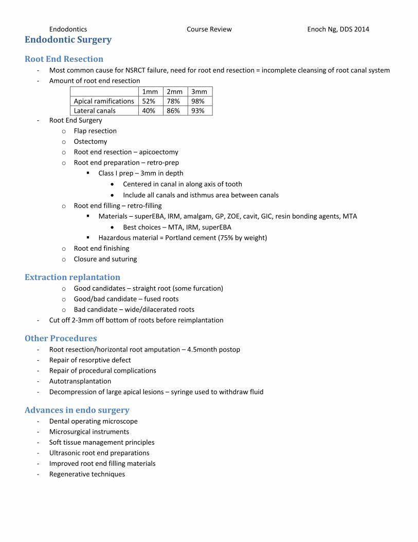
Endodontics Course Review Enoch Ng, DDS 2014
Endodontic Surgery
Root End Resection - Most common cause for NSRCT failure, need for root end resection = incomplete cleansing of root canal system
- Amount of root end resection
1mm 2mm 3mm
Apical ramifications 52% 78% 98%
Lateral canals 40% 86% 93%
- Root End Surgery
o Flap resection
o Ostectomy
o Root end resection – apicoectomy
o Root end preparation – retro-prep
Class I prep – 3mm in depth
Centered in canal in along axis of tooth
Include all canals and isthmus area between canals
o Root end filling – retro-filling
Materials – superEBA, IRM, amalgam, GP, ZOE, cavit, GIC, resin bonding agents, MTA
Best choices – MTA, IRM, superEBA
Hazardous material = Portland cement (75% by weight)
o Root end finishing
o Closure and suturing
Extraction replantation o Good candidates – straight root (some furcation)
o Good/bad candidate – fused roots
o Bad candidate – wide/dilacerated roots
- Cut off 2-3mm off bottom of roots before reimplantation
Other Procedures - Root resection/horizontal root amputation – 4.5month postop
- Repair of resorptive defect
- Repair of procedural complications
- Autotransplantation
- Decompression of large apical lesions – syringe used to withdraw fluid
Advances in endo surgery - Dental operating microscope
- Microsurgical instruments
- Soft tissue management principles
- Ultrasonic root end preparations
- Improved root end filling materials
- Regenerative techniques
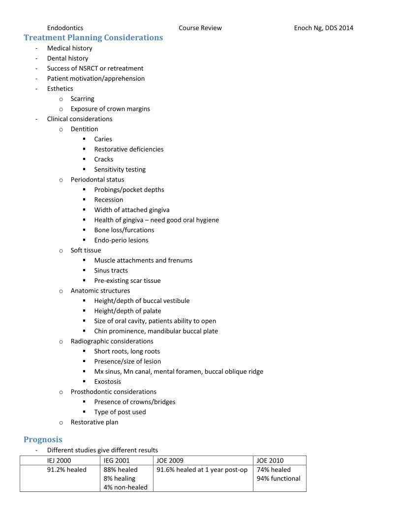
Endodontics Course Review Enoch Ng, DDS 2014
Treatment Planning Considerations - Medical history
- Dental history
- Success of NSRCT or retreatment
- Patient motivation/apprehension
- Esthetics
o Scarring
o Exposure of crown margins
- Clinical considerations
o Dentition
Caries
Restorative deficiencies
Cracks
Sensitivity testing
o Periodontal status
Probings/pocket depths
Recession
Width of attached gingiva
Health of gingiva – need good oral hygiene
Bone loss/furcations
Endo-perio lesions
o Soft tissue
Muscle attachments and frenums
Sinus tracts
Pre-existing scar tissue
o Anatomic structures
Height/depth of buccal vestibule
Height/depth of palate
Size of oral cavity, patients ability to open
Chin prominence, mandibular buccal plate
o Radiographic considerations
Short roots, long roots
Presence/size of lesion
Mx sinus, Mn canal, mental foramen, buccal oblique ridge
Exostosis
o Prosthodontic considerations
Presence of crowns/bridges
Type of post used
o Restorative plan
Prognosis - Different studies give different results
IEJ 2000 IEG 2001 JOE 2009 JOE 2010
91.2% healed 88% healed 8% healing 4% non-healed
91.6% healed at 1 year post-op 74% healed 94% functional

Endodontics Course Review Enoch Ng, DDS 2014
Endo-Perio
Pulpal/Perio Communication - Dentinal tubules
- Accessory canals
o 27.4% of teeth have accessory canals
Apical area – 17%
Middle third – 8.8%
Coronal third – 1.6%
o 28.4% molars (Mx and Mn) have accessory furcation canals
- Apical foramina
- Palatal groove
Pulpal Perio Disease - Bacterial infection of the pulp system induces significant inflammatory and immune response in apical tissues
- Untreated endodontic disease may support an increase in:
o Pocket depth
o Bone loss
- Perio treatment of teeth with pulpal necrosis and ARL resulted in delayed or impaired perio healing
- If blood supply through apical foramen is intact, perio disease rarely jeopardizes vital function of pulp
- Pulpal inflammation can come from exposure of lateral canals
- Pulpal necrosis results from main apical foramen invaded by bacteria
- Potential exists for S&RP to open dentinal tubules – indirectly induce localized pulpitis
- Microorganisms found in infected root canals of caries-free teeth with advanced perio usually resemble those
found in adjacent perio pockets
Endodontic Lesions - Endo lesions associated with inflamed/necrotic pulp with distinct etiology for pathosis
o Caries, restorations, cracks, trauma, attrition, abrasion, erosion
- Perio lesions usually associated with local factors that induce inflammation
o Bacteria, plaque, calculus
- Periodontal origin – generalized, broad lesions
- Pulpal origin – narrow coronally, isolated
Glickman’s Classification - Grade I – engaged flutes – pocket formation into the flute of the furca, but interradicular bone intact
- Grade II – engaged roof – loss of interradicular bone, pocket formation of varying depths into furca but not
completely through (dead end, cul de sac)
- Grade III – probe thru – complete loss of interradicular bone with a pocket probable to opposite side of tooth
- Grade IV – see thru – grade III with advanced gingival tissue recession that has made furca clearly visible during
clinical Exam

Endodontics Course Review Enoch Ng, DDS 2014
Lesion Classification - Primary endo
o Inflamed/necrotic pulp
o Possible isolated perio defect
o Osseous destruction localized to involved tooth
o Healing via regeneration of perio and osseous structures
o Endo treatment only
- Primary perio
o Generalized bone loss
o Local factors present
o Healing usually via reattachment
o Vital pulp
o Perio treatment only
- Primary endo with secondary perio
o Endo disease caused a perio communication
o Endo treat first, evaluate after 2 months, perio treat if needed
- Primary perio with secondary endo
o Perio disease, then necrotic pulp
o Osseous destruction exposes dentinal tubules, accessory canals, apical foramen
o Endo treat first, then perio treat 2 months later
- Concomitant endo-perio
o Endo and perio disease exist separately
o Endo treat first, then perio treat 2 months later
- True combined
o Endo and perio lesions eventually joined at a position on the root
o Endo treat first, then perio treat 2 months later
- Summary
o Endo treat completed before perio start
o Perio treatment 2 months after endo, only if needed
o Perio condition generally dictates overall prognosis
Longitudinal Tooth Fractures - Craze Lines
o Confined to enamel – no discomfort
o Natural or due to trauma – no treatment necessary, maybe for esthetics
- Fractured Cusp
o Lack of cusp support from weakened marginal ridge
o Brief sharp pain on biting, variable cold sensitivity
o Transillumination and bite tests to ID cusp
o Pulp test, remove fractured segment, restore tooth
o 79% molar fractures
Mx – 66% buccal, 34% lingual
Mn – 75% lingual, 25% buccal
o 21% premolar fractures

Endodontics Course Review Enoch Ng, DDS 2014 - Cracked Tooth
o Incomplete fracture
o May or may not involve pulp
o Extends from occlusal to apical
o Mesiodistal direction
o Excursive interference precursor for fracture
o Restored teeth 29x more likely to fracture than unrestored teeth
o Dental History
Repeated occlusal adjustments with minimal/transient decrease in symptoms
Vague/elusive symptoms
Extensive restorative history
Parafunctional habits
History of cracked teeth, history of trauma
o Subjective Exam
Episodic discomfort on biting
Patient remembering precipitating incident
Patient may not localize or ID tooth accurately
o Clinical Exam
Visual – restoration integrity, marginal discoloration
Tactile exam with explorer
Perio probings – isolated defect
Percussion – might have sensitivity
Bite test – sensitive on bite or release
transillumination
o Radiographics
Variable detection
Fractures not usually visible
o Restoration removal
Allows access
Aids in placement of stain to determine extent of crack
Methylene blue = caries indicator helps visualize location, direction, extent of crack
Necessary to determine mobility of segments
o Treatment
Cuspal coverage restorations may impede propagation of racks
Orthodontic bands
Occlusion reduction
Reduce height of non-functional cusps
Eliminate occlusal contacts on non-functional cusp
Re-contour outer incline of non-functional cusp
NSRCT when indicated by diagnosis – sensitivity testing shows pulpal damage is irreversible
Tooth prognosis decreases as crack propagation continues
21% of teeth with reversible pulpitis from cracks will require NSRCT in 6 months
Cuspal coverage = almost 0% failure
No cuspal coverage, composite restorations instead = 6% annual failure
-

Endodontics Course Review Enoch Ng, DDS 2014 - Split Tooth
o Progresses from cracked tooth
o Segments are mobile
o Usually extracted
For some Mx molars, sometimes can NSRCT, mobile segment can be removed, then restored
- Vertical Root Fracture
o Longitudinal fracture originating from root
o Usually involves previously RCT treated tooth
Force from impaction of GP (84%)
Operative and post-space errors second most likely cause (too long, too wide)
o Buccal-lingual fractures initiated at root
Factors beyond Operator Control - Canal shape and size
- Dentin thickness
- Much of fracture susceptibility is intrinsic to root and canal morphology – beyond clinicial control
Dental History and Exam for Vertical Root Fractures o Repeated occlusal adjustments with temporary symptom relief
o Variable discomfort with biting/touch
o Patient reports gum-bump/boil, bad taste, drainage
- Clinical Exam
o Palpate gingiva to assess for fenestrations, dehiscence
o Variable percussion sensitivity
o Sinus tract(s)
Perio pockets – 78%
Isolated perio probing pocket – suggestive of deep crack or VRF
Must rule out endo perio lesion with drainage through sulcus
Radiolucency – 72%
Diffuse longitudinal radiolucency (J-shaped or halo-like appearance) suggests VRF
Take PAs from multiple angles
Fracture not visualized unless beam passes through exactly the same plane as fracture
Swelling – 53%
Sinus tracts – 42%
Average time from RCT to VRF = 10.8 years
- Surgical Assessment
o Allows for visual assessment of root surface if a crack is highly suspected and cannot be confirmed by
other diagnostic means
- Treatment – extraction (or for some multirooted Mx molars, root amputation)
Longitudinal Root Fracture Summary - Loose tooth, endo treated, operative procedures, post space
- Factors that induce stress – post placement, obturation, parafunctional habits
- Cracked teeth – present with variable symptoms – multiple tests most predictable way to reach sound diagnosis
- Prognosis of cracked tooth depends on extent of crack
- If symptoms have been unresolved by dental treatment(s), should suspect cracks and fractures