SKELETAL, MUSCULAR AND … Skeletal Muscular Integumentary Cardiovascular Respiratory Immune...
26
This X-ray shows a color- enhanced image of the human skull, mandible, teeth, and neck. SECTION 1 The Human Body Plan SECTION 2 Skeletal System SECTION 3 Muscular System SECTION 4 Integumentary System CHAPTER 45 906 45 CHAPTER S KELETAL , M USCULAR , AND I NTEGUMENTARY S YSTEMS S KELETAL , M USCULAR , AND I NTEGUMENTARY S YSTEMS Copyright © by Holt, Rinehart and Winston. All rights reserved.
Transcript of SKELETAL, MUSCULAR AND … Skeletal Muscular Integumentary Cardiovascular Respiratory Immune...

This X-ray shows a color-enhanced image of thehuman skull, mandible,teeth, and neck.
SECTION 1 The Human Body Plan
SECTION 2 Skeletal System
SECTION 3 Muscular System
SECTION 4 Integumentary System
C H A P T E R 4 5906
45CHAPTER SKELETAL, MUSCULAR, AND
INTEGUMENTARY SYSTEMSSKELETAL, MUSCULAR, ANDINTEGUMENTARY SYSTEMS
Copyright © by Holt, Rinehart and Winston. All rights reserved.

907S K E L E T A L , M U S C U L A R , A N D I N T E G U M E N T A R Y S Y S T E M S
T H E H U M A N B O DY P L A NThe human body begins to take shape during the earliest
stages of embryonic development. While the embryo is a tiny
ball of dividing cells, it begins forming the tissues and organs
that compose the human body. By the end of its third week, the
human embryo has bilateral symmetry and is developing
vertebrate characteristics that will support an upright body
position.
BODY TISSUESA tissue is a collection of cells that are similar in structure and thatwork together to perform a particular function. The human bodyhas four main types of tissues: muscle, nervous, epithelial, andconnective.
Muscle TissueMuscle tissue is composed of cells that can contract. Every func-tion that muscle tissue performs—from creating a facial expressionto keeping the eyes in focus—is carried out by groups of musclecells that contract in a coordinated fashion. The human body hasthree types of muscle tissue: skeletal, smooth, and cardiac.Skeletal muscle moves the bones in your trunk, limbs, and face.Smooth muscle handles body functions that you cannot controlconsciously, such as the movement of food through your digestivesystem. Cardiac muscle, found in your heart, pumps bloodthrough your body. Figure 45-1a, on the following page, shows anillustration of cells of skeletal muscle tissue.
Nervous TissueNervous tissue contains cells that receive and transmit messagesin the form of electrical impulses. These cells, called neurons(NOO-rahnz), are specialized to send and receive messages through-out the body. Nervous tissue makes up your brain, spinal cord, andnerves. It is also found in parts of sensory organs, such as theretina in your eye. Some nervous tissue senses changes in theinternal and external environment. Other nervous tissue interpretsthe meaning of sensory information. Still other types of nervoustissue cause the body to move in response to sensory information.Coordination of voluntary and involuntary activities and regulationof some body processes are also accomplished by nervous tissue.Figure 45-1b, on the following page, shows an illustration of cells ofnervous tissue.
SECTION 1
O B J E C T I V E S● Describe four types of tissues that
make up the human body.● Explain how tissues, organs, and
organ systems are organized.● Summarize the functions of the
primary organ systems in thehuman body.
● Identify the five human bodycavities and the organs that eachcontains.
V O C A B U L A R Y muscle tissueskeletal musclesmooth musclecardiac musclenervous tissueneuronsepithelial tissueconnective tissuematrixorgancranial cavityspinal cavitydiaphragmthoracic cavityabdominal cavitypelvic cavity
www.scilinks.orgTopic: TissuesKeyword: HM61529
Copyright © by Holt, Rinehart and Winston. All rights reserved.
C H A P T E R 4 5908
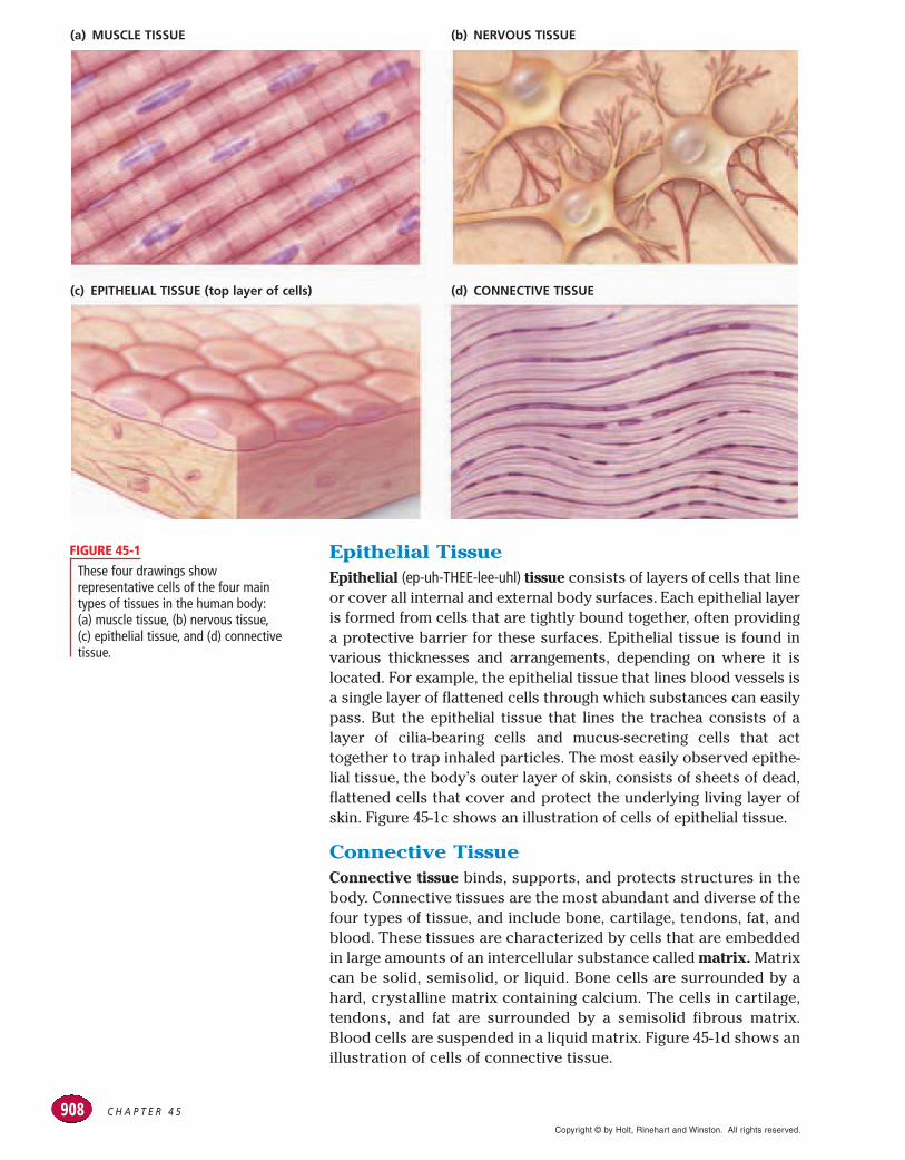
Epithelial TissueEpithelial (ep-uh-THEE-lee-uhl) tissue consists of layers of cells that lineor cover all internal and external body surfaces. Each epithelial layeris formed from cells that are tightly bound together, often providinga protective barrier for these surfaces. Epithelial tissue is found invarious thicknesses and arrangements, depending on where it islocated. For example, the epithelial tissue that lines blood vessels isa single layer of flattened cells through which substances can easilypass. But the epithelial tissue that lines the trachea consists of alayer of cilia-bearing cells and mucus-secreting cells that acttogether to trap inhaled particles. The most easily observed epithe-lial tissue, the body’s outer layer of skin, consists of sheets of dead,flattened cells that cover and protect the underlying living layer ofskin. Figure 45-1c shows an illustration of cells of epithelial tissue.
Connective TissueConnective tissue binds, supports, and protects structures in thebody. Connective tissues are the most abundant and diverse of thefour types of tissue, and include bone, cartilage, tendons, fat, andblood. These tissues are characterized by cells that are embeddedin large amounts of an intercellular substance called matrix. Matrixcan be solid, semisolid, or liquid. Bone cells are surrounded by ahard, crystalline matrix containing calcium. The cells in cartilage,tendons, and fat are surrounded by a semisolid fibrous matrix.Blood cells are suspended in a liquid matrix. Figure 45-1d shows anillustration of cells of connective tissue.
These four drawings showrepresentative cells of the four maintypes of tissues in the human body:(a) muscle tissue, (b) nervous tissue,(c) epithelial tissue, and (d) connectivetissue.
FIGURE 45-1
(c) EPITHELIAL TISSUE (top layer of cells) (d) CONNECTIVE TISSUE
(a) MUSCLE TISSUE (b) NERVOUS TISSUE
Copyright © by Holt, Rinehart and Winston. All rights reserved.

909S K E L E T A L , M U S C U L A R , A N D I N T E G U M E N T A R Y S Y S T E M S
ORGANS AND ORGANSYSTEMS
An organ consists of various tissues that work together to carryout a specific function. The stomach, a saclike organ in which foodis mixed with digestive enzymes, is composed of the four types oftissues. A single organ, such as the stomach, usually does not func-tion in isolation. Rather, groups of organs interact in an organ sys-tem. For example, in the digestive system, the stomach, smallintestine, liver, and pancreas all work together to break down foodinto molecules the body can use for energy. Table 45-1 lists thebody’s organ systems and names their major structures and func-tions. As you study the table, think about the ways in which the dif-ferent organ systems work together to function in an efficient,integrated manner.
TABLE 45-1 Summary of Organ Systems
System
Skeletal
Muscular
Integumentary
Cardiovascular
Respiratory
Immune
Digestive
Excretory
Nervous
Endocrine
Reproductive
Major structures
bones
muscles (skeletal, cardiac, and smooth)
skin, hair, nails
heart, blood vessels, blood
air passages, lungs
lymph nodes and vessels, white blood cells
mouth, esophagus, stomach, liver,pancreas, small and large intestines
kidneys, ureters, bladder, urethra, skin,lungs
brain, spinal cord, nerves, sense organs,receptors
glands (such as adrenal, thyroid, pituitary,and pancreas); hypothalamus andspecialized cells in the brain, heart,stomach, and other organs
ovaries, uterus, mammary glands (in females), testes (in males)
Functions
provides structure; supports and protectsinternal organs
provides structure; supports and movestrunk and limbs; moves substancesthrough body
protects against pathogens; helps regulatebody temperature
transports nutrients and wastes to andfrom all body tissues
carries air into and out of lungs, wheregases (oxygen and carbon dioxide) areexchanged
provides protection against infection anddisease
stores and breaks down food; absorbsnutrients; eliminates waste
eliminates waste; maintains water andchemical balance
controls and coordinates body movementsand senses; controls consciousness andcreativity; helps monitor and maintainother body systems
maintains homeostasis; regulatesmetabolism, water and mineral balance,growth, behavior, development, andreproduction
produces eggs and milk in females, spermin males, and offspring after fertilization
Copyright © by Holt, Rinehart and Winston. All rights reserved.

C H A P T E R 4 5910
Integration of Organ SystemsAn even higher level of organization is the integration of organ sys-tems. Each organ system has organs associated with it accordingto the organ’s primary function. However, the boundaries are notalways well defined. For example, nearly all of the juices producedby the pancreas are designed to aid in digestion. But because thepancreas produces vitally important hormones, it is also consid-ered a component of the endocrine system. Each organ system car-ries out its own specific function, but for the organism to survive,the organ systems must work together. For example, nutrients fromthe digestive system are distributed by the cardiovascular system.The efficiency of the cardiovascular system depends on nutrientsfrom the digestive system and oxygen from the respiratory system.
BODY CAVITIESMany organs and organ systems in the human body are housed incompartments called body cavities. These cavities protect internalorgans from injuries and permit organs such as the lungs to expandand contract while remaining securely supported. As shown inFigure 45-2, the human body has five main body cavities. Each cav-ity contains one or more organs. The cranial cavity contains thebrain. The spinal cavity surrounds the spinal cord.
The two main cavities in the trunk of the human body are sepa-rated by a wall of muscle called the diaphragm (DIE-uh-FRAM). Theupper compartment, or thoracic (thoh-RAS-ik) cavity, contains theheart, the esophagus, and the organs of the respiratory system.The lower compartment, or abdominal (ab-DAHM-uh-nuhl) cavity,contains organs of the digestive system. The pelvic cavity containsthe organs of the reproductive and excretory systems.
1. Name the four types of tissues in the humanbody, and give an example of each.
2. Explain the difference between muscle tissueand nervous tissue.
3. How are tissues, organs, and organ systemsorganized in the body?
4. How do the organ systems function together inthe human body?
5. Give an example of interaction between theendocrine system and another organ system.
6. Identify the organs each body cavity contains.
CRITICAL THINKING7. Applying Information Describe how the skele-
tal, muscular, nervous, respiratory, and circulatorysystems function in a person swimming in a pool.
8. Analyzing Concepts Explain how the functionof the body’s organs might be affected if thebody were not divided into cavities?
9. Forming Reasoned Opinions The body cavitythat protects the brain is encased in bone. Whydo you think the abdominal cavity is notencased in bone?
SECTION 1 REVIEW
Cranialcavity
Spinalcavity
Thoraciccavity
Diaphram
Abdominalcavity
Pelviccavity
The human body has five main cavitiesthat house and protect delicate internalorgans.
FIGURE 45-2
Copyright © by Holt, Rinehart and Winston. All rights reserved.

911S K E L E T A L , M U S C U L A R , A N D I N T E G U M E N T A R Y S Y S T E M S
S K E L E T A L S Y S T E MThe adult human body consists of approximately 206 bones,
which are organized into an internal framework called the
skeleton. The variation in size and shape among the bones that
make up the skeleton reflects their different roles in the body.
THE SKELETONAs shown in Figure 45-3, the human skeleton is composed of twoparts—the axial skeleton and the appendicular (AP-uhn-DIK-yuh-luhr)skeleton. The bones of the skull, ribs, spine, and sternum form theaxial skeleton. The bones of the arms and legs, along with thescapula, clavicle, and pelvis, make up the appendicular skeleton.
SECTION 2
O B J E C T I V E S● Distinguish between the axial
skeleton and the appendicularskeleton.
● Explain the function and structureof bones.
● Summarize how bones developand elongate.
● List three types of joints, and givean example of each.
● Describe a common disorder thataffects the skeletal system.
V O C A B U L A R Yskeletonaxial skeletonappendicular skeletonperiosteumcompact boneHaversian canalosteocytespongy bonebone marrowfractureossificationepiphyseal platejointfixed jointsemimovable jointmovable jointligamentsynovial fluidrheumatoid arthritisosteoarthritis
Cranium
Maxilla
Mandible
Cervicalvertebrae
Clavicle
Scapula
Sternum
Ribs
Humerus
Lumbarvertebrae
Radius
Ulna
Pelvis
Carpals
Metacarpals
Phalanges
Sacralvertebrae
Pubis
Femur
Patella
Tibia
Fibula
Tarsals
Metatarsals
Phalanges
HUMAN SKELETON
The skeleton is the framework thatsupports and protects the body. Thebones of the axial skeleton are coloredpurple. The bones of the appendicularskeleton are colored yellow.
FIGURE 45-3
Copyright © by Holt, Rinehart and Winston. All rights reserved.

Blood vessels
Femur (long bone)
PeriosteumOsteocyte
Compact boneSpongy bone
Bone marrow
Haversian canal
C H A P T E R 4 5912
BONE FUNCTION ANDSTRUCTURE
The bones that make up the skeleton function in a variety of ways.Bones provide a rigid framework against which muscles can pull,give shape and structure to the body, and support and protect del-icate internal organs. Notice, for example, that the ribs curve toform a cage that contains the heart and lungs. Similarly, bones inthe skull form the cranium, a dome-shaped case that protects thebrain. Bones also store minerals, such as calcium and phosphorus,which play vital roles in important metabolic processes. In addi-tion, the internal portion of many bones produces red blood cells,platelets, and white blood cells.
Despite their number and size, bones make up less than 20 per-cent of the body’s mass. The reason for their having relatively lit-tle mass can be better understood by looking at bone structure.Bones are not dry, rigid structures, as they may appear in amuseum exhibit. They are moist, living tissues.
Long Bone StructureAs shown in Figure 45-4, a long bone consists of a porous centralcavity surrounded by a ring of dense material. The bone’s surfaceis covered by a tough membrane called the periosteum(PER-ee-AHS-tee-uhm). This membrane contains a network of bloodvessels, which supply nutrients, and nerves, which signal pain.
Long bones, found in the limbs of thebody, are hollow and cylindrical. Theouter shell of hard compact boneconsists of closely packed rings ofminerals and protein fibers. Narrowcanals running through these ringscontain blood vessels and nerves.Spongy bone is found in small flat bonesand in the ends of long bones. Thecentral shaft of the long bone containsmarrow and blood vessels.
FIGURE 45-4
periostium
from the Greek peri,meaning “around,” and osteon,
meaning “bone”
Word Roots and Origins
Copyright © by Holt, Rinehart and Winston. All rights reserved.

913S K E L E T A L , M U S C U L A R , A N D I N T E G U M E N T A R Y S Y S T E M S
ConnectionConnectionEcoEcoEcoBones of LeadMillions of Americans have beenexposed to lead in the environ-ment. Following exposure to lead,the kidneys excrete most of themetal. But 7 to 10 percent of theremaining lead in the body isstored in bone and can stay therefor a lifetime. The rapid boneuptake of lead acts as a detoxifyingmechanism. But lead may not bepermanently locked in bone. Aspeople age, bone degeneration mayoccur, releasing lead into the blood-stream. Even very small concentra-tions of lead in the bloodstreamcan cause damage to kidneys, andhigh blood pressure.
The United States has outlawedthe addition of lead to gasoline,water pipes, and paint. As a result,people who are now under age 25may not accumulate as much leadin their bones as people from ear-lier generations.
OsteocytesPeriosteum
Vein
Artery
Haversian canal
Haversian canals
Mineral rings(lamellae)
Spongy bone
Bone marrow
Under the periosteum is a hard material called compact bone. Athick layer of compact bone enables the shaft of the long bone toendure the large amount of stress it receives during activities suchas jumping. In the cross section shown in Figure 45-5a, notice thatcompact bone is composed of cylinders of mineral crystals andprotein fibers called lamellae. In the center of each cylinder is anarrow channel called a Haversian (huh-VER-shuhn) canal, as shownin Figure 45-5b. Blood vessels run through interconnectedHaversian canals, creating a network that carries nourishment tothe living bone tissue. Several layers of protein fibers wrap aroundeach Haversian canal. Embedded within the gaps between the pro-tein layers are bone cells called osteocytes (AHS-tee-uh-SIETS).
Beneath some compact bone is a network of connective tissuecalled spongy bone. Although its name suggests that it is soft, this tis-sue is hard and strong. As shown in Figure 45-4, spongy bone has a lat-ticework structure that consists of bony spikes arranged along pointsof pressure or stress, making bones both light and strong.
Bone MarrowMany bones also contain a soft tissue called bone marrow, whichcan be either red or yellow. Red bone marrow—found in spongybone, the ends of long bones, ribs, vertebrae, the sternum, and thepelvis—produces red blood cells, platelets, and white blood cells.Yellow bone marrow fills the shafts of long bones. It consistsmostly of fat cells and serves as an energy reserve. It can also beconverted to red bone marrow and produce blood cells whensevere blood loss occurs.
Injury and RepairDespite their strength, bones will crack or even break if they aresubjected to extreme loads, sudden impacts, or stresses fromunusual directions. The crack or break is referred to as a fracture.If circulation is maintained and the periosteum survives, healingwill occur even if the damage to the bone is severe.
The cross section in (a) shows theinternal structure of compact bone. Amicrograph of a Haversian canal (380!)surrounded by lamellae in compactbone is shown in (b).
FIGURE 45-5(a)
(b)
Copyright © by Holt, Rinehart and Winston. All rights reserved.

C H A P T E R 4 5914
Epiphyseal plate
Cartilage cells
New bone
New blood vessels
(a)
(b)
LONG-BONE GROWTH
BONE DEVELOPMENTMost bones develop from cartilage, a tough but flexible connectivetissue. In the second month of fetal development, much of the skele-ton is made of cartilage. During the third month, osteocytes beginto develop and release minerals that lodge in the spaces betweenthe cartilage cells, turning the cartilage to bone. The process bywhich cartilage is slowly replaced by bone as a result of the depo-sition of minerals is called ossification (AHS-uh-fuh-KAY-shuhn). Mostfetal cartilage is eventually replaced by bone. However, some carti-lage remains, lending flexibility to the areas between bones, at theend of the nose, in the outer ear, and along the inside of the trachea.
A few bones, such as some parts of the skull, develop directlyinto hard bone without forming cartilage first. In these cases, theosteocytes are initially scattered randomly throughout the embry-onic connective tissue but soon fuse into layers and become flatplates of bone. In the skull, suture lines can be seen where theplates of bone meet.
Bone ElongationBones continue to develop after a person’s birth. Between earlychildhood and late adolescence, bone cells gradually replace thecartilage in long bones of limbs, such as the arms and legs. Boneelongation takes place near the ends of long bones in an areaknown as the epiphyseal (EP-uh-FIZ-ee-uhl) plate. As shown in Figure45-6a, the epiphyseal plate is composed of cartilage cells thatdivide and form columns, pushing older cells toward the middle ofthe bone. As these older cells die, they are replaced by new bonecells. Growth continues, as shown in Figure 45-6b, until bone hasreplaced all the cartilage in the epiphyseal plate. At this point,bones no longer elongate and a person has usually reached fullheight. The epiphyseal plates then become epiphyseal lines.
The epiphyseal plate, found at the ends of immature long bones, such asthe fibula shown above, is the site ofbone elongation. This region is rich withcartilage cells, which divide, enlarge,and push older cells toward the middleof the bone shaft. As older cells moveback, they are replaced by new bonecells, forming new regions of bone.A long bone (a) will grow in length,circumference, and density in thismanner, as shown in (b).
FIGURE 45-6
www.scilinks.orgTopic: Bones and JointsKeyword: HM60187
Copyright © by Holt, Rinehart and Winston. All rights reserved.

915S K E L E T A L , M U S C U L A R , A N D I N T E G U M E N T A R Y S Y S T E M S
JOINTSThe place where two bones meet is known as a joint. Three majorkinds of joints are found in the human body—fixed, semimovable,and movable. Examples of these joints are shown in Figure 45-7.
Fixed JointsFixed joints prevent movement. They are found in the skull, wherethey securely connect the bony plates and permit no movement ofthose bones. A small amount of connective tissue in a fixed jointhelps absorb impact to prevent the bones from breaking.
Semimovable JointsSemimovable joints permit limited movement. For example, semi-movable joints hold the bones of the vertebral column in place andallow the body to bend and twist. The vertebrae of the spine areseparated by disks of cartilaginous tissue. These tough, springydisks compress and absorb shocks that could damage the fragilespinal cord. Semimovable joints are also found in the rib cage,where long strands of cartilage connect the upper ten pairs of ribsto the sternum, allowing the chest to expand during breathing.
Movable JointsAll other joints in the body are movable joints. These joints enablethe body to perform a wide range of movements and activities.Movable joints include hinge, ball-and-socket, pivot, saddle, andgliding joints. An example of a hinge joint is found in the elbow,which allows you to move your forearm upward and downward,like a hinged door. An example of a ball-and-socket joint is the shoul-der joint, which enables you to move your arm up, down, forward,and backward, as well as to rotate it in a complete circle. The jointformed by the top two vertebrae of your spine is an example of apivot joint; it allows you to turn your head from side to side, aswhen shaking your head “no.” The saddle joint, found at the base ofeach thumb, allows you to rotate your thumbs and helps you graspobjects with your hand. Finally, gliding joints allow bones to slideover one another. Examples are the joints between the small bonesof your foot, which allow your foot to flex when you walk.
Joint StructureJoints, such as the knee, are often subjected to a great deal of pres-sure and stress, but their structure is well suited to meet thesedemands. As in all movable joints, the parts of the bones that comein contact with each other are covered with cartilage, which pro-tects the bones’ surface from friction. Tough bands of connectivetissue, called ligaments, hold the bones of the joint in place. Thesurfaces of the joints that are subjected to a great deal of pressureare lined with tissue that secretes a lubricating substance calledsynovial (sih-NOH-vee-uhl) fluid. Synovial fluid helps protect theends of bones from damage by friction.
In addition to fixed joints andsemimovable joints, the human bodyhas five types of movable joints: pivot,hinge, saddle, ball-and-socket, andgliding.
FIGURE 45-7
Fixed joint
Pivot joint
Semimovable joints
Hinge joint
Saddle joint
Ball-and-socket joint
Gliding joint
Copyright © by Holt, Rinehart and Winston. All rights reserved.

C H A P T E R 4 5916
1. List the major parts of the axial skeleton and themajor parts of the appendicular skeleton.
2. Name five functions of bones.
3. Illustrate the structure of a long bone.
4. When does the ossification of most of the bonesin the body begin and end?
5. Describe the function of the three major typesof joints, and give an example of each.
6. Differentiate between the two types of arthritis.
CRITICAL THINKING 7. Applying Information What is the advantage
of a cartilaginous skeleton during prenataldevelopment?
8. Analyzing Information Which type of arthritisis not related to age?
9. Relating Concepts How are the structures ofcartilage and bone related to the function eachperforms in the body?
SECTION 2 REVIEW
Femur
Patella
Fibula
Ligament
CartilageSynovialfluid
Tibia
Sometimes these protective structures are not enough to pre-vent a joint from becoming injured. Of all the joints in the body, theknee joint is the most susceptible to injury because it carries thebody’s weight and relies on many ligaments for stability. Damage tothe knee joint can cause swelling in the compartment that containsthe synovial fluid. Figure 45-8 shows the internal structures of theknee joint.
The term arthritis is used to describe disorders that causepainful, swollen joints. There are two forms of arthritis that affectjoints. Rheumatoid arthritis develops when the immune systembegins to attack body tissues. The joints become inflamed,swollen, stiff, and deformed. Osteoarthritis is a degenerative jointdisease in which the cartilage covering the surface of bonebecomes thinner and rougher. As a result, bone surfaces rubagainst each other, which is sensed by the nerves in the perios-teum, and causes severe discomfort.
The knee is a movable joint formed bythe ends of the femur, the tibia, andthe patella. Many cordlike ligamentsstabilize the joint, especially duringmovement. Pads of cartilage protectthe ends of bones and act as shockabsorbers. Like many joints in thebody, the knee is a synovial joint. Itcontains membranes that secretesynovial fluid, which lubricates andnourishes the tissues inside the joint.
FIGURE 45-8
Copyright © by Holt, Rinehart and Winston. All rights reserved.

917S K E L E T A L , M U S C U L A R , A N D I N T E G U M E N T A R Y S Y S T E M S
M U S C U L A R S Y S T E MMuscles make up the bulk of the body and account for about
one-third of its weight. Their ability to contract and relax not
only enables the body to move, but also provides the force that
pushes substances, such as blood and food, through the body.
MUSCLE TYPESA muscle is an organ that can contract in a coordinated fashion andincludes muscle tissue, blood vessels, nerves, and connective tis-sue. Some of the major muscles of the human body are shown inFigure 45-9. Recall that the human body has three types of muscletissues: skeletal, smooth, and cardiac.
Skeletal muscle is responsible for moving parts of the body,such as the limbs, trunk, and face. Skeletal muscle tissue is madeup of elongated cells called muscle fibers. Each muscle fiber con-tains many nuclei and is crossed by light and dark stripes, calledstriations, as shown on the following page in Figure 45-10a. Skeletalmuscle fibers are grouped into dense bundles called fascicles. Agroup of fascicles are bound together by connective tissue to forma muscle. Because their contractions can usually be consciouslycontrolled, skeletal muscles are described as voluntary muscles.
SECTION 3
O B J E C T I V E S● Distinguish between the three
types of muscle tissues.● Describe the structure of skeletal
muscle fibers.● Explain how skeletal muscles
contract.● Describe how muscles move bones.● Explain the process in which a
muscle becomes fatigued.
V O C A B U L A R Ymuscle fiberstriationfasciclevoluntary muscleinvoluntary musclemyofibrilmyosinactinZ linesarcomeretendonorigininsertionflexorextensormuscle fatigueoxygen debt
Frontalis
Trapezius
Pectoralismajor
Triceps brachii
Bicepsbrachii
Latissimus dorsi
Abdominalmuscles
Gluteus maximus
Sartorius
Biceps femoris
Rectus femoris
Gastrocnemius
Achilles tendon
Deltoid
Skeletal muscle tissue is shown in thesediagrams of some of the major musclesin the human body.
FIGURE 45-9
Copyright © by Holt, Rinehart and Winston. All rights reserved.

C H A P T E R 4 5918
Job Description A certified ath-letic trainer (ATC) is a highly educatedand skilled professional who specializesin athletic health care. Certified athletictrainers must have a bachelor’s degree,usually in athletic training, health, physi-cal education, or exercise science. Inaddition, an ATC must pass a certifica-tion exam. The job includes the preven-tion, identification, evaluation, treatment,referral, and rehabilitation of sports-related injuries.
Focus on Certified Athletic TrainerIn high school, Veronica Ampey wasinterested in science, medicine, rehabili-tation, and community service. Today,she has found a career that brings
together all of these interests and herlove for sports. “I like the fact that I am not tied to a desk,” says Ampey.“I get paid to watch athletes practiceand compete.”
Ampey works as an ATC at a smallhigh school in Washington, D.C. Ampeysays “The bonus is using my educationand experience to address incidentswhen they occur. Additionally, there is agreat deal of satisfaction in seeingsomeone you’ve worked with return tofull athletic participation.”
Education and Skills• High School—at least three years of
science courses and four years ofmath courses.
• College—bachelor’s degree from acollege with an accredited athletictraining curriculum, including coursework in biology; a master’s degree forsome jobs; and certification.
• Skills—patience, good organizationalskills, and ability to work as a mem-ber of a team.
Careersin BIOLOGY
Certified Athletic Trainer
For more about careers, visitgo.hrw.com and type in thekeyword HM6 Careers.
(a) SKELETAL MUSCLE TISSUE (b) SMOOTH MUSCLE TISSUE (c) CARDIAC MUSCLE TISSUE
These light micrographs show the threetypes of muscle tissue. Skeletal muscletissue (a) has a striped appearancewhen viewed under a microscope(430!). Smooth muscle tissue (b) isfound in the digestive tract, the uterus,the bladder, and the blood vessels(400!). Cardiac muscle tissue (c) isfound only in the heart (270!).
FIGURE 45-10
Smooth muscle forms the walls of the stomach, intestines, bloodvessels, and other internal organs. Individual smooth muscle cellsare spindle-shaped, have a single nucleus, and interlace to formsheets, as shown in Figure 45-10b. Notice that smooth muscle lacksthe striations found in skeletal muscle tissue. Smooth musclefibers are surrounded by connective tissue, but the connective tis-sue does not unite to form tendons as it does in skeletal muscles.Because most of its movements cannot be consciously controlled,smooth muscle is referred to as involuntary muscle.
Cardiac muscle, shown in Figure 45-10c, makes up the walls ofthe heart. Cardiac muscle shares some characteristics with bothskeletal muscle and smooth muscle. As with skeletal muscle, car-diac muscle tissue is striated; as with smooth muscle, it is invol-untary and each cell has one nucleus.
Copyright © by Holt, Rinehart and Winston. All rights reserved.

919S K E L E T A L , M U S C U L A R , A N D I N T E G U M E N T A R Y S Y S T E M S
Deltoid muscle
Fascicle
Connective tissue
Muscle fiber (cell)
Z line Myofibril
Myosin
ActinSarcomere
MUSCLE STRUCTUREA skeletal muscle fiber is a single, multinucleated muscle cell. Askeletal muscle may be made up of hundreds or even thousands ofmuscle fibers, depending on the muscle’s size. Although musclefibers make up most of the muscle tissue, a large amount of con-nective tissue, blood vessels and nerves are also present. Like allbody cells, muscle cells are soft and easy to injure. Connective tis-sue covers and supports each muscle fiber and reinforces the mus-cle as a whole.
The health of a muscle depends on a sufficient nerve and bloodsupply. Each skeletal muscle fiber has a nerve ending that controlsits activity. Active muscles use a lot of energy and therefore requirea continuous supply of oxygen and nutrients, which are suppliedby arteries. Muscles produce large amounts of metabolic wastethat must be removed through veins.
A skeletal muscle fiber, such as the one shown in Figure 45-11,contains bundles of threadlike structures called myofibrils(MIE-oh-FIE-bruhlz). Each myofibril is made up of two types of proteinfilaments—thick ones and thin ones. Thick filaments are made ofthe protein myosin (MIE-uh-suhn), and thin filaments are made of theprotein actin. Myosin and actin filaments are arranged to form anoverlapping pattern, which gives striated muscle tissue its stripedappearance. Thin actin filaments are anchored at their endpointsto a structure called the Z line. The region from one Z line to thenext is called a sarcomere (SAHR-kuh-MIR).
Skeletal muscles consist of denselypacked groups of elongated cells,called fascicles, that are held togetherby connective tissue. Muscle fibersconsist of protein filaments calledmyofibrils. Two types of filaments arefound in muscle fibers—actin andmyosin. The complementary structuresof actin and myosin interact to contractand relax muscles.
FIGURE 45-11
Copyright © by Holt, Rinehart and Winston. All rights reserved.

C H A P T E R 4 5920
Sarcomere
MyosinMUSCLERELAXED
MUSCLECONTRACTS
MUSCLEFULLYCONTRACTED
Actin
Z line
In a relaxed muscle, the actin andmyosin filaments overlap. During amuscle contraction, the filaments slidepast each other and the zone of overlapincreases. As a result, the length of thesarcomere shortens.
FIGURE 45-12
MUSCULAR CONTRACTIONThe sarcomere is the functional unit of muscle contraction. Whena muscle contracts, myosin filaments and actin filaments interactto shorten the sarcomere. Myosin filaments have extensionsshaped like oval “heads.” Actin filaments look like a twisted strandof beads. When a nerve impulse stimulates a muscle to contract,the myosin filaments’ heads attach to points between the beads ofthe actin filaments. The myosin heads then bend inward, pullingthe actin with them. The myosin heads then let go, bend back intotheir original position, attach to a new point on the actin filament,and pull again. This action shortens the sarcomere. The synchro-nized shortening of sarcomeres along the length of a muscle fibercauses the whole fiber, and hence the muscle, to contract. Figure45-12 shows a sarcomere’s structures.
Muscle contraction requires energy, which is supplied by ATP.This energy is used to detach the myosin heads from the actin fila-ments. Because myosin heads must attach and detach a number oftimes during a single muscle contraction, muscle cells must have acontinuous supply of ATP. Without ATP, the myosin would remainattached to the actin, keeping a muscle permanently contracted.
Muscle contraction is an all-or-none response—either the fiberscontract or they remain relaxed. How, then, are you able to con-tract your muscles tightly enough to lift a dumbbell or gentlyenough to lift a pen? The force of a muscle contraction is deter-mined by the number of muscle fibers that are stimulated. As morefibers are activated, the force of the contraction increases.
Testing Muscle Staminaand Strength
Materials bathroom scale,notepad, pencil
Procedure1. Create a chart that coompares
the strength of pectoral musclesat four different time intervals,each 1 minute apart.
2. Press the scale between thepalms of your hands. Have yourpartner record the amount of pressure applied by yourpectoral muscles.
3. Set the scale down and pressyour hands together in front ofyou for 1 minute. Press the scalebetween your hands again andhave your partner record thepressure.
4. Repeat steps 2 and 3 two moretimes. Then repeat the experi-ment with your partner pressingthe scale while you record thepressure.
Analysis What trends did younotice, if any, in the amount of pres-sure recorded? What might be areason for this trend?
Quick Lab
Copyright © by Holt, Rinehart and Winston. All rights reserved.
921S K E L E T A L , M U S C U L A R , A N D I N T E G U M E N T A R Y S Y S T E M S
MUSCULAR MOVEMENT OF BONES
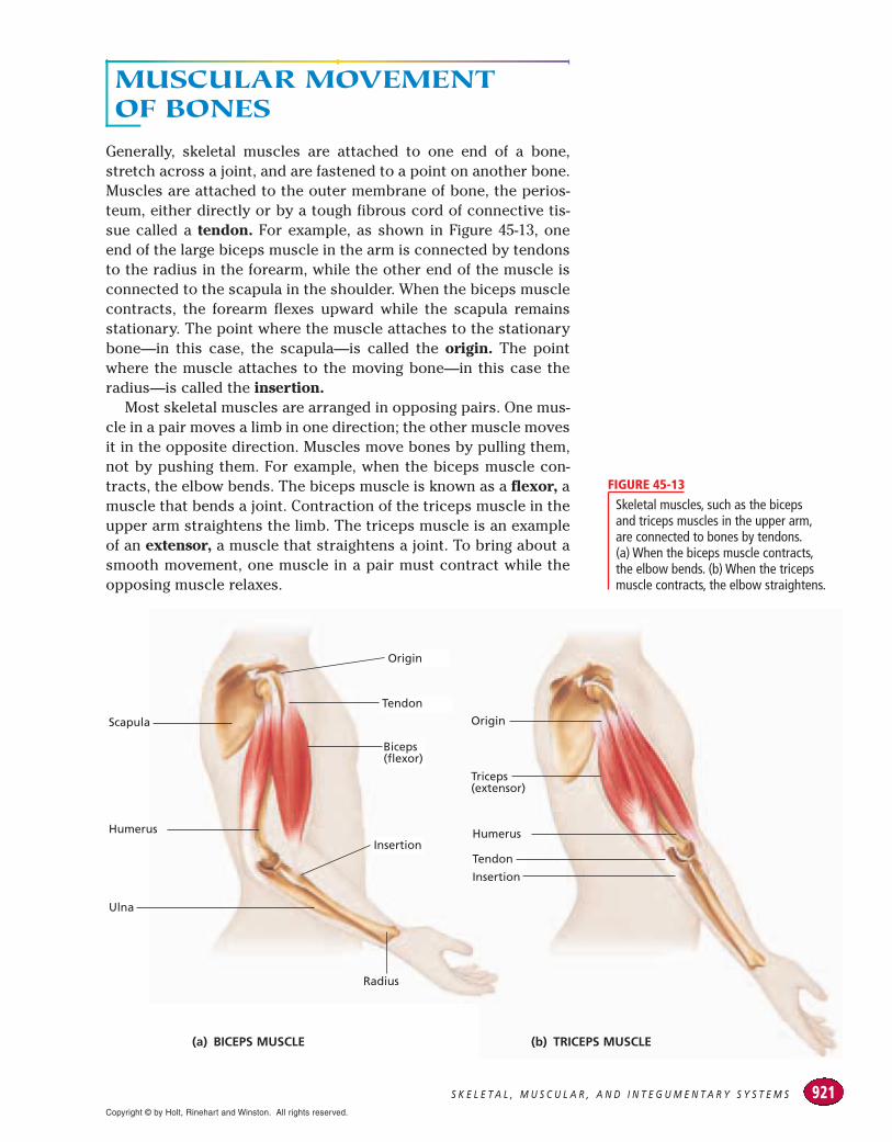
Generally, skeletal muscles are attached to one end of a bone,stretch across a joint, and are fastened to a point on another bone.Muscles are attached to the outer membrane of bone, the perios-teum, either directly or by a tough fibrous cord of connective tis-sue called a tendon. For example, as shown in Figure 45-13, oneend of the large biceps muscle in the arm is connected by tendonsto the radius in the forearm, while the other end of the muscle isconnected to the scapula in the shoulder. When the biceps musclecontracts, the forearm flexes upward while the scapula remainsstationary. The point where the muscle attaches to the stationarybone—in this case, the scapula—is called the origin. The pointwhere the muscle attaches to the moving bone—in this case theradius—is called the insertion.
Most skeletal muscles are arranged in opposing pairs. One mus-cle in a pair moves a limb in one direction; the other muscle movesit in the opposite direction. Muscles move bones by pulling them,not by pushing them. For example, when the biceps muscle con-tracts, the elbow bends. The biceps muscle is known as a flexor, amuscle that bends a joint. Contraction of the triceps muscle in theupper arm straightens the limb. The triceps muscle is an exampleof an extensor, a muscle that straightens a joint. To bring about asmooth movement, one muscle in a pair must contract while theopposing muscle relaxes.
(a) BICEPS MUSCLE
Origin
Tendon
Insertion
Radius
Biceps(flexor)
Tendon
Humerus
Scapula
Ulna
Humerus
Insertion
Triceps(extensor)
Origin
(b) TRICEPS MUSCLE
Skeletal muscles, such as the biceps and triceps muscles in the upper arm,are connected to bones by tendons.(a) When the biceps muscle contracts,the elbow bends. (b) When the tricepsmuscle contracts, the elbow straightens.
FIGURE 45-13
Copyright © by Holt, Rinehart and Winston. All rights reserved.

C H A P T E R 4 5922
MUSCLE FATIGUEMuscle cells store glycogen, which is used as a source of energywhen the blood cannot deliver adequate amounts of glucose. Thebreakdown of glycogen releases large amounts of energy, but some-times even those reserves are used up. During prolonged and vig-orous exertion, fat molecules are utilized for energy. Fat moleculescontain a greater concentration of potential energy than any othermolecule in the body. When energy availability fails to keep pacewith its use, muscle fatigue sets in and controlled muscle activityceases, even though the muscle may still receive nerve stimulationto move. Muscle fatigue is the physiological inability of a muscleto contract. Muscle fatigue is a result of a relative depletion of ATP.When ATP is absent, a state of continuous contraction occurs. Anexample of depletion of ATP is when a marathon runner collapsesduring a race, suffering from severe muscle cramps.
Oxygen DebtOxygen is used during cellular respiration in the synthesis of ATP.Large amounts of oxygen are needed to maintain the rate of maxi-mum ATP production required to sustain strenuous exercise. How-ever, after several minutes of heavy exertion, the circulatorysystem and the respiratory system are not able to bring in enoughoxygen to meet the demands of energy production. Oxygen levelsin the body become depleted. This temporary lack of oxygen avail-ability is called oxygen debt. Oxygen debt leads to an accumula-tion of lactic acid as metabolic waste in the muscle fibers. Thepresence of lactic acid produces the soreness you may experienceafter prolonged exercise. Oxygen debt causes a person to spendtime in rapid, deep breathing after strenuous exercise, as the ath-letes shown in Figure 45-14 are doing. The oxygen debt is repaidquickly as additional oxygen becomes available, but muscle sore-ness may persist until all of the metabolic wastes that have accu-mulated in the muscle fibers are carried away or converted.
1. Compare the three main types of muscle tissuesfound in the body.
2. Why is smooth muscle referred to as involuntarymuscle?
3. Why do skeletal muscle fibers appear striated?
4. How do skeletal muscles contract?
5. How do muscles work together to move bones?
6. Contrast the functions of flexors and extensors.
7. What causes muscles to become fatigued?
CRITICAL THINKING8. Analyzing Information Rigor mortis is a condi-
tion in which all of the body muscles becomerigid shortly after a person dies. Why does rigormortis develop?
9. Applying Information What causes musclecramping after vigorous exercise or repeatedmovement?
10. Applying Information Why do you think theheart muscle never suffers from fatigue?
SECTION 3 REVIEW
www.scilinks.orgTopic: Muscle FatigueKeyword: HM61005
These athletes are in the process ofrepaying their oxygen debts. Oxygendebt occurs frequently after strenuous,sustained exertion.
FIGURE 45-14
Copyright © by Holt, Rinehart and Winston. All rights reserved.

S C I E N C ET E C H N O L O G Y
S O C I E T Y
www.scilinks.orgTopic: HolographyKeyword: HM60752
Looking Inside the Human Body
In 1895, the development ofX-ray equipment providedphysicians a way to look at
images of dense tissue in thebody, such as bones. Modernimaging techniques rely oncomputers.
Computerized Tomography
Computerized tomography (CT)uses a focused beam of low-dose X-rays to obtain cross-sectional images of structuresin the body. Tomography is thetechnique used to take imagesof a specific “slice” or plane oftissue. Computerized tomogra-phy can differentiate tissues ofvarious densities.
Positrons and Brain Imaging
Still newer imaging technologyis positron emission technology,or PET. Positrons are positivelycharged particles with the samemass as electrons that resultfrom the disintegration ofradioisotopes. Michael M.TerPogossian and his col-leagues at Johns Hopkins sug-gested using short-livedradioisotopes in medicalresearch. They developed scan-ners to detect the positronsreleased by radioisotopes thathad been injected into apatient’s bloodstream. As tech-nology improved, biomedicalengineers redesigned scanningequipment to create three-dimensional images from thepositron emissions. Positronemission technology is used tomap areas of the brain that areinvolved in memory, sensation,perception, speech, and infor-mation processing. In addition,positron emission technologyprovides clues about the causesof psychiatric disorders, such asdepression.
Holographic Imaging
A new holographic imagingsystem combines imagesobtained by computerizedtomography or magnetic reso-nance scanners and displays an accurate three-dimensionalimage of anatomical structures.Magnetic resonance imaging(MRI) creates images of softtissues. MRI uses radio wavesemitted by the nuclei of hydro-gen atoms that are activated by amagnetic scanner. A holographis a method of photographythat uses laser light to produce
a three-dimensional image. Thetransparent but solid lookingimage floats in front of theholographic film and can bestudied from all sides.
Holograms in Medicine
Physicians can make surgicalplans by studying the trueappearance of a patient’sorgans, such as the threedimensional PET scan thathighlights the verbal center inthe brain (a). Compare the threedimensional PET scan with theX-ray of the same part of thebody (b). The X-ray may not beas helpful as the three dimen-sional PET scan when a physi-cian must diagnose an illnessor injury of the brain.
1. How do X-rays differ from PETscans?
2. How might a holograph bemore useful than a PET scan?
3. Justifying Conclusions If asurgeon had to remove a pieceof metal lodged in a patient’sskull, which type of imagingwould you choose? Supportyour answer, and give reasonswhy you did not choose theother imaging techniques.
R E V I E W
923
(a) A three dimensional PET scanshows the surface detail andcellular activity of the brain. (b) ThisX-ray shows the bones of the skull,mandible, and neck.
(a)
(b)
Copyright © by Holt, Rinehart and Winston. All rights reserved.

C H A P T E R 4 5924
I N T E G U M E N T A R Y S Y S T E MThe integumentary system, consisting of the skin, hair, and
nails, acts as a barrier to protect the body from the outside
world. It also functions to retain body fluids, protect against
disease, eliminate waste products, and regulate body
temperature.
SKINThe skin is one of the human body’s largest organs. Subjected to alifetime of wear and tear, the layers of skin are capable of repairingthemselves. Skin contains sensory receptors that monitor the exter-nal environment, and mechanisms that rid the body of wastes. Theskin is composed of two layers—the epidermis and the dermis.
EpidermisThe epidermis, or outer layer of skin, is composed of many sheetsof flattened, scaly epithelial cells. Its top layers are made of mostlydead cells. These cells are exposed to the dangers of the externalenvironment. Scraped or rubbed away on a daily basis, they arereplaced by new cells made in the rapidly dividing lower layers. Thecells of the epidermis are filled with a protein called keratin, whichgives skin its rough, leathery texture and its waterproof quality.
There is a great variety in skin color among humans. The color of skin is mainly determined by a brown pigment called melanin(MEL-uh-nin), which is produced by cells in the lower layers of the epi-dermis. Melanin absorbs harmful ultraviolet radiation. The amount ofmelanin produced in skin depends on two factors: heredity and thelength of time the skin is exposed to ultraviolet radiation. Increasedamounts of melanin are produced in a person’s skin in response toultraviolet radiation. All people, but especially people with light skin,need to minimize exposure to the sun and protect themselves fromits ultraviolet radiation, which can damage the DNA in skin cells andlead to deadly forms of skin cancer.
DermisThe dermis, the inner layer of skin, is composed of living cells andspecialized structures, such as sensory neurons, blood vessels,muscle fibers, hair follicles, and glands. Sensory neurons make itpossible for you to sense many kinds of conditions and signalsfrom the environment, such as heat and pressure. Blood vesselsprovide nourishment to the living cells and help regulate bodytemperature.
SECTION 4
O B J E C T I V E S● Describe the functions of the skin.● Distinguish between the two
layers that form the skin.● Identify two types of glands found
in the skin, and describe theirfunctions.
● Describe the structure of nails.● Describe the structure of hair.
V O C A B U L A R Yepidermiskeratinmelanindermisexocrine glandsweat glandoil glandsebum
Epidermis
from the Greek epi, meaning“on” or “upon,” and derma,
meaning “skin”
Word Roots and Origins
Copyright © by Holt, Rinehart and Winston. All rights reserved.

925S K E L E T A L , M U S C U L A R , A N D I N T E G U M E N T A R Y S Y S T E M S
Tiny muscle fibers attached to hair follicles contract and pullhair upright when you are cold or afraid, producing what are com-monly called goose bumps. Glands produce sweat, which helpscool your body, and oil, which helps soften your skin. A layer of fatcells lies below the dermis. These cells act as energy reserves; adda protective, shock-absorbing layer; and insulate the body againstheat loss. Study the structures of the skin in Figure 45-15.
GlandsThe skin contains exocrine glands, glands that release secretionsthrough ducts. The main exocrine glands of the skin are the sweatglands and the oil glands.
The skin functions as an excretory organ by releasing excesswater, salts, and urea through the sweat glands. By releasingexcess water, the skin also helps regulate body temperature. Whenthe body’s temperature rises, circulation increases, and the skinbecomes warm and flushed, as shown in Figure 45-16. The sweatglands then release more sweat. As the water in sweat evaporates,the skin is cooled.
Oil glands, found in large numbers on the face and scalp,secrete the fatty substance sebum. Oil glands are usually con-nected by tiny ducts to hair follicles. Sebum coats the surface ofthe skin and the shafts of hairs, preventing excess water loss andsoftening the skin and hair. Sebum is also mildly toxic to some bac-teria. The production of sebum is controlled by hormones. Duringadolescence, high levels of sex hormones increase the activity ofthe oil glands. If the ducts of oil glands become clogged with exces-sive amounts of sebum, dead cells, and bacteria, the skin disorderacne can result.
Hair
Corneal layer
Basal layer
Oil gland
Hair follicle
Fat cells
VeinArtery
Sweatgland
Musclefibers
Nerveendings
Sensoryneuron
Epidermis
Dermis
Subcutaneouslayer
Pore
Skin is composed of two layers: theepidermis and the dermis. The top of theepidermis consists of dead, flattenedcells that are shed and replaced everyday. The dermis contains specializedstructures that protect the body frominfectious diseases, regulate bodytemperature, sense the environment, andsecrete oil, sweat, and some wastes.
FIGURE 45-15
Skin acts as a temperature-controllingdevice. It contains millions of sweatglands that secrete microscopic dropletsof water. The water droplets help coolthe body when its temperature rises,such as after a rigorous workout.
FIGURE 45-16
Copyright © by Holt, Rinehart and Winston. All rights reserved.

C H A P T E R 4 5926
1. What are the names and functions of the twolayers of skin?
2. Identify the reason the dermis is considered theliving layer of skin.
3. What are the functions of the two types ofexocrine glands found in the dermis?
4. Illustrate and label the structure of a fingernail.
5. Describe the structure of hair.
CRITICAL THINKING6. Relating Concepts Why can sunbathing be con-
sidered dangerous?
7. Analyzing Information A third-degree burnmay be surrounded by painful areas of second-and first- degree burns. However, a third-degreeburn is often painless. Why?
8. Recognizing Relationships How might musclesin the dermis benefit mammals in cold weather?
SECTION 4 REVIEW
Nail rootEponychium (cuticle)
Lunula
Nail body
Nail bedFree edge
Hyponychium
Phalanx(bone of thefingertip)
This illustration of the structure of afingernail shows that the nail root,from which the nail is constantlyregenerated, is protected well beneaththe surface of the finger, next to thebone of the fingertip.
FIGURE 45-17
NAILSNails, which protect the ends of the fingers and toes, form from nailroots under skin folds at the base and sides of the nail. As new cellsform, the nail grows longer. Like hair, nails are composed primarilyof keratin. The nail body is about 0.5 mm (0.02 in.) thick. Nails growat about 1 mm (0.04 in.) per week. Nails rest on a bed of tissue filledwith blood vessels, giving the nails a pinkish color. The structureof a fingernail can be seen in Figure 45-17.
Changes in the shape, structure, and appearance of the nailsmay be an indicator of a disease somewhere in the body. They mayturn yellow in patients with chronic respiratory disorders, or theymay grow concave in certain blood disorders.
HAIRHair, which protects and insulates the body, is produced by a clus-ter of cells at the base of deep dermal pits called hair follicles. Thehair shaft is composed of dead, keratin-filled cells that overlap likeroof shingles. Oil glands associated with hair follicles prevent hairfrom drying out. Most individual hairs grow for several years andthen fall out.
Hair color is the result of the presence of the pigment melanin inthe hair shaft. Black, brown, and yellow variants of melanin com-bine to determine an individual’s hair color. Hair color is influ-enced by hereditary factors.
Copyright © by Holt, Rinehart and Winston. All rights reserved.

The Human Body Plan SECTION 1
CHAPTER HIGHLIGHTS
927S K E L E T A L , M U S C U L A R , A N D I N T E G U M E N T A R Y S Y S T E M S
muscle tissue (p. 907)skeletal muscle (p. 907)smooth muscle (p. 907)cardiac muscle (p. 907)
nervous tissue (p. 907)neurons (p. 907)epithelial tissue (p. 908)connective tissue (p. 908)
matrix (p. 908)organ (p. 909)cranial cavity (p. 910)spinal cavity (p. 910)
diaphragm (p. 910)thoracic cavity (p. 910)abdominal cavity (p. 910)pelvic cavity (p. 910)
Vocabulary
skeleton (p. 911)axial skeleton (p. 911)appendicular skeleton (p. 911)periosteum (p. 912)compact bone (p. 913)
Haversian canal (p. 913)osteocyte (p. 913)spongy bone (p. 913)bone marrow (p. 913)fracture (p. 913)
ossification (p. 914)epiphyseal plate (p. 914)joint (p. 915)fixed joint (p. 915)semimovable joint (p. 915)
movable joint (p. 915)ligament (p. 915)synovial fluid (p. 915)rheumatoid arthritis (p. 916)osteoarthritis (p. 916)
Vocabulary
muscle fiber (p. 917)striation (p. 917)voluntary muscle (p. 917)involuntary muscle (p. 918)
myofibril (p. 919)myosin (p. 919)actin (p. 919)Z line (p. 919)
sarcomere (p. 919)tendon (p. 921)origin (p. 921)insertion (p. 921)
flexor (p. 921)extensor (p. 921)muscle fatigue (p. 922)oxygen debt (p. 922)
Vocabulary
● The human body has four main types of tissue: muscle,nervous, epithelial, and connective.
● A tissue is a collection of cells, an organ is a collection oftissues, and an organ system is a collection of organs.
● Some of the primary organ systems in the body includethe integumentary, nervous, and cardiovascular systems.
● Many organs are located in the body’s five main cavities:abdominal, cranial, spinal, thoracic, and pelvic.
Skeletal SystemSECTION 2
● The human skeleton is composed of the axial skeleton(skull, ribs, spine, and sternum) and the appendicularskeleton (arms and legs, scapula, clavicle, and pelvis).
● Bones support muscles, give structure to the body,protect organs, store minerals, and make blood cells.
● Bones are made up of minerals, protein fibers, and cells.
● Most bones develop from cartilage through a processcalled ossification.
● The human body has three types of joints: fixed joints,semimovable joints, and movable joints. The joints can beaffected by a disease called arthritis.
Muscular SystemSECTION 3
● The human body has three types of muscle tissues:skeletal, smooth, and cardiac.
● Skeletal muscles consist of groups of fibers. Muscle fiberscontain myofibrils made up of protein filaments.
● During a muscle contraction, myosin and actin filamentsinteract to shorten the length of a sarcomere.
● Most skeletal muscles are arranged in opposing pairs.
Integumentary SystemSECTION 4
● Skin, hair, and nails act as barriers that protect the bodyfrom the environment.
● Skin is composed of two layers, which are the epidermisand the dermis.
● Hair and nails are composed of the protein keratin; theygrow from a root of rapidly dividing cells.
● Sweat glands produce sweat, which cools the body. Oilglands secrete sebum, which softens the skin.
epidermis (p. 924)keratin (p. 924)
melanin (p. 924)dermis (p. 924)
exocrine gland (p. 925)sweat gland (p. 925)
oil gland (p. 925)sebum (p. 925)
Vocabulary
Copyright © by Holt, Rinehart and Winston. All rights reserved.
CHAPTER REVIEW
C H A P T E R 4 5928
USING VOCABULARY1. Choose the term that does not belong in the fol-
lowing group, and explain why it does not belong:saddle joint, pivot joint, fixed joint, and hingejoint, and ball-and-socket joint.
2. Distinguish between compact bone andspongy bone.
3. Use the following key terms in the same sen-tence: actin, muscle fiber, myofibrils, and myosin.
4. Word Roots and Origins The word epidermis isderived from the Greek derma, which means“skin.” The prefix epi means “on.” Using thisinformation, explain why the term epidermisis a good name for the anatomical structure it describes.
UNDERSTANDING KEY CONCEPTS5. Define epithelial tissue.6. Explain the relationship between cells, tissues,
organs, and organ systems.7. Summarize the functions of the primary organ
systems in the human body.8. Describe the organs that can be found in the
abdominal cavity.9. List all of the bones in the axial skeleton.
10. Identify the five functions of the skeletal system.11. Explain the role the Haversian canals play in
compact bone.12. Define red bone marrow. Where is it produced,
and what is its function?13. Summarize how bones develop and elongate.14. State three types of joints, and give examples of
each type.15. Describe the cause and symptoms of the disease
rheumatoid arthritis.16. Explain the difference between skeletal muscle,
smooth muscle, and cardiac muscle.17. Describe the components of a sarcomere.18. Illustrate how a skeletal muscle contracts.19. Explain how muscles move bones.20. Name the functions of tendons and ligaments.21. List four functions of the skin.22. Identify the difference between the epidermis and
the dermis.23. Define melanin. What is its role in the body?
24. Explain the similarities and differences betweennails and hair.
25. Identify the substance that prevents the hair andskin from drying out, and the gland where thissubstance is produced.
26. CONCEPT MAPPING Use the followingterms to create a concept map that illus-
trates the body’s four levels of structural organi-zation: muscle tissue, connective tisue, epithelialtissue, nervous tissue, organ, and organ system.
CRITICAL THINKING27. Inferring Relationships Young thoroughbred
horses that are raced too early in life have anincreased risk of breaking the bones in their legs.What can you infer about the process of ossifica-tion in horses?
28. Evaluating Information During a normal birth, a baby passes through the mother’s pelvis. Awoman’s pelvis has a larger diameter and is moreoval-shaped than a man’s pelvis. In addition, anewborn’s skull bones are not completely ossi-fied. How are these skeletal properties advanta-geous to the birthing process?
29. Analyzing Concepts Oil glands secrete an oilysubstance that helps keep the skin soft and flexi-ble. They also secrete fatty acids, which help killbacteria. How can the function of oil glands beaffected if you wash your skin too frequently?
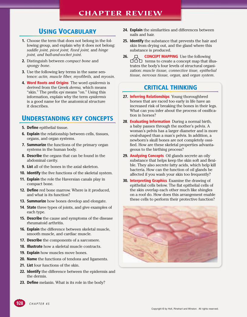
30. Interpreting Graphics Examine the drawing ofepithelial cells below. The flat epithelial cells ofthe skin overlap each other much like shingles on a roof do. How does this arrangement enablethese cells to perform their protective function?
Copyright © by Holt, Rinehart and Winston. All rights reserved.

For a question involving experi-mental data, determine the constants, variables, andcontrol before answering the questions.
929S K E L E T A L , M U S C U L A R , A N D I N T E G U M E N T A R Y S Y S T E M S
Standardized Test PreparationDIRECTIONS: Choose the letter that best answers thequestion or completes the sentence.
1. The thoracic cavity contains which organs?A. brainB. spineC. organs of the digestive systemD. organs of the respiratory system
2. The cells of connective tissue are embedded inwhat substance?F. matrixG. keratinH. marrowJ. synovial fluid
3. The periosteum is a membrane that does whichof the following?A. covers the boneB. contains marrowC. produces red blood cellsD. increases the length of long bones
4. Which of the following is true about the dermis?F. It is the top layer of skin.G. It contains cardiac muscle.H. It is made up of dead cells.J. It contains nerves and blood vessels.
INTERPRETING GRAPHICS: The graph below showsthe relationship between skin type, UV index, and sunburns. Use the table to answer the question thatfollows.
5. Which of the following statements about skintype 1 is true?A. Skin type 4 will never sunburn.B. Skin type 1 will always burn in less than
20 minutes.C. Skin type 1 is less sensitive to UV exposure
than skin type 4 is.D. Skin type 1 is more sensitive to UV exposure
than skin type 4 is.
DIRECTIONS: Complete the following analogy.6. nerve : neuron :: bone :
F. marrowG. skeletonH. osteocyteJ. Haversian canal
INTERPRETING GRAPHICS: The figure below showsa sarcomere and an enlargement of actin andmyosin filaments. Use the figure to answer thequestion that follows.
7. Which part of the sarcomere represents the Z line? A. feature 1B. feature 2C. feature 3D. feature 4
SHORT RESPONSERed bone marrow inside spongy bone produces redblood cells, which are specialized cells used to carryoxygen throughout the body.
How are red blood cells transported around thebody?
EXTENDED RESPONSEA single layer of smooth muscle encircles the walls ofblood vessels. The walls of the stomach and smallintestine have a layer of circular smooth muscle anda layer of longitudinal smooth muscle.
Part A How does the muscle arrangement of bloodvessels reflect the function of this structure?
Part B How does the muscle arrangement of thestomach and small intestine reflect the func-tion of these structures?
Relationship of UV Index and Sunburns
Minutes before Minutes before UV index Skin Type 1 burns Skin Type 4 burns
0–2 30 ! 120
3 20 90
5 12 60
7 8.5 40
9 7 33
1
2
34
Copyright © by Holt, Rinehart and Winston. All rights reserved.

SKILLS PRACTICE LAB
C H A P T E R 4 5930
Dehydrating and Demineralizing Bone
■ Determine the amount of water and minerals in bone.■ Identify structures in bone cells.
■ observing■ identifying■ calculating
Background
1. Dehydration is the process of removing the waterfrom a substance.
2. Demineralization is the process of removing the minerals from a substance.
Dehydrating a Bone1. In your lab report, prepare a data table similar to
Table A.2. Put on safety goggles, a lab
apron, and gloves. Wear this pro-tective gear during all parts of this investigation.
3. Obtain a bone from your teacher. Test the flexibilityof the bone by trying to bend and twist it.
4. Place the bone on a balance. Measure the mass ofthe bone to the nearest 0.1 g, and record it in yourdata table. Then, use a permanent marker to writethe initials of each member of your group on aspecimen tag, and tie the tag to the bone.
5. Place the bone in a drying oven at 100°C for 30 minutes. While the bone is in the oven,complete Part C.
6. CAUTION Do not touch hot objects withyour bare hands. Use insulated gloves
and tongs as appropriate. Using tongs, remove the bone from the oven and place it on a heat-resistant pad to cool for 10 minutes.
7. Use tongs to place the cooled bone on the balance.Measure the mass of the bone to the nearest 0.1 g,and record it in your data table.
8. Use the equation below to calculate the percentageof the bone’s mass that was lost during heating.
Percentage mass lost !
mass before heating " mass after heating # 100
mass before heating
Demineralizing a Bone
9. In your lab report, prepare a data table similar toTable B.
10. Obtain a second bone from your teacher. Test theflexibility of the bone by trying to bend and twist it.
PART B
PART A
MATERIALS
PROCESS SKILLS
OBJECTIVES TABLE A DEHYDRATION OF BONE
Mass before drying
Mass after drying
Percentage ofbone mass lost
■ balance■ beaker, 250 mL ■ beakers, 500 mL (2)■ bones (2)■ bone slides, prepared ■ drying oven■ gauze, circular piece■ glass plate or parafilm■ hot pad
■ hydrochloric acid, 1 M(300 mL)
■ lens paper■ marker, permanent ■ microscope, compound ■ pencil, wax ■ plastic bag, resealable ■ specimen tag■ tongs
Copyright © by Holt, Rinehart and Winston. All rights reserved.
931S K E L E T A L , M U S C U L A R , A N D I N T E G U M E N T A R Y S Y S T E M S
11. Place the bone on a balance. Measure the mass of thebone, and record it in your data table.
12. CAUTION Glassware is fragile. Notify yourteacher promptly of any broken glass or cuts.
Do not clean up broken glass or spills unless yourteacher tells you to do so. Using a wax pencil, label a500 mL beaker “1 M HCl.” Also label the beaker withthe initials of all group members. Place a piece ofgauze in the bottom of the beaker.
13. CAUTION If you get an acid on your skin orclothing, wash it off at the sink immediately
while calling to your teacher. Place the bone on topof the gauze in the beaker, and add enough 1 M HClto cover the bone. Use a glass plate or parafilm tocover the beaker.
14. Place the beaker under a fume hood, and allow thebone to soak in the acid until it softens and becomesspongy. This should take 5 to 7 days. Periodically usetongs to test the hardness of the bone. Note: Do nottouch the bone with your fingers while it is soaking inacid. Rinse the tongs with water thoroughly each timeyou finish testing the bone.
15. When the bone becomes spongy, use tongs to carefullyremove it from the beaker, and rinse it under running water for two minutes.
16. After the bone has been thoroughly rinsed, test thebone for hardness by twisting and bending it withyour fingers. Note: Be sure you are wearing gloves.
17. Then, use a permanent marker to write the initials ofeach member of your group on a specimen tag, and tiethe tag to the bone. Place the bone in a drying oven at100°C for 30 minutes.
18. CAUTION Do not touch hot objects with yourbare hands. Use insulated gloves and tongs
as appropriate. Using tongs, remove the bone fromthe oven and place it on a heat-resistant pad. Allowthe bone to cool for 10 minutes.
19. Use tongs to place the cooled bone on the balance.Measure the mass of the bone to the nearest 0.1 g,and record the measurement in your data table.
20. Use the equation below to calculate the percentage ofthe bone’s mass that was lost through demineraliza-tion and dehydration.
Percentage of mass lost !
mass before mass after demineralizing " demineralizing and drying
# 100mass before demineralizing
Observing Prepared Slides of Bone
21. CAUTION Do not use electrical equipmentnear water or with wet hands or clothing.
Using a compound light microscope, focus on aprepared slide of bone by using low power, and thenswitch to high power. Locate a Haversian canal, thedarkly stained circle in the center of a set of lamellae.Find the darkly stained osteocytes between thelamellae.
22. In your lab report, draw and label the following bonestructures: Haversian canal, lamella, and osteocyte.
Analysis and Conclusions1. What effect did water loss have on the bone? What
effect did mineral loss have on the bone?2. Why did you have to dehydrate the bone before
measuring its mass in Part B?3. What percentage of bone is water? What percentage
of bone is made of minerals?4. If you were to prepare a slide using the dehydrated
and demineralized bone, what do you think the bonewould look like?
5. What happened when the demineralized bone wasdried? Why do you think this happened?
6. If a person’s diet lacked calcium, how could this affecthis or her bones?
Further InquiryResearch the differences in the amount and distribution of different bone types from various parts of the humanskeleton.
PART C
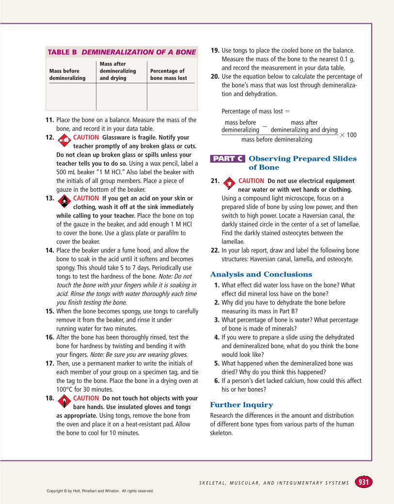
TABLE B DEMINERALIZATION OF A BONE
Mass before demineralizing
Mass after demineralizingand drying
Percentage ofbone mass lost
Copyright © by Holt, Rinehart and Winston. All rights reserved.