Scottish Adaptation of the European Pressure Ulcer ... Grading Tool.pdf · NATVNS Scottish...
1
NATVNS Scottish Adaptation of the European Pressure Ulcer Advisory Panel (EPUAP) Pressure Ulcer Classification Tool Early warning sign - Blanching erythema Areas of discoloured tissue that blanch when fingertip pressure is applied and the colour recovers when pressure released, indicating damage is starting to occur but can be reversed. On darkly pigmented skin blanching does not occur and changes to colour, temperature and texture of skin are the main indicators. Grade 1 - Non Blanchable Erythema Intact skin with non-blanchable redness, usually over a bony prominence. Darker skin tones may not have visible blanching but the colour may differ from the surrounding area. The affected area may be painful, firmer, softer, warmer or cooler than the surrounding tissue. Grade 2 - Partial thickness skin loss Loss of the epidermis/dermis presenting as a shallow open ulcer with a red/pink wound bed without slough or bruising.* May also present as an intact or open/ruptured blister. Grade 3 - Full thickness skin loss Subcutaneous fat may be visible but bone, tendon or muscle is not visible or palpable. Slough may be present but does not obscure the depth of tissue loss. May include undermining or tunnelling. ** Grade 4 - Full Thickness Tissue Loss Extensive destruction with exposed or palpable bone, tendon or muscle. Slough may be present but does not obscure the depth of tissue loss. Often includes undermining or tunnelling.** Suspected Deep Tissue Injury: Epidermis will be intact but the affected area can appear purple or maroon or be a blood filled blister over a dark wound bed. Over time this skin will degrade and develop into deeper tissue loss. Once grade can be established this must be documented. Ungradable: Full thickness skin / tissue loss where the depth of the ulcer is completely obscured by slough and / or necrotic tissue. Until enough slough and necrotic tissue is removed to expose the base of the wound the true depth cannot be determined. It may be a Grade 3 or 4 once debrided. Once grade can be established this must be documented. Combination Lesions: These are lesions where a combination of pressure and moisture contribute to the tissue breakdown. They still need to be graded as pressure damage as above but awareness of other causes and treatments is needed. See Excoriation & Moisture Related Skin Damage Tool *Bruising can indicate deep tissue injury **The depth of a Grade 3 or 4 pressure ulcer varies by anatomical location. Areas such as the bridge of the nose, ears, occiput and malleolus do not have fatty tissue so the depth of these ulcers may be shallow. In contrast areas which have excess fatty tissue can develop deep Grade 3 pressure ulcers where bone, tendon, muscle is not directly visible or palpable. Ref: European Pressure Ulcer Advisory Panel and National Pressure Ulcer Advisory Panel. (2009) Prevention and treatment of pressure ulcers: quick reference guide. National Pressure Ulcer Advisory Panel, Washington DC NHS Quality Improvement Scotland (2009) Best Practice Statement: Prevention and management of pressure ulcers. NHS Quality Improvement Scotland, Edinburgh 268608
Transcript of Scottish Adaptation of the European Pressure Ulcer ... Grading Tool.pdf · NATVNS Scottish...
NATVNS
Scottish Adaptation of the European Pressure Ulcer Advisory Panel (EPUAP) Pressure Ulcer Classification Tool
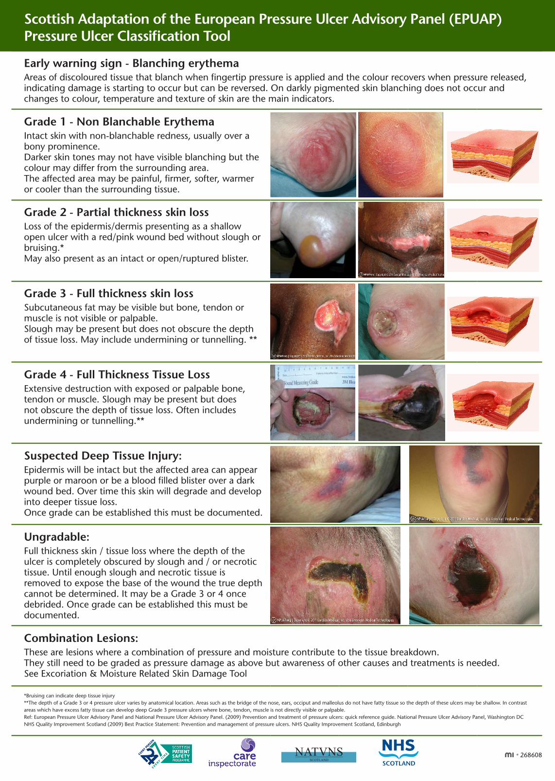
Early warning sign - Blanching erythemaAreas of discoloured tissue that blanch when fingertip pressure is applied and the colour recovers when pressure released, indicating damage is starting to occur but can be reversed. On darkly pigmented skin blanching does not occur and changes to colour, temperature and texture of skin are the main indicators.
Grade 1 - Non Blanchable ErythemaIntact skin with non-blanchable redness, usually over a bony prominence.Darker skin tones may not have visible blanching but the colour may differ from the surrounding area.The affected area may be painful, firmer, softer, warmer or cooler than the surrounding tissue.
Grade 2 - Partial thickness skin lossLoss of the epidermis/dermis presenting as a shallow open ulcer with a red/pink wound bed without slough or bruising.*May also present as an intact or open/ruptured blister.
Grade 3 - Full thickness skin lossSubcutaneous fat may be visible but bone, tendon or muscle is not visible or palpable.Slough may be present but does not obscure the depth of tissue loss. May include undermining or tunnelling. **
Grade 4 - Full Thickness Tissue LossExtensive destruction with exposed or palpable bone, tendon or muscle. Slough may be present but does not obscure the depth of tissue loss. Often includes undermining or tunnelling.**
Suspected Deep Tissue Injury:Epidermis will be intact but the affected area can appear purple or maroon or be a blood filled blister over a dark wound bed. Over time this skin will degrade and develop into deeper tissue loss. Once grade can be established this must be documented.
Ungradable:Full thickness skin / tissue loss where the depth of the ulcer is completely obscured by slough and / or necrotic tissue. Until enough slough and necrotic tissue is removed to expose the base of the wound the true depth cannot be determined. It may be a Grade 3 or 4 once debrided. Once grade can be established this must be documented.
Combination Lesions:These are lesions where a combination of pressure and moisture contribute to the tissue breakdown.They still need to be graded as pressure damage as above but awareness of other causes and treatments is needed. See Excoriation & Moisture Related Skin Damage Tool
*Bruising can indicate deep tissue injury**The depth of a Grade 3 or 4 pressure ulcer varies by anatomical location. Areas such as the bridge of the nose, ears, occiput and malleolus do not have fatty tissue so the depth of these ulcers may be shallow. In contrast areas which have excess fatty tissue can develop deep Grade 3 pressure ulcers where bone, tendon, muscle is not directly visible or palpable.Ref: European Pressure Ulcer Advisory Panel and National Pressure Ulcer Advisory Panel. (2009) Prevention and treatment of pressure ulcers: quick reference guide. National Pressure Ulcer Advisory Panel, Washington DCNHS Quality Improvement Scotland (2009) Best Practice Statement: Prevention and management of pressure ulcers. NHS Quality Improvement Scotland, Edinburgh
Scottish Adapted European Pressure Ulcer Advisory Panel (EPUAP)
Grading Tool
Grade 2Partial thickness skin loss involving epidermis, dermis, or both. The ulcer is superficial and presents clinically as an abrasion or blister
Grade 3Full thickness skin loss involving damage to or necrosis of subcutaneous tissue that may extend down to, but not through underlying fascia
Grade 4 Extensive destruction, tissue necrosis, or damage to muscle, bone, or supporting structures with or without full thickness skin loss
Epidermis
Dermis
Hypodermis
Bone
Grade 1Non-blanchable erythema (redness) of intact skin.Discolouration of the skin, warmth, oedema, induration or hardness may also be used as indicators, particularly on individuals with darker skin
Progression of a pressure ulcer
Epidermis
Dermis
Hypodermis
Bone
www.tissueviabilityonline.com/pu Images: Colin Blain Medical Photographer Inverclyde Royal Hospital (IRH) Greenock / Science Photo Library
Epidermis
Dermis
Hypodermis
Bone
Epidermis
Dermis
Hypodermis
Bone
268608


![Pressure ulcer prevention[2]](https://static.fdocuments.net/doc/165x107/55894026d8b42ab55b8b467a/pressure-ulcer-prevention2.jpg)