PRESSURE ULCER PREVENTION, ASSESSMENT AND MANAGEMENT · PDF filePRESSURE ULCER PREVENTION,...
24
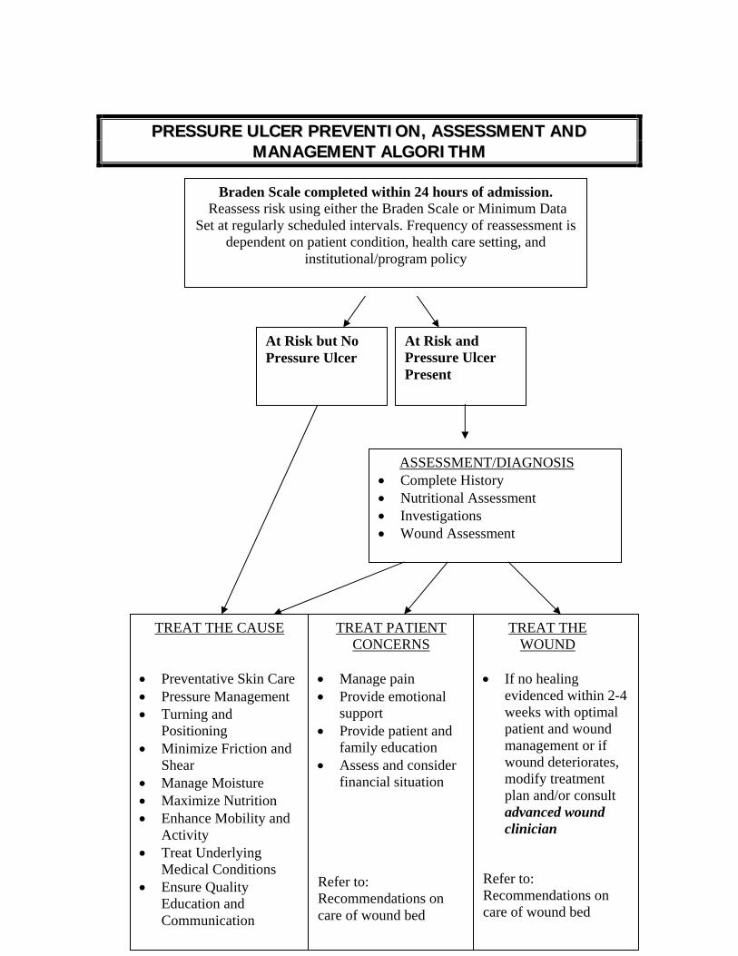
PRESSURE ULCER PREVENTION, ASSESSMENT AND MANAGEMENT ALGORITHM Braden Scale completed within 24 hours of admission. Reassess risk using either the Braden Scale or Minimum Data Set at regularly scheduled intervals. Frequency of reassessment is dependent on patient condition, health care setting, and institutional/program policy At Risk but No Pressure Ulcer At Risk and Pressure Ulcer Present TREAT THE CAUSE • Preventative Skin Care • Pressure Management • Turning and Positioning • Minimize Friction and Shear • Manage Moisture • Maximize Nutrition • Enhance Mobility and Activity • Treat Underlying Medical Conditions • Ensure Quality Education and Communication TREAT PATIENT CONCERNS • Manage pain • Provide emotional support • Provide patient and family education • Assess and consider financial situation Refer to: Recommendations on care of wound bed TREAT THE WOUND • If no healing evidenced within 2-4 weeks with optimal patient and wound management or if wound deteriorates, modify treatment plan and/or consult advanced wound clinician Refer to: Recommendations on care of wound bed ASSESSMENT/DIAGNOSIS • Complete History • Nutritional Assessment • Investigations • Wound Assessment
Transcript of PRESSURE ULCER PREVENTION, ASSESSMENT AND MANAGEMENT · PDF filePRESSURE ULCER PREVENTION,...
34
PPRREESSSSUURREE UULLCCEERR PPRREEVVEENNTTIIOONN,, AASSSSEESSSSMMEENNTT AANNDD MMAANNAAGGEEMMEENNTT AALLGGOORRIITTHHMM
//
Braden Scale completed within 24 hours of admission. Reassess risk using either the Braden Scale or Minimum Data
Set at regularly scheduled intervals. Frequency of reassessment is dependent on patient condition, health care setting, and
institutional/program policy
At Risk but No Pressure Ulcer
At Risk and Pressure Ulcer Present
TREAT THE CAUSE • Preventative Skin Care • Pressure Management • Turning and
Positioning • Minimize Friction and
Shear • Manage Moisture • Maximize Nutrition • Enhance Mobility and
Activity • Treat Underlying
Medical Conditions • Ensure Quality
Education and Communication
TREAT PATIENT CONCERNS
• Manage pain • Provide emotional
support • Provide patient and
family education • Assess and consider
financial situation Refer to: Recommendations on care of wound bed
TREAT THE WOUND
• If no healing
evidenced within 2-4 weeks with optimal patient and wound management or if wound deteriorates, modify treatment plan and/or consult advanced wound clinician
Refer to: Recommendations on care of wound bed
ASSESSMENT/DIAGNOSIS • Complete History • Nutritional Assessment • Investigations • Wound Assessment
35
PPRREESSSSUURREE UULLCCEERR INTRODUCTION • Prevention of pressure ulcers is of utmost importance due to the significant impact on
quality of life and health care resources. Most pressure ulcers can be prevented. • A pressure ulcer is any lesion caused by unrelieved pressure, friction and/or shear
that results in damage to the skin and underlying tissue. • Tissues overlying bony prominences are at highest risk of pressure damage especially
tissues overlying the sacrum, coccyx, heel, ischial tuberosity, malleolus, greater trochanter, occiput, scapula, vertebrae, knee and elbow. Previous surgical sites/scars are also at risk for pressure ulcer development.
• Consider all bed-or chair-bound patients, or those whose ability to independently reposition is impaired, to be at risk for pressure ulcers. Key predisposing risk factors include:
Intrinsic Factors: Previous history of pressure ulcer, malnutrition, dehydration, excessive perspiration/wound exudate, urinary/fecal incontinence, decreased sensory perception, altered mental status, decreased mobility, premature infants, age>70 years, altered blood pressure, impaired circulation, increased temperature (either internal to the patient or at the patient/surface interface), gender, body build, and co-existing health conditions/acute illness (malignancy, diabetes, stroke, pneumonia, heart failure, sepsis, hypotension, renal failure, anemia, immune compromised)
Extrinsic Factors: Treatment protocols, failure to recognize risk, patient handling techniques, use of restraints, hygiene, medications, emotional stress, and smoking
ASSESSMENT AND DIAGNOSIS • Complete History
• Cause, duration, history, and treatment of previous and current pressure ulcers
• Co-existing health conditions • Medications especially those that may impair healing (e.g. systemic
corticosteroids, chemotherapeutic agents and nonsteroidal anti-inflammatories) or cause sedation (e.g. opioids, benzodiazepines, muscle relaxants, hypnotics)
• Positioning, posture, and related equipment
36
• Patient’s ability and motivation to comprehend and adhere to the treatment program including cognition, learning ability and depression
• Available resources including caregiver support and finances • Pain (refer to recommendations for care of wound bed and
recommendations for malignant wounds) • Impact of patient’s quality of life
• Nutritional Assessment • Measure height, monitor weight at regularly scheduled intervals (weekly if
possible in acute care setting, monthly in long term care) • Monitor fluid and nutrient intake (% of meals eaten, calorie counts, etc.) • Refer to Registered Dietitian if:
• Patient has stage III or stage IV pressure ulcer(s) • Patient has stage I or stage II pressure ulcer(s) and a history of
weight loss greater than 10%. To calculate: usual body weight – current body weight x 100 = %weight loss
usual body weight • Patient is at high risk for pressure ulcer development and nutritional
concerns are present • Complete nutritional assessment by a Registered Dietitian includes
biochemical assessment, diet/intake history, weight history, physical exam, nutritional diagnosis, estimation of nutrient requirements, nutrition planning, and on-going evaluation.
• Investigations • Should be based on patient assessment, identified risk factors, severity of
pressure ulcers and may include any of the following: • Physical Exam • Blood Pressure • CBC, Urinalysis if indicated • Pre-albumin in serial measurements (once weekly until normal
values achieved) to assess nutritional status where opportunity to improve nutritional status exists
• Hgb A1c, Glucose (to determine adequacy of glycemic control where appropriate)
• Wound Culture (refer to recommendations on care of wound bed) • X-Ray/Erythrocyte Sedimentation Rate (ESR) (if osteomyelitis suspected);
Bone Scan (if X-ray/ESR inconclusive) Note: ESR, bonescan, and X-ray may be inconclusive as other inflammatory conditions may affect results
• Risk Assessment • Complete a risk assessment using the Braden Scale for Predicting Pressure
Ulcer Risk within 24 hours of admission • Reassess risk using either the Braden Scale or the Minimum Data Set at
regularly scheduled intervals. Frequency of re-assessment dependent on
37
patient’s condition, health care setting, and institutional/program policy (refer to Appendix A for a copy of the Braden Scale)
• Risk should be interpreted in the context of the full patient profile (age, acuity of illness, co-morbidity, medications, psychosocial well-being, surface support, posture) and the patient’s goals
• Risk assessments should be documented and made accessible to all members of the health care team
• Wound Assessment
• Stage ulcer according to the National Pressure Ulcer Advisory Panel (NPUAP) injury severity guidelines, 2003. Staging can only occur after necrotic tissue has been removed allowing complete visualization of the ulcer bed.
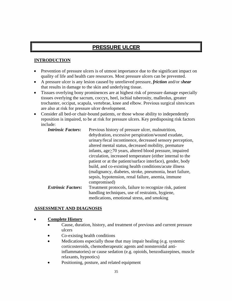
Stage I: Pressure ulcer is an observable pressure-related alteration of intact skin whose indicators as compared to an adjacent or opposite area on the body may include changes in one or more of the following: skin temperature (warmth or coolness), tissue consistency (firm or boggy feel), and/or sensation (pain, itching).
The ulcer appears as a defined area of persistent redness in lightly
pigmented skin, whereas in darker skin tones, the ulcer may appear with persistent red, blue, or purple hues.
Stage I pressure ulcer
38
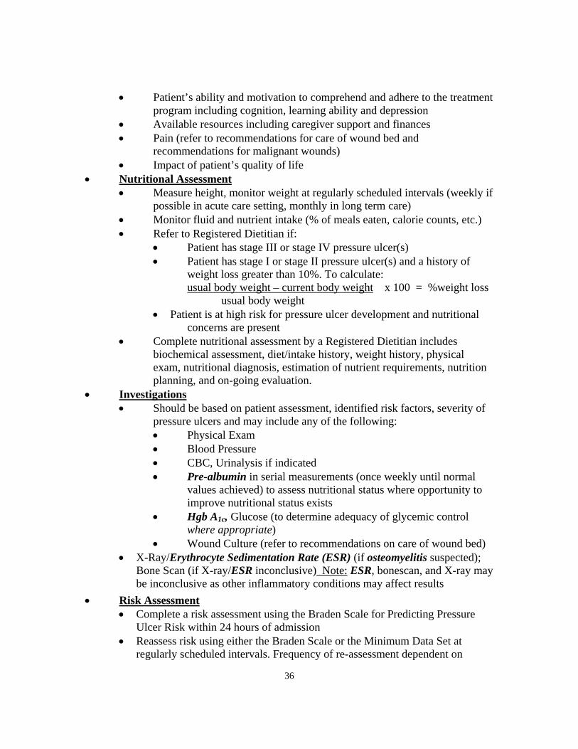
Stage II: Partial-thickness skin loss involving epidermis and/or dermis. The ulcer is superficial and presents clinically as an abrasion, blister, or shallow crater
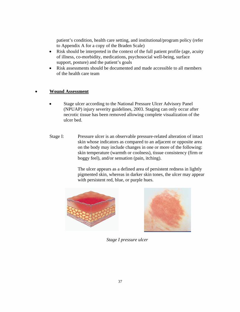
Stage II pressure ulcer Stage III: Full thickness skin loss involving damage or necrosis of
subcutaneous tissue that may extend down to, but not through, underlying fascia. The ulcer presents clinically as a deep crater with or without undermining of adjacent tissue
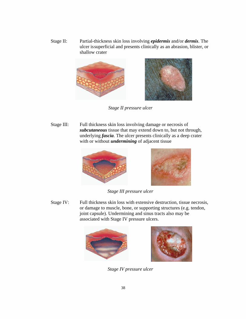
Stage III pressure ulcer Stage IV: Full thickness skin loss with extensive destruction, tissue necrosis,
or damage to muscle, bone, or supporting structures (e.g. tendon, joint capsule). Undermining and sinus tracts also may be associated with Stage IV pressure ulcers.
Stage IV pressure ulcer
39
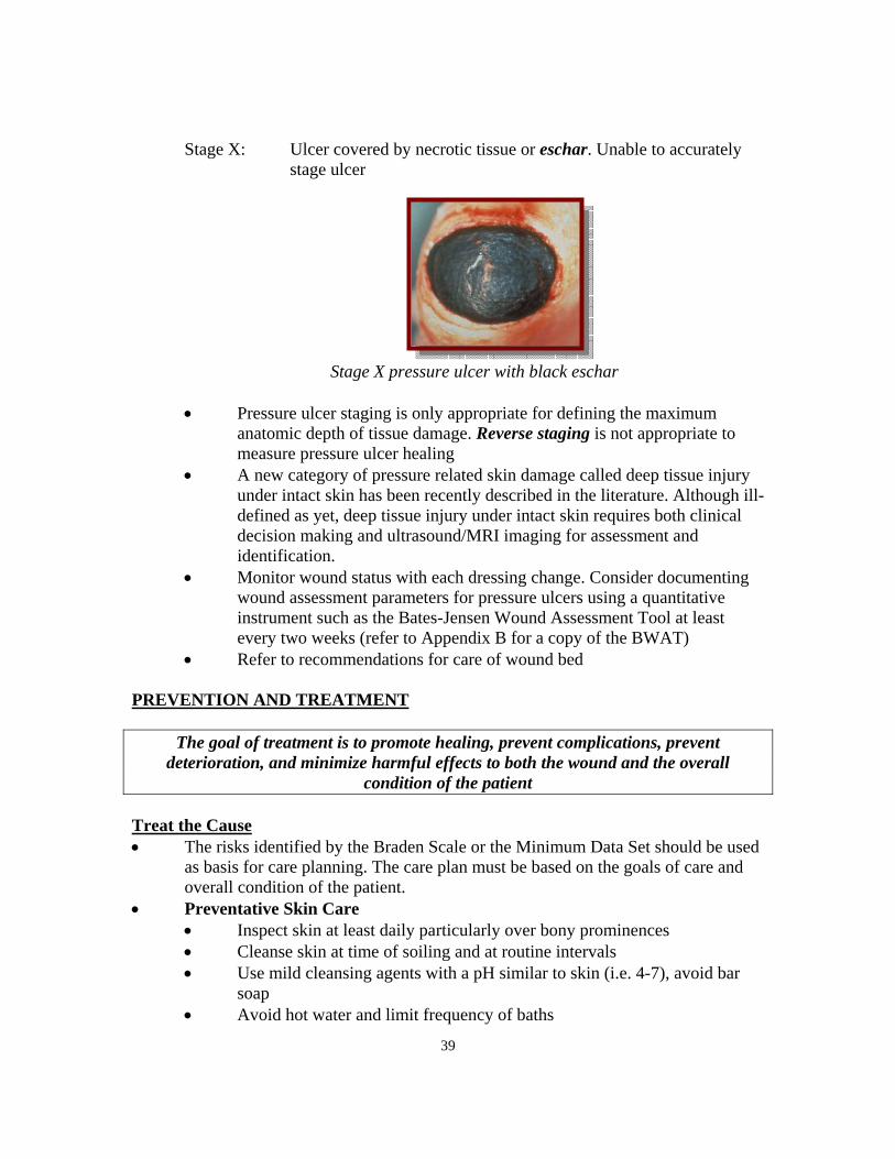
Stage X: Ulcer covered by necrotic tissue or eschar. Unable to accurately stage ulcer
Stage X pressure ulcer with black eschar
• Pressure ulcer staging is only appropriate for defining the maximum anatomic depth of tissue damage. Reverse staging is not appropriate to measure pressure ulcer healing
• A new category of pressure related skin damage called deep tissue injury under intact skin has been recently described in the literature. Although ill-defined as yet, deep tissue injury under intact skin requires both clinical decision making and ultrasound/MRI imaging for assessment and identification.
• Monitor wound status with each dressing change. Consider documenting wound assessment parameters for pressure ulcers using a quantitative instrument such as the Bates-Jensen Wound Assessment Tool at least every two weeks (refer to Appendix B for a copy of the BWAT)
• Refer to recommendations for care of wound bed
PREVENTION AND TREATMENT
The goal of treatment is to promote healing, prevent complications, prevent deterioration, and minimize harmful effects to both the wound and the overall
condition of the patient Treat the Cause • The risks identified by the Braden Scale or the Minimum Data Set should be used
as basis for care planning. The care plan must be based on the goals of care and overall condition of the patient.
• Preventative Skin Care • Inspect skin at least daily particularly over bony prominences • Cleanse skin at time of soiling and at routine intervals • Use mild cleansing agents with a pH similar to skin (i.e. 4-7), avoid bar
soap • Avoid hot water and limit frequency of baths
40
• Apply moisturizers to skin at least daily • For sensitive skin, avoid all products containing alcohol, perfumes, lanolin
and other potential sensitizers to avoid sensitivity or allergic reactions • Minimize environmental factors leading to skin drying when possible (low
humidity, exposure to cold). Heat lamps should be avoided. • Do not massage red or bony prominences
• Pressure Management • Relieve constant pressure over at risk areas, at site of existing ulcer, at site
of previously healed ulcer, and over scars • Investigate all possible sources of pressure. Assess all surfaces used by the
patient including bed, wheelchair, dining room chairs, recliners, toilet, stretchers, operating room tables, etc.
• Avoid use of donut type devices, water-filled gloves, IV bags, and synthetic sheepskin for pressure reduction
• Use Preventative pressure management mattress/seat cushion for all at risk patients (refer to Appendix C)
• Therapeutic pressure management mattress/seat cushion may be indicated depending on location and severity of pressure ulcer(s), number of available turning surfaces, pain and mobility (refer to Appendix C)
• If preventative or therapeutic pressure management mattress/seat cushion are in place, ensure sheets/covers are loose and extensible. Avoid multiple layers of sheets, soakers, mattress toppers, overlays, etc. as will impede the pressure redistributing ability of the surface.
• Check conditions of all mattresses and cushions. For pressure management mattress overlays and seat cushions, check for “bottoming out”
• Advance notice of the transfer should be given when transferring a patient between settings if pressure management equipment is required to be in place at time of transfer, e.g. mattresses, seating, special transfer equipment. Transfer to another setting may require a site visit, client/family conference, and/or assessment for funding of resources to prevent the development of pressure ulcers.
• Consult Occupational Therapist, Physiotherapist, Advanced Wound Clinician for those patients who are at moderate to high risk of developing a pressure ulcer or who have existing or recently healed pressure ulcers
• Turning and Positioning • Evaluate bed mobility and develop a turning schedule based on identified
risk. Individualized positioning regime and repositioning schedule must be documented and displayed.
• If the patient is able to make large body movements easily and frequently: Monitor bed mobility and ensure adequate turning every 3-4 hours
• If the patient is able to make small body shifts but is unable to make large body movements: Reposition every 2 hours. Use positioning
41
devices to position the patient in a 30-degree laterally inclined position when repositioned to either side (see picture below). Avoid 900 side-lying position
• If the patient is unable to make any independent movement: Turn every 2 hours or more frequently if indicated. May require therapeutic pressure management mattress. Please note that a patient on a therapeutic pressure management mattress should still be turned and repositioned regularly as per individualized positioning regime.
• Use positioning devices to prevent contact between bony prominences • Completely relieve heel pressure when in bed. Support length of legs with
a pillow and allow heels to drop off pillow. Alternatively, consult Occupational Therapy or Physiotherapy for heel positioning devices. Monitor to prevent foot drop.
• For patients restricted to chairs: • Consider postural alignment, distribution of weight, balance,
stability, and pressure reduction capabilities of all seating surfaces used by patient (wheelchair, recliner, dining chair, etc.)
• Avoid positioning the wheelchair seated patient directly on a pressure ulcer
• Teach patient to shift weight every 15 minutes. The “forward lean” (i.e. bringing one’s chest towards one’s knees/lap) is the most effective and easiest method of weight shift.
• If the patient is unable to perform weight shifts, reposition q 1 hour. If this is not possible, return the patient to bed.
• Ensure the wheelchair cushion is positioned and functioning properly
• Consult Occupational Therapy or Physiotherapy for seating assessment
• Minimize Friction and Shear • Maintain head of the bed at the lowest elevation consistent with medical
conditions and restrictions. A 30 degree elevation or lower is recommended. If the head of the bed is elevated higher than 30 degrees, flex knee gatch slightly to prevent sliding and closely monitor skin on sacrum. As well, after elevating the head of the bed, briefly lifting the trunk away from the bed surface releases skin tension and reduces shearing forces.
• Use transfer techniques that decrease shear when indicated (i.e. nylon sliders, transfer board, trapeze, mechanical lifts). Avoid leaving slings under the patient
• Keep linens flat, free from stray objects • Use turning sheets, do not drag the patient when repositioning
42
• Protect elbows and heels if being exposed to friction (i.e. transparent films, socks, pillows, foam blocks, heel booties, etc.)
• Manage Moisture • Use commercial moisture barriers, barrier films, transparent dressings • Assess and treat urinary and fecal incontinence • If patient has diarrhea, identify and treat the cause (i.e. C. difficile) • Use absorbent pads or briefs that wick and hold moisture away from the
skin • Avoid use of plastic sheets • Wear breathable clothing
• Maximize Nutrition • Consult dietitian as indicated • Provide adequate nutrients to promote wound healing
• Calories 30-35 kcal/kg/day • Protein 1.2-1.5 g/kg/day • Fluid 30-35 ml/kg/day or 1 ml/kcal/day • Micronutrients per Recommended Dietary Allowance (RDA) or
Dietary Reference Intake (DRI) – (refer to http://www.hc-sc.gc.ca/fn-an/nutrition/reference/index_e.html)
• Ensure balanced nutrition with consumption of foods from all food groups per Canada’s Food Guide for Healthy Eating (refer to http://www.hc-sc.gc.ca/fn-an/food-guide-aliment/fg_rainbow-arc_en_ciel_ga_e.html)
• A patient who consistently consumes less than 75% of his/her meals may benefit from an oral nutritional supplement such as Ensure, Boost, Carnation Breakfast Anytime, Resource, etc.
• Offer fluids when turning, repositioning, administering medications, etc. • Consider the need for a multivitamin with minerals • Additional micronutrients such as zinc and vitamin C may be considered
with clinical suspicion of deficiency or inadequate intake of foods rich in these micronutrients
• Identify and address possible causes of inadequate intake (e.g. ensure teeth are in good condition and fit properly; consult Speech Language Pathologist if difficulties swallowing; provide assistance with meals as needed, etc.)
• Enhance Mobility/Activity • Consult Physiotherapist, Occupational Therapist, Recreational Therapist,
Activity Worker as indicated • Encourage walking, activity as indicated to prevent further deconditioning
• Treat Underlying Medical Conditions • Wherever possible, treat specific medical conditions that may be causing
or contributing to wound development or impeding wound healing • Ensure Quality Education and Communication
43
• Educational programs for the prevention of pressure ulcers should be structured, organized, and comprehensive and directed at all levels of health care providers.
• Educational programs for the prevention of pressure ulcers should include information on the following items: • The etiology and risk factors predisposing to pressure ulcer
development • The Braden Scale and the Minimum Data Set and their relevance
to planning care • Skin assessment • Staging of pressure ulcers • Selection and/or use of support surfaces • Development and implementation of an individualized skin care
program • Demonstration of positioning/transferring techniques to decrease
risk of tissue breakdown • Instruction on accurate documentation of pertinent data
• Patients moving between care settings should have the following information provided: • Risk factors for pressure ulcer development • Skin condition prior to discharge • Type of bed/mattress and seating the patient requires • History of ulcers, previous treatments and dressings used • Stage, site, and size of existing ulcers • Type and frequency of current dressing • Any sensitivities or allergies to dressing products • Need for on-going nutritional support
Treat Patient Concerns • Manage pain (refer to recommendations on care of wound bed and malignant
wounds) • Provide emotional support, assess and consider financial situation. Consult Social
Work if indicated. Refer for peer counselling, support groups as appropriate. • Ensure patient is actively involved in developing care plan. • Provide patient and family education regarding:
• Etiology of pressure ulcers • How to inspect skin • Protection of skin • Proper, safe cleansing techniques and agents • Reduction of pressure ulcer risk • Role of nutrition in pressure ulcer prevention • Proper positioning techniques, proper use of positioning devices
44
• Skin and other health status changes to be reported to health care professionals
Treat the Wound • Refer to recommendations on care of wound bed • Assess and manage complications as indicated (e.g. infection, pain) • If no healing evidenced within 2-4 weeks with optimal patient and wound
management or if wound deteriorates, modify treatment plan and/or consult an advanced wound clinician
• Surgical repair of pressure ulcers may be indicated for patients with complex, stage III pressure ulcers (i.e. undermining, tracts) or stage IV pressure ulcers unresponsive to optimal care. The decision to refer a patient for surgical evaluation should be based on the patient’s overall burden of illness and prognosis, care goals, quality of life, and the expected functional outcomes.
• Electrical stimulation of chronic pressure ulcers that are not responsive to conventional therapy has been shown to be effective. Other adjuvant therapies that may be effective include: • Negative pressure therapy and normothermic therapies • Therapeutic ultrasound • Ultraviolet light • Pulsed electromagnetic fields • Growth factors and skin equivalents
45
References American Medical Directors Association. (1999). Pressure ulcer therapy companion. Columbia, MD: Author. Arnold, M. (2003). Pressure ulcer prevention and management: The current evidence for care. AACN Clinical Issues, 14(4), 411-28. Ayello, EA. (2003). Preventing pressure ulcers and skin tears. In: Mezey M, Fulmer T, Abraham, I, Zwicker, DA (eds), Geriatric nursing protocols for best practice (2nd edition). New York, NY: Springer Publishing Company Inc. Barton, P. & Parslow, N. (1996). Pressure ulcers. In Wound care: A comprehensive guide for community nurses (pp. 19-29). Markham, ON: Saint Elizabeth Health Care. Bergstrom, N., Allman, R.M., Alvarez, O.M., Bennett, M.A., Carlson, C.E., Frantz, C.E. et. al. (1995). Pressure ulcer treatment: Quick reference guide for clinicians. Advances in Wound Care, 8, 22-24. Cullum, N., Deeks, J., Sheldon, T.A., Song, F., & Fletcher, A.W. (2002). Beds, mattresses and cushions for pressure sore prevention and treatment (Cochrane Review). In The Cochrane Library, Issue 2. Oxford: Update Software. Dolynchuk, K., Keast, D., Campbell, K., Houghton, P., Orsted, H., Sibbald, G., & Atkinson A. (2000). Best practices for the prevention and treatment of pressure ulcers. Ostomy/Wound Management, 46, 38-52. Folkedahl BA, & Frantz, R. (2002). Prevention of pressure ulcers. Iowa City, IA: University of Iowa Gerontological Nursing Interventions Research Centre, Research Dissemination Core. Folkedahl BA, & Frantz, R. (2002). Treatment of pressure ulcers. Iowa City, IA: University of Iowa Gerontological Nursing Interventions Research Centre, Research Dissemination Core. Frias, L, Vazquez, M, & Perez-Portabella, C. (2004). The effectiveness of oral nutritional supplementation in the healing of pressure ulcers. Journal of Wound Care, 1(13), 319-22. Gray, M. (2003). Does oral zinc supplementation promote healing of chronic wounds? Journal of Wound, Ostomy, Continence Nursing, 30, 296-8. Gray, M. (2003). Does vitamin C supplementation promote pressure ulcer healing? Journal of Wound, Ostomy, Continence Nursing, 30, 245-9.

46
Guenter, P., Malyszek, R., Zimmaro, D., et al. (2000). Survey of nutritional status in newly hospitalized patients with stage III or stage IV pressure ulcers. Advances in Skin and Wound Care, 13, 164-8. Joanna Briggs Institute for Evidence Based Nursing and Midwifery (1997). Pressure sores B Part I: Prevention of pressure related damage. Best Practice: Information Sheets for Health Professionals, 1(1), 1-6. Retrieved from http://www.joannabriggs.edu.au/bpwound1.pdf Joanna Briggs Institute for Evidence Based Nursing and Midwifery (1997). Pressure sores B Part II: Management of pressure related tissue damage. Best Practice: Information Sheets for Health Professionals, 1(2), 1-6. Retrieved from http://www.joannabriggs.edu.au/bpwound2.pdf Makelbust, J., and Siegreen, M. Pressure Ulcers: Guidelines for Prevention and Nursing Management, 78-85. National Collaborating Centre for Nursing and Supportive Care. (2003). Pressure ulcer risk assessment and prevention, including the use of pressure relieving devices (beds, mattresses and overlays) for the prevention of pressure ulcers in primary and secondary care. London, UK: National Institute for Clinical Excellence. National Pressure Ulcer Advisory Panel (2003). NPUAP Staging Report. Reston, VA: Author. Retrieved from http://www.npuap.org/positn6.html National Pressure Ulcer Advisory Panel (2000). The facts about Reverse staging in 2000: The NPUAP position statement. Reston, VA: Author. Retrieved from http://www.npuap.org/positn5.htm National Pressure Ulcer Advisory Panel (1992). Statement on Pressure Ulcer Prevention. Reston, VA: Author. Retrieved from http://www.npuap.org/positn1.html Nova Scotia Department of Health. (2000). Pressure Ulcers. In Evidence-based wound management protocol (pp.21-30). Halifax, NS: Department of Health. Ovington, L.G. (1999). Dressings and adjunctive therapies: AHCPR guidelines revisited. Ostomy/Wound Management, 45, 94S-106S Paralyzed Veterans of America. (2000). Pressure ulcer prevention and treatment following spinal cord injury: A clinical practice guideline for health care professionals. Washington, DC: Author.
47
Peel Region Wound Management Committee (1998). Pressure ulcers. In P. Jackson (Ed.), An integrated approach to wound management manual: A Peel region initiative (pp. 9-11). Mississauga, ON: Author & Caremark Ltd., 1998. Pressure ulcers prevalence, cost and risk assessment: consensus development conference statement - The National Pressure Ulcer Advisory Panel (1989). Decubitus, 2, 24-28. Regional Wound Care Guidelines Working Group (1998). Pressure ulcers. In Regional Wound Care Guidelines (pp. 1-8). Edmonton, AB: Capital Health Authority. Registered Nurses Association of Ontario. (2002). Assessment and management of stage I to IV pressure ulcers. Toronto, ON: Author. Registered Nurses Association of Ontario. (2001). Risk assessment and the prevention of pressure ulcers. Toronto, ON: Author. Retrieved from http://www.rnao.org/bestpractices/PDF/BPG_Pressure_Ulcers.pdf Royal College of Nursing. (2001). Pressure ulcer risk assessment and prevention. London: Author. Schmidt, T. (2002). Pressure ulcers: Nutrition strategies that make a difference. Caring, U.S. Department of Health and Human Services (1992). Pressure ulcers in adults: Prediction and prevention (AHCPR Publication No. 92-0047). Rockville, MD: Author. Van Rijswijk, L. & Braden, B.J. (1999). Pressure ulcer patient and wound assessment: an AHCPR clinical practice guideline update. Ostomy/Wound Management, 4, 56S-67S Weir, D. (2001). Pressure ulcers. In Diane Krasner, Gary Sibbald, & Geroge T. Rodeheaver (Eds.), Chronic wound care: A clinical source book for health care professionals (3rd ed., pp. 619-628). Malvern, PA: Health Management Publications. Williams, L. (2002). Assessing patients’ nutritional needs in the wound-healing process. Journal of Wound Care, 1, 225-8. Woodbury, M.G., Houghton, P.E., Campbell, K.E., & Keast, D.H. (1999). Pressure ulcer assessment instruments: A critical appraisal. Ostomy/Wound Management, 45, 42-55. Wound, Ostomy, and Continence Nurses Society. (2003). Guideline for prevention and management of pressure ulcers. Glenview, IL: Author.
48
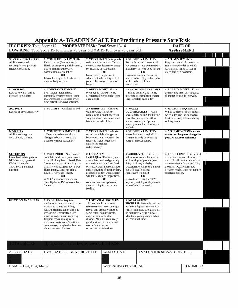
Appendix A- BRADEN SCALE For Predicting Pressure Sore Risk HIGH RISK: Total Score<12 MODERATE RISK: Total Score 13-14 LOW RISK: Total Score 15-16 if under 75 years old OR 15-18 if over 75 years old.
DATE OF ASSESSMENT
RISK FACTOR SCORE/DESCRIPTION 1 2 3 4 SENSORY PERCEPTION
Ability to respond meaningfully to pressure-related discomfort.
1. COMPLETELY LIMITED- Unresponsive (does not moan, flinch, or grasp) to painful stimuli, due to diminished level of consciousness or sedation
OR Limited ability to feel pain over most of body surface.
2. VERY LIMITED-Responds only to painful stimuli. Cannot communicate discomfort except by moaning or restlessness,
OR has a sensory impairment which limits the ability to feel pain or discomfort over ½ of body.
3. SLIGHTLY LIMITED-Responds to verbal commands but cannot always communicate discomfort or need to be turned,
OR Has some sensory impairment which limits ability to feel pain or discomfort in 1 or 2 extremities.
4. NO IMPAIRMENT-Responds to verbal commands. Has no sensory deficit which would limit ability to feel or voice pain or discomfort.
MOISTURE Degree to which skin is exposed to moisture
1. CONSTANTLY MOIST- Skin is kept moist almost constantly by perspiration, urine, etc. Dampness is detected every time patient is moved or turned.
2. OFTEN MOIST- Skin is often but not always moist. Linen must be changed at least once a shift.
3. OCCASIONALLY MOIST – Skin is occasionally moist, requiring an extra linen change approximately once a day.
4. RARELY MOIST – Skin is usually dry; linen only requires changing at routine intervals.
ACTIVITY Degree of physical activity.
1. BEDFAST – Confined to bed. 2. CHAIRFAST – Ability to walk severely limited or nonexistent. Cannot bear own weight and/or must be assisted into chair or wheelchair..
3. WALKS OCCASIONALLY – Walks occasionally during day but for very short distances, with or without assistance. Spends majority of each shift in bed or in chair.
4. WALKS FREQUENTLY – Walks outside the room at least twice a day and inside room at least once every 2 hours during waking hours.
MOBILITY Ability to change and control body position.
1. COMPETELY IMMOBILE – Does not make even slight changes in body or extremity position without assistance.
2. VERY LIMITED – Makes occasional slight changes in body or extremity position but unable to make frequent or significant changes independently.
3. SLIGHTLY LIMITED- makes frequent though slight changes in body or extremity position independently.
4. NO LIMITATIONS- makes major and frequent changes in position without assistance
NUTRITION Usual food intake pattern NPO:Nothing by mouth IV: Intravenously TPN: Total parenteral nutrition
1. VERY POOR – Never eats a complete meal. Rarely eats more than 1/3 of any food offered. Eats 2 servings or less of protein (meat or dairy products) per day. Takes fluids poorly. Does not take a liquid dietary supplement,
OR is NPO1 and/or maintained on clear liquids or IV2 for more than 5 days.
2. PROBABLY INADEQUATE – Rarely eats a complete meal and generally eats only about ½ of any food offered. Protein intake includes only 3 servings of meat or dairy products per day. Occasionally will take a dietary supplement,
OR receives less than optimum amount of liquid diet or tube feeding.
3. ADEQUATE – Eats over half of most meals. Eats a total of 4 servings of protein (meat, dairy products) each day. Occasionally will refuse a meal, but will usually take a supplement if offered
OR is on a tube feeding or TPN3 regimen, which probably meets most of nutrition needs.
4. EXCELLENT – Eats most of every meal. Never refuses a meal. Usually eats a total of 4 or more servings of meat and dairy products. Occasionally eats between meals. Does not require supplementation.
FRICTION AND SHEAR 1. PROBLEM – Requires moderate to maximum assistance in moving. Complete lifting without sliding against sheets is impossible. Frequently slides down in bed or chair, requiring frequent repositioning with maximum assistance. Spasticity, contractures, or agitation leads to almost constant friction.
2. POTENTIAL PROBLEM – Moves feebly or requires minimum assistance. During a move, skin probably slides to some extent against sheets, chair restraints, or other devices. Maintains relatively good position in chair or bed most of the time but occasionally slides down.
3. NO APPARENT PROBLEM- Moves in bed and in chair independently and has sufficient muscle strength to lift up completely during move. Maintains good position in bed or chair at all times.
TOTAL SCORE Total score of 12 or less represents HIGH RISK
ASSESS DATE EVALUATOR SIGNATURE/TITLE ASSESS DATE EVALUATOR SIGNATURE/TITLE 1 3 2 4
NAME – Last, First, Middle ATTENDING PHYSICIAN
ID NUMBER
49
Appendix B
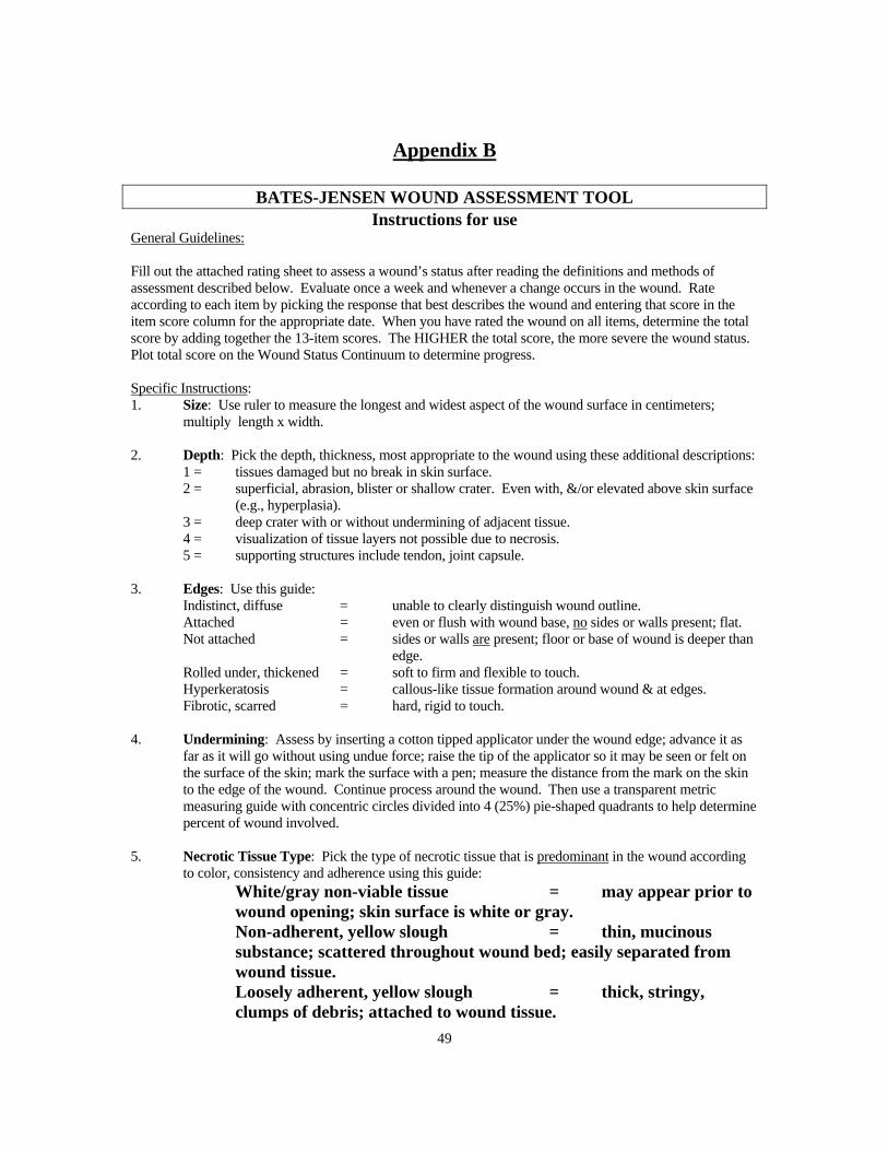
BATES-JENSEN WOUND ASSESSMENT TOOL Instructions for use
General Guidelines: Fill out the attached rating sheet to assess a wound’s status after reading the definitions and methods of assessment described below. Evaluate once a week and whenever a change occurs in the wound. Rate according to each item by picking the response that best describes the wound and entering that score in the item score column for the appropriate date. When you have rated the wound on all items, determine the total score by adding together the 13-item scores. The HIGHER the total score, the more severe the wound status. Plot total score on the Wound Status Continuum to determine progress. Specific Instructions: 1. Size: Use ruler to measure the longest and widest aspect of the wound surface in centimeters;
multiply length x width. 2. Depth: Pick the depth, thickness, most appropriate to the wound using these additional descriptions: 1 = tissues damaged but no break in skin surface. 2 = superficial, abrasion, blister or shallow crater. Even with, &/or elevated above skin surface
(e.g., hyperplasia). 3 = deep crater with or without undermining of adjacent tissue. 4 = visualization of tissue layers not possible due to necrosis. 5 = supporting structures include tendon, joint capsule. 3. Edges: Use this guide: Indistinct, diffuse = unable to clearly distinguish wound outline. Attached = even or flush with wound base, no sides or walls present; flat. Not attached = sides or walls are present; floor or base of wound is deeper than
edge. Rolled under, thickened = soft to firm and flexible to touch. Hyperkeratosis = callous-like tissue formation around wound & at edges. Fibrotic, scarred = hard, rigid to touch. 4. Undermining: Assess by inserting a cotton tipped applicator under the wound edge; advance it as
far as it will go without using undue force; raise the tip of the applicator so it may be seen or felt on the surface of the skin; mark the surface with a pen; measure the distance from the mark on the skin to the edge of the wound. Continue process around the wound. Then use a transparent metric measuring guide with concentric circles divided into 4 (25%) pie-shaped quadrants to help determine percent of wound involved.
5. Necrotic Tissue Type: Pick the type of necrotic tissue that is predominant in the wound according
to color, consistency and adherence using this guide: White/gray non-viable tissue = may appear prior to
wound opening; skin surface is white or gray. Non-adherent, yellow slough = thin, mucinous
substance; scattered throughout wound bed; easily separated from wound tissue.
Loosely adherent, yellow slough = thick, stringy, clumps of debris; attached to wound tissue.
50
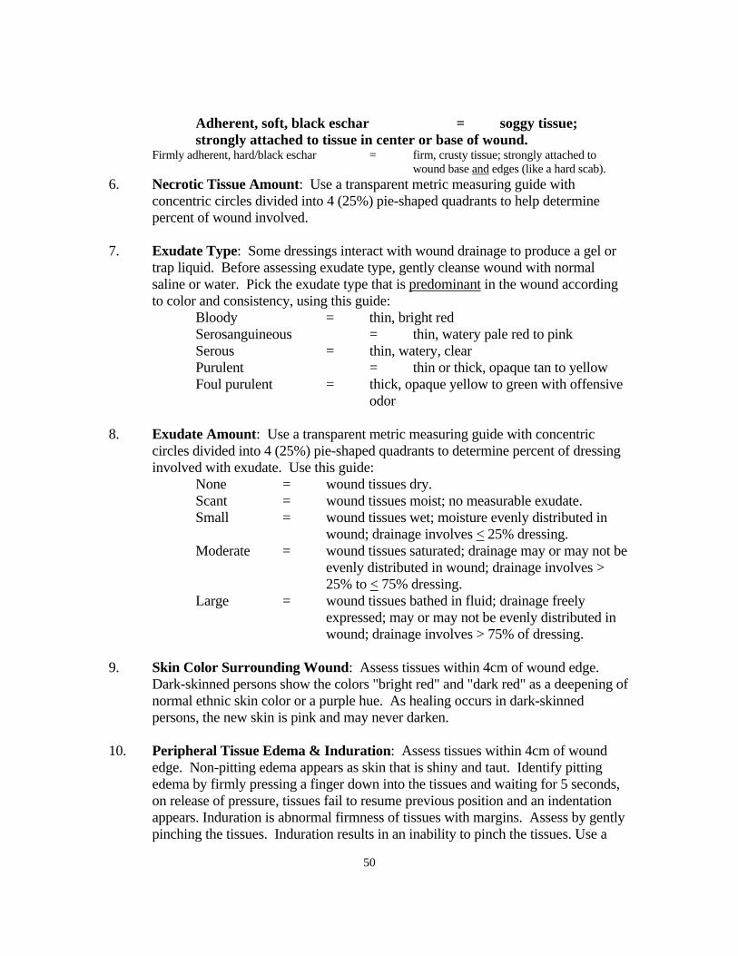
Adherent, soft, black eschar = soggy tissue; strongly attached to tissue in center or base of wound.
Firmly adherent, hard/black eschar = firm, crusty tissue; strongly attached to wound base and edges (like a hard scab).
6. Necrotic Tissue Amount: Use a transparent metric measuring guide with concentric circles divided into 4 (25%) pie-shaped quadrants to help determine percent of wound involved.
7. Exudate Type: Some dressings interact with wound drainage to produce a gel or
trap liquid. Before assessing exudate type, gently cleanse wound with normal saline or water. Pick the exudate type that is predominant in the wound according to color and consistency, using this guide:
Bloody = thin, bright red Serosanguineous = thin, watery pale red to pink Serous = thin, watery, clear Purulent = thin or thick, opaque tan to yellow Foul purulent = thick, opaque yellow to green with offensive
odor 8. Exudate Amount: Use a transparent metric measuring guide with concentric
circles divided into 4 (25%) pie-shaped quadrants to determine percent of dressing involved with exudate. Use this guide:
None = wound tissues dry. Scant = wound tissues moist; no measurable exudate. Small = wound tissues wet; moisture evenly distributed in
wound; drainage involves < 25% dressing. Moderate = wound tissues saturated; drainage may or may not be
evenly distributed in wound; drainage involves > 25% to < 75% dressing.
Large = wound tissues bathed in fluid; drainage freely expressed; may or may not be evenly distributed in wound; drainage involves > 75% of dressing.
9. Skin Color Surrounding Wound: Assess tissues within 4cm of wound edge.
Dark-skinned persons show the colors "bright red" and "dark red" as a deepening of normal ethnic skin color or a purple hue. As healing occurs in dark-skinned persons, the new skin is pink and may never darken.
10. Peripheral Tissue Edema & Induration: Assess tissues within 4cm of wound
edge. Non-pitting edema appears as skin that is shiny and taut. Identify pitting edema by firmly pressing a finger down into the tissues and waiting for 5 seconds, on release of pressure, tissues fail to resume previous position and an indentation appears. Induration is abnormal firmness of tissues with margins. Assess by gently pinching the tissues. Induration results in an inability to pinch the tissues. Use a

51
transparent metric measuring guide to determine how far edema or induration extends beyond wound.
11. Granulation Tissue: Granulation tissue is the growth of small blood vessels and
connective tissue to fill in full thickness wounds. Tissue is healthy when bright, beefy red, shiny and granular with a velvety appearance. Poor vascular supply appears as pale pink or blanched to dull, dusky red color.
12. Epithelialization: Epithelialization is the process of epidermal resurfacing and
appears as pink or red skin. In partial thickness wounds it can occur throughout the wound bed as well as from the wound edges. In full thickness wounds it occurs from the edges only. Use a transparent metric measuring guide with concentric circles divided into 4 (25%) pie-shaped quadrants to help determine percent of wound involved and to measure the distance the epithelial tissue extends into the wound.
© 2001 Barbara Bates-Jensen
52
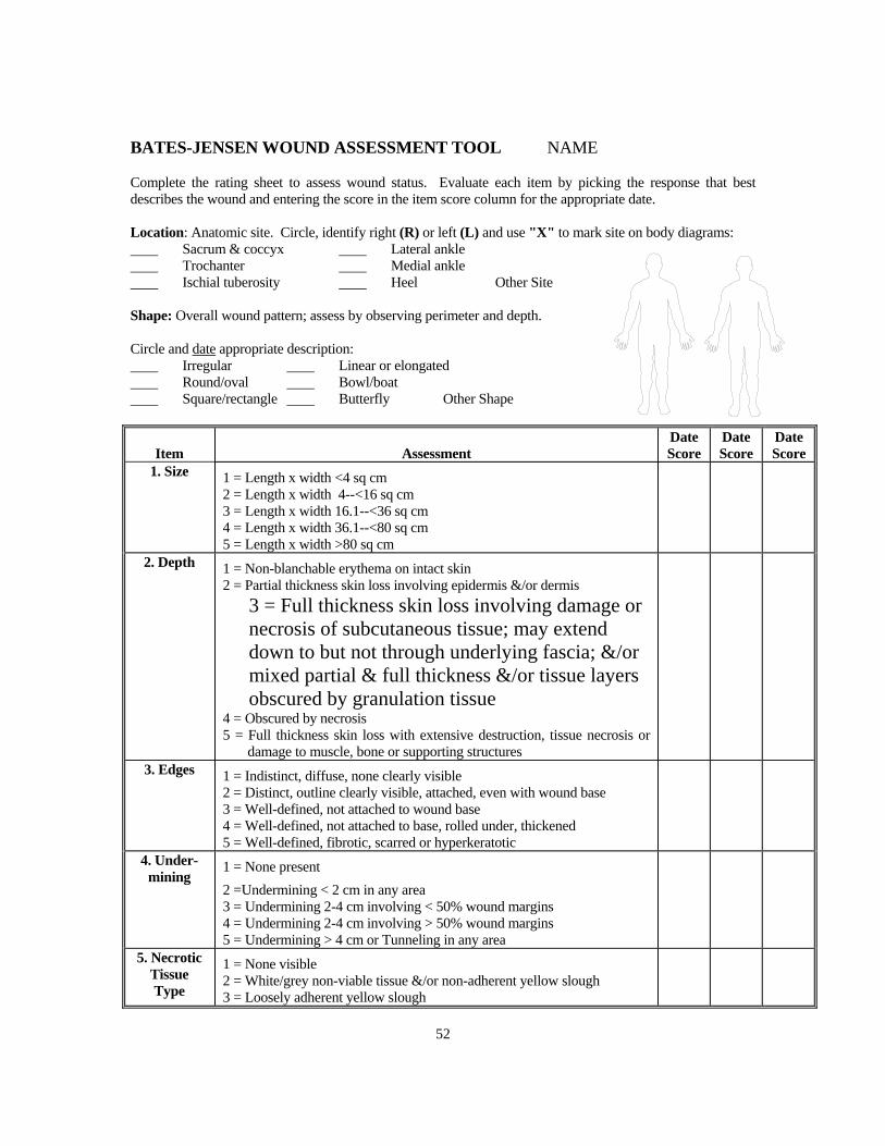
BATES-JENSEN WOUND ASSESSMENT TOOL NAME Complete the rating sheet to assess wound status. Evaluate each item by picking the response that best describes the wound and entering the score in the item score column for the appropriate date. Location: Anatomic site. Circle, identify right (R) or left (L) and use "X" to mark site on body diagrams: Sacrum & coccyx Lateral ankle Trochanter Medial ankle Ischial tuberosity Heel Other Site Shape: Overall wound pattern; assess by observing perimeter and depth. Circle and date appropriate description: Irregular Linear or elongated Round/oval Bowl/boat Square/rectangle Butterfly Other Shape
Item
Assessment
Date Score
Date Score
Date Score
1. Size 1 = Length x width <4 sq cm 2 = Length x width 4--<16 sq cm 3 = Length x width 16.1--<36 sq cm 4 = Length x width 36.1--<80 sq cm 5 = Length x width >80 sq cm
2. Depth 1 = Non-blanchable erythema on intact skin 2 = Partial thickness skin loss involving epidermis &/or dermis
3 = Full thickness skin loss involving damage or necrosis of subcutaneous tissue; may extend down to but not through underlying fascia; &/or mixed partial & full thickness &/or tissue layers obscured by granulation tissue
4 = Obscured by necrosis 5 = Full thickness skin loss with extensive destruction, tissue necrosis or
damage to muscle, bone or supporting structures
3. Edges 1 = Indistinct, diffuse, none clearly visible 2 = Distinct, outline clearly visible, attached, even with wound base 3 = Well-defined, not attached to wound base 4 = Well-defined, not attached to base, rolled under, thickened 5 = Well-defined, fibrotic, scarred or hyperkeratotic
4. Under-mining
1 = None present 2 =Undermining < 2 cm in any area 3 = Undermining 2-4 cm involving < 50% wound margins 4 = Undermining 2-4 cm involving > 50% wound margins 5 = Undermining > 4 cm or Tunneling in any area
5. Necrotic Tissue Type
1 = None visible 2 = White/grey non-viable tissue &/or non-adherent yellow slough 3 = Loosely adherent yellow slough

53
Item
Assessment
Date Score
Date Score
Date Score
4 = Adherent, soft, black eschar 5 = Firmly adherent, hard, black eschar
6. Necrotic Tissue
Amount
1 = None visible 2 = < 25% of wound bed covered 3 = 25% to 50% of wound covered 4 = > 50% and < 75% of wound covered 5 = 75% to 100% of wound covered
7. Exudate Type
1 = None 2 = Bloody 3 = Serosanguineous: thin, watery, pale red/pink 4 = Serous: thin, watery, clear 5 = Purulent: thin or thick, opaque, tan/yellow, with or without odor
8. Exudate Amount
1 = None, dry wound 2 = Scant, wound moist but no observable exudate 3 = Small 4 = Moderate 5 = Large
9. Skin Color Sur-
rounding Wound
1 = Pink or normal for ethnic group 2 = Bright red &/or blanches to touch 3 = White or grey pallor or hypopigmented 4 = Dark red or purple &/or non-blanchable 5 = Black or hyperpigmented
10. Peripheral
Tissue Edema
1 = No swelling or edema 2 = Non-pitting edema extends <4 cm around wound 3 = Non-pitting edema extends >4 cm around wound 4 = Pitting edema extends < 4 cm around wound 5 = Crepitus and/or pitting edema extends >4 cm around wound
11. Peripheral
Tissue Induration
1 = None present 2 = Induration, < 2 cm around wound 3 = Induration 2-4 cm extending < 50% around wound 4 = Induration 2-4 cm extending > 50% around wound 5 = Induration > 4 cm in any area around wound
12. Granu-lation Tissue
1 = Skin intact or partial thickness wound 2 = Bright, beefy red; 75% to 100% of wound filled &/or tissue overgrowth
3 = Bright, beefy red; < 75% & > 25% of wound filled 4 = Pink, &/or dull, dusky red &/or fills < 25% of wound 5 = No granulation tissue present
13. Epithe-lializa-
tion
1 = 100% wound covered, surface intact 2 = 75% to <100% wound covered &/or epithelial tissue extends >0.5cm into wound bed 3 = 50% to <75% wound covered &/or epithelial tissue extends to <0.5cm into wound bed 4 = 25% to < 50% wound covered

54
Item
Assessment
Date Score
Date Score
Date Score
5 = < 25% wound covered
TOTAL SCORE
SIGNATURE
WOUND STATUS CONTINUUM 1 5 10 13 15 20 25 30 35 40 45 50 55 60 Tissue Wound Wound Health Regeneration Degeneration Plot the total score on the Wound Status Continuum by putting an "X" on the line and the date beneath the line. Plot multiple scores with their dates to see-at-a-glance regeneration or degeneration of the wound.
© 2001Barbara Bates-Jensen
55
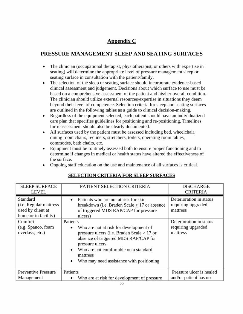
Appendix C
PRESSURE MANAGEMENT SLEEP AND SEATING SURFACES
• The clinician (occupational therapist, physiotherapist, or others with expertise in seating) will determine the appropriate level of pressure management sleep or seating surface in consultation with the patient/family.
• The selection of the sleep or seating surface should incorporate evidence-based clinical assessment and judgement. Decisions about which surface to use must be based on a comprehensive assessment of the patient and his/her overall condition. The clinician should utilize external resources/expertise in situations they deem beyond their level of competence. Selection criteria for sleep and seating surfaces are outlined in the following tables as a guide to clinical decision-making.
• Regardless of the equipment selected, each patient should have an individualized care plan that specifies guidelines for positioning and re-positioning. Timelines for reassessment should also be clearly documented.
• All surfaces used by the patient must be assessed including bed, wheelchair, dining room chairs, recliners, stretchers, toilets, operating room tables, commodes, bath chairs, etc.
• Equipment must be routinely assessed both to ensure proper functioning and to determine if changes in medical or health status have altered the effectiveness of the surface.
• Ongoing staff education on the use and maintenance of all surfaces is critical.
SELECTION CRITERIA FOR SLEEP SURFACES
SLEEP SURFACE LEVEL
PATIENT SELECTION CRITERIA DISCHARGE CRITERIA
Standard (i.e. Regular mattress used by client at home or in facility)
• Patients who are not at risk for skin breakdown (i.e. Braden Scale > 17 or absence of triggered MDS RAP/CAP for pressure ulcers)
Deterioration in status requiring upgraded mattress
Comfort (e.g. Spanco, foam overlays, etc.)
Patients • Who are not at risk for development of
pressure ulcers (i.e. Braden Scale > 17 or absence of triggered MDS RAP/CAP for pressure ulcers
• Who are not comfortable on a standard mattress
• Who may need assistance with positioning
Deterioration in status requiring upgraded mattress
Preventive Pressure Management
Patients • Who are at risk for development of pressure
Pressure ulcer is healed and/or patient has no
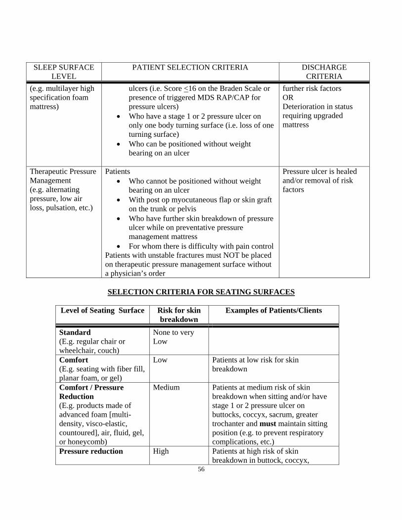
56
SLEEP SURFACE LEVEL
PATIENT SELECTION CRITERIA DISCHARGE CRITERIA
(e.g. multilayer high specification foam mattress)
ulcers (i.e. Score <16 on the Braden Scale or presence of triggered MDS RAP/CAP for pressure ulcers)
• Who have a stage 1 or 2 pressure ulcer on only one body turning surface (i.e. loss of one turning surface)
• Who can be positioned without weight bearing on an ulcer
further risk factors OR Deterioration in status requiring upgraded mattress
Therapeutic Pressure Management (e.g. alternating pressure, low air loss, pulsation, etc.)
Patients • Who cannot be positioned without weight
bearing on an ulcer • With post op myocutaneous flap or skin graft
on the trunk or pelvis • Who have further skin breakdown of pressure
ulcer while on preventative pressure management mattress
• For whom there is difficulty with pain control Patients with unstable fractures must NOT be placed on therapeutic pressure management surface without a physician’s order
Pressure ulcer is healed and/or removal of risk factors
SELECTION CRITERIA FOR SEATING SURFACES
Level of Seating Surface Risk for skin
breakdown Examples of Patients/Clients
Standard (E.g. regular chair or wheelchair, couch)
None to very Low
Comfort (E.g. seating with fiber fill, planar foam, or gel)
Low Patients at low risk for skin breakdown
Comfort / Pressure Reduction (E.g. products made of advanced foam [multi-density, visco-elastic, countoured], air, fluid, gel, or honeycomb)
Medium Patients at medium risk of skin breakdown when sitting and/or have stage 1 or 2 pressure ulcer on buttocks, coccyx, sacrum, greater trochanter and must maintain sitting position (e.g. to prevent respiratory complications, etc.)
Pressure reduction
High Patients at high risk of skin breakdown in buttock, coccyx,

57
sacrum, greater trochanter and/or who have stage 3 or 4 pressure ulcer in this area and must maintain sitting position (e.g. to prevent respiratory complications, etc.).