
Quantification of sulfated polysaccharides in urine by the ...

1
A NOVEL ALLOSTERIC PATHWAY OF THROMBIN INHIBITION Exosite II Mediated Potent Inhibition of Thrombin by Chemo-Enzymatic, Sulfated
Dehydropolymers of 4-Hydroxycinnamic Acids
Brian L. Henry,1 Bernhard H. Monien,1 Paul E. Bock2 and Umesh R. Desai*,1
From the 1Department of Medicinal Chemistry and, Institute for Structural Biology and Drug
Discovery, Virginia Commonwealth University, Richmond, VA 23298 and
2Department of Pathology, Vanderbilt University School of Medicine, Nashville, TN 37232
Running Title: Mechanism of Action of Cinnamic Acid DHPs Address correspondence to: Umesh R. Desai, Department of Medicinal Chemistry, Virginia Commonwealth University, 410 N. 12th Street, PO Box 980540, Richmond, VA 23298-0540. Ph. 804-828-7328, Fax 804-827-3664, e-mail: [email protected]
Thrombin and factor Xa, two important pro-coagulant proteinases, can be regulated through direct and indirect inhibition mechanisms. Recently, we designed sulfated dehydropolymers (DHPs) of 4-hydroxycinnamic acids that displayed interesting anticoagulant properties (Monien et al. Bioorg. Med. Chem. (2006) 14, 7988). To better understand their mechanism of action, we studied the direct inhibition of thrombin, factor Xa, factor IXa, and factor VIIa by CDSO3, FDSO3 and SDSO3, three analogs of sulfated DHPs. All three sulfated DHPs displayed a 2–3-fold preference for direct inhibition of thrombin over factor Xa, while this preference for inhibiting thrombin over factor IXa and factor VIIa increased to 17–300-fold suggesting a high level of selectivity. Competitive binding studies with a thrombin-specific chromogenic substrate, a fluorescein-labeled hirudin peptide, bovine heparin, enoxaparin, and a heparin octasaccharide suggest that CDSO3 preferentially binds in or near anion-binding exosite II of thrombin. Studies of the hydrolysis of H-D-hexahydrotyrosol-Ala-Arg-p-nitroanilide indicate that CDSO3 inhibits thrombin through allosteric disruption of the catalytic apparatus, specifically through the catalytic step. Overall, designed sulfated DHPs appear to be the first molecules that bind primarily in the region defined by exosite II and allosterically induce thrombin inhibition. The molecules are radically different in structure from all the current clinically used anticoagulants and thus, represent a novel class of potent dual thrombin and factor Xa inhibitors.
The coagulation cascade is composed of two intertwined pathways, called the extrinsic and the intrinsic pathways, which operate in a highly complex, but tightly-regulated, manner to bring about controlled formation of the fibrin polymer. Several enzymes participate in this process, including factor IXa and factor VIIa, which belong to the intrinsic and extrinsic pathways, respectively, and thrombin and factor Xa, which belong to the common pathway (1,2). The cascade is regulated by several proteins present naturally in the plasma, of which antithrombin is a major regulator (3,4).
Antithrombin, a member of the serpin (serine proteinase inhibitor) family of proteins, primarily inhibits thrombin, factor Xa, and factor IXa, while also possibly inhibiting several other enzymes to a lesser extent. Yet, antithrombin is a rather poor inhibitor of these pro-coagulant enzymes and requires the presence of heparin to exhibit its anticoagulant potential (3,4). Heparin is a highly sulfated, polysaccharide that greatly enhances the rate of antithrombin inhibition of thrombin, factor Xa and factor IXa under physiological conditions (5). This acceleration is the primary reason for heparin’s continued use as an effective anticoagulant for the past eight decades. Yet, heparin suffers from several limitations including enhanced risk for bleeding, variable patient response, heparin-induced thrombocytopenia and the inability to inhibit clot-bound thrombin (6,7). Low molecular weight heparins (LMWHs#), derivatives of heparin with reduced polymeric length, and fondaparinux, a specific sequence of five saccharide residues (Fig. 1A), have been introduced in the past decade as better mimics of full-length heparin. These new

2
anticoagulants still possess risk for bleeding and are unable to inhibit clot-bound thrombin (8,9).
A major reason for the limitations of heparin therapy is its high negative charge density. Heparin (and LMWH) is a linear, co-polymer of glucosamine and uronic acid residues that are decorated with a large number of sulfate groups generating a complex, heterogeneous, polyanionic macromolecule (Fig. 1A) (10). This highly polyanionic polymer is capable of interacting with a large number of plasma proteins and cells, which likely induce many of the adverse effects of heparin (11).
Apart from the indirectly acting heparins, several direct inhibitors have been put forward including argatroban and ximelagatran for thrombin and, DX9065a and razaxaban for factor Xa (12,13). Structurally, most direct inhibitors of thrombin (DTIs) and factor Xa contain a guanidine or an amidine group that mimic the critical arginine residue at the P-1$ site of the proteinase recognition sequence (14,15). DTIs and direct factor Xa inhibitors form major classes of clotting regulators that are considered to be superior to heparins primarily because of the expectation that they are likely to inhibit both circulating and clot-bound enzymes. Yet, challenges exist in the development of these inhibitors including establishing enzyme-binding affinity that is not associated with excessive bleeding and avoiding liver toxicity (13).
Mechanistically, the direct and indirect anticoagulants utilize different pathways of inhibition. While heparins require antithrombin to mediate their effect, DTIs and direct factor Xa inhibitors either bind in the active site or to an exosite on the enzyme to inhibit its proteolytic function (14,15). With a goal of developing inhibitors of factor Xa and thrombin that are less polyanionic and more hydrophobic than the heparins, we designed sulfated dehydropolymers (DHPs) (Fig. 1B). Three sulfated DHPs – CDSO3, FDSO3 and SDSO3 – were prepared in a simple, two-step chemo-enzymatic process involving horseradish peroxidase catalyzed oligomerization of 4-hydroxycinnamic acid monomers followed by the chemical sulfation of the resulting DHPs with triethylamine-sulfur trioxide complex (16). Preliminary studies suggested that the chemo-enzymatic sulfated DHPs were potent anticoagulants in vitro. The sulfated DHPs prolonged activated partial thromboplastin time and prothrombin time at
concentrations equivalent to LMWH. Interestingly, although being designed as heparin mimics, CDSO3, FDSO3 and SDSO3 were found to inhibit factor Xa and thrombin in an antithrombin–independent (direct) manner suggesting a potentially novel mode of inhibition (16).
In this paper, we report that CDSO3, FDSO3, and SDSO3 possess high selectivity for inhibiting thrombin and factor Xa over other enzymes of the coagulation cascade; CDSO3 inhibits thrombin through allosteric disruption of its catalytic apparatus; and preferentially binds the enzyme in or near the region formed by anion-binding exosite II. Sulfated DHPs appear to be the first molecules that induce inhibition of pro-coagulant proteinases, thrombin and factor Xa, through exosite II. Our work suggests that sulfated DHPs are structurally, functionally and mechanistically a very interesting class of molecules that may lead to novel anticoagulants.
EXPERIMENTAL PROCEDURES Proteins and Chemicals. Sulfated
dehydropolymers CDSO3, FDSO3 and SDSO3 (Fig. 1B) were prepared in two steps from 4-hydroxycinnamic acid monomers, caffeic acid (CA), ferulic acid (FA) and sinapic acid (SA) using chemo-enzymatic synthesis described by Monien et al (16). Human plasma proteinases, factor VIIa, factor IXa, factor Xa and α-thrombin, were purchased from Haematologic Technologies (Essex Junction, VT) and used as such. Stock solutions of proteins were prepared in 20 mM sodium phosphate buffer, pH 7.4, containing 100 mM NaCl and 2.5 mM CaCl2 (AT and thrombin) or 5 mM MES buffer, pH 6.0 (factor Xa). Factor VIIa stock solutions of proteins were prepared in 25 mM HEPES buffer, pH 7.4, containing 100 mM NaCl and 5 mM CaCl2, while factor IXa stock solutions were prepared in 5 mM MES buffer, pH 5.5 containing 150 mM NaCl. Chromogenic substrates Spectrozyme TH (H-D-hexahydrotyrosol-Ala-Arg-p-nitroanilide), Spectrozyme FXa (Methoxycarbonyl-D-cyclohexylglycyl-Gyl-Arg-p-nitroanilide), Spectrozyme FIXa (D-Leu-Phe-Gly-Arg-p-nitroanilide) and Spectrozyme FVIIa (methanesulphonyl-D-cyclohexylalanyl-butyl-Arg-p-nitroanilide) were purchased from American Diagnostica (Greenwich, CT). A low molecular weight heparin (MW 5,060 Da) used in plasma assays was purchased from Sigma (St. Louis, MO),

3
while enoxaparin (MW 4,500 Da, from Aventis Pharmaceuticals) and fondaparinux (MW 1,727 Da, from GlaxoSmithKline) were pharmaceutical grade. Heparin octasaccharide H8 was from Dextra Laboratories (Reading, UK). Thrombin substrate p-nitrophenyl-p’-guanidinobenzoate (NPGB) and active site fluorophore p-aminobenzamidine (PABA) were purchased from Sigma Chemicals (St. Louis, MO) and used as such. Tyr63-sulfated hirudin-(54-65) labeled with 5-(carboxy)fluorescein ([5F]-Hir[54-65](SO3
-)) was prepared as described earlier (22). All other chemicals were analytical reagent grade from either Sigma Chemicals (St. Louis, MO) or Fisher (Pittsburgh, PA) and used without further purification.
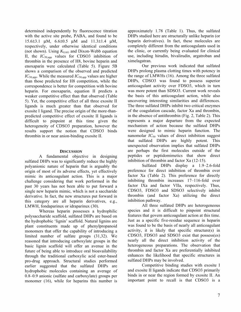
Physico-chemical Properties of CDSO3, FDSO3 and SDSO3. The weight average molecular weight (MW) of the unsulfated parent dehydropolymers CD, FD and SD were determined by Monien et al. (16) using non-aqueous size-exclusion chromatography (Table 1). The MW values suggest that an average of 12.7, 15.5, and 14.4 monomer units are present in CD, FD and SD, respectively. Sulfate composition of the sulfated DHPs was determined by elemental analysis and found to be 0.40, 0.30 and 0.38 sulfate groups per monomer unit (16). This implies that an average of 5.1, 4.7, and 5.5 sulfate groups per average DHP chain are present in CDSO3, FDSO3, and SDSO3, respectively. Thus, the MW value of the sulfated DHPs was calculated to be 3320, 4120, and 3550 Da for CDSO3, FDSO3, and SDSO3, respectively (Table 1).
Direct Inhibition of Coagulation Proteinases. Direct inhibition of thrombin, factor Xa, factor IXa, and FVIIa by sulfated DHPs was determined using chromogenic substrate hydrolysis assays (4,5,17,18). For these assays, 10 µL DHP at concentrations ranging from 0.035 to 10,000 µg/mL was diluted with 930 µL of the appropriate buffer in PEG 20,000-coated polystyrene cuvettes. The buffers used in these experiments include 20 mM Tris-HCl buffer, pH 7.4, containing 100 mM NaCl, 2.5 mM CaCl2 and 0.1 % polyethylene glycol (PEG) 8000 for thrombin and factor Xa; 100 mM HEPES buffer, pH 8, containing 100 mM NaCl and 10 mM CaCl2 for factor IXa (18); and 25 mM HEPES buffer, pH 7.4, containing 100 mM NaCl and 5 mM CaCl2 for factor VIIa (17). Following the preparation of the sulfated DHP solution, 10 µL of the proteinase solution was added to give 1 to 10 nM
initial enzyme concentration and the cuvette incubated for 10 minutes. Thrombin, factor Xa and factor VIIa assays were incubated at 25°C, while factor IXa assays were incubated at 20°C. Following incubation, 50 µL of 2 mM chromogenic substrate, Spectrozyme TH, FXa, FVIIa or Spectrozyme FIXa, was rapidly added and the residual enzyme activity was determined from the initial rate of increase in absorbance at 405 nm. Relative residual proteinase activity at each concentration was calculated using the activity measured under otherwise identical conditions, except for the absence of the sulfated DHP. Logistic equation I was used to fit the dose-dependence of residual proteinase activity to obtain IC50.
HSICDHPOM
OO
YYYY )log](log[ 50101 −+
−+= Eq. I
In this equation Y is the ratio of residual proteinase activity in the presence of inhibitor to its absence (fractional residual activity), YM and YO are the maximum and minimum possible values of the fractional residual proteinase activity, IC50 is the concentration of the inhibitor that results in 50% inhibition of enzyme activity, and HS is the Hill slope. HS does not represent co-operativity because sulfated DHPs are highly complex species that may possess multiple binding modes and geometries. Sigmaplot 8.0 (SPSS, Inc. Chicago, IL) was used to perform non-linear curve fitting in which YM, YO, IC50 and HS were allowed to float. The values of each of these parameters returned by curve fitting are reported in Table 2.
Michaelis-Menten Kinetics of Spectrozyme TH Hydrolysis by Thrombin in the Presence of CDSO3. The initial rate of Spectrozyme TH hydrolysis by 1 nM thrombin was monitored from the linear increase in absorbance at 405 nm corresponding to less than 20% consumption of the substrate. The initial rate was measured as a function of various concentrations of the substrate (0.6 to 20 µM) in the presence of fixed concentration of CDSO3 (10–300 nM) in 20 mM Tris-HCl buffer, pH 7.4, containing 100 mM NaCl, 2.5 mM CaCl2 and 0.1 % PEG8000 at 25 OC. The data was fitted by the Michaelis-Menten equation to determine KM,app and VMAX. To calculate kCAT from VMAX, active site titration of thrombin with NPGB was performed, according to the reported procedure (19), and the change in extinction co-efficient of 9920 M-1cm-1 (20,21) was used for the release of p-nitroaniline.

4
Michaelis-Menten Kinetics of Spectrozyme TH Hydrolysis by Thrombin in the Presence of [5F]-Hir[54-65](SO3
-). The initial rate of Spectrozyme TH hydrolysis by 1 nM thrombin was monitored from the linear increase in absorbance at 405 nm corresponding to less than 20% consumption of the substrate. The initial rate was measured as a function of various concentrations of the substrate (0.6 to 20 µM) in the presence of fixed concentration of [5F]-Hir[54-65](SO3
-) (8.6–103.2 nM) in 20 mM Tris-HCl buffer, pH 7.4, containing 100 mM NaCl, 2.5 mM CaCl2 and 0.1 % PEG8000 at 25 OC. The data were analyzed as described above.
Competitive Binding Studies with [5F]-Hir[54-65](SO3
-), an Exosite I ligand. CDSO3-dependent thrombin inhibition studies in the presence of [5F]-Hir[54-65](SO3
-) were performed in a manner similar to that described above for direct thrombin inhibition using the chromogenic substrate hydrolysis assay. A 950 µL solution containing CDSO3, [5F]-Hir[54-65](SO3
-) and thrombin, each at their required concentrations, in 20 mM Tris-HCl buffer, pH 7.4, containing 100 mM NaCl, 2.5 mM CaCl2 and 0.1 % PEG 8000 was incubated at 25 OC in PEG 20,000-coated polystyrene cuvettes for 10 minutes. Following incubation, 50 µL of 2 mM Spectrozyme TH was added and the initial change in absorbance at 405 measured. A 4 nM concentration of thrombin was found to give sufficient signal at various concentrations of the fluorescent peptide for reproducible results. The dose-dependence of the fractional residual proteinase activity at each concentration of the competitor was fitted by equation I to obtain the apparent concentration of CDSO3 required to reduce thrombin activity to 50% of its initial value (IC50,app).
Competitive Binding Studies with Anion-Binding Exosite II Ligands. Exosite II competition experiments were performed in a manner similar to that described above, except for the presence exosite II competitors. Briefly, residual thrombin activity was measured in a spectrophotometric assay following 10 minute incubation of CDSO3, exosite II competitor and thrombin, each at the required concentration, in 20 mM Tris-HCl buffer, pH 7.4, as described above. Exosite II competitor ligands included bovine heparin, enoxaparin and H8. The MW of bovine heparin and enoxaparin were assumed to be 15000 and 4500 Da, respectively, as reported in the literature (22-24). The dose-dependence of the
fractional residual proteinase activity at each concentration of the competitor was fitted by equation I to obtain IC50,app. The affinities of bovine heparin, enoxaparin and H8 for thrombin under the conditions utilized for inhibition experiments were measured spectrofluorometrically using p-aminobenzamidine (PABA), as reporter of interaction following published procedures (25). The interaction of exosite II GAG ligands with thrombin–PABA complex results in a saturable quenching in fluorescence at 370 nm (~16 %, λEX = 345 nm), which can be fitted by the quadratic binding equation to derive the KD for the interaction.
RESULTS Structure of Sulfated Dehydropolymers
(DHPs). The sulfated DHP molecules studied in this work were prepared chemo-enzymatically in two steps from 4-hydroxy cinnamic acid monomers, caffeic acid, ferulic acid and sinapic (Fig. 1B). Horseradish peroxidase-catalyzed oxidative coupling followed by sulfation of the available hydroxyl and phenolic groups gives the corresponding sulfated DHPs, CDSO3, FDSO3, and SDSO3, in reproducibly good yields (16). Overall, these sulfated DHPs are a mixture of many oligomeric species that range in size from 4–15 monomer units and contain several inter-monomer linkages, including β-O-4, β-5, β-β and 5-5. Of these, β-O-4 and β-5 linkages form the major proportion (Fig. 1B), except for SDSO3 (see below).
Although heparins and sulfated DHPs belong to structurally distinct class of molecules, with respect to the properties of polydispersity and microheterogeneity, they have much in common. Each preparation of both these types of molecules contains numerous sequences possessing different chain lengths and fine structure. Yet, the level of sulfation in the sulfated DHPs is significantly lower than that found in heparins. Whereas the three sulfated DHPs studied here have an average of 1 sulfate group for every 2–3 monomer residues, heparins possess an average of 2–2.5 sulfate groups for every disaccharide (Table 1). More importantly, the backbone of sulfated DHPs is composed of a number of aromatic rings, a feature completely absent in heparin. Thus, the sulfated DHPs are significantly more hydrophobic than heparins, while heparins have significantly more anionic character. Finally, whereas unfractionated heparin with a MW of ~15,000 Da is considerably larger, sulfated DHPs

5
(~2500–4000 Da) are comparable to enoxaparin (4,500 Da) and fondaparinux (~1,700 Da) (Table 1).
Although all three sulfated DHPs possess several types of inter-monomer linkages, structural constraints in the SA monomer do not allow the formation of 5-5 and β-5 inter-monomer linkages in the oligomer (16). Thus, the predominant inter-monomeric linkage in SDSO3 is β-O-4 with some proportion of β-β. This implies that the SDSO3 dehydropolymer is structurally more homogeneous, or less diverse, than CDSO3 and FDSO3.
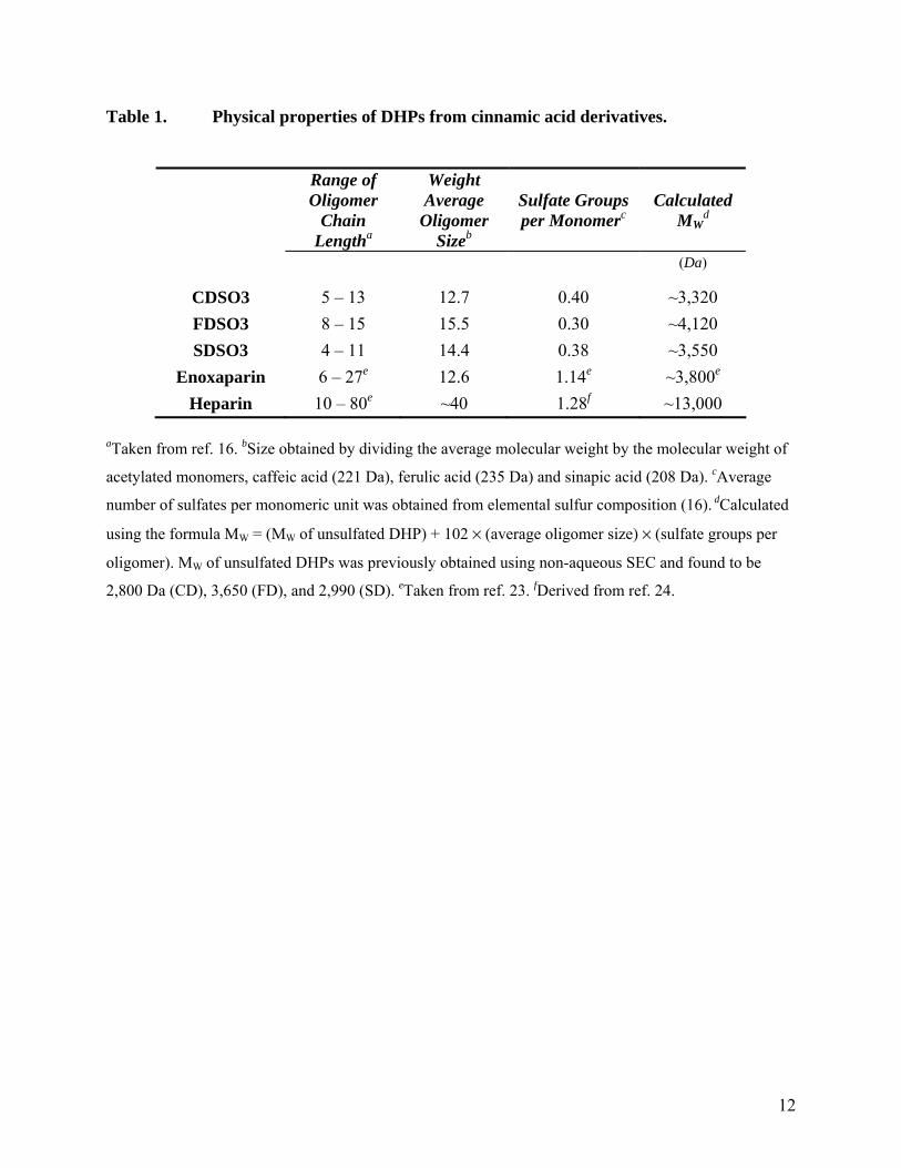
Sulfated DHPs Inhibit Factor Xa and Thrombin in the Absence of Antithrombin. Our initial results on inhibition of thrombin and factor Xa in the presence and absence of antithrombin suggested the possibility of a dual inhibition mechanism (16). The discovery of a direct inhibition mechanism was unexpected and hence interesting. To better define this initial observation, inhibition of thrombin and factor Xa by sulfated DHPs was studied under pseudo-first order conditions in the absence of human plasma antithrombin. The inhibition of enzyme activity was followed by spectrophotometric determination of the initial rate of hydrolysis of appropriate chromogenic substrate.
As the concentration of the sulfated DHP was increased, the residual factor Xa or thrombin activity progressively decreased (Fig. 2). In contrast, enoxaparin and fondaparinux, two sulfated molecules known to activate antithrombin, displayed no direct inhibition of thrombin and factor Xa even at concentrations higher than 100 µM (not shown). The decrease in enzyme activity by sulfate DHPs was fitted by the logistic dose–response equation I to derive the IC50 value, the concentration of the inhibitor that results in 50% reduction in enzyme activity (Table 2). The three sulfated DHPs inhibited factor Xa and thrombin with IC50 values in the range of 34–244 nM and 18–94 nM, respectively (Fig. 2, Table 2). Of the three sulfated DHPs, CDSO3 and FDSO3 are nearly 3.2–7.2-fold better than SDSO3. Thus, the sulfated DHPs studied here are potent direct inhibitors of factor Xa and thrombin.
Table 2 also provides Hill slopes of the inhibition curves (see Equation 1). Hill slope refers to the steepness of the inhibition profile and does not imply Hill co-operativity because of the significant complexity of the system. Each sulfated DHP studied herein is a complex mixture of structural species, which may possess multiple modes of
binding with multiple geometries in same binding site. Thus, a priori a multivalent molecular analysis of Hill-type is not advisable. Despite this complexity, the analysis of direct inhibition profiles by sulfated DHPs shows that Hill slopes are generally closer to 1.0, except for SDSO3 inhibiting thrombin (Table 2). The uniformity of Hill slopes for both factor Xa and thrombin suggests the possibility that the underlying nature of interaction is similar for the sulfated DHPs.
Effect of Sulfated DHPs on Direct Inhibition of Factor IXa and Factor VIIa. To determine whether the sulfated DHPs inhibit other enzymes of the coagulation cascade directly, we studied inhibition of factor IXa and factor VIIa, enzymes of the intrinsic and extrinsic pathways, respectively. The inhibition was studied in a manner similar to that reported in the literature, except for the presence of sulfated DHPs (or reference LMWH) in the reaction mixture (17,18). CDSO3 and FDSO3 inhibited factor IXa with IC50 values of 3.4 and 0.5 µM, while inhibition of factor VIIa was not detectable (Table 2). SDSO3 was essentially inactive against both factor IXa and factor VIIa at concentrations as high as 28 µM (not shown). These results suggest that CDSO3 and FDSO3 are better direct inhibitors of factor Xa and thrombin with 7–99-fold and 17–187-fold, respectively, higher selectivity over factor IXa. The level of selectivity of direct inhibition against factor VIIa is even greater (>319-fold).
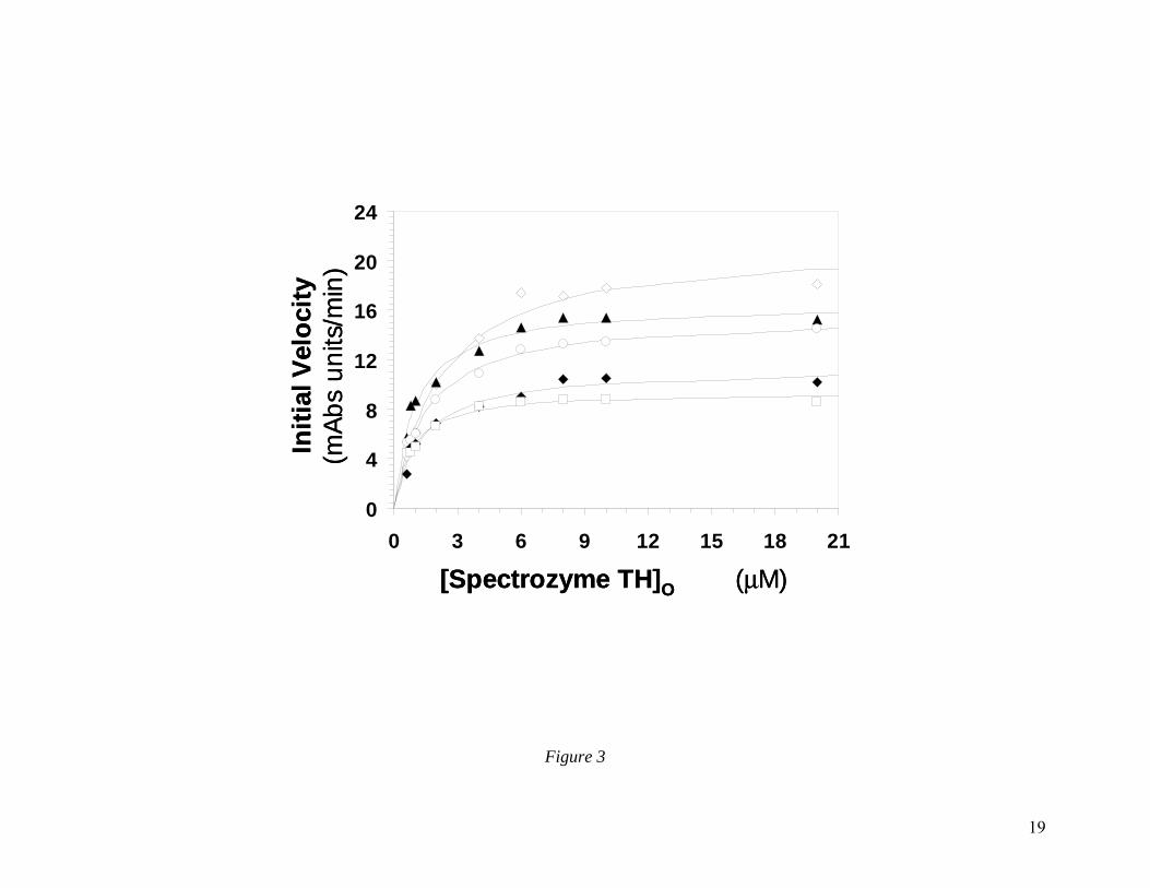
CDSO3 Inhibits Thrombin by Disrupting its Catalytic Apparatus. To understand the molecular basis for sulfated DHPs inhibiting thrombin, we studied the Michaelis-Menten kinetics of Spectrozyme TH hydrolysis at pH 7.4 and 25 OC in the presence of CDSO3, a representative sulfated DHP. Plots of the initial rates versus Spectrozyme TH concentration were hyperbolic, as expected (Fig. 3), from which the apparent Michaelis constant (KM,app) and maximal velocity of the reaction (VMAX) were derived (Table 3). The results show that although the concentration of CDSO3 increased from 0 nM to 300 nM, the KM,app value fluctuated around 1.5 µM, or alternatively remained essentially invariant in the range of 2.2 to 0.8 µM. This suggests that the presence of CDSO3 does not much affect the binding of the chromogenic substrate to the active site of the enzyme. In contrast, the VMAX value decreased steadily from a high of 21.5 mAbsU/min in the absence of CDSO3 to a low of 9.5

6
mAbsU/min at 300 nM CDSO3 (Fig. 3, Table 3) corresponding to a decrease in kCAT value from 36.1 to 16.0 s-1, respectively. Thus, the presence of CDSO3 brings about structural changes in the active site of thrombin, which do not alter the formation of the thrombin–Spectrozyme TH Michaelis complex, but significantly reduce the rate of conversion of the complex into products.
CDSO3 Does Not Interact with Thrombin in the Anion-Binding Exosite I. To test whether CDSO3 binds in the anion-binding exosite I, we sought to measure the effect of a hirudin-based peptide, [5F]-Hir[54-65](SO3
-), on the IC50 value of CDSO3-inhibition of thrombin. Previous studies indicate that [5F]-Hir[54-65](SO3
-) binds thrombin with 28 nM affinity in exosite I with 1:1 stoichiometry (26,27). However, the effect of this exosite I ligand on the catalytic apparatus of thrombin was not clear. Literature reports on studies with the parent Hir[54-65](SO3
-), which is also known to bind in exosite I of thrombin, show that the kCAT/KM value increases or decreases approximately 2-fold depending on the type of chromogenic substrate (28,29). Thus, we first determined the effect of [5F]-Hir[54-65](SO3
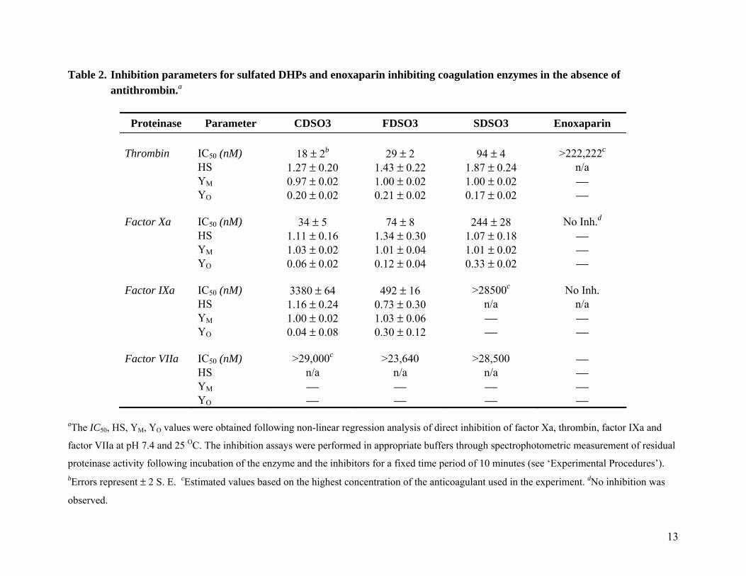
-) on the thrombin hydrolysis of Spectrozyme TH using the standard Michaelis-Menten conditions at pH 7.4. As the concentration of the exosite I ligand was increased to 103.6 nM, the KM remained essentially constant in the range of 1.2 to 2.2 µM, while the VMAX increased steadily from 21.5 to 30.9 mAbs/min (Fig. 4A, Table 4). This suggested that [5F]-Hir[54-65](SO3
-) increased the catalytic efficiency of Spectrozyme TH hydrolysis arising specifically from a kCAT effect.
The [5F]-Hir[54-65](SO3-)-dependent
enhancement and the CDSO3-dependent reduction in rate of Spectrozyme TH hydrolysis (kCAT) afforded a fine experimental setup to study competition between these two ligands. Thus, we measured the IC50 values of thrombin inhibition by CDSO3 in the presence of the dodecapeptide over a concentration range up to 3.7-fold higher than the KD of the thrombin-[5F]-Hir[54-65](SO3
-) complex (22,23). The IC50,app values were measured in the standard dose-response assay, which we had used to detect thrombin inhibition. Figure 4B shows the change in the dose-response profile of CDSO3 inhibiting thrombin in the presence of [5F]-Hir[54-65](SO3
-) at pH 7.4 and 25 OC. As the concentration of the dodecapeptide was increased from 0 to 103.6
nM, IC50,app increased from 28 to 57.8 nM (Table 4). This represents a 2.1-fold change in IC50,app value for a 3.7-fold increase in concentration over the KD of the exosite I competitor. Additionally, the change in IC50,app value appears to be not linear with the concentration of [5F]-Hir[54-65](SO3
-). For example, the IC50,app value increases ~1.7-fold at 8.6 nM dodecapeptide (0.3×KD), which is followed by much slower increases (Table 4). These small changes suggest that that the interaction of [5F]-Hir[54-65](SO3
-) with thrombin does not affect CDSO3 inhibition of thrombin to a significant extent. Thus, it appears that CDSO3 does not preferentially bind thrombin in anion-binding exosite I.
CDSO3 Interacts with Thrombin in or Near Anion-Binding Exosite II. To assess whether CDSO3 binds in the region formed by anion-binding exosite II of thrombin, we resorted to the enzyme inhibition assay described above. Exosite II ligands, bovine heparin, enoxaparin and heparin octasaccharide H8, did not affect the proteolytic activity of thrombin (not shown), while CDSO3 is a potent inhibitor. Thus, if CDSO3 binds in or near the GAG binding site, its inhibition potency is expected to decrease as a function of the concentration of the GAG competitor. Figure 5A shows the change in the dose-response curve of CDSO3 inhibiting thrombin in the presence of H8 at pH 7.4 and 25 OC. As the concentration of H8 was increased to 26.3 µM, the IC50 value of thrombin inhibition increases from 28 to 414 nM (Table 5). Less dramatic, but significant, changes in dose-response profiles were also observed for bovine heparin and enoxaparin (not shown), corresponding to increases in apparent IC50 values (Table 5), suggesting that all three GAG ligands compete with CDSO3.
A more quantitative test of competitive binding is the Dixon-Webb relationship (Eq. II), which predicts the effect of competition on a measured parameter, e.g., KD or IC50. In this equation, KGAG is the dissociation constant of thrombin–GAG (bovine heparin, H8 or enoxaparin) interaction.
)][
1(50,50GAG
Oapp K
GAGICIC += Eq. II
The equilibrium dissociation constants
(KGAG) of bovine heparin, enoxaparin and H8 were
7
determined independently by fluorescence titration with the active site probe, PABA, and found to be 15.6±3.1 µM, 6.6±0.5 µM and 11.3±1.4 µM, respectively, under otherwise identical conditions (not shown). Using KGAG and Dixon-Webb equation II, the IC50,app values for CDSO3 inhibition of thrombin in the presence of H8, bovine heparin and enoxaparin were calculated (Table 5). Figure 5B shows a comparison of the observed and predicted IC50,app. While the measured IC50,app values are higher than those predicted for H8 competition, while the correspondence is better for competition with bovine heparin. For enoxaparin, equation II predicts a weaker competitive effect than that observed (Table 5). Yet, the competitive effect of all three exosite II ligands is much greater than that observed for exosite I ligand. The precise origin of the more-than-predicted competitive effect of exosite II ligands is difficult to pinpoint at this time given the heterogeneity of CDSO3 preparation, however the results support the notion that CDSO3 binds thrombin in or near anion-binding exosite II.
DISCUSSION A fundamental objective in designing
sulfated DHPs was to significantly reduce the highly polyanionic nature of heparin that is arguably the origin of most of its adverse effects, yet effectively mimic its anticoagulant action. This is a major challenge considering that work performed in the past 30 years has not been able to put forward a single new heparin mimic, which is not a saccharide derivative. In fact, the new structures put forward in this category are all heparin derivatives, e.g., LMWH, fondaparinux or idraparinux (30).
Whereas heparin possesses a hydrophilic polysaccharide scaffold, sulfated DHPs are based on the hydrophobic ‘lignin’ scaffold. Natural lignins are plant constituents made up of phenylpropanoid monomers that offer the capability of introducing a limited number of sulfate groups (31,32). We reasoned that introducing carboxylate groups in the basic lignin scaffold will offer an avenue in the future of being able to introduce oral bioavailability through the traditional carboxylic acid ester-based pro-drug approach. Structural studies performed earlier suggested that the sulfated DHPs are hydrophobic molecules containing an average of 0.8–0.9 anionic (sulfate and carboxylate) groups per monomer (16), while for heparins this number is
approximately 1.78 (Table 1). Thus, the sulfated DHPs studied here are structurally unlike heparin (or heparin derivatives). In fact, these molecules are completely different from the anticoagulants used in the clinic, or currently being evaluated for clinical use, including hirudin, bivalirudin, argatroban and ximelagatran.
Our previous work indicated that sulfated DHPs prolong plasma clotting times with potency in the range of LMWHs (16). Among the three sulfated DHPs, CDSO3 was found to possess superior anticoagulant activity over FDSO3, which in turn was more potent than SDSO3. Current work reveals the basis of this anticoagulant action, while also uncovering interesting similarities and differences. The three sulfated DHPs inhibit two critical enzymes of the coagulation cascade, factor Xa and thrombin, in the absence of antithrombin (Fig. 2, Table 2). This represents a major departure from the expected mechanism of action because the sulfated DHPs were designed to mimic heparin function. The nanomolar IC50 values of direct inhibition suggest that sulfated DHPs are highly potent. This unexpected observation implies that sulfated DHPs are perhaps the first molecules outside of the peptides or peptidomimetics that show direct inhibition of thrombin and factor Xa (12-15).
Sulfated DHPs display a 1.9–2.6-fold preference for direct inhibition of thrombin over factor Xa (Table 2). This preference for directly inhibiting thrombin increases 17–116-fold over factor IXa and factor VIIa, respectively. Thus, CDSO3, FDSO3 and SDSO3 selectively inhibit thrombin (and factor Xa) utilizing the direct inhibition pathway.
All three sulfated DHPs are heterogeneous species and it is difficult to pinpoint structural features that govern anticoagulant action at this time. Just as a specific five-residue sequence in heparin was found to be the basis of nearly all anticoagulant activity, it is likely that specific structure(s) in CDSO3, FDSO3 and SDSO3 exist that possess(es) nearly all the direct inhibition activity of the heterogeneous preparations. The observation that thrombin and factor Xa are preferentially inhibited enhances the likelihood that specific structures in sulfated DHPs may be involved.
Competitive binding studies with exosite I and exosite II ligands indicate that CDSO3 primarily binds in or near the region formed by exosite II. An important point to recall is that CDSO3 is a

8
heterogeneous anionic molecule implying that certain sequences in CDSO3 may be still interacting with exosite I of thrombin. However, the competitive binding data suggest that such an interaction with exosite I is likely to be of lower affinity.
Studies on the hydrolysis of Spectrozyme TH indicate that CDSO3 disrupts the catalytic apparatus without binding to the active site. Thus, CDSO3 is an allosteric inhibitor of thrombin function. Although FDSO3 and SDSO3 differ from CDSO3 in terms of the fine structure, their overall similarity suggests that these oligomers may also bind thrombin exosite II. In a similar manner, these sulfated DHPs may be interacting with factor Xa in its anion-binding exosite II, which is known to recognize GAG ligands with an affinity similar to thrombin (33). A supporting evidence for exosite II recognition is the observation that, unlike thrombin, exosite I in factor Xa is not an anion-binding site, but a cation-binding site (34).
Exosite II binding of CDSO3 also partially explains the observed weak direct inhibition of factor IXa and essentially no inhibition of factor VIIa. Factor IXa is known to possess a heparin-binding exosite II (5,18), analogous to thrombin, which is likely to be the CDSO3 recognition site. However, differences in the structures of exosite II in thrombin and factor IXa may introduce differences in the binding affinities of the sulfated
DHPs. Finally, factor VIIa is not known to possess an anion-binding region, similar to exosite II of thrombin, explaining the lack of inhibition induced by CDSO3.
Exosite II in thrombin is known to bind GAGs and haemadin (35-37). GAG binding to thrombin is non-specific, not high-affinity and non-inhibitory (25,38). On the other hand, haemadin is a peptide that binds thrombin with nM affinity, inhibits its proteolytic activity, but also binds in the active site (39). Thus, our work puts forward perhaps the first organic molecules that primarily recognize anion-binding exosite II of thrombin (and possibly factor Xa) with nM potency and induce inhibition.
In conclusion, our work demonstrates that chemo-enzymatically prepared sulfated DHPs display interesting anticoagulant properties. All three novel anticoagulants selectively inhibit thrombin and factor Xa of the coagulation cascade through an interaction with anion-binding exosite II that allosterically disrupts the catalytic apparatus of the enzyme. This represents a novel mechanism of thrombin (and factor Xa) inhibition. Sulfated DHP molecules possess a novel structural scaffold, which is completely different from all the current clinically used anticoagulants, including the heparins, coumarins, hirudins and arginine-peptidomimetics. Thus, sulfated DHPs represent a novel class of potent dual factor Xa and thrombin inhibitors.

9
REFERENCES
1. Butenas, S., and Mann, K. G. (2002) Biochemistry (Moscow) 67, 3-12 2. Jenny, N. S., and Mann, K. G. (2003) In Thrombosis and Hemorrhage (Loscalzo, J., and Schafer,
A. I., Ed.) 3rd ed., Lippincott Williams & Wilkins, Philadelphia, pg. 1-21 3. Björk, I., and Olson, S. T. (1997) Adv. Exp. Med. Biol. 425, 17-33 4. Olson, S. T., Björk, I., and Shore, J. D. (1993) Meth. Enzymol. 222, 525-559 5. Olson, S. T., Swanson, R., Raub-Segall, E., Bedsted, T., Sadri, M., Petitou, M., Herault, J. P.,
Herbert, J. M., and Bjork, I. (2004) Thromb. Haemost. 92, 929-939 6. Menajovsky, L. B. (2005) Am. J. Med. 118 Suppl 8A, 21S-30S 7. van Dongen, C. J., van den Belt, A. G., Prins, M. H., and Lensing, A. W. (2004) Cochrane
Database Syst Rev. (4), CD001100 8. Bauersachs, R. M. (2005) Eur. J. Clin. Invest. 35 Suppl 1, 27-32 9. Turpie, A. G. (2005) Expert Opin. Drug. Saf. 4, 707-721 10. Rabenstein, D. L. (2002) Nat. Prod. Rep. 19, 312-331 11. Capila, I., and Linhardt, R. J. (2002) Angew. Chem. Int. Ed. 41, 391-412 12. Weitz, J. I., and Buller, H. R. (2002) Circulation 105, 1004-1011 13. Nutescu, E. A., Shapiro, N. L., and Chevalier, A. (2006) Clin. Geriatr. Med. 22, 33-56 14. Kikelj, D. (2003) Pathophysiol. Haemost. Thromb. 33, 487-491 15. Gerotziafas, G. T., and Samama, M. M. (2005) Curr. Pharm. Des. 11, 3855-3876 16. Monien, B. H., Henry, B. L., Raghuraman, A., Hindle, M., and Desai, U. R. (2006) Bioorg. Med.
Chem. 14, 7988-7998 17. Neuenschwander, P. F., Branam, D. E., and Morrissey, J. H. (1993) Thromb. Haemost. 70, 970-977 18. Bedsted, T., Swanson, R., Chuang, Y. J., Bock, P. E., Bjork, I., and Olson, S. T. (2003)
Biochemistry 42, 8143-8152 19. Chase, T., Jr., and Shaw, E. (1969) Biochemistry 8, 2212–2224 20. Sonder, S. A., and Fenton, J. W. IInd (1986) Clin. Chem. 32, 934-937 21. Berliner L. J. and Shen, Y. Y. L. (1977) Biochemistry 16, 4622-4626 22. Desai, U. R. (2005) In Chemistry and Biology of Heparin and Heparan Sulfate (Eds. Garg, H. G.,
Linhardt, R. J., and Hales, C. A.), Elsevier, NY, pp. 483-512 23. Linhardt, R. J., Loganathan. D., Al-Hakim, A., Wang, H. –M., Walenga, J. M., Hoppensteadt, D.,
and Fareed, J. (1990) J. Med. Chem. 33, 1639-1645 24. Vongchan, P., Warda, M., Toyoda, H., Toida, T., Marks, M., and Linhardt, R. J. (2005) Biochim.
Biophys. Acta 1721, 1–8 25. Olson, S. T., Halvorson, H. R., and Björk, I. (1991) J. Biol. Chem. 266, 6342-6352 26. Bock, P. E., Olson, S. T., and Björk, I. (1997) J. Biol. Chem. 272, 19837–19845 27. Verhamme, I. M., Olson, S. T., Tollefsen, D. M., and Bock, P. E. (2002) J. Biol. Chem. 277, 6788-
6798 28. Ye, J., Liu, L. W., Esmon, C. T., and Johnson, A. E. (1992) J. Biol. Chem. 267, 11023-11028 29. Liu, L. W., Vu, T. K., Esmon, C. T., and Coughlin, S. R. (1991) J. Biol. Chem. 266, 16977-16980 30. Desai, U. R. (2004) Med. Res. Rev. 24, 151-181 31. Davin, L. B., and Lewis, N. G. (2005) Curr. Opin. Biotechnol. 16, 407-415 32. Reale, S., Di Tullio, A., Spreti, N., and De Angelis, F. (2004) Mass Spectrom. Rev. 23, 87-126 33. Rezaie, A. R. (2000) J. Biol. Chem. 275, 3320-3327 34. Bianchini, E. P., Pike, R. N., and Le Bonniec, B. F. (2004) J. Biol. Chem. 279, 3671-3679 35. Huntington, J. A. (2005) J. Thromb. Haemost. 3, 1861–1872 36. Davie, E. W. and Kulman, J. D. (2006) Semin. Thromb. Hemost. 32, 3-15 37. Lane, D. A., Philippou, H., and Huntington, J. A. (2005) Blood 106, 2605-2612 38. Carter, W. J., Cama, E., and Huntington, J. A. (2005) J. Biol. Chem. 280, 2745-2749 39. Richardson, J. L.; Fuentes-Prior, P.; Sadler, J. E.; Huber, R.; Bode, W. (2002) Biochemistry 41,
2535-2542

10
FOOTNOTES
This work was supported by the grants RO1 HL069975 and R41 HL081972 from the National Institutes of Health and grant EIA 0640053N from the American Heart Association National Center to U. R. D. and grant HL038779 from the National Institutes of Health to P.E.B. #CDSO3, sulfated dehydropolymer of caffeic acid; DHP, dehydrogenation polymer; DTI, direct thrombin inhibitor; FDSO3, sulfated dehydropolymer of ferulic acid; [5F]-Hir[54-65](SO3
-), Tyr63-sulfated hirudin-(54-65) labeled with fluorescein; GAG, glycosaminoglycan; H8, heparin octasaccharide; IC50, concentration of inhibitor that results in 50% inhibition; LMWH, low-molecular-weight heparin; MW, weight average molecular weights; NPGB, p-nitrophenyl-p’-guanidinobenzoate; PABA, p-aminobenzamidine; PEG, polyethylene glycol; SDSO3, sulfated dehydropolymer of sinapic acid $Nomenclature follows the convention introduced by Schechter and Berger ((1967) Biochem. Biophys. Res. Com., 27, 157-162) in which the substrate residues are denoted P with the amino acid residues of the N-terminal side of the scissile bond are numbered P3, P2, P1; those of the C-terminal side are numbered P1', P2', P3'; and P1–P1' residues form the scissile bond.

11
FIGURE LEGENDS Figure 1. Structures of heparins (A) and sulfated DHPs synthesized from 4-hydroxycinammic
acid monomers (B). Clinically used fondaparinux is based on the structure of heparin pentasaccharide, while clinically used heparin and LMW heparins are a polydisperse, heterogeneous mixture of millions of polysaccharide species with average molecular weight of ~13,000 and ~4,000 Da arising due to variations in X, Y, Z and R groups (shown in A). Sulfated DHPs (shown in B) possess a novel, radically different structure from the current clinically used anticoagulants and were chemo-enzymatically synthesized in two steps from the corresponding starting 4-hydroxycinnamic acid monomers, caffeic acid (CA), ferulic acid (FA) or sinapic acid (SA). Two types of common linkages present in sulfated DHPs include the β-O-4 and β-5 linkages (shown as shaded ovals).
Figure 2 Direct inhibition of factor Xa (A) and thrombin (B) by sulfated DHPs. The inhibition of thrombin and factor Xa by CDSO3 (▲), FDSO3 ( ) and SDSO3 (•) in the absence of antithrombin was studied as described under ‘Experimental Procedures’. Solid lines represent sigmoidal dose-response fits (Eq. 1) to the data to obtain values of IC50 and HS.
Figure 3 Michaelis-Menten kinetics of Spectrozyme TH hydrolysis by thrombin in the presence of CDSO3. The initial rate of hydrolysis at various substrate concentrations was measured in pH 7.4 buffer as described in ‘Experimental Procedures’. The concentrations of CDSO3 chosen for study include 0 (◊), 10.5 (▲), 30 (○), 105 (♦) and 300 nM ( ). Solid lines represent non-linear regressional fits to the data by the Michaelis-Menten equation.
Figure 4 A) Influence of [5F]-Hir[54-65](SO3-) on the hydrolysis of Spectrozyme TH by
thrombin. The Michaelis-Menten kinetics of Spectrozyme TH hydrolysis by thrombin in the presence of 0 (□), 8.6 (♦), 25.8 (○), 51.6 (▲) and 103.6 nM (◊) [5F]-Hir[54-65](SO3
-
) was studied at pH 7.4 and 25 OC. Solid lines represents non-linear regressional fits to the data by Michaelis-Menten equation. B) Competitive effect of [5F]-Hir[54-65](SO3
-) on the inhibition of thrombin by CDSO3. Thrombin inhibition by CDSO3 in the presence of [5F]-Hir[54-65](SO3
-) was determined spectrophotometrically through the Spectrozyme TH hydrolysis assay at pH 7.4 and 25 OC. Solid lines represent fits by the dose-response equation to obtain IC50,app, as described in ‘Experimental Procedures’.
Figure 5. A) Competitive direct inhibition of thrombin by CDSO3 in the presence of heparin octasaccharide H8. The inhibition of thrombin by CDSO3 in the presence of H8 was determined spectrophotometrically through the Spectrozyme TH hydrolysis assay at pH 7.4 and 25 OC. Solid lines represent fits by the dose-response equation to obtain IC50,app, as described in ‘Experimental Procedures’. The concentrations of H8 chosen for study include 0 (♦), 0.26 (∆), 3.0 (●) and 26.3 µM (□). B) Comparison of the predicted and experimentally measured IC50 values of CDSO3 inhibition of thrombin in the presence of H8 and bovine heparin. Open bars represent the measured values, while closed bars are the values predicted using Dixon-Webb equation II. Error bars show ±2 S.E.
12
Table 1. Physical properties of DHPs from cinnamic acid derivatives.
Range of Oligomer
Chain Lengtha
Weight Average
Oligomer Sizeb
Sulfate Groups per Monomerc
Calculated MW
d
(Da)
CDSO3 5 – 13 12.7 0.40 ~3,320 FDSO3 8 – 15 15.5 0.30 ~4,120 SDSO3 4 – 11 14.4 0.38 ~3,550
Enoxaparin 6 – 27e 12.6 1.14e ~3,800e
Heparin 10 – 80e ~40 1.28f ~13,000 aTaken from ref. 16. bSize obtained by dividing the average molecular weight by the molecular weight of
acetylated monomers, caffeic acid (221 Da), ferulic acid (235 Da) and sinapic acid (208 Da). cAverage
number of sulfates per monomeric unit was obtained from elemental sulfur composition (16). dCalculated
using the formula MW = (MW of unsulfated DHP) + 102 × (average oligomer size) × (sulfate groups per
oligomer). MW of unsulfated DHPs was previously obtained using non-aqueous SEC and found to be
2,800 Da (CD), 3,650 (FD), and 2,990 (SD). eTaken from ref. 23. fDerived from ref. 24.
13
Table 2. Inhibition parameters for sulfated DHPs and enoxaparin inhibiting coagulation enzymes in the absence of antithrombin.a
Proteinase Parameter CDSO3 FDSO3 SDSO3 Enoxaparin
Thrombin IC50 (nM) 18 ± 2b 29 ± 2 94 ± 4 >222,222c
HS 1.27 ± 0.20 1.43 ± 0.22 1.87 ± 0.24 n/a YM 0.97 ± 0.02 1.00 ± 0.02 1.00 ± 0.02 ⎯ YO 0.20 ± 0.02 0.21 ± 0.02 0.17 ± 0.02 ⎯ Factor Xa IC50 (nM) 34 ± 5 74 ± 8 244 ± 28 No Inh.d HS 1.11 ± 0.16 1.34 ± 0.30 1.07 ± 0.18 ⎯ YM 1.03 ± 0.02 1.01 ± 0.04 1.01 ± 0.02 ⎯ YO 0.06 ± 0.02 0.12 ± 0.04 0.33 ± 0.02 ⎯ Factor IXa IC50 (nM) 3380 ± 64 492 ± 16 >28500c No Inh. HS 1.16 ± 0.24 0.73 ± 0.30 n/a n/a YM 1.00 ± 0.02 1.03 ± 0.06 ⎯ ⎯ YO 0.04 ± 0.08 0.30 ± 0.12 ⎯ ⎯ Factor VIIa IC50 (nM) >29,000c >23,640 >28,500 ⎯ HS n/a n/a n/a ⎯ YM ⎯ ⎯ ⎯ ⎯ YO ⎯ ⎯ ⎯ ⎯
aThe IC50, HS, YM, YO values were obtained following non-linear regression analysis of direct inhibition of factor Xa, thrombin, factor IXa and
factor VIIa at pH 7.4 and 25 OC. The inhibition assays were performed in appropriate buffers through spectrophotometric measurement of residual
proteinase activity following incubation of the enzyme and the inhibitors for a fixed time period of 10 minutes (see ‘Experimental Procedures’).
bErrors represent ± 2 S. E. cEstimated values based on the highest concentration of the anticoagulant used in the experiment. dNo inhibition was
observed.

Table 3. Hydrolysis of Spectrozyme TH by human α-thrombin in the presence of
CDSO3.a
[CDSO3]O KM VMAX kCATb
(nM) (µM) (mAbsU/min) (s-1)
0 2.2 ± 0.6c 21.5 ± 1.8 36.1 ± 3.0
10.5 1.0 ± 0.2 16.5 ± 1.6 27.7 ± 2.7 30 1.4 ± 0.2 15.5 ± 0.6 26.0 ± 1.0 105 1.3 ± 0.4 11.4 ± 0.8 19.2 ± 1.3 300 0.8 ± 0.2 9.5 ± 0.4 16.0 ± 0.7
aKM and VMAX of Spectrozyme TH substrate hydrolysis by thrombin were measured as described in
‘Experimental Procedures’. bkCAT was derived from VMAX using a conversion factor of 1.68 s-1 obtained
from 1 nM enzyme and 9920 AU per mole of product formed. cError represents ±2 S.E.
15
Table 4. Michaelis-Menten parameters for Spectrozyme TH hydrolysis and CDSO3-
dependent thrombin inhibition parameters in the presence [5F]-Hir[54-65](SO3-).a
[5F-Hir[54-65](SO3-)]O KM
VMAX kCAT
b IC50,appc
(nM) (µM) (mAbsU/min) (s-1) (nM)
0 2.2±0.6d 21.5±1.8 36.1±3.0 28.0±0.4 8.6 1.5±0.3 23.7±1.2 39.8±2.0 48.4±0.6 25.8 1.2±0.2 25.7±1.0 43.2±1.7 46.8±0.6 51.6 1.2±0.3 27.3±1.8 45.9±3.0 50.2±0.6 103.6 1.5±0.2 30.9±1.2 51.9±2.0 57.8±0.8
aKM and VMAX values of Spectrozyme TH substrate hydrolysis by thrombin in the presence of [5F]-
Hir[54-65](SO3-) were measured as described in ‘Experimental Procedures’. bkCAT was derived from VMAX
using a conversion factor of 1.68 s-1 obtained from 1 nM enzyme and 9920 AU per mole of product
formed. cIC50,app values of CDSO3 inhibition of thrombin in the presence of [5F]-Hir[54-65](SO3-) were
measured as described in ‘Experimental Procedures’. dError represents ± 2 S.E.

16
Table 5. Inhibition of human α-thrombin with CDSO3 in the presence of exosite II
ligands.a
IC50,app (nM) Exosite II ligand [Ligand]O
(µM) Measured Dixon-Webb Predicted
Octasaccharide H8 0 28 ± 4b 28 0.255 112 ± 12 29 3.0 203 ± 10 35 26.3 414 ± 20 93
Bovine Heparin 0 28 ± 2 29 4.2 44 ± 1 37 12.8 50 ± 2 53 25.0 67 ± 1 75 42.6 71 ± 2 108
Enoxaparin 20.0 348 ± 10 113
aInhibition of thrombin by CDSO3 in the presence of exosite II ligands was studied as described in
‘Experimental Procedures’. bError represents ±2 S.E.

Figure 1
O
O
CH2OSO3
NHSO3
OH
OMe
O
OSO3
OHO
COOO
O
CH2OSO3
NHSO3
OSO3
O
OH
OH
O
COOO
OH
CH2OSO3
NHSO3
OH
_ _ _ _
_
_ _ _ _
_
Heparin Pentasaccharide
O
O
CH2OX
NHR
OZO
OY
OH
O
COOO
O
CH2OX
NHR
OZO
OY
OH
O
COO_ _
X, Y and Z = –H or –OSO3
_R = –COCH3 or –OSO3
_
Heparin (MW = ~15,000 Da); LMW-heparin (MW = ~5,000 Da)
A)
B)1) HRP, H2O2
OxidativeCoupling
CA: R = OH; R’ = HFA: R = OMe; R’ = HSA: R = OMe; R’ = OMe
OH R
R'
COOH_
O R
COO
O
R
R'O3SO
OOCO
R
R'
R'O
OSO3
COO
R'
A representative structure of sulfated DHPs – CDSO3, FDSO3, and SDSO3 – (MW = ~2,500 – 4,000 Da)
__
_
R and R’ = -H, -OH or -OMe
2) Et3N:SO3, DMF, 60 OC
Sulfation
4-Hydroxy cinnamic acids
_β
β
β
44
5
4
O
O
CH2OSO3
NHSO3
OH
OMe
O
OSO3
OHO
COOO
O
CH2OSO3
NHSO3
OSO3
O
OH
OH
O
COOO
OH
CH2OSO3
NHSO3
OH
_ _ _ _
_
_ _ _ _
_
Heparin Pentasaccharide
O
O
CH2OSO3
NHSO3
OH
OMe
O
OSO3
OHO
COOO
O
CH2OSO3
NHSO3
OSO3
O
OH
OH
O
COOO
OH
CH2OSO3
NHSO3
OH
_ _ _ _
_
_ _ _ _
_
Heparin Pentasaccharide
O
O
CH2OX
NHR
OZO
OY
OH
O
COOO
O
CH2OX
NHR
OZO
OY
OH
O
COO_ _
X, Y and Z = –H or –OSO3
_R = –COCH3 or –OSO3
_
Heparin (MW = ~15,000 Da); LMW-heparin (MW = ~5,000 Da)
O
O
CH2OX
NHR
OZO
OY
OH
O
COOO
O
CH2OX
NHR
OZO
OY
OH
O
COO_ _
X, Y and Z = –H or –OSO3
_X, Y and Z = –H or –OSO3
_R = –COCH3 or –OSO3
_R = –COCH3 or –OSO3
_
Heparin (MW = ~15,000 Da); LMW-heparin (MW = ~5,000 Da)
A)
B)1) HRP, H2O2
OxidativeCoupling
CA: R = OH; R’ = HFA: R = OMe; R’ = HSA: R = OMe; R’ = OMe
OH R
R'
COOH_
O R
COO
O
R
R'O3SO
OOCO
R
R'
R'O
OSO3
COO
R'
A representative structure of sulfated DHPs – CDSO3, FDSO3, and SDSO3 – (MW = ~2,500 – 4,000 Da)
__
_
R and R’ = -H, -OH or -OMe
2) Et3N:SO3, DMF, 60 OC
Sulfation
4-Hydroxy cinnamic acids
_β
β
β
44
5
4

18
Figure 2
0
0.2
0.4
0.6
0.8
1
-10 -9 -8 -7 -6 -5 -4
0
0.2
0.4
0.6
0.8
1
-10 -9 -8 -7 -6 -5 -4
Frac
tiona
l Res
idua
l Pro
tein
ase
Act
ivity
log [Sulfated DHP]O (M)
A) Factor Xa
B) Thrombin
0
0.2
0.4
0.6
0.8
1
-10 -9 -8 -7 -6 -5 -4
0
0.2
0.4
0.6
0.8
1
-10 -9 -8 -7 -6 -5 -4
Frac
tiona
l Res
idua
l Pro
tein
ase
Act
ivity
log [Sulfated DHP]O (M)
A) Factor Xa
B) Thrombin
19
Figure 3
Initi
al V
eloc
ity
(mAb
sun
its/m
in)
[Spectrozyme TH]O (µM)
0
4
8
12
16
20
24
0 3 6 9 12 15 18 21
Initi
al V
eloc
ity
(mAb
sun
its/m
in)
[Spectrozyme TH]O (µM)
0
4
8
12
16
20
24
0 3 6 9 12 15 18 21

Figure 4
0
6
12
18
24
30
0 3 6 9 12 15 18 21[Spectrozyme TH]O (µM)
Initi
al V
eloc
ity(m
AU/m
in)
0
0.2
0.4
0.6
0.8
1
-10 -9 -8 -7 -6 -5log [CDSO3]O (M)
Frac
tiona
l Res
idua
l Pr
otei
nase
Act
ivity
A)
B)
0
6
12
18
24
30
0 3 6 9 12 15 18 21[Spectrozyme TH]O (µM)
Initi
al V
eloc
ity(m
AU/m
in)
0
6
12
18
24
30
0 3 6 9 12 15 18 21[Spectrozyme TH]O (µM)
Initi
al V
eloc
ity(m
AU/m
in)
0
0.2
0.4
0.6
0.8
1
-10 -9 -8 -7 -6 -5log [CDSO3]O (M)
Frac
tiona
l Res
idua
l Pr
otei
nase
Act
ivity
A)
B)
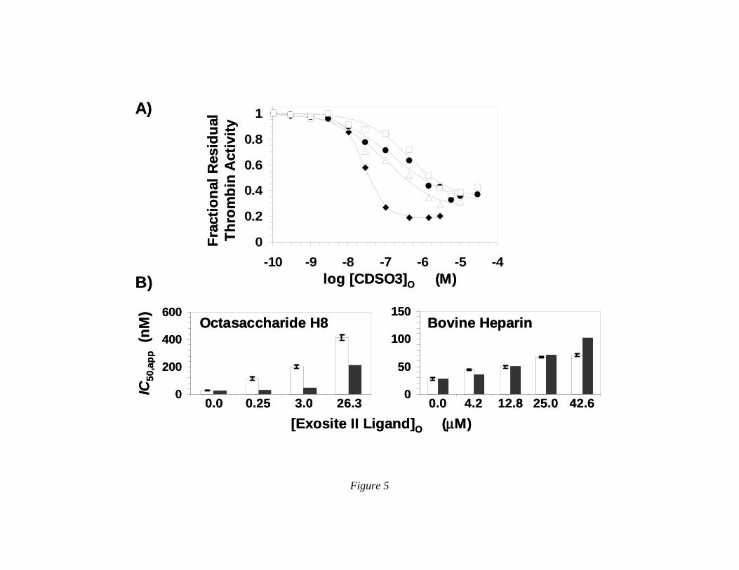
Figure 5
0
0.2
0.4
0.6
0.8
1
-10 -9 -8 -7 -6 -5 -4log [CDSO3]O (M)
Frac
tiona
l Res
idua
l Th
rom
bin
Act
ivity
[Exosite II Ligand]O (µM)
IC50
,app
(nM
)
0.0 0.25 3.0 26.3 0.0 4.2 12.8 25.0 42.6
Octasaccharide H8 Bovine Heparin
A)
B)
0
200
400
600
0
50
100
150
0
0.2
0.4
0.6
0.8
1
-10 -9 -8 -7 -6 -5 -4log [CDSO3]O (M)
Frac
tiona
l Res
idua
l Th
rom
bin
Act
ivity
[Exosite II Ligand]O (µM)
IC50
,app
(nM
)
0.0 0.25 3.0 26.3 0.0 4.2 12.8 25.0 42.6
Octasaccharide H8 Bovine Heparin
A)
B)
0
200
400
600
0
50
100
150


![Sulfated zirconia[1]](https://static.fdocuments.net/doc/165x107/5568f2ecd8b42aff2e8b4932/sulfated-zirconia1.jpg)


