Fucose-containing sulfated polysaccharidesfrom brown ...orbit.dtu.dk/files/9648779/PhD...
140
General rights Copyright and moral rights for the publications made accessible in the public portal are retained by the authors and/or other copyright owners and it is a condition of accessing publications that users recognise and abide by the legal requirements associated with these rights. • Users may download and print one copy of any publication from the public portal for the purpose of private study or research. • You may not further distribute the material or use it for any profit-making activity or commercial gain • You may freely distribute the URL identifying the publication in the public portal If you believe that this document breaches copyright please contact us providing details, and we will remove access to the work immediately and investigate your claim. Downloaded from orbit.dtu.dk on: May 07, 2018 Fucose-containing sulfated polysaccharides from brown seaweed: Extraction technolgy and bioactivity assessment Ale, Marcel Tutor; Meyer, Anne S.; Mikkelsen, Jørn Dalgaard Publication date: 2012 Document Version Publisher's PDF, also known as Version of record Link back to DTU Orbit Citation (APA): Ale, M. T., Meyer, A. S., & Mikkelsen, J. D. (2012). Fucose-containing sulfated polysaccharides from brown seaweed: Extraction technolgy and bioactivity assessment. Kgs.Lyngby: Technical University of Denmark, Department of Chemical Engineering.
Transcript of Fucose-containing sulfated polysaccharidesfrom brown ...orbit.dtu.dk/files/9648779/PhD...
General rights Copyright and moral rights for the publications made accessible in the public portal are retained by the authors and/or other copyright owners and it is a condition of accessing publications that users recognise and abide by the legal requirements associated with these rights.
• Users may download and print one copy of any publication from the public portal for the purpose of private study or research. • You may not further distribute the material or use it for any profit-making activity or commercial gain • You may freely distribute the URL identifying the publication in the public portal
If you believe that this document breaches copyright please contact us providing details, and we will remove access to the work immediately and investigate your claim.
Downloaded from orbit.dtu.dk on: May 07, 2018
Fucose-containing sulfated polysaccharides from brown seaweed: Extractiontechnolgy and bioactivity assessment
Ale, Marcel Tutor; Meyer, Anne S.; Mikkelsen, Jørn Dalgaard
Publication date:2012
Document VersionPublisher's PDF, also known as Version of record
Link back to DTU Orbit
Citation (APA):Ale, M. T., Meyer, A. S., & Mikkelsen, J. D. (2012). Fucose-containing sulfated polysaccharides from brownseaweed: Extraction technolgy and bioactivity assessment. Kgs.Lyngby: Technical University of Denmark,Department of Chemical Engineering.
Fucose-containing sulfated polysaccharidesfrom brown seaweed: Extraction technologyand biological activity assessment
PhD 2
mata
Typewritten Text
mata
Typewritten Text
mata
Typewritten Text
mata
Typewritten Text
mata
Typewritten Text
mata
Typewritten Text
mata
Typewritten Text
mata
Typewritten Text
Fucose containing sulfatedpolysaccharides from brown seaweed:Extraction technology and biological
activity assessment
Marcel Tutor Ale
PhD Thesis2012
1
Fucose containing sulfatedpolysaccharides from brown seaweed:Extraction technology and biological
activity assessment
Marcel Tutor Ale
PhD Thesis2012
1
Copyright©: Marcel Tutor Ale
Center For Bioprocess Engineering
Department of Chemical and Biochemical Engineering
Technical University of Denmark
Søltofts Plads, Building 229
DK-2800 Kgs. Lyngby
Denmark
Phone: +45 4525 2800
Web: www.bioeng.kt.dtu.dk
Print: J&R Frydenberg A/S
København
May 2012
ISBN: 978-87-92481-70-2
2
February 2012
i
Preface
Enormous amounts of seaweed resource still remain unexploited. Examining new applications of
this unexploited seaweed by developing state of the art solutions; and innovation of seaweed
products are the primary interest and motivation of this PhD study.
The PhD work was initiated at the Department of System Biology, Technical University of Denmark
(DTU) and was completed at Center for Bioprocess Engineering (BioEng), Department of Chemical
and Biochemical Engineering System Biology, Technical University of Denmark (DTU), Lyngby
Denmark. The main research and experimental activities were performed in BioEng laboratory
facilities located at Bldg. 227 DTU Søltoft Plads, Kgs. Lyngby. In fulfillment of the PhD programme
various courses, seminars, conference participations and research activities were taken both
locally and internationally. External research activity was also accomplished at School of Allied
Health Sciences, Kitasato University, Sagamihara, Kanagawa, Japan.
In this PhD thesis, published works are cited and the chapters are based on published research and
scientific papers completed by the author during the PhD study:
Chapter 1 is an introduction to the state of the art FCSP methods. It also outlines the problems,
hypotheses, and core objectives of this PhD study. This chapter provides an overall outline of
different scientific investigations and research activities in the form of project phases.
Chapter 2 is based on Paper 1, which highlights FCSP structure function relationships and
extraction methods. It also includes an overview of seaweed potentials based on recent
development and reports from published journals.
Chapter 3 is about optimized single step extraction of FCSPs based on Paper 2. This chapter
provides an in depth investigation of the influence of different extraction parameters on the
chemical nature and structural features of FCSPs.
Chapter 4 shows the FCSP bioactivity. The anti proliferative and immune response activity of
fucoidan extracted from Sargassum sp. using minimal processes based on Papers 3 and 4 is also
presented.
2
3
iiPhD Thesis2011
Chapter 5 is based on Paper 5. Successful exploitation of seaweed for commercial applications is
accomplished provided that growth parameters are optimized; hence, growth and nutrient
assimilation monitoring of seaweed has also been the subject of this study using U. lactuca
seaweed as a model.
Chapter 6 contains the final remarks about the present work. It also gives some future
perspectives and prospective research areas.
This whole PhD study was undertaken with superior guidance and supervision of Prof. Anne S.
Meyer, head of Center for Bioprocess Engineering and tireless encouragement of Prof. Jørn
Dalgaard Mikkelsen, Center for Bioprocess Engineering as co supervisor. Moreover, the diligent
assistance and supervision of Dr. Hiroko Maruyama and Dr. Hidezaku Tamauchi during my research
work in Kitasato University, Kanagawa Japan was so beneficial for the advancement of this PhD
study.
The PhD project was fully financed by the Technical University of Denmark (DTU) for a period from
March 2008 to October 2011.
This thesis is submitted for the fulfillment of the PhD degree requirements at Technical University
of Denmark.
Marcel Tutor AleTechnical University of Denmark (DTU)February 2012
2
4
iiiP
A
Abstract
Marine seaweed that is washed up on the coastline is a nuisance as its degradation produces a foul
a smell and generates waste problems. Exploitation of coastline polluting seaweeds such as
Sargassum sp., Ulva sp., and other beach cast seaweed species for various commercial
applications will generate new valuable products that may help lessen coastal pollution by
seaweeds and create new seaweed based resources. Thus, utilization of these natural resources is
of great importance. The objectives of this PhD study were to develop a technology to extract
bioactive compounds from nuisance brown seaweeds, and investigate their bioactivity. To this
effect, designed optimized extraction of fucose containing sulfated polysaccharides (FCSPs) and/or
crude fucoidan from brown seaweed were performed, and the bioactivity of the isolated FCSPs
was investigated. Moreover, to assess the potential of seaweed to assimilate nitrogen based
nutrients, a technology for accurate monitoring of differential seaweed growth responses to
nutrient assimilation was also developed.
Fucoidan is a term used to describe a class of sulfated polysaccharides extracted from brown
seaweed, which contains substantial amounts of fucose; varying amounts of galactose, xylose, and
glucuronic acid; and differing glycosidic linkages, and are variously substituted with sulfate and
acetyl groups and side branches containing fucose or other glycosyl units. These FCSPs principally
consist of a backbone of (1 3) and/or (1 4) linked L fucopyranose residues that may be
substituted with sulfate (SO3 ) on C 2, C 3, or C 4 and acetyl groups at C 4 on the main chain or
may have short fucoside side chains that are usually linked from the O 4 of one or several of the
fucopyranose backbone residues. FCSPs are known to exhibit crucial biological activities including
anti tumor activity. Although differently extracted, purified, fucose rich, modified fucoidans have
been reported to exert bioactive properties such as anti coagulant and enhance immune response
activity, few studies have investigated the bioactivity of unfractionated FCSPs, notably FCSPs
extracted using milder and fewer processing steps. Crude fucoidan from Sargassum sp. and Fucus
vesiculosus were examined for their bioactivity against lung and skin cancer cell lines in both in
vitro and in vivo studies. This study showed that unfractionated FCSPs hinder the in vitro
proliferation of Lewis lung carcinoma and melanoma B16 cell lines by induction of apoptosis.
Moreover, the anti tumor activity of crude fucoidan seems to be associated with an enhanced
immune response as depicted by an increase in natural killer cell activity in mice.
2
5
ivPhD Thesis 2011
The classical extraction of FCSPs involving long, repetitive, multi step acid and alkaline treatments
is detrimental to its structural properties, yield, and compositional attributes. In this study,
statistically designed, optimized extraction of a single step extraction of FCSPs from Sargassum sp.
was carried out. The effects of the different extraction parameters on the natural chemical
composition of the isolated sulfated polysaccharides were also investigated. The data showed that
classical multi step extraction using 0.2 M HCl at elevated temperature and extended time had a
detrimental effect on the FCSPs yield, as this treatment apparently disrupted the structural
integrity of the polymer and evidently degraded carbohydrate chains of fucose residues during
extraction. The results also revealed a maximal FCSPs yield of approximately 7% dry weight with
Sargassum sp. using 0.03 M HCl at 90°C and 4 h extraction conditions.
Accurate monitoring of the differential growth response of seaweed to different nutrient
assimilation is crucial to explore various applications of seaweed resources, such as biomass for
bioenergy production and source of functional healthy components and bioactive compounds. A
major prerequisite for the successful exploitation of cultivated seaweed like Ulva lactuca for
commercial purposes is that the growth rate and yields should be optimized. In this study, the
growth response of U. lactuca to ammonium and nitrate assimilation was investigated using a
photoscanning technique to monitor the growth kinetics in U. lactuca. Photoscanning images
revealed differential increases in the surface area of U. lactuca discs over time in response to
different nitrogen based nutrient sources. The results also showed a favorable growth response to
ammonium as a nitrogen source, and the presence of ammonium discriminated the nitrate uptake
by U. lactuca upon exposure to ammonium nitrate. This study exhibits the applicability of a
photoscanning approach for acquiring precise quantitative growth data for U. lactuca.
In conclusion, we demonstrated that nuisance seaweed can be a potential source of biomass and
bioactive compound notably FCSPs. This study proved the hypotheses that different extraction
conditions have crucial influenced to the chemical nature of FCSPs. The study also demonstrated
that unfractionated FCSPs are able to exert bioactive actions such as anti tumor and immune
modulating properties in both in vitro and in vivo studies. This study illustrates the importance of a
precise monitoring technique of the growth of U. lactuca in order to successfully exploit it for
commercial application.
2
6
vPhD Thesis 2011
Dansk Sammenfatning
Tang på stranden er en plage, først og fremmest på grund af de lugtgener, som kommer når
tangen går i forrådnelse. En udnyttelse af dette tang, såsom Sargassum sp., Ulva sp., og andre
lignende kyst nære typer af tang, kan frembringe helt nye værdifulde produkter og måske samtidig
mindske de uønskede lugtgener fra tang som skyller op på stranden. Udnyttelse af tang som
resource er udgangspunktet for dette PhD studium. PhD studiets formål har været dels at udvikle
en ekstraktionsmetode til at isolere bioaktive produkter fra brunt tang, dels at undersøge disse
produkters bioaktivitet. For at opfylde dette formål blev der i PhD arbejdet udviklet en statistisk
designet, optimeret ekstraktionsmetode til ekstraktion af såkaldte fukose indeholdende
sulfaterede polysakkarider (eng. fucose containing sulfated polysaccharides), forkortet FCSPs,
henholdsvis grov fucoidan fra brun tang, primært Sargassum sp., og bioaktiviteten af
ekstraktionsproduktet blev undersøgt. For ydermere at vurdere tangs evne til at assimilere, og
dermed vokse på nitrogen holdige salte, blev der udviklet en teknologi til at monitorere
differential vækst af Ulva lactuca, på forskellige næringsstoffer i vandet.
Fucoidan er en betegnelse, som dækker over en gruppe af sulfaterede, fukose holdige,
polysakkarider fra tang. Udover fukose indeholder fucoidan forkellige mængder galaktose, xylose,
glukuronsyre, som er forbundet via forskellige typer glykosidbindinger, og som derudover er
substitueret i forskellig grad med sulfat og acetyl grupper og som kan have sidekæder
indeholdende fukose eller andre glykosyl substituenter. Disse FCSPs består principielt af en
rygrad, eller en hovedkæde, af (1 3) og/eller (1 4) linkede L fucopyranose enheder, som kan
være substitueret med sulfate (SO3 ) på C 2, C 3, eller C 4 foruden acetyl grupper på C 4 på fucose
enhenderne i hovedkæden, og/eller som har korte fukose kæder, der normalt er bundet via O 4
fra en eller flere fukose enheder i hovedkæden. Det er kendt, at FCSPs isoleret fra tang har
forskellige gavnlige, bioaktive effekter, herunder anti tumor aktivitet. Selvom det har været
rapporteret, at oprensede, fucose rige, modificerede fucoidan prøver har bioaktive effekter,
såsom anti koagulerende, og immun respons forøgende egenskaber, er bioaktiviteten af mere
grove, ufraktionerede FCSPs – ekstraheret med mildere og færre ekstraktionstrin ikke undersøgt.
Groft oprensede fucoidan prøver fra Sargassum sp. og Fucus vesiculosus blev i dette PhD arbejde
undersøgt for deres bioaktivitet mod lunge og hudcancer cellelinjer vækst både in vitro og in vivo.
2
7
viPhD Thesis 2011
Arbejdet viste, at ufraktionerede FCSPs ekstrakter hindrer proliferation in vitro af Lewis lung
carcinoma og melanoma B16 celle linjer via induktion af apoptosis. Desuden blev det vist, at
denne anti tumor aktivitet af grov fucoidan, tilsyneladende er associeret med et øget
immunrespons, målt som et forøget niveau af naturlige ”killer” cellers aktivitet i mus.
Klassisk ekstraktion af FCSPs fra tang involverer adskillige langsommelige, behandlinger med syre
og base, hvilket er ødelæggende for deres specifikke struktur og sammensætning. I dete PhD
arbejde blev der udviklet en statistisk designet, optimeret enkelt trins ekstraktionsmetode til at
udtrække FCSPs fra Sargassum sp.. Effekten af de forskellige ekstraktionsparametre på
sammensætningen af de ekstraherede polysaccharider belv også vurderet. Resultaterne viste, at
klassisk, multi trins ekstraktion ved brug af 0.2 M HCl ved høj temperature havde en
ødelæggende effect på udbyttet af FCSPs og viste desuden at en sådan behandling tilsyneladende
ødelagde polysaccharidstrukturen og at fukose kæderne blev nedbrudt under behandlingen.
Resultaterne viste også, at et maximal FCSPS udbytte, på ca. 7% af tørvægten for Sargassum sp.
kunne opnås ved et trins ekstraktion med 0.03 M HCl ved 90°C i 4 timer.
Nøjagtig monitorering af vækstrespons af tang på forskellige næringsstoffer er afgørende for at
undersøge forskellige anvendelser af tang, primært biomassevækst til bioenergi produktion og til
udnyttelse af tang som kilde til produktion af f.eks. funktionelle fødevarekomponenter eller
bioaktive stoffer. En vigtig forudsætning for kommerciel udnyttelse af kultiveret tang såsom Ulva
lactuca er, at vækstraten og udbyttet optimeres. I dette studie blev vækstrespons af U. lactuca I
forhold til ammonium og nitrat assimilation undersøgt ved hjælp af en foto scannings teknik. Foto
scannede billeder afslørede forskellige vækst inkrementer udfra måling af det fotograferede areal
af udstukne skiver af U. lactuca over tid som respons på forskellige nitrogen baserede
næringskilder. Resultaterne viste også en favorabel vækstøgning på ammonium som
nitrogenkilde, og desuden at tilstedeværelsen af ammonium diskriminerede U. lactucas nitrat
optag i forhold til optaget af ammonium nitrat. Studiet viste anvendeligheden af phot scanning til
præcis kvantitativ vækst monitorering af U. lactuca.
Den samelde konklusien er således at vi demonstrerede, at tang kan være en potential kilde til
biomasse og bioaktive komponenter, især FCSPs. PhD studiet viste desuden at hypotesen, at
forskellgie ekstraktionsbetingelser har afgørende indflydelse på den kemiske natur af FCSPs er
sand. Studiet demonstrerede også, at ufraktionerede FCSPs har bioaktive egenskaber såsom anti
tumor og immun modulerende effecter, vist i både in vitro og in vivo. Studiet ilustrerer desuden
vigtigheden af en præcis monitoreringsmetode til måling af U. lactuca vækst.
2
8
viiPhD Thesis 2011
List of publications
The PhD thesis is based on the work presented in the following papers:
I. Important determinants for fucoidan bioactivity: A critical review of structure functionrelations and extraction methods for fucose containing sulfated polysaccharides frombrown seaweedAle MT, Mikkelsen JD and Meyer ASMarine Drugs (2011) DOI:10.3390/md9 , Published online
II. Designed optimization of a single step extraction of fucose containing sulfatedpolysaccharides from Sargassum sp.Ale MT, Mikkelsen JD and Meyer ASJournal of Applied Phycology (2011)DOI: 10.1007/s1081 011 9690 3, Published online
III. Fucoidan from Sargassum sp. and Fucus vesiculosus reduces cell viability of lung carcinomaand melanoma cells in vitro and activates natural killer cells in mice in vivoAle MT, Maruyama H, Tamauchi H, Mikkelsen JD and Meyer ASInternational Journal of Biological Macromolecules 49 (2011) 331 – 336DOI: 10.1016/j.ibiomac.2011.05.009, Published online
IV. Fucose containing sulfated polysaccharides from brown seaweed inhibit proliferation ofmelanoma cells and induce apoptosis by activation of caspase 3 in vitroAle MT, Maruyama H, Tamauchi H, Mikkelsen JD and Meyer ASMarine Drugs (2011) DOI:10.3390/md , Published online
V. Differential growth response of Ulva lactuca to ammonium and nitrate assimilationAle MT, Mikkelsen JD and Meyer ASJournal of Applied Phycology (2011) 23: 345–351, Published
2
9

10 10
ixPhD Thesis 2011
Table of Contents
Preface ............................................................................................................................... ...................... i
Abstract............................................................................................................................... ................... iii
Dansk Sammenfatning ............................................................................................................................ v
List of publications ............................................................................................................................... . vii
Table of Contents............................................................................................................................... .... ix
1 Introduction ............................................................................................................................... ..........1
1.1 Problems statement ................................................................................................................. 3
1.2 Hypotheses ............................................................................................................................... 4
1.3 Core objectives and project phases.......................................................................................... 5
2 Seaweed potentials: a general overview ............................................................................................. 9
2.1 Bioactive seaweed compounds: sulfated polysaccharides.....................................................10
2.1.1 FCSPs structure ............................................................................................................. 10
2.1.2 Anti tumor bioactivity of FCSPs .................................................................................... 13
2.2 FCSPs extraction and chemical composition: past and present .............................................15
2.2.1 FCSPs composition studies, 1930–1950........................................................................17
2.2.2 Lab scale extraction of fucoidan, 1950s........................................................................18
2.2.3 FCSPs extraction, 1970s–present .................................................................................. 19
2.3 Seaweed products and biomass potential.............................................................................. 19
2.3.1 Phycocolloids................................................................................................................. 20
2.3.2 Seaweed biomass for bioenergy production ................................................................21
2.4 Seaweed production............................................................................................................... 21
2.5 Paper 1: Important determinants for fucoidan bioactivity: a critical review of structurefunction correlations and extraction methods for FCSPs from brown seaweeds ..............22
3 Extraction of FCSPs ............................................................................................................................ 4
3.1 Extraction methods................................................................................................................. 4
3.2 Single step extraction of FCSPs...............................................................................................
2
11
xPhD Thesis 2011
3.2.1 Relevance ......................................................................................................................
3.2.2 Hypotheses and objectives ...........................................................................................
3.2.3 Result highlights ............................................................................................................
3.2.4 Consideration and justification .....................................................................................
3.3 Paper 2: Designed optimization of single step extraction of FCSPs from Sargassum sp........5
4 Bioactivity of FCSPs ............................................................................................................................ 6
4.1 Anti tumor activity of FCSPs ...................................................................................................6
4.2 Anti proliferative and immune response activities................................................................6
4.2.1 Relevance ...................................................................................................................... 6
4.2.2 Hypotheses and objectives ...........................................................................................6
4.2.3 Result highlights ............................................................................................................ 6
4.2.4 Consideration and justification .....................................................................................6
4.3 Paper 3: Fucoidan from Sargassum sp. and Fucus vesiculosus reduces cell viability of lungcarcinoma and melanoma cells in vitro and activates natural killer cells in mice in vivo ..
4.4 Paper 4: Fucose containing sulfated polysaccharides inhibits the proliferation of melanomacells and induces apoptosis by activation of caspase 3 in vitro .........................................
5 Seaweed nutrient assimilation and growth response .......................................................................9
5.1 Evaluation of growth and assimilation ...................................................................................9
5.2 Growth response of U. lactuca ...............................................................................................9
5.2.1 Relevance ...................................................................................................................... 9
5.2.2 Hypotheses and objectives ...........................................................................................9
5.2.3 Result highlights ............................................................................................................ 9
5.2.4 Consideration and justification .....................................................................................9
5.3 Paper 5: Differential growth response of Ulva lactuca to ammonium and nitrate assimilation............................................................................................................................... .............9
6 Conclusions and perspectives ..........................................................................................................10
6.1 Future perspectives .............................................................................................................. 1
7 References..................................................................................................................... ...................11
2
12
1PhD Thesis 2011
1
1 Introduction
In the vast coastal areas worldwide, seaweed has played a major role in maintaining the ecological
balance of the environment and marine bio ecosystem. Seaweeds, including various brown
seaweeds such as Undaria and Laminaria spp., are part of the food culture in many Asian
countries, notably Japan, the Philippines, and Korea, and seaweed extracts have also been used as
remedies in traditional medicine. In recent years, harvesting and monoculture farming of certain
seaweed species have become an important livelihood for fishermen in Southeast Asia owing to
the progressively increasing demand for raw seaweed worldwide. Many seaweed species, notably
beach cast seaweeds, still need to be examined for their characteristics and properties, which will
determine their commercial applications, before they can be recognized as important
commodities.
Seaweed that washes up on the coastline often generates waste problems for populations residing
seaside owing to microbial accumulation and unpleasant odors. Utilization of beach cast seaweeds
such as Ulva sp., Sargassum sp., and other nuisance seaweed species for advantageous
applications may alleviate these problems and create valuable seaweed based products. It is
widely known that seaweed may contain unique components that have potential commercial
applications; however, few seaweed species are commercially utilized and others remain
unexploited. Some seaweed species could be potential sources of functional dietary fiber and
polysaccharides with bioactive properties (Lahaye 1991; Takahashi 1983). Recent developments in
seaweed utilization include applications involving naturally derived seaweed extracts and bioactive
compounds such as fucose containing sulfated polysaccharides (FCSPs) and/or fucoidan in some
cosmetic products and food supplements.
Fucoidan is a term used to described a class of sulfated polysaccharide extracted from the
seaweed class Phaeophyceae, which consist almost entirely of fucose and ester sulfate (Percival
and McDowell 1967). This FCSP principally consists of a backbone of (1 3) and/or (1 4) linked
L fucopyranose residues that may be organized in stretches of (1 3) fucan or with alternating
(1 3) and (1 4) bonded L fucopyranose residues. The L fucopyranose residues may be
substituted with sulfate (SO3 ) on C 2 or C 4 (rarely on C 3) single L fucosyl residues and/or short
fucoside (fuco oligosaccharide) side chains. If present, the fucoside side chains are usually O 4,
linked to the L fucopyranose backbone residues. Apart from variations in the sulfate content and
2
13
2PhD Thesis 2011
substitutions, the monosaccharide composition of FCSPs varies among different species of brown
seaweeds. Hence, in addition to fucose, different types of FCSPs may also contain galactose,
mannose, xylose, glucose, and/or glucuronic acid, usually in minor amounts (Percival and
McDowell, 1967; Bilan and Usov, 2008). It appears that FCSPs cover a broader range of complex
polysaccharides than those having only fucan backbones. Fucoidan—or more correctly, FCSPs
extracted from brown seaweeds like Sargassum sp. and Fucus vesiculosus—were documented to
have a wide range of biological activities including anticoagulant (Nardella et al., 1996); anti
inflammatory (Blondin et al., 1994); antiviral (Adhikari et al., 2006; Trinchero et al., 2009); and,
notably, anti tumoral effects (Zhuang et al., 1995; Ale et al., 2011).
Typical extraction of FCSPs from brown seaweed involves a harsh processing condition and several
purification steps. Purification of FCSPs by column chromatography was effective for isolating
polysaccharide fractions as they had higher fucose contents than those FCSPs obtained using
minimal processing steps (Li et al., 2006; Ale et al., 2011). The conditions for obtaining FCSPs from
brown seaweed generally consist of multiple, long, repetitive steps using acid (e.g., HCl) and other
solvents at elevated temperatures (Chizhov et al., 1999; Bilan et al., 2002). The influence of the
extraction methods on the chemical nature of sulfated polysaccharide has already been
demonstrated by Black et al. (1952). On the other hand, the FCSP yield of F. evanescens extracted
4 times using 2% CaCl2 solution at 85°C for 5 h was 12.9% dry weight (DW) (Bilan et al., 2002),
while extraction at 25°C using 0.4% HCl for 5 h yielded 12.0% DW (Zvyagintseva et al., 1999).
Despite the existence of early seminal studies about FCSP or fucoidan extraction, there is only
limited evidence about the influences and apparently complex interactions of extraction
parameters, such as acid solvents, temperature, and time, on FCSP yield and composition. FCSP
extraction procedures with fewer steps are milder on the brown seaweeds than are other though
they may yield a heterogeneous sulfated polysaccharide product. Nevertheless, fewer steps
extraction approach minimizes the structural alteration of algal sulfated polysaccharides and,
thereby, maintains the natural bioactive characteristics of FCSPs.
Although differently extracted and purified FCSPs have been reported to exert bioactivity
(Holtkamp et al., 2009), unfractionated FCSPs has also been found to reduce cell proliferation of
lung carcinoma and melanoma cells, exert immunopotentiating effects in tumor bearing animals,
and to activate natural killer (NK) cells in mice, leading to anti tumor activity efficacy (Takahashi,
1983; Ale et al., 2011; Foley et al., 2011). Kim et al. (2010) applied a crude polysaccharide
composed predominantly of sulfated fucose from F. vesiculosus to human colon cancer cells in
2
14
3PhD Thesis 2011
1
vitro and concluded that this polysaccharide from brown seaweed induces apoptosis. Moreover,
commercially available crude fucoidan (Sigma Inc.) was tested on human lymphoma HS Sultan cell
lines and was found to inhibit proliferation and induce apoptosis by activating caspase 3 (Aisa et
al., 2005). It was reported recently that FCSPs from Sargassum sp. and crude fucoidan (Sigma Inc.)
from F. vesiculosus induced apoptosis in melanoma cells (Ale et al., 2011).
Besides the bioactive compounds, seaweeds also possess other valuable components, such as
soluble dietary fibers and carbohydrates for hydrocolloid applications. The green seaweed (Ulva
species) are particularly rich in rare cell wall polysaccharides and have been proposed as being
important sources of dietary fiber, mainly soluble fiber (Lahaye 1991; Lahaye and Axelos 1993).
Furthermore, U. lactuca is also a good source of vitamins A, B2, B12, and C and is rich in
tocopherol (Abd El Baky et al., 2008; Ortiz et al., 2006). It has been shown that seaweed, notably
U. lactuca, was suitable for propagation under controlled conditions (Vermaat and Sand Jensen,
1987; Lee, 2000; Sato et al., 2006). For this reason, U. lactuca cultivation in tanks for either crude
biomass production for bioenergy or for the production of biologically active compounds is
currently receiving increased attention (Hiraoka and Oka, 2008). However, a major prerequisite for
the successful exploitation of cultivated U. lactuca for commercial applications is optimization of
growth rates and yields. This in turn requires both an understanding of the influence of different
nutrients on the growth response and a precise methodology to measure the growth. Nuisance
green seaweeds like U. lactuca showed bioremediation ability in nitrogen and phosphate rich
waste water (Copertino et al., 2008). Nevertheless, limited information is known about the growth
response and nutrient uptake assimilation of U. lactuca when expose to combine concentrations of
ammonium and nitrate.
This PhD thesis delivers the most recent study involving FCSPs extraction technology and evaluates
FCSPs biological activity. A single step extraction method for the removal of FCSPs from Sargassum
sp. was developed in the course of this study. FCSPs bioactivity studies have been conducted in
lung and skin cancer cell models in vitro and immune response activity in vivo. In addition to
extraction and bioactivity studies, evaluation of the growth response of U. lactuca to nutrients
such as NH4 and NO3 was also performed.
1.1 Problems statementPurified fractions from FCSPs are commonly used for structural and bioactivity analysis. These
samples contain high fucose sulfate levels and are free from other contaminant saccharide
2
15
4PhD Thesis 2011
residues. Hence, many seminal studies have shown that purified fucoidan has high bioactivity as a
result of multi step extraction and further purification and fractionation.
Can an unpurified FCSPs product extracted using minimal steps exert bioactivity?
What is the effect of unpurified FCSPs against certain cancer cell lines and what is its
influence on immune response activity?
The present extraction technology using aqueous alkali solution or dilute acid at ambient or
slightly elevated temperature has always been the most convenient method to produced FCSPs.
Would different extraction parameters, i.e., acid, temperature, and time, influence the
structural features and chemical nature of FCSPs?
What are the effects of the interactions of different extraction treatments?
Exploitation of seaweed resources has recently received special attention for its potential for both
the production of bioactive compounds and as a biomass source for bioenergy production. To
successfully exploit seaweed for commercial applications, the growth rate and yields must be
optimized. Hence, a precise monitoring technology to evaluate the seaweed growth response is
required.
What are the different monitoring techniques that are used to evaluate the U. lactuca
growth response to nutrient assimilation? How does it affect the measurements’
precision?
How is the growth of U. lactuca influenced by the assimilation of different nutrients (NH4
and NO3)?
1.2 HypothesesNuisance marine seaweed that has washed up on the coastlines is a potential starting material for
producing bioactive compounds like fucoidan or FCSPs. The precise assessment of seaweed growth
is crucial in our understanding of seaweed nutrient assimilation mechanisms. In this thesis, some
specific hypotheses were outlined:
Minimal extraction step of FCSPs will preserve the polysaccharides’ structural integrity
and, thus, increase the yield and improve its biological properties
2
16
5
FCSP products from a single step extraction process (i.e., crude fucoidan) from brown
seaweed can exert bioactivity against certain types of cancer cell lines (e.g., by inducing
apoptosis) and can promote immune responses
Classical extraction of FCSPs involving long, repetitive, multi step acid and alkaline
treatments can be detrimental to its structural makeup, yield, and compositional
attributes and, thus, may influence its bioactive properties
To exploit marine seaweed for commercial applications, growth monitoring is crucial to
the accurate evaluation of differential growth responses and nutrient (NH4 or NaNO3)
assimilations
1.3 Core objectives and project phasesSeaweed resources generally cover wide potential applications for different industries including
food and nutrition, pharmaceuticals, cosmetics, and bioenergy. Therefore, their study requires
extensive in depth research involving an interdisciplinary approach and a considerable amount of
time to accomplish certain achievable objectives. This PhD study narrows the subjects into more
focused areas with very realistic aims or specific objectives to produce novel technology while
attaining basic scientific understanding.
To test the hypotheses, various specific objectives were applied in this study. The core objectives
were primarily concentrated on the investigation of different FCSPs extraction methods and
developing innovative FCSPs extraction technology; the potential of seaweeds as a source of
bioactive compounds, notably FCSPs; and to assess the bioactivity and mechanism of FCPS
products against certain types of cancer. Furthermore, we evaluate seaweeds’ differential growth
responses to nutrient assimilation. Along with the core objectives, the specific aims of the study
were as follows:
Development of a new process for producing bioactive compound like fucose containing
sulfated polysaccharides from brown seaweed by optimized designed extraction
parameters using of state of the art analytical methods and quantification analyses
Investigation of the bioactivity of single step extracted unpurified FCSPs products (i.e.,
crude fucoidan) in cancer cell lines using in vitro and in vivo experiments
Evaluation of the seaweed growth response to nutrient assimilation (i.e., NH4 and NO3)
2
17
6PhD Thesis 2011
To ensure that the work was conducted in the right direction, workloads were organized and
narrowed down to specific scopes in a number of project phases:
Phase 1: Survey of seaweed bioactive compound structure and bioactivity
A literature research and gathering of review articles was conducted with the aim of gaining an
overall perspective of the subject. Special focus was placed on the available technical and scientific
information regarding bioactive compounds’ structural makeup and composition, the influence of
the extraction process, and factors affecting bioactivity.
Phase 2: Optimized extraction and hydrolysis of seaweed polysaccharides (i.e., FCSPs)
Fucoidan can be obtained by extraction from brown seaweeds like Sargassum sp. The typical
extraction of FCSPs involves multi step extraction using different temperatures and acid and
alkaline concentrations in an extended period. In this study, it was hypothesized that harsh
extraction treatment is detrimental to the integrity of the chemical nature of these
polysaccharides; thus, preservation of its structure during extraction can only be done using a mild
extraction technique. The main objective of this project was to design an optimized extraction of
FCSPs using a minimal step method compared to existing classical extraction methods with special
attention placed on the influence on chemical composition.
Phase 3: Assessment of FCSP bioactivity
FSCPs are known to possess bioactive properties such as anti tumor activity, apoptosis induction in
cancer cells, and immune potentiation. The efficacy of FCSPs derived from brown seaweed remains
a matter of debate; nevertheless, several reports attribute its efficacy to its structural makeup,
substitutions, and content of sulfate. It was hypothesized that mild extracted FCSPs must possess
high bioactive properties because their structural makeup remains intact. The objective of this
study was to examine the different contributing factors influencing FCSP bioactivity on some
cancer cell lines. The bioactivities of FCSPs extracted from nuisance seaweed were evaluated using
cancer cell lines in vitro and in vivo experiments.
Phase 4: Seaweed growth response to nutrient assimilation
The proliferation of seaweed is mostly influenced by photosynthesis and available nutrients that
can be utilized and assimilated. Seaweed growth is tantamount to increased biomass production.
Seaweed can potentially be utilized for the production of bioactive compounds and healthy
2
18
7PhD Thesis 2011
1
components. Monitoring the seaweed growth and nutrient uptake is a crucial step for evaluating
the seaweed growth rate, nutrient uptake rate, and biomass yield. Thus, developing a method to
properly monitor seaweed growth will contribute to our understanding of the seaweed growth
response to nutrient and assimilation patterns. The objective of this study is to examine the U.
lactuca growth response to nutrients, i.e., NH4 or NaNO3, with the special aim of developing a
growth monitoring technique to accurately evaluate the differential growth response and nutrient
assimilation and to analyze different biomass accumulation related parameters.
2
19

20 20
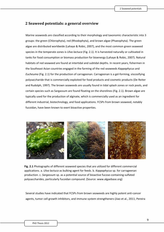
9
2 Seaweed potentials: a general overview
Marine seaweeds are classified according to their morphology and taxonomic characteristic into 3
groups: the green (Chlorophyta), red (Rhodophyta), and brown algae (Phaeophyta). The green
algae are distributed worldwide (Lahaye & Robic, 2007), and the most common green seaweed
species in the temperate zones is Ulva lactuca (Fig. 2.1). It is harvested naturally or cultivated in
tanks for food consumption or biomass production for bioenergy (Lahaye & Robic, 2007). Natural
habitats of red seaweed are found at intertidal and subtidal depths. In recent years, fishermen in
the Southeast Asian countries engaged in the farming of the red seaweeds Kappaphycus and
Eucheuma (Fig. 2.1) for the production of carrageenan. Carrageenan is a gel forming, viscosifying
polysaccharide that is commercially exploited for food products and cosmetic products (De Reiter
and Rudolph, 1997). The brown seaweeds are usually found in tidal splash zones or rock pools, and
certain species such as Sargassum are found floating on the shorelines (Fig. 2.1). Brown algae are
typically used for the production of alginate, which is commercially used as an ingredient for
different industrial, biotechnology, and food applications. FCSPs from brown seaweed, notably
fucoidan, have been known to exert bioactive properties.
Fig. 2.1 Photographs of different seaweed species that are utilized for different commercialapplications. a. Ulva lactuca as bulking agent for feeds. b. Kappaphycus sp. for carrageenanproduction. c. Sargassum sp. as a potential source of bioactive fucose containing sulfatedpolysaccharides, particularly fucoidan compound. (Source: www.algaebase.org)
Several studies have indicated that FCSPs from brown seaweeds are highly potent anti cancer
agents, tumor cell growth inhibitors, and immune system strengtheners (Jiao et al., 2011; Pereira
2 Seaweed otentials
2
21
10PhD Thesis 2011
et al., 1999). It has been suggested that the lower breast cancer and obesity rates in Japan may be
related to regular intake of brown seaweed (Teas 1983). Brown seaweeds are very abundant and
constitute unexploited resources worldwide, thus making them viable sources of polysaccharides,
especially those with bioactive properties such as fucoidan or other FCSPs. Nevertheless, the red
and green seaweeds are also good sources of functional food ingredients, including phycocolloids
and dietary fibers.
2.1 Bioactive seaweed compounds: sulfated polysaccharides
Sulfated polysaccharides are a family of compounds containing ester sulfate groups in their sugar
residues. These polysaccharides are commonly found in the marine algal groups Phaeophyta,
Rhodophyta, and Chlorophyta. Fucans is the general term for sulfated polysaccharides that are
present in the class Phaeophyta, which includes FCSPs. The sulfated polysaccharides in the class
Rhodophyta are galactans consisting entirely of galactose or modified galactose units such as agar
and carrageenans. The major polysaccharides in the class Chlorophyta are polydisperse
heteropolysaccharides known as ulvans.
Ulva, the green seaweed species of the class Chlorophyta, are particularly rich in rare cell wall
polysaccharides and have been proposed as being an important source of dietary fiber, mainly
soluble fiber, which could be a potential prebiotic substrate (Lahaye 1991; Lahaye and Axelos
1993). Brown seaweed is a source of a unique compounds, notably FCSPs, which have shown high
potency against certain cancers in some in vitro studies and exhibited immune response activity in
vivo (Cumashi et al., 2007; Maruyama et al., 2003). Red seaweed, on the other hand, is known for
its hydrocolloid characteristics; however, published reports have shown that sulfated
polysaccharides from red seaweed also possessed some bioactive properties (Pereira et al., 2005;
Talarico et al., 2007).
2.1.1 FCSPs structureFucoidan, an FCSP that is extracted from brown algae, may contain differing glycosidic linkages and
is variously substituted with acetate and side branches containing fucose or other glycosyl units.
Brown seaweed in the order of Fucales such as F. evanescens and F. serratus possess a large
proportion of both (1 3) and (1 4) linked L fucopyranose residues may be substituted with
sulfate (SO3 ) on C 2 and C 4 (Fig. 2.2; Bilan et al., 2002, 2006; Cumashi et al., 2007). The alga
Ascophyllum nodosum (Fucales) has a predominant repeating structure (1 3) L fucopyranose
residues with sulfate at C 2 position linked (1 4) L fucopyranose residues with disulfate at C 2
2
22
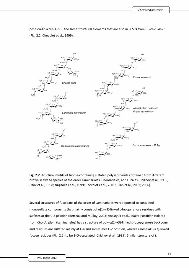
11PhD Thesis 2011
position linked (1 3), the same structural elements that are also in FCSPs from F. vesiculosus
(Fig. 2.2; Chevolot et al., 1999).
Cladosiphon okamuranus
O
O
O
OSO3-CH3
O
-O3SO
OOH
CH3
-O3SO
-O3SOO
OO
CH3
OOH
CH3
HO
OH
Laminaria saccharina
O
O
O
OHCH3
O
HO
OOH
CH3
-O3SO
HOO
OO
CH3
OOH
OH
COO-HO
O
O
O
OSO3-CH3
-O3SO
OOH
CH3
-O3SO
-O3SOO
OOH
CH3
O
OCH3
-O3SO
O
OOH
CH3
HO
OH
O
O
OH
OSO3-CH3
-O3SO
Chorda filum
Ascophyllum nodosumFucus vesiculosus
O
OH O
O
OSO3-CH3
O
O OSO3-OSO3-
CH3
O
OSO3-
OH
OSO3-CH3
O
OH OOSO3-
CH3
O
OHOSO3-
OSO3-CH3
OOH
O
O
OSO3-CH3
O
OOH
OSO3-CH3
-O3SO
OOSO3-
CH3
OHO
OOSO3-
CH3
O
Fucus evanescens C.Ag
Fucus serratus L.
OOSO3-
CH3
O
OOH
O
O
OSO3-CH3
O
OOH
OSO3-CH3
O
OOSO3-
CH3
-O3SO
OOSO3-
CH3
OHOH
OH
Fig. 2.2 Structural motifs of fucose containing sulfated polysaccharides obtained from differentbrown seaweed species of the order Laminariales, Chordariales, and Fucales (Chizhov et al., 1999;Usov et al., 1998; Nagaoka et al., 1999; Chevolot et al., 2001; Bilan et al., 2002; 2006).
Several structures of fucoidans of the order of Laminariales were reported to contained
monosulfate components that mainly consist of (1 3) linked L fucopyranose residues with
sulfates at the C 2 position (Berteau and Mulloy, 2003; Anastyuk et al., 2009). Fucoidan isolated
from Chorda filum (Laminariales) has a structure of poly (1 3) linked L fucopyranose backbone
and residues are sulfated mainly at C 4 and sometimes C 2 position, whereas some (1 3) linked
fucose residues (Fig. 2.2) to be 2 O acetylated (Chizhov et al., 1999). Similar structure of L.
2 Seaweed otentials
2
23
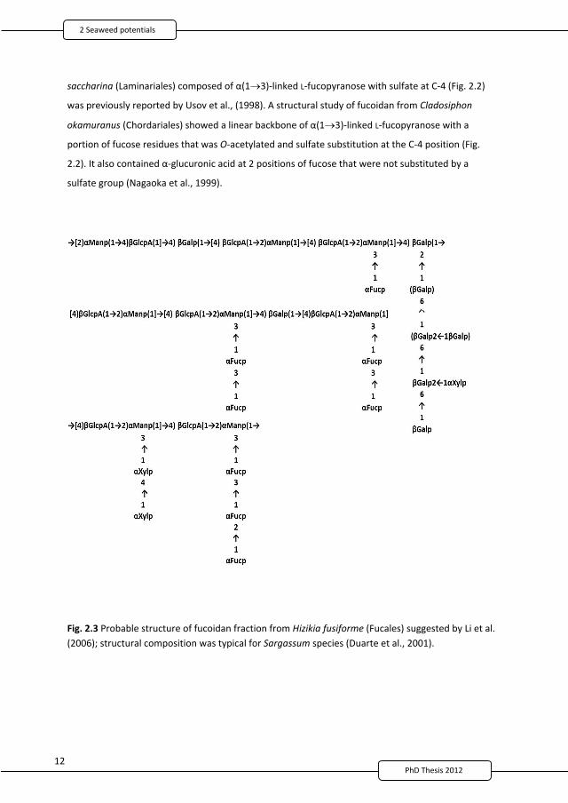
12PhD Thesis 2011
saccharina (Laminariales) composed of (1 3) linked L fucopyranose with sulfate at C 4 (Fig. 2.2)
was previously reported by Usov et al., (1998). A structural study of fucoidan from Cladosiphon
okamuranus (Chordariales) showed a linear backbone of (1 3) linked L fucopyranose with a
portion of fucose residues that was O acetylated and sulfate substitution at the C 4 position (Fig.
2.2). It also contained glucuronic acid at 2 positions of fucose that were not substituted by a
sulfate group (Nagaoka et al., 1999).
Fig. 2.3 Probable structure of fucoidan fraction from Hizikia fusiforme (Fucales) suggested by Li et al.(2006); structural composition was typical for Sargassum species (Duarte et al., 2001).
2
24
13PhD Thesis 2011
Complex fucoidan structures such as sulfated galactofucans and heterofucans brown algae have
been reported (Bilan and Usov, 2008). A complex mixture of polysaccharides extracted from
Sargassum stenophyllum was mainly built of (1 6) D galactose and/or (1 2) D mannose units
with branching points formed by (1 3) and/or (1 4) L fucose, (1 4) D glucuronic acid,
terminal D xylose, and sometimes (1 4) D glucose. Sulfate groups on the fucans are located at
C 4 of a (1 3) linked unit or C 2 of a (1 4) linked residue (Fig. 2.3; Duarte et al., 2001). Galactose
is found in trace amounts in the fucans from Himanthalia lorea and Macrocystis pyrifera (Mian and
Percival, 1973) and has been reported as a major constituent, but the polysaccharides also had
substantial quantities of fucose, xylose, and glucuronic acid (Percival and Young, 1974). A sulfated
D galactan isolated from L. angustata in trace amounts was considered a fucoidan with D
galactose as the major sugar and L fucose and D glucuronic acid as trace components (Nishino et
al., 1994).
2.1.2 Anti tumor bioactivity of FCSPsFCSPs in brown seaweed have been subject to many scientific studies due to their diverse biological
functions including anti tumor and immunomodulatory activities (Alekseyenko et al., 2007;
Maruyama et al., 2006). There are very few published reports on the relationship between chemical
properties and anti tumor activity of FCSPs. (Li et al., 2008; Alekseyenko et al., 2007; Koyanagi et al.,
2003). However, it was suggested that the bioactive properties of FCSPs are mainly determined by the
fucose sulfated chains (Nishino et al., 1994; Mourão et al., 1996); nevertheless, the anti cancer
activity of FCSPs was recently revealed to not be a function of a single but a combination of many
factors such as the amount of sulfate groups, monosaccharide residues ratio, and the linkage type of
the sugar residues (Ermakova et al., 2011; Ale et al., 2011). The available findings indicate that the
anti tumor activity of FCSPs may be associated with a significant enhancement of the cytolytic activity
of NK cells augmented by increased production of macrophage mediated immune response signaling
molecules (Maruyama et al., 2003; Takahashi et al., 1983; Teruya et al., 2009), namely interleukin (IL)
2, interferon (IFN) , and IL 12 (Ale et al., 2011; Maruyama et al., 2003), and induction of apoptosis
(Ale et al., 2011).
Macrophage activation by polysaccharides is mediated through specific membrane receptors. The
major receptors reported for polysaccharide recognition in macrophages are glycoproteins including
Toll like receptor 4 (TLR 4), cluster of differentiation 14 (CD14), competent receptor 3 (CR 3), and
scavenging receptor (SR) (Teruya et al., 2009). Activation of these receptors is mediated by
intracellular signaling pathways, and the family of mitogen activated protein kinases (MAPKs) plays a
2 Seaweed otentials
2
25
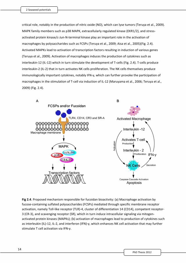
14
critical role, notably in the production of nitric oxide (NO), which can lyse tumors (Teruya et al., 2009).
MAPK family members such as p38 MAPK, extracellularly regulated kinase (EKR1/2), and stress
activated protein kinase/c Jun N terminal kinase play an important role in the activation of
macrophages by polysaccharides such as FCSPs (Teruya et al., 2009; Aisa et al., 2005)(Fig. 2.4).
Activated MAPKs lead to activation of transcription factors resulting in induction of various genes
(Teruya et al., 2009). Activation of macrophages induces the production of cytokines such as
interleukin 12 (IL 12) which in turn stimulate the development of T cells (Fig. 2.4). T cells produce
interleukin 2 (IL 2) that in turn activates NK cells proliferation. The NK cells themselves produce
immunologically important cytokines, notably IFN , which can further provoke the participation of
macrophages in the stimulation of T cell via induction of IL 12 (Maruyama et al., 2006; Teruya et al.,
2009) (Fig. 2.4).
Fig 2.4. Proposed mechanism responsible for fucoidan bioactivity: (a) Macrophage activation byfucose containing sulfated polysaccharides (FCSPs) mediated through specific membrane receptoractivation, namely Toll like receptor (TLR) 4, cluster of differentiation 14 (CD14), competent receptor3 (CR 3), and scavenging receptor (SR), which in turn induce intracellular signaling via mitogenactivated protein kinases (MAPKs); (b) activation of macrophages lead to production of cytokines suchas interleukin (IL) 12, IL 2, and interferon (IFN) , which enhances NK cell activation that may furtherstimulate T cell activation via IFN .
2
26
15PhD Thesis 2011
The anti tumor mechanism of FCSPs appears to be associated with the significant enhancement of the
cytolytic activity of NK cells augmented by increased production of the macrophage mediated
immune response, namely IL 2, type II IFN , and IL 12 (Maruyama et al., 2003; Maruyama et al.,
2006). NK cells are large granular lymphocytes that are found throughout the body that contain
cytotoxic substances which are important for the protection against some tumors. The slated NK cell
killing occurs via release of granules containing perforin, which effectively opens up pores in target
cell membranes through which the granzymes can enter and induce apoptosis (Kindt et al., 2007;
Lydyard et al., 2000). FCSPs was reported to induce apoptosis in HT 29 colon cancer cells (Kim et al.,
2010), MCF 7 human breast cancer cells (Yamasaki Miyamoto et al., 2009), and HS Sultan human
lymphoma cells (Aisa et al., 2005). The importance of apoptosis as a killing mechanism used by the
immune system is that targeted cells can be rapidly removed by phagocytes without induction of an
inflammatory response.
IL 2, which is made by T cells, is a critical autocrine growth factor that is required for proliferation of T
cells and NK cells (Lydyard et al., 2000). NK cell secretion of type II IFN activates macrophages,
inducing IL 12 secretion, activating NK cells, and creating a system of positive feedback that increases
the activation of both cell types within an infected cell or tissue (Parham 2009; Kindt et al., 2007).
Hence, it was suggested that stimulation with IL 2 and IL 12 promotes IFN secretion by NK cells,
probably due to enhancing NK cell activity by FCSPs (Maruyama et al., 2006). IL 12 stimulation alone
was reported to produce only moderate augmentation of NK cell cytotoxicity. However, it increases
the catalytic activity of lymphocytes against autologous targets in synergy with IL 2 (Nastala et al.,
1994).
2.2 FCSPs extraction and chemical composition: past and present
Several extraction and purification procedures have been used for many years to isolate fucoidan
from brown seaweed. Extraction using dilute acetic acid and subsequent purification was first
performed by Kylin in 1913 to isolate the substance from various species of Laminaria and Fucus
(Kylin, 1913). Kylin reported that fucoidan extracted in this way mainly contained fucose and also
observed that the fucose occurred together with mannitol, alginic acid, and laminarin (Kylin, 1913)
(Table 1); we now know that this interpretation was a result of co extraction of these latter
contaminants with fucoidan. Two years later, Kylin reported that fucoidan isolated from L. digitata
contained methylpentose, interpreted as L fucose, as well as some other pentoses (Kylin, 1915). A
2 Seaweed otentials
2
27
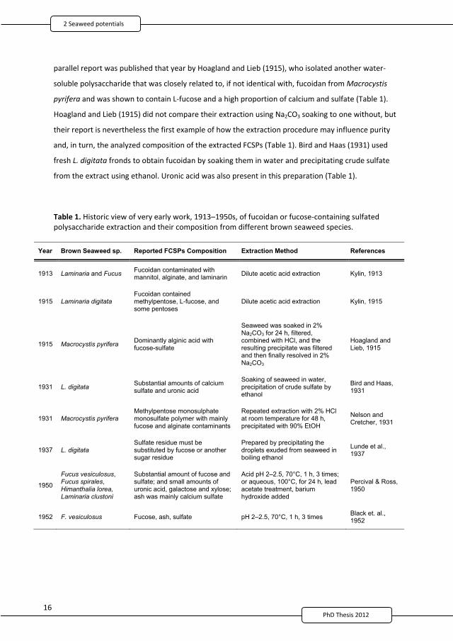
16PhD Thesis 2011
parallel report was published that year by Hoagland and Lieb (1915), who isolated another water
soluble polysaccharide that was closely related to, if not identical with, fucoidan from Macrocystis
pyrifera and was shown to contain L fucose and a high proportion of calcium and sulfate (Table 1).
Hoagland and Lieb (1915) did not compare their extraction using Na2CO3 soaking to one without, but
their report is nevertheless the first example of how the extraction procedure may influence purity
and, in turn, the analyzed composition of the extracted FCSPs (Table 1). Bird and Haas (1931) used
fresh L. digitata fronds to obtain fucoidan by soaking them in water and precipitating crude sulfate
from the extract using ethanol. Uronic acid was also present in this preparation (Table 1).
Table 1. Historic view of very early work, 1913–1950s, of fucoidan or fucose containing sulfatedpolysaccharide extraction and their composition from different brown seaweed species.
Year Brown Seaweed sp. Reported FCSPs Composition Extraction Method References
1913 Laminaria and Fucus Fucoidan contaminated with mannitol, alginate, and laminarin Dilute acetic acid extraction Kylin, 1913
1915 Laminaria digitata Fucoidan contained methylpentose, L-fucose, and some pentoses
Dilute acetic acid extraction Kylin, 1915
1915 Macrocystis pyrifera Dominantly alginic acid with fucose-sulfate
Seaweed was soaked in 2% Na2CO3 for 24 h, filtered, combined with HCl, and the resulting precipitate was filtered and then finally resolved in 2% Na2CO3
Hoagland and Lieb, 1915
1931 L. digitata Substantial amounts of calcium sulfate and uronic acid
Soaking of seaweed in water, precipitation of crude sulfate by ethanol
Bird and Haas, 1931
1931 Macrocystis pyrifera Methylpentose monosulphate monosulfate polymer with mainly fucose and alginate contaminants
Repeated extraction with 2% HCl at room temperature for 48 h, precipitated with 90% EtOH
Nelson and Cretcher, 1931
1937 L. digitata Sulfate residue must be substituted by fucose or another sugar residue
Prepared by precipitating the droplets exuded from seaweed in boiling ethanol
Lunde et al., 1937
1950
Fucus vesiculosus,Fucus spirales,Himanthalia lorea,Laminaria clustoni
Substantial amount of fucose and sulfate; and small amounts of uronic acid, galactose and xylose; ash was mainly calcium sulfate
Acid pH 2–2.5, 70°C, 1 h, 3 times; or aqueous, 100°C, for 24 h, lead acetate treatment, barium hydroxide added
Percival & Ross, 1950
1952 F. vesiculosus Fucose, ash, sulfate pH 2–2.5, 70°C, 1 h, 3 times Black et. al., 1952
2
28
17PhD Thesis 2011
The early FCSPs extraction procedures involved dilute acid treatment either with acetic or
hydrochloric acid as the first extraction step with the purpose of hydrolyzing the non FCSP
polysaccharides (Table 1). However, the extraction and purification methods employed in different
studies to isolate fucoidan/FCSPs from brown seaweed biomass have been modified to different
extents since the first reports in 1913 and 1915. For example, Nelson and Cretcher (1931) extracted
fucoidan from Macrocystis pyrifera by repeated extended (48 h) extraction with dilute HCl followed
by FCSP isolation by ethanol precipitation and revealed the presence of sulfate in the form of ester
groupings in the precipitated product. They also confirmed that fucose was the only sugar identified
in the unhydrolyzed residue after acid hydrolysis, even though their product contained uronic acid,
which was considered to be due to alginate contamination (Table 1).
2.2.1 FCSPs composition studies, 1930–1950The studies by Nelson and Cretcher (1931) revealed the presence of sulfate in the form of ester
groupings and confirmed that fucose was the only sugar identified after hydrolysis, although their
product contained uronic acid, which was considered to be due to alginic acid contamination
(Table 1). Please note the terminology used is quite confusing; nevertheless, with recent advances
in chemical analyses, we now know that alginic acid and alginate comprise guluronic and
mannuronic acids. Our understanding today that alginic acid, or alginate, is a linear hydrocolloid
polymer that consist of blocks of (1 4) linked D mannuronate and its L guluronate residues,
that these 2 monomers are C 5 epimers, and that the detailed structure of alginate may have
mannuronate and guluronate in homopolymeric blocks of consecutive mannuronate residues (M
blocks), consecutive guluronate residues (G blocks), or in structural units of alternating
mannuronate and guluronate residues (MG blocks). Uronic acids were determined to cover several
different structures. This work addressed the significance of the differences and introduced
consistent terminology by grouping these as uronic acids, especially since they do not belong to
the FCSPs. Later, Lunde et al. (1937) prepared fucoidan by directly precipitating the droplets
exuded from freshly gathered L. digitata fronds in ethanol (Table 1). After purification via product
precipitation using boiling ethanol, they obtained a FCSP specimen that contained 33–37%
methylpentose (interpreted correctly as fucose) and 26–30% ash in which the sulfate content was
17–19%, which made up approximately half of the total estimated sulfate in the polysaccharide
(35–38%). They proposed a structural unit formula, (R R’ O SO2 OM)n, for fucoidan and suggested
that R was fucose or another pentose sugar residue, R’ was unknown, and M was Na+, K+, (½)Ca2+,
or (½)Mg2+ (Lunde et al., 1937) (Table 1).
2 Seaweed otentials
2
29
18PhD Thesi 2011
Specimens from F. vesiculosus, F. spirales, H. lorea, and L. clustoni were prepared by Percival and
Ross (1950) for FCSPs extraction using boiling water for 24 h, and the alginates and protein were
removed with lead acetate and the addition of barium hydroxide. The resulting lead hydroxide
fucoidan complex was then decomposed using dilute sulfuric acid and the fucoidan was isolated
after prolonged hydrolysis and filtration. The purest specimen was from H. lorea, which contained
the following: a substantial amount of fucose and sulfate; small quantities of uronic acid,
galactose, and xylose; metals; and ash, which was mainly calcium sulfate. These workers believed
that the principal constituent of fucoidan is a polyfucose monosulfate and that other constituents
arise from impurities (Table 1). A parallel work was done by Conchie and Percival (1950) in which
fucoidan from Fucus vesiculosus was methylated. It was believed that the main residue in fucoidan
was 1 2 linked of L fucopyranose units carrying a sulfate group in C 4 (Conchie and Percival
1950). However, we now know that this is not the case because advanced analyses have verified
that the backbone of fucoidan from F. vesiculosus consists of alterna ng (1 3) and (1 4)
linkages of L fucopyranose residues (Fig. 2.2).
2.2.2 Lab scale extraction of fucoidan, 1950sIn the pursuit of obtaining extensive quantities, a laboratory scale extraction of fucoidan was
performed by Black et al. (1952). Interestingly, they referred to the extracted product as a
“polyfucose ethereal sulfate occurring in the Phaeophyceae.” Their optimal fucoidan extraction
procedure was as follows: one part by weight of dried ground seaweed and 10 parts by volume of
0.1 M hydrochloric acid at pH 2.0–2.5 contacted at 70°C with constant stirring for 1 h. A single acid
hydrolysis extraction treatment using this method recovered about 50% by weight (w/w) of the
theoretical maximum of fucoidan (recovered yield measured as % fucose obtained as % of total
fucose in the seaweed DW), whereas 3 rounds of acid extraction recovered >80% of the fucose
present; the triple acid hydrolysis treatment (0.1 M HCl, pH 2.0–2.5, 70°C, 1 h x 3) was therefore
selected as the optimal extraction method (Table 1). After the acid hydrolysis treatment the crude
fucoidan was isolated by fractional precipitation with alcohol and further purified by precipitation
after the addition of formaldehyde (Black et al., 1952). Using this procedure, crude fucoidan
samples containing 30–36% fucose were obtained; for example, the fucoidan recovered from F.
vesiculosus using the optimal extraction protocol was analyzed to contain (by weight) 44% fucose,
26% total sulfate, and 31% ash (Black et al., 1952). In terms of yields of percentage of total fucose,
the results obtained for the 4 different algal species were: Pelvetia canaliculata, 76%; F.
vesiculosus, 62%; Ascophyllum nodosum, 54%; and L. cloustoni, 20% (Black et al., 1952). The
authors suggested that a more efficient extraction methodology, i.e., extracting higher fucose
2
30
19PhD Thesis 2011
yields, could be achieved by increasing the water/seaweed ratio, extraction time, or extraction
number.
2.2.3 FCSPs extraction, 1970s–presentCarbohydrates of the brown seaweeds were successively extracted from H. lorea, Bifurcaria
bifurcata, and Padina pavonia using dilute acid followed by alkaline or using a water, acid, and
alkali sequence. Prior to extraction, the seaweed frond was pretreated with formaldehyde to
polymerize phenolic constituents that could otherwise be contaminant to various extracts (Mian
and Percival, 1973). This technique produced complex mixture of glucan, FCPSs, and alginic acid,
which could be separated by fractional precipitation with ethanol, calcium salts, or
cetyltrimethylammonium hydroxide, or by fractionation on resin columns. Fucoidan extracted in
this fashion consisted of heteropolysaccharide comprising different proportions of fucose,
glucuronic acid, xylose, and half ester sulfate together with a trace of galactose. The result of this
extraction method also showed that sulfate and uronic acid contents in the fucoidan separated
from aqueous calcium chloride and acid extracts varied significantly by species.
A study was conducted on the purification of a sulfated heteropolysaccharide from S. linifolium to
elucidate its structural components. The extraction was done using hydrochloric acid pH 1.0 for 3 h
at 80°C; the extract was neutralized using aqueous sodium carbonate and precipitated with
ethanol. The resulting sulfated polysaccharide material was acid hydrolyzed and it was proposed
that the backbone was composed of glucuronic acid, mannose, and galactose residues with
partially sulfated side chains of galactose, xylose, and fucose residues (Abdel Fattah et al., 1974).
Extraction using an aqueous alkali solution or dilute acid at ambient or slightly elevated
temperatures has been the convenient method to produce fucoidan for many years. In recent
years, pretreatment of seaweed biomass has been found advantageous to eliminate low molecular
components (e.g., phenols) using a mixture of methanol, chloroform, and water (Bilan et al., 2006);
removal of protein can be facilitated using proteolytic enzymes (Rocha et al., 2005). A useful
procedure to transform alginate in the residual biomass into insoluble calcium salts uses aqueous
calcium chloride (Bilan et al., 2002).
2.3 Seaweed products and biomass potentialThe growth of seaweed production and developments of advanced technological farming indicates
that the seaweed industry is progressively evolving and thus paves the way to new application
opportunities (Buck and Buchholz, 2004). For many years, seaweed has been utilized as a source of
2 Seaweed otentials
2
31
20PhD Thesis 2011
dietary nourishment among Asian constituents. In recent years, more applications have used
seaweed resources because of their unique characteristics and properties. Seaweed has been
proposed as a source of important compound that can be incorporated in the production of
functional food ingredients, pharmaceuticals, and cosmetics (Bodin Dubigeon et al., 1997; Bixler,
1996; De Roeck Holtzhauer, 1991). Gel forming polysaccharides such as carrageenan and alginate are
mostly known and commercially important seaweed products. Production of alternative fuels from
non starch biomass has recently directed the attention to utilization of marine macroalgae as sources
of biomass for biofuel production (Knauf and Moniruzzaman, 2004). Moreover, healthy and bioactive
components, notably the FCSPs found in brown seaweed biomass, have also become subject of much
research and product development.
2.3.1 PhycocolloidsThe global market for phycocolloids such as agar, carrageenans, and alginates is estimated to be
worth annually $585 million US (McHugh, 2003). To date, the red seaweed has been harvested for
food consumption in some regions of Asia, but many of these areas have engaged in farming red
seaweed that is intended for the production of agar and carrageenan, while brown seaweed is most
harvested naturally for alginate production (Crawford, 2002).
Carrageenan is composed of a linear galactose backbone with varying degrees of sulfation (15–40%)
and are mainly composed of disaccharide repeating units of an (1 4) linked D galactopyranose or
3,6 anhydro D galactopyranose residue and a (1 3) linked D galactopyranose. The sulfated groups
are covalently attached to individual galactose residues via ester linkages to the carbon atoms C 2, C
4, or C 6 (De Ruiter and Rudolph, 1997). Carrageenan is applied as a stabilizing agent to many food
products and other industrial and pharmaceutical applications (van de Velde and De Ruiter, 2002).
Among the leading species of red algae responsible for most of today’s commercial carrageenan
production are K. alvarezii and E. denticulatum.
In addition, alginate another gel forming sulfated polysaccharide extracted from brown seaweed has
been utilized for various applications including foods and feeds, pharma/medical, and industrial
preparations. Alginate is composed of mannuronic (M) and guluronic (G) acids with (1, 4) linkages,
and its structure varies according to the monomer position on the chain, forming either
homopolymeric (MM or GG) or heteropolymeric (MG or GM) segments (Percival and McDowell,
1967). The molecular weight of alginate is generally 500–1000kDa. Its solubility is influenced by
factors such as pH, concentration, ions in solution, and the presence of divalent ions such as calcium
(Morris and Norton, 1983).
2
32
21PhD Thesis 2011
2.3.2 Seaweed biomass for bioenergy productionOnly few seaweed species have been exploited for commercial production (i.e., red and brown
seaweed); hence, this study opens the way for other species to provide a basis for additional potential
applications of seaweed biomass, namely bioenergy production. The cultivation of seaweed poses
advantages to terrestrial crops since they have high growth rates and can be continuously harvested
(Rasmussen et al., 2009). The estimated annual production of U. lactuca per hectare was 45 tons DW,
which is 3× greater the yield of conventional food or energy crops (Bruhn et al., 2011). However, the
production is believed to be significantly higher once growth conditions (exploiting flue gas as a
source of carbon) and nutrients (fishery effluents as a source of nitrogen) are optimized. A study on
the anaerobic digestion of sea lettuce (Ulva sp.) suggested that the methane gas yield from washed
and grinded sea lettuce biomass is about 180 mL g 1 VS (volatile solid based), while that from non
pretreated biomass is about 70% (Otsuka and Yoshino, 2004). Biogas production from fresh and
macerated U. lactuca yielded up to 271 mL CH4 g 1 VS, which is in the range of the methane
production from cattle manure and land based energy crops such as grass clover. Drying of the
biomass resulted in a 5–9 fold increase in weight specific methane production compared to wet
biomass (Bruhn et al., 2011).
2.4 Seaweed production
Seaweed has been traditionally cultured for centuries in several Asian countries including China,
Japan, and Korea (Crawford, 2002). Most seaweed production came from the harvest of wild stocks in
these countries, although limited culture had been established in countries such as the Philippines
and Indonesia (Trono, 1990). The species cultivated include Kappaphycus and Eucheuma. The leading
seaweed exporter, the Philippines, increased production from 675 tons in 1967 to 65,617 tons in 2009
and 80,000 tons in 2010, but the country is still importing raw seaweed materials from Indonesia
(www.siap.com.ph, 2011). The world demand for seaweed in 2003 was 220,000 tons for K. alvarezii
and E. denticulatum with an expected 10% annual increase in demand (Sievanen et al., 2005).
Indonesia, on the other hand, raised its production from 1,000 million tons (MT) in 1966 to
approximately 27,874 tons in 2001 and estimated 10 MT in 2015 (Sievanen et al., 2005). The
production levels of algae in 2002 reached 18.6 MT (FAO, 2004). Europe had only a 6.3% share of the
global world production of brown algae (362,000 tons FW) and about 0.3% of red algae (9,400 tons
FW) with <200 tons of macroalgae produced in aquaculture in 2002 (FAO, 2004).
Several sea based cultivation design methods have been developed and tested. The most commonly
used was a fixed off bottom long line, hanging long line, and combination of the 2 (Hurtado et al.,
2 Seaweed otentials
2
33
22PhD Thesis 2011
2001). System off shore designs for Laminaria culture tested within the area of Helgoland farm,
Germany (Buck and Buchholz, 2004) used longline, ladder (tandem longline), and ring shaped designs
for attachment of algae seeded culture lines. The off shore cultivation ring developed by Buch and
Buchholz (2004) proved resistant to rough weather conditions (6 m wave heights) and current
velocities (2 m s 1) and also permitted easy handling. About 75% of the cultivation ring culture lines
were fully covered by L. saccharina, with a total fresh weight of 304 kg after a 6 month grow out
phase in the sea. Harvesting of the ring construction is performed by towing it to the shore and lifting
it by cranes, or harvest at sea can be performed using boat based cranes. However, the cost of a fully
mounted ring is quite high (€ 1000) as suggested by Buch and Buchholz (2004).
On the other hand, the nuisance seaweed U. lactuca has been shown to be suitable for propagation
under controlled conditions (Lee, 2000). For this reason, cultivation of U. lactuca in tanks for either
crude biomass production for bioenergy or production of biologically active compounds is receiving
increased attention. The green algae Ulva sp. is cultivated in tanks in Denmark for bioenergy
production (Rasmussen et al., 2009). The production of Ulva in Denmark is estimated to be an
average of 45 tons DW/ha per annum (200 days of sufficient light conditions). Critical consideration
when cultivating Ulva spp. in tanks is the frequently occurring sporulation events result in the loss of
algal tissue (Werner et al., 2003).
2.5 Paper 1: Important determinants for fucoidan bioactivity: a criticalreview of structure function correlations and extraction methods for FCSPsfrom brown seaweeds
Marine Drugs, 2011, Published online
Authors:Marcel Tutor Ale, Jørn Dalgaard Mikkelsen, Anne S. Meyer
Address:Center for Bioprocess Engineering, Department of Chemical and Biochemical EngineeringTechnical University of Denmark (DTU)Soeltofts Plads Bldg. 229, 2800 Kgs. Lyngby, Denmar
2
34
23PhD Thesis 2011
23PhD Thesis 2011
Mar. Drugs 2011, 9, 2106-2130; doi:10.3390/md9102106
Marine Drugs ISSN 1660-3397
www.mdpi.com/journal/marinedrugs Review
Important Determinants for Fucoidan Bioactivity: A Critical Review of Structure-Function Relations and Extraction Methods for Fucose-Containing Sulfated Polysaccharides from Brown Seaweeds
Marcel Tutor Ale, Jørn D. Mikkelsen and Anne S. Meyer *
Center for Bioprocess Engineering, Department of Chemical and Biochemical Engineering, Technical University of Denmark (DTU), Søltofts Plads, Building 229, DK-2800 Kgs. Lyngby, Denmark; E-Mails: [email protected] (M.T.A.); [email protected] (J.D.M.)
* Author to whom correspondence should be addressed; E-Mail: [email protected]; Tel.: +45-4525-2800; Fax: +45-4593-2906.
Received: 14 September 2011; in revised form: 3 October 2011 / Accepted: 13 October 2011 / Published: 24 October 2011
Abstract: Seaweeds—or marine macroalgae—notably brown seaweeds in the class Phaeophyceae, contain fucoidan. Fucoidan designates a group of certain fucose-containing sulfated polysaccharides (FCSPs) that have a backbone built of (1 3)-linked
-L-fucopyranosyl or of alternating (1 3)- and (1 4)-linked -L-fucopyranosyl residues, but also include sulfated galactofucans with backbones built of (1 6)- -D-galacto- and/or (1 2)- -D-mannopyranosyl units with fucose or fuco-oligosaccharide branching, and/or glucuronic acid, xylose or glucose substitutions. These FCSPs offer several potentially beneficial bioactive functions for humans. The bioactive properties may vary depending on the source of seaweed, the compositional and structural traits, the content (charge density), distribution, and bonding of the sulfate substitutions, and the purity of the FCSP product. The preservation of the structural integrity of the FCSP molecules essentially depends on the extraction methodology which has a crucial, but partly overlooked, significance for obtaining the relevant structural features required for specific biological activities and for elucidating structure-function relations. The aim of this review is to provide information on the most recent developments in the chemistry of fucoidan/FCSPs emphasizing the significance of different extraction techniques for the structural composition and biological activity with particular focus on sulfate groups.
OPEN ACCESS
2
35
23PhD Thesis 2011
23PhD Thesis 2011
Mar. Drugs 2011, 9, 2106-2130; doi:10.3390/md9102106
Marine Drugs ISSN 1660-3397
www.mdpi.com/journal/marinedrugs Review
Important Determinants for Fucoidan Bioactivity: A Critical Review of Structure-Function Relations and Extraction Methods for Fucose-Containing Sulfated Polysaccharides from Brown Seaweeds
Marcel Tutor Ale, Jørn D. Mikkelsen and Anne S. Meyer *
Center for Bioprocess Engineering, Department of Chemical and Biochemical Engineering, Technical University of Denmark (DTU), Søltofts Plads, Building 229, DK-2800 Kgs. Lyngby, Denmark; E-Mails: [email protected] (M.T.A.); [email protected] (J.D.M.)
* Author to whom correspondence should be addressed; E-Mail: [email protected]; Tel.: +45-4525-2800; Fax: +45-4593-2906.
Received: 14 September 2011; in revised form: 3 October 2011 / Accepted: 13 October 2011 / Published: 24 October 2011
Abstract: Seaweeds—or marine macroalgae—notably brown seaweeds in the class Phaeophyceae, contain fucoidan. Fucoidan designates a group of certain fucose-containing sulfated polysaccharides (FCSPs) that have a backbone built of (1 3)-linked
-L-fucopyranosyl or of alternating (1 3)- and (1 4)-linked -L-fucopyranosyl residues, but also include sulfated galactofucans with backbones built of (1 6)- -D-galacto- and/or (1 2)- -D-mannopyranosyl units with fucose or fuco-oligosaccharide branching, and/or glucuronic acid, xylose or glucose substitutions. These FCSPs offer several potentially beneficial bioactive functions for humans. The bioactive properties may vary depending on the source of seaweed, the compositional and structural traits, the content (charge density), distribution, and bonding of the sulfate substitutions, and the purity of the FCSP product. The preservation of the structural integrity of the FCSP molecules essentially depends on the extraction methodology which has a crucial, but partly overlooked, significance for obtaining the relevant structural features required for specific biological activities and for elucidating structure-function relations. The aim of this review is to provide information on the most recent developments in the chemistry of fucoidan/FCSPs emphasizing the significance of different extraction techniques for the structural composition and biological activity with particular focus on sulfate groups.
OPEN ACCESS
2
35
24PhD Thesis 2011PhD Thesis 2011
Mar. Drugs 2011, 9
2107
Keywords: fucoidan; antitumor; anticoagulant; extraction; sulfated polysaccharides
1. Introduction
Fucoidan is a term used for a class of sulfated, fucose rich, polysaccharides found in the fibrillar cell walls and intercellular spaces of brown seaweeds of the class Phaeophyceae. These fucose-containing sulfated polysaccharides (FCSPs) principally consist of a backbone of (1 3)- and (1 4)-linked -L-fucopyranose residues, that may be organized in stretches of (1 3)- -fucan or of alternating (1 3)- and (1 4)-bonded L-fucopyranose residues. The L-fucopyranose residues may be substituted with sulfate (SO3 ) on the C-2 or C-4 (rarely on C-3), with single L-fucosyl residues and/or with short fucoside (fuco-oligosaccharide) side chains. If present, the fucoside side chains are usually O-4 linked to the -L-fucopyranose backbone residues. However, as FCSPs structures of more brown seaweeds are being elucidated, as discussed further in the present review, it appears that FCSPs cover a broader range of complex polysaccharides than only those having fucan backbones. Apart from variations in the sulfate content and substitutions, also the monosaccharide composition of FCSPs varies among different species of brown seaweeds. Hence, in addition to fucose, different types of FCSPs may also contain galactose, mannose, xylose, glucose and/or glucuronic acid—usually in minor amounts [1].
According to the ISI Web of Knowledge (Thomson Reuters) the number of published articles with the topic assigned as “fucoidan” has increased significantly since fucoidan, or “fucoidin” as it was first called, was first isolated from brown algae in 1913 [2]; in particular, a profound increase in the number of papers has taken place during the last 5–10 years. By now, the published papers related to fucoidan hit approximately 1800 (August 2011, Figure 1). The recent interest has mainly focused on the potentially beneficial biological activities of fucoidan and FCSPs in humans including antitumor, immunomodulatory, anti-inflammatory, antiviral, antithrombotic, anticoagulant, and antioxidant effects as well as specific activities against kidney, liver and urinary system disorders.
While the development of research efforts involving FCSPs and their potential applications are advancing, the understanding of the mechanisms and the particular structural features of the FCSPs being responsible for the various biological activities is still incomplete. Seaweeds, including various brown seaweeds such as Undaria and Laminaria spp., are part of the food culture in Asia, notably in Japan, the Philippines, and Korea, and seaweed extracts have also been used as a remedy in traditional medicine. However, no standardized FCSPs extraction or purification protocols exist, and no specific pharmaceutical, dermatological, or nutraceutical applications have as yet been officially approved for these polysaccharides or their lower molar mass oligosaccharide derivatives.
It is our proposition that more focus should be directed to the extraction and purification processes in order to obtain consistent protocols that account for the biodiversity of FCSPs from different seaweeds and to retain the structural features of significance for the specific bioactivity properties of FCSPs. The development and use of such consistent extraction procedures would also help in achieving a better understanding of structure-function relationships of FCSPs. The aim of this review is to bring attention to the detailed structural features of FCSPs in relation to their marine algal sources
2
36
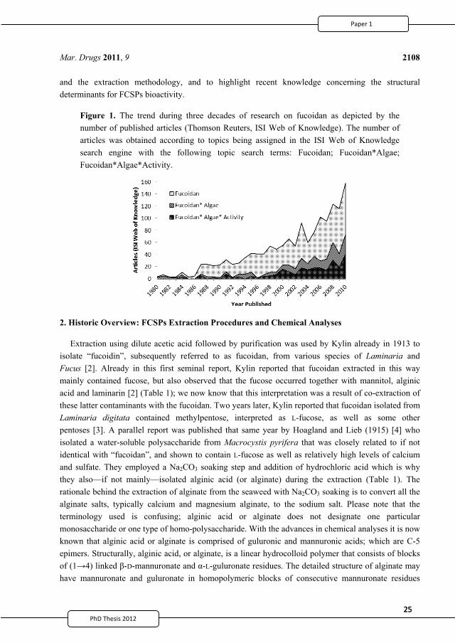
25PhD Thesis 2011
25PhD Thesis 2011
Mar. Drugs 2011, 9
2108
and the extraction methodology, and to highlight recent knowledge concerning the structural determinants for FCSPs bioactivity.
Figure 1. The trend during three decades of research on fucoidan as depicted by the number of published articles (Thomson Reuters, ISI Web of Knowledge). The number of articles was obtained according to topics being assigned in the ISI Web of Knowledge search engine with the following topic search terms: Fucoidan; Fucoidan*Algae; Fucoidan*Algae*Activity.
2. Historic Overview: FCSPs Extraction Procedures and Chemical Analyses
Extraction using dilute acetic acid followed by purification was used by Kylin already in 1913 to isolate “fucoidin”, subsequently referred to as fucoidan, from various species of Laminaria and Fucus [2]. Already in this first seminal report, Kylin reported that fucoidan extracted in this way mainly contained fucose, but also observed that the fucose occurred together with mannitol, alginic acid and laminarin [2] (Table 1); we now know that this interpretation was a result of co-extraction of these latter contaminants with the fucoidan. Two years later, Kylin reported that fucoidan isolated from Laminaria digitata contained methylpentose, interpreted as L-fucose, as well as some other pentoses [3]. A parallel report was published that same year by Hoagland and Lieb (1915) [4] who isolated a water-soluble polysaccharide from Macrocystis pyrifera that was closely related to if not identical with “fucoidan”, and shown to contain L-fucose as well as relatively high levels of calcium and sulfate. They employed a Na2CO3 soaking step and addition of hydrochloric acid which is why they also—if not mainly—isolated alginic acid (or alginate) during the extraction (Table 1). The rationale behind the extraction of alginate from the seaweed with Na2CO3 soaking is to convert all the alginate salts, typically calcium and magnesium alginate, to the sodium salt. Please note that the terminology used is confusing; alginic acid or alginate does not designate one particular monosaccharide or one type of homo-polysaccharide. With the advances in chemical analyses it is now known that alginic acid or alginate is comprised of guluronic and mannuronic acids; which are C-5 epimers. Structurally, alginic acid, or alginate, is a linear hydrocolloid polymer that consists of blocks of (1 4) linked -D-mannuronate and -L-guluronate residues. The detailed structure of alginate may have mannuronate and guluronate in homopolymeric blocks of consecutive mannuronate residues
2
37
25PhD Thesis 2011
25PhD Thesis 2011
Mar. Drugs 2011, 9
2108
and the extraction methodology, and to highlight recent knowledge concerning the structural determinants for FCSPs bioactivity.
Figure 1. The trend during three decades of research on fucoidan as depicted by the number of published articles (Thomson Reuters, ISI Web of Knowledge). The number of articles was obtained according to topics being assigned in the ISI Web of Knowledge search engine with the following topic search terms: Fucoidan; Fucoidan*Algae; Fucoidan*Algae*Activity.
2. Historic Overview: FCSPs Extraction Procedures and Chemical Analyses
Extraction using dilute acetic acid followed by purification was used by Kylin already in 1913 to isolate “fucoidin”, subsequently referred to as fucoidan, from various species of Laminaria and Fucus [2]. Already in this first seminal report, Kylin reported that fucoidan extracted in this way mainly contained fucose, but also observed that the fucose occurred together with mannitol, alginic acid and laminarin [2] (Table 1); we now know that this interpretation was a result of co-extraction of these latter contaminants with the fucoidan. Two years later, Kylin reported that fucoidan isolated from Laminaria digitata contained methylpentose, interpreted as L-fucose, as well as some other pentoses [3]. A parallel report was published that same year by Hoagland and Lieb (1915) [4] who isolated a water-soluble polysaccharide from Macrocystis pyrifera that was closely related to if not identical with “fucoidan”, and shown to contain L-fucose as well as relatively high levels of calcium and sulfate. They employed a Na2CO3 soaking step and addition of hydrochloric acid which is why they also—if not mainly—isolated alginic acid (or alginate) during the extraction (Table 1). The rationale behind the extraction of alginate from the seaweed with Na2CO3 soaking is to convert all the alginate salts, typically calcium and magnesium alginate, to the sodium salt. Please note that the terminology used is confusing; alginic acid or alginate does not designate one particular monosaccharide or one type of homo-polysaccharide. With the advances in chemical analyses it is now known that alginic acid or alginate is comprised of guluronic and mannuronic acids; which are C-5 epimers. Structurally, alginic acid, or alginate, is a linear hydrocolloid polymer that consists of blocks of (1 4) linked -D-mannuronate and -L-guluronate residues. The detailed structure of alginate may have mannuronate and guluronate in homopolymeric blocks of consecutive mannuronate residues
2
37
26PhD Thesis 2011PhD Thesis 2011
Mar. Drugs 2011, 9
2109
(M-blocks), in consecutive guluronate residues (G-blocks), or in structural units of alternating mannuronate and -L-guluronate residues (MG-blocks). Both mannuronic acid and -L-guluronic acids are uronic acids, and they have in the past been analytically determined as uronic acids. However, uronic acids also include several other structures, e.g., glucuronic and galacturonic acids. In this review, we will address the significance of the compositional and structural differences, but also attempt to introduce a consistent terminology by grouping these compounds as uronic acids, especially as homopolymers do not belong to the group of FCSPs. Hoagland and Lieb (1915) [4] did not compare their extraction with Na2CO3 soaking to one without, but their report is nevertheless the first example of how the extraction procedure may influence the purity and, in turn, the analyzed composition of the extracted FCSPs (see Table 1).
Table 1. Historic view of very early work, from 1913–1950, of fucoidan or FCSPs extraction and their reported composition from different brown seaweed species.
The early FCSPs extraction procedures were based on using a dilute acid treatment, with either acetic or hydrochloric acid used as a first “extraction” step with the purpose of hydrolyzing the non-FCSP polysaccharides (Table 1). However, the extraction and purification methodologies employed in
Year Brown seaweed sp. Reported FCSPs composition Extraction method References
1913 Laminaria and Fucus Fucoidan contains fucose, that occurs together with mannitol, alginate and laminaran
Dilute acetic acid extraction Kylin, 1913 [2]
1915 Laminaria digitata Fucoidan contains L-fucose and other pentoses
Dilute acetic acid extraction Kylin, 1915 [3]
1915 Macrocystis pyrifera Mainly alginic acid, with some fucose-sulfate
Soaking in 2% Na2CO3 for 24 h, filtration, HCl addition, recovery of precipitate by filtration, redissolution in 2% Na2CO3
Hoagland and Lieb, 1915 [4]
1931 Laminaria digitata Substantial amounts of calcium sulfate and uronic acid
Soaking of the seaweed in water, precipitation of crude, sulfated polysaccharides by ethanol
Bird and Haas, 1931 [5]
1931 Macrocystis pyrifera Methylpentose monosulphate polymer with fucose and alginate contaminants
Repeated extraction with 2% HCl at room temperature for 48 h, precipitated with 90% ethanol
Nelson and Cretcher, 1931 [6]
1937 Laminaria digitata Proposed (R-R -O-SO2-OM)n with R = fucose, R as unknown, M being Na, K, Ca0.5, or Mg0.5
Precipitation of droplets exuded from seaweed in boiling ethanol
Lunde et al., 1937 [7]
1950
Fucus vesiculosus, Fucus spirales,
Himanthalia lorea, Laminaria clustoni
Substantial amounts of fucose and sulfate; small amounts of uronic acid, galactose and xylose; metals and ash were also detected, ash was mainly calcium sulfate
Aqueous extraction at ∼100 °C for 24 h, extract treated with lead acetate (to precipitate alginate and proteins), filtrate solution treated with Ba(OH)2 to precipitate a “hydroxide-fucoidin complex”
Percival and Ross, 1950 [8]
1952 Fucus vesiculosus Fucose, ash, sulfate 0.1 M HCl at pH 2–2.5 and 70 °C for 1 h, 3-times, fractional precipitation with ethanol
Black et. al., 1952 [9]
2
38
27PhD Thesis 2011
27PhD Thesis 2011
Mar. Drugs 2011, 9
2110
different studies to isolate fucoidan/FCSPs from brown seaweed biomass have been modified to different extents since the first reports from 1913 and 1915. Bird and Haas (1931) [5], for example, used soaking of the brown alga biomass in water and precipitation of crude sulfate from the extract with ethanol to obtain fucoidan from L. digitata (Table 1). The product of this extraction was also found to contain relatively high levels of ash. The high ash levels were presumably chiefly a result of the presence of calcium sulfate in the algal polysaccharides. For the compositional analysis, acid hydrolysis using H2SO4 was typically used. This acid hydrolysis step might by itself have contributed a substantial amount of sulfate. As a consequence, the use of H2SO4 clearly biased the interpretation of the compositional analysis. Hence, the fucoidan isolated by Bird and Haas (1931) [5] was designated as carbohydrate sulfate (i.e., containing esterified sulfate) since the total sulfate content was approximately the double of that found in the ash. Uronic acid was also present in the FCSPs preparation extracted from L. digitata [5] (Table 1).
Nelson and Cretcher (1931) [6] extracted fucoidan from Macrocystis pyrifera by repeated, extended (48 h) extraction with dilute HCl followed by isolation of the FCSPs by ethanol precipitation, and revealed the presence of sulfate in the form of ester groups in the precipitated product. They also confirmed that fucose was the only sugar identified in the unhydrolyzed residue after acid hydrolysis, even though their product contained uronic acid, considered to be due to alginate contamination (Table 1). Later, Lunde et al. [7] prepared fucoidan by directly precipitating the droplets exuded from freshly gathered L. digitata fronds in ethanol (Table 1). After purification via precipitation of the product from boiling ethanol they obtained a FCSPs specimen that contained 33–37% methylpentose (interpreted correctly as fucose), and 26–30% ash in which the sulfate content was 17–19%, which made up approximately half of the total sulfate estimated in the polysaccharide (35–38%). They proposed a structural unit formula, (R-R -O-SO2-OM)n, for fucoidan and suggested that R was fucose or another pentose sugar residue, R was unknown, and M was Na+, K+, (½)Ca2+ or (½)Mg2+ [7] (Table 1).
“In an attempt to reconcile some of the conflicting views on the nature of fucoidin” crude fucoidan extracts from F. vesiculosus, F. spirales, Himanthalia lorea and Laminaria clustoni were prepared by Percival and Ross (1950) [8]. Their methodology involved boiling of the seaweed biomass in neat boiling water for 24 h (hydrolysis treatment) followed by removal of alginates and protein by addition of lead acetate, then, after addition of barium hydroxide (presumably to precipitate alginate) the fucoidan was isolated as a crude lead hydroxide complex (Table 1). In order to isolate lead free fucoidan, the lead hydroxide complex was treated with dilute H2SO4 and fucoidan was then isolated after prolonged dialysis and filtration. The purest fucoidan specimen obtained was from H. lorea. This fucoidan isolated from H. lorea contained substantial amounts of fucose and sulfate; as well as small quantities of uronic acid, galactose and xylose. Metals and ash were also detected, and the ash was mainly made up of calcium sulfate [8]. Based on the data obtained the authors believed that the principal constituent of fucoidan was a polyfucose with one sulfate substitution on each fucose residue and that other constituents arose from adventitious impurities. A parallel paper was published in which it was proposed that the core structure of fucoidan from F. vesiculosus was built of 1 2 linked L-fucopyranose units, each carrying a sulfate group on C-4 [10]. As discussed later, the interpretation that the fucosyl units in the fucoidan were 1 2 linked turned out to be incorrect, as more advanced
2
39
27PhD Thesis 2011
27PhD Thesis 2011
Mar. Drugs 2011, 9
2110
different studies to isolate fucoidan/FCSPs from brown seaweed biomass have been modified to different extents since the first reports from 1913 and 1915. Bird and Haas (1931) [5], for example, used soaking of the brown alga biomass in water and precipitation of crude sulfate from the extract with ethanol to obtain fucoidan from L. digitata (Table 1). The product of this extraction was also found to contain relatively high levels of ash. The high ash levels were presumably chiefly a result of the presence of calcium sulfate in the algal polysaccharides. For the compositional analysis, acid hydrolysis using H2SO4 was typically used. This acid hydrolysis step might by itself have contributed a substantial amount of sulfate. As a consequence, the use of H2SO4 clearly biased the interpretation of the compositional analysis. Hence, the fucoidan isolated by Bird and Haas (1931) [5] was designated as carbohydrate sulfate (i.e., containing esterified sulfate) since the total sulfate content was approximately the double of that found in the ash. Uronic acid was also present in the FCSPs preparation extracted from L. digitata [5] (Table 1).
Nelson and Cretcher (1931) [6] extracted fucoidan from Macrocystis pyrifera by repeated, extended (48 h) extraction with dilute HCl followed by isolation of the FCSPs by ethanol precipitation, and revealed the presence of sulfate in the form of ester groups in the precipitated product. They also confirmed that fucose was the only sugar identified in the unhydrolyzed residue after acid hydrolysis, even though their product contained uronic acid, considered to be due to alginate contamination (Table 1). Later, Lunde et al. [7] prepared fucoidan by directly precipitating the droplets exuded from freshly gathered L. digitata fronds in ethanol (Table 1). After purification via precipitation of the product from boiling ethanol they obtained a FCSPs specimen that contained 33–37% methylpentose (interpreted correctly as fucose), and 26–30% ash in which the sulfate content was 17–19%, which made up approximately half of the total sulfate estimated in the polysaccharide (35–38%). They proposed a structural unit formula, (R-R -O-SO2-OM)n, for fucoidan and suggested that R was fucose or another pentose sugar residue, R was unknown, and M was Na+, K+, (½)Ca2+ or (½)Mg2+ [7] (Table 1).
“In an attempt to reconcile some of the conflicting views on the nature of fucoidin” crude fucoidan extracts from F. vesiculosus, F. spirales, Himanthalia lorea and Laminaria clustoni were prepared by Percival and Ross (1950) [8]. Their methodology involved boiling of the seaweed biomass in neat boiling water for 24 h (hydrolysis treatment) followed by removal of alginates and protein by addition of lead acetate, then, after addition of barium hydroxide (presumably to precipitate alginate) the fucoidan was isolated as a crude lead hydroxide complex (Table 1). In order to isolate lead free fucoidan, the lead hydroxide complex was treated with dilute H2SO4 and fucoidan was then isolated after prolonged dialysis and filtration. The purest fucoidan specimen obtained was from H. lorea. This fucoidan isolated from H. lorea contained substantial amounts of fucose and sulfate; as well as small quantities of uronic acid, galactose and xylose. Metals and ash were also detected, and the ash was mainly made up of calcium sulfate [8]. Based on the data obtained the authors believed that the principal constituent of fucoidan was a polyfucose with one sulfate substitution on each fucose residue and that other constituents arose from adventitious impurities. A parallel paper was published in which it was proposed that the core structure of fucoidan from F. vesiculosus was built of 1 2 linked L-fucopyranose units, each carrying a sulfate group on C-4 [10]. As discussed later, the interpretation that the fucosyl units in the fucoidan were 1 2 linked turned out to be incorrect, as more advanced
2
39
28PhD Thesis 2011PhD Thesis 2011
Mar. Drugs 2011, 9
2111
analyses have now verified that the backbone of fucoidan from F. vesiculosus consists of alternating (1 3) and (1 4) linkages [11].
In the pursuit to obtain extensive quantities, a laboratory-scale extraction of fucoidan was reported by Black et al. [9]. Interestingly, the extracted product was referred to as a “polyfucose ethereal sulphate occurring in the Phaeophyceae”. Their optimal fucoidan extraction procedure was as follows: One part by weight of dried ground seaweed and 10 parts by volume of 0.1 M hydrochloric acid at pH 2.0–2.5 contacted at 70 °C with constant stirring for 1 h. A single acid hydrolysis extraction treatment using this method recovered about 50% by weight (w/w) of the theoretical maximum of fucoidan (recovered yield measured as % fucose obtained as % of total fucose in the seaweed dry weight), whereas three rounds of the acid extraction recovered more than 80% of the fucose present; the triple acid hydrolysis treatment (0.1 M HCl, pH 2.0–2.5, 70 °C, 1 h × 3) was therefore selected as the optimal extraction method (Table 1). After the acid hydrolysis treatment the crude fucoidan was isolated by fractional precipitation with alcohol and further purified by precipitation after addition of formaldehyde [9]. By this procedure samples of crude fucoidan containing 30–36% fucose were obtained; for example, the fucoidan recovered from F. vesiculosus using the optimal extraction protocol was analyzed to contain (by weight) 44% fucose; 26% total sulfate, and 31% ash [9]. In terms of yields, calculated as fucose as % of total fucose, the results obtained for the four different algal species were: Pelvetia canaliculata 76%; F. vesiculosus 62%; Ascophyllum nodusum 54%, and L. cloustoni 20% [9]. The authors suggested that a more efficient extraction methodology, i.e., extracting higher fucose yields, could be achieved by increasing the water/seaweed ratio, extraction time or number of extractions.
In a study about 20 years later, FCSPs from the brown seaweeds Himanthalia lorea, Bifurcaria bifurcata and Padina pavonia were extracted successively using dilute acid, followed by alkaline or neat water extraction, acid, and alkali in sequence [12] (Table 2). Prior to extraction, the seaweed fronds were treated with formaldehyde to polymerize phenolic constituents which might otherwise contaminate the extracted saccharides [12]. This extraction protocol produced a complex mixture of glucans, fucose-containing polysaccharides, and alginic acid which could be separated by fractional precipitation with ethanol, calcium salts (CaCl2) or by fractionation on resin columns. The FCSPs extracted in this fashion were reported to be heteropolysaccharides containing different levels of fucose, glucuronic acid, xylose, and esterified sulfate, together with traces of galactose [12]. The results also showed that the sulfate and uronic acid contents in the FCSPs separated from the aqueous calcium chloride and acid extracts varied significantly according to the seaweed species [12].
Another study was conducted on the purification of a sulfated heteropolysaccharide substance from Sargassum linifolium to elucidate its structural components [13]. The extraction was done using hydrochloric acid at pH 1.0 for 3 h at 80 °C (Table 2); the extract was then neutralized with aqueous sodium carbonate and precipitated with ethanol [13]. The resulting sulfated polysaccharide material, termed “sargassan”, was proposed to be built of glucuronic acid, mannose, and galactose residues with partially sulfated side-chains composed of galactose, xylose and fucose residues [13]. As discussed later, we now know that Sargassum spp. do indeed contain highly complex FCSPs structures built from this array of monosaccharides.
2
40
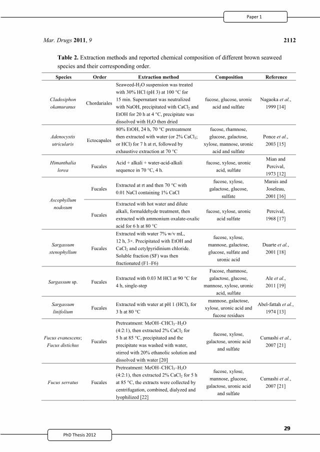
29PhD Thesis 2011
29PhD Thesis 2011
Mar. Drugs 2011, 9
2112
Table 2. Extraction methods and reported chemical composition of different brown seaweed species and their corresponding order.
Species Order Extraction method Composition Reference
Cladosiphon okamuranus
Chordariales
Seaweed-H2O suspension was treated with 30% HCl (pH 3) at 100 °C for 15 min. Supernatant was neutralized with NaOH, precipitated with CaCl2 and EtOH for 20 h at 4 °C, precipitate was dissolved with H2O then dried
fucose, glucose, uronic acid and sulfate
Nagaoka et al., 1999 [14]
Adenocystis utricularis
Ectocapales
80% EtOH, 24 h, 70 °C pretreatment then extracted with water (or 2% CaCl2; or HCl) for 7 h at rt, followed by exhaustive extraction at 70 °C
fucose, rhamnose, glucose, galactose,
xylose, mannose, uronic acid and sulfate
Ponce et al., 2003 [15]
Himanthalia lorea
Fucales Acid + alkali + water-acid-alkali sequence in 70 °C, 4 h.
fucose, xylose, uronic acid, sulfate
Mian and Percival, 1973 [12]
Ascophyllum nodosum
Fucales Extracted at rt and then 70 °C with 0.01 NaCl containing 1% CaCl
fucose, xylose, galactose, glucose,
sulfate
Marais and Joseleau, 2001 [16]
Fucales
Extracted with hot water and dilute alkali, formaldehyde treatment, then extracted with ammonium oxalate-oxalic acid for 6 h at 80 °C
fucose, xylose, uronic acid sulfate
Percival, 1968 [17]
Sargassum stenophyllum
Fucales
Extracted with water 7% w/v mL, 12 h, 3×. Precipitated with EtOH and CaCl2 and cetylpyridinium chloride. Soluble fraction (SF) was then fractionated (F1–F6)
fucose, xylose, mannose, galactose, glucose, sulfate and
uronic acid
Duarte et al., 2001 [18]
Sargassum sp. Fucales Extracted with 0.03 M HCl at 90 °C for 4 h, single-step
Fucose, rhamnose, galactose, glucose,
mannose, xylose, uronic acid, sulfate
Ale et al., 2011 [19]
Sargassum linifolium
Fucales Extracted with water at pH 1 (HCl), for 3 h at 80 °C
mannose, galactose, xylose, uronic acid and
fucose residues
Abel-fattah et al., 1974 [13]
Fucus evanescens; Fucus distichus
Fucales
Pretreatment: MeOH–CHCl3–H2O (4:2:1), then extracted 2% CaCl2 for 5 h at 85 °C, precipitated and the precipitate was washed with water, stirred with 20% ethanolic solution and dissolved with water [20]
fucose, xylose, galactose, uronic acid
and sulfate
Cumashi et al., 2007 [21]
Fucus serratus Fucales
Pretreatment: MeOH–CHCl3–H2O (4:2:1), then extracted 2% CaCl2 for 5 h at 85 °C, the extracts were collected by centrifugation, combined, dialyzed and lyophilized [22]
fucose, xylose, mannose, glucose,
galactose, uronic acid and sulfate
Cumashi et al., 2007 [21]
2
41
29PhD Thesis 2011
29PhD Thesis 2011
Mar. Drugs 2011, 9
2112
Table 2. Extraction methods and reported chemical composition of different brown seaweed species and their corresponding order.
Species Order Extraction method Composition Reference
Cladosiphon okamuranus
Chordariales
Seaweed-H2O suspension was treated with 30% HCl (pH 3) at 100 °C for 15 min. Supernatant was neutralized with NaOH, precipitated with CaCl2 and EtOH for 20 h at 4 °C, precipitate was dissolved with H2O then dried
fucose, glucose, uronic acid and sulfate
Nagaoka et al., 1999 [14]
Adenocystis utricularis
Ectocapales
80% EtOH, 24 h, 70 °C pretreatment then extracted with water (or 2% CaCl2; or HCl) for 7 h at rt, followed by exhaustive extraction at 70 °C
fucose, rhamnose, glucose, galactose,
xylose, mannose, uronic acid and sulfate
Ponce et al., 2003 [15]
Himanthalia lorea
Fucales Acid + alkali + water-acid-alkali sequence in 70 °C, 4 h.
fucose, xylose, uronic acid, sulfate
Mian and Percival, 1973 [12]
Ascophyllum nodosum
Fucales Extracted at rt and then 70 °C with 0.01 NaCl containing 1% CaCl
fucose, xylose, galactose, glucose,
sulfate
Marais and Joseleau, 2001 [16]
Fucales
Extracted with hot water and dilute alkali, formaldehyde treatment, then extracted with ammonium oxalate-oxalic acid for 6 h at 80 °C
fucose, xylose, uronic acid sulfate
Percival, 1968 [17]
Sargassum stenophyllum
Fucales
Extracted with water 7% w/v mL, 12 h, 3×. Precipitated with EtOH and CaCl2 and cetylpyridinium chloride. Soluble fraction (SF) was then fractionated (F1–F6)
fucose, xylose, mannose, galactose, glucose, sulfate and
uronic acid
Duarte et al., 2001 [18]
Sargassum sp. Fucales Extracted with 0.03 M HCl at 90 °C for 4 h, single-step
Fucose, rhamnose, galactose, glucose,
mannose, xylose, uronic acid, sulfate
Ale et al., 2011 [19]
Sargassum linifolium
Fucales Extracted with water at pH 1 (HCl), for 3 h at 80 °C
mannose, galactose, xylose, uronic acid and
fucose residues
Abel-fattah et al., 1974 [13]
Fucus evanescens; Fucus distichus
Fucales
Pretreatment: MeOH–CHCl3–H2O (4:2:1), then extracted 2% CaCl2 for 5 h at 85 °C, precipitated and the precipitate was washed with water, stirred with 20% ethanolic solution and dissolved with water [20]
fucose, xylose, galactose, uronic acid
and sulfate
Cumashi et al., 2007 [21]
Fucus serratus Fucales
Pretreatment: MeOH–CHCl3–H2O (4:2:1), then extracted 2% CaCl2 for 5 h at 85 °C, the extracts were collected by centrifugation, combined, dialyzed and lyophilized [22]
fucose, xylose, mannose, glucose,
galactose, uronic acid and sulfate
Cumashi et al., 2007 [21]
2
41
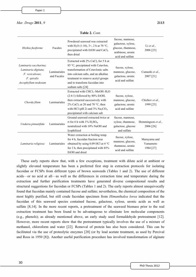
30
Mar. Drugs 2011, 9
2113
Table 2. Cont.
Hizikia fusiforme Fucales
Powdered seaweed was extracted with H2O (1:10), 3×, 2 h at 70 °C, precipitated with EtOH and CaCl2 then dried
fucose, mannose, galactose, xylose,
glucose, rhamnose, arabinose, uronic acid and sulfate
Li et al., 2006 [23]
Laminaria saccharina; Laminaria digitata;
F. vesiculosus; F. spiralis
Ascophyllum nodosum
Laminariales and Fucales
Extracted with 2% CaCl2 for 5 h at 85 °C, precipitated with Cetavlon, transformation of Cetavlonic salts into calcium salts, and an alkaline treatment to remove acetyl groups and to transform fucoidan into sodium salts [24]
fucose, xylose, mannose, glucose, galactose, uronic acid and sulfate
Cumashi et al., 2007 [21]
Chorda filum Laminariales
Extracted with CHCl3–MeOH–H2O (2:4:1) followed by 80% EtOH, then extracted successively with 2% CaCl2 at 20 and 70 °C, then with HCl (pH 2) and 3% Na2CO3, precipitated with calcium salt
fucose, xylose, mannose, glucose, galactose, uronic acid and sulfate
Chizhov et al., 1999 [25]
Undaria pinnatifida Laminariales
Ground seaweed extracted twice at rt for 6 h with 1% H2SO4, neutralized with 10% NaOH and lyophilized
fucose, mannose, xylose, rhamnose, galactose, glucose
and sulfate
Hemmingson et al., 2006 [26]
Laminaria religiosa Laminariales
Water extraction at boiling temp. for 4 h, fucoidan fraction was obtained by using 0.09 HCl at 4 °C for 2 h, then precipitated with 85% EtOH and dried
fucose, xylose, mannose, glucose, rhamnose, uronic acid and sulfate
Maruyama and Yamamoto 1984 [27]
These early reports show that, with a few exceptions, treatment with dilute acid at ambient or slightly elevated temperature has been a preferred first step in extraction protocols for isolating fucoidan or FCSPs from different types of brown seaweeds (Tables 1 and 2). The use of different acids—or no acid at all—as well as the differences in extraction time and temperature during the extraction and further purification treatments have generated diverse compositional results and structural suggestions for fucoidan or FCSPs (Tables 1 and 2). The early reports almost unequivocally found that fucoidan mainly contained fucose and sulfate; nevertheless, the chemical composition of the most highly purified, but still crude fucoidan specimen from Himanthalea lorea indicated that the fucoidan of this seaweed species contained fucose, galactose, xylose, uronic acids as well as sulfate [8,14]. In the more recent reports, a pretreatment of the seaweed biomass prior to the real extraction treatment has been found to be advantageous to eliminate low molecular components (e.g., phenols); as already mentioned above, an early study used formaldehyde pretreatment [12]. However, more recent reports show that the pretreatment typically involves the use of a mixture of methanol, chloroform and water [22]. Removal of protein has also been considered. This can be facilitated via the use of proteolytic enzymes [28] (or by lead acetate treatment, as used by Percival and Roos in 1950 [8]). Another useful purification procedure has involved transformation of alginate
2
42
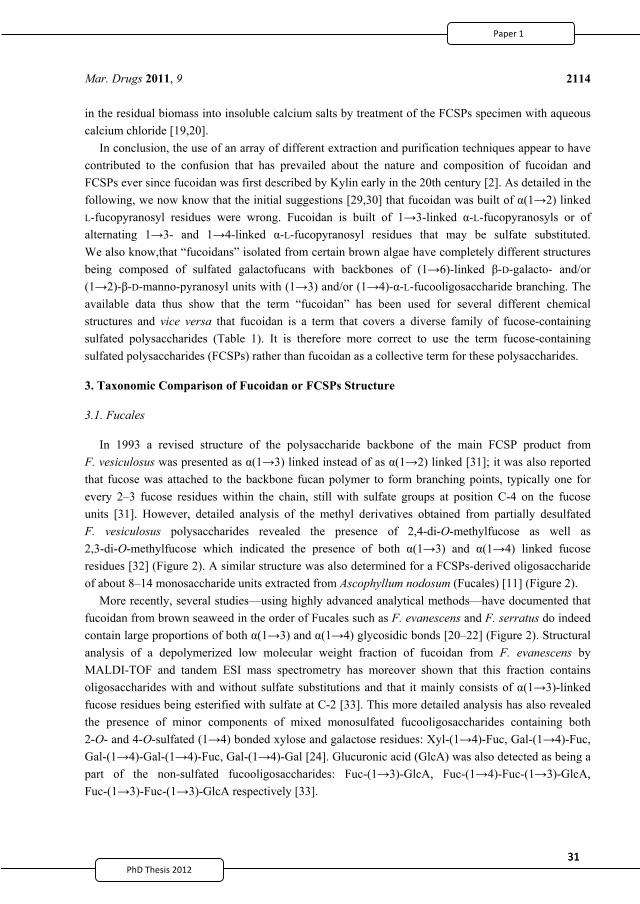
31PhD Thesis 2011
31PhD Thesis 2011
Mar. Drugs 2011, 9
2114
in the residual biomass into insoluble calcium salts by treatment of the FCSPs specimen with aqueous calcium chloride [19,20].
In conclusion, the use of an array of different extraction and purification techniques appear to have contributed to the confusion that has prevailed about the nature and composition of fucoidan and FCSPs ever since fucoidan was first described by Kylin early in the 20th century [2]. As detailed in the following, we now know that the initial suggestions [29,30] that fucoidan was built of (1 2) linked L-fucopyranosyl residues were wrong. Fucoidan is built of 1 3-linked -L-fucopyranosyls or of alternating 1 3- and 1 4-linked -L-fucopyranosyl residues that may be sulfate substituted. We also know,that “fucoidans” isolated from certain brown algae have completely different structures being composed of sulfated galactofucans with backbones of (1 6)-linked -D-galacto- and/or (1 2)- -D-manno-pyranosyl units with (1 3) and/or (1 4)- -L-fucooligosaccharide branching. The available data thus show that the term “fucoidan” has been used for several different chemical structures and vice versa that fucoidan is a term that covers a diverse family of fucose-containing sulfated polysaccharides (Table 1). It is therefore more correct to use the term fucose-containing sulfated polysaccharides (FCSPs) rather than fucoidan as a collective term for these polysaccharides.
3. Taxonomic Comparison of Fucoidan or FCSPs Structure
3.1. Fucales
In 1993 a revised structure of the polysaccharide backbone of the main FCSP product from F. vesiculosus was presented as (1 3) linked instead of as (1 2) linked [31]; it was also reported that fucose was attached to the backbone fucan polymer to form branching points, typically one for every 2–3 fucose residues within the chain, still with sulfate groups at position C-4 on the fucose units [31]. However, detailed analysis of the methyl derivatives obtained from partially desulfated F. vesiculosus polysaccharides revealed the presence of 2,4-di-O-methylfucose as well as 2,3-di-O-methylfucose which indicated the presence of both (1 3) and (1 4) linked fucose residues [32] (Figure 2). A similar structure was also determined for a FCSPs-derived oligosaccharide of about 8–14 monosaccharide units extracted from Ascophyllum nodosum (Fucales) [11] (Figure 2).
More recently, several studies—using highly advanced analytical methods—have documented that fucoidan from brown seaweed in the order of Fucales such as F. evanescens and F. serratus do indeed contain large proportions of both (1 3) and (1 4) glycosidic bonds [20–22] (Figure 2). Structural analysis of a depolymerized low molecular weight fraction of fucoidan from F. evanescens by MALDI-TOF and tandem ESI mass spectrometry has moreover shown that this fraction contains oligosaccharides with and without sulfate substitutions and that it mainly consists of (1 3)-linked fucose residues being esterified with sulfate at C-2 [33]. This more detailed analysis has also revealed the presence of minor components of mixed monosulfated fucooligosaccharides containing both 2-O- and 4-O-sulfated (1 4) bonded xylose and galactose residues: Xyl-(1 4)-Fuc, Gal-(1 4)-Fuc, Gal-(1 4)-Gal-(1 4)-Fuc, Gal-(1 4)-Gal [24]. Glucuronic acid (GlcA) was also detected as being a part of the non-sulfated fucooligosaccharides: Fuc-(1 3)-GlcA, Fuc-(1 4)-Fuc-(1 3)-GlcA, Fuc-(1 3)-Fuc-(1 3)-GlcA respectively [33].
2
43
31PhD Thesis 2011
31PhD Thesis 2011
Mar. Drugs 2011, 9
2114
in the residual biomass into insoluble calcium salts by treatment of the FCSPs specimen with aqueous calcium chloride [19,20].
In conclusion, the use of an array of different extraction and purification techniques appear to have contributed to the confusion that has prevailed about the nature and composition of fucoidan and FCSPs ever since fucoidan was first described by Kylin early in the 20th century [2]. As detailed in the following, we now know that the initial suggestions [29,30] that fucoidan was built of (1 2) linked L-fucopyranosyl residues were wrong. Fucoidan is built of 1 3-linked -L-fucopyranosyls or of alternating 1 3- and 1 4-linked -L-fucopyranosyl residues that may be sulfate substituted. We also know,that “fucoidans” isolated from certain brown algae have completely different structures being composed of sulfated galactofucans with backbones of (1 6)-linked -D-galacto- and/or (1 2)- -D-manno-pyranosyl units with (1 3) and/or (1 4)- -L-fucooligosaccharide branching. The available data thus show that the term “fucoidan” has been used for several different chemical structures and vice versa that fucoidan is a term that covers a diverse family of fucose-containing sulfated polysaccharides (Table 1). It is therefore more correct to use the term fucose-containing sulfated polysaccharides (FCSPs) rather than fucoidan as a collective term for these polysaccharides.
3. Taxonomic Comparison of Fucoidan or FCSPs Structure
3.1. Fucales
In 1993 a revised structure of the polysaccharide backbone of the main FCSP product from F. vesiculosus was presented as (1 3) linked instead of as (1 2) linked [31]; it was also reported that fucose was attached to the backbone fucan polymer to form branching points, typically one for every 2–3 fucose residues within the chain, still with sulfate groups at position C-4 on the fucose units [31]. However, detailed analysis of the methyl derivatives obtained from partially desulfated F. vesiculosus polysaccharides revealed the presence of 2,4-di-O-methylfucose as well as 2,3-di-O-methylfucose which indicated the presence of both (1 3) and (1 4) linked fucose residues [32] (Figure 2). A similar structure was also determined for a FCSPs-derived oligosaccharide of about 8–14 monosaccharide units extracted from Ascophyllum nodosum (Fucales) [11] (Figure 2).
More recently, several studies—using highly advanced analytical methods—have documented that fucoidan from brown seaweed in the order of Fucales such as F. evanescens and F. serratus do indeed contain large proportions of both (1 3) and (1 4) glycosidic bonds [20–22] (Figure 2). Structural analysis of a depolymerized low molecular weight fraction of fucoidan from F. evanescens by MALDI-TOF and tandem ESI mass spectrometry has moreover shown that this fraction contains oligosaccharides with and without sulfate substitutions and that it mainly consists of (1 3)-linked fucose residues being esterified with sulfate at C-2 [33]. This more detailed analysis has also revealed the presence of minor components of mixed monosulfated fucooligosaccharides containing both 2-O- and 4-O-sulfated (1 4) bonded xylose and galactose residues: Xyl-(1 4)-Fuc, Gal-(1 4)-Fuc, Gal-(1 4)-Gal-(1 4)-Fuc, Gal-(1 4)-Gal [24]. Glucuronic acid (GlcA) was also detected as being a part of the non-sulfated fucooligosaccharides: Fuc-(1 3)-GlcA, Fuc-(1 4)-Fuc-(1 3)-GlcA, Fuc-(1 3)-Fuc-(1 3)-GlcA respectively [33].
2
43
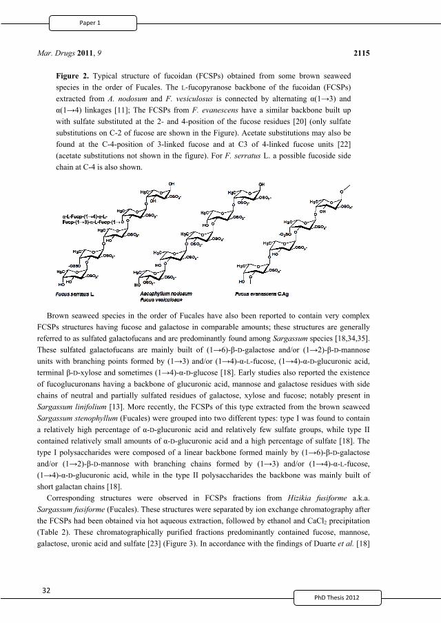
32
Mar. Drugs 2011, 9
2115
Figure 2. Typical structure of fucoidan (FCSPs) obtained from some brown seaweed species in the order of Fucales. The L-fucopyranose backbone of the fucoidan (FCSPs) extracted from A. nodosum and F. vesiculosus is connected by alternating (1 3) and
(1 4) linkages [11]; The FCSPs from F. evanescens have a similar backbone built up with sulfate substituted at the 2- and 4-position of the fucose residues [20] (only sulfate substitutions on C-2 of fucose are shown in the Figure). Acetate substitutions may also be found at the C-4-position of 3-linked fucose and at C3 of 4-linked fucose units [22] (acetate substitutions not shown in the figure). For F. serratus L. a possible fucoside side chain at C-4 is also shown.
Brown seaweed species in the order of Fucales have also been reported to contain very complex FCSPs structures having fucose and galactose in comparable amounts; these structures are generally referred to as sulfated galactofucans and are predominantly found among Sargassum species [18,34,35]. These sulfated galactofucans are mainly built of (1 6)- -D-galactose and/or (1 2)- -D-mannose units with branching points formed by (1 3) and/or (1 4)- -L-fucose, (1 4)- -D-glucuronic acid, terminal -D-xylose and sometimes (1 4)- -D-glucose [18]. Early studies also reported the existence of fucoglucuronans having a backbone of glucuronic acid, mannose and galactose residues with side chains of neutral and partially sulfated residues of galactose, xylose and fucose; notably present in Sargassum linifolium [13]. More recently, the FCSPs of this type extracted from the brown seaweed Sargassum stenophyllum (Fucales) were grouped into two different types: type I was found to contain a relatively high percentage of -D-glucuronic acid and relatively few sulfate groups, while type II contained relatively small amounts of -D-glucuronic acid and a high percentage of sulfate [18]. The type I polysaccharides were composed of a linear backbone formed mainly by (1 6)- -D-galactose and/or (1 2)- -D-mannose with branching chains formed by (1 3) and/or (1 4)- -L-fucose, (1 4)- -D-glucuronic acid, while in the type II polysaccharides the backbone was mainly built of short galactan chains [18].
Corresponding structures were observed in FCSPs fractions from Hizikia fusiforme a.k.a. Sargassum fusiforme (Fucales). These structures were separated by ion exchange chromatography after the FCSPs had been obtained via hot aqueous extraction, followed by ethanol and CaCl2 precipitation (Table 2). These chromatographically purified fractions predominantly contained fucose, mannose, galactose, uronic acid and sulfate [23] (Figure 3). In accordance with the findings of Duarte et al. [18]
2
44
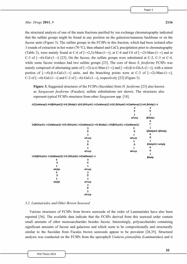
33PhD Thesis 2011
33PhD Thesis 2011
Mar. Drugs 2011, 9
2116
the structural analysis of one of the main fractions purified by ion exchange chromatography indicated that the sulfate groups might be found in any position on the galactose/mannose backbone or on the fucose units (Figure 3). The sulfate groups in the FCSPs in this fraction, which had been isolated after 3 rounds of extraction in hot water (70 °C), then ethanol and CaCl2 precipitation prior to chromatography (Table 2), were mainly found at C-6 of [ 2,3)-Man-(1 ], at C-4 and C6 of [ 2)-Man-(1 ] and at C-3 of [ 6)-Gal-(1 ] [23]. On the fucose, the sulfate groups were substituted at C-2, C-3 or C-4, while some fucose residues had two sulfate groups [23]. The core of these S. fusiforme FCSPs was mainly composed of alternating units of [ 2)- -D-Man-(1 ] and [ 4)- -D-GlcA-(1 ], with a minor portion of [ 4)- -D-Gal-(1 ] units, and the branching points were at C-3 of [ 2)-Man-(1 ], C-2 of [ 4)-Gal-(1 ] and C-2 of [ 6)-Gal-(1 ], respectively [23] (Figure 3).
Figure 3. Suggested structures of the FCSPs (fucoidan) from H. fusiforme [23] also known as Sargassum fusiforme (Fucales); sulfate substitutions not shown. The structures also represent typical FCSPs structures from other Sargassum spp. [18].
3.2. Laminariales and Other Brown Seaweed
Various structures of FCSPs from brown seaweeds of the order of Laminariales have also been reported [36]. The available data indicate that the FCSPs derived from this seaweed order contain small amounts of other monosaccharides besides fucose. Interestingly, polysaccharides containing significant amounts of fucose and galactose and which seem to be compositionally and structurally similar to the fucoidan from Fucales brown seaweeds appear to be prevalent [26,35]. Structural analysis was conducted on the FCSPs from the sporophyll Undaria pinnatifida (Laminariales) and it
2
45
33PhD Thesis 2011
33PhD Thesis 2011
Mar. Drugs 2011, 9
2116
the structural analysis of one of the main fractions purified by ion exchange chromatography indicated that the sulfate groups might be found in any position on the galactose/mannose backbone or on the fucose units (Figure 3). The sulfate groups in the FCSPs in this fraction, which had been isolated after 3 rounds of extraction in hot water (70 °C), then ethanol and CaCl2 precipitation prior to chromatography (Table 2), were mainly found at C-6 of [ 2,3)-Man-(1 ], at C-4 and C6 of [ 2)-Man-(1 ] and at C-3 of [ 6)-Gal-(1 ] [23]. On the fucose, the sulfate groups were substituted at C-2, C-3 or C-4, while some fucose residues had two sulfate groups [23]. The core of these S. fusiforme FCSPs was mainly composed of alternating units of [ 2)- -D-Man-(1 ] and [ 4)- -D-GlcA-(1 ], with a minor portion of [ 4)- -D-Gal-(1 ] units, and the branching points were at C-3 of [ 2)-Man-(1 ], C-2 of [ 4)-Gal-(1 ] and C-2 of [ 6)-Gal-(1 ], respectively [23] (Figure 3).
Figure 3. Suggested structures of the FCSPs (fucoidan) from H. fusiforme [23] also known as Sargassum fusiforme (Fucales); sulfate substitutions not shown. The structures also represent typical FCSPs structures from other Sargassum spp. [18].
3.2. Laminariales and Other Brown Seaweed
Various structures of FCSPs from brown seaweeds of the order of Laminariales have also been reported [36]. The available data indicate that the FCSPs derived from this seaweed order contain small amounts of other monosaccharides besides fucose. Interestingly, polysaccharides containing significant amounts of fucose and galactose and which seem to be compositionally and structurally similar to the fucoidan from Fucales brown seaweeds appear to be prevalent [26,35]. Structural analysis was conducted on the FCSPs from the sporophyll Undaria pinnatifida (Laminariales) and it
2
45
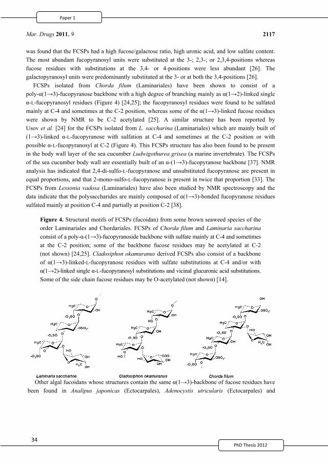
342
Mar. Drugs 2011, 9
2117
was found that the FCSPs had a high fucose/galactose ratio, high uronic acid, and low sulfate content. The most abundant fucopyranosyl units were substituted at the 3-; 2,3-; or 2,3,4-positions whereas fucose residues with substitutions at the 3,4- or 4-positions were less abundant [26]. The galactopyranosyl units were predominantly substituted at the 3- or at both the 3,4-positions [26].
FCSPs isolated from Chorda filum (Laminariales) have been shown to consist of a poly- (1 3)-fucopyranose backbone with a high degree of branching mainly as (1 2)-linked single
-L-fucopyranosyl residues (Figure 4) [24,25]; the fucopyranosyl residues were found to be sulfated mainly at C-4 and sometimes at the C-2 position, whereas some of the (1 3)-linked fucose residues were shown by NMR to be C-2 acetylated [25]. A similar structure has been reported by Usov et al. [24] for the FCSPs isolated from L. saccharina (Laminariales) which are mainly built of (1 3)-linked -L-fucopyranose with sulfation at C-4 and sometimes at the C-2 position or with possible -L-fucopyranosyl at C-2 (Figure 4). This FCSPs structure has also been found to be present in the body wall layer of the sea cucumber Ludwigothurea grisea (a marine invertebrate). The FCSPs of the sea cucumber body wall are essentially built of an -(1 3)-fucopyranose backbone [37]. NMR analysis has indicated that 2,4-di-sulfo-L-fucopyranose and unsubstituted fucopyranose are present in equal proportions, and that 2-mono-sulfo-L-fucopyranose is present in twice that proportion [33]. The FCSPs from Lessonia vadosa (Laminariales) have also been studied by NMR spectroscopy and the data indicate that the polysaccharides are mainly composed of (1 3)-bonded fucopyranose residues sulfated mainly at position C-4 and partially at position C-2 [38].
Figure 4. Structural motifs of FCSPs (fucoidan) from some brown seaweed species of the order Laminariales and Chordariales. FCSPs of Chorda filum and Laminaria saccharina consist of a poly- -(1 3)-fucopyranoside backbone with sulfate mainly at C-4 and sometimes at the C-2 position; some of the backbone fucose residues may be acetylated at C-2 (not shown) [24,25]. Cladosiphon okamuranus derived FCSPs also consist of a backbone of (1 3)-linked-L-fucopyranose residues with sulfate substitutions at C-4 and/or with
(1 2)-linked single -L-fucopyranosyl substitutions and vicinal glucuronic acid substitutions. Some of the side chain fucose residues may be O-acetylated (not shown) [14].
Other algal fucoidans whose structures contain the same (1 3)-backbone of fucose residues have been found in Analipus japonicas (Ectocarpales), Adenocystis utricularis (Ectocarpales) and
2
46
35PhD Thesis 2011
35PhD Thesis 2011
Mar. Drugs 2011, 9
2118
Cladosiphon okamuranus (Chordariales) [14,15,39], but the FCSPs from these brown algae also appear to contain other monosaccharides than fucose (Table 2). The detailed structural elucidation of the FCSPs from C. okamuranus (Chordariales) confirmed that this product was made up of a linear backbone of (1 3)-fucopyranose units with a portion of the fucose residues carrying sulfate substitutions at C-4 but some of the fucose residues have also been found to be O-acetylated (Figure 4). The C. okamuranus FCSPs may also contain -glucuronic acid substitutions at the C-2-positions of those backbone fucose residues that are not substituted by a sulfate group [14] (Figure 4).
Methylation analysis, desulfation and NMR spectroscopy of the FCSPs fractions from Adenocystis utricularis (Ectocarpales) showed that these FCSPs contained the same -(1 3)-fucopyranose backbone as that found in Chorda filum and Laminaria saccharina FCSPs, and that the fucopyranosyl units were mostly sulfated at C-4, and branched at C-2 with non-sulfated fucopyranosyl units; the galactan moiety, which was also present, was predominantly found to be a backbone structure of (1 3) and (1 6) D-galactopyranose units with sulfation mostly on C-4 [15]. Later, a similar structure was found in FCSPs extracted from Analipus japonicas (Ectocarpales) [39].
The relatively large variations in the reported compositional and structural properties of the FCPSs extracted from different brown seaweed species thus clearly confirm the natural biodiversity of FCSPs notably as exemplified by the structures found in Fucales, e.g., in the Fucus sp. and Sargassum sp. (Figures 2 and 3). The (1 3)-linked -L-fucopyranosyl backbone structure, with various extents of sulfate substitutions, is prevalent as the core backbone structure in the majority of the currently analyzed FCSPs. Nevertheless, the reported structural data for FCSPs from different brown seaweed species clearly indicate that there is no consistent basic structure of “fucoidan”. It also seems clear that FCSPs extracted from seaweeds under the same order have different composition, and in turn that the structural traits of FCSPs cannot be categorized or predicted according to algal order (Tables 1 and 2).
When assessing the available compositional data the large variation in the composition of the FCSPs products obtained from different extraction methods is evident (Table 2). Recently, we optimized the extraction yields of FCSPs from Sargassum sp. by developing a single-step extraction procedure. While doing so, we also systematically examined the effects of different extraction parameters (i.e., acid concentration, time, and temperature) on the yields and composition of the FCSPs products obtained [19] (Table 2). All extraction factors affected the FCSPs yield. Lower total FCSPs yields, but higher fucose contents in the products were obtained with shorter extraction time [19]. The work also revealed that classical extraction treatment with HCl at elevated temperature and during extended time, i.e., a procedure similar to the one used by Black et al. [9], had a detrimental effect on the FCSPs yield as this treatment apparently disrupted the structural integrity of the fucose-containing polymer and caused degradation of the chains built of fucose residues [19]. Hence, some of the classic methods, employing relatively harsh acid treatments, may in fact have affected the composition and structure of the target FCSP products to different extents, and may have contributed to the prevailing “conflicting views on the nature of fucoidin” recognized already in 1950 by Percival and Ross [8]. A consensus to employ defined extraction protocols for extraction of FCSPs, or at least an agreement among scientists in the field to include a benchmark extraction procedure in their studies, would help to advance the understanding of these intriguing FCSPs substances.
2
47
35PhD Thesis 2011
35PhD Thesis 2011
Mar. Drugs 2011, 9
2118
Cladosiphon okamuranus (Chordariales) [14,15,39], but the FCSPs from these brown algae also appear to contain other monosaccharides than fucose (Table 2). The detailed structural elucidation of the FCSPs from C. okamuranus (Chordariales) confirmed that this product was made up of a linear backbone of (1 3)-fucopyranose units with a portion of the fucose residues carrying sulfate substitutions at C-4 but some of the fucose residues have also been found to be O-acetylated (Figure 4). The C. okamuranus FCSPs may also contain -glucuronic acid substitutions at the C-2-positions of those backbone fucose residues that are not substituted by a sulfate group [14] (Figure 4).
Methylation analysis, desulfation and NMR spectroscopy of the FCSPs fractions from Adenocystis utricularis (Ectocarpales) showed that these FCSPs contained the same -(1 3)-fucopyranose backbone as that found in Chorda filum and Laminaria saccharina FCSPs, and that the fucopyranosyl units were mostly sulfated at C-4, and branched at C-2 with non-sulfated fucopyranosyl units; the galactan moiety, which was also present, was predominantly found to be a backbone structure of (1 3) and (1 6) D-galactopyranose units with sulfation mostly on C-4 [15]. Later, a similar structure was found in FCSPs extracted from Analipus japonicas (Ectocarpales) [39].
The relatively large variations in the reported compositional and structural properties of the FCPSs extracted from different brown seaweed species thus clearly confirm the natural biodiversity of FCSPs notably as exemplified by the structures found in Fucales, e.g., in the Fucus sp. and Sargassum sp. (Figures 2 and 3). The (1 3)-linked -L-fucopyranosyl backbone structure, with various extents of sulfate substitutions, is prevalent as the core backbone structure in the majority of the currently analyzed FCSPs. Nevertheless, the reported structural data for FCSPs from different brown seaweed species clearly indicate that there is no consistent basic structure of “fucoidan”. It also seems clear that FCSPs extracted from seaweeds under the same order have different composition, and in turn that the structural traits of FCSPs cannot be categorized or predicted according to algal order (Tables 1 and 2).
When assessing the available compositional data the large variation in the composition of the FCSPs products obtained from different extraction methods is evident (Table 2). Recently, we optimized the extraction yields of FCSPs from Sargassum sp. by developing a single-step extraction procedure. While doing so, we also systematically examined the effects of different extraction parameters (i.e., acid concentration, time, and temperature) on the yields and composition of the FCSPs products obtained [19] (Table 2). All extraction factors affected the FCSPs yield. Lower total FCSPs yields, but higher fucose contents in the products were obtained with shorter extraction time [19]. The work also revealed that classical extraction treatment with HCl at elevated temperature and during extended time, i.e., a procedure similar to the one used by Black et al. [9], had a detrimental effect on the FCSPs yield as this treatment apparently disrupted the structural integrity of the fucose-containing polymer and caused degradation of the chains built of fucose residues [19]. Hence, some of the classic methods, employing relatively harsh acid treatments, may in fact have affected the composition and structure of the target FCSP products to different extents, and may have contributed to the prevailing “conflicting views on the nature of fucoidin” recognized already in 1950 by Percival and Ross [8]. A consensus to employ defined extraction protocols for extraction of FCSPs, or at least an agreement among scientists in the field to include a benchmark extraction procedure in their studies, would help to advance the understanding of these intriguing FCSPs substances.
2
47
36
Mar. Drugs 2011, 9
2119
4. Bioactivity of Fucoidan or FCSPs
In recent years, fucoidan or FCSPs from seaweed biomass have been the subject of many scientific studies aiming at assessing their potential biological activities including antitumor and immunomodulatory [40–42], antivirus [43], antithrombotic and anticoagulant [44], anti-inflammatory [45], and antioxidant effects [46], as well as their effects against various renal [47], hepatic [48] and uropathic disorders [49].
Recently, low molecular weight FCSPs have been shown to have therapeutic potential in preventing intimal hyperplasia in both in vivo and in vitro studies: Contact with low molecular weight FCSPs (“fucoidan”) thus increased the migration of human vascular endothelial cells and induced decreased migration of vascular smooth muscle cells in vitro [50]. In an in vivo rat experiment FCSPs reduced the intimal hyperplasia in the rat aortic wall after balloon injury [50]. Furthermore, an in vivo efficacy study of fucoidan films conducted using a rat model showed that during cecal-sidewall surgery a fucoidan film wound healing treatment reduced the adhesion scores by approximately 90% and resulted in 50% to 100% of animals being adhesion free [51].
In this review, the most significant bioactivities of FCSPs, including antitumor and immunomodulatory, anticoagulant and antithrombotic effects will be presented with special focus on the relationship between the FCSP structural features and biological activity.
4.1. Antitumor and Immune-Response Activities
Several different therapeutic strategies such as chemotherapy, radiation therapy, surgery or combinations hereof have been used to treat different types of cancer. Unfortunately, several of these treatments provide only minimal benefits; moreover, there are undesirable complications and long term side effects of the treatments [52,53]. Consequently the quest for potential preventive or therapeutic measures against cancer has been going on for years and recently the focus has been directed towards bioactive compounds of natural origin, including FCSPs from brown seaweeds [1]. Many reports have been published which indicate the antitumor and immune-response modulating activity of FCSPs in both in vivo and in vitro studies [40–42,54,55].
Sulfated polysaccharide fractions from Sargassum fulvellum, S. kjellmanianum, L. angustata, L. angustata var. longissima, L. japonica, Ecklonia cava, and Eisenia bicyclis have been evaluated for their bioactivities, and they have been found to exert remarkable growth inhibitory activities on Sarcoma-180 cells implanted into mice and to possess antitumor activity against L-1210 leukemia in mice [56–58]. Recently, we reported the potent in vitro bioactivity of FCSPs extracted from Sargassum sp. and F. vesiculosus against lung and skin cancer cell growth [42]. The antitumor mechanism of FCSPs from sporophylls of Undaria pinnatifida has been described by Maruyama et al. [41,59]. The available findings indicate that antitumor activity of FCSPs may be associated with a significant enhancement of the cytolytic activity of natural killer (NK) cells augmented by increased production of macrophage-mediated immune response signaling molecules [59–61], namely interleukins (IL)-2, IFN- and IL-12 [42,59], and induction of apoptosis [42].
Macrophage activation by polysaccharides is mediated through specific membrane receptors. The major receptors reported for polysaccharides recognition in macrophages are glycoproteins including
2
48
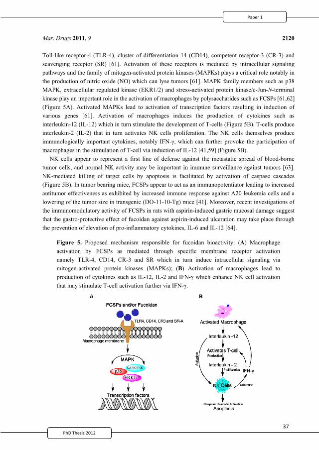
37
Mar. Drugs 2011, 9
2120
Toll-like receptor-4 (TLR-4), cluster of differentiation 14 (CD14), competent receptor-3 (CR-3) and scavenging receptor (SR) [61]. Activation of these receptors is mediated by intracellular signaling pathways and the family of mitogen-activated protein kinases (MAPKs) plays a critical role notably in the production of nitric oxide (NO) which can lyse tumors [61]. MAPK family members such as p38 MAPK, extracellular regulated kinase (EKR1/2) and stress-activated protein kinase/c-Jun-N-terminal kinase play an important role in the activation of macrophages by polysaccharides such as FCSPs [61,62] (Figure 5A). Activated MAPKs lead to activation of transcription factors resulting in induction of various genes [61]. Activation of macrophages induces the production of cytokines such as interleukin-12 (IL-12) which in turn stimulate the development of T-cells (Figure 5B). T-cells produce interleukin-2 (IL-2) that in turn activates NK cells proliferation. The NK cells themselves produce immunologically important cytokines, notably IFN- , which can further provoke the participation of macrophages in the stimulation of T-cell via induction of IL-12 [41,59] (Figure 5B).
NK cells appear to represent a first line of defense against the metastatic spread of blood-borne tumor cells, and normal NK activity may be important in immune surveillance against tumors [63]. NK-mediated killing of target cells by apoptosis is facilitated by activation of caspase cascades (Figure 5B). In tumor bearing mice, FCSPs appear to act as an immunopotentiator leading to increased antitumor effectiveness as exhibited by increased immune response against A20 leukemia cells and a lowering of the tumor size in transgenic (DO-11-10-Tg) mice [41]. Moreover, recent investigations of the immunomodulatory activity of FCSPs in rats with aspirin-induced gastric mucosal damage suggest that the gastro-protective effect of fucoidan against aspirin-induced ulceration may take place through the prevention of elevation of pro-inflammatory cytokines, IL-6 and IL-12 [64].
Figure 5. Proposed mechanism responsible for fucoidan bioactivity: (A) Macrophage activation by FCSPs as mediated through specific membrane receptor activation namely TLR-4, CD14, CR-3 and SR which in turn induce intracellular signaling via mitogen-activated protein kinases (MAPKs); (B) Activation of macrophages lead to production of cytokines such as IL-12, IL-2 and IFN- which enhance NK cell activation that may stimulate T-cell activation further via IFN- .
a
2
49
38
Mar. Drugs 2011, 9
2121
Apoptosis is one of the most prevalent pathways through which FCSPs can inhibit the overall growth of cancer. Previous studies have shown that different types of FCSPs can induce apoptosis in melanoma cells [42], HT-29 colon cancer cells [65], MCF-7 human breast cancer cells [66], and HS-Sultan human lymphoma cells [62]. In human HS-Sultan cells, the apoptosis may occur via activation of caspase-3 [62]; and in MCF-7 cells via caspase-8 dependent pathways [66]. Alternatively, FCSPs induced apoptosis may take place through activation of caspases via both death receptor-mediated and mitochondria-mediated apoptotic pathways [65].
4.2. Anticoagulant and Antithrombotic Activities
The earliest published report describing the anticoagulant activity of fucoidan was published in 1957 [67]. In that report it was shown that a certain fraction of fucoidan from F. vesiculosus possessed powerful anticoagulant activity that qualified fucoidan to belong to the group of heparinoids [67]. Heparin is a biomolecule containing highly sulfated glucosaminoglycan that is widely used as an injectable anticoagulant. It has been reported that the anticoagulant mechanisms of fucoidan are related to both antithrombin and heparin cofactor II-mediated activity [68,69], but the mechanisms by which fucoidan exerts anticoagulant activity remain controversial [32]. Hence, any possible relations between the physical and chemical properties, the structure, and the anticoagulant activity of fucoidan remain to be firmly established. The uncertainties are mainly due to the structural variation of fucoidan between algal species, but most likely also a result of the different extraction methodologies employed to isolate FCSPs that appear to have produced FCSPs of different composition, structure, and size, which have given rise to conflicting results in the detailed studies of mechanisms of anticoagulant activity [32,70].
Results obtained using the so called activated partial thromboplastin time assay (APTT) have strongly indicated that FCSPs from F. vesiculosus have specific anticoagulant activity. Comparable results have been obtained in two independent studies using FCSPs dosages equivalent to 9–13 U/mg versus 167 U/mg for heparin; and 16 U/mg versus 193 U/mg heparin, respectively [71,72]. When FCSPs samples isolated from nine brown seaweed species were tested for anticoagulant activities, the APTT results were significant at 12–38 U/mg as compared to at 167 U/mg for heparin [73]. A remarkable finding for anticoagulant action was also reported by Kitamura et al. [74], who showed that a FCSPs fraction from L. angustata var. longissima (Laminariales) had antithrombin activity at 200 U/mg, equivalent to a dose of 140 U/mg heparin. The particular FCSPs fraction having anticoagulant activity had a molecular weight of ~21–23 kDa and contained fucose-galactose-sulfate at a ratio of 9:1:9 with the sulfate substitutions at C-4 of the fucose residues [74].
It has been postulated that it is not a specific structural trait that determines fucoidan’s ability to elicit anticoagulant activity, but rather that the anticoagulant effect is due to a multitude of structural features including monosaccharide composition, molecular weight, sulfation level, and position of sulfate groups on the main chain of the polysaccharide [69,75–77]. The comprehensive study of the anticoagulant activity of fucoidan by Cumashi et al. [21] also noted that neither the content of fucose and sulfate nor other structural features affected the anticoagulant efficacy. FCSPs from C. okamuranus (Chordariales) have been reported to exert virtually no anticoagulant effect, and this could be due to the low amount of sulfate in its polymer backbone and/or the presence of vicinal branching points formed by 2-O- -D-glucuronyl substituents (Figure 4) [21,14]. On the other hand, the concentrations
2
50
39PhD Thesis 2011
Mar. Drugs 2011, 9
2122
of C-2 sulfate and C-2,3-disulfated sugar residues (Figure 2) have been reported to be a common structural feature for fucoidan anticoagulant activity [11]. The anticoagulant activity of high molecular weight FCSPs from Ecklonia kurome were thus reported to be dependent on both molecular weight and sulfate content [76]: Higher molecular weight FCSPs (i.e., 27 and 58 kDa) showed higher anticoagulant activity than lower molecular weight FCSPs (i.e., 10 kDa); and FCSPs samples having a high molar ratio of sulfate to total sugar residues were found to exhibit inhibitory effects on fibrinogen clotting by thrombin reaction [76]. These results were supported by data reported by Haroun-Bouhedja et al. [78] who reported a relationship between the extent of sulfate group substitutions and the biological activities of fucoidan. The anticoagulant activity of low molecular weight (LMW) fucoidan, i.e., MW < 18 kDa was thus found to decrease with decreasing degree of sulfation, and very low-sulfate (<20%) or desulfated LMW fucoidan lost its anticoagulant activity, but retained some antiproliferative activity on CCL39 fibroblast cells [78]. In contrast, LMW fucoidan with sulfate content higher than 20% was found to exert profound anticoagulant activity as well as antiproliferative effects on fibroblast cell line CCL39 cells in a dose-dependent fashion [78].
Some studies suggest that also the sugar composition (e.g., fucose, galactose, mannose, etc.) or the type of oligo- or polysaccharides of the FCSPs may play an important role for anticoagulant activity [75,79]. However, the series of investigations conducted by Pereira et al. [32,80,81] indicated that a 2-sulfated, 3-linked -L-galactan, but not -L-fucan, was the potent thrombin inhibitor mediated by anti-thrombin of heparin cofactor II. These findings have however also pointed out that it is not necessarily the sugar composition but rather the sulfate substitutions on the sugars that determine the anticoagulant activity of fucoidan—or both [82].
Most of the reported studies were carried out with crude, diverse and complex FCSPs obtained via extraction from brown seaweeds as opposed to being chemically well defined structures. For this reason it is not easy to identify a structure versus activity relationship because of the presence of highly branched portions and the complex distributions of sulfate and acetyl groups in algal FCSPs. This aspect was attempted resolved by use of invertebrate polysaccharides [83]. The data obtained indicated that regular, linear sulfated -L-fucans and sulfated -L-galactans express anticoagulant activity, which is not simply a function of charge density, but critically dependent on the pattern of sulfation as well as monosaccharide composition. Sulfated -L-fucans and fucosylated chondroitin sulfate were also shown to elicit antithrombotic activity when tested on in vivo models of venous and arterial thrombosis in experimental animals [83].
4.3. Bioactivities and Oversulfation of FCSPs
In 1984 crude FCSPs fractions from Sargassum kjellmanianum were prepared in order to investigate the influence of the sulfation levels on the survival of L-1210 leukemia bearing mice; and on the growth of Sarcoma-180 cells [58]. The study showed that the fraction with the highest sulfation was the most effective against L-1210 leukemia bearing mice and it produced an increase in life span of 26%. On the other hand, this particular FCSPs fraction was also less effective in inhibiting growth of Sarcoma-180 cells subcutaneously implanted into mice [58].
Since then, several investigations have focused on the effect of oversulfation of FCSPs on biological activity [54,78,84–89]. Oversulfated FCSPs may be obtained by further sulfation of native FCSP
2
51
40
Mar. Drugs 2011, 9
2123
molecules using dimethylformamide as solvent and a sulfur trioxide-trimethylamine complex as the sulfating agent [86]. The inhibitory effects of such oversulfated FCSPs were investigated on the invasion of Murine Lewis Lung Carcinoma cells through a reconstituted membrane basement fragment, socalled laminin [86]. Oversulfated FCSPs were found to be the most potent inhibitor of tumor cell invasion and were also, in particular, found to inhibit tumor cell adhesion to laminin better than native and desulfated FCSPs. The most potent oversulfated FCSP structures had sulfate groups on both the C-3 and C-4 positions of the fucose units; hence, the particular spatial orientation of the negative charges in the FCSPs molecules may also be an important determinant of bioactivity [86]. The study did not allow firm conclusions to be drawn with respect to mechanisms of action, but it was suggested that the increased negative charge resulting from oversulfation might promote the formation of FCSPs-protein complexes involved in cell proliferation, in turn suppressing cell growth [86].
When the importance of the spatial orientation of the negative charges on the FCSPs was investigated in more depth it was confirmed that this feature plays a major role in determining the binding potency of FCSPs to vascular endothelial growth factor 165 (VEGF165) [88]. Both native and oversulfated FCSPs have been tested for their anti-angiogenic actions in vivo and for their in vitro anti-proliferative effects against B16 melanoma cells, Sarcoma-180 and Lewis lung carcinoma cells: The interaction of oversulfated FCSPs with VEGF165 occurred with high affinity and resulted in the formation of highly stable complexes, thereby interfering with the binding of VEGF165 to vascular endothelial growth factor receptor-2 (VEGFR-2). The results showed that both native and oversulfated FCSPs were able to suppress neovascularization in mice implanted Sarcoma-180 cells; and that both FCSPs types inhibited tumor growth through the prevention of tumor-induced angiogenesis, but the data indicated that sulfation tended to give more potent effects [88].
Native and oversulfated FCSPs derived from Cladosiphon okamuranus (Chordariales) were analyzed using 1H NMR spectroscopy and it was suggested that whereas natural sulfation produced 4-mono-O-sulfo-L-fucopyranose the oversulfated FCSPs contained 2,4-di-, 2-mono-, and 4-mono-O-sulfo-L-fucopyranose [89]. It was also suggested that sulfate content and the positioning of sulfate groups, e.g., 2,4-di- vs. 4-mono, might be important for the anti-proliferative activity of fucoidan in a human leukemia cell line (U937), an effect which is presumed to take place via induction of apoptosis associated with activation of caspase-3 and -7 [89].
The effects of oversulfation of low and high molecular weight FCSPs derivatives from F. vesiculosus and heparin on lipopolysaccharide (LPS)-induced release of plasminogen activator inhibitor-1 (PAI-1) from cultured human umbilical vein endothelial cells (HUVEC) were examined by Soeda et al. [87]. Their study demonstrated that all oversulfated FCSPs derivatives including high molecular weight derivatives of 100–130 kDa were effective in suppressing the LPS-induced PAI-1 antigen, and supported an important role of the degree of sulfation for bioactivity [87].
The correlation of oversulfation and conformation of molecular sizes of FCSPs for anticancer activity using human stomach cancer cell lines AGS was evaluated recently for FCSPs isolated from dried Undaria pinnatifada FCSPs [54]. The data showed that the oversulfated, low molecular weight FCSP derivatives increased the inhibition of cell growth, while the growth inhibition was less for native, high molecular weight FCSPs and for oversulfated high molecular weight FCSPs [54]. The differences were suggested to be a result of the smaller molecular weight fractions having a less
2
52
41PhD Thesis 2011
41PhD Thesis 2011
Mar. Drugs 2011, 9
2124
compact conformation than the higher, which may have allowed a higher extent of sulfate substitution to occur during oversulfation.
5. Conclusions
Fucoidan—or FCSPs—are an important group of polysaccharides that show remarkable biological actions notably anticoagulant, antitumor and immune-response activities. Despite intensive research, the exact correlation between the bioactivity and the structural molecular features of FCSPs—which vary depending on seaweed species and extraction methodology—has yet to be clarified.
The preservation of the structural integrity of the FCSPs molecules nevertheless appears crucial for maintaining the biological properties and it has been clearly shown that the extraction treatment employed affects the composition and thus the structural features of the FCSPs substances.
The diverse structures and varied chemical composition of FCSPs may have hindered the development of an in-depth understanding of the precise properties of significance for specific bioactivity effects.
Important structural issues for bioactivity appear to include the degree of sulfation and the size of the FCSP molecules. Oversulfated FCSPs have thus been found to be potent inhibitors of tumor cell invasion compared to desulfated native FCSPs. Low molecular weight FCSPs have been shown to be effective in inhibiting human stomach cancer cell growth and to exert anticoagulant activity provided that the extent of the degree of sulfation was relatively high. Loss of anticoagulant activity has been observed with decreasing degree of sulfation, although anti-proliferative effects on fibroblast cell lines were retained.
Undoubtedly, the presence of impurities influences the biological properties of FCSPs and therefore may currently hinder our full understanding of the biological activity of fucoidan or FCSPs. Hence the development of standard extraction procedures for FCSPs including hydrolysis treatment, purification and fractionation methodology, preferably with specific steps adapted to the particular botanical order of the seaweed, will generate a better, common basis for analysis and understanding of bioactivities and the mechanisms determining the bioactivities of FCSPs. On this basis it may even be possible to target specific structural features and in turn tune the extraction procedure to obtain specific bioactivities via the use of targeted extraction methodologies.
Despite the availability of early, seminal studies of the extraction of FCSPs from brown seaweeds the understanding of the complex structures of FCSPs, is far from complete.
References
1. Jiao, G.; Yu, G.; Zhang, J.; Ewart, S.E. Chemical structure and bioactivities of sulfated polysaccharides from marine algae. Mar. Drugs 2011, 9, 196–223.
2. Kylin, H. Biochemistry of sea algae. H. Z. Physiol. Chem. 1913, 83, 171–197. 3. Kylin, H. Analysis of the biochemistry of the seaweed. H. Z. Physiol. Chem. 1915, 94, 337–425. 4. Hoagland, D.R.; Lieb, L.L. The complex carbohydrates and forms of sulphur in marine algae of
the Pacific coast. J. Biol. Chem. 1915, 23, 287–297. 5. Bird, G.M.; Hass, P. On the nature of the cell wall constituents of Laminaria sp. mannuronic acid.
Biochem. J. 1931, 25, 403–411.
2
53
41PhD Thesis 2011
41PhD Thesis 2011
Mar. Drugs 2011, 9
2124
compact conformation than the higher, which may have allowed a higher extent of sulfate substitution to occur during oversulfation.
5. Conclusions
Fucoidan—or FCSPs—are an important group of polysaccharides that show remarkable biological actions notably anticoagulant, antitumor and immune-response activities. Despite intensive research, the exact correlation between the bioactivity and the structural molecular features of FCSPs—which vary depending on seaweed species and extraction methodology—has yet to be clarified.
The preservation of the structural integrity of the FCSPs molecules nevertheless appears crucial for maintaining the biological properties and it has been clearly shown that the extraction treatment employed affects the composition and thus the structural features of the FCSPs substances.
The diverse structures and varied chemical composition of FCSPs may have hindered the development of an in-depth understanding of the precise properties of significance for specific bioactivity effects.
Important structural issues for bioactivity appear to include the degree of sulfation and the size of the FCSP molecules. Oversulfated FCSPs have thus been found to be potent inhibitors of tumor cell invasion compared to desulfated native FCSPs. Low molecular weight FCSPs have been shown to be effective in inhibiting human stomach cancer cell growth and to exert anticoagulant activity provided that the extent of the degree of sulfation was relatively high. Loss of anticoagulant activity has been observed with decreasing degree of sulfation, although anti-proliferative effects on fibroblast cell lines were retained.
Undoubtedly, the presence of impurities influences the biological properties of FCSPs and therefore may currently hinder our full understanding of the biological activity of fucoidan or FCSPs. Hence the development of standard extraction procedures for FCSPs including hydrolysis treatment, purification and fractionation methodology, preferably with specific steps adapted to the particular botanical order of the seaweed, will generate a better, common basis for analysis and understanding of bioactivities and the mechanisms determining the bioactivities of FCSPs. On this basis it may even be possible to target specific structural features and in turn tune the extraction procedure to obtain specific bioactivities via the use of targeted extraction methodologies.
Despite the availability of early, seminal studies of the extraction of FCSPs from brown seaweeds the understanding of the complex structures of FCSPs, is far from complete.
References
1. Jiao, G.; Yu, G.; Zhang, J.; Ewart, S.E. Chemical structure and bioactivities of sulfated polysaccharides from marine algae. Mar. Drugs 2011, 9, 196–223.
2. Kylin, H. Biochemistry of sea algae. H. Z. Physiol. Chem. 1913, 83, 171–197. 3. Kylin, H. Analysis of the biochemistry of the seaweed. H. Z. Physiol. Chem. 1915, 94, 337–425. 4. Hoagland, D.R.; Lieb, L.L. The complex carbohydrates and forms of sulphur in marine algae of
the Pacific coast. J. Biol. Chem. 1915, 23, 287–297. 5. Bird, G.M.; Hass, P. On the nature of the cell wall constituents of Laminaria sp. mannuronic acid.
Biochem. J. 1931, 25, 403–411.
2
53
42PhD Thesis 2011
Mar. Drugs 2011, 9
2125
6. Nelson, W.L.; Cretcher, L.H. The carbohydrate acid sulfate of Macrocystis pyrifera. J. Biol. Chem. 1931, 94, 147–154.
7. Lunde, G.; Heen, E.; Oy, E. Uber fucoidin. H. Z. Physiol. Chem. 1937, 247, 189–196. 8. Percival, E.G.V.; Ross, A.G. Fucoidin. Part 1. The isolation and purification of fucoidin from
brown seaweeds. J. Chem. Soc. 1950, 717–720. 9. Black, W.A.P.; Dewar, E.T.; Woodward, F.N. Manufacturing of algal chemicals 4: Laboratory
scale isolation of fucoidan from brown marine algae. J. Sci. Food Agric. 1952, 3, 122–129. 10. Conchie, J.; Percival, E.G.V. Fucoidin. Part II. The hydrolysis of a methylated fucoidin prepared
from Fucus vesiculosus. J. Chem. Soc. 1950, 827–832. 11. Chevolot, L.; Foucault, A.; Chaubet, F.; Kervarec, N.; Sinquin, C.; Fisher, A.M.; Boisson-Vidal, C.
Further data on the structure of brown seaweed fucans: Relationships with anticoagulant activity. Carbohydr. Res. 1999, 319, 154–165.
12. Mian, J.; Percival, E. Carbohydrates of the brown seaweeds Himanthalia lorea and Bifurcaria bifurcate Part II. Structural studies of “fucans”. Carbohydr. Res. 1973, 26, 147–161.
13. Abdel-fattah, A.F.; Hussein, M.D.; Salem, H.M. Studies of purification and some properties of Sargassan, a sulfated heteropolysaccharide from Sargassum linifolium. Carbohydr. Res. 1974, 33, 9–17.
14. Nagaoka, M.; Shibata, H.; Kimura-Takagi, I.; Hashimoto, S.; Kimura, K.; Makino, T.; Aiyama, R.; Ueyama, S.; Yokokura, T. Structural study of fucoidan from Cladosiphon okamuranus TOKIDA. Glycoconj. J. 1999, 16, 19–26.
15. Ponce, N.M.; Pujol, C.A.; Damonte, E.B.; Flores, M.L.; Stortz, C.A. Fucoidans from the brown seaweed Adenocystis utricularis: Extraction methods, antiviral activity and structural studies. Carbohydr. Res. 2003, 338, 153–165.
16. Marais, M.F.; Joseleau, J.P. A fucoidan fraction from Ascophyllum nodosum. Carbohydr. Res. 2001, 336, 155–159.
17. Percival, E. Glucuronoxylofucan, a cell-wall component of Ascophyllum nodosum. Carbohydr. Res. 1968, 7, 272–277.
18. Duarte, M.E.; Cardoso, M.A.; Noseda, M.D.; Cerezo, A.S. Structural studies on fucoidans from the brown seaweed Sargassum stenophyllum. Carbohydr. Res. 2001, 333, 281–293.
19. Ale, M.T.; Mikkelsen, J.D.; Meyer, A.S. Designed optimization of a single-step extraction of fucose-containing sulfated polysaccharides from Sargassum sp. J. Appl. Phycol. 2011, doi:10.1007/s10811-011-9690-3.
20. Bilan, M.I.; Grachev, A.A.; Ustuzhanina, N.E.; Shashkov, A.S.; Nifantiev, N.E.; Usov, A.I. Structure of a fucoidan from brown seaweed Fucus evanescens. Carbohydr. Res. 2002, 337, 719–730.
21. Cumashi, A.; Ushakova, N.A.; Preobrazhenskaya, M.E.; D’Incecco, A.; Piccoli, A.; Totani, L.; Tinari, N.; Morozevich, G.E.; Berman, A.E.; Bilan, M.I.; et al. A comparative study of the anti-inflammatory, anticoagulant, antiangiogenic, and antiadhesive activities of nine different fucoidans from brown seaweeds. Glycobiology 2007, 5, 541–552.
22. Bilan, M.I.; Grachev, A.A.; Shashkov, A.S.; Nifantiev; N.E.; Usov, A.I. Structure of a fucoidan from the brown seaweed Fucus serratus L. Carbohydr. Res. 2006, 341, 238–245.
2
54
4PhD Thesis 2011
Mar. Drugs 2011, 9
2126
23. Li, B.; Wei, X.J.; Sun, J.L.; Xu, S.Y. Structural investigation of a fucoidan containing a fucose-free core from the brown seaweed, Hizikia fusiforme. Carbohydr. Res. 2006, 341, 1135–1146.
24. Usov, A.I.; Smirnova, G.P.; Bilan, M.I.; Shashkov, A.S. Polysaccharides of algae: 53. Brown alga Laminaria saccharina (L.) Lam. as a source of fucoidan. Bioorg. Khim. 1998, 24, 382–389.
25. Chizhov, A.O.; Dell, A.; Morris, H.R.; Haslam, S.M.; McDowell, R.A.; Shashkov, A.S.; Nifant’ev, N.E.; Khatuntseva, E.A.; Usov, A.I. A study of fucoidan from the brown seaweed Chorda filum. Carbohydr. Res. 1999, 320, 108–119.
26. Hemmingson, J.A.; Falshow, R.; Furneaux, R.H.; Thompsom, K. Structure and antiviral activity of the galactofucans sulfates extracted from Undaria pinnatifida (Phaeophyta). J. Appl. Phycol. 2006, 18, 185–193.
27. Maruyama, H.; Yamamoto, I. An antitumor fraction from an edible brown seaweed, Laminaria religiosa. Hydrobiologia 1984, 116/177, 534–536.
28. Rocha, H.A.O.; Moraes, F.A.; Trindade, E.S.; Franco, C.R.C.; Torquato, R.J.S.; Veiga, S.S.; Valente, A.P.; Mourao, P.A.S.; Leite, E.L.; Nader, H.B.; et al. Structural and hemostatic activities of a sulfated galactofucan from the brown alga Spatoglossum schroederi—An ideal antithrombotic agent? J. Biol. Chem. 2005, 280, 41278–41288.
29. O’Neill, A.N. Degradative studies on fucoidin. J. Am. Chem. Soc. 1954, 76, 5074–5076. 30. Côte, R.H. Disaccharides from fucoidin. J. Chem. Soc. 1959, 2248–2254. 31. Patankar, M.S.; Oehninger, S.; Barnett, T.; Williams, R.L.; Clark, G.F. A revised structure for
fucoidan may explain some of its biological activities. J. Biol. Chem. 1993, 268, 21770–21776. 32. Pereira, M.S.; Mulloy, B.; Mourão, P.A. Structure and anticoagulant activity of sulfated fucans.
Comparison between the regular, repetitive, and linear fucans from echinoderms with the more heterogeneous and branched polymers from brown algae. J. Biol. Chem. 1999, 274, 7656–7667.
33. Anastyuk, S.D.; Shevchenko, N.M.; Nazarenko, E.L. Dmitrenok, P.S.; Zvyagintseva, T.N. Structural analysis of a fucoidan from the brown alga Fucus evanescens by MALDI-TOF and tandem ESI mass spectrometry. Carbohydr. Res. 2009, 21, 779–787.
34. Bilan, M.I.; Usov, A.I. Structural analysis of fucoidans. Nat. Prod. Commun. 2008, 3, 1639–1648. 35. Zhu, W.; Ooi, V.E.C.; Chan, P.K.S.; Ang, P.O. Isolation and characterization of a sulfated
polysaccharide from the brown alga Sargassum patens and determination of its anti-herpes activity. Biochem. Cell Biol. 2003, 81, 25–33.
36. Berteau, O.; Mulloy, B. Sulfated fucans, fresh perspectives: Structures, functions, and biological properties of sulfated fucans and an overview of enzymes active toward this class of polysaccharide. Glycobiology 2003, 13, 29–40.
37. Ribeiro, A.C.; Vieira, R.P.; Mourão, P.A.; Mulloy, B. A sulfated alpha-L-fucan from sea cucumber. Carbohydr. Res. 1994, 255, 225–240.
38. Chandía, N.P.; Matsuhiro, B. Characterization of a fucoidan from Lessonia vadosa (Phaeophyta) and its anticoagulant and elicitor properties. Int. J. Biol. Macromol. 2008, 42, 235–240.
39. Bilan, M.I.; Zakharova, A.N.; Grachev, A.A.; Shashkov, A.S.; Nifantiev, N.E.; Usov, A.I. Polysaccharides of algae: 60. Fucoidan from the pacific brown alga Analipus japonicus (Harv.) winne (Ectocarpales, Scytosiphonaceae). Russ. J. Bioorg. Chem. 2007, 33, 38–46.
2
55
4PhD Thesis 2011
Mar. Drugs 2011, 9
2127
40. Alekseyenko, T.V.; Zhanayeva, S.Y.; Venediktova, A.A.; Zvyagintseva, T.N.; Kuznetsova, T.A.; Besednova, N.N.; Korolenko, T.A. Antitumor and antimetastatic activity of fucoidan, a sulfated polysaccharide isolated from the Okhotsk Sea Fucus evanescens brown alga. Bull. Exp. Biol. Med. 2007, 143, 730–732.
41. Maruyama, H.; Tamauchi, H.; Iizuka, M.; Nakano, T. The role of NK cells in antitumor activity of dietary fucoidan from Undaria pinnatifida sporophylls (Mekabu). Planta Med. 2006, 72, 1415–1417.
42. Ale, M.T.; Maruyama, H.; Tamauchi, H.; Mikkelsen, J.D.; Meyer, A. Fucoidan from Sargassum sp. and Fucus vesiculosus reduces cell viability of lung carcinoma and melanoma cells in vitro and activates natural killer cells in mice in vivo. Int. J. Biol. Macromol. 2011, 49, 331–336.
43. Makarenkova, I.D.; Deriabin, P.G.; L’vov, D.K.; Zviagintseva, T.N.; Besednova, N.N. Antiviral activity of sulfated polysaccharide from the brown algae Laminaria japonica against avian influenza A (H5N1) virus infection in the cultured cells. Vopr. Virusol. 2010, 55, 41–45.
44. Zhu, Z.; Zhang, Q.; Chen, L.; Ren, S.; Xu, P.; Tang, Y.; Luo, D. Higher specificity of the activity of low molecular weight fucoidan for thrombin-induced platelet aggregation. Thromb. Res. 2010, 125, 419–426.
45. Semenov, A.V.; Mazurov, A.V.; Preobrazhenskaia, M.E.; Ushakova, N.A.; Mikha lov, V.I.; Berman, A.E.; Usov, A.I.; Nifant’ev, N.E.; Bovin, N.V. Sulfated polysaccharides as inhibitors of receptor activity of P-selectin and P-selectin-dependent inflammation. Vopr. Med. Khim. 1998, 44, 135–144 (in Russian).
46. Wang, J.; Zhang, Q.; Zhang, Z.; Song, H.; Li, P. Potential antioxidant and anticoagulant capacity of low molecular weight fucoidan fractions extracted from Laminaria japonica. Int. J. Biol. Macromol. 2010, 46, 6–12.
47. Veena, C.K.; Josephine, A.; Preetha, S.P.; Varalakshmi, P.; Sundarapandiyan, R. Renal peroxidative changes mediated by oxalate: The protective role of fucoidan. Life Sci. 2006, 79, 1789–1795.
48. Hayakawa, K.; Nagamine, T. Effect of fucoidan on the biotinidase kinetics in human hepatocellular carcinoma. Anticancer Res. 2009, 4, 1211–1217.
49. Zhang, Q.; Li, N.; Zhao, T.; Qi, H.; Xu, Z.; Li, Z. Fucoidan inhibits the development of proteinuria in active Heymann nephritis. Phytother. Res. 2005, 19, 50–53.
50. Hlawaty, H.; Suffee, N.; Sutton, A.; Oudar, O.; Haddad, O.; Ollivier, V.; Laguillier-Morizot, C.; Gattegno, L.; Letourneur, D.; Charnaux, N. Low molecular weight fucoidan prevents intimal hyperplasia in rat injured thoracic aorta through the modulation of matrix metalloproteinase-2 expression. Biochem. Pharmacol. 2011, 81, 233–243.
51. Cashman, J.D.; Kennah, E.; Shuto, A.; Winternitz, C.; Springate, C.M. Fucoidan film safely inhibits surgical adhesions in a rat model. J. Surg. Res. 2011, doi:10.1016/j.jss.2010.04.043.
52. Schneider, U.; Stipper, A.; Besserer, J. Dose-response relationship for lung cancer induction at radiotherapy dose. Z. Med. Phys. 2010, 20, 206–214.
53. Grossi, F.; Kubota, K.; Cappuzzo, F.; de Marinis, F.; Gridelli, C.; Aita, M.; Douillard, J.Y. Future scenarios for the treatment of advanced non-small cell lung cancer: Focus on taxane-containing regimens. Oncologist 2010, 15, 1102–1112.
2
56
4PhD Thesis 2011
Mar. Drugs 2011, 9
2128
54. Cho, M.L.; Lee, B.Y.; You, S.G. Relationship between oversulfation and conformation of low and high molecular weight fucoidans and evaluation of their in vitro anticancer activity. Molecules 2011, 16, 291–297.
55. Synytsya, A.; Kim, W.J.; Kim, S.M.; Pohl, R.; Synytsya, A.; Kvasnicka, F.; Copikova, J.; Park, Y.I. Structure and antitumor activity of fucoidan isolated from sporophyll of Korean brown seaweed Undaria pinnatifida. Carbohydr. Polym. 2010, 81, 41–48.
56. Yamamoto, I.; Nagumo, T.; Yagi, K.; Tominaga, H.; Aoki, M. Antitumor effect of seaweeds, 1. Antitumor effect of extract from Sargasum and Laminaria. Jpn. J. Exp. Med. 1974, 44, 543–546.
57. Yamamoto, I.; Nagumo, T.; Takahashi, M.; Fujihara, M.; Suzuki, Y.; Iizima, N. Antitumor effect of seaweeds, 3. Antitumor effect of an extract from Sargassum kjellmanianum. Jpn. J. Exp. Med. 1981, 51, 187–189.
58. Yamamoto, I.; Takahashi, M.; Tamura, E.; Maruyama, H.; Mori, H. Antitumor activity of edible marine algae: Effect of crude fucoidan fractions prepared from edible brown seaweed against L-1210 leukemia. Hydrobiology 1984, 116/117, 145–148.
59. Maruyama, H.; Tamauchi, H.; Hashimoto, M.; Nakano, T. Antitumor activity and immune response of Mekabu fucoidan extracted from Sporophyll of Undaria pinnatifida. Vivo 2003, 17, 245–249.
60. Takahashi, M. Studies on the mechanism of host mediated antitumor action of fucoidan from a brown alga Eisenia bicyclis. J. Jpn. Soc. Reticuloendothel. Syst. 1983, 22, 269–283.
61. Teruya, T.; Tatemoto, H.; Konishi, T.; Tako, M. Structural characteristics and in vitro macrophage activation of acetyl fucoidan from Cladosiphon okamuranus. Glycoconj. J. 2009, 26, 1019–1028.
62. Aisa, Y.; Miyakawa, Y.; Nakazato, T.; Shibata, H.; Saito, K.; Ikeda, Y.; Kizaki, M. Fucoidan induces apoptosis of human HS-sultan cells accompanied by activation of caspase-3 and down-regulation of ERK pathways. Am. J. Hematol. 2005, 78, 7–14.
63. Whiteside, T.L.; Herberman, R.B. The role of natural killer cells in human disease. Clin. Immunol. Immunopathol. 1989, 53, 1–23.
64. Raghavendran, H.R.; Srinivasan, P.; Rekha, S. Immunomodulatory activity of fucoidan against aspirin-induced gastric mucosal damage in rats. Int. Immunopharmacol. 2011, 11, 157–163.
65. Kim, E.J.; Park, S.Y.; Lee, J.Y.; Park, J.H. Fucoidan present in brown algae induces apoptosis of human colon cancer cells. BMC Gastroenterol. 2010, 10, 96.
66. Yamasaki-Miyamoto, Y.; Yamasaki, M.; Tachibana, H.; Yamada, K. Fucoidan induces apoptosis through activation of caspase-8 on human breast cancer MCF-7 cells. J. Agric. Food Chem. 2009, 57, 8677–8682.
67. Springer, G.F.; Wurzel, H.A.; McNeal, G.M.; Ansell, N.J.; Doughty, M.F. Isolation of anticoagulant fractions from crude fucoidin. Proc. Soc. Exp. Biol. Med. 1957, 94, 404–409.
68. Grauffel, V.; Kloareg, B.; Mabeau, S.; Durand, P.; Jozefonvicz, J. New natural polysaccharides with potent antithrombic activity: Fucans from brown algae. Biomaterials 1989, 10, 363–368.
69. Nishino, T.; Nagumo, T. The sulfate-content dependence of the anticoagulant activity of a fucan sulfate from the brown seaweed Ecklonia kurome. Carbohydr. Res. 1991, 214, 193–197.
70. Nishino, T.; Kiyohara, H.; Yamada, H.; Nagumo, T. An anticoagulant fucoidan from the brown seaweed Ecklonia kurome. Phytochemistry 1991, 30, 535–539.
2
57
4PhD Thesis 2011
Mar. Drugs 2011, 9
2129
71. Nishino, T.; Nishioka, C.; Ura, H.; Nagumo, T. Isolation and partial characterization of a novel amino sugar-containing fucan sulfate from commercial Fucus vesiculosus fucoidan. Carbohydr. Res. 1994, 255, 213–224.
72. Mourão, P.A.; Pereira, M.S. Searching for alternatives to heparin: Sulfated fucans from marine invertebrates. Trends Cardiovasc. Med. 1999, 9, 225–232.
73. Nishino, T.; Nagumo, T. Sugar constituents and blood coagulant activities of fucose-containing sulfated polysaccharide in nine brown seaweed species. Nippon Nog. Kaishi 1987, 61, 361–363.
74. Kitamura, K.; Matsuo, M.; Yasui, T. Enzymic degradation of fucoidan by fucoidanase from the hepatopancreas of Patinopecten yessoensis. Biosci. Biotechnol. Biochem. 1992, 56, 490–494.
75. Nishino, T.; Yokoyama, G.; Dobashi, K.; Fujihara, M.; Nagumo, T. Isolation, purification, and characterization of fucose-containing sulfated polysaccharides from the brown seaweed Ecklonia kurome and their blood-anticoagulant activities. Carbohydr. Res. 1989, 186, 119–129.
76. Nishino, T.; Aizu, Y.; Nagumo, T. The influence of sulfate content and molecular weight of a fucan sulfate from the brown seaweed Ecklonia kurome on its antithrombin activity. Thromb. Res. 1991, 64, 723–731.
77. Pomin, V.H.; Pereira, M.S.; Valente, A.P.; Tollefsen, D.M.; Pavao, M.S.G.; Mourao, P.A.S. Selective cleavage and anticoagulant activity of a sulfated fucan: Stereospecific removal of a 2-sulfate ester from the polysaccharide by mild acid hydrolysis, preparation of oligosaccharides, and heparin cofactor II-dependent anticoagulant activity. Glycobiology 2005, 15, 369–381.
78. Haroun-Bouhedja, F.; Ellouali, M.; Sinquin, C.; Boisson-Vidal, C. Relationship between sulfate groups and biological activities of fucans. Thromb. Res. 2000, 100, 453–459.
79. Dobashi, K.; Nishino, T.; Fujihara, M.; Nagumo, T. Isolation and preliminary characterization of fucose-containing sulfated polysaccharides with blood-anticoagulant activity from the brown seaweed Hizikia fusiforme. Carbohydr. Res. 1989, 194, 315–320.
80. Pereira, M.S.; Melo, F.R.; Mourão, P.A.S. Is there a correlation between structure and anticoagulant action of sulfated galactans and sulfated fucans? Glycobiology 2002, 12, 573–580.
81. Pereira, M.S.; Vilela-Silva A.E.S.; Valente, A.; Mourão, P.A.S. A 2-sulfated, 3-linked -L-galactan is an anticoagulant polysaccharide. Carbohydr. Res. 2002, 337, 2231–2238.
82. Li, B.; Lu, F.; Wei, X.; Zhao, R. Fucoidan: Structure and bioactivity. Molecules 2008, 13, 1671–1695.
83. Mourão, P.A.S. Use of sulfated fucans as anticoagulant and antithrombotic agents: Future perspectives. Curr. Pharmaceut. Des. 2004, 10, 967–981.
84. Boisson-Vidal, C.; Chaubet, F.; Chevolot, L.; Sinquin, C.; Theveniaux, J.; Millet, J.; Sternberg, C.; Mulloy, B.; Fischer, A.M. Relationship between antithrombotic activities of fucans and their structure. Drug Dev. Res. 2000, 51, 216–224.
85. Qiu, X.D.; Amarasekara, A.; Doctor, V. Effect of oversulfation on the chemical and biological properties of fucoidan. Carbohydr. Polym. 2006, 63, 224–228.
86. Soeda, S.; Ishia, S.; Shimeno, H.; Nagamatsu, A. Inhibitory effect of oversulfated fucoidan on invasion through reconstituted basement membrane by murine Lewis lung carcinoma. Jpn. J. Cancer Res. 1994, 85, 1144–1150.
2
58
PhD Thesis 2011
Mar. Drugs 2011, 9
2130
87. Soeda, S.; Nobuaki, F.; Shimeno, H.; Nagamatsu, A. Oversulfated fucoidan and heparin suppress endotoxin induction of plasminogen activator inhivitor-1 in cultured human endothelial cells: Their possible mechanism of action. Biochim. Biophys. Acta 1995, 1269, 85–90.
88. Koyanagi, S.; Tanigawa, N.; Nakagawa, H.; Soeda, S.; Shimeno, H. Oversulfation of fucoidan enhances its anti-angiogenic and antitumor activities. Biochem. Pharmacol. 2003, 65, 173–179.
89. Teruya, T.; Konishi, T.; Uechi, S.; Tamaki, H.; Tako, M. Anti-proliferative activity of oversulfated fucoidan from commercially cultured Cladosiphon okamuranus TOKIDA in U937 cells. Int. J. Biol. Macromol. 2007, 41, 221–226.
© 2011 by the authors; licensee MDPI, Basel, Switzerland. This article is an open access article distributed under the terms and conditions of the Creative Commons Attribution license (http://creativecommons.org/licenses/by/3.0/).
2
59

60 60
4PhD Thesis 2011
3 Extraction of FCSPs
Brown seaweeds in the class of Phaeophyceae are excellent sources of sulfated polysaccharides
notably FCSPs (Shanmugam and Mody, 2000; Abdel fattah et al., 1974). FCSPs comprise families of
polydisperse heterogeneous molecules based on L fucose, D xylose, D glucuronic acid, D mannose,
and D galactose. Fucoidan is part of a group of FCSPs that consists almost entirely of fucose and
ester sulfate (Percival and McDowell, 1967), have a backbone of (1 3) linked L fucopyranosyl or
of alternating (1 3) and (1 4) linked L fucopyranosyl residues. Nevertheless, we now know
that sulfated galactofucans with backbones of (1 6) D galacto or (1 2) D mannopyranosyl
units with fucose or fuco oligosaccharide branching and/or glucuronic acid, xylose, or glucose
substitutions that could be considered fucoidans (Nishino et al., 1994). It has long been known that
cell wall polymers of brown seaweed are complex; hence, extraction poses a challenge because
the yield and chemical nature of the polysaccharides recovered from such seaweeds are markedly
influenced by the conditions used to extract them.
Early reports show that treatment with dilute acid at ambient or slightly elevated temperature has
been a preferred first step in extraction protocols for isolating fucoidan or FCSPs from different
types of brown seaweed. The use of different acids or no acid at all as well as the differences in
extraction time and temperature during extraction and further purification treatments have
generated varying compositional results and structural suggestions for fucoidan and FCSPs. The
early reports almost unequivocally found that fucoidan mainly contained fucose and sulfate;
nevertheless, the chemical composition of the most highly purified but still crude fucoidan
specimen from H. lorea indicated that the fucoidan of this seaweed species contained fucose,
galactose, xylose, uronic acid, and sulfate (Percival and Ross, 1950; Percival, 1968).
3.1 Extraction methodsExtraction of brown seaweed fucans generally involves multiple extended aqueous extractions,
usually with hot acid, and may include calcium addition to promote alginate precipitation (Chizhov
et al., 1999; Marais et al., 2001). It has long been known that extraction time, temperature, and
acid concentration/pH may influence both the yield and the composition of the resulting fucans (Li
et al., 2008; Black et al., 1952). Hence, Black et al. (1952) reported how the use of different
extraction methods influenced the fucose quantity with a disparity of 20–80% of the total fucose
of F. vesiculosus. The influence of extraction method on fucan yield is further exemplified by
2
61
PhD Thesis 2011
fucoidan yield data from Laminaria japonica: the yield was only 1.5% of the DW of the seaweed
when extracted using alkaline solution at 95°C for 2 h (Sakai et al., 2002) but was 2.3% DW when
the extraction was done using water in an autoclave at 120°C for 3 h (Wang et al., 2008). However,
the maximal yield of brown seaweed fucans is typically in the range of 5–7% DW. Fucoidan yields
from F. vesiculosus have thus been reported to be 7.0% DW, while the yields obtained from
Sargassum horneri and Undaria pinnatifida were found to be 5.2% and 6.8% DW, respectively
(Kuda et al., 2002).
3.2 Single step extraction of FCSPsThis section is an extended elucidation of Paper 1 and concerns designed optimization of a single
step extraction of FCSPs from Sargassum sp.
3.2.1 RelevanceBrown seaweed fucans have been reported to possess bioactive properties (Chevolot et al., 1999).
Acquiring this valuable compound from non commercially important seaweed material is of the
outmost interest. Sargassum sp. is an unexploited brown seaweed, belonging to class
Phaeophyceae, which grows wildly almost worldwide in abundance; hence, it is considered a
nuisance seaweed. To initially assess the possible use of Sargassum sp. as an FCSP source, we
wanted to systematically evaluate the influence of the extraction parameters (i.e., acid
concentration, time, temperature) and maximize the yield while attempting a relatively mild
treatment.
3.2.2 Hypotheses and objectivesThe effect of different extraction treatment conditions on FCSP yield and composition was evident,
especially in early seminal studies about fucoidan extraction (Black et al., 1952), which
recommended the use of a 3 step hot acid extraction procedure. Furthermore, 4× CaCl2 treatment
for 5 h at 85°C for the extraction of fucoidan was found to be remarkably useful (Bilan et al., 2002).
Nevertheless, repetitive extractions of FCSPs for several hours are probably not necessary since it
could generate variations on the chemical nature of polysaccharides, thus influencing its yield and
structural integrity.
On the other hand, single step extraction of FCSPs using optimized parameters (i.e., acid
concentration, time, temperature) is sufficient to obtain considerable yield while conserving
polysaccharide integrity. Nonetheless, there is limited systematic information about the influences
and apparently complex interactions of extraction parameters on fucose containing sulfated
2
62
PhD Thesis 2011
polysaccharide yield. It is our proposition that more focus should be directed to the extraction and
purification processes to obtain consistent protocols that account for the biodiversity of FCSPs
from different seaweeds and retain the structurally significant features of the specific bioactivity
properties of FCSPs. The development and use of such consistent extraction procedures would also
help achieve a better understanding of the structure function relationships of FCSPs.
Therefore, the objectives of this study were to optimize the extraction of FCSPs while
systematically examining the effects of treatments using different acids, reaction times, and
temperature on its yield from a Sargassum sp. seaweed. We compared the composition of the
extracted polysaccharides obtained using the final yield optimized one step extraction procedure
as well as an analogous two step extraction procedure to those obtained with the classical state
of the art multi step fucoidan extraction methods of Black et al. (1952) and Bilan et al. (2002).
3.2.3 Result highlightsFCSP extraction parameters significantly affect yield: acid concentration has negative effects, while
both time and temperature exert positive effects. The optimized single step extraction was
achieved by the statistically designed optimal extraction procedure of 0.03 M HCl, 90°C, and 4 h
with a maximal fucoidan yield of approximately 7% DW. The influenced of extended extraction
time was evident on polysaccharide composition: the fucose and sulfate content of fucoidan
decreased as extraction time increased, while glucuronic acid content increased. The results
indicated that obtaining a high yield FCSP with relatively high fucose content in a single step
extraction was a compromise, and the data confirmed that a shorter extraction time was required
to obtain a high fucose yield, while 3 h extraction time was the best compromise to achieve high
polysaccharides yield and high fucose levels of FCSP from Sargassum sp. seaweed.
The data from this study demonstrated the vulnerability of FCSPs to harsh extraction conditions
and confirmed that the extraction methods significantly influence both the yields and the chemical
nature of polysaccharides recovered from extraction.
3.2.4 Consideration and justificationSeveral brown seaweed species could be used as source materials for the production of FCSPs.
Nevertheless, Sargassum sp. was chosen in this study for the following reasons: it is a highly
invasive seaweed species that is abundant worldwide; no commercial application is yet firmly
established, thus its commercial value is very low; and the use of Sargassum sp. for the production
of bioactive compounds such as fucoidan will add value to it.
2
63
5PhD Thesis 2011
The main message of the work was that the yields and chemical nature of the polysaccharides
were clearly affected by extraction treatment. Apart from highlighting the effects of the extraction
parameters and their interactions, this work provides a novel approach to defining milder
extraction conditions and reveals that the current state of the art extraction methods that involve
multiple steps and harsh acid and temperature conditions partly degrade the FCSPs.
A sulfated fucan (a fucoidan) may occur in Sargassum sp., but no evidence in this present study to
prove that L fucose comprised the backbone of a significant component of the preparation; as
such, the term fucoidan may not reasonably be applied to the isolated product. One could
interpret the evidence as indicating that the product isolated contained a sulfated fucose
containing heteroglycan and possibly had a glucuronan primary structure with extensive fucosyl
side branches that are cleaved and lost as extraction time increases. In the absence of more direct
indication for a fucan structure, it is preferable to refer to the extracted product as fucose
containing sulfated polysaccharides (FCSPs).
Based on the simple and practical method for recovering a suite of complex sulfated
polysaccharides from Sargassum sp. established in this work, we can, therefore, conclude that
product yield and chemical composition are strongly affected by extraction method. An optimized
one step extraction treatment to obtain high yields of FCSP from Sargassum sp. was developed,
and the effect of different treatment parameters on polysaccharide integrity was established. The
evidence presented in this study (Paper 2) shows that the extracted polysaccharide product is
heterogeneous at any time it is analyzed, although the composition varies with extraction
duration. The monomeric composition shows that fucose and sulfate were important components
of the polysaccharide mixture as isolated (Paper 2). The results confirmed that Sargassum sp. may
be a good source of FCSPs. The data also demonstrated the vulnerability of FCSPs to harsh
extraction conditions and confirmed that extraction method significantly influences FCSP
composition and yield. We strongly emphasize this point as it has a major bearing on any study in
which such products are being evaluated for biological activity. It is our belief that the model
obtained may be applied to other fucoidan containing brown seaweeds.
3.3 Paper 2: Designed optimization of single step extraction of FCSPs fromSargassum sp.
Journal of Applied Phycology, 2011, in Press
2
64
5PhD Thesis 2011
Authors:Marcel Tutor Ale, Jørn Dalgaard Mikkelsen, Anne S. Meyer
Address:Center for Bioprocess Engineering, Department of Chemical and Biochemical EngineeringTechnical University of Denmark (DTU)Soeltofts Plads Bldg. 229, 2800 Kgs. Lyngby, Denmark
2
65
5PhD Thesis 2011
Authors:Marcel Tutor Ale, Jørn Dalgaard Mikkelsen, Anne S. Meyer
Address:Center for Bioprocess Engineering, Department of Chemical and Biochemical EngineeringTechnical University of Denmark (DTU)Soeltofts Plads Bldg. 229, 2800 Kgs. Lyngby, Denmark
2
65

66 66
5PhD Thesis 2011
Designed optimization of a single-step extractionof fucose-containing sulfated polysaccharidesfrom Sargassum sp.
Marcel Tutor Ale & Jørn Dalgaard Mikkelsen &
Anne S. Meyer
Received: 20 March 2011 /Revised and accepted: 18 May 2011# Springer Science+Business Media B.V. 2011
Abstract Fucose-containing sulfated polysaccharides canbe extracted from the brown seaweed, Sargassum sp. It hasbeen reported that fucose-rich sulfated polysaccharidesfrom brown seaweeds exert different beneficial biologicalactivities including anti-inflammatory, anticoagulant, andanti-viral effects. Classical extraction of fucose-containingsulfated polysaccharides from brown seaweed species typi-cally involves extended, multiple-step, hot acid, or CaCl2treatments, each step lasting several hours. In this work, wesystematically examined the influence of acid concentration(HCl), time, and temperature on the yield of fucose-containing sulfated polysaccharides (FCSPs) in statisticallydesigned two-step and single-step multifactorial extractionexperiments. All extraction factors had significant effects onthe fucose-containing sulfated polysaccharides yield, withthe temperature and time exerting positive effects, and theacid concentration having a negative effect. The modeldefined an optimized single-step FCSPs extraction procedurefor Sargassum sp. (a brown seaweed). A maximal fucose-containing sulfated polysaccharides yield of ∼7% of theSargassum sp. dry matter was achieved by the optimalextraction procedure of: 0.03 M HCl, 90°C, 4 h. HPAEC-PAD analysis confirmed that fucose, galactose, and glucur-onic acid were the major constituents of the polysaccharidesobtained by the optimized method. Lower polysaccharideyield, but relatively higher fucose content was obtained withshorter extraction time. The data also revealed that classicalmulti-step extraction with acid ≥0.2 M HCl at elevated
temperature and extended time had a detrimental effect onthe FCSPs yield as this treatment apparently disrupted thestructural integrity of the polymer and evidently causeddegradation of the carbohydrate chains built up of fucoseresidues.
Keywords Fucoidan . Sargassum . Brown seaweed .
Fucose . Bioactive compound . Extraction method
Introduction
Fucose-containing sulfated polysaccharides, or “fucoidan”,from brown algae may contain differing glycosidic linkagesand are variously substituted with acetate and side branchescontaining fucose or other glycosyl units. These fucose-containing sulfated polysaccharides (FCSPs) can be extractedfrom brown seaweed species such as Fucus, Laminaria, andSargassum (Li et al. 2008; Mori and Nisizawa 1982). A rangeof biological activities have been attributed to FCSPs includinganti-tumoral (Zhuang et al. 1995), anti-viral (Adhikari et al.2006; Trinchero et al. 2009), anti-inflammatory (Blondin et al.1994); and notably anticoagulant effects (Nardella et al. 1996).The potential pharmaceutical and medical applications ofFCSPs have recently directed special interest into utilizationof brown seaweeds as a source of FCSPs (Li et al. 2008;Cumashi et al. 2007).
Some brown seaweed FCSPs have a backbone of 3-linked α-L-fucopyranose, while others have a backbone ofalternating 3- and 4-linked α-L-fucopyranose residues andsulfated galactofucans (Bilan and Usov 2008). The sulfatedgalactofucans are prominently found in various Sargassumspecies (Duarte et al. 2001; Zhu et al. 2003). These sulfatedgalactofucans are mainly built of (1→6)-β-D-galactoseand/or (1→2)-β-D-mannose units with branching points
M. T. Ale : J. D. Mikkelsen :A. S. Meyer (*)Center for Bioprocess Engineering,Department of Chemical and Biochemical Engineering,Technical University of Denmark (DTU),Søltofts Plads—Building 229,DK-2800 Kgs. Lyngby, Denmarke-mail: [email protected]
J Appl PhycolDOI 10.1007/s10811-011-9690-3
2
67
5PhD Thesis 2011
formed by (1→3) and/or (1→4)-α-L-fucose, (1→4)-α-D-glucuronic acid, terminal β-D-xylose and sometimes (1→4)-α-D-glucose (Duarte et al. 2001). Early studies alsoreported the existence of fucoglucuronans having a back-bone of glucuronic acid, mannose, and galactose residueswith side chains of neutral and partially sulfated residuesof galactose, xylose, and fucose; these supposed fuco-glucuronans have been reported to be present inSargassum linifolium (Abdel-Fattah et al. 1974).
Extraction of fucose-containing sulfated polysaccharidesfrom brown seaweeds generally involves multiple, extend-ed aqueous extractions, usually with hot acid (hydrochloricacid), and may include calcium addition to promote alginateprecipitation (Chizhov et al. 1999; Marais and Joseleau2001). It has long been known that extraction time,temperature, and acid concentration/pH may influence bothyields and composition of the resulting fucoidan or FCSPs(Li et al. 2008; Black et al. 1952). Already in 1952, Blacket al. (1952) reported how the use of different extractionmethods influenced the quantity of fucose with a disparityof 20% to 80% of total fucose of Fucus vesiculosusfucoidan and 20% to 55% of total fucose of Pelvetiacanaliculata fucoidan (Black et al. 1952). The influence ofextraction method on fucose-containing sulfated poly-saccharides yield is further exemplified by data for theyield from Laminaria japonica: the yield was only 1.5%of the dry weight (DW) of the seaweed when extractedwith alkaline solution at 95°C for 2 h (Sakai et al. 2002),but 2.3% DW when the extraction was done with water inan autoclave at 120°C for 3 h (Wang et al. 2008). On theother hand, the FCSPs notably fucoidan yield of acombined extract of F. evanescens was 12.9% DW whenextracted four times with 2% CaCl2 solution at 85°C for5 h (Bilan et al. 2002), while cold extraction with 0.4%HCl at 25°C for 5 h yielded 12.0% DW (Zvyagintseva etal. 1999). Typically, the maximum FCSPs yields from(dried) brown seaweeds range from 5–7% DW. Fucoidanyields extracted from F. vesiculosus have thus beenreported to be 7.0% DW; while the fucoidan yieldsobtained from Sargassum horneri and Undaria pinnatifidawere found to be 5.2% DW and 6.8% DW, respectively(Kuda et al. 2002). Despite the existence of early seminalstudies about FCSPs extraction, notably Black et al.(1952), that recommended the use of a three-step hot acidextraction procedure, there is only limited systematicinformation about the influences and apparently complexinteractions of extraction parameters acid, temperature andtime on fucose-containing sulfated polysaccharides yield.
Sargassum is an unexploited brown seaweed genus inthe Phaeophyceae which grows wildly in enormousquantities almost all over the world, but it is particularlyabundant along the coastal regions in south East Asia,where members of this genus are considered as nuisance
seaweeds. In order to initially assess the possible use ofSargassum sp. as a source of FCSPs, we wanted to evaluatesystematically the influence of the extraction parameters,i.e., acid concentration, time, temperature, and maximizethe FCSPs yields, while at the same time attempt arelatively mild extraction procedure. In this present work,we therefore systematically examined the effects ofdifferent combinations of acid, reaction time and temperatureon the fucose-containing sulfated polysaccharides yields froma Sargassum sp. obtained from Vietnam. We also comparedthe composition of the extracted polysaccharides obtained bythe final yield-optimized one-step extraction procedure aswell as an analogous two-step extraction procedure tothose obtained with the classical, state-of-the art multi-step fucoidan extraction methods of Black et al. (1952)and Bilan et al. (2002).
Materials and methods
Chemicals Hydrochloric acid 37%, D-glucose andD-xylose were purchased from Merck. Ethanol 99.8%,trifluoracetic acid 99% (TFA), trichloroacetic acid 99%,diethyl ether, CaCl2, Na2SO4, BaCl2, L-arabinose,L-rhamnose, D-galactose, L-fucose, D-mannose andD-glucuronic acid were from Sigma-Aldrich (Germany).Agarose D-2 was obtained from Hispanagar (Spain). Allchemicals used were analytical grade. Dried Sargassumsp. was obtained from the company Viet Delta Co. Ltd.(Ho Chi Minh City, Vietnam).
Design of experiment Two-step and one-step extractionexperiments were evaluated according to an experimentaldesign objective of response surface modeling (RSM). Acentral composite face centered design was used withprocess modeling and optimization using multiple linearregression modeling. The number of different parametercombinations in each design was 14 with 3 replications ofthe center point. The varying factors were as follows: acidconcentrations, 0, 0.1, and 0.2 M of HCl; extractiontemperatures, 30, 60, and 90°C; and reaction times were1, 3 and 5 h. All extraction experiments were performed induplicate.
General extraction process Sargassum sp. was ground bymilling using an OBH Nordica Coffee Mill 100 watts (OBHNordica Denmark A/S, Denmark) to pass through a 500-μmsieve and pretreated with a MeOH-CHCl3-H2O (4:2:1) mixtureat room temperature to remove colored matter and phenolcompounds prior to extraction. All extractions (Fig. 1a and b)were done in a water bath (Julabo SW22—Germany) withshaking at 200 rpm. 20 mL (HCl) of different concentration(according to the experimental design) were added to a
J Appl Phycol
2
68
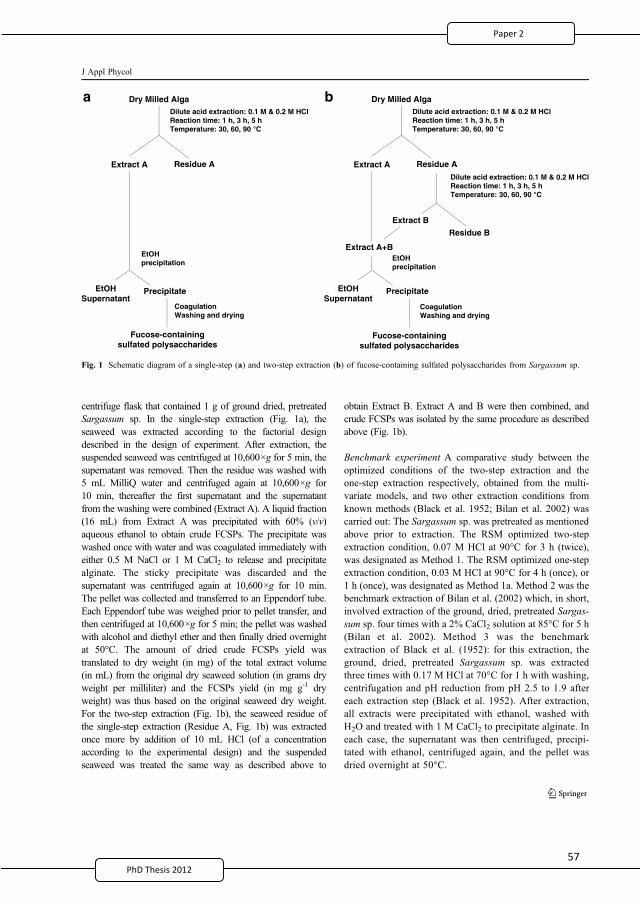
5PhD Thesis 2011
centrifuge flask that contained 1 g of ground dried, pretreatedSargassum sp. In the single-step extraction (Fig. 1a), theseaweed was extracted according to the factorial designdescribed in the design of experiment. After extraction, thesuspended seaweed was centrifuged at 10,600×g for 5 min, thesupernatant was removed. Then the residue was washed with5 mL MilliQ water and centrifuged again at 10,600×g for10 min, thereafter the first supernatant and the supernatantfrom the washing were combined (Extract A). A liquid fraction(16 mL) from Extract A was precipitated with 60% (v/v)aqueous ethanol to obtain crude FCSPs. The precipitate waswashed once with water and was coagulated immediately witheither 0.5 M NaCl or 1 M CaCl2 to release and precipitatealginate. The sticky precipitate was discarded and thesupernatant was centrifuged again at 10,600×g for 10 min.The pellet was collected and transferred to an Eppendorf tube.Each Eppendorf tube was weighed prior to pellet transfer, andthen centrifuged at 10,600×g for 5 min; the pellet was washedwith alcohol and diethyl ether and then finally dried overnightat 50°C. The amount of dried crude FCSPs yield wastranslated to dry weight (in mg) of the total extract volume(in mL) from the original dry seaweed solution (in grams dryweight per milliliter) and the FCSPs yield (in mg g-1 dryweight) was thus based on the original seaweed dry weight.For the two-step extraction (Fig. 1b), the seaweed residue ofthe single-step extraction (Residue A, Fig. 1b) was extractedonce more by addition of 10 mL HCl (of a concentrationaccording to the experimental design) and the suspendedseaweed was treated the same way as described above to
obtain Extract B. Extract A and B were then combined, andcrude FCSPs was isolated by the same procedure as describedabove (Fig. 1b).
Benchmark experiment A comparative study between theoptimized conditions of the two-step extraction and theone-step extraction respectively, obtained from the multi-variate models, and two other extraction conditions fromknown methods (Black et al. 1952; Bilan et al. 2002) wascarried out: The Sargassum sp. was pretreated as mentionedabove prior to extraction. The RSM optimized two-stepextraction condition, 0.07 M HCl at 90°C for 3 h (twice),was designated as Method 1. The RSM optimized one-stepextraction condition, 0.03 M HCl at 90°C for 4 h (once), or1 h (once), was designated as Method 1a. Method 2 was thebenchmark extraction of Bilan et al. (2002) which, in short,involved extraction of the ground, dried, pretreated Sargas-sum sp. four times with a 2% CaCl2 solution at 85°C for 5 h(Bilan et al. 2002). Method 3 was the benchmarkextraction of Black et al. (1952): for this extraction, theground, dried, pretreated Sargassum sp. was extractedthree times with 0.17 M HCl at 70°C for 1 h with washing,centrifugation and pH reduction from pH 2.5 to 1.9 aftereach extraction step (Black et al. 1952). After extraction,all extracts were precipitated with ethanol, washed withH2O and treated with 1 M CaCl2 to precipitate alginate. Ineach case, the supernatant was then centrifuged, precipi-tated with ethanol, centrifuged again, and the pellet wasdried overnight at 50°C.
Dry Milled AlgaDilute acid extraction: 0.1 M & 0.2 M HClReaction time: 1 h, 3 h, 5 hTemperature: 30, 60, 90 °C
Dilute acid extraction: 0.1 M & 0.2 M HClReaction time: 1 h, 3 h, 5 hTemperature: 30, 60, 90 °C
Extract A Residue A
Extract B
Residue B
Extract A+B
EtOHSupernatant
Precipitate
Dry Milled AlgaDilute acid extraction: 0.1 M & 0.2 M HClReaction time: 1 h, 3 h, 5 hTemperature: 30, 60, 90 °C
Extract A Residue A
EtOHprecipitation
EtOHSupernatant
Precipitate
CoagulationWashing and drying
Fucose-containingsulfated polysaccharides
ba
EtOHprecipitation
CoagulationWashing and drying
Fucose-containingsulfated polysaccharides
Fig. 1 Schematic diagram of a single-step (a) and two-step extraction (b) of fucose-containing sulfated polysaccharides from Sargassum sp.
J Appl Phycol
2
69
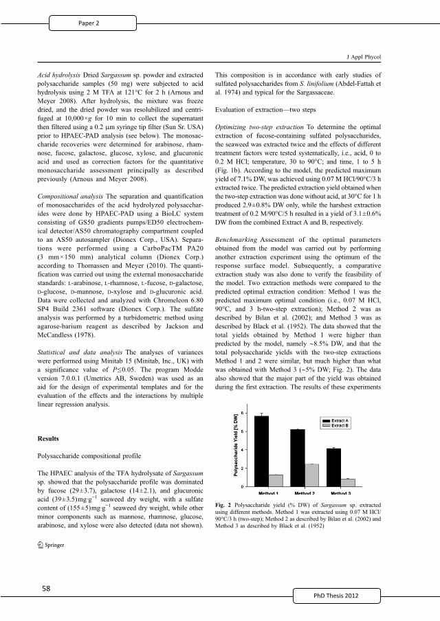
5PhD Thesis 2011
Acid hydrolysis Dried Sargassum sp. powder and extractedpolysaccharide samples (50 mg) were subjected to acidhydrolysis using 2 M TFA at 121°C for 2 h (Arnous andMeyer 2008). After hydrolysis, the mixture was freezedried, and the dried powder was resolubilized and centri-fuged at 10,000×g for 10 min to collect the supernatantthen filtered using a 0.2 μm syringe tip filter (Sun Sr. USA)prior to HPAEC-PAD analysis (see below). The monosac-charide recoveries were determined for arabinose, rham-nose, fucose, galactose, glucose, xylose, and glucuronicacid and used as correction factors for the quantitativemonosaccharide assessment principally as describedpreviously (Arnous and Meyer 2008).
Compositional analysis The separation and quantificationof monosaccharides of the acid hydrolyzed polysacchar-ides were done by HPAEC-PAD using a BioLC systemconsisting of GS50 gradients pumps/ED50 electrochem-ical detector/AS50 chromatography compartment coupledto an AS50 autosampler (Dionex Corp., USA). Separa-tions were performed using a CarboPacTM PA20(3 mm×150 mm) analytical column (Dionex Corp.)according to Thomassen and Meyer (2010). The quanti-fication was carried out using the external monosaccharidestandards: L-arabinose, L-rhamnose, L-fucose, D-galactose,D-glucose, D-mannose, D-xylose and D-glucuronic acid.Data were collected and analyzed with Chromeleon 6.80SP4 Build 2361 software (Dionex Corp.). The sulfateanalysis was performed by a turbidometric method usingagarose-barium reagent as described by Jackson andMcCandless (1978).
Statistical and data analysis The analyses of varianceswere performed using Minitab 15 (Minitab, Inc., UK) witha significance value of P≤0.05. The program Moddeversion 7.0.0.1 (Umetrics AB, Sweden) was used as anaid for the design of experimental templates and for theevaluation of the effects and the interactions by multiplelinear regression analysis.
Results
Polysaccharide compositional profile
The HPAEC analysis of the TFA hydrolysate of Sargassumsp. showed that the polysaccharide profile was dominatedby fucose (29±3.7), galactose (14±2.1), and glucuronicacid (39±3.5)mg·g−1 seaweed dry weight, with a sulfatecontent of (155±5)mg·g−1 seaweed dry weight, while otherminor components such as mannose, rhamnose, glucose,arabinose, and xylose were also detected (data not shown).
This composition is in accordance with early studies ofsulfated polysaccharides from S. linifolium (Abdel-Fattah etal. 1974) and typical for the Sargassaceae.
Evaluation of extraction—two steps
Optimizing two-step extraction To determine the optimalextraction of fucose-containing sulfated polysaccharides,the seaweed was extracted twice and the effects of differenttreatment factors were tested systematically, i.e., acid, 0 to0.2 M HCl; temperature, 30 to 90°C; and time, 1 to 5 h(Fig. 1b). According to the model, the predicted maximumyield of 7.1% DW, was achieved using 0.07 M HCl/90°C/3 hextracted twice. The predicted extraction yield obtained whenthe two-step extraction was done without acid, at 30°C for 1 hproduced 2.9±0.8% DW only, while the harshest extractiontreatment of 0.2 M/90°C/5 h resulted in a yield of 3.1±0.6%DW from the combined Extract A and B, respectively.
Benchmarking Assessment of the optimal parametersobtained from the model was carried out by performinganother extraction experiment using the optimum of theresponse surface model. Subsequently, a comparativeextraction study was also done to verify the feasibility ofthe model. Two extraction methods were compared to thepredicted optimal extraction condition: Method 1 was thepredicted maximum optimal condition (i.e., 0.07 M HCl,90°C, and 3 h-two-step extraction); Method 2 was asdescribed by Bilan et al. (2002); and Method 3 was asdescribed by Black et al. (1952). The data showed that thetotal yields obtained by Method 1 were higher thanpredicted by the model, namely ∼8.5% DW, and that thetotal polysaccharide yields with the two-step extractionsMethod 1 and 2 were similar, but much higher than whatwas obtained with Method 3 (∼5% DW; Fig. 2). The dataalso showed that the major part of the yield was obtainedduring the first extraction. The results of these experiments
Fig. 2 Polysaccharide yield (% DW) of Sargassum sp. extractedusing different methods. Method 1 was extracted using 0.07 M HCl/90°C/3 h (two-step); Method 2 as described by Bilan et al. (2002) andMethod 3 as described by Black et al. (1952)
J Appl Phycol
2
70
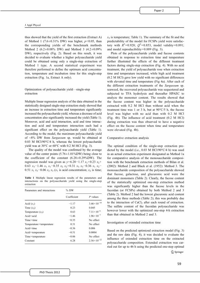
5
PhD Thesis 2011
thus showed that the yield of the first extraction (Extract A)of Method 1 (7.6±0.31% DW) was higher, p<0.05, thanthe corresponding yields of the benchmark methodsMethod 2 (6.2±0.08% DW) and Method 3 (4.2±0.09%DW), respectively (Fig. 2). Based on this result, it wasdecided to evaluate whether a higher polysaccharide yieldcould be obtained using only a single-step extraction ofMethod 1 type. A second statistical experiment wastherefore performed to define the optimum acid concentra-tion, temperature and incubation time for this single-stepextraction (Fig. 1a, Extract A only).
Optimization of polysaccharide yield—single-stepextraction
Multiple linear regression analysis of the data obtained in thestatistically designed single-step extraction study showed thatan increase in extraction time and temperature significantlyincreased the polysaccharide yield, whereas a decrease of acidconcentration also significantly increased the yield (Table 1).Moreover, acid and acid interaction, acid and time interac-tion and acid and temperature interaction each had asignificant effect on the polysaccharide yield (Table 1).According to the model, the maximum polysaccharide yieldof ∼8% DW from Sargassum sp. would be obtained at0.03 M HCl/90°C/4 h, whereas the lowest polysaccharideyield was at 30°C or 60°C with 0.2 M HCl (Fig. 3).
The quality of the model was confirmed by the averagevalue of the center points (5.76±1.01%DW) being close tothe coefficient of the constant (6.28±0.29%DW). Theregression model was given as y=6.28−1.17 x1+0.23 x2+0.63 x3−1.46 x1 x1−0.35 x2 x2+0.31 x3 x3−0.36 x1 x2−0.51 x1 x3−0.06 x2 x3 (x1 is acid concentration; x2 is time;
x3 is temperature; Table 1). The summary of the fit and thepredictability of the model for FCSPs yield were satisfac-tory with R2=0.928; Q2=0.853, model validity=0.891;and model reproducibility=0.889 (Fig. 3).
Plots of the polysaccharide yields and fucose contentsobtained in response to extraction time and temperaturefurther illustrated the effects of the different treatmentfactors during single-step extraction (Fig. 4). With no acidtreatment, the yield of polysaccharide rose when extractiontime and temperature increased, while high acid treatment(0.2 M HCl) gave low yield with no significant differenceswith elevated time and temperature (Fig 4a). After each ofthe different extraction treatments of the Sargassum sp.seaweed, the recovered polysaccharide was sequestered andsubjected to TFA hydrolysis and thereafter HPAEC toanalyze the monomer content. The results showed thatthe fucose content was higher in the polysaccharideextracted with 0.2 M HCl than without acid when thetreatment time was 1 or 3 h, but at 90°C/5 h, the fucoselevel was higher with no acid than with 0.2 M HCl(Fig. 4b). The influence of acid treatment (0.2 M HCl)during extraction was thus observed to have a negativeeffect on the fucose content when time and temperaturewere elevated (Fig. 4b).
Comparative extraction analysis
The optimal condition of the single-step extraction pre-dicted by the model (i.e., 0.03 M HCl/90°C/4 h) was usedin an actual extraction experiment designated as Method 1afor comparative analysis of the monosaccharide composi-tion with the benchmark extraction methods of Bilan et al.(2002): Method 2 and Black et al. (1952): Method 3. Themonosaccharide composition of the polysaccharide showedthat fucose, galactose, and glucuronic acid were thedominant monomers (Table 2). Clearly, the fucose contentof the statistically optimized one-step extraction methodwas significantly higher than the fucose levels in thefucoidan (or FCSPs) obtained by both Method 2 and 3(Table 2). Method 2 had the lowest glucuronic acid contentamong the three methods (Table 2); this was probably dueto the interaction of CaCl2 after each round of extraction.The sulfate content of the fucoidan polysaccharide washowever lower with the optimized one-step 4-h extractionthan that obtained in Method 2 and 3.
Investigation of extended extraction time
Based on the predicted optimized extraction model (Fig. 3)and the raw data (Fig. 4), it was decided to evaluate theinfluence of extended extraction time on the extractedpolysaccharide composition. Extended extraction was car-ried out for up to 46 h using the predicted one-step optimal
Table 1 Multiple linear regression results of the parameters andinteractions on the polysaccharide yield using the single-stepextraction
Parameters and interactions % DW
Coefficient P values
Acid (x1) −1.17 3.46×10⁻¹⁰
Time (x2) 0.23 0.045
Temperature (x3) 0.63 7.11×10⁻⁶
Acid×acid −1.46 1.96×10⁻⁷
Time×time −0.35 No effect
Temperature×temperature 0.31 No effect
Acid×time −0.36 0.006
Acid×temperature −0.51 0.0004
Time×temperature −0.06 No effect
Constant 6.28 2.56×10⁻²²
J Appl Phycol
2
71
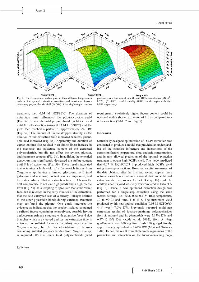
treatment, i.e., 0.03 M HCl/90°C. The duration ofextraction time influenced the polysaccharide yield(Fig. 5a). Hence, the total polysaccharide yield increaseduntil 8 h of extraction (using 0.03 M HCl/90°C) and theyield then reached a plateau of approximately 9% DW(Fig. 5a). The amount of fucose dropped steadily as theduration of the extraction time increased whereas glucur-onic acid increased (Fig. 5a). Apparently, the duration ofextraction time also resulted in an almost linear increase inthe mannose and galactose content of the extractedpolysaccharide, but did not affect the xylose, glucose,and rhamnose contents (Fig. 5b). In addition, the extendedextraction time significantly decreased the sulfate contentuntil 8 h of extraction (Fig. 5b). These results indicatedthat obtaining a high yield of a fucose-rich fucans fromSargassum sp. having a limited glucuronic acid (andgalactose and mannose) content was a compromise, andthe data confirmed that an extraction time of 3 h was thebest compromise to achieve high yields and a high fucoselevel (Fig. 5a). It is tempting to speculate that some “true”fucoidan is released in the early minutes of the extraction,that the acid catalyzed loss of α-fucosyl linkages relativeto the other glycosidic bonds during extended treatmentmay confound the picture. One could interpret theevidence as indicating that the product isolated containeda sulfated fucose-containing heteroglycan, possibly havinga glucuronan primary structure with extensive fucosyl sidebranches which are cleaved and lost as extraction time isextended. A sulfated fucan (a fucoidan) may occur inSargassum sp., but further elucidation of fucose-containing sulfated polysaccharides from Sargassum sp.is required. With a lower total polysaccharide yield
requirement, a relatively higher fucose content could beobtained with a shorter extraction of 1 h as compared to a4 h extraction (Table 2 and Fig. 5).
Discussion
Statistically designed optimization of FCSPs extraction wasconducted to produce a model that provided an understand-ing of the complex influences and interactions of theextraction factors temperature, time, and acid concentration,and in turn allowed prediction of the optimal extractiontreatment to obtain high FCSPs yield. The model predictedthat 0.07 M HCl/90°C/3 h produced high FCSPs yieldusing two-step extractions. However, careful assessment ofthe data obtained after the first and second steps at theseoptimal extraction conditions showed that an additionalextraction step to produce Extract B (Fig. 1b) could beomitted since its yield was very low compared to Extract A(Fig. 2). Hence, a new optimized extraction design wasperformed for a single-step extraction using the samefactors settings, i.e., acid, 0 to 0.2 M HCl; temperature,30 to 90°C; and time, 1 to 5 h. The maximum yieldproduced by this new optimal condition (0.03 M HCl/90°C/4 h) was ∼7.0% DW. Previously reported multi-stepextraction results of fucose-containing polysaccharidesfrom S. horneri and U. pinnatifida were 5.17% DW and6.77–15.10% DW (Kuda et al. 2002); from S. ring-goldianum it was 200 mg from fresh 150 g algal fronds,approximately equivalent to 0.67% DW (Mori and Nisizawa1982). Hence, the result of multiple linear regressions of theparameters and interaction on the fucose-containing poly-
Fig. 3 The 3D response surface plots at three different temperatureseach at the optimal extraction condition and maximum fucose-containing polysaccharide yield (% DW) of the single-step extraction
procedure as a function of time (h) and HCl concentration (M). R2=0.928; Q2=0.853; model validity=0.891; model reproducibility=0.889 respectively
J Appl Phycol
2
72
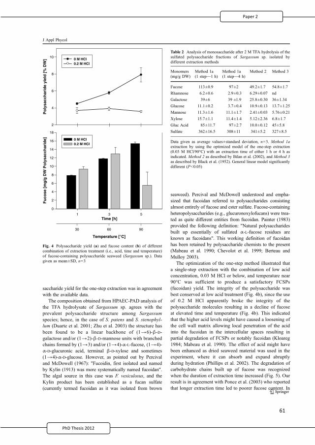
PhD Thesis 2011
saccharide yield for the one-step extraction was in agreementwith the available data.
The composition obtained from HPAEC-PAD analysis ofthe TFA hydrolysate of Sargassum sp. agrees with theprevalent polysaccharide structure among Sargassumspecies; hence, in the case of S. patens and S. stenophyl-lum (Duarte et al. 2001; Zhu et al. 2003) the structure hasbeen found to be a linear backbone of (1→6)-β-D-galactose and/or (1→2)-β-D-mannose units with branchedchains formed by (1→3) and/or (1→4)-α-L-fucose, (1→4)-α-D-glucuronic acid, terminal β-D-xylose and sometimes(1→4)-α-D-glucose. However, as pointed out by Percivaland McDowell (1967): "Fucoidin, first isolated and namedby Kylin (1913) was more systematically named fucoidan".The algal source in this case was F. vesiculosus, and theKylin product has been established as a fucan sulfate(currently termed fucoidan as it was isolated from brown
seaweed). Percival and McDowell understood and empha-sized that fucoidan referred to polysaccharides consistingalmost entirely of fucose and ester sulfate. Fucose-containingheteropolysaccharides (e.g., glucuronoxylofucans) were trea-ted as quite different entities from fucoidan. Painter (1983)provided the following definition: "Natural polysaccharidesbuilt up essentially of sulfated α-L-fucose residues areknown as fucoidans". This working definition of fucoidanhas been retained by polysaccharide chemists to the present(Mabeau et al. 1990; Chevolot et al. 1999; Berteau andMulloy 2003).
The optimization of the one-step method illustrated thata single-step extraction with the combination of low acidconcentration, 0.03 M HCl or below, and temperature near90°C was sufficient to produce a satisfactory FCSPs(fucoidan) yield. The integrity of the polysaccharide wasbest conserved at low acid treatment (Fig. 4b), since the useof 0.2 M HCl apparently broke the integrity of thepolysaccharide molecules resulting in a decline of fucoseat elevated time and temperature (Fig. 4b). This indicatedthat the higher acid levels might have caused a loosening ofthe cell wall matrix allowing local penetration of the acidinto the fucoidan in the intercellular spaces resulting inpartial degradation of FCSPs or notably fucoidan (Kloareg1984; Mabeau et al. 1990). The effect of acid might havebeen enhanced as dried seaweed material was used in theexperiment, where it can absorb and expand abruptlyduring hydration (Phillips et al. 2002). The degradation ofcarbohydrate chains built up of fucose was recognizedwhen the duration of extraction time increased (Fig. 5). Ourresult is in agreement with Ponce et al. (2003) who reportedthat longer extraction time led to poorer fucose content. In
Table 2 Analysis of monosaccharide after 2 M TFA hydrolysis of thesulfated polysaccharide fractions of Sargassum sp. isolated bydifferent extraction methods
Monomers(mg/g DW)
Method 1a(1 step—1 h)
Method 1a(1 step—4 h)
Method 2 Method 3
Fucose 113±0.9 97±2 49.2±1.7 54.8±1.7
Rhamnose 6.2±0.6 2.9±0.3 6.29±0.07 nd
Galactose 39±6 39 ±1.9 25.8±0.30 36±1.34
Glucose 11.1±0.2 3.7±0.4 10.9±0.13 13.7±1.25
Mannose 11.3±1.6 11.1±1.7 2.41±0.03 5.76±0.21
Xylose 15.7±1.1 11.4±1.4 5.12±2.36 6.8±1.7
Gluc Acid 85±11.7 97±2.7 10.0±0.12 45±5.8
Sulfate 362±16.5 308±11 341±5.2 327±8.5
Data given as average values±standard deviation, n=3. Method 1aextraction by using the optimized model of the one-step extraction(0.03 M HCl/90°C) with an extraction time of either 1 h or 4 h asindicated. Method 2 as described by Bilan et al. (2002), and Method 3as described by Black et al. (1952). General linear model significantlydifferent (P<0.05)
Temperature [°C]
Po
lysa
cch
arid
e yi
eld
[%
DW
]
2
4
6
8
10 0 M HCl0.2 M HCl
Time [h]
30 60 90
1 3 5
Fu
cose
[m
g/g
DW
Po
lysa
cch
arid
e]
0
2
4
6
8
10
12
14
16
18
0 M HCl0.2 M HCl
Fig. 4 Polysaccharide yield (a) and fucose content (b) of differentcombination of extraction treatment (i.e., acid, time and temperature)of fucose-containing polysaccharide seaweed (Sargassum sp.). Datagiven as mean±SD, n=3
J Appl Phycol
2
73
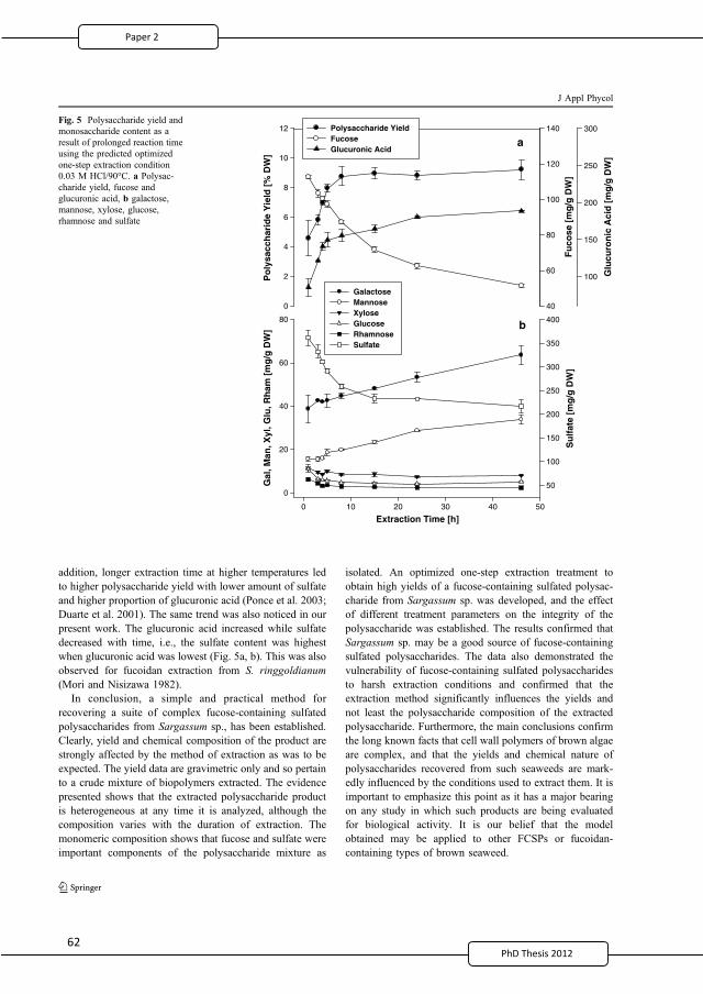
6PhD T201
addition, longer extraction time at higher temperatures ledto higher polysaccharide yield with lower amount of sulfateand higher proportion of glucuronic acid (Ponce et al. 2003;Duarte et al. 2001). The same trend was also noticed in ourpresent work. The glucuronic acid increased while sulfatedecreased with time, i.e., the sulfate content was highestwhen glucuronic acid was lowest (Fig. 5a, b). This was alsoobserved for fucoidan extraction from S. ringgoldianum(Mori and Nisizawa 1982).
In conclusion, a simple and practical method forrecovering a suite of complex fucose-containing sulfatedpolysaccharides from Sargassum sp., has been established.Clearly, yield and chemical composition of the product arestrongly affected by the method of extraction as was to beexpected. The yield data are gravimetric only and so pertainto a crude mixture of biopolymers extracted. The evidencepresented shows that the extracted polysaccharide productis heterogeneous at any time it is analyzed, although thecomposition varies with the duration of extraction. Themonomeric composition shows that fucose and sulfate wereimportant components of the polysaccharide mixture as
isolated. An optimized one-step extraction treatment toobtain high yields of a fucose-containing sulfated polysac-charide from Sargassum sp. was developed, and the effectof different treatment parameters on the integrity of thepolysaccharide was established. The results confirmed thatSargassum sp. may be a good source of fucose-containingsulfated polysaccharides. The data also demonstrated thevulnerability of fucose-containing sulfated polysaccharidesto harsh extraction conditions and confirmed that theextraction method significantly influences the yields andnot least the polysaccharide composition of the extractedpolysaccharide. Furthermore, the main conclusions confirmthe long known facts that cell wall polymers of brown algaeare complex, and that the yields and chemical nature ofpolysaccharides recovered from such seaweeds are mark-edly influenced by the conditions used to extract them. It isimportant to emphasize this point as it has a major bearingon any study in which such products are being evaluatedfor biological activity. It is our belief that the modelobtained may be applied to other FCSPs or fucoidan-containing types of brown seaweed.
Po
lysa
cch
arid
e Y
ield
[%
DW
]
0
2
4
6
8
10
12
Fu
cose
[m
g/g
DW
]
40
60
80
100
120
140
Glu
curo
nic
Aci
d [
mg
/g D
W]
100
150
200
250
300Polysaccharide YieldFucoseGlucuronic Acid
Extraction Time [h]0 10 20 30 40 50
Gal
, Man
, Xyl
, Glu
, Rh
am [
mg
/g D
W]
0
20
40
60
80
Su
lfat
e [m
g/g
DW
]
50
100
150
200
250
300
350
400
GalactoseMannoseXylose GlucoseRhamnoseSulfate
a
b
Fig. 5 Polysaccharide yield andmonosaccharide content as aresult of prolonged reaction timeusing the predicted optimizedone-step extraction condition0.03 M HCl/90°C. a Polysac-charide yield, fucose andglucuronic acid, b galactose,mannose, xylose, glucose,rhamnose and sulfate
J Appl Phycol
2
74
6PhD Thesis 2011
References
Abdel-Fattah AF, Hussein MD, Salem HM (1974) Studies ofpurification and some properties of sargassan, a sulfatedheteropolysaccharide from Sargassum linifolium. CarbohydrRes 33:9–17
Adhikari U, Mateii CG, Chattopadhyay K, Pujol CA, Damonte EB,Ray B (2006) Structure and antiviral activity of sulfatedfucans from Stoechospermum marginatum. Phytochemistry67:2474–2482
Arnous A, Meyer AS (2008) Comparison of methods for composi-tional characterization of grape (Vitis vinifera) and apple (Malusdomestica) skins. Food Bioprod Proc 86:79–86
Berteau O, Mulloy B (2003) Sulfated fucans, fresh perspectives:structures, functions, and biological properties of sulfated fucansand an overview of enzymes active toward this class ofpolysaccharide. Glycobiology 13:29R–40R
Bilan MI, Usov AI (2008) Structural analysis of fucoidans. NatProdCommun 3:1639–1648
Bilan MI, Grachev AA, Ustuzhanina NE, Shashkov AS, NifantievNE, Usov AI (2002) Structure of a fucoidan from the brownseaweed Fucus evanescens C.Ag. Carbohydr Res 337:719–730
Black WAP, Dewar ET, Woodward FN (1952) Manufacturing of algalchemicals 4: laboratory scale isolation of fucoidan from brownmarine algae. J Sci Food Agric 3:122–129
Blondin C, Fischer E, Boisson-Vidal C, Kazatchkine MD, JozefonviczJ (1994) Inhibition of complement activation by natural sulfatedpolysaccharides (fucans) from brown seaweed. Mol Immunol31:247–253
Chevolot L, Foucault A, Chaubet F, Kervarec N, Sinquin C, FisherAM, Boisson-Vidal C (1999) Further data on the structure ofbrown seaweed fucans: relationships with anticoagulant activity.Carbohydr Res 319:154–165
Chizhov AO, Dell A, Morris HR, Haslam SM, McDowell RA,Shashkov AS, Nivan’ev NE, Khatsuntseva ES, Usov AI (1999)A study of fucoidan from the brown seaweed Chorda filum.Carbohydr Res 320:108–119
Cumashi A, Ushakova NA, Preobrazhenskaya ME et al (2007) Acomparative study of the anti-inflammatory, anticoagulant, anti-angiogenic, and antiadhesive activities of nine different fucoidansfrom brown seaweeds. Glycobiology 17:541–552
Duarte MER, Cardoso MA, Noseda MD, Cerezo AS (2001) Structuralstudies on fucoidans from the brown seaweed Sargassumstenophyllum. Carbohydr Res 333:281–293
Jackson SG, McCandless EL (1978) Simple, rapid, turbidometricdetermination of inorganic sulfate and/or protein. Anal Biochem90:802–808
Kloareg B (1984) Isolation and analysis of cell walls of the brownmarine algae Pelvetia canaliculata and Ascophyllum nodosum.Physiol Vegetale 22:47–56
Kuda T, Taniguchi E, Nishizawa M, Araki Y (2002) Fate of water-soluble polysaccharides in dried Chorda filum a brown algaduring water washing. J Food Comp Anal 15:3–9
Kylin K (1913) Zur Biochemie der Meeresalgen. HoppeSeyler’s ZPhysiol Chem 83:171–197
Li B, Lu F, Wei X, Zhao R (2008) Fucoidan: structure and bioactivity.Molecules 13:1671–1695
Mabeau S, Kloareg B, Joseleau JP (1990) Fractionation andanalysis of fucans from brown algae. Phytochemistry29:2441–2445
Marais MF, Joseleau JP (2001) A fucoidan fraction from Ascophyllumnodosum. Carbohydr Res 336:155–159
Mori H, Nisizawa K (1982) Sugar constituents of sulfated poly-saccharides from the fronds of Sargassum ringgoldianum.Nippon Suisan Gakkaishi 48:981–986
Nardella A, Chaubet F, Boisson-Vidal C, Blondin C, Durand P,Jozefonvicz J (1996) Anticoagulant low molecular weightfucans produced by radical process and ion exchangechromatography of high molecular weight fucans extractedfrom the brown seaweed Ascophyllum nodosum. CarbohydrRes 289:201–208
Painter (1983) Algal polysaccharides. In: Aspinall GO (ed) Thepolysaccharides (vol. 2). Academic, New York, pp 195–285
Percival, McDowell (1967) Chemistry and enzymology of marinealgal polysaccharides. Chapter 7. Academic, London
Phillips JR, Oliver MJ, Bartels D, Black M (2002) Molecular geneticsof desiccation and tolerant systems. In: Black MJ, Pritchard HW(eds) Desiccation and survival in plants: drying without dying.CABI, UK, pp 319–341
Ponce NMA, Pujol CA, Damonte EB, Flores ML, Stortz CA (2003)Fucoidans from the brown seaweed Adenocystis utricularis:extraction methods, antiviral activity and structural studies.Carbohydr Res 338:153–165
Sakai T, Kimura H, Kato I (2002) A marine strain of Flavobacter-iaceae utilizes brown seaweed fucoidan. Mar Biotechnol4:399–405
Thomassen LV, Meyer AS (2010) Statistically designed optimizationof enzyme catalyzed starch removal from potato pulp. EnzymeMicrob Technol 46:297–303
Trinchero J, Ponce NMA, Cordoba OL, Flores ML, Pampuro S, StortzCA, Salomon H, Turk G (2009) Antiretroviral activity offucoidans extracted from the brown seaweed Adenocystisutricularis. Phytother Res 23:707–712
Wang J, Zhang QB, Zhang ZS, Li Z (2008) Antioxidant activity ofsulfated polysaccharide fractions extracted from Laminariajaponica. Int J Biol Macromol 42:127–132
Zhu W, Ooi VEC, Chan PKS, Ang PO (2003) Isolation andcharacterization of a sulfated polysaccharide from the brownalga Sargassum patens and determination of its anti-herpesactivity. Biochem Cell Biol 81:25–33
Zhuang C, Itoh H, Mizuno T, Ito H (1995) Antitumor active fucoidanfrom brown seaweed, Umitoranoo (Sargassum thunbergii).Biosci Biotech Biochem 59:563–567
Zvyagintseva TN, Shevchenko NM, Popivnich IB, Isakov VV, ScobunEV, Elyakova LA (1999) A new procedure for the separation ofwater-soluble polysaccharides from brown seaweeds. CarbohydrRes 322:32–39
J Appl Phycol
2
75

76 76
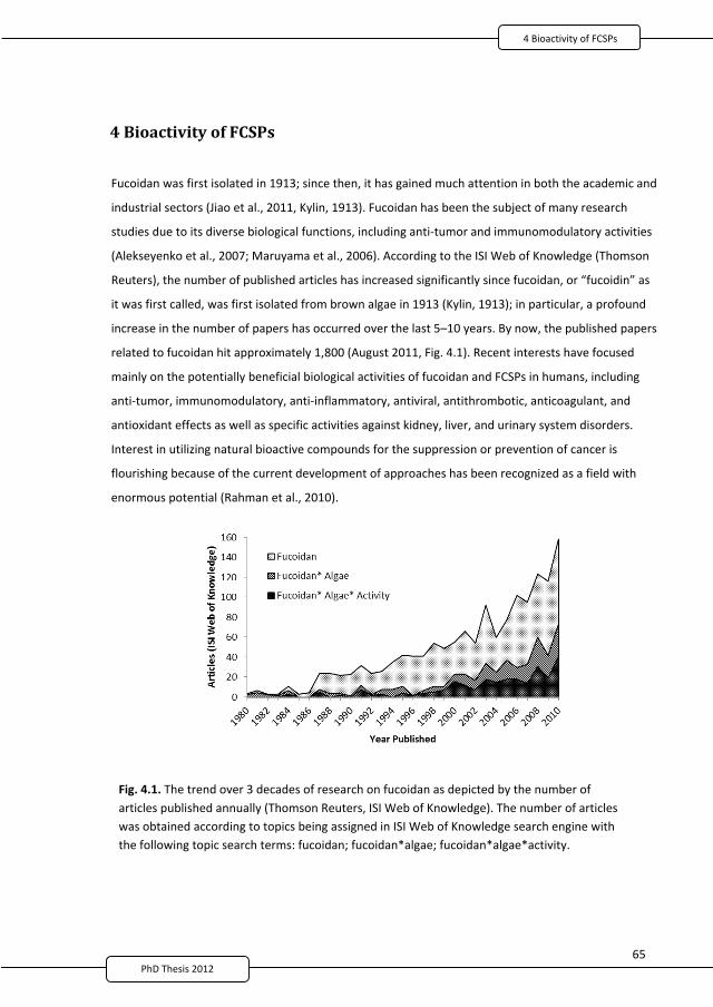
6PhD Thesis 2011
4 Bioactivity of FCSPs
Fucoidan was first isolated in 1913; since then, it has gained much attention in both the academic and
industrial sectors (Jiao et al., 2011, Kylin, 1913). Fucoidan has been the subject of many research
studies due to its diverse biological functions, including anti tumor and immunomodulatory activities
(Alekseyenko et al., 2007; Maruyama et al., 2006). According to the ISI Web of Knowledge (Thomson
Reuters), the number of published articles has increased significantly since fucoidan, or “fucoidin” as
it was first called, was first isolated from brown algae in 1913 (Kylin, 1913); in particular, a profound
increase in the number of papers has occurred over the last 5–10 years. By now, the published papers
related to fucoidan hit approximately 1,800 (August 2011, Fig. 4.1). Recent interests have focused
mainly on the potentially beneficial biological activities of fucoidan and FCSPs in humans, including
anti tumor, immunomodulatory, anti inflammatory, antiviral, antithrombotic, anticoagulant, and
antioxidant effects as well as specific activities against kidney, liver, and urinary system disorders.
Interest in utilizing natural bioactive compounds for the suppression or prevention of cancer is
flourishing because of the current development of approaches has been recognized as a field with
enormous potential (Rahman et al., 2010).
Fig. 4.1. The trend over 3 decades of research on fucoidan as depicted by the number ofarticles published annually (Thomson Reuters, ISI Web of Knowledge). The number of articleswas obtained according to topics being assigned in ISI Web of Knowledge search engine withthe following topic search terms: fucoidan; fucoidan*algae; fucoidan*algae*activity.
2
77
6PhD Thesis 2011
While the development of research efforts involving FCSPs and their potential applications continue
to advance, understanding of the mechanisms and the particular structural features of the FCSPs
being responsible for the various biological activities remains incomplete. Seaweeds, including various
brown seaweeds such as Undaria and Laminaria spp., are part of the food culture in Asia, notably in
Japan, the Philippines, and Korea, and seaweed extracts have also been used as remedies in
traditional medicine. However, there currently exist no standardized FCSPs extraction or purification
protocols, and no specific pharmaceutical, dermatological, or nutraceutical applications have as yet
been officially approved for these polysaccharides or their lower molar mass oligosaccharide
derivatives. FCSPs that were isolated in multi step processes and then purified and fractionated
demonstrated essential bioactivities (Holtkamp et al., 2009), while unpurified fucoidan that is isolated
using milder and fewer processing step has been found to induce anti tumor activity and act as an
immunopotentiator in tumor bearing animals (Takahashi, 1983).
4.1 Anti tumor activity of FCSPsFCSPs from different brown seaweed species have shown remarkable growth inhibition of
Sarcoma 180 cells implanted into mice and possess anti tumor activity against L 1210 leukemia in
mice (Yamamoto et al., 1974, 1981, 1984). The anti tumor mechanism of fucoidan from sporophyll
of Undaria pinnatifida was described by Maruyama et al. (2003), who indicated that the anti
tumor activity of fucoidan appears to be associated with significant enhancement of the cytolytic
activity of NK cells. The effectiveness of fucoidan as an immunopotentiator was exhibited by an
increased immune response against A20 leukemia cells and a significantly lowered tumor size in
transgenic (DO 11 10 Tg) mice (Maruyama et al., 2006). The enhancement of NK cell activity by
fucoidan was augmented through increase production of macrophage mediated immune
responses, namely IL 2, IFN , and IL 12 (Maruyama et al., 2003). Moreover, the most prevalent
pathway through which fucoidan can inhibit cancer growth is apoptosis. Fucoidan induces
apoptosis via the activation of caspase 3 in human HS Sultan cells (Aisa et al., 2005); in MCF 7 cells
via caspase 8–dependent pathways (Yamasaki Miyamoto et al., 2009); and through the activation
of caspases via both the death receptor mediated and the mitochondria mediated apoptotic
pathways (Kim et al., 2010).
4.2 Anti proliferative and immune response activitiesThis section is an extended elucidation of Paper 3: fucoidan from Sargassum sp. and F. vesiculosus
reduces cell viability of lung carcinoma and melanoma cells in vitro and activates NK cells in mice in
2
78
6PhD Thesis 2011
vivo. In addition, highlights of Paper 4: FCSPs inhibit the proliferation of melanoma cells and induce
apoptosis by activating caspase 3 in vitro will also be presented in this section.
4.2.1 RelevanceLung cancer is among the most prevalent types of cancer worldwide and is a prime contributor to
cancer related mortality. Melanoma incidence rates for both males and females are also increasing
in the United States (Jemal et al., 2010). To date, therapeutic strategies such as chemotherapy,
radiation therapy, surgery, or combinations thereof have been implemented for many cancer
patients; however, they still provide only minimal survival benefits due to factors such as toxicity,
complications, and long term side effects (Schneider et al., 2010; Grossi et al., 2010). As a
consequence, the need for chemopreventive agents from natural sources with minimal or no
harmful side effects is of ardent importance. Hence, FCSPs, notably fucoidan, from brown seaweed
may prove to be excellent contenders for the prevention or control of lung and skin
carcinogenesis.
4.2.2 Hypotheses and objectivesIsolating fucoidan from brown seaweed using minimal processing will preserve its structural
integrity and, thereby, help maintain its bioactive characteristics but results in crude fucoidan
products. The chemical nature of polysaccharides recovered from seaweed is influenced by the
technology used to extract them. Crude fucoidan obtained via single step extraction has been
subjected to fewer and milder process conditions. Therefore, the well defined structural features
of fucoidans are likely be conserved and, thus, retain their biological activity.
It has been reported that crude fucoidan fractions from edible brown seaweeds affect L 1210
leukemia cell development in vivo, while sulfation of crude fucoidan fractions from Sargassum
kjellmanianum enhances their anti tumor activity (Yamamoto et al., 1983). Moreover, in vivo
studies confirmed that the feeding of ground brown seaweed to animals, the oral administration
of hot water extraction of seaweed, and the intraperitoneal injection of crude fucoidan fractions
resulted in an inhibitory effect on mammary tumorigenesis and intestinal carcinogenesis
(Yamamoto et al., 1987; Yamamoto and Maruyama, 1985). Hence, other forms of cancer such as
lung and skin cancer may alternatively be prevented or controlled through the use of crude
fucoidan from brown seaweed.
Therefore, the objectives of the present works were to determine the potency of unfractionated
FCSPs to inhibit the growth of skin and lung cancer cells in vitro, evaluate the immune response
2
79
6PhD Thesis 2011
activity of FCSPs in mice in vivo, and elucidate the contributing factors behind this effect (Paper 3).
Furthermore, we investigated the different structural features of unfractionated FCSPs from
Sargassum sp. and F. vesiculosus using Fourier transform infrared (FT IR) and proton nuclear
magnetic resonance (1H NMR) spectroscopy to determine whether its contribution is crucial to its
bioactive effectiveness. We also conducted an in vitro study to examine the influence of FCSP
products from Sargassum sp. and F. vesiculosus on melanoma B16 cell (MC) proliferation and
caspase 3 activity mediating the apoptosis of melanoma B16 cells (Paper 4).
4.2.3 Result highlightsThe influence of 2 crude fucoidans extracted from Sargassum sp. (MTA) using a minimal number of
processing steps and obtained commercially from F. vesiculosus (SIG) on Lewis lung carcinoma cells
(LCC) and MC was examined. The compositions of the SIG and MTA fucoidans were significantly
different with respect to fucose, galactose, and glucuronic acid, unlike the sulfate content (Paper
3). The FT IR spectra indicated that the sulfate in the FCSPs from Sargassum sp. (FSAR) was located
in the equatorial C 2 and/or C 3 positions as depicted by the absorption bands at 817 cm 1,
whereas the IR spectra of FCSPs from F. vesiculosus (FVES) displayed an absorption band at 838
cm 1 with a small shoulder absorption band at 822 cm 1, indicating sulfate groups at the C 4 and C
2 positions (Paper 4). This finding corresponds to the 1H NMR spectra of the unfractionated FVES
sample from F. vesiculosus, indicating a typical structure of algal fucoidan consisting of 3 linked 2
mono O sulfated L fucopyranose residues and/or 3 linked 2,4 di O sulfated L fucopyranose
residues (Pereira et al., 1999; Patankar et al., 1993).
In vitro studies showed the anti proliferative effect of crude fucoidan on LCC and MC cells in a
dose dependent manner. Male C57BL/6JJCL mice were subjected to daily intraperitoneal injections
over 4 days with either SIG or MTA fucoidan (50 mg/kg body wt) to evaluate immune response
augmentation. The cytolytic activity of NK cells was enhanced by crude fucoidan as indicated by51Cr labeled YAC 1 target cell release. Histochemical staining showed morphologic changes of MC
cells after exposure to crude fucoidan. Fragmentation and condensation of chromatin, illustrated
as an intense dark brown color within the cell nuclei, was indicative of crude fucoidan induced
apoptosis (Paper 3). In this work (Paper 4), we noted based on flow cytometric analysis that FSCP
samples from Sargassum sp. and F. vesiculosus induced apoptosis through activation of caspase 3
in a dose dependent manner (Paper 4).
The mechanism behind FCSP anti tumor activity and how it enhances the immune response has
yet to be determined. Nevertheless, this study provides substantial indications that FCSP exerts
2
80
6PhD Thesis 2011
bioactive characteristics on lung and skin cancer model cells and that its anti tumor activity was
due to the enhancement of NK cell activity (Paper 3). The crucial bioactive effectiveness of these
unfractionated FCSPs from Sargassum sp. and F. vesiculosus may be attributed to their distinct
structural features, such as level of sulfation (charge density) and position and bonding of the
sulfate substitutions or sulfated fucans and sulfated galactan complexes. This present study
demonstrated the early and later apoptosis stages by FACScan, which could indicate that FCSPs
have a direct apoptotic effect on MC cells (Paper 4) and that the presence of 3 linked galactan in
the structure of FCSPs from Sargassum sp. may somehow contribute to its anti proliferative
effects.
4.2.4 Consideration and justificationThe monosaccharide profiles of the acid hydrolyzed polysaccharides from Sargassum sp. and F.
vesiculosus suggest that both products are contaminated with components other than fucose and
sulfate (Paper 3). The commercially obtained sample from F. vesiculosus was a crude fucoidan, as
indicated in the product specification from the supplier (Sigma Aldrich Inc., Germany).
Alternatively, the product extracted from Sargassum sp. contained a low amount of fucose
compare to that of F. vesiculosus, but they have the same chemical composition profile. The low
amount of recovered fucose was probably attributed by the condition used to extract them or the
extent of the acid hydrolysis, which perhaps has a major influence in the disappearance of fucose.
In any case, the isolated product from Sargassum sp. showed the same monosaccharide profile as
the commercially obtained sample from F. vesiculosus (Sigma Aldrich Inc.); hence, it is safe to say
that the isolated FCSP product from Sargassum sp. was also a crude fucoidan.
Moreover, based on previous published papers, the term crude fucoidan was applied for FCSPs
from brown seaweeds in which impurities are present in the isolated products (Yamamoto et al.,
1983; Takahashi, 1983). Nevertheless, crude fucoidan from these samples have shown potent anti
tumor activity and cellular immunity associated with T cells (Takahashi, 1983). However, it should
be noted that evidence establishing that fucose comprised the backbone of a significant
component of the preparation was necessary to reasonably apply the term fucoidan. In the
absence of this evidence, it is preferable to term the isolated product from Sargassum sp. as crude
fucoidan or, to be more accurate, FCSP (Papers 3 and 4). The main active component may be a
FCSP (e.g., fucoidan) that is present in brown seaweed such as Sargassum sp., but further
structural elucidation is essential, as it has major bearing on any biological activity study.
Nevertheless, there are many indications in this present work that FCSP samples may contain
2
81
PhD Thesis 2011
fucoidan like structures (Paper 4). This study showed that both FCSP samples induced apoptosis by
activating caspases 3 and exerted anti tumor activity by inhibiting the growth of MC cells. It
appears that unfractionated FCSPs from Sargassum sp. and F. vesiculosus are potent skin cancer
preventive agents, a fact that was demonstrated by a direct in vitro study of melanoma B16 cells
(Paper 4).
4.3 Paper 3: Fucoidan from Sargassum sp. and Fucus vesiculosus reduces cellviability of lung carcinoma and melanoma cells in vitro and activates naturalkiller cells in mice in vivo
International Journal of Biological Macromolecules, 2011, in Press
Authors:Marcel Tutor Ale1, Hiroko Maruyama2, Hidekazu Tamauchi3, Jørn Dalgaard Mikkelsen1, Anne S.Meyer1
4.4 Paper 4: Fucose containing sulfated polysaccharides inhibits theproliferation of melanoma cells and induces apoptosis by activation ofcaspase 3 in vitro
Marine Drugs, 2011, Submitted
Authors:Marcel Tutor Ale1, Hiroko Maruyama2, Hidekazu Tamauchi3, Jørn Dalgaard Mikkelsen1, Anne S.Meyer1
Address:1Center for Bioprocess Engineering, Department of Chemical and Biochemical EngineeringTechnical University of Denmark (DTU)Soeltofts Plads Bldg. 229, 2800 Kgs. Lyngby, Denmark
2Department of Pathology, School of Allied Science and 3Department of Microbiology, School ofMedicineKitasato University, Kitasato 1 15 1Sagamihara, Kanagawa 228 8555 Japan
2
82
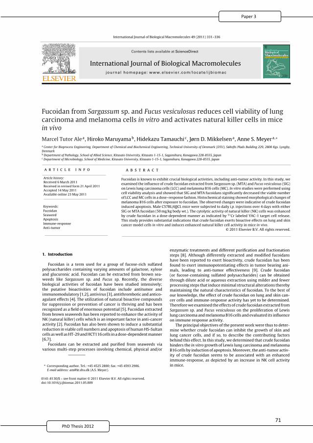
International Journal of Biological Macromolecules 49 (2011) 331– 336
Contents lists available at ScienceDirect
International Journal of Biological Macromoleculesjo u r n al hom epa ge: ww w.elsev ier .com/ locate / i jb iomac
Fucoidan from Sargassum sp. and Fucus vesiculosus reduces cell viability of lung
carcinoma and melanoma cells in vitro and activates natural killer cells in mice
in vivo
Marcel Tutor Alea, Hiroko Maruyamab, Hidekazu Tamauchic, Jørn D. Mikkelsena, Anne S. Meyera,∗
a Center for Bioprocess Engineering, Department of Chemical and Biochemical Engineering, Technical University of Denmark (DTU), Søltofts Plads Building 229, 2800 Kgs. Lyngby,
Denmarkb Department of Pathology, School of Allied Science, Kitasato University, Kitasato 1-15-1, Sagamihara, Kanagawa 228-8555, Japanc Department of Microbiology, School of Medicine, Kitasato University, Kitasato 1-15-1, Sagamihara, Kanagawa 228-8555, Japan
a r t i c l e i n f o
Article history:
Received 6 March 2011
Received in revised form 21 April 2011
Accepted 14 May 2011
Available online 23 May 2011
Keywords:
Fucoidan
Seaweed
Apoptosis
Immune-response
Anti-tumor
a b s t r a c t
Fucoidan is known to exhibit crucial biological activities, including anti-tumor activity. In this study, we
examined the influence of crude fucoidan extracted from Sargassum sp. (MTA) and Fucus vesiculosus (SIG)
on Lewis lung carcinoma cells (LCC) and melanoma B16 cells (MC). In vitro studies were performed using
cell viability analysis and showed that SIG and MTA fucoidans significantly decreased the viable number
of LCC and MC cells in a dose–response fashion. Histochemical staining showed morphological changes of
melanoma B16 cells after exposure to fucoidan. The observed changes were indicative of crude fucoidan
induced apoptosis. Male C57BL/6JJCL mice were subjected to daily i.p. injections over 4 days with either
SIG or MTA fucoidan (50 mg/kg body wt.). The cytolytic activity of natural killer (NK) cells was enhanced
by crude fucoidan in a dose-dependent manner as indicated by 51Cr labeled YAC-1 target cell release.
This study provides substantial indications that crude fucoidan exerts bioactive effects on lung and skin
cancer model cells in vitro and induces enhanced natural killer cell activity in mice in vivo.
© 2011 Elsevier B.V. All rights reserved.
1. Introduction
Fucoidan is a term used for a group of fucose-rich sulfated
polysaccharides containing varying amounts of galactose, xylose
and glucuronic acid. Fucoidan can be extracted from brown sea-
weeds like Sargassum sp. and Fucus sp. Recently, the diverse
biological activities of fucoidan have been studied intensively;
the putative bioactivities of fucoidan include antitumor and
immunomodulatory [1,2], antivirus [3], antithrombotic and antico-
agulant effects [4]. The utilization of natural bioactive compounds
for suppression or prevention of cancer is thriving and has been
recognized as a field of enormous potential [5]. Fucoidan extracted
from brown seaweeds has been reported to enhance the activity of
NK (natural killer) cells which is an important factor in anti-cancer
activity [2]. Fucoidan has also been shown to induce a substantial
reduction in viable cell numbers and apoptosis of human HS-Sultan
cells as well as HT-29 and HCT116 cells in a dose-dependent manner
[6,7].
Fucoidans can be extracted and purified from seaweeds via
various multi-step processes involving chemical, physical and/or
∗ Corresponding author. Tel.: +45 4525 2800; fax: +45 4593 2906.
E-mail address: [email protected] (A.S. Meyer).
enzymatic treatments and different purification and fractionation
steps [8]. Although differently extracted and modified fucoidans
have been reported to exert bioactivity, crude fucoidan has been
found to exert immunopotentiating effects in tumor bearing ani-
mals, leading to anti-tumor effectiveness [9]. Crude fucoidan
(or fucose-containing sulfated polysaccharides) can be obtained
through dilute acid or aqueous extraction using milder and fewer
processing steps that induce minimal structural alterations thereby
maintaining the natural characteristics of fucoidan. To the best of
our knowledge, the effect of crude fucoidan on lung and skin can-
cer cells and immune-response activity has yet to be determined.
Therefore we examined the effects of crude fucoidan extracted from
Sargassum sp. and Fucus vesiculosus on the proliferation of Lewis
lung carcinoma and melanoma B16 cells and evaluated its influence
on immune response activity.
The principal objectives of the present work were thus to deter-
mine whether crude fucoidan can inhibit the growth of skin and
lung cancer cells, and if so, to describe the contributing factors
behind this effect. In this study, we determined that crude fucoidan
hinders the in vitro growth of Lewis lung carcinoma and melanoma
B16 cells by induction of apoptosis. Moreover, the anti-tumor activ-
ity of crude fucoidan seems to be associated with an enhanced
immune-response, as depicted by an increase in NK cell activity
in mice.
0141-8130/$ – see front matter © 2011 Elsevier B.V. All rights reserved.doi:10.1016/j.ijbiomac.2011.05.009
PhD Thesis 2011 2
83
7PhD Thesis 2011
332 M.T. Ale et al. / International Journal of Biological Macromolecules 49 (2011) 331– 336
2. Materials and methods
2.1. Chemicals
Two crude fucoidan samples were used throughout this work.
Firstly, crude fucoidan from Sargassum sp. (MTA) was extracted
in our laboratory (see Section 2.2) while crude fucoidan from
F. vesiculosus (SIG) was obtained commercially (Sigma–Aldrich
Co., Steinheim, Germany). Dried Sargassum sp. was obtained
from Viet Delta Ltd. (Ho Chi Minh, Vietnam). Minimal essential
medium eagle (MEM-eagle) cell culture media was purchased
from Sigma–Aldrich Co. (Steinheim, Germany); foetal bovine
serum (FBS) was from Flow Laboratories (North Ryde, N.S.W.,
Australia); streptomycin–penicillin and Trypan Blue were from
Gibco (Canada). Hydrochloric acid (37%), d-glucose and d-xylose
were purchased from Merck (Darmstadt, Germany). Trifluo-
racetic acid (99%, TFA), trichloroacetic acid (99%, TCA), CaCl2,
Na2SO4, BaCl2, arabinose, rhamnose, d-galactose and l-fucose were
from Sigma–Aldrich Co. (Steinheim, Germany). Agarose D-2 was
obtained from Hispanagar (Burgos, Spain). All chemicals used were
analytical grade.
2.2. Extraction of crude fucoidan (MTA)
Seaweed from Sargassum sp. was ground and sieved to pass
through a 500 �m sieve. Crude fucoidan extraction was carried out
by adding 100 g of dried ground seaweed to a 5 L flask containing
2 L of 0.03 M HCl. The mixture was then placed in a 90 ◦C water bath
(Julabo, Germany) with continuous mixing at 200 rpm for 4 h. The
suspended seaweed was filtered (Whatman filter paper, no. 1) to
obtain a fucoidan extract. The extract was then precipitated using
60% ethanol (EtOH), and the precipitate was collected after cen-
trifugation (Sigma Laboratory Centrifuge 4K15, VWR, Denmark) at
10,600 rpm for 10 min, and the resulting pellet was freeze dried.
This pellet was the crude fucoidan and is referred to as MTA in
this study. The crude fucoidan (SIG) derived from F. vesiculosus
was commercially obtained from Sigma–Aldrich Co. (Steinheim,
Germany). According to the product description the F. vesiculosus
fucoidan had been prepared via the extraction method described
by Black [10].
2.3. Acid hydrolysis
The freeze dried crude fucoidan powder (MTA, 20 mg) and
the commercially obtained crude fucoidan (SIG, 20 mg) were
hydrolyzed separately using 2 M TFA (final concentration) at 121 ◦C
for 2 h [11]. The hydrolyzed mixtures were then freeze dried at
−57 ◦C (Heto Lyolab 3000, England). The dried powders were res-
olubilized in doubly distilled water and centrifuged at 10,000 rpm
for 10 min to collect the supernatants. Each supernatant was fil-
tered through a 0.2 �m syringe tip filter (SUN-SRi, Rockwood, TN)
prior to injection into the HPAEC-PAD for monosaccharide analysis
(see Section 2.4).
2.4. Fucoidan composition analysis
The supernatant of each of the acid hydrolyzed crude fucoidan
samples was analyzed for monosaccharides and sulfate con-
tents. The separation and quantification of monosaccharides were
performed by HPAEC-PAD analysis using an ICS-3000 system
consisting of gradient pumps (model DP-1), an electrochemical
detector/chromatography module (model DC-1) and an autosam-
pler (Dionex Corp., Sunnyvale, CA). Separation was achieved using
a CarboPacTM PA20 (3 mm × 150 mm) analytical column following
the method described by Arnous and Meyer [11]. Data quan-
tification was carried out using the Chromeleon 6.8 SP4 Build
2361 chromatography software (Dionex Corp., Sunnyvale, CA).
Recovery values of the monosaccharides were estimated from
parallel runs, i.e., TFA hydrolysis, freeze drying, and HPAEC-PAD
analysis, of monosaccharide standards as described previously [11].
Analysis of sulfate content was done according to the method
described by Jackson and McCandless [12].
2.5. Cell culture and anti-tumor activity assay
Lewis Lung Carcinoma (LLC) and melanoma B16 cells (MC) were
grown in MEM eagle medium supplemented with 10% (v/v) heat-
inactivated FBS, 1% streptomycin–penicillin and 1% of 200 mM
l-glutamine. The cells were maintained at 37 ◦C under 5% CO2. An
aliquot (10 �l) of the cell-MEM-FBS medium mixture was diluted
with 90 �l of 0.4% Trypan Blue for cell counting. Monolayer cul-
tivation was carried out by adding 100 �l of the cell-MEM-FBS
mixture in 96-flat well plates at a density of 3 × 10⁴ cells per
well. Plates were then incubated for 24 h in 5% CO2 at 37 ◦C.
Afterwards the medium was removed and replaced with 100 �l
of MEM medium containing 2% FBS and varying concentrations
(0.1–1.0 mg/ml) of Sargassum sp. crude fucoidan (MTA) and com-
mercial crude fucoidan from F. vesiculosus (SIG) and then incubated
for 24 h. A 20 �l MTT (5 mg/ml) solution was added to the cultures
and they were then re-incubated for 4 h. Finally, 100 �l of stabiliza-
tion solution was added to each well and the plates were incubated
overnight at 37 ◦C under 5% CO2. Absorbance was measured using
an Elisa reader at 550–690 nm.
2.6. Natural killer (NK) cell activity based on 51Chromium (51Cr)
release assay
Male C57BL/6J mice (Clea Japan Inc., Tokyo, Japan) were
weighed prior to intraperitoneal injection (i.p.) of fucoidan sam-
ples (50 mg/kg body wt.) from Sargassum sp. (MTA) and commercial
fucoidan from F. vesiculosus (SIG) in 0.1 ml saline for 4 successive
days; neat saline was used as control, and Poly I:C as positive con-
trol. 3 mice were used per treatment (n = 3). The mouse spleen was
removed and suspended in complete RPMI-1640 medium supple-
mented with 100 U/ml of penicillin, 100 �g/ml of streptomycin,
2 mM of l-glutamine and 10% FBS. Spleen cells were seeded in
0.1 ml of this complete medium per well in 96-well microtiter
plates (V-typed, Nalge Nunc, Tokyo, Japan) at densities of 1 × 10⁷,
0.5 × 10⁶, and 0.25 × 10⁶ cells/ml. 51Cr (with sodium chromate,
37 MBq/ml, Dupont, NEN Research Products, DE, USA) was used
to label YAC-1 cells using an exposure time of 1 h after which
the cells were washed, brought to a density of 1 × 10⁴/ml and
added in 0.1 ml aliquots to the wells. The plate was centrifuged
for 3 min at 800 rpm and room temperature, and then incubated
for 4 h in 5% CO2 at 37 ◦C. Percent specific lysis was calculated
using the formula: % specific 51Cr release = [(mean cpm experi-
mental release − mean cpm spontaneous release)/(mean cpm total
releasable counts − mean cpm spontaneous release)] × 100, based
on results from radioactivity measurements of the supernatant
using a �-counter (Gamma 5500B, Beckman Instruments Inc., CA,
USA). The experiments adhered to the National Institute of Health’s
guidelines for the use of experimental animals and the experi-
mental protocol was approved by The Animal Use Committee of
the Kitasato University School of Allied Health Sciences before the
study was initiated.
2.7. Apoptosis detection assay
Both the MTA and SIG crude fucoidan samples (0.8 mg/ml
dose concentration) and a control (no fucoidan) were tested
on melanoma B16 cells for programmed cell death by the
terminal deoxynucleotidyl transferase-mediated deoxyuridine
2
84
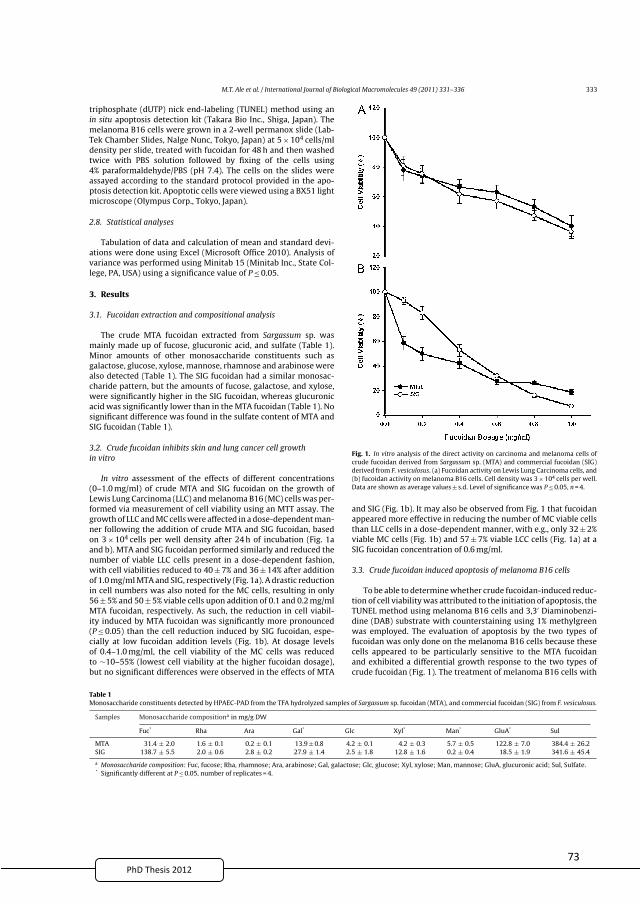
7PhD Thesis 2011
M.T. Ale et al. / International Journal of Biological Macromolecules 49 (2011) 331– 336 333
triphosphate (dUTP) nick end-labeling (TUNEL) method using an
in situ apoptosis detection kit (Takara Bio Inc., Shiga, Japan). The
melanoma B16 cells were grown in a 2-well permanox slide (Lab-
Tek Chamber Slides, Nalge Nunc, Tokyo, Japan) at 5 × 10⁴ cells/ml
density per slide, treated with fucoidan for 48 h and then washed
twice with PBS solution followed by fixing of the cells using
4% paraformaldehyde/PBS (pH 7.4). The cells on the slides were
assayed according to the standard protocol provided in the apo-
ptosis detection kit. Apoptotic cells were viewed using a BX51 light
microscope (Olympus Corp., Tokyo, Japan).
2.8. Statistical analyses
Tabulation of data and calculation of mean and standard devi-
ations were done using Excel (Microsoft Office 2010). Analysis of
variance was performed using Minitab 15 (Minitab Inc., State Col-
lege, PA, USA) using a significance value of P ≤ 0.05.
3. Results
3.1. Fucoidan extraction and compositional analysis
The crude MTA fucoidan extracted from Sargassum sp. was
mainly made up of fucose, glucuronic acid, and sulfate (Table 1).
Minor amounts of other monosaccharide constituents such as
galactose, glucose, xylose, mannose, rhamnose and arabinose were
also detected (Table 1). The SIG fucoidan had a similar monosac-
charide pattern, but the amounts of fucose, galactose, and xylose,
were significantly higher in the SIG fucoidan, whereas glucuronic
acid was significantly lower than in the MTA fucoidan (Table 1). No
significant difference was found in the sulfate content of MTA and
SIG fucoidan (Table 1).
3.2. Crude fucoidan inhibits skin and lung cancer cell growth
in vitro
In vitro assessment of the effects of different concentrations
(0–1.0 mg/ml) of crude MTA and SIG fucoidan on the growth of
Lewis Lung Carcinoma (LLC) and melanoma B16 (MC) cells was per-
formed via measurement of cell viability using an MTT assay. The
growth of LLC and MC cells were affected in a dose-dependent man-
ner following the addition of crude MTA and SIG fucoidan, based
on 3 × 10⁴ cells per well density after 24 h of incubation (Fig. 1a
and b). MTA and SIG fucoidan performed similarly and reduced the
number of viable LLC cells present in a dose-dependent fashion,
with cell viabilities reduced to 40 ± 7% and 36 ± 14% after addition
of 1.0 mg/ml MTA and SIG, respectively (Fig. 1a). A drastic reduction
in cell numbers was also noted for the MC cells, resulting in only
56 ± 5% and 50 ± 5% viable cells upon addition of 0.1 and 0.2 mg/ml
MTA fucoidan, respectively. As such, the reduction in cell viabil-
ity induced by MTA fucoidan was significantly more pronounced
(P ≤ 0.05) than the cell reduction induced by SIG fucoidan, espe-
cially at low fucoidan addition levels (Fig. 1b). At dosage levels
of 0.4–1.0 mg/ml, the cell viability of the MC cells was reduced
to ∼10–55% (lowest cell viability at the higher fucoidan dosage),
but no significant differences were observed in the effects of MTA
Fig. 1. In vitro analysis of the direct activity on carcinoma and melanoma cells of
crude fucoidan derived from Sargassum sp. (MTA) and commercial fucoidan (SIG)
derived from F. vesiculosus. (a) Fucoidan activity on Lewis Lung Carcinoma cells, and
(b) fucoidan activity on melanoma B16 cells. Cell density was 3 × 10⁴ cells per well.
Data are shown as average values ± s.d. Level of significance was P ≤ 0.05, n = 4.
and SIG (Fig. 1b). It may also be observed from Fig. 1 that fucoidan
appeared more effective in reducing the number of MC viable cells
than LLC cells in a dose-dependent manner, with e.g., only 32 ± 2%
viable MC cells (Fig. 1b) and 57 ± 7% viable LCC cells (Fig. 1a) at a
SIG fucoidan concentration of 0.6 mg/ml.
3.3. Crude fucoidan induced apoptosis of melanoma B16 cells
To be able to determine whether crude fucoidan-induced reduc-
tion of cell viability was attributed to the initiation of apoptosis, the
TUNEL method using melanoma B16 cells and 3,3′ Diaminobenzi-
dine (DAB) substrate with counterstaining using 1% methylgreen
was employed. The evaluation of apoptosis by the two types of
fucoidan was only done on the melanoma B16 cells because these
cells appeared to be particularly sensitive to the MTA fucoidan
and exhibited a differential growth response to the two types of
crude fucoidan (Fig. 1). The treatment of melanoma B16 cells with
Table 1Monosaccharide constituents detected by HPAEC-PAD from the TFA hydrolyzed samples of Sargassum sp. fucoidan (MTA), and commercial fucoidan (SIG) from F. vesiculosus.
Samples Monosaccharide compositiona in mg/g DW
Fuc* Rha Ara Gal* Glc Xyl* Man* GluA* Sul
MTA 31.4 ± 2.0 1.6 ± 0.1 0.2 ± 0.1 13.9 ± 0.8 4.2 ± 0.1 4.2 ± 0.3 5.7 ± 0.5 122.8 ± 7.0 384.4 ± 26.2
SIG 138.7 ± 5.5 2.0 ± 0.6 2.8 ± 0.2 27.9 ± 1.4 2.5 ± 1.8 12.8 ± 1.6 0.2 ± 0.4 18.5 ± 1.9 341.6 ± 45.4
a Monosaccharide composition: Fuc, fucose; Rha, rhamnose; Ara, arabinose; Gal, galactose; Glc, glucose; Xyl, xylose; Man, mannose; GluA, glucuronic acid; Sul, Sulfate.* Significantly different at P ≤ 0.05, number of replicates = 4.
2
85
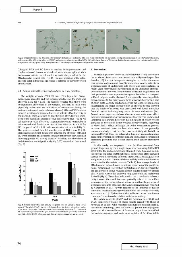
7PhD Thesis 2011
334 M.T. Ale et al. / International Journal of Biological Macromolecules 49 (2011) 331– 336
Fig. 2. Images of melanoma B16 cells after exposure to fucoidan: melanoma B16 cells were plated onto cell cultured 2-well permanox slides at a 5 × 10⁴ cells/ml density,
and incubated for 48 h in the absence (CONT) and presence of crude fucoidan (MTA, SIG) added at a dosage of 0.8 mg/ml. DAB substrate was used to stain the cells and the
images were photographed using an Olympus BX51 microscope following two independent experiments.
0.8 mg/ml MTA and SIG fucoidan resulted in fragmentation and
condensation of chromatin, visualized as an intense pyknotic dark
brown color within the cell nuclei, as particularly evident for the
MTA fucoidan treated cells (Fig. 2). (For interpretation of the refer-
ences to color in this text, the reader is referred to the web version
of the article.)
3.4. Natural killer (NK) cell activity induced by crude fucoidans
The weights of male C57BL/6J mice (Clea Japan Inc., Tokyo,
Japan) were recorded and the inherent alertness of the mice was
observed daily for 4 days. The records revealed that there were
no significant differences in the weights, and that all mice were
physically active with no indications of weaknesses during the
entire experimental period (data not shown). MTA and SIG fucoidan
induced significantly increased levels of splenic NK cell activity in
the C57BL/6J mice, assessed as specific lysis after daily i.p. injec-
tions of the fucoidan samples for four consecutive days (Fig. 3). NK
cell activity at 100:1 effector to target ratio increased remarkably in
mice treated with fucoidan to 14 ± 3.8% for MTA and 11 ± 1.7% for
SIG compared to 5.1 ± 2.1% in the non-treated mice control (Fig. 3).
The positive control Poly I:C specific lysis at 100:1 was 26 ± 9%.
Statistically significant differences between the effects of MTA and
SIG were detected at all effector to target ratios with MTA fucoidan
inducing greater NK activity than SIG fucoidan, and the effects of
both fucoidans were significantly (P ≤ 0.05) better than the control
(Fig. 3).
Fig. 3. Natural killer (NK) cell activity in spleen cells of C57BL/6J mice (n = 3)
against 51Cr-labeled YAC-1 target cells injected i.p. for 4 days with either saline
or fucoidan (50 mg/kg). Cytotoxicity was determined by measuring 51Cr release at
4 h expressed as percent specific lysis. Positive control Poly I:C specific lysis at 100:1
was 26.2 ± 8.9%. (E)/(T); effector/target. Data are shown as average values ± s.d.
4. Discussion
The leading cause of cancer deaths worldwide is lung cancer and
the incidence of melanoma has risen dramatically over the past few
decades [13]. Current therapeutic strategies to combat these can-
cers provide only minimal benefits and expose cancer patients to
significant risks of undesirable side effects and complications. In
recent years many studies have focused on the utilization of bioac-
tive compounds derived from biomass of natural origin based on
their potential as cancer preventive agents. Fucoidan is a complex
sulfated polysaccharide obtained from naturally occurring edible
brown seaweeds. For many years, these seaweeds have been a part
of Asian diets. A study conducted across the Japanese population
investigating the major impact of diet on chronic disease showed
that the intake of seaweed was associated with lower mortality
from all causes, including lung cancer for men and women [14].
Animal model experiments have shown a direct anti-cancer effect
following incorporation of brown seaweeds of the type Undaria and
Laminaria into animal diets with no indications of either weight
gain/loss or alterations in the weights of body organs, signifying
no direct lethal effect. Although the active components present
in these seaweeds have not been determined, it has generally
been acknowledged that the effects are most likely attributable to
fucoidan [15,16]. Thus, the potential of fucoidan as an outstanding
agent for prevention or control of lung and skin cancer is considered
promising providing that it does indeed exert cancer-preventive
effects.
In this study, we employed crude fucoidan extracted from
ground Sargassum sp. via a single step extraction using 0.03 M HCl
at 90 ◦C for 4 h, and commercially obtained crude fucoidan from F.
vesiculosus. We noted that the chemical composition of the seaweed
species were distinctively different. In particular, fucose, galactose
and glucuronic acid contents differed widely while no differences
were noted in the sulfate content (Table 1). Low dosage levels of
MTA fucoidan induced more significant reduction of the prolifera-
tion of melanoma B16 cells than the SIG fucoidan, but in general the
cell proliferation assays revealed almost similar bioactivity effects
of MTA and SIG fucoidan on Lewis lung carcinoma and melanoma
B16 cells (Fig. 1). These data indicate that the crude fucoidan bioac-
tivity towards these cell lines was probably related to the sulfate
groups present in the fucoidan structure rather than the presence of
significant amounts of fucose. The same observation was reported
by Yamamoto et al. [17] with respect to the influence of fucose
content of fucoidan on the growth inhibition of Sarcoma-180 cells;
Yamamoto et al. [17] thus found that sulfation rather than fucose
content of crude fucoidan elicited anti-tumor activity.
The sulfate contents of MTA and SIG fucoidan were 38.4% and
34.2%, respectively (Table 1). These results agreed with those of
Koyanagi et al. [18] who reported that purified fucoidan from F.
vesiculosus containing 32.6% sulfate was a potent anti-angiogenic
agent and that oversulfation of fucoidan might have intensified
the anti-angiogenesis and anti-tumor activity of fucoidan. Addi-
2
86
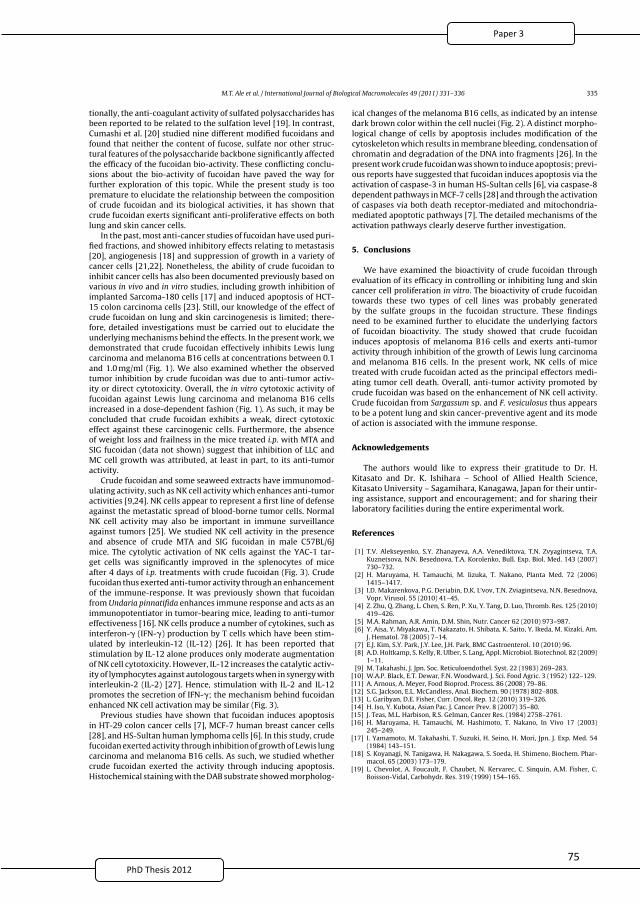
7PhD Thesis 2011
M.T. Ale et al. / International Journal of Biological Macromolecules 49 (2011) 331– 336 335
tionally, the anti-coagulant activity of sulfated polysaccharides has
been reported to be related to the sulfation level [19]. In contrast,
Cumashi et al. [20] studied nine different modified fucoidans and
found that neither the content of fucose, sulfate nor other struc-
tural features of the polysaccharide backbone significantly affected
the efficacy of the fucoidan bio-activity. These conflicting conclu-
sions about the bio-activity of fucoidan have paved the way for
further exploration of this topic. While the present study is too
premature to elucidate the relationship between the composition
of crude fucoidan and its biological activities, it has shown that
crude fucoidan exerts significant anti-proliferative effects on both
lung and skin cancer cells.
In the past, most anti-cancer studies of fucoidan have used puri-
fied fractions, and showed inhibitory effects relating to metastasis
[20], angiogenesis [18] and suppression of growth in a variety of
cancer cells [21,22]. Nonetheless, the ability of crude fucoidan to
inhibit cancer cells has also been documented previously based on
various in vivo and in vitro studies, including growth inhibition of
implanted Sarcoma-180 cells [17] and induced apoptosis of HCT-
15 colon carcinoma cells [23]. Still, our knowledge of the effect of
crude fucoidan on lung and skin carcinogenesis is limited; there-
fore, detailed investigations must be carried out to elucidate the
underlying mechanisms behind the effects. In the present work, we
demonstrated that crude fucoidan effectively inhibits Lewis lung
carcinoma and melanoma B16 cells at concentrations between 0.1
and 1.0 mg/ml (Fig. 1). We also examined whether the observed
tumor inhibition by crude fucoidan was due to anti-tumor activ-
ity or direct cytotoxicity. Overall, the in vitro cytotoxic activity of
fucoidan against Lewis lung carcinoma and melanoma B16 cells
increased in a dose-dependent fashion (Fig. 1). As such, it may be
concluded that crude fucoidan exhibits a weak, direct cytotoxic
effect against these carcinogenic cells. Furthermore, the absence
of weight loss and frailness in the mice treated i.p. with MTA and
SIG fucoidan (data not shown) suggest that inhibition of LLC and
MC cell growth was attributed, at least in part, to its anti-tumor
activity.
Crude fucoidan and some seaweed extracts have immunomod-
ulating activity, such as NK cell activity which enhances anti-tumor
activities [9,24]. NK cells appear to represent a first line of defense
against the metastatic spread of blood-borne tumor cells. Normal
NK cell activity may also be important in immune surveillance
against tumors [25]. We studied NK cell activity in the presence
and absence of crude MTA and SIG fucoidan in male C57BL/6J
mice. The cytolytic activation of NK cells against the YAC-1 tar-
get cells was significantly improved in the splenocytes of mice
after 4 days of i.p. treatments with crude fucoidan (Fig. 3). Crude
fucoidan thus exerted anti-tumor activity through an enhancement
of the immune-response. It was previously shown that fucoidan
from Undaria pinnatifida enhances immune response and acts as an
immunopotentiator in tumor-bearing mice, leading to anti-tumor
effectiveness [16]. NK cells produce a number of cytokines, such as
interferon-� (IFN-�) production by T cells which have been stim-
ulated by interleukin-12 (IL-12) [26]. It has been reported that
stimulation by IL-12 alone produces only moderate augmentation
of NK cell cytotoxicity. However, IL-12 increases the catalytic activ-
ity of lymphocytes against autologous targets when in synergy with
interleukin-2 (IL-2) [27]. Hence, stimulation with IL-2 and IL-12
promotes the secretion of IFN-�; the mechanism behind fucoidan
enhanced NK cell activation may be similar (Fig. 3).
Previous studies have shown that fucoidan induces apoptosis
in HT-29 colon cancer cells [7], MCF-7 human breast cancer cells
[28], and HS-Sultan human lymphoma cells [6]. In this study, crude
fucoidan exerted activity through inhibition of growth of Lewis lung
carcinoma and melanoma B16 cells. As such, we studied whether
crude fucoidan exerted the activity through inducing apoptosis.
Histochemical staining with the DAB substrate showed morpholog-
ical changes of the melanoma B16 cells, as indicated by an intense
dark brown color within the cell nuclei (Fig. 2). A distinct morpho-
logical change of cells by apoptosis includes modification of the
cytoskeleton which results in membrane bleeding, condensation of
chromatin and degradation of the DNA into fragments [26]. In the
present work crude fucoidan was shown to induce apoptosis; previ-
ous reports have suggested that fucoidan induces apoptosis via the
activation of caspase-3 in human HS-Sultan cells [6], via caspase-8
dependent pathways in MCF-7 cells [28] and through the activation
of caspases via both death receptor-mediated and mitochondria-
mediated apoptotic pathways [7]. The detailed mechanisms of the
activation pathways clearly deserve further investigation.
5. Conclusions
We have examined the bioactivity of crude fucoidan through
evaluation of its efficacy in controlling or inhibiting lung and skin
cancer cell proliferation in vitro. The bioactivity of crude fucoidan
towards these two types of cell lines was probably generated
by the sulfate groups in the fucoidan structure. These findings
need to be examined further to elucidate the underlying factors
of fucoidan bioactivity. The study showed that crude fucoidan
induces apoptosis of melanoma B16 cells and exerts anti-tumor
activity through inhibition of the growth of Lewis lung carcinoma
and melanoma B16 cells. In the present work, NK cells of mice
treated with crude fucoidan acted as the principal effectors medi-
ating tumor cell death. Overall, anti-tumor activity promoted by
crude fucoidan was based on the enhancement of NK cell activity.
Crude fucoidan from Sargassum sp. and F. vesiculosus thus appears
to be a potent lung and skin cancer-preventive agent and its mode
of action is associated with the immune response.
Acknowledgements
The authors would like to express their gratitude to Dr. H.
Kitasato and Dr. K. Ishihara – School of Allied Health Science,
Kitasato University – Sagamihara, Kanagawa, Japan for their untir-
ing assistance, support and encouragement; and for sharing their
laboratory facilities during the entire experimental work.
References
[1] T.V. Alekseyenko, S.Y. Zhanayeva, A.A. Venediktova, T.N. Zvyagintseva, T.A.Kuznetsova, N.N. Besednova, T.A. Korolenko, Bull. Exp. Biol. Med. 143 (2007)730–732.
[2] H. Maruyama, H. Tamauchi, M. Iizuka, T. Nakano, Planta Med. 72 (2006)1415–1417.
[3] I.D. Makarenkova, P.G. Deriabin, D.K. L’vov, T.N. Zviagintseva, N.N. Besednova,Vopr. Virusol. 55 (2010) 41–45.
[4] Z. Zhu, Q. Zhang, L. Chen, S. Ren, P. Xu, Y. Tang, D. Luo, Thromb. Res. 125 (2010)419–426.
[5] M.A. Rahman, A.R. Amin, D.M. Shin, Nutr. Cancer 62 (2010) 973–987.[6] Y. Aisa, Y. Miyakawa, T. Nakazato, H. Shibata, K. Saito, Y. Ikeda, M. Kizaki, Am.
J. Hematol. 78 (2005) 7–14.[7] E.J. Kim, S.Y. Park, J.Y. Lee, J.H. Park, BMC Gastroenterol. 10 (2010) 96.[8] A.D. Holtkamp, S. Kelly, R. Ulber, S. Lang, Appl. Microbiol. Biotechnol. 82 (2009)
1–11.[9] M. Takahashi, J. Jpn. Soc. Reticuloendothel. Syst. 22 (1983) 269–283.
[10] W.A.P. Black, E.T. Dewar, F.N. Woodward, J. Sci. Food Agric. 3 (1952) 122–129.[11] A. Arnous, A. Meyer, Food Bioprod. Process. 86 (2008) 79–86.[12] S.G. Jackson, E.L. McCandless, Anal. Biochem. 90 (1978) 802–808.[13] L. Garibyan, D.E. Fisher, Curr. Oncol. Rep. 12 (2010) 319–326.[14] H. Iso, Y. Kubota, Asian Pac. J. Cancer Prev. 8 (2007) 35–80.[15] J. Teas, M.L. Harbison, R.S. Gelman, Cancer Res. (1984) 2758–2761.[16] H. Maruyama, H. Tamauchi, M. Hashimoto, T. Nakano, In Vivo 17 (2003)
245–249.[17] I. Yamamoto, M. Takahashi, T. Suzuki, H. Seino, H. Mori, Jpn. J. Exp. Med. 54
(1984) 143–151.[18] S. Koyanagi, N. Tanigawa, H. Nakagawa, S. Soeda, H. Shimeno, Biochem. Phar-
macol. 65 (2003) 173–179.[19] L. Chevolot, A. Foucault, F. Chaubet, N. Kervarec, C. Sinquin, A.M. Fisher, C.
Boisson-Vidal, Carbohydr. Res. 319 (1999) 154–165.
2
87
7PhD Thesis 2011
336 M.T. Ale et al. / International Journal of Biological Macromolecules 49 (2011) 331– 336
[20] A. Cumashi, N.A. Ushakova, M.E. Preobrazhenskaya, A. D’Incecco, A. Piccoli,L. Totani, N. Tinari, G.E. Morozevich, A.E. Berman, M.I. Bilan, A.I. Usov, N.E.Ustyuzhanina, A.A. Grachev, C.J. Sanderson, M. Kelly, G.A. Rabinovich, S. Iaco-belli, N.E. Nifantiev, Glycobiology 17 (2007) 541–552.
[21] N.Y Lee, S.P. Ermakova, T.N. Zvyagintseva, K.W. Kang, Z. Dong, H.S. Choi, FoodChem. Toxicol. 46 (2008) 1793–1800.
[22] T. Teruya, T. Konishi, S. Uechi, H. Tamaki, M. Tako, Int. J. Biol. Macromol. 41(2007) 221–226.
[23] J.H. Hyun, S.C. Kim, J.I. Kang, M.K. Kim, H.J. Boo, J.M. Kwon, Y.S. Koh, J.W. Hyun,D.B. Park, E.S. Yoo, H.K. Kang, Biol. Pharm. Bull. 32 (2009) 1760–1764.
[24] B.E. Shan, Y. Yoshida, E. Kuroda, U. Yamashita, Int. J. Immunopharmacol. 21(1999) 59–70.
[25] T.L. Whiteside, R.B. Herberman, Clin. Immunol. Immunopathol. 53 (1989)1–23.
[26] T.J. Kindt, R.A. Goldsby, B.A. Osborne, in: W.H. Freeman and Company (Ed.),Cell-Mediated Cytotoxic Responses, Kuby Immunology, New York, 2007, pp.360–363.
[27] C.L. Nastala, H.D. Edington, T.G. McKinney, H. Tahara, M.A. Nalesnik, M.J. Brunda,M.K. Gately, S.F. Wolf, R.D. Schreiber, W.J. Storkus, M.T. Lotza, J. Immunol. 151(1994) 1697–1706.
[28] Y. Yamasaki-Miyamoto, M. Yamasaki, H. Tachibana, K. Yamada, J. Agric. FoodChem. 57 (2009) 8677–8682.
2
88
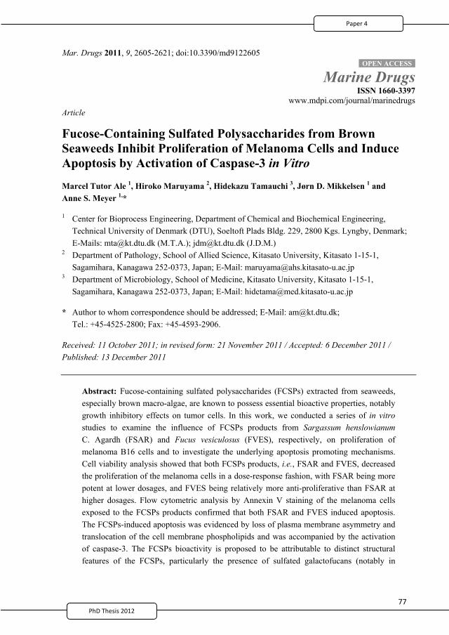
7PhD Thesis 2011
Mar. Drugs 2011, 9, 2605-2621; doi:10.3390/md9122605
Marine Drugs ISSN 1660-3397
www.mdpi.com/journal/marinedrugs Article
Fucose-Containing Sulfated Polysaccharides from Brown Seaweeds Inhibit Proliferation of Melanoma Cells and Induce Apoptosis by Activation of Caspase-3 in Vitro
Marcel Tutor Ale 1, Hiroko Maruyama 2, Hidekazu Tamauchi 3, Jørn D. Mikkelsen 1 and Anne S. Meyer 1,*
1 Center for Bioprocess Engineering, Department of Chemical and Biochemical Engineering, Technical University of Denmark (DTU), Soeltoft Plads Bldg. 229, 2800 Kgs. Lyngby, Denmark; E-Mails: [email protected] (M.T.A.); [email protected] (J.D.M.)
2 Department of Pathology, School of Allied Science, Kitasato University, Kitasato 1-15-1, Sagamihara, Kanagawa 252-0373, Japan; E-Mail: [email protected]
3 Department of Microbiology, School of Medicine, Kitasato University, Kitasato 1-15-1, Sagamihara, Kanagawa 252-0373, Japan; E-Mail: [email protected]
* Author to whom correspondence should be addressed; E-Mail: [email protected]; Tel.: +45-4525-2800; Fax: +45-4593-2906.
Received: 11 October 2011; in revised form: 21 November 2011 / Accepted: 6 December 2011 / Published: 13 December 2011
Abstract: Fucose-containing sulfated polysaccharides (FCSPs) extracted from seaweeds, especially brown macro-algae, are known to possess essential bioactive properties, notably growth inhibitory effects on tumor cells. In this work, we conducted a series of in vitro studies to examine the influence of FCSPs products from Sargassum henslowianum C. Agardh (FSAR) and Fucus vesiculosus (FVES), respectively, on proliferation of melanoma B16 cells and to investigate the underlying apoptosis promoting mechanisms. Cell viability analysis showed that both FCSPs products, i.e., FSAR and FVES, decreased the proliferation of the melanoma cells in a dose-response fashion, with FSAR being more potent at lower dosages, and FVES being relatively more anti-proliferative than FSAR at higher dosages. Flow cytometric analysis by Annexin V staining of the melanoma cells exposed to the FCSPs products confirmed that both FSAR and FVES induced apoptosis. The FCSPs-induced apoptosis was evidenced by loss of plasma membrane asymmetry and translocation of the cell membrane phospholipids and was accompanied by the activation of caspase-3. The FCSPs bioactivity is proposed to be attributable to distinct structural features of the FCSPs, particularly the presence of sulfated galactofucans (notably in
OPEN ACCESS
2
89

7PhD Thesis 2011
Mar. Drugs 2011, 9
2606
S. henslowianum) and sulfated fucans (notably in F. vesiculosus). This study thus indicates that unfractionated FCSPs may exert bioactive effects on skin cancer cells via induction of apoptosis through cascades of reactions that involve activation of caspase-3.
Keywords: fucoidan; anti-tumor; sulfated polysaccharides; bio-activity; apoptosis; fucose
1. Introduction
Fucose-containing sulfated polysaccharides (FCSPs) designate a group of diverse polysaccharides that can be extracted from brown seaweeds of the class Phaeophyceae. This seaweed class includes the order Fucales, in which seaweed species such as Fucus sp. and Sargassum sp. belong. The most studied FCSPs, originally called fucoidin, fucoidan or just fucans, have a backbone built of (1 3)-linked
-L-fucopyranosyl residues or of alternating (1 3)- and (1 4)-linked -L-fucopyranosyl residues [1,2]. These fucopyranosyl residues may be substituted with short fucoside side chains or sulfate groups at C-2 or C-4, and may also carry other minor substitutions, e.g., acetate, xylose, mannose, glucuronic acid, galactose, or glucose [3–5]. Brown seaweed FCSPs also include sulfated galactofucans with backbones built of (1 6)- -D-galacto- and/or (1 2)- -D-mannopyranosyl units. In addition to sulfate these backbone residues may be substituted with fucosides, single fucose substitutions, and/or glucuronic acid, xylose or glucose substitutions [4]. Recently it has been understood that the compositional and structural features of FCSPs differ significantly among seaweed species and that these features are markedly influenced by the conditions used to extract them [3,6].
FCSPs of different degrees of purity and composition, extracted from brown seaweeds such as Sargassum sp. and Fucus sp., have been documented to have a wide range of biological activities including anticoagulant [7,8], antithrombotic [8], anti-inflammatory [9], anti-viral [10,11]; and notably anti-tumoral effects [8,12,13]. Unfractionated FCSPs have thus specifically been found to reduce cell proliferation of lung carcinoma and melanoma cells in vitro; to exert immunopotentiating effects in tumor bearing animals; and to activate natural killer cells in mice leading to increased anti-tumor effectiveness [13–16]. Kim et al. [17] applied a crude polysaccharide composed predominantly of sulfated fucose from Fucus vesiculosus to human colon cancer cells in vitro, and concluded that this crude brown seaweed polysaccharide extract can induce apoptosis, and provided data that suggested that the apoptosis was induced via activation of caspases. Moreover, commercially available crude FCSPs (“fucoidan”) extracted from F. vesiculosus have been reported to inhibit proliferation and induce apoptosis on human lymphoma HS-Sultan cells lines by activation of caspase-3 [18]. Recently, we have reported that crude FCSPs extracted from a Sargassum sp. and from F. vesiculosus, respectively, induce growth inhibition and apoptosis of melanoma B16 cells in vitro [13]. When injected intraperitoneally into mice over four days, these same unfractionated FCSPs were found to induce enhanced natural killer cells (NK cells) activity to result in specific lysis of YAC-1 cells (a murine T-lymphoma cell line sensitive to NK cells) [13]. Previous reports with human HS-Sultan cells and MCF-7 cells, respectively, have suggested that the FCSPs induced apoptosis initiation may take place via activation of caspase-3 and caspase-8 dependent pathways, respectively [18,19], but no firm evidence has been established regarding the exact mechanism responsible for the apoptotic action
2
90
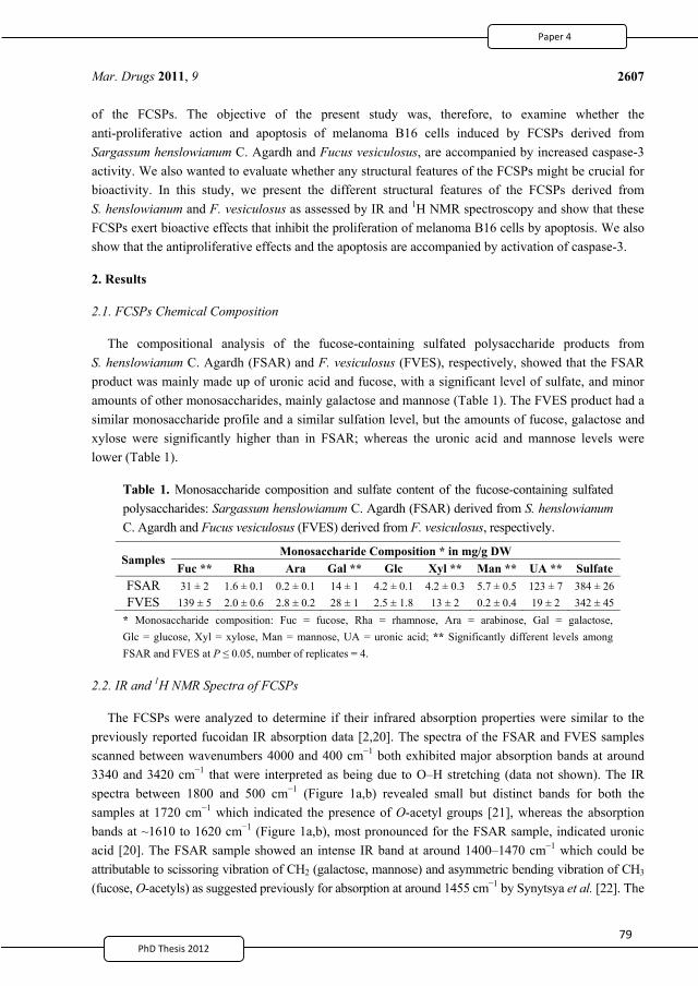
7PhD Thesis 2011
Mar. Drugs 2011, 9
2607
of the FCSPs. The objective of the present study was, therefore, to examine whether the anti-proliferative action and apoptosis of melanoma B16 cells induced by FCSPs derived from Sargassum henslowianum C. Agardh and Fucus vesiculosus, are accompanied by increased caspase-3 activity. We also wanted to evaluate whether any structural features of the FCSPs might be crucial for bioactivity. In this study, we present the different structural features of the FCSPs derived from S. henslowianum and F. vesiculosus as assessed by IR and 1H NMR spectroscopy and show that these FCSPs exert bioactive effects that inhibit the proliferation of melanoma B16 cells by apoptosis. We also show that the antiproliferative effects and the apoptosis are accompanied by activation of caspase-3.
2. Results
2.1. FCSPs Chemical Composition
The compositional analysis of the fucose-containing sulfated polysaccharide products from S. henslowianum C. Agardh (FSAR) and F. vesiculosus (FVES), respectively, showed that the FSAR product was mainly made up of uronic acid and fucose, with a significant level of sulfate, and minor amounts of other monosaccharides, mainly galactose and mannose (Table 1). The FVES product had a similar monosaccharide profile and a similar sulfation level, but the amounts of fucose, galactose and xylose were significantly higher than in FSAR; whereas the uronic acid and mannose levels were lower (Table 1).
Table 1. Monosaccharide composition and sulfate content of the fucose-containing sulfated polysaccharides: Sargassum henslowianum C. Agardh (FSAR) derived from S. henslowianum C. Agardh and Fucus vesiculosus (FVES) derived from F. vesiculosus, respectively.
Samples Monosaccharide Composition * in mg/g DW
Fuc ** Rha Ara Gal ** Glc Xyl ** Man ** UA ** Sulfate FSAR 31 ± 2 1.6 ± 0.1 0.2 ± 0.1 14 ± 1 4.2 ± 0.1 4.2 ± 0.3 5.7 ± 0.5 123 ± 7 384 ± 26 FVES 139 ± 5 2.0 ± 0.6 2.8 ± 0.2 28 ± 1 2.5 ± 1.8 13 ± 2 0.2 ± 0.4 19 ± 2 342 ± 45
* Monosaccharide composition: Fuc = fucose, Rha = rhamnose, Ara = arabinose, Gal = galactose, Glc = glucose, Xyl = xylose, Man = mannose, UA = uronic acid; ** Significantly different levels among FSAR and FVES at P 0.05, number of replicates = 4.
2.2. IR and 1H NMR Spectra of FCSPs
The FCSPs were analyzed to determine if their infrared absorption properties were similar to the previously reported fucoidan IR absorption data [2,20]. The spectra of the FSAR and FVES samples scanned between wavenumbers 4000 and 400 cm 1 both exhibited major absorption bands at around 3340 and 3420 cm 1 that were interpreted as being due to O–H stretching (data not shown). The IR spectra between 1800 and 500 cm 1 (Figure 1a,b) revealed small but distinct bands for both the samples at 1720 cm 1 which indicated the presence of O-acetyl groups [21], whereas the absorption bands at ~1610 to 1620 cm 1 (Figure 1a,b), most pronounced for the FSAR sample, indicated uronic acid [20]. The FSAR sample showed an intense IR band at around 1400–1470 cm 1 which could be attributable to scissoring vibration of CH2 (galactose, mannose) and asymmetric bending vibration of CH3 (fucose, O-acetyls) as suggested previously for absorption at around 1455 cm 1 by Synytsya et al. [22]. The
2
91
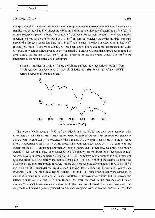
PhD Thesis 2011
Mar. Drugs 2011, 9
2608
absorption band at 1240 cm 1 observed for both samples, but being particularly prevalent for the FVES sample, was assigned as S=O stretching vibration, indicating the presence of esterified sulfate [20]. A similar absorption pattern around 820–840 cm 1 was observed for both FCSPs: The FSAR infrared spectrum showed an absorption band at 817 cm 1 (Figure 1a) whereas the FVES infrared spectrum displayed a broader absorption band at 838 cm 1 and a small shoulder of absorption at 822 cm 1 (Figure 1b). Since IR adsorption at 840 cm 1 has been reported to be due to sulfate groups at the axial C-4 position whereas sulfate groups at the equatorial C-2 and/or C-3 positions have been reported to give a small absorption at 820 cm 1 [2], the observed absorption bands at 820–840 cm 1 were interpreted as being indicative of sulfate groups.
Figure 1. Infrared analysis of fucose-containing sulfated polysaccharides (FCSPs) from (a) Sargassum henslowianum C. Agardh (FSAR) and (b) Fucus vesiculosus (FVES) scanned between 1800 and 550 cm 1.
The proton NMR spectra (2H2O) of the FSAR and the FVES samples were complex with broad signals and with several signals in the chemical shift of the envelope of anomeric signals at 5.0–5.5 ppm (Figure 2a,b). The presence of the signals at 5.0–5.5 ppm is consistent with the presence of -L-fucopyranosyl [23]. The 1H-NMR spectra also both contained peaks at 1.1–1.3 ppm, with the signals for the FVES sample being particularly strong (Figure 2a,b). Previously, such high-field region signals at 1.1–1.3 ppm have been assigned to a C6 methyl proton group of L-fucopyranose [22] whereas several intense and narrow signals at 2.14–2.21 ppm have been attributed to CH3 protons of O-acetyl groups [5]. The narrow and intense signals at 5.10 and 5.18 ppm in the chemical shift of the envelope of the anomeric proton of FSAR (Figure 2a) were reported earlier and assigned to 3-linked and 3,4-linked L-fucopyranose residues for fucoidan from Hizikia fusiformis a.k.a Sargassum fusiformis [24]. The high field region signals 1.24 and 1.20 ppm (Figure 2a) were assigned to
3-linked 2-mono-O-sulfated and 3-linked unsulfated L-fucopyranose residues [21]. Moreover the intense signals at 4.37 and 3.99 ppm (Figure 2a) were assigned to the presence of 4-linked 2-mono-O-sulfated L-fucopyranose residues [21]. The independent signal, 4.61 ppm (Figure 2a) was assigned to a 3-linked D-galactopyranosyl residue when compared with the data of Farias et al. [25]. The
2
92
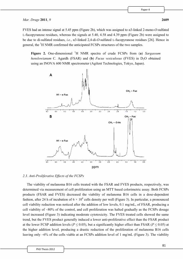
PhD Thesis 2011
Mar. Drugs 2011, 9
2609
FVES had an intense signal at 5.45 ppm (Figure 2b), which was assigned to 3-linked 2-mono-O-sulfated L-fucopyranose residues, whereas the signals at 5.40, 4.58 and 4.39 ppm (Figure 2b) were assigned to be due to di-sulfated residues, i.e., 3-linked 2,4-di-O-sulfated L-fucopyranose residues [26]. Hence in general, the 1H NMR confirmed the anticipated FCSPs structures of the two samples.
Figure 2. One-dimensional 1H NMR spectra of crude FCSPs from (a) Sargassum henslowianum C. Agardh (FSAR) and (b) Fucus vesiculosus (FVES) in D2O obtained using an INOVA 600 NMR spectrometer (Agilent Technologies, Tokyo, Japan).
2.3. Anti-Proliferative Effects of the FCSPs
The viability of melanoma B16 cells treated with the FSAR and FVES products, respectively, was determined via measurement of cell proliferation using an MTT based colorimetric assay. Both FCSPs products (FSAR and FVES) decreased the viability of melanoma B16 cells in a dose-dependent fashion, after 24 h of incubation of 6 × 10⁴ cells density per well (Figure 3). In particular, a pronounced cell viability reduction was noticed after the addition of low levels, 0.1 mg/mL, of FSAR, producing a cell viability of ~80% of the control, and cell proliferation was halted gradually as the FCSPs dosage level increased (Figure 3) indicating moderate cytotoxicity. The FVES treated cells showed the same trend, but the FVES product generally induced a lower anti-proliferative effect than the FSAR product at the lower FCSP addition levels (P 0.05), but a significantly higher effect than FSAR (P 0.05) at the higher addition level, producing a drastic reduction of the proliferation of melanoma B16 cells leaving only ~6% of the cells viable at an FCSPs addition level of 1 mg/mL (Figure 3). The viability
2
93
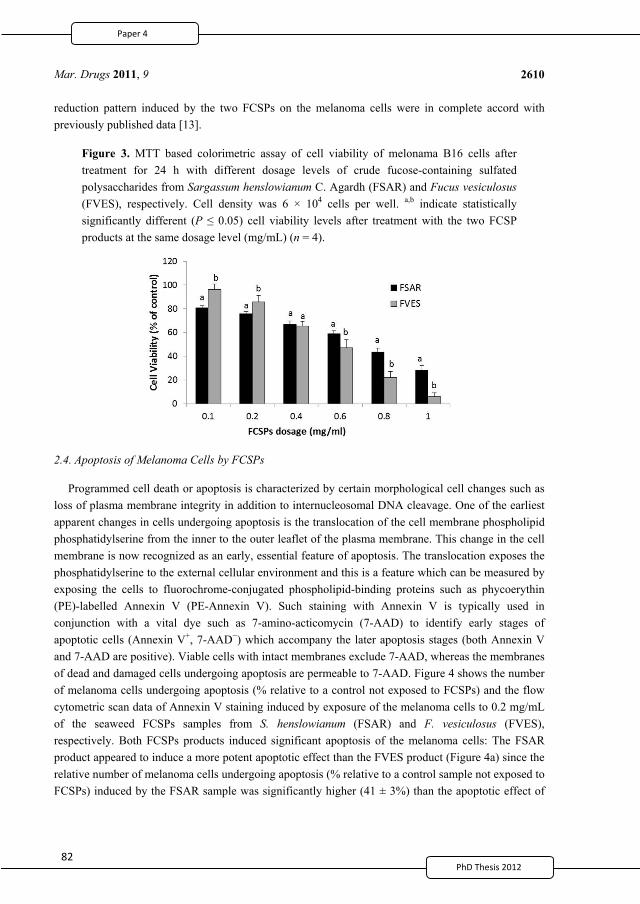
8PhD Thesis 2011
Mar. Drugs 2011, 9
2610
reduction pattern induced by the two FCSPs on the melanoma cells were in complete accord with previously published data [13].
Figure 3. MTT based colorimetric assay of cell viability of melonama B16 cells after treatment for 24 h with different dosage levels of crude fucose-containing sulfated polysaccharides from Sargassum henslowianum C. Agardh (FSAR) and Fucus vesiculosus (FVES), respectively. Cell density was 6 × 10⁴ cells per well. a,b indicate statistically significantly different (P 0.05) cell viability levels after treatment with the two FCSP products at the same dosage level (mg/mL) (n = 4).
2.4. Apoptosis of Melanoma Cells by FCSPs
Programmed cell death or apoptosis is characterized by certain morphological cell changes such as loss of plasma membrane integrity in addition to internucleosomal DNA cleavage. One of the earliest apparent changes in cells undergoing apoptosis is the translocation of the cell membrane phospholipid phosphatidylserine from the inner to the outer leaflet of the plasma membrane. This change in the cell membrane is now recognized as an early, essential feature of apoptosis. The translocation exposes the phosphatidylserine to the external cellular environment and this is a feature which can be measured by exposing the cells to fluorochrome-conjugated phospholipid-binding proteins such as phycoerythin (PE)-labelled Annexin V (PE-Annexin V). Such staining with Annexin V is typically used in conjunction with a vital dye such as 7-amino-acticomycin (7-AAD) to identify early stages of apoptotic cells (Annexin V+, 7-AAD ) which accompany the later apoptosis stages (both Annexin V and 7-AAD are positive). Viable cells with intact membranes exclude 7-AAD, whereas the membranes of dead and damaged cells undergoing apoptosis are permeable to 7-AAD. Figure 4 shows the number of melanoma cells undergoing apoptosis (% relative to a control not exposed to FCSPs) and the flow cytometric scan data of Annexin V staining induced by exposure of the melanoma cells to 0.2 mg/mL of the seaweed FCSPs samples from S. henslowianum (FSAR) and F. vesiculosus (FVES), respectively. Both FCSPs products induced significant apoptosis of the melanoma cells: The FSAR product appeared to induce a more potent apoptotic effect than the FVES product (Figure 4a) since the relative number of melanoma cells undergoing apoptosis (% relative to a control sample not exposed to FCSPs) induced by the FSAR sample was significantly higher (41 ± 3%) than the apoptotic effect of
2
94
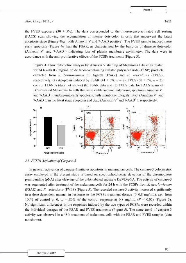
8PhD Thesis 2011
Mar. Drugs 2011, 9
2611
the FVES exposure (30 ± 5%). The data corresponded to the fluorescence-activated cell sorting (FACS) scan showing the accumulation of intense dots-color in cells that underwent the latest apoptosis stage (Figure 4b,c: both Annexin V and 7-AAD positive). The FVES sample induced more early apoptosis (Figure 4c than the FSAR, as characterized by the build-up of disperse dots-color (Annexin V+ and 7-AAD ) indicating loss of plasma membrane asymmetry. The data were in accordance with the anti-proliferative effects of the FCSPs treatments (Figure 3).
Figure 4. Flow cytometric analysis by Annexin V staining of Melanoma B16 cells treated for 24 h with 0.2 mg/mL crude fucose-containing sulfated polysaccharide (FCSP) products extracted from S. henslowianum C. Agardh (FSAR) and F. vesiculosus (FVES), respectively. (a) Apoptosis induced by FSAR (41 ± 3%, n = 2), FVES (30 ± 5%, n = 2); control 11.66 % (data not shown) (b) FSAR data and (c) FVES data for FACS scans of FCSP treated Melanoma 16 cells that were viable and not undergoing apoptosis (Annexin V and 7-AAD ); undergoing early apoptosis, with membrane integrity intact (Annexin V+ and 7-AAD ); in the latest stage apoptosis and dead (Annexin V+ and 7-AAD+ ), respectively.
2.5. FCSPs Activation of Caspase-3
In general, activation of caspase-3 initiates apoptosis in mammalian cells. The caspase-3 colorimetric assay employed in the present study is based on spectrophotometric detection of the chromophore p-nitroaniline (pNA) after cleavage of the pNA-labeled substrate DEVD-pNA. The activity of caspase-3 was augmented after treatment of the melanoma cells for 24 h with the FCSPs from S. henslowianum (FSAR) and F. vesiculosus (FVES) (Figure 5). The recorded caspase-3 activity increased significantly in a dose-dependent manner in response to the FCSPs treatment dosage (0–0.8 mg/mL), i.e., from 100% of control at 0, to ~180% of the control response at 0.8 mg/mL (P 0.05) (Figure 5). No significant differences in the responses induced by the two types of FCSPs were recorded within the individual dosages of the FSAR and FVES treatments (Figure 5). The same trend of caspase-3 activity was observed in a 48 h treatment of melanoma cells with the FSAR and FVES samples (data not shown).
2
95
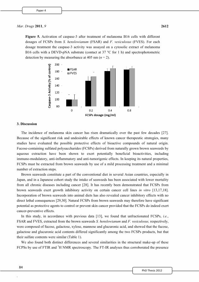
8PhD Thesis 2011
.
Mar. Drugs 2011, 9
2612
Figure 5. Activation of caspase-3 after treatment of melanoma B16 cells with different dosages of FCSPs from S. henslowianum (FSAR) and F. vesiculosus (FVES). For each dosage treatment the caspase-3 activity was assayed on a cytosolic extract of melanoma B16 cells with a DEVD-pNA substrate (contact at 37 °C for 1 h) and spectrophotometric detection by measuring the absorbance at 405 nm (n = 2).
3. Discussion
The incidence of melanoma skin cancer has risen dramatically over the past few decades [27]. Because of the significant risk and undesirable effects of known cancer therapeutic strategies, many studies have evaluated the possible protective effects of bioactive compounds of natural origin. Fucose-containing sulfated polysaccharides (FCSPs) derived from naturally grown brown seaweeds by aqueous extraction have been shown to exert potentially beneficial bioactivities, including immuno-modulatory, anti-inflammatory and anti-tumorigenic effects. In keeping its natural properties, FCSPs must be extracted from brown seaweeds by use of a mild processing treatment and a minimal number of extraction steps.
Brown seaweeds constitute a part of the conventional diet in several Asian countries, especially in Japan, and in a Japanese cohort study the intake of seaweeds has been associated with lower mortality from all chronic diseases including cancer [28]. It has recently been demonstrated that FCSPs from brown seaweeds exert growth inhibitory activity on certain cancer cell lines in vitro [13,17,18]. Incorporation of brown seaweeds into animal diets has also revealed cancer inhibitory effects with no direct lethal consequences [29,30]. Natural FCSPs from brown seaweeds may therefore have significant potential as protective agents to control or prevent skin cancer provided that the FCSPs do indeed exert cancer-preventive effects.
In this study, in accordance with previous data [13], we found that unfractionated FCSPs, i.e., FSAR and FVES, extracted from the brown seaweeds S. henslowianum and F. vesiculosus, respectively, were composed of fucose, galactose, xylose, mannose and glucuronic acid, and showed that the fucose, galactose and glucuronic acid contents differed significantly among the two FCSPs products, but that their sulfate contents were similar (Table 1).
We also found both distinct differences and several similarities in the structural make-up of these FCPSs by use of FTIR and 1H NMR spectroscopy. The FT-IR analyses thus corroborated the presence
2
96
8PhD Thesis 2011
Mar. Drugs 2011, 9
2613
of sulfate groups in both the FSAR and the FVES sample (Figure 1). The IR spectra indicated that the sulfate substitutions of the FCSPs extracted from the Sargassum sp. (FSAR) were located in the equatorial C-2 and/or C-3 positions as depicted by absorption bands at 817 cm 1. This finding was in agreement with data reported for fucoidan fractions isolated from Sargassum stenophyllum [4]. However, Duarte et al. [4] also reported that two other saccharide fractions from S. stenophyllum had an absorption band at 837 cm 1 indicating sulfate groups at the C-4 positions of the structural monosaccharides [4]. The spectra of the FCSPs from F. vesiculosus (FVES) displayed an absorption band at 838 cm 1 with a small shoulder at ~822 cm 1 indicating sulfate groups at both the C-4 and the C-2 position (Figure 1). This finding corresponds to previously reported 1H NMR data of FCSPs from F. vesiculosus that have indicated a typical structure of algal fucoidan consisting of
3-linked 2-mono-O-sulfated L-fucopyranose residues, and/or 3-linked 2,4-di-O-sulfated L-fucopyranose residues [2,26]. Small disparities in the IR spectra from different published reports can be due to factors such as sample handling and the FCSPs extraction procedure employed.
The present study also aimed at establishing whether crude FCSPs extracted from Sargassum henslowianum C. Agardh (FSAR) contained fucoidan-like structures composed of -3-linked or/and
-3,4-linked L-fucopyranose residues. Even though signals consistent with the presence of -L-fucopyranose entities were recorded (with 1H NMR signals at 5.10 and 5.18 ppm, Figure 2a), the
probability that the FSAR may contain a cocktail of polysaccharides is likely. Hence, the 1H NMR spectra also showed that the FSAR sample contained 3-linked D-galactopyranose residues as indicated by an independent signal at 4.61 ppm (Figure 2a). -(1 )3-linked galactopyranose residues are known to be a typical structural feature of seaweed polysaccharides, from e.g., Laminaria angustata var. longissima, Botryocladia occidentalis [25,31]. However, another possibility might be that the FSAR sample was not composed of a mixture of different types of polysaccharides but rather, that the sample consisted of one type of a highly complex hetero-polysaccharide as suggested by Duarte et al. [4] for the fucoidans from Sargassum stenophyllum. It can safely be said that the 1H NMR spectra of the FCSPs samples were complex and overlapping. It is therefore difficult to draw any definite conclusions about the detailed structural features and differences among the two FCSPs; the detailed elucidations of the definite structural details were also beyond the scope of this present study, but clearly deserve further investigation. Nonetheless, the data confirmed that the diversity, i.e., the compositional and structural complexity of (potentially bioactive) algal fucose-containing sulfated polysaccharides, is much wider than originally believed.
The biological activities of the FCSPS against skin cancer cells were investigated in vitro, and the results revealed that both FSAR and FVES can exert anti-proliferative effects on melanoma B16 cells in vitro. The FSAR sample induced more significant reductions of the cell viability of melanoma cells than the FVES sample at low dosage levels (Figure 3). At higher dosages, the FSAR treatment still induced gradually more loss of cell viability, but the FVES had more potent anti-proliferative effects at higher dosages than FSAR which could indicate direct cell toxicity. The bioactivities of these FCSPs may be attributable to their distinct structural features, notably the level of sulfation (charge density), the distribution (e.g., random versus clustered) and bonding of the sulfate substitutions, as well as other specific structural features of the sulfated fucans and the sulfated galactofucan complexes. The sulfate groups of FVES were substituted at the C-2 and C-4 position of the fucose substituents, typical for fucoidan from F. vesiculosus, and consistent with previously published data indicating that the sulfate
2
97
8PhD Thesis 2011
Mar. Drugs 2011, 9
2614
groups were substituted at C-2 of 3-linked L-fucopyranose residues in fucoidan from e.g., Fucus evanescens [21]. In contrast, the sulfate substitutions in the FSAR were interpreted to be mainly at the C-2 and/or C-3 positions of the monosaccharides according to the IR spectra (Figure 1); the observation of C-2 linked sulfate groups agreed with the data mentioned above for fucoidan from Fucus spp. [3,21], but is also in agreement with the proposition that the sulfate groups were substituted at C-2 on the 3-linked galactopyranose residues [25]. The possible presence of sulfated, 3-linked galactan in the structure of FSAR may contribute to the efficacy of FSAR to induce anti-proliferative effects as it has been reported that 2-O-sulfated 3-linked galactan is more bioactive than 2-O-sulfated 3-linked fucans and 3-O-sulfated 4-linked galactan [32–34].
The findings that the FCSPs induced apoptosis of the melanoma B16 cells in vitro were in agreement with recent reports [13,16,35], but the differential apoptotic efficacies, and the dose-response effects of differently structured FCSPs (Figure 4) have not been reported earlier. In particular, it is a novel finding that significantly different sulfated, polysaccharide structures from brown seaweeds—as evaluated in the present work—exert relatively similar apoptotic effects on melanoma cells. The results of this work thus indicate that not only the well-studied, classical type of FCSPs having a backbone made up of (1 3)-linked -L-fucopyranosyl or of alternating (1 3)- and (1 4)-linked
-L-fucopyranosyl residues have potential tumor-preventing effecs, but also that the more complex sulfated fucose-rich galacto-mannans from Sargassum spp. exert promising cancer-preventive effects. The principal objective of this study was to assess whether any structural features of the FCSPs might be crucial for bioactivity, and the data suggest that the sulfate substitutions, and not necessarily only the fucose-backbone structure itself, confer this decisive bioactivity. It is however important to investigate whether other differently structured FCSPs may exert similar growth inhibitory and apoptosis inducing effects on cancer cells.
In this work we noted that both FCSPs activate caspase-3 in a dosage-response fashion (Figure 5). These findings affirmed the results reported previously which have shown that FCSPs (“fucoidan”) from F. vesiculosus induce apoptosis in human lymphoma HS-Sultan cell lines and in HT-29 and HCT116 human colon cancer cells in vitro, and moreover that the exposure of these cells to the F. vesiculosus FCSPs appear to activate caspase-3 [17,18]. The F. vesiculosus FCSPs treatment was also shown to enhance mitochondrial membrane permeability of human colon cancer cells in vitro, and to induce cytochrome c and Smac/Diablo release from the mitochondria [17]. It has also been reported that pretreatment of HT-29 and HCT116 colon cancer cells with individual caspase-8 or caspase-9 inhibitors (Z-IETD-FMK and Z-LEHD-FMK, respectively) prior to fucoidan exposure reduced the levels of caspases, including caspase-3 [17]. It has likewise been shown that pretreatment of human lymphoma HS Sultan cells with a pan-caspase inhibitor, z-VAD-FMK, reduced fucoidan-induced apoptosis [18]. Hence, the available data support the proposition that fucoidan-induced apoptosis occurs through caspase activation pathways. The cascade mechanism by which the caspase-activation is presumed to take place via the mitochondria-mediated apoptotic pathway is illustrated in Figure 6.
Loss of plasma membrane is one of the earliest features of apoptosis and Annexin V staining can identify apoptosis at an early stage. However this assay does not distinguish between cells that have undergone apoptotic death versus those that have died as a result of a necrotic pathway, because in either case the dead cells will stain with both Annexin V and 7-AAD. Both early and later apoptosis stages were observed by the FACS scanning indicating that the FCSPs had a direct apoptotic effect on
2
98
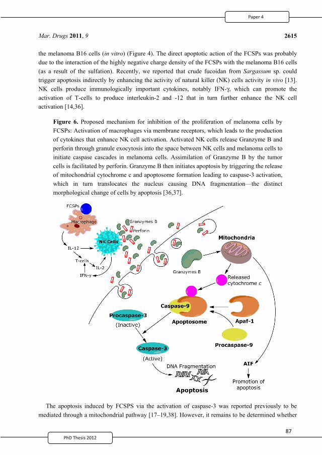
8PhD Thesis 2011
Mar. Drugs 2011, 9
2615
the melanoma B16 cells (in vitro) (Figure 4). The direct apoptotic action of the FCSPs was probably due to the interaction of the highly negative charge density of the FCSPs with the melanoma B16 cells (as a result of the sulfation). Recently, we reported that crude fucoidan from Sargassum sp. could trigger apoptosis indirectly by enhancing the activity of natural killer (NK) cells activity in vivo [13]. NK cells produce immunologically important cytokines, notably IFN-γ, which can promote the activation of T-cells to produce interleukin-2 and -12 that in turn further enhance the NK cell activation [14,36].
Figure 6. Proposed mechanism for inhibition of the proliferation of melanoma cells by FCSPs: Activation of macrophages via membrane receptors, which leads to the production of cytokines that enhance NK cell activation. Activated NK cells release Granzyme B and perforin through granule exocytosis into the space between NK cells and melanoma cells to initiate caspase cascades in melanoma cells. Assimilation of Granzyme B by the tumor cells is facilitated by perforin. Granzyme B then initiates apoptosis by triggering the release of mitochondrial cytochrome c and apoptosome formation leading to caspase-3 activation, which in turn translocates the nucleus causing DNA fragmentation—the distinct morphological change of cells by apoptosis [36,37].
The apoptosis induced by FCSPS via the activation of caspase-3 was reported previously to be mediated through a mitochondrial pathway [17–19,38]. However, it remains to be determined whether
2
99
8PhD Thesis 2011
Mar. Drugs 2011, 9
2616
differences in FCSPs structures will influence the apoptotic mechanism, including the mitochondrial pathway apoptosis cascade. The route of the mitochondrially dependent apoptotic pathway is the release of apoptosis-inducing factor (AIF) and cytochrome c from the inner mitochondrial membrane into the cytosol. Cytochrome c interacts with Apaf-1 (apoptotic protease activating factor 1) and procaspase-9 to form the active apoptosome. The apoptosome then initiates the cleavage of procaspase-3, producing active caspase-3, which initiates the execution phase of apoptosis by proteolysis of substances whose cleavage commits the cell to apoptosis [39] (Figure 6). The influence of the different FCSPs structures on the mitochondrial membrane permeability and electric potential requires further study. We hope in the future to investigate the bioactivity and mechanism of FCSPs on certain degenerative diseases in vivo and to further elucidate specific molecular targets of FSCPs for inhibition of cancer cells.
4. Experimental Section
4.1. Chemicals
Dried S. henslowianum C. Agardh was obtained from Viet Delta Ltd. (Ho Chi Menh, Vietnam) and the Fucose-containing sulfated polysaccharides (FCSPs) from the S. henslowianum (FSAR) were extracted in our laboratory (see below). Crude fucoidan from F. vesiculosus (FVES) was obtained from Sigma-Aldrich (Steinheim, Germany); according to the product description the FVES had been prepared from F. vesiculosus via the extraction method described by Black and Dewar [40]. Hydrochloric acid (37%), D-glucose and D-xylose were purchased from Merck (Darmstadt, Germany). Trifluoracetic acid (99%, TFA), trichloroacetic acid (99%, TCA), CaCl2, Na2SO4, BaCl2, arabinose, rhamnose, D-galactose and L-fucose were from Sigma–Aldrich Co. (Steinheim, Germany). Agarose D-2 was obtained from Hispanagar (Burgos, Spain). Caspase-3 colorimetric assay kit was obtained from Biovision, Inc. (Mountain View, CA. USA). Minimal essential medium eagle (MEM-eagle) cell culture media was purchased from Sigma–Aldrich Co. (Steinheim, Germany); foetal bovine serum (FBS) was from Flow Laboratories (North Ryde, N.S.W., Australia); streptomycin–penicillin and Trypan Blue were from Gibco (Canada). Cell Proliferation Kit 1 was obtained from Roche Applied Science, Germany. The PE Annexin V Apoptosis Detection Kit 1 was obtained from BD Biosciences (Franklin Lakes, NJ, USA). All chemicals used were analytical grade.
4.2. Extraction of FCSPs from S. henslowianum C. Agardh
The Sargassum FCSP product (FSAR) used was extracted from S. henslowianum C. Agardh by use of an optimized single-step extraction procedure described previously [6]. Briefly, the dried S. henslowianum seaweed was ground and sieved to pass through a 500 μm sieve and 100 g of dried ground seaweed was extracted in 2 L of 0.03 M HCl with continuous stirring at 200 rpm for 4 h at 90 °C water bath (Julabo, Germany). The suspended seaweed was filtered, and the extract was precipitated using 60% ethanol, the precipitate collected after centrifugation at 10,600 rpm for 10 min (Sigma Laboratory Centrifuge 4K15, VWR, Denmark), and the resulting pellet was freeze dried. This freeze dried pellet constituted the fucose-containing sulfated polysaccharides (FSAR).
2
100
8PhD Thesis 2011
Mar. Drugs 2011, 9
2617
4.3. Acid Hydrolysis and FCSPs Composition Analysis
The freeze dried FSAR and FVES samples (20 mg) were hydrolyzed separately in 2 M TFA (final concentration) at 121 °C for 2 h, then the hydrolyzed mixture were freeze dried at 57 °C (Heto Lyolab 3000, England). Each dried powder sample was resolubilized in doubly distilled water and centrifuged at 10,000 rpm for 10 min to collect the supernatant (Sigma Laboratory Centrifuge 4K15, VWR, Denmark). Each supernatant was filtered through a 0.2 μm syringe tip filter (SUN-Sri, Rockwood, TN) prior to injection into the HPAEC-PAD for monosaccharide analysis [41]. Analysis of sulfate content was done according to the method described by Jackson and McCandless [42].
4.4. 1H NMR and FTIR Spectroscopy
The 1H NMR spectra were obtained using an INOVA 600 NMR spectrometer (Agilent Technologies Japan, Ltd., Tokyo Japan) equipped with a 1H[15N-31P] pulse field gradient indirect-detecting probe. Standard pulse sequences were used in all operations. The 1H chemical shift ( H) was referenced to HOD ( H 4.76 ppm, 2H2O). The 1H NMR spectrum was assigned through the 1H–1H decoupling technique. An NMR spectrum of L-fucose was utilized as a reference for chemical shift assignment. The lyophilized FCSPs powders were dissolved in deuterium oxide (2H2O) and evaporated to exchange the unstable 1H with 2H. The evaporation and dissolution step was repeated five times, and the samples (10 mg) were finally dissolved in 0.75 mL 2H2O and then subjected to NMR spectroscopy. The IR spectra were obtained using a Spectrum One FT-IR spectrometer (Perkin Elmer, Waltham, MA, USA) equipped with universal attenuated total reflectance (UATR) accessories. Analysis of each of the FSAR and FVES powders, ~1 mg of each, was done using diffuse reflectance infrared transform spectroscopy (DRIFTS) and the spectrum was evaluated by Perkin Elmer Spectrum software version 5 (Perkin Elmer, Waltham, MA, USA).
4.5. Cell Culture and Anti-Proliferative Assay
Melanoma B16 cells (MC) were grown in MEM eagle medium supplemented with 10% (v/v) heat inactivated FBS, 1% (w/v) streptomycin–penicillin and 1% (v/v) of 200 mM L-glutamine at 37 °C under 5% CO2. Monolayer cultivation was carried out by adding 100 μL of the cell-MEM-FBS mixture into separate wells in 96-flat well plates at a density of 6 × 10⁴ cells per well followed by incubation for 24 h in 5% CO2 at 37 °C. For the anti-proliferation assay the medium was removed after the 24 h of monolayer cell cultivation and replaced with 100 μL of MEM medium containing 2% FBS and varying concentrations (0.1–1.0 mg/mL) of the crude FCSPs, i.e., FSAR and FVES, respectively, and the mixtures were then incubated for 24 h. Quantification of cell proliferation was carried out using a tetrazolium salt (MTT (3-(4,5-dimethyl-thiazolyl-2)-2,5-diphenyltetrazolium bromide)) based colorimetric assay following the protocol supplied with the Cell Proliferation Kit 1 (Roche Applied Science, Germany). Briefly, 20 μL MTT solution (5 mg/mL) was added to the cell cultures after the 24 h of incubation with the FCSPs, and the cell cultures were then re-incubated for 4 h. Finally, 100 μL of stabilization solution was added to each well and the plates were incubated overnight at 37 °C under 5% CO2. Absorbance was measured using an Elisa reader at 550–690 nm.
2
101
PhD Thesis 2011
Mar. Drugs 2011, 9
2618
4.6. Cell Culture and Caspase-3 Assay
Melanoma B16 cells (MC) were grown in MEM eagle culture medium supplemented with 10% (v/v) heat inactivated FBS, 1% (w/v) streptomycin–penicillin and 1% (v/v) of 200 mM L-glutamine maintained at 37 °C under 5% CO2. For the caspase-3 assay, monolayer cultivation was carried out in a petri dish (60 × 15 mm) by adding 5 mL culture medium containing melanoma cells at a density of 1 × 10⁵ per mL and varying concentrations (0.2, 0.4 and 0.8 mg/mL) of the FSAR and FVES, respectively. The mixtures were then incubated for 24 and 48 h in 5% CO2 at 37 °C. The caspase-3 assay was performed according to the protocol supplied with the assay kit (Biovision Inc., Mountain View, CA, USA) used to assay the activity of caspases that recognize the amino acid sequence DEVD. The assay was based on spectrophotometric detection of the chromophore p-nitroaniline (pNA) after cleavage from the labeled substrate DEVD-pNA. Concisely, the melanoma B16 cells exposed to FSAR and FVES, respectively, were harvested and resuspended in 50 μL of cell lysis buffer and incubated on ice for 10 min. and the mixture centrifuged for 1 min (14,000 × g, 4 °C). Each supernatant was then transferred to a fresh tube, and reaction buffer (50 μL) and 4 mM DEVD-pNA substrate (5 μL) were added, and this reaction mixture was then incubated at 37 °C for 1 h. Absorbance of pNA light emission was quantified using a microtiter plate reader at 405 nm.
4.7. Apoptosis Assay by Fluorescence-Activated Cell Sorting (FACS)
After 24 h of monolayer cultivation of melanoma B16 cells with 0.2 mg/mL of FSAR or FVES, and no FCSPs addition as control, the culture medium was removed, and the cells harvested by addition of 1 mL Trypsin-EDTA. The harvested cells were washed twice with 0.1 M PBS and then resuspended in binding buffer according to the protocol for the Annexin V Apoptosis Detection Kit I (BD Biosciences, Franklin Lakes, NJ, USA). 100 μL of this solution at 1 × 10⁵ cells was transferred to a culture tube and 5 μL of Annexin V and 5 μL of 7-amino-actinomycin (7-ADD) were added, and the mixture incubated at room temperature for 25 min. Then, 400 L of binding buffer was added and the extent of apoptosis and staining pattern of the cells were tracked by flow cytometric analysis on a FACScan instrument (Becton Dickinson).
5. Conclusions
The tumor inhibitory bioactivity of fucose-containing sulfated polysaccharides (FCSPs) from Sargassum henslowianum C. Agardh (FSAR) and F. vesiculosus (FVES) was demonstrated through evaluation of inhibition of melanoma cell proliferation, activation of caspase-3, and apoptosis of melanoma B-16 cells in vitro. The structural traits of the FCSPs products were shown to be complex and to differ among the two FCSPs making it delicate to draw definite conclusions about structural effects and mechanisms. However, since the sulfate levels were relatively high as well as relatively similar among the two FCSPs, we propose that the bioactivity effects of the FSAR and FVES might be attributable to the sulfation (charge density), positioning and bonding of the sulfate substitutions in the FCSPs. The work clearly indicates that unfractionated fucose-containing sulfated polysaccharides from both Sargassum henslowianum C. Agardh and Fucus vesiculosus may have therapeutic potential as skin-cancer preventive agents.
2
102
9PhD Thesis 2011
Mar. Drugs 2011, 9
2619
Acknowledgments
The authors would like to express their gratitude to D. Tsubokawa, H. Kitasato and K. Ishihara—School of Allied Health Science, Kitasato University—Sagamihara, Kanagawa, Japan for sharing their laboratory facilities; and for their untiring assistance, support and encouragement during the entire experimental activities of this present study.
References
1. Percival, E.; McDowell, R. Chemistry and Enzymology of Marine Algal Polysaccharides; Academic Press: London, UK, 1967; p. 157.
2. Patankar, M.S.; Oehninger, S.; Barnett, T.; Williams, R.L.; Clark, G.F. A revised structure for fucoidan may explain some of its biological activities. J. Biol. Chem. 1993, 268, 21770–21776.
3. Bilan, M.I.; Usov, A.I. Structural analysis of fucoidans. Nat. Prod. Comm. 2008, 3, 1639–1648. 4. Duarte, M.E.; Cardoso, M.A.; Noseda, M.D.; Cerezo, A.S. Structural studies on fucoidans from
the brown seaweed Sargassum stenophyllum. Carbohydr. Res. 2001, 333, 281–293. 5. Tako, M.; Yoza, E.; Tohma, S. Chemical characterization of acetyl fucoidan and alginate from
commercially cultured Cladosiphon okamuranus. Bot. Mar. 2000, 43, 393–398. 6. Ale, M.T.; Mikkelsen, J.D.; Meyer, A.S. Designed optimization of a single-step extraction of
fucose-containing sulfated polysaccharides from Sargassum sp. J. Appl. Phycol. 2011, doi:10.1007/s10811-011-9690-3.
7. Nardella, A.; Chaubet, F.; Boisson-Vidal, C.; Blondin, C.; Durand, P.; Jozefonvicz, J. Anticoagulant low molecular weight fucans produced by radical process and ion exchange chromatography of high molecular weight fucans extracted from the brown seaweed Ascophyllum nodosum. Carbohydr. Res. 1996, 289, 201–208.
8. Jiao, G.; Yu, G.; Zhang, J.; Ewart, S.E. Chemical structure and bioactivities of sulfated polysaccharides from marine algae. Mar. Drugs 2011, 9, 196–223.
9. Blondin, C.; Fischer, E.; Boisson-Vidal, C.; Kazatchkine, M.D.; Jozefonvicz, J. Inhibition of complement activation by natural sulfated polysaccharides (fucans) from brown seaweed. Mol. Immunol. 1994, 31, 247–253.
10. Adhikari, U.; Mateii, C.G.; Chattopadhyay, K.; Pujol, C.A.; Damonte, E.B.; Ray, B. Structure and antiviral activity of sulfated fucans from Stoechospermum marginatum. Phytochemistry 2006, 67, 2474–2482.
11. Trinchero, J.; Ponce, N.M.A.; Cordoba, O.L.; Flores, M.L.; Pampuro, S.; Stortz, C.A.; Salomon, H.; Turk, G. Antiretroviral activity of fucoidans extracted from the brown seaweed Adenocystis utricularis. Phytother. Res. 2009, 23, 707–712.
12. Zhuang, C.; Itoh, H.; Mizuno, T.; Ito, H. Antitumor active fucoidan from brown seaweed, Umitoranoo (Sargassum thunbergii). Biol. Biotechnol. Biochem. 1995, 59, 563–567.
13. Ale, M.T.; Maruyama, H.; Tamauchi, H.; Mikkelsen, J.D.; Meyer, A.S. Fucoidan from Sargassum sp. and Fucus vesiculosus reduces cell viability of lung carcinoma and melanoma cells in vitro and activates natural killer cells in mice in vivo. Int. J. Biol. Macromol. 2011, 49, 331–336.
2
103
9PhD Thesis 2011
Mar. Drugs 2011, 9
2620
14. Maruyama, H.; Tamauchi, H.; Iizuka, M.; Nakano, T. The role of NK cells in antitumor activity of dietary fucoidan from Undaria pinnatifida sporophylls (Mekabu). Planta Med. 2006, 72, 1415–1417.
15. Takahashi, M. Studies on the mechanism of host mediated antitumor action of fucoidan from a brown alga Eisenia bicyclis. J. Jpn. Soc. Reticuloendothel. Syst. 1983, 22, 269–283.
16. Foley, S.A.; Mulloy, B.; Tuohy, M.G. An unfractionated fucoidan from Ascophyllum nodosum: Extraction, characterization, and apoptotic effects in vitro. J. Nat. Prod. 2011, doi:10.1021/ np200124m.
17. Kim, E.J.; Park, S.Y.; Lee, J.Y.; Park, J.H. Fucoidan present in brown algae induces apoptosis of human colon cancer cells. BMC Gastroenterol. 2010, 10, doi:10.1186/1471-230X-10-96.
18. Aisa, Y.; Miyakawa, Y.; Nakazato, T.; Shibata, H.; Saito, K.; Ikeda, Y.; Kizaki, M. Fucoidan induces apoptosis of human HS-sultan cells accompanied by activation of caspase-3 and down-regulation of ERK pathways. Am. J. Hematol. 2005, 78, 7–14.
19. Yamasaki-Miyamoto, Y.; Yamasaki, M.; Tachibana, H.; Yamada, K. Fucoidan induces apoptosis through activation of caspase-8 on human breast cancer MCF-7 cells. J. Agric. Food Chem. 2009, 57, 8677–8682.
20. Mori, H.; Nisizawa, K. Sugar constituents of sulfated polysaccharides from the fronds of Sargassum ringgoldianum. Bull. Jpn. Soc. Sci. Fish. 1982, 48, 981–986.
21. Bilan, M.I.; Grachev, A.A.; Ustuzhanina, N.E.; Shashkov, A.S.; Nifantiev, N.E.; Usov, A.I. Structure of a fucoidan from brown seaweed Fucus evanescens. Carbohydr. Res. 2002, 337, 719–730.
22. Synytsya, A.; Kim, W.J.; Kim, S.M.; Pohl, R.; Synytsya, A.; Kvasnicka, F.; Copikova, J.; Park, YI. Structure and antitumor activity of fucoidan isolated from sporophyll of Korean brown seaweed Undaria pinnatifida. Carbohydr. Pol. 2010, 81, 41–48.
23. Mulloy, B.; Ribeiro, A.C.; Alves, A.P.; Vieira, R.P.; Mourao, P.A.S. Sulfated fucans from Echinoderms have a regular tetrasaccharide repeating unit defined by specific patterns of sulfation at the 0-2 and 0-4 positions. J. Biol. Chem. 1994, 269, 22113–22123.
24. Shiroma, R.; Konishi, T.; Uechi, S.; Tako, M. Structural study of fucoidan from the brown seaweed Hizikia fusiformis. Food Sci. Technol. Res. 2008, 14, 176–182.
25. Farias, W.R.L.; Valente, A.P.; Pereira, M.S.; Maurão, P.A.S. Structure and anticoagulant activity of sulfated galactan. J. Biol. Chem. 2000, 275, 29299–29307.
26. Periera, M.S.; Mulloy, B.; Mourão, P.A.S. Structure and anticoagulant activity of sulfated fucans. J. Biol. Chem. 1999, 274, 7656–7667.
27. Garibyan, L.; Fisher, D.E. How sunlight causes melanoma. Curr. Oncol. Rep. 2010, 12, 319–326. 28. Iso, H.; Kubota, Y. Nutrition and disease in the Japan Collaborative Cohort Study for Evaluation
of Cancer (JACC). Asian Pac. J. Cancer Prev. 2007, 8, 35–80. 29. Teas, J.; Harbison, M.L.; Gelman, R.S. Dietary seaweed (Laminaria) and mammary carcinogenesis
in rates. Cancer Res. 1984, 7, 2758–2761. 30. Yamamoto, I.; Maruyama, H. Effect of dietary seaweed preparations on 1,2-dimethylhydrazine-
induced intestinal carcinogenesis in rats. Cancer Lett. 1985, 26, 241–251.
2
104
3PhD Thesis 2011
Mar. Drugs 2011, 9
2621
31. Nishino, T.; Nishioka, C.; Ura, H.; Nagumo, T. Isolation and partial characterization of a novel amino sugar-containing fucan sulfate from commercial Fucus vesiculosus fucoidan. Carbohydr. Res. 1994, 255, 213–224.
32. Berteau, O.; Mulloy, B. Sulfated fucans, fresh perspectives: Structures, functions, and biological properties of sulfated fucans and an overview of enzymes active toward this class of polysaccharide. Glycobiology 2003, 13, 29–40.
33. Periera, M.S.; Vilela-Silva, A.E.S.; Valente, A.; Mourão, P.A.S. A 2-sulfated, 3-linked -L-galactan is an anticoagulant polysaccharide. Carbohydr. Res. 2002, 337, 2231–2238.
34. Periera, M.S.; Melo, F.R.; Mourão, P.A.S. Is there a correlation between stricture and anticoagulant action of sulfated galactans and sulfated fucans? Glycobiology 2002, 12, 573–580.
35. Silva Costa, L.; Silva Telles, C.B.; Oliveira, R.M.; Nobre, L.T.D.B.; Dantas-Santos, N.; Camara, R.B.G.; Costa, M.S.S.P.; Almeida-Lima, J.; Melo-Silveira, R.F.; Albuquerque, I.R.L.; et al. Heterofucan from Sargassum filipendula induces apoptosis in Hela cells. Mar. Drugs 2011, 9, 603–614.
36. Ale, M.T.; Mikkelsen, J.D.; Meyer, A.S. Important determinants for fucoidan bioactivity: A critical review of structure-function relations and extraction methods for fucose-containing sulfated polysaccharides from brown seaweeds. Mar. Drugs 2011, 9, 2106–2130.
37. Kindt, T.J.; Goldsby, R.A.; Osborne, B.A. Cell-Mediated Cytotoxic Responses. In Immunology Sixth Edition, 6th ed.; Kuby, J., Ed.; W.H. Freeman and Company: New York, NY, USA, 2007; Chapter 14, p. 360.
38. Teruya, T.; Konishi, T.; Uechi, S.; Tamaki, H.; Tako, M. Anti-proliferative activity of oversulfated fucoidan from commercially cultured Cladosiphon okamuranus TOKIDA in U937 cells. Int. J. Biol. Macromol. 2007, 41, 221–226.
39. Kaufmann, S.H.; Hengartner, M.O. Programmed cell death: Alive and well in the new millennium. Trends Cell. Biol. 2001, 11, 526–534.
40. Black, W.A.P.; Dewar, E.T.; Woodward, F.N. Manufacturing of algal chemicals 4: Laboratory scale isolation of fucoidan from brown marine algae. J. Sci. Food Agric. 1952, 3,122–129.
41. Arnous, A.; Meyer, A.S. Comparison of methods for compositional characterization of grape (Vitis vinifera) and apple (Malus domestica) skins. Food Bioprod. Proc. 2008, 86, 79–86.
42. Jackson, S.G.; McCandless, E.L. Simple, rapid, turbidometric determination of inorganic sulfate and/or protein. Anal. Biochem. 1978, 90, 802–808.
Samples Availability: Available from the authors.
© 2011 by the authors; licensee MDPI, Basel, Switzerland. This article is an open access article distributed under the terms and conditions of the Creative Commons Attribution license (http://creativecommons.org/licenses/by/3.0/).
2
105

106 106
9PhD Thesis 2011
5 Seaweed nutrient assimilation and growth response
Industrial sewage, fish farm effluents, and agricultural runoff are the major causes of
eutrophication of aquatic systems. Eutrophication has adverse environmental effects including
hypoxia; on the other hand, some aquatic organisms may experience increased population,
promoted growth, and the production of algae, including fast growing seaweed. The use of large
scale seaweed populations is thought to provide new and cost effective technologies for
sequestering and preventing the distribution of waterborne pollutants. Seaweeds can remove
several pollutants through assimilation, bioaccumulation, and detoxification. Integration of
seaweed cultivation with fish aquaculture has been proposed to reduce the nutrient burden of fish
effluents (Chopin et al., 2001). Ulva sp. has been considered the integrated biofilter system and
has shown reasonably high efficiency in the removal of waste inorganic nutrients (Chung et al.,
2002). Seaweed accumulates a wide range of heavy metals regardless of radioactivity status. In
highly polluted seawaters, bioaccumulation can be as high as 1 g per dry gram of seaweed tissue,
while approximately 5–100 mg/g in normal seawaters (Burdin and Bird, 1994; Grüven et al., 1993).
Opportunistic green macroalgae such as U. lactuca can rapidly consume the available inorganic
nutrients at a rate that depend on the availability of ammonium and nitrate (Pedersen and Borum,
1996) and, hence, influence their differential growth responses. U. lactuca has a high growth
potential doubling time of approximately 2 days with a production potential of 45 T (total solids)
ha 1 yr 1 (Christensen and Sand Jensen, 1990; Bruhn et al., 2011). Nitrogen enrichment studies have
shown uptake rates and growth responses that were more in favor of ammonium than nitrate
since they require energy demanding nitrate reductions (Pedersen and Borum, 1996; Solomonson
and Barber, 1990). U. lactuca was known to have high affinity for dissolved inorganic carbon (DIC).
Rapid photosynthesis in dense floating mats leads to DIC depletion and increases pH and oxygen
levels; as a consequence, these changes may all inhibit carbon fixation and, thus, reduce growth.
Photosynthetic experiments have shown that the growth response of Ulva species was weakly
affected by oxygen levels, whereas pH approaching 10 was highly inhibitory (Christensen and
Sand Jensen, 1990).
2
107

9PhD Thesis 2011
5
5.1 Evaluation of growth and assimilationQuantitative determination and accurate measurement of productivity responses are essential
aspects for understanding the primary production and the response to different transient
nutrients. The common method and technique to determine seaweed growths in culture systems
are gravimetric, and direct contact to the algal tissue when removing of surface moisture (Msuya
et al., 2008). Measurement of the surface area of circular disks from algal thalli has been used in
some growth monitoring studies (Sand Jensen, 1988; Christensen and Sand Jensen, 1990). The
traditional method for determining uptake rates has been to measure changes in nitrogen
concentration in the incubation medium over time (Harlin and Wheeler, 1985). More recently,
incorporation of the nitrogen stable isotope 15N into algal tissue has been used to calculate uptake
rates (Naldi and Wheeler, 2002). Specific growth rates were mostly expressed in percentage day 1,
while uptake rates were expressed as μM N g Dw 1 day 1. The uptake rate was calculated using the
changes in ammonium and nitrate concentration during each sampling interval according to V= [(S0
× vol0) (Si voli)]/(t × B), where S0 is the ammonium concentration, vol0 is the water volume at the
beginning of a sampling interval, Si is the ammonium concentration, voli is the water volume at the
end of the sampling interval, t is the time elapsed between 2 successive samplings, and B is the
amount of biomass DW (Pedersen, 1994). A specific growth rate (μ) as a percent increase in disc
area expansion was calculated according to μ = 100 × Ln (Ai/A0)/t, where A0 is the initial disc area
and Ai is the disc area at time t (DeBoer et al., 1978).
5.2 Growth response of U. lactucaThis section is an extended elucidation of Paper 1: differential growth response of U. lactuca to
ammonium and nitrate assimilation.
5.2.1 RelevanceIncreased fluctuating levels of nutrients in the estuarine ecosystem stimulate the abundance and
production of fast growing algae like the ephemeral macroalgae U. lactuca (Twilley et al., 1985).
The abundant growth of ephemeral macroalgae is so invasive that it could lead to oxygen
depletion and shading among other marine habitats. Enormous quantities of this seaweed create
environmental concerns (e.g., microbial waste and rancid odors) when cast away on the beach. U.
lactuca a common ephemeral macroalga from the tropical to polar climates that has been
harvested from natural populations or cultivated in land based systems and as part of integrated
multi trophic aquaculture systems. Nevertheless, most of today’s U. lactuca biomass is unused,
2
108
9PhD Thesis 2011
dumped or left stranded to decompose in the shore creating waste problem (Morand et al., 2006).
Hence, utilization of U. lactuca is of ardent importance.
Production of alternative biofuels from non starch biomass has directed attention to the utilization
of U. lactuca as primary substrates for anaerobic digestion to biogas. Moreover, Ulva species are
rich in rare cell wall polysaccharides and vitamins A, B2, B12, and C, and they exert antioxidant,
antimicrobial, and antiviral activities (Ivanova et al., 1993; Abd El Baky et al., 2008; Ortiz et al.,
2006). For this reason, cultivation of U. lactuca for either crude biomass production or for the
production of biologically active compounds is currently receiving increased attention (Hiraoka and
Oka, 2008). Nonetheless, a major prerequisite for the successful exploitation of cultivated U.
lactuca for commercial applications is that both the growth rate and the yield are optimized. This
in turn requires both an understanding of the influence of different nutrients on the growth
response and a precise methodology to measure the growth.
5.2.2 Hypotheses and objectivesThe utilization of ammonium and nitrate by seaweed varies among species, and the assimilation of
these nutrients influences the growth. It has been observed that the presence of ammonium
inhibits nitrate uptake (Thomas and Harrison, 1987). These interactions, however, were only
discussed in relation to nitrogen uptake rates and the understanding of the influence of these
interactions on seaweed growth responses, including those of U. lactuca, remains limited. The
common method to determine growth rate and biomass yield in seaweed culture systems involves
determination of the initial and final weight of seaweed samples. This requires removal of surface
moisture either by wiping with a filter cloth or by centrifugation. The extent of remaining surface
moisture induces varying degrees of measurement inaccuracy (Msuya and Neori, 2008;
Vandermeulen and Gordin, 1990). In continuous culture monitoring, the harvested seaweeds are
dehydrated prior to weighing, and then a stock strain is taken and used as the initial inoculum
material for further culture. However, seaweed needs to recuperate from the harsh dehydration
hydration treatment before it can resume growth. Therefore, the interruption of cellular growth is
almost unavoidable (Phillips et al., 2002).
Therefore, the objective of this work was to evaluate the growth response of U. lactuca cultured in
artificial seawater exposed to different sources and levels of nitrogen (NH4+ and NO3 ) using a more
accurate growth monitoring technique. To achieve the precise evaluation, a photo scanning
technology was used to obtain digital images of the sizes of the frond discs, and in this way
2
109
9PhD Thesis 2011
5
examined the growth kinetics by measuring the surface area expansion of the seaweed discs using
digital image processing software.
5.2.3 Result highlightsThe growth response of U. lactuca exposed to different sources of nitrogen (NH4
+, NO3 , and the
combination NH4NO3) was examined using photo scanning technology. Differential increases of
the surface area of U. lactuca discs with time in response to different N nutrient enrichments were
expected. The NH4Cl and NaNO3 rich media (50 μM of N) accelerated U. lactuca growth to a
maximum specific growth rate of 16.4 ± 0.18% d 1 and 9.4 ± 0.72% d 1, respectively. The highest
obtained biomass production was 22.6 ± 0.24 mg DW m 2 d 1. The U. lactuca growth response
favored ammonium as the nitrogen source, and its presence apparently discriminated nitrate
uptake by U. lactuca when exposed to NH4NO3.
Apart from showing a significant differential growth response of U. lactuca to different nitrogen
sources, the work exhibits the applicability of a photo scanning approach for acquiring precise
quantitative growth data for U. lactuca as exemplified by assessment of the growth response to
two different N sources.
5.2.4 Consideration and justificationThe exploitation of land based resources for bioenergy production created global concern for its
impact on the food production, availability, and price of commodities. Meanwhile, the utilization
of marine based resources, notably non commercially important seaweed, is gaining attention.
This leads macroalgae like U. lactuca to be considered for the production of bioenergy because it
exhibits production potential for either land based culture or off shore cultivation (Bruhn et al.,
2011). Furthermore, other potential applications of U. lactuca biomass (e.g., source of functional
and bioactive compounds) must be established to add more value to the seaweed biomass.
Nevertheless, the growth rate, yields, and culture condition must primarily be optimized to
successfully exploit U. lactuca for commercial applications. Therefore, understanding the effect of
different nutrients on the growth response is critical, and it requires precise methodology to
measure the growth with the purpose of effectively evaluating nutrient assimilation.
Over the growth experiment duration, an artificial seawater medium was used without renewal,
which may have influenced the growth kinetics of the algal frond. However, the results indicate
that U. lactuca can grow even without renewing the artificial seawater medium for a certain
period. The maximum growth rate was close to that of a published study (Neori et al., 1991),
2
110
9PhD Thesis 2011
where they used fresh seawater medium and extensive medium renewal. However, the amount of
biomass accumulated during this experiment was relatively low compared to those of other earlier
works (Neori et al., 1991). This variation was probably attributed to different monitoring
techniques and culture conditions, such as water flow velocity, temperature, density, and
illumination intensity.
5.3 Paper 5: Differential growth response of Ulva lactuca to ammonium andnitrate assimilation
Journal of Applied Phycology, 2011, DOI 10.1007/s10811 010 9546 2, published: Online first
Authors:Marcel Tutor Ale, Jørn Dalgaard Mikkelsen, Anne S. Meyer§
Address:Center for Bioprocess Engineering, Department of Chemical and Biochemical EngineeringTechnical University of Denmark (DTU)Soeltofts Plads Bldg. 229, 2800 Kgs. Lyngby, Denmark
2
111

112 112
PhD Thesis 2011
Differential growth response of Ulva lactucato ammonium and nitrate assimilation
Marcel Tutor Ale & Jørn Dalgaard Mikkelsen &
Anne S. Meyer
Received: 15 March 2010 /Revised and accepted: 11 June 2010 /Published online: 7 July 2010# Springer Science+Business Media B.V. 2010
Abstract Controlled cultivation of marine macroalgalbiomass such as Ulva species, notably Ulva lactuca, iscurrently studied for production of biofuels or functionalfood ingredients. In a eutrophic environment, this macro-phyte is exposed to varying types of nutrient supply,including different and fluctuating levels of nitrogensources. Our understanding of the influences of this varyingcondition on the uptake and growth responses of U. lactucais limited. In this present work, we examined the growthresponse of U. lactuca exposed to different sources ofnitrogen (NH4
+; NO3−; and the combination NH4NO3) by
using photo-scanning technology for monitoring the growthkinetics of U. lactuca. The images revealed differentialincreases of the surface area of U. lactuca disks with timein response to different N-nutrient enrichments. The resultsshowed a favorable growth response to ammonium as thenitrogen source. The NH4Cl and NaNO3 rich media(50 μM of N) accelerated U. lactuca growth to a maximumspecific growth rate of 16.4±0.18% day−1 and 9.4±0.72%day−1, respectively. The highest biomass production rateobtained was 22.5±0.24 mg DW m−2·day−1. The presenceof ammonium apparently discriminated the nitrate uptakeby U. lactuca when exposed to NH4NO3. Apart fromshowing the significant differential growth response of U.lactuca to different nitrogen sources, the work exhibits theapplicability of a photo-scanning approach for acquiringprecise quantitative growth data for U. lactuca as exempli-fied by assessment of the growth response to two differentN-sources.
Keywords Ammonium . Nitrate . Growth monitoring .
Seaweed cultivation .Ulva . Nutrient uptake
Introduction
Ulva lactuca is an important macroalga in marine ecology.Its fronds are soft, sheet-like structures that are two cellsthick, and this morphology is the reason for its commonname “sea lettuce”. Recently, production of alternative fuelsfrom non-starch biomass has also directed the attention toutilization of marine algae, including seaweed or macroalgae,as sources of biomass for biofuels production (Knauf andMoniruzzaman 2004). The rapid growth of U. lactuca isattributed to its high photosynthetic rates and high C and N-nutrient uptake capacity (Magnusson et al. 1996; Naldi andWheeler 2002; Sand-Jensen 1988). Seaweed has also beenproposed as a biomass source for production of functionalfood ingredients, pharmaceuticals, and cosmetics (Bodin-Dubigeon et al. 1997; Cumashi et al. 2007; Bixler 1996; DeRoeck-Holtzhauer 1991). Ulva species are particularly richin rare cell-wall polysaccharides and have been proposed asbeing an important source of dietary fiber, mainly solublefiber (Lahaye 1991; Lahaye and Axelos 1993). Ulva lactucais also a good source of Vitamin A, B2, B12, and C and isrich in γ-tocopherol and U. lactuca extracts have beenshown to exert antioxidant, anti-microbial, and anti-viralactivities in various in vitro assays (Ivanova et al. 1994; AbdEl-Baky et al. 2008; Ortiz et al. 2006).
It has been shown previously, that U. lactuca is suitablefor propagation under controlled conditions (Vermaat andSand-Jensen 1987; Lee 2000; Sato et al. 2006a, b). For thisreason, cultivation of U. lactuca in tanks for either crudebiomass production or for production of biologically activecompounds is currently receiving increased attention
M. T. Ale (*) : J. D. Mikkelsen :A. S. MeyerCenter for Bioprocess Engineering, Department of Chemical andBiochemical Engineering, Technical University of Denmark,Søltofts Plads, Building 229,2800 Kgs. Lyngby, Denmarke-mail: [email protected]
J Appl Phycol (2011) 23:345–351DOI 10.1007/s10811-010-9546-2
2
113
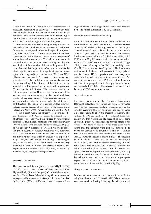
PhD Thesis 2011
(Hiraoka and Oka 2008). However, a major prerequisite forsuccessful exploitation of cultivated U. lactuca for com-mercial applications is that the growth rate and yields areoptimized. This in turn requires both an understanding ofthe influence of different nutrients on the growth responseand a precise methodology to measure the growth.
Ammonium and nitrate are the major nitrogen sources ofseaweeds in the natural habitat and are used as nourishmentfor seaweed in integrated multi-trophic aquaculture systems(Copertino et al. 2008). Several experiments have beenconducted for several seaweed species on the interaction ofammonium and nitrate uptake. The utilization of ammoni-um and nitrate by seaweed varies among species andassimilation of these nutrients influences the growth. It hasbeen observed that the presence of ammonium inhibits thenitrate uptake; thus ammonium uptake dominated nitrateuptake when exposed to a combination of NH4
+ and NO3−
(Thomas and Harrison 1987). However, these interactionswere discussed only in relation to nitrogen uptake rates andthe understanding of the influence of these interactions onseaweed growth responses, including growth responses ofU. lactuca, is still limited. The common method todetermine growth rate and biomass yield in seaweed culturesystems involves determination of the initial and finalweight of seaweed samples. This requires removal ofsurface moisture either by wiping with filter cloth or bycentrifugation. The extent of remaining surface moistureinduces varying degrees of inaccuracy to the measurement(Msuya and Neori 2008; Vandermeulen and Gordin 1990).
In this present work, the objective is to evaluate thegrowth response of U. lactuca exposed to different sourcesof nitrogen (NH4
+ and NO3−). We cultured U. lactuca frond
disks for 10 days in small containers with artificial seawater(ASW) enriched with equimolar levels of nitrogen (50 μM)of two different N-nutrients, NH4Cl and NaNO3, to assessthe growth responses. Another experiment was conductedin the same set-up for 4 days to evaluate the ammoniumand nitrate uptake rates when U. lactuca was exposed to50 μM NH4NO3. We used photo-scanning to obtain digitalimages of the sizes of the frond disks, and in this wayexamined the growth kinetics by measuring the surface areaexpansion of the seaweed disks daily using commerciallyavailable digital image processing software.
Materials and methods
The chemicals used for nitrogen source were NH4Cl (99.5%),NH4NO3 (99.5%), and NaNO3 (99.0%), purchased fromSigma-Aldrich, (Bornem, Belgium). Commercial marine seasalt (Sera Marine Basic Salt—Heinsberg, Germany) was usedto prepare artificial seawater (ASW) principally as describedby Sato et al. (2006a, b). For nitrate determination, a low-
range lab nitrate test kit supplied with nitrate reductase wasused (The Nitrate Elimination Co., Inc., Michigan).
Aquarium culture: condition and set-up
Fresh Ulva lactuca fronds were obtained from the NationalEnvironmental Research Institute of Denmark (DMI),University of Aarhus (Silkeborg, Denmark). The originalseaweed material was cultured in ponds with naturalseawater. Upon arrival to our laboratory, the seaweedfronds were transferred into a 2 L beaker containing 1.5 LASW with a 33 g L−1 concentration of marine sea salt tohabituate. The ASW medium had a pH of 8.35 and 21.5 pptsalinity measured using a handheld conductivity meter(Con 11, Eutech Instruments—Singapore). The fronds wereacclimatized in the beaker with aeration for 5 days prior totransfer into a 112-L aquarium tank for long termcultivation. The water at ambient temperature in the 112-Laquarium was led directly to a 45-L reservoir tank and thewater was then pumped back to the aquarium at a rate ofapproximately 1,700 L h−1. The reservoir was aerated andthe water (ASW) was renewed weekly.
Flask culture set-up
The growth monitoring of the U. lactuca disks duringdifferential cultivation was carried out using a perforatedflask (500 mL) positioned inside a rectangular canister thatserved as a catchment basin. The culture flask was perforatedat a certain height to allow the medium to overflow afterreaching the 300 mL level into the catchment basin. Themedium was then re-circulated at a speed of 1.5 L h−1 usinga peristaltic pump. A small magnetic bar was placed at thebottom of the flask to mix the water twice daily and toensure that the nutrients were properly distributed. Toprevent the contact of the magnetic bar and the U. lactucadisks, a 5-mm mesh was fitted inside in the middle of theflask. A schematic diagram is shown in Fig. 1. The media inthe individual flasks were enriched with 50 μM NH4Cl,50 μM NaNO3 and ASW as control, respectively. A 5-mLwater sample was collected daily to assess the ammoniumand nitrate uptake of U. lactuca. From this set-up, twoseparate cultivation experiments were conducted, a 10-daycultivation period was used for growth monitoring while a 4-day cultivation was used to evaluate the nitrogen uptakeresponse of U. lactuca to the interaction of equimolarammonium and nitrate supply (50 μM NH4NO3).
Nitrogen uptake measurement
Ammonium concentration was determined with theendophenol-blue method (Koroleff 1970). Nitrate measure-ment was conducted using low-range lab nitrate test kit
346 J Appl Phycol (2011) 23:345–351
2
114
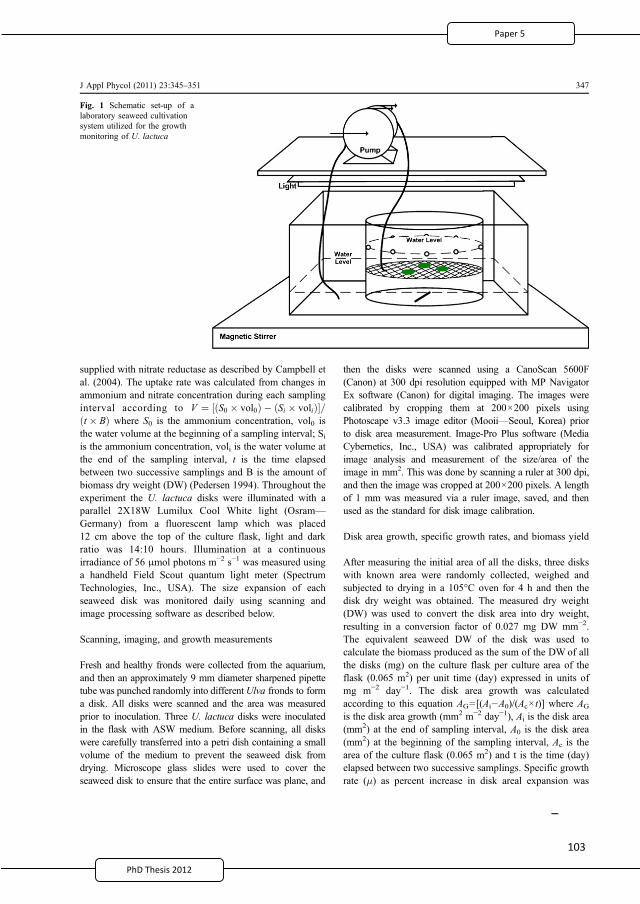
supplied with nitrate reductase as described by Campbell etal. (2004). The uptake rate was calculated from changes inammonium and nitrate concentration during each samplinginterval according to V ¼ S0 � vol0ð Þ � Si � volið Þ½ �=t � Bð Þ where S0 is the ammonium concentration, vol0 isthe water volume at the beginning of a sampling interval; Siis the ammonium concentration, voli is the water volume atthe end of the sampling interval, t is the time elapsedbetween two successive samplings and B is the amount ofbiomass dry weight (DW) (Pedersen 1994). Throughout theexperiment the U. lactuca disks were illuminated with aparallel 2X18W Lumilux Cool White light (Osram—Germany) from a fluorescent lamp which was placed12 cm above the top of the culture flask, light and darkratio was 14:10 hours. Illumination at a continuousirradiance of 56 μmol photons m−2 s−1 was measured usinga handheld Field Scout quantum light meter (SpectrumTechnologies, Inc., USA). The size expansion of eachseaweed disk was monitored daily using scanning andimage processing software as described below.
Scanning, imaging, and growth measurements
Fresh and healthy fronds were collected from the aquarium,and then an approximately 9 mm diameter sharpened pipettetube was punched randomly into differentUlva fronds to forma disk. All disks were scanned and the area was measuredprior to inoculation. Three U. lactuca disks were inoculatedin the flask with ASW medium. Before scanning, all diskswere carefully transferred into a petri dish containing a smallvolume of the medium to prevent the seaweed disk fromdrying. Microscope glass slides were used to cover theseaweed disk to ensure that the entire surface was plane, and
then the disks were scanned using a CanoScan 5600F(Canon) at 300 dpi resolution equipped with MP NavigatorEx software (Canon) for digital imaging. The images werecalibrated by cropping them at 200×200 pixels usingPhotoscape v3.3 image editor (Mooii—Seoul, Korea) priorto disk area measurement. Image-Pro Plus software (MediaCybernetics, Inc., USA) was calibrated appropriately forimage analysis and measurement of the size/area of theimage in mm2. This was done by scanning a ruler at 300 dpi,and then the image was cropped at 200×200 pixels. A lengthof 1 mm was measured via a ruler image, saved, and thenused as the standard for disk image calibration.
Disk area growth, specific growth rates, and biomass yield
After measuring the initial area of all the disks, three diskswith known area were randomly collected, weighed andsubjected to drying in a 105°C oven for 4 h and then thedisk dry weight was obtained. The measured dry weight(DW) was used to convert the disk area into dry weight,resulting in a conversion factor of 0.027 mg DW mm−2.The equivalent seaweed DW of the disk was used tocalculate the biomass produced as the sum of the DW of allthe disks (mg) on the culture flask per culture area of theflask (0.065 m2) per unit time (day) expressed in units ofmg m−2 day−1. The disk area growth was calculatedaccording to this equation AG=[(Ai−A0)/(Ac× t)] where AG
is the disk area growth (mm2 m−2 day−1), Ai is the disk area(mm2) at the end of sampling interval, A0 is the disk area(mm2) at the beginning of the sampling interval, Ac is thearea of the culture flask (0.065 m2) and t is the time (day)elapsed between two successive samplings. Specific growthrate (μ) as percent increase in disk areal expansion was
Fig. 1 Schematic set-up of alaboratory seaweed cultivationsystem utilized for the growthmonitoring of U. lactuca
J Appl Phycol (2011) 23:345–351 347
2
115
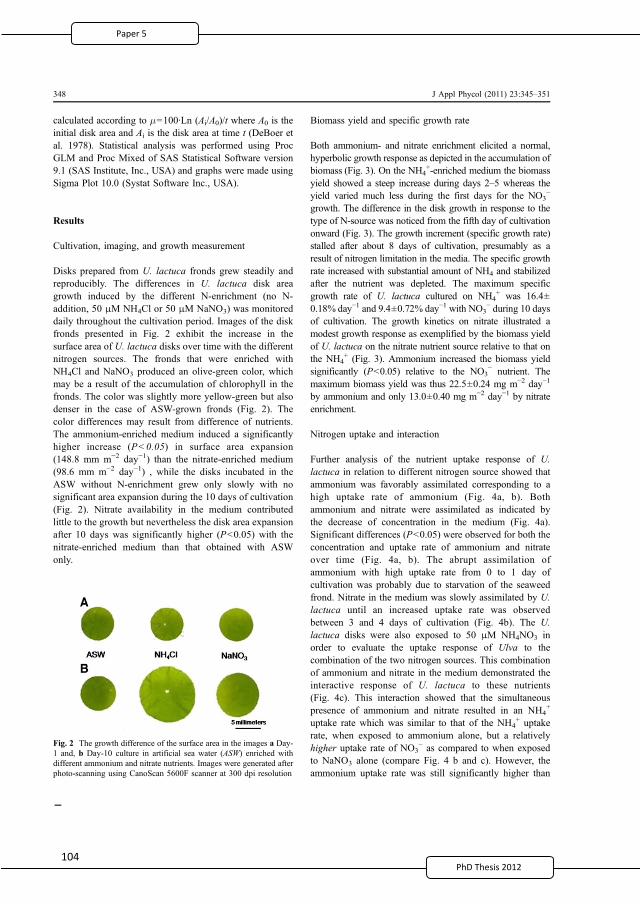
10
calculated according to μ=100·Ln (Ai/A0)/t where A0 is theinitial disk area and Ai is the disk area at time t (DeBoer etal. 1978). Statistical analysis was performed using ProcGLM and Proc Mixed of SAS Statistical Software version9.1 (SAS Institute, Inc., USA) and graphs were made usingSigma Plot 10.0 (Systat Software Inc., USA).
Results
Cultivation, imaging, and growth measurement
Disks prepared from U. lactuca fronds grew steadily andreproducibly. The differences in U. lactuca disk areagrowth induced by the different N-enrichment (no N-addition, 50 μM NH4Cl or 50 μM NaNO3) was monitoreddaily throughout the cultivation period. Images of the diskfronds presented in Fig. 2 exhibit the increase in thesurface area of U. lactuca disks over time with the differentnitrogen sources. The fronds that were enriched withNH4Cl and NaNO3 produced an olive-green color, whichmay be a result of the accumulation of chlorophyll in thefronds. The color was slightly more yellow-green but alsodenser in the case of ASW-grown fronds (Fig. 2). Thecolor differences may result from difference of nutrients.The ammonium-enriched medium induced a significantlyhigher increase (P<0.05) in surface area expansion(148.8 mm m−2 day−1) than the nitrate-enriched medium(98.6 mm m−2 day−1) , while the disks incubated in theASW without N-enrichment grew only slowly with nosignificant area expansion during the 10 days of cultivation(Fig. 2). Nitrate availability in the medium contributedlittle to the growth but nevertheless the disk area expansionafter 10 days was significantly higher (P<0.05) with thenitrate-enriched medium than that obtained with ASWonly.
Biomass yield and specific growth rate
Both ammonium- and nitrate enrichment elicited a normal,hyperbolic growth response as depicted in the accumulation ofbiomass (Fig. 3). On the NH4
+-enriched medium the biomassyield showed a steep increase during days 2–5 whereas theyield varied much less during the first days for the NO3
−
growth. The difference in the disk growth in response to thetype of N-source was noticed from the fifth day of cultivationonward (Fig. 3). The growth increment (specific growth rate)stalled after about 8 days of cultivation, presumably as aresult of nitrogen limitation in the media. The specific growthrate increased with substantial amount of NH4 and stabilizedafter the nutrient was depleted. The maximum specificgrowth rate of U. lactuca cultured on NH4
+ was 16.4±0.18% day−1 and 9.4±0.72% day−1 with NO3
− during 10 daysof cultivation. The growth kinetics on nitrate illustrated amodest growth response as exemplified by the biomass yieldof U. lactuca on the nitrate nutrient source relative to that onthe NH4
+ (Fig. 3). Ammonium increased the biomass yieldsignificantly (P<0.05) relative to the NO3
− nutrient. Themaximum biomass yield was thus 22.5±0.24 mg m−2 day−1
by ammonium and only 13.0±0.40 mg m−2 day−1 by nitrateenrichment.
Nitrogen uptake and interaction
Further analysis of the nutrient uptake response of U.lactuca in relation to different nitrogen source showed thatammonium was favorably assimilated corresponding to ahigh uptake rate of ammonium (Fig. 4a, b). Bothammonium and nitrate were assimilated as indicated bythe decrease of concentration in the medium (Fig. 4a).Significant differences (P<0.05) were observed for both theconcentration and uptake rate of ammonium and nitrateover time (Fig. 4a, b). The abrupt assimilation ofammonium with high uptake rate from 0 to 1 day ofcultivation was probably due to starvation of the seaweedfrond. Nitrate in the medium was slowly assimilated by U.lactuca until an increased uptake rate was observedbetween 3 and 4 days of cultivation (Fig. 4b). The U.lactuca disks were also exposed to 50 μM NH4NO3 inorder to evaluate the uptake response of Ulva to thecombination of the two nitrogen sources. This combinationof ammonium and nitrate in the medium demonstrated theinteractive response of U. lactuca to these nutrients(Fig. 4c). This interaction showed that the simultaneouspresence of ammonium and nitrate resulted in an NH4
+
uptake rate which was similar to that of the NH4+ uptake
rate, when exposed to ammonium alone, but a relativelyhigher uptake rate of NO3
− as compared to when exposedto NaNO3 alone (compare Fig. 4 b and c). However, theammonium uptake rate was still significantly higher than
Fig. 2 The growth difference of the surface area in the images a Day-1 and, b Day-10 culture in artificial sea water (ASW) enriched withdifferent ammonium and nitrate nutrients. Images were generated afterphoto-scanning using CanoScan 5600F scanner at 300 dpi resolution
348 J Appl Phycol (2011) 23:345–351
2
116
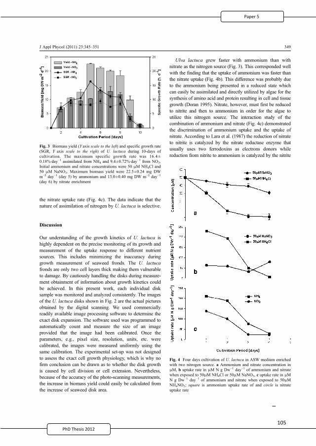
10PhD Thesis 2011
the nitrate uptake rate (Fig. 4c). The data indicate that thenature of assimilation of nitrogen by U. lactuca is selective.
Discussion
Our understanding of the growth kinetics of U. lactuca ishighly dependent on the precise monitoring of its growth andmeasurement of the uptake response to different nutrientsources. This includes minimizing the inaccuracy duringgrowth measurement of seaweed fronds. The U. lactucafronds are only two cell layers thick making them vulnerableto damage. By cautiously handling the disks during measure-ment obtainment of information about growth kinetics couldbe achieved. In this present work, each individual disksample was monitored and analyzed consistently. The imagesof the U. lactuca disks shown in Fig. 2 are the actual picturesobtained by the digital scanning. We used commerciallyreadily available image processing software to determine theexact disk expansion. The software used was programmed toautomatically count and measure the size of an imageprovided that the image had been calibrated. Once theparameters, e.g., pixel size, resolution, units, etc. werecalibrated, the images were measured uniformly using thesame calibration. The experimental set-up was not designedto assess the exact cell growth physiology, which is why nofirm conclusion can be drawn as to whether the disk growthis caused by cell division or cell extension. Nevertheless,because of the accuracy of the photo-scanning measurements,the increase in biomass yield could easily be calculated fromthe increase of seaweed disk area.
Ulva lactuca grew faster with ammonium than withnitrate as the nitrogen source (Fig. 3). This corresponded wellwith the finding that the uptake of ammonium was faster thanthe nitrate uptake (Fig. 4b). This difference was probably dueto the ammonium being presented in a reduced state whichcan easily be assimilated and directly utilized by algae for thesynthesis of amino acid and protein resulting in cell and tissuegrowth (Doran 1995). Nitrate, however, must first be reducedto nitrite and then to ammonium in order for the algae toutilize this nitrogen source. The interaction study of thecombination of ammonium and nitrate (Fig. 4c) demonstratedthe discrimination of ammonium uptake and the uptake ofnitrate. According to Lara et al. (1987) the reduction of nitrateto nitrite is catalyzed by the nitrate reductase enzyme thatusually uses two ferrodoxins as electrons donors whilereduction from nitrite to ammonium is catalyzed by the nitrite
Fig. 4 Four days cultivation of U. lactuca in ASW medium enrichedwith two nitrogen source. a Ammonium and nitrate concentration inμM, b uptake rate in μM N g Dw−1 day−1 of ammonium and nitratewhen exposed to 50μM NH4Cl or 50μM NaNO3, c uptake rate in μMN g Dw−1 day−1 of ammonium and nitrate when exposed to 50μMNH4NO3; square is ammonium uptake rate of and circle is nitrateuptake rate
Fig. 3 Biomass yield (Y axis scale to the left) and specific growth rate(SGR, Y axis scale to the righ) of U. lactuca during 10-days ofcultivation. The maximum specific growth rate was 16.4±0.18%·day−1 assimilated from NH4 and 9.4±0.72%·day−1 from NO3.Initial ammonium and nitrate concentrations were 50 μM NH4Cl and50 μM NaNO3. Maximum biomass yield were 22.5±0.24 mg DWm−2 day−1 (day 5) by ammonium and 13.0±0.40 mg DW m−2 day−1
(day 6) by nitrate enrichment
J Appl Phycol (2011) 23:345–351 349
2
117
10
reductase, a process that needs six ferrodoxins. This reductionmechanism as a consequence requires energy for thebioconversion process. Thus, U. lactuca growth responsesare different given the same concentration of nitrate nitrogenas they would on ammonium nitrogen.
The use of ASW medium over the duration of thisexperiment without renewal may have influenced thegrowth kinetics. The common experimental practice inindoor seaweed culture is water renewal (Naldi andWheeler 2002; Vermaat and Sand-Jensen 1987; Neori etal. 1991; Christensen and Sand-Jensen 1990), however, theeffect of the water renewal is unclear. Nevertheless, ourresults indicate that U. lactuca can grow even withoutrenewing the medium for a certain period. The maximumgrowth rate (μmax) in this condition was 16.39±0.18%day−1 that is close (μmax) reported in a previously publishedstudy (Neori et al. 1991), where they used fresh seawatermedium and extensive renewal of medium.
The amount of biomass accumulated during thisexperiment was relatively low as compared to otherearlier works where they harvested considerable amountsof biomass (Neori et al. 1991; Msuya and Neori 2008).The water flow velocity may be the cause of the relativelylower biomass production. In this work, the watercirculation was controlled to be 1.5 L h−1, this circulationwas almost equivalent to the water flow in the shallowbenthic zone, which should be beneficial for U. lactucagrowth. This assumption is supported by the previousworks of Doty (1971) and Parker (1981) that showed theapplication of simulated current consistently enhancedgrowth rates of U. lactuca under laboratory conditions.This was also the case in a study conducted by Msuya andNeori (2008) in which they concluded that water velocityaffected biomass yields and biofiltration performance ofU. lactuca under low nutrient concentration in laboratoryexperiments.
Acknowledgment The authors are grateful to Michael Bo Rasmussenof the National Environmental Research Institute (DMI), Univer-sity of Aarhus—Silkeborg, Denmark for providing the U. lactucastrain.
References
Abd El-Baky HH, El Baz FK, El-Baroty GS (2008) Evaluation ofmarine alga Ulva lactuca L. as a source of natural preservativeingredient. EJEAFChe 7:3353–3367
Bixler HJ (1996) Recent developments in manufacturing andmarketing carrageenan. Hydrobiologia 326(327):35–57
Bodin-Dubigeon C, Lahaye M, Barry JL (1997) Human colonicbacterial degradability of dietary fibres from sea-lettuce (Ulvasp.). J Sci Food Agric 73:149–159
Campbell WH, Kinnunen-Skidmore T, Brodeur-Campbell MJ, CampbellER (2004) New and improved nitrate reductase for enzymaticnitrate analysis. Am Lab 22:12
Christensen HF, Sand-Jensen K (1990) Growth rate and carbonaffinity of Ulva lactuca under controlled levels of carbon, pH andoxygen. Mar Biol 104:497–501
Cumashi A, Ushakova NA, Preobrazhenskaya ME, D’Incecco A,Piccoli A, Totani L, Tinari N, Morozevich GE, Berman AE,Bilan MI, Usov AI, Ustyuzhanina NE, Grachev AA, SandersonCJ, Kelly M, Rabinovich GA, Iacobelli S, Nifantiev NE(2007) A comparative study of the anti-inflammatory, antico-agulant, antiangiogenic, and antiadhesive activities of ninedifferent fucoidans from brown seaweeds. Glycobiology17:541–552
Copertino MS, Tormena T, Seeliger U (2008) Biofiltering efficiency,uptake and assimilation rates of Ulva clathrata (Roth) J. Agardh(Clorophyceae) cultivated in shrimp aquaculture waste water. JAppl Phycol 21:31–45
DeBoer JA, Guigly HJ, Isarael TL, D’Elia CF (1978) Nutritionalstudies of two red algae. I. Growth rate as a function of nitrogensource and concentration. J Phycol 14:261–266
De Roeck-Holtzhauer Y (1991) Uses of seaweed in cosmetics. In:Guiry MD, Blunden G (eds) Seaweed resources in Europe: usesand potential. Wiley, Chichester, pp 83–94
Doran PM (1995) Homogeneous reactions. Bioprocess engineeringprinciples. Academic, San Diego, pp 277–278
Doty MS (1971) Measurement of water movement in reference tobenthic algal growth. Bot Mar 14:32–35
Hiraoka M, Oka N (2008) Tank cultivation of Ulva prolifera in deepseawater using a new germling cluster method. J Appl Phycol20:97–102
Ivanova I, Rouseva R, Kolarova M, Serkedjieva J, Rachev R,Manolova N (1994) Isolation of a polysaccharide with antiviraleffect from Ulva lactuca. Prep Biochem 24:83–97
Knauf M, Moniruzzaman M (2004) Lignocellulosic biomass process-ing: a perspective. Int Sugar J 106:147–150
Koroleff F (1970) Direct determination of ammonia in natural watersas indophenol blue. In Information on techniques and methodsfor seawater analysis. Interlaboratory report No. 3. I.C.E.S.Charlottenlund, pp 19–22
Lahaye M (1991) Marine algae as sources of fibres: determination ofsoluble and insoluble dietary fibre contents in some seavegetables. J Sci Food Agric 54:587–594
Lahaye M, Axelos MAV (1993) Gelling properties of water-solublepolysaccharides from proliferating marine green seaweeds(Ulva spp.) Carbohydrate. Polymer 22:261–265
Lara C, Romero JM, Guerrero MG (1987) Regulated nitrate transportin the cyanobacterium Anacystis nidulans. J Bacteriol 169:4376–4378
Lee T-M (2000) Phosphate starvation induction of acid phosphatase inUlva lactuca L. (Ulvales, Chlorophyta). Bot Bull Acad Sin41:19–25
Magnusson G, Larsson C, Axelsson L (1996) Effects of high CO2
treatment on nitrate and ammonium uptake by Ulva lactucagrown in different nutrient regimes. Sci Mar 60:179–189
Msuya FE, Neori A (2008) Effect of water aeration and nutrient loadlevel on biomass yield, N uptake and protein content of theseaweed Ulva lactuca cultured in seawater tanks. J Appl Phycol20:1021–1031
Naldi M, Wheeler PA (2002) 15N Measurement of ammonium andnitrate uptake by Ulva fenestrata (Chlorophyta) and Gracilariapacifica (Rhodophyta): comparison of net nutrient disappearance,release of ammonium and nitrate and 15N accumulation in algaltissue. J Phycol 38:135–144
350 J Appl Phycol (2011) 23:345–351
2
118
10PhD Thesis 2011
Neori A, Cohen I, Gordin H (1991) Ulva lactuca biofilters for marinefishfond effluents II. Growth rate, yield and C:N ratio. Bot Mar34:483–489
Ortiz J, Romero N, Robert P, Araya J, Lopez-Hernandez J, Bozzo C,Navarrete E, Osorio A, Rios A (2006) Dietary fiber, aminoacid, fatty acid and tocopherol contents of the edible seaweedsUlva lactuca and Durvillaea antarctica. Food Chem 99:98–104
Parker HS (1981) Influence of relative water motion on the growth,ammonium uptake and carbon and nitrogen composition of Ulvalactuca (Chlorophyta). Mar Biol 63:309–318
Pedersen FP (1994) Transient ammonium uptake in the macroalgaUlva lactuca (Chlorophyta): nature, regulation, and the conse-quences for choice of measuring technique. J Phycol 30:980–986
Sand-Jensen K (1988) Minimum light requirements for growth inUlva lactuca. Mar Ecol Prog Ser 50:187–193
Sato K, Eksangsri T, Egashira R (2006a) Ammonia-nitrogen uptakeby seaweed for water quality control in intensive maricultureponds. J Chem Eng Japan 39:247–255
Sato K, Ueno Y, Egashira R (2006b) Uptake of nitrate-nitrogen inintensive shrimp culture ponds by sterile Ulva sp. J Chem EngJapan 39:1128–1131
Thomas TE, Harrison PJ (1987) Rapid ammonium uptake andnitrogen interactions in five intertidal seaweeds grown underfield condition. J Exp Mar Biol 107:1–8
Vandermeulen H, Gordin H (1990) Ammonium uptake using Ulva(Chlorophyta) in intensive fishpond systems: mass culture andtreatment of effluent. J Appl Phycol 2:363–374
Vermaat JE, Sand-Jensen K (1987) Survival, metabolism and growthof Ulva lactuca under winter conditions: a laboratory study ofbottlenecks in the life cycle. Mar Biol 95:55–61
J Appl Phycol (2011) 23:345–351 351
2
119

120 120
10PhD Thesis 2011
6 Conclusions and perspectives
As already stated, seaweeds are the subject of a wide range of interesting research topics;
therefore, it entails crossing beyond another border of academic discipline to elucidate the
problems and accomplish specific objectives. The present study provided information about the
most recent developments in the chemistry of fucoidan/FCSPs, emphasizing the significance of
different extraction techniques for the structural composition and biological activity using an
interdisciplinary approach. The utilization of marine seaweeds that have washed up on the
coastline as a source of bioactive compounds like FCSPs and the growth response of U. lactuca to
nutrient assimilation were thoroughly investigated.
The use of different extraction and purification techniques appears to have contributed to the
confusion about the nature and composition of FCSPs ever since it was first described by Kylin
early in the 20th century. As detailed in the following, we now know that fucoidan is built of 1 3
linked L fucopyranosyl or of alternating 1 3 and 1 4 linked L fucopyranosyl residues that
may be sulfate substituted, and that some fucoidans isolated from certain brown algae have
completely different structures composed of sulfated galactofucans with backbones of (1 6)
linked D galacto and/or (1 2) D mannopyranosyl units with (1 3) and/or (1 4) L fuco
oligosaccharide branching. The available data thus show that the term fucoidan has been used for
several different chemical structures and vice versa, that fucoidan is a term that covers a diverse
family of FCSPs (Paper 1). It is, therefore, more correct to use the term FCSPs, rather than
fucoidan, as a collective term for these polysaccharides.
The varied chemical composition and diverse structures of FCSPs from brown seaweed may have
hindered the development of an in depth understanding of the precise properties of significance
for structure function correlations. Nevertheless, important structural bioactivity issues appear to
include the degree of sulfation of the FCSP molecules. Oversulfated FCSPs have thus been found to
be excellent potent inhibitors of tumor cell invasion compared to desulfated native FCSPs. Loss of
anticoagulant activity has been observed with decreasing degrees of sulfation, although anti
proliferative effects on fibroblast cell lines were retained (Paper 1).
A simple and practical method for recovering a suite of complex FCSPs from Sargassum sp. and the
effect of different treatment parameters on the integrity of the polysaccharide have been
2
121
1 0PhD Thesis 2011
established (Paper 2). The preservation of the structural integrity of the FCSP molecules appears to
be crucial for maintaining its biological properties, and it has been clearly shown that the
extraction treatment employed affects the composition and thus the structural features of the
FCSP substances (Paper 2). Evidently, the chemical composition and yield of the isolated products
are strongly influenced by the method of extraction, as was expected. The data presented (Paper
2) showed that the polysaccharide obtained from a single step extraction method may be
heterogeneous, and that the composition varies with the length of the extraction process. Fucose,
sulfate, and glucuronic acid were the important components of the polysaccharide mixture and
this is typical for a fucoidan compound (Paper 2). The results (Paper 2) also demonstrated that
FCSPs were vulnerable to harsh extraction conditions. Hence, we confirmed that extraction
condition significantly influenced FCSP composition, and structural alteration may have occurred.
Undoubtedly, the presence of impurities could influence the biological properties of FCSPs and,
therefore, may currently hinder our complete understanding of the biological activity of fucoidan
or FCSPs. Hence, the development of standard extraction procedures for FCSPs including hydrolysis
treatment, purification, and fractionation methodology, preferably with specific steps adapted to
the particular botanical order of the seaweed, will generate a better common basis for the analysis
and understanding of bioactivities and the mechanisms determining FCSP bioactivities. On this
basis, it may even be possible to target specific structural features and, in turn, tune the extraction
procedure to obtain specific bioactivities via the use of targeted extraction methodologies.
The bioactivity of the isolated FCSP products from Sargassum sp. against LLC and MC was
investigated (Paper 3). The study showed that FCSPs induce apoptosis of MC cells (Papers 3 and 4)
and exert anti tumor activity through the inhibition of the growth of LLC and MC, which was
probably due to the enhancement of NK cell activity as the principal effector mediating tumor cell
death. We showed that both FCSPs samples from Sargassum sp. and F. vesiculosus induces
apoptosis by activating caspase 3 and exerts anti tumor activity by inhibiting the growth of cells
(Paper 4). FCSPs from Sargassum sp. and Fucus vesiculosus thus appear to be potent against lung
and skin cancer cell lines, and its mode of action is associated with the immune response (Paper 3).
Furthermore, the bioactivity of crude fucoidan toward these 2 types of cancer cell lines was
possibly augmented by the sulfate groups in the fucoidan structure. Nevertheless, further
examination about these findings is needed to elucidate the underlying factors of FCSP bioactivity.
The unfractionated FCSP structures from Sargassum sp. could probably be heterogeneous and
branched as expected; however, another possibility could be that these FCSPs were not mixtures
2
122
1PhD Thesis 2011
of different types of polysaccharides but members of the same polysaccharide family. Our
structural investigation presented by 1H NMR spectra indicate that in both Sargassum sp. and F.
vesiculosus, FSCPs samples contained fucoidan like structures, but definite structural information
about whether the structure is heterogeneous or members of the same polysaccharide family
should further be investigated (Paper 4). The quantitative variation of its components and
distribution patterns as well as the differences in its structural details was probably not due to
sample heterogeneity but rather to extreme compositional and structural dispersion. At any rate,
the crucial bioactive effectiveness of these unfractionated FCSPs may be attributed to their distinct
structural features, such as level of sulfation (charge density) and the position and bonding of the
sulfate substitutions or sulfated fucans (i.e., C 2 and/or C 4 of 3 linked L fucopyranose residues)
and sulfated galactans (i.e., C 2 of 3 linked galactopyranose residues) complexes (Paper 4).
Nonetheless, we now understand that the type and variety of algal FCSPs is much wider than
originally believed.
In addition to the investigation of the potential of the brown seaweeds Sargassum sp. and F.
vesiculosus as natural sources of bioactive compounds, the washed up nuisance green seaweed U.
lactuca was also examined for its growth and nutrient assimilation potential. This study was
performed to illustrate the need for a precise monitoring method of the growth of U. lactuca in
order to successfully exploit it for commercial application. Our understanding of the growth
kinetics of U. lactuca is highly dependent on the precise monitoring of its growth and
measurement of the uptake response to different nutrient sources. This includes minimizing
inaccuracies during the growth measurement of seaweed fronds. This work exhibited the
applicability of the photo scanning approach for attaining accurate quantitative growth data of U.
lactuca as demonstrated by evaluation of the growth response to ammonium and nitrate (Paper
5). The experimental set up was not designed to assess the exact cell growth physiology, which is
why no firm conclusion can be drawn as to whether the U. lactuca disc growth was caused by cell
division or cell extension. Nevertheless, because of the accuracy of the photo scanning
measurements, the increase in biomass yield could easily be calculated from the increase of
seaweed disc area (Paper 5). This result showed that U. lactuca grew faster with ammonium than
with nitrate as the nitrogen source (Paper 5). This corresponded well with the finding that the
ammonium uptake was faster than the nitrate uptake. This difference was probably due to the
ammonium being presented in a reduced state that can be easily assimilated and directly utilized
by algae for the synthesis of amino acids and proteins resulting in cell and tissue growth. Nitrate,
however, must first be reduced to nitrite and then to ammonium for the algae to utilize this
2
123
1PhD Thesis 2011
nitrogen source (Paper 5). The interaction study of the combination of ammonium and nitrate
demonstrated that the presence of ammonium discriminate the nitrate uptake when U. lactuca
was exposed to ammonium nitrate. This study provides new technique for evaluating the growth
of U. lactuca and outlines the possibility of U. lactuca as a good agent for bioremediation.
6.1 Future perspectivesMarine seaweed has gained a lot of attention in the scientific community and in industries; thus,
the potential biological effects of seaweeds have been exploited has been examined extensively in
recent years. Still, there are many avenues in seaweed research, that need to be considered,
studied, and understood to successfully exploit this abundant resource. This PhD study highlighted
a few of the crucially important factors in the utilization of seaweed products, notably the
extraction procedure and bioactivity analysis of FCSPs. Nevertheless, there are other research
areas concerning seaweed resources that need further investigation; they may include
bioprocesses and purification involving enzyme technology coupled with membrane technology
coupled with the concept of seaweed biorefinery.
A typical technology for isolating valuable products from seaweeds, notably bioactive compounds
like FCSPs, involves the use of chemicals. This technique has been employed since the first
extraction of fucoidan by Kylin in 1913. This present study demonstrated that chemical extraction
of fucans from brown seaweeds contained impurities. The apparent impurities (i.e., saccharides
other than sulfated fucose) may or may not contribute to the bioactivity efficacy of fucoidan. To
reach a definitive conclusion, this hypothesis must be examined strategically using enzymes that
cleave specific sites. Extraction using enzymes coupled with membrane technology could be a
future strategy for recovering bioactive seaweed compounds. Mono component activity enzymes
may be employed to eliminate and/or hydrolyze specific impurities, including certain
monosaccharides and structural polysaccharides. Chemically and/or enzymatically isolated FCSPs
compounds can be further purified by filtration using membranes to separate the molecular size of
interest. Purified components will be investigated to determine whether they possess any
biological active properties against certain diseases using in vitro, in vivo, animal models, and/or
possibly clinical testing.
Seaweed cultivation and processing is an important industry in Southeast Asia, but it is still new to
Scandinavia, especially Denmark. As described in this study, only selected components are
obtained from seaweeds (i.e., FCSPs, fucoidan, alginate, carrageenan, and agar), and the remaining
are considered waste. This present study proposed a new type of strategy to improve the
2
124
1PhD Thesis 2011
utilization of seaweed by using the entire seaweed frond for the production of chemicals and/or
biochemicals and energy. Thus, a concept of seaweed biorefinery should be developed.
The first phase of this concept includes isolation of commercially important
chemicals/biochemicals such as hydrocolloids and the bioactive sulfated polysaccharide
compounds. In the second phase, the remaining residues are collected as bulk biomass and then
converted into chemicals and energy carriers (heat, liquid, gas, and electricity) by catalytic and
enzymatic conversions and/or by digestion and pyrolysis. The residue containing minerals are
targeted for use in fertilizer applications in the final phase of the biorefinery cycle.
Many potential applications exist for the products acquired from biorefineries. Seaweed
components derived thereof can be used as hydrocolloids for food and feed applications or can be
formulated into natural health supplements; in contrast, mineral containing seaweed residues are
incorporated into fertilizer products.
The detailed processes, procedures, and specific working conditions of this seaweed biorefinery
concept are yet to be determined.
2
125

126 126
11PhD Thesis 2011
7 References
Abd El Baky HH, El Baz FK, El Baroty GS (2008) Evaluation of marine alga Ulva lactuca L. as a sourceof natural preservative ingredient. EJEAFChe 7(11): 3353 3367
Abdel fattah AF, Hussein MD, Salem HM (1974) Studies of purification and some properties ofsargassan, a sulfated heteropolysaccharide from Sargassum linifolium. Carbohydrate Research33:9 17
Adhikari, U.; Mateii, C.G.; Chattopadhyay, K.; Pujol, C.A.; Damonte, E.B.; Ray, B. (2006) Structureand antiviral activity of sulfated fucans from Stoechospermum marginatum. Phytochemistry. 67,2474–2482.
Aisa Y, Miyakawa Y, Nakazato T, Shibata H, Saito K, Ikeda Y, Kizaki M (2005) Fucoidan inducesapoptosis of human HS sultan cells accompanied by activation of caspase 3 and down regulation ofERK pathways. Am J Hematol. 78: 7 14
Ale, M.T.; Maruyama, H.; Tamauchi, H.; Mikkelsen, J.D.; Meyer, A.S. (2011) Fucoidan fromSargassum sp. and Fucus vesiculosus reduces cell viability of lung carcinoma and melanoma cells invitro and activates natural killer cells in mice in vivo. Intl. J. Biol. Macromol., 49, 331 336
Ale, M.T.; Mikkelsen, J.D.; Meyer, A.S. (3992011) Designed optimization of a single step extractionof fucose containing sulfated polysaccharides from Sargassum sp. J. Appl. Phycol., DOI 10.1007/s10811 011 9690 3
Alekseyenko TV, Zhanayeva SY, Venediktova AA, et al. (2007) Antitumor and antimetastatic activityof fucoidan, a sulfated polysaccharide isolated from the Okhotsk Sea Fucus evanescens brown alga.Bull Exp Biol Med.143(6):730 2
Anastyuk SD, Shevchenko NM, Nazarenko EL, et al., (2009) Structural analysis of a fucoidan fromthe brown alga Fucus evanescens by MALDI TOF and tandem ESI mass spectrometry. CarbohydrRes. 344: 779 87
Berteau O, Mulloy B (2003) Sulfated fucans, fresh perspectives: structures, functions, and biologicalproperties of sulfated fucans and an overview of enzymes active toward this class ofpolysaccharide. Glycobiology 13: 29R 40R
Bilan, M.I.; Usov, A.I. (2008) Structural Analysis of Fucoidans. Nat. Prod. Comm. 3,1639 1648.
Bilan MI, Grachev AA, Shashkov AS, et al., (2006) Structure of a fucoidan from the brown seaweedFucus serratus L. Carbohydr Res. 341: 238 45
Bilan MI, Grachev AA, Ustuzhanina NE et al., (2002) Structure of a fucoidan from brown seaweedFucus evanescens. Carbohydr. Res., 337, 719–730
2
127
11PhD Thesis 2011
Bilan MI, Usov AI (2008) Structural Analysis of Fucoidans. Natural Product Communications 3:16391648
Bird, GM, Hass, P (1931) On the nature of the cell wall constituents of Laminaria spp . mannuronicacid. Biochem . J. 25 : 403 411
Bixler HJ (1996) Recent developments in manufacturing and marketing carrageenan. Hydrobiologia326: 35–57
Black WAP, Dewar ET, Woodward FN (1952) Manufacturing of algal chemicals 4: Laboratory scaleisolation of fucoidan from brown marine algae. Journal of the Science of Food and Agriculture3:122 129
Blondin, C.; Fischer, E.; Boisson Vidal, C.; Kazatchkine, M.D.; Jozefonvicz, J. (1994) Inhibition ofcomplement activation by natural sulfated polysaccharides (fucans) from brown seaweed. Mol.Immunol., 31: 247–253
Bodin Dubigeon C, Lahaye M, Barry JL (1997) Human colonic bacterial degradability of dietary fibresfrom sea lettuce (Ulva sp.). J Sci Food Agric 73: 149–159
Boisson Vidal C, Chaubet F, Chevolot L, et al., (2000) Relationship between antithrombotic activitiesof fucans and their structure. Drug Dev. Res. 51: 216–224
Bruhn A, Dahl J, Nielsen HB, et al. (2011) Bioenergy potential of Ulva lactuca: Biomass yield,methane production and combustion. Bioresource Technology 102: 2595 2604
Buck BH, Bucholz CM (2004) The offshore ring: A new system for the open ocean aquaculture ofmacroalgae. Journal of Applied Phycology 16: 355 368
Burdin KS, Bird KT (1994) Heavy metals accumulation by carrageenan and agar producing algae.Bot. mar. 37: 467 470
Chevolot L, Foucault A, Chaubet F, Kervarec N, Sinquin C, Fisher AM, Boisson Vidal C (1999) Furtherdata on the structure of brown seaweed fucans: relationships with anticoagulant activity.Carbohydr Res. 319(1 4):154 65.
Chizhov AO, Dell A, Morris HR, Haslam SM, McDowell RA et al (1999) A study of fucoidan from thebrown seaweed Chorda filum. Carbohydrate Research 320:108 119
Chopin T, Bushmann AH, Halling C, et al. (2001) Integrated seaweeds into marine aquaculturesystems: a key toward sustainability J. Phycol. 37: 975 986
Christensen H F, Sand Jensen K (1990) Growth rate and carbon affinity of Ulva lactuca undercontrolled levels of carbon, pH and oxygen. Mar. Biol. 104: 497 501.
Chung IK, Kang YH, Yarish C, et al. (2002) Application of seaweed cultivation to the bioremediationof nutrient rich effluents. Algae 17: 187 194
2
128
11PhD Thesis 2011
Conchie J, Percival EGV (1950) Fucoidan part II. The hydrolysis of a methylated fucoidin preparedfrom Fucus vesiculosus, Journal of Chemical Society 1950, pp. 827–833
Copertino MS, Tormena T, Seeliger U (2008) Biofiltering efficiency, uptake and assimilation rates ofUlva clathrata (Roth) J. Agardh (Clorophyceae) cultivated in shrimp aquaculture waste water. J ApplPhycol 21:31–45
Crawford, B. 2002. Seaweed Farming: An Alternative Livelihood for Small Scale Fishers? CoastalResources Center, University of Rhode Island, USA.
Cumashi A, Ushakova NA, Preobrazhenskaya ME, et al., (2007) A comparative study of the antiinflammatory, anticoagulant, antiangiogenic, and antiadhesive activities of nine different fucoidansfrom brown seaweeds. Glycobiology 17: 541–552
De Reiter GA, Rudolph B (1997) Carrageenan biotechnology. Trends in foods science & technology8, 389–395
De Roeck Holtzhauer Y (1991) Uses of seaweed in cosmetics. In: Guiry MD, Blunden G (eds)Seaweed resources in Europe: uses and potential. Wiley, Chichester, pp 83–94
DeBoer JA, Guigly HJ, Isarael TL, D’Elia CF (1978) Nutritional studies of two red algae. I. Growth rateas a function of nitrogen source and concentration. J Phycol 14:261 266
Duarte ME, Cardoso MA, Noseda MD, Cerezo AS (2001) Structural studies on fucoidans from thebrown seaweed Sargassum stenophyllum. Carbohydr Res. 333(4):281 93
Ermakova S, Sokolova R, Kim S M, Um B H, Isakov V, Zvyagintseva T (2011) Fucoidans from brownseaweeds Sargassum hornery, Eclonia cava, Costaria costata: Structural characteristics andanticancer activity. Appl Biochem Biotechnol 164, 841 – 850.
FAO (2004) FAO Fisheries Department, Fishery Information, Data and Statistics Unit. FISHSTAT Plus.(Universal Software for fishery statistical time series). Version 2.3. last updated March 2004. Foodand Agriculture Organisation. United Nations. Rome, Italy.
Foley, S.A.; Mulloy, B.; Tuohy, M.G. (2011) An unfractionated fucoidan from Ascophyllum nodosum:Extraction, characterization, and apoptotic effects in vitro. J. Nat. Prod., DOI: 10.1021/ np200124m
Grauffel V, Kloareg B, Mabeau S, et al., (1989) New natural polysaccharides with potentantithrombic activity: Fucans from brown algae. Biomaterials 1989, 10, 363–368
Grossi F, Kubota K, Cappuzzo F, de Marinis F, Gridelli C, Aita M, Douillard JY (2010): Future scenariosfor the treatment of advanced non small cell lung cancer: focus on taxane containing regimens.Oncologist 15:1102 12
Grüven KC, Saygi N, Öztürk B (1993) Surveys of metals contents of Bosphorus algae, Zostera marinaand sediments. Bot. Mar. 36:175 178
2
129
11PhD Thesis 2011
Harlin MM, Wheeler PA (1985) Nutrient uptake. In Littler MM and Littler D eds. Ecological fieldmethods: Macroalgae. Handbook of phycological methods. vol. 4. Cambridge University Press, NewYork pp 493 508
Hayakawa K, Nagamine T (2009): Effect of fucoidan on the biotinidase kinetics in humanhepatocellular carcinoma. Anticancer Res. 4: 1211 7
Hiraoka M, Oka N (2008) Tank cultivation of Ulva prolifera in deep seawater using a new germlingcluster method. J Appl Phycol 20: 97–102
Hoagland, DR, Lieb, LL (1915) The complex carbohydrates and forms of sulphur in marine algae ofthe Pacific coast . J . biol. Chem . 23 : 287 297
Holtkamp AD, Kelly S, Ulber R, Lang S (2009) Fucoidans and fucoidanases focus on techniques formolecular structure elucidation and modification of marine polysaccharides. Appl MicrobiolBiotechnol. 82(1):1 11
Hurtado AQ, Agbayani RF, Sanares R, et al., (2001) The seasonality and economic feasibility ofcultivating Kappaphycus alvarezii in Panagatan Cays, Caluya, Antique, Philippines. Aquaculture. 199:295 310
Ivanova I, Rouseva R, Kolarova M, Serkedjieva J, Rachev R, Manolova N (1994) Isolation of apolysaccharide with antiviral effect from Ulva lactuca. Prep Biochem 24(2):83 97
Jemal A, Siegel R, Xu J, Ward E (2010) Cancer Statistics 2010. CA Cancer J Clin, doi:10.3322/caac.20073
Jiao G, Yu G, Zhang J, Ewart SE (2011) Chemical structure and bioactivities of sulfatedpolysaccharides from marine algae. Marine Drugs 9, 196 – 223; doi: 10.3390/md9020196.
Kim EJ, Park SY, Lee JY, Park JH (2010) Fucoidan present in brown algae induces apoptosis of humancolon cancer cells. BMC Gastroenterol.10:96
Kindt TJ, Goldsby RA, Osborne BA (2007): Cell mediated cytotoxic responses. In Kuby Immunologyby W.H. Freeman and Company, NY. Chap. 14, p360 – 363.
Knauf M, Moniruzzaman M (2004) Lignocellulosic biomass processing: a perspective. Int Sugar J106:147–150
Koyanagi, S.; Tanigawa, N.; Nakagawa, H.; Soeda, S.; Shimeno, H. (2003) Oversulfation of fucoidanenhances its anti angiogenic and antitumor activities. Biochem. Pharmacol., 65, 173 – 179.
Kuda T, Taniguchi E, et al. (2002) Fate of water soluble polysaccharides in dried Chorda filum abrown alga during water washing. Journal of Food Composition and Analysis 15:3 9
Kylin K. (1913) Zur Biochemie der Meeresalgen. Hoppe Seyler's Z. Physiol. Chem. 83: 171 197
Kylin, H. (1915) Analysis of the biochemistry of the seaweed. H. Z. Physiol. Chem. 94, 337 – 425
2
130
11PhD Thesis 2011
Lahaye M (1991) Marine algae as sources of fibres: determination of soluble and insoluble dietaryfibre contents in some sea vegetables. J Sci Food Agric 54:587–594
Lahaye M, Axelos MAV (1993) Gelling properties of water soluble polysaccharides fromproliferating marine green seaweeds (Ulva spp.) Carbohydrate. Polymer 22: 261–265
Lahaye M, Robic A (2007) Review: Structure and functional properties of Ulvan, a polysaccharidefrom green seaweeds. American Chemical Society. 8: No. 6
Lee T M (2000) Phosphate starvation induction of acid phosphatase in Ulva lactuca L. (Ulvales,chlorophyta). Bot Bull Acad Sin 41:19–25
Li, B.; Wei, X.J.; Sun, J.L.; Xu, S.Y. (2006) Structural investigation of a fucoidan containing a fucosefree core from the brown seaweed, Hizikia fusiforme. Carbohydr. Res., 341, 1135 – 1146
Li B, Lu F, Wei X, Zhao R (2008) Fucoidan: structure and bioactivity. Molecules 13: 1671 – 1695
Lunde G, Heen E, Oy E, (1937) Uber fucoidin . Hoppe Seyler's Z . Physiol . Chem. 247 : 189 196
Lydyard PM, Whelan A, Fanger MW (2000) Instant Notes Immunology. BIOS Scientific PublisherLimited, Oxford, UK p13, 88, 89.
Makarenkova ID, Deriabin PG, L'vov DK, Zviagintseva TN, Besednova NN (2010): Antiviral activity ofsulfated polysaccharide from the brown algae Laminaria japonica against avian influenza A (H5N1)virus infection in the cultured cells. Vopr Virusol. 55(1):41 5
Marais MF, Joseleau JP (2001) A fucoidan fraction from Ascophyllum nodosum. CarbohydrateResearch 336:155 159
Maruyama H, Tamauchi H, Hashimoto M, Nakano T (2003): Antitumor activity and immuneresponse of Mekabu fucoidan extracted from Sporophyll of Undaria pinnatifida. In Vivo 17(3):2459.
Maruyama H, Tamauchi H, Iizuka M, Nakano T (2006) The role of NK cells in antitumor activity ofdietary fucoidan from Undaria pinnatifida sporophylls (Mekabu). Planta Med. 72(15):1415 7
McHugh DJ (2003) A Guide to the Seaweed Industry, Food and Agriculture Organisation, UnitedNations, Fisheries Technical Paper 441, Rome, Italy, 123 pp.
Mian AJ, Percival E (1973) Carbohydrates of the brown seaweds Himanthalia lorea, Bifurcariabifurcata, and Padina pavonia. Carbohydr. Res. 26: 133 146
Morand P, Briand X, Charlier RH (2006) Anaerobic digestion of Ulva sp 3. Liquefaction juicesextraction by pressing and a technico economic budget. J. Appl. Phycol. 18, 741 755.
Morris, E. R., & Norton, A. N. 1983. Polysaccharide aggregation in solutions and gels. In E. WynJones & J. Gormally (Eds.), Aggregation processes in solution (pp. 549–593). Amsterdam: Elsevier.
2
131
1PhD Thesis 2011
Mourão, PAS (2004) Use of sulfated fucans as anticoagulant and antithrombotic agents: futureperspectives. Curr. Pharmaceut. Des., 10, 967 981
Mourão, P.A.S., Pereira, M.S., Pavão, M.S.G., Mulloy, B., Tollefsen, D.M., Mowinckel, M.C.,Abildgaard, U. (1996) Structure and anticoagulant activity of a fucosylated chondroitin sulfate fromechinoderm: sulfated fucose branches on the polysaccharide accounts for its high anticoagulantaction. Journal of Biological Chemistry, 271, 23973 – 23984
Msuya FE, Neori A: Effect of water aeration and nutrient load level on biomass yield, N uptake andprotein content of the seaweed Ulva lactuca cultured in seawater tanks. J. Appl. Phycol. 2008, 20:1021–1031.
Nagaoka M, Shibata H, Kimura Takagi I, et al., (1999) Structural study of fucoidan from Cladosiphonokamuranus TOKIDA. Glycoconj J. 16(1):19 26.
Naldi M, Wheeler PA (2002) 15N Measurement of ammonium and nitrate uptake by Ulvafenestrata (Chlorophyta) and Glacilaria pacifica (Rhodophyta): Comparison of net nutrientdisappearance, release of ammonium and nitrate and 15N accumulation in algal tissue. J Phycol38:135–144
Nardella, A.; Chaubet, F.; Boisson Vidal, C.; Blondin, C.; Durand, P.; Jozefonvicz, J. (1996)Anticoagulant low molecular weight fucans produced by radical process and ion exchangechromatography of high molecular weight fucans extracted from the brown seaweed Ascophyllumnodosum. Carbohydr. Res. 289, 201–208
Nastala CL, Edington HD, McKinnet TG, et al., (1994) Recombinant IL 12 administration inducestumor regression in association with IFN gamma production. J. Immunol., 153, 1697 1706.
Nelson, WL, Cretcher, LH (1931) The carbohydrate acid sulfate of Macrocystis pyrifera . J. biol .Chem. 94 : 147 154
Neori A, Cohen I, Gordin H (1991) Ulva lactuca biofilters for marine fishfond effluents II. Growthrate, yield and C:N ratio. Bot Mar 34:483 489
Nishino T, Aizu Y, Nagumo T (1991) The influence of sulfate content and molecular weight of afucan sulfate from the brown seaweed Ecklonia kurome on its antithrombin activity. Thromb Res.64(6):723 31.
Nishino T, Kiyohara H, Yamada H, Nagumo T (1991) An anticoagulant fucoidan from the brownseaweed Ecklonia kurome. Phytochemistry 1991, 30, 535–539
Nishino T, Nagumo T (1987) Sugar constituents and blood coagulant activities of fucose containingsulfated polysaccharide in nine brown seaweed species. Nippon Nogeikagaku Kaishi, 61 361 363.
Nishino T, Nagumo T (1991) The sulfate content dependence of the anticoagulant activity of afucan sulfate from the brown seaweed Ecklonia kurome. Carbohydr Res. 214:193 197.
2
132
1PhD Thesis 2011
Nishino T, Yokoyama G, Dobash K, Fujihara M, Nagumo T (1989) Isolation, purification, andcharacterization of fucose containing sulfated polysaccharides from the brown seaweed Eckloniakurome and their blood anticoagulant activities. Carbohydr. Res., 186, 119–129
Nishino T, Takabe Y, Nagumo T (1994) isolation and partial characterization of a novel D galactanfrom the brown seaweed Laminaria angustata var. longissima. Carbohydrate Res. 23, 165 – 173.
Ortiz J, Romero N, Robert P, Araya J, Lopez Hernandez J, Bozzo C, Navarrete E, Osorio A, Rios A(2006) Dietary fiber, amino acid, fatty acid and tocopherol contents of the edible seaweeds Ulvalactuca and Durvillaea Antarctica. Food Chem 99:98 104.
Otsuka, K., Yoshino, A., 2004. A fundamental study on anaerobic digestion of sea lettuce. In: Oceans‘04 Mts/Ieee Techno Ocean ‘04, vols. 1–2. Conference Proceedings, vols. 1–4. pp. 1770–1773.
Patankar, M.S.; Oehninger, S.; Barnett, T.; Williams, R.L.; Clark, G.F. (1993) A revised structure forfucoidan may explain some of its biological activities. J. Biol. Chem., 268, 21770 6
Parham P (2009) The immune system. Garland Science, Taylor and Francis Group, LLC. New York,USA
Pedersen FP (1994) Transient ammonium uptake in the macroalga Ulva lactuca (Chlorophyta):nature, regulation, and the consequences for choice of measuring technique. J Phycol 30:980 986
Pedersen MF, Borum J (1996) Nutrient control of algal growth in estuarine waters: nutrientlimitation and the importance of nitrogen requirements and nitrogen storage amongphytoplankton and species of macroalgae. Mar Ecol prog Ser 142: 261 272
Percival EGV, McDowell RH (1967) Chemistry and Enzymology of Marine Algal Polysaccharides.Academic press London, UK
Percival EGV and Ross AG (1950) Fucoidin. Part 1. The Isolation and purification of fucoidan frombrown seaweeds J. Chem. Soc. (1950), 717
Pereira MG, Benevides NMB, Melo MRS, et al., (2005) Structure and anticoagulant activity of asulfated galactan from the red alga, Gelidium crinale. Is there a specific structural requirement forthe anticoagulant action? Carbohydr. Res., 340, 2015–2023.
Pereira MG, Mulloy B, Mourão PA (1999) Structure and anticoagulant activity of sulfated fucans.Comparison between the regular, repetitive, and linear fucans from echinoderms with the moreheterogeneous and branched polymers from brown algae. J Biol Chem. 274:7656 67.
Phillips JR, Oliver MJ, Bartels D (2002) Molecular Genetics of desiccation and tolerant systems. InDesiccation and survival in plants: drying without dying; Black, M.; Pritchard, H.W., Eds.; CABIPublishing: UK: pp 263 291.
Pomin VH, Pereira MS, Valente AP, et al (2005) Selective cleavage and anticoagulant activity of asulfated fucan: Stereospecific removal of a 2 sulfate ester from the polysaccharide by mild acid
2
133
1PhD Thesis 2011
hydrolysis, preparation of oligosaccharides, and heparin cofactor II dependent anticoagulantactivity. Glycobiology, 15, 369–381
Qiu XD, Amarasekara A, Doctor V (2006) Effect of oversulfation on the chemical and biologicalproperties of fucoidan. Carbohydrate Polymers, 63, 224 228
Rahman MA, Amin AR, Shin DM (2010) Chemopreventive potential of natural compounds in headand neck cancer. Nutr Cancer 62(7):973 87
Rasmussen M B, Jensen PD, Bruhn A, et al., (2009) Aquatic biomass for biofuels. Earth andEnvironmental Science. 6: 182010.
Rasmussen MB, Fossing H, Markager S, Bruhn A (2009) Anvendelse af marine algaer til fremstillingaf biobrændstof. 15. Danske Havforskermøde, Helsingør, January 2009. Abstracts p.67
Rocha HAO, Moraes FA, Trindade ES, et al. (2005) Structural and hemostatic activities of a sulfatedgalactofucan from the brown alga Spatoglossum schroederi An ideal antithrombotic agent? J BiolChem 280: 41278 41288
Sakai T, Kimura H, Kato I (2002) A marine strain of Flavobacteriaceae utilizes brown seaweedfucoidan. Marine Biotechnology 4:399 405
Sand Jensen K (1988) Minimum light requirements for growth in Ulva lactuca. Mar Ecol Prog Ser50:187 193
Sato K, Eksangsri T, Egashira R (2006) Ammonia nitrogen uptake by seaweed for water qualitycontrol in intensive mariculture ponds. J Chem Eng Japan 39:247 255
Schneider U, Stipper A, Besserer J (2010) Dose response relationship for lung cancer induction atradiotherapy dose. Zeitschrift für Medizinische Physik Vol 20, Issue 3, 2010
Semenov AV, Mazurov AV, Preobrazhenskaia ME, et al., (1998): Sulfated polysaccharides asinhibitors of receptor activity of P selectin and P selectin dependent inflammation. Vopr Med Khim.44(2):135 44
Shanmugam M, Mody KH (2000) Heparinoid active sulphated polysaccharides from marine algae aspotential blood anticoagulant agents. Current Science 79:1672–1683
Sievanen L, Crawford B, Pollnac R, Lowe C (2005) Weeding through assumptions of livelihoodapproaches in ICM: Seaweed farming in the Philippines and Indonesia. Ocean & CoastalManagement. 48: 297 313
Solomonson LP, Barber MJ (1990) Assimilatory nitrate reductase: functional properties andregulation. Annu. Rev Plant Physiol Plant Mol Biol 41: 225 253
Springer GF, Wurzel HA, McNeal Jr GM, Ansell NJ, Doughty MF (1957) Isolation of anticoagulantfractions from crude fucoidin. Proc Soc Exp Biol Med. 94: 404 9
2
134
1PhD Thesis 2011
Takahashi M (1983) Studies on the mechanism of host mediated antitumor action of fucoidan froma brown alga Eisenia bicyclis. J. Jpn Soc Reticuloendothel Sys 22: 269 – 283
Talarico LB; Damonte EB (2007) Interference in dengue virus adsorption and uncoating bycarrageenans. Virology, 363, 473–485
Teas J (1983) The dietary intake of Laminaria, a brown seaweed , and breast cancer prevention.Nutr Cancer 4: 217 22
Thomas TE, Harrison PJ (1987) Rapid ammonium uptake and nitrogen interactions in five intertidalseaweeds grown under field condition. J Exp. Mar. Biol 107:1–8
Trinchero, J.; Ponce, N.M.A.; Cordoba, O.L.; Flores, M.L.; Pampuro, S.; Stortz, C.A., Salomon, H.;Turk, G. (2009) Antiretroviral activity of fucoidans extracted from the brown seaweed Adenocystisutricularis. Phytother. Res., 23, 707–712
Trono, G. C. (1990) Seaweed Resources in the Developing countries of Asia: Production andSocioeconomic implications. In: Dogma, I.J., G.C. Trono and R.A. Tabbada (eds.). Culture and Use ofAlgae in Southeast Asia. Proceedings of a symposium on culture and utilization of algae inSoutheast Asia, 8 11 December 1981. Aquaculture Department, Southeast Asia FisheriesDevelopment Center. Tigbauan, Iloilo, Philippines. pp. 1 8.
Twilley RR, Kemp WM, Staver KW, et al. (1985) Nutrient enrichment of estuarine submersedvascular plant communities. 1. Algal growth and effects on production of plants and associatedcommunities. Marine Ecology Progress Series 23: 179 191
Usov AI, Bilan MI, Klochkova NG (1998) Polysaccharides of algae. 53. Brown alga Laminariasaccharina (L.) Lam. as a source of fucoidan. Russian Journal of Bioorganic Chemistry, 24, 382 389
van de Velde, F. & De Reuiter, G. A. 2002. Carrageenan. Biopolymers. p. 245 273
Vandermeulen H, Gordin H (1990) Ammonium uptake using Ulva (Chlorophyta) in intensivefishpond systems: mass culture and treatment of effluent. J App Phycol 2: 363 374
Veena CK, Josephine A, Preetha SP, Varalakshmi P, Sundarapandiyan R(2006): Renal peroxidativechanges mediated by oxalate: the protective role of fucoidan. Life Sci.79(19):1789 95
Vermaat JE, Sand Jensen K (1987) Survival, metabolism and growth of Ulva lactuca under winterconditions: a laboratory study of bottlenecks in the life cycle. Mar Biol 95:55 61
Wang J, Zhang QB, Zhang Z, Song H, Li P (2010): Potential antioxidant and anticoagulant capacity oflow molecular weight fucoidan fractions extracted from Laminaria japonica. Int J Biol Macromol.2010 Jan 1;46(1):6 12.
Wang J, Zhang QB, Zhang ZS, Li Z (2008) Antioxidant activity of sulfated polysaccharide fractionsextracted from Laminaria japonica. International Journal of Biological Macromolecules 42:127 132
2
135
12PhD Thesis 2011
Werner A, Clarke D, Kraan S (2003) Strategic Review of the Feasibility of Seaweed Aquaculture inIreland. Marine Institute, Technology Park, Parkmore, Galway, Ireland (www.marine.ie/marinertdi)
Yamamoto I, Maruyama H (1985): Effect of dietary seaweed preparations on 1,2dimethylhydrazine induced intestinal carcinogenesis in rats. Cancer Lett. 26(3):241 51
Yamamoto I, Maruyama H, Moriguchi M (1987): The effect of dietary seaweeds on 7,12 dimethylbenz[ ]anthracene induced mammary tumorigenesis in rats. Cancer Lett. 35(2):109 18
Yamamoto I, Nagumo T, Takahashi M, Fujihara M, Suzuki Y, Iizima N (1981) Antitumor effect ofseaweeds, 3. Antitumor effect of an extract from Sargassum kjellmanianum. Jpn J Exp Med. 51:187189
Yamamoto I, Nagumo T, Yagi K, Tominaga H, Aoki M (1974) Antitumor effect of seaweeds. 1.Antitumor effect of extract from Sargasum and Laminaria. Jpn J Exp Med. 44:543 546
Yamamoto I, Takahashi M, Tamura E, Maruyama H, Mori H (1984) Antitumor activity of ediblemarine algae: Effect of crude fucoidan fractions prepared from edible brown seaweed against L1210 leukemia. Hydrobiologia 116/117
Yamasaki Miyamoto Y, Yamasaki M, Tachibana H, Yamada K: Fucoidan induces apoptosis throughactivation of caspase 8 on human breast cancer MCF 7 cells. J Agric Food Chem 2009, 57: 86778682
Zhang Q, Li N, Zhao T, Qi H, Xu Z, Li Z (2005) Fucoidan inhibits the development of proteinuria inactive Heymann nephritis. Phytother Res. 19(1):50 3
Zhu W, Ooi VEC, Chan PKS, Ang PO (2003) Isolation and characterization of a sulfatedpolysaccharide from the brown alga Sargassum patens and determination of its anti herpesactivity. Biochemistry and Cell Biology Biochimie Et Biologie Cellulaire 81:25 33
Zhu Z, Zhang Q, Chen L, Ren S, Xu P, Tang Y, Luo D (2010) Higher specificity of the activity of lowmolecular weight fucoidan for thrombin induced platelet aggregation. Thromb Res. 125(5):419 26
Zhuang, C.; Itoh, H.; Mizuno, T.; Ito, H. (1995) Antitumor active fucoidan from brown seaweed,Umitoranoo (Sargassum thunbergii). Bio. Biotechnol. Biochem., 59, 563–567
Zvyagintseva TN, Shevchenko NM, Popivnich IB, Isakov VV et al (1999) A new procedure for theseparation of water soluble polysaccharides from brown seaweeds. Carbohydrate Research 322:3239
2
136

138
138
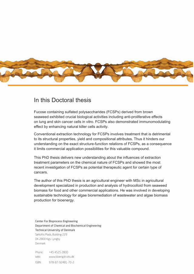
octoral
Center For Bioprocess Engineering
Department of Chemical and Biochemical Engineering
Technical University of Denmark
Søltofts Plads, Building 229
DK-2800 Kgs. Lyngby
Denmark
Phone: +45 4525 2800
Web: www.bioeng.kt.dtu.dk
ISBN :
978-87-92481-70-2
mata
Typewritten Text
mata
Typewritten Text
mata
Typewritten Text
mata
Typewritten Text
mata
Typewritten Text
mata
Typewritten Text
mata
Typewritten Text
mata
Typewritten Text


![Sulfated zirconia[1]](https://static.fdocuments.net/doc/165x107/5568f2ecd8b42aff2e8b4932/sulfated-zirconia1.jpg)