Role of Microwave in Cytology, Quick HP Diagnosis And Cell...
69
ACKNOWLEDGEMENTS I thank Dr. Mathilde Boon of Leids Cytology and Histopathology centre , Leiden, Netherlands and Dr. Lambrecht Kok, Physicist at Groienghen University Netherlands for sharing all their knowledge.
Transcript of Role of Microwave in Cytology, Quick HP Diagnosis And Cell...

ACKNOWLEDGEMENTS
I thank Dr. Mathilde Boon of Leids Cytology andHistopathology centre , Leiden, Netherlands and
Dr. Lambrecht Kok, Physicist at GroienghenUniversity Netherlands for sharing all their
knowledge.

Role of Microwave inCytology, Cell Block and QuickHP Diagnosis : An alternative
to frozen section.
Dr.Meera Govindarajan

Properties of microwave
Electro magnetic waves. Frequency 2.45GHz Dipolar molecules oscillate at 2450
million times a second They move in a straight line as waves They show reflection, refraction and
absorption
Electro magnetic waves. Frequency 2.45GHz Dipolar molecules oscillate at 2450
million times a second They move in a straight line as waves They show reflection, refraction and
absorption

Wave pattern if waves moved in single direction– a two dimensional representation

Properties of Microwave
Microwave transparent material : eg.Glass, Plastic used as containers.
Microwave resistant materials : eg.Metals
Microwave absorbable materials : eg.Biological tissue, water, Alcohol etc
Microwave transparent material : eg.Glass, Plastic used as containers.
Microwave resistant materials : eg.Metals
Microwave absorbable materials : eg.Biological tissue, water, Alcohol etc

Penetration depth of Microwave in chemical mediums
MEDIUM Depth per cm
Water at 37*C 4.4
at 45*C 5.4
at 55*C 6.6at 55*C 6.6
Paraffin wax 15,000
Methyl alcohol 1.5
Ethyl Alcohol 7
Propyl Alcohol 3.5
Butyl Alcohol 5

Penetration depth of Microwave into Tissue
mediums
Tissue MediumDepth per cm
at 37*C
Muscle 2
Skin 1.5
Liver 2.1
Lung 2.5Lung 2.5
Spleen 1.8
Kidney 2
Brain 2.5
Bone marrow 10
Fat 6
Bone 12

Cycle time of Microwave. Once the Magnetron isheated it releases 2.45 hertz MW with a 800Wattheating capacity till the Magnetron switches off

Comparison of routine Tissue Fixationprocedures with Microwave induced
fixation
Fixation –in formalinfor all tissues.
All tissues need 10%Formalin
Time depends onsize and may varyfrom 2-3 hours to8hours.
Fixation not requiredfor small biopsies.
Rest of the tissueneeds 5% formalin
Time depends onsize and may varyfrom 3mins to 30mins
Fixation –in formalinfor all tissues.
All tissues need 10%Formalin
Time depends onsize and may varyfrom 2-3 hours to8hours.
Fixation not requiredfor small biopsies.
Rest of the tissueneeds 5% formalin
Time depends onsize and may varyfrom 3mins to 30mins


COMPARISON OF ROUTINEDEHYDRATION PROCEDURE WITH
MICROWAVE HEATING
Graded Alcohol is used.
Usually either Ethyl orIsopropyl Alcohol areused.
Procedure is slow.2 to 6 hours
Speed of the proceduredepends on size/load oftissue/ temperature andVacuum
Two different Alcoholsare used sequentially
The two alcohols usedare E.g. Ethyl/Isopropylor Methyl/Isopropyl
Procedure is fast .10 mins to 30 mins.
Time can be furthershortened by use ofVacuum

Use of Clearing agents and their rolein the two procedures
Xylene or chloroformis essential
The tissue may gethardened
A minimum of 1-2hours of processingin two changes isneeded
Not required
Costly chemicals likeXylene can beavoided.
Isopropyl Alcoholacts as theintermediate .
Xylene or chloroformis essential
The tissue may gethardened
A minimum of 1-2hours of processingin two changes isneeded
Not required
Costly chemicals likeXylene can beavoided.
Isopropyl Alcoholacts as theintermediate .

IMPREGNATION WITH PARAFFIN WAX –Comparison of the two procedures
Paraffin Wax takes alonger time forimpregnation.
Good impregnation isdependent on clearingand dehydration
The wax quality isaltered as xylene mixeswith it
The tissue in the blockis comparatively harder
The impregnation timeis shortened to 10-30minutes.
Good impregnation isachieved fast anddepends on MW
The wax is reused asthe quality does notdeteriorate.
The tissue in the blockis softer and goodquality sections areeasily obtained.

Tissue Processing: (Total time 21 – 24mins) forRapid processing of less then 1mm thick tissue/
or cell block only
Immerse the Cassettes in Methyl Alcohol andmicrowave for 4 mins at 450 W.
Drain the alcohol from the cassettes and immerse inIsopropyl Alcohol. Microwave for 4 min at 450 W.
Drain the alcohol from the cassettes. Immerse cassettes in hot liquid paraffin wax and
microwave for 7 mins at 750-800 W. Embed the tissues in wax. Keep the block on ice for
cooling. Cut 5 micron sections and stain with Haematoxylin
and Eosin.CAUTION: If the tissue is very soft and 1mm thick
bit is not easy to gross then fix the bit in 10%Formalin for 30 secs at 450W MW beforeproceeding to step1

Various advantages of Microwaveprocessing
Cost of setting up theprocessing unit – less thanRs. 10,000/- for routine aswell as Quick diagnosis ifMW is used.
Technicians find it an easy,quick and clean procedure.
Pathologist is more familiarwith HPE sections. Alsoabundant time is availablefor discussions and analysis.
Cost varies betweenRs.60,000 to Rs.5 lakhsdepending on the instrumentpurchased i.e. Tissueprocessor/cryostat.
Time consuming. Large areais used.
Frozen sections need moreexperienced Pathologists.Routine HPE may not allowenough time for discussions.

Various advantages of Microwaveprocessing (contd)
Frozen sections have torefrigerated for storing.Remaining tissue is tobe stored in liquidnitrogen.
Technical support of theEngineer is essential incase of breakdown.Magnetron/ Electronic/Mechanical parts needsto imported
Wax blocks are availablefor Immuno-histochemistry, specialstains / Other studies.Storage is simple
Technical support iseasily available and costsless for repair. A newinstrument may bepurchased immediatelywhen the magnetron lifeis over.

Various disadvantages of DomesticMicrowave oven
The time taken for quick diagnosis is around30 minutes. An experienced department cangive frozen section reports in 5-15 minutes.
The domestic oven lacks temperature/ outputcontrols as compared to Microwave histologyprocessors.
Vacuum facility is not present Hotspot
The time taken for quick diagnosis is around30 minutes. An experienced department cangive frozen section reports in 5-15 minutes.
The domestic oven lacks temperature/ outputcontrols as compared to Microwave histologyprocessors.
Vacuum facility is not present Hotspot

Wave pattern in three dimensionalrepresentation with the formation of hot spots

Hot Spot
A hotspot is formed whenthe temperature rises to140-200 degrees C.
The spot shows signs ofexcess heating as in theadjacent figure
If the area covered in ahotspot is crucial fordiagnosis then destain theslide. Heat the slide in MWat 400 W in Xylene for5mins. Remove from Xyleneand Stain.
A hotspot is formed whenthe temperature rises to140-200 degrees C.
The spot shows signs ofexcess heating as in theadjacent figure
If the area covered in ahotspot is crucial fordiagnosis then destain theslide. Heat the slide in MWat 400 W in Xylene for5mins. Remove from Xyleneand Stain.

62 Year old man with 25 years history of smoking/ pan paragconsumption came with complaints of difficulty in swallowing liquids for
2 days. Endoscopy showed total obstruction of the Esophagus at the 25cmlevel. Emergency surgical intervention was essential. More then 70
sections at the end of 42 mins, helped in diagnosis.

Factors which influence Tissue processing
Size of the bits Methodology of packing in the beaker Total processing load / position in the
Microwave oven Type of tissue and its absorption/penetration
qualities Output of Microwave oven - lower
800W/900W output better then 1200W Power supply - specially if less then 200Volts.
Size of the bits Methodology of packing in the beaker Total processing load / position in the
Microwave oven Type of tissue and its absorption/penetration
qualities Output of Microwave oven - lower
800W/900W output better then 1200W Power supply - specially if less then 200Volts.

Hazards of Microwave Oven
A. Accidental malfunction of the doormay lead to:1. burn injury of the hand –skin/ deepmuscle injury with sparing of fat2. Anesthesia of the injured part3. Dysthesia
B. Injury secondary to inhalation of fumesC. Potential to develop Cataract
A. Accidental malfunction of the doormay lead to:1. burn injury of the hand –skin/ deepmuscle injury with sparing of fat2. Anesthesia of the injured part3. Dysthesia
B. Injury secondary to inhalation of fumesC. Potential to develop Cataract

Various practical applications of Microwaveprocessing in Histopathology department
Fixation of largespecimen
Quick diagnosis of anysoft tissue biopsymaterial
Cell block study H&E and other special
stains of Cytology andhistology material.
Decalcification
Large macro tissueprocessing.
Immunohistochemistry Antigen retrieval In situ hybridisation
study Immunofluorescence
staining Electron microscopy
processing/ staining

36 Year old male with history of smoking and drinking for17 years was brought with complaints of haematemesis. AEndoscope was cautiously passed and revealed a bleeding
ulcer with necrotic debris in the stomach. The gastriccontents were removed . No biopsy taken.

A 62 year old male with obstructive jaundice with an ulceratedgrowth at the Ampulla of vater. Attempt to biopsy the growthcaused bleeding. Only brush smears sent. Benign and malignantcells in a background of necrosis and Mucinous material seen.Patient operated at some other centre was diagnosed to haveAdenocarcinoma of the Ampulla.

A 94 Year old female presented with blood and mucus instool with diarrhea. Patient was uncooperative.
Colonoscope could not be passed beyond rectum becauseof spasm. Only necrotic tissue could be removed. A cell
block preparation of the necrotic tissue

Inguinal mass in a 76yr old man suspected to havemetastatic deposits from tumor arising in a non healed
ulcer of foot. FNAC revealed pleomorphic cells with largepleomorphic nuclei.

Histology revealed a pleomorphic spindle cellsarcoma.

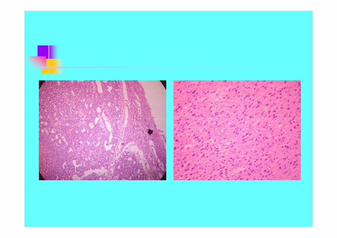
33 year old healthy female presenting with hard gritty mass5x5 cms in the inner quadrant of breast . Mammogram suggested
a malignancy. FNAC done. Fungal infection was suggested.

FNAC from Breast mass with biopsyof the mass for quick diagnosis
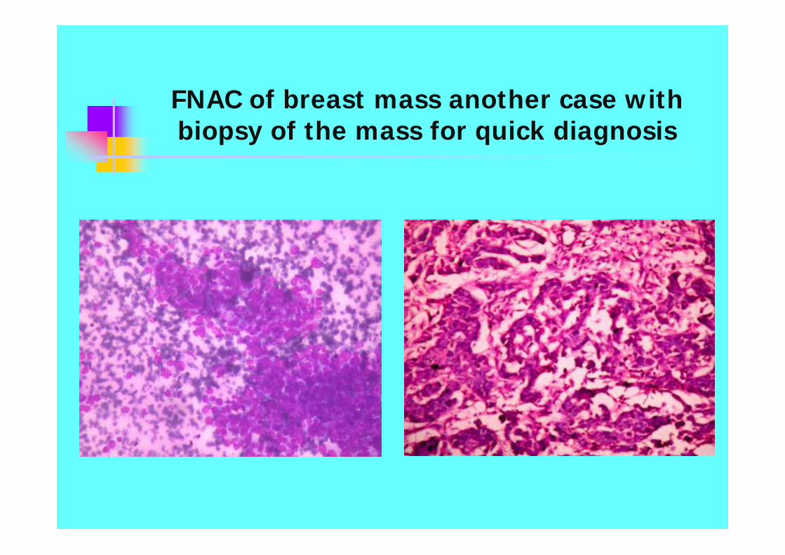
FNAC of breast mass another case withbiopsy of the mass for quick diagnosis

Pleural effusion metastasis

Ovarian Cyst aspiration1. Follicular cyst aspirate
2. Cell block - papillary serous adenoma3. Mucinous carcinoma aspirate

62 year Old female with history of back pain and nonhealing wound at the L2- L4 level following a road Traffic
accident showed an osteolytic lesion in the vertebra.Necrotic debris from the wound was reported as caseous
material and a slide sent for second opinion.

PAS stain of the same slide was carried out.Thefoamy cells revealed fungal spores of
Histoplasma Capsulatum

75 Year Old female with a discharging sinus tract in theabdominal wall was unfit for surgery due to cardiac
problems. Discharge sent for culture and cytology. A cellblock of the discharge revealed dense clusters of fungal
hyphae and necrotic tissue.

Fungal stains were carried out. A picture of the pigmentedconidiophore( fruiting bodies) seen in the block and
dichotomous branching of Hyphae suggested AspergillusFumigatus.

52 year old Male with an inguinal node 2x2 cms . FNACdone . 2cc of haemorrhagic aspirate received showed
brown sediments. The cell bock sections are shown below.Smears were haemorrhagic.

A melanin bleach was carried out on cell blocksections. Biopsy of the node was carried out.
Sections of the node are shown below.

Ultra sound guided Para Aortic mass aspiratewith evidence of granulomatous reaction
probably Tuberculous. Note the langhan’s giantcells.

FNAC from a Para Aortic mass in a 50 year oldmale – Clinical suspicion of Tuberculosis. A
diagnosis of Massive Lymphadenopathy withsinus histiocytosis was suggested.

FNAC was carried out on 58 Year old Male withpain abdomen and left sided Para Aortic mass
with necrosis. Clinical diagnosis was Renal Cellcarcinoma with metastasis.

CT and Ultrasound study ruled out renalPathology. The cytology on smear and cell block
section of necrotic material is shown below.

A diagnosis of metstatic deposits probablyfrom left Testis was considered based on
clinicopathological analysis

Clinical examination of Testis was normal. Ultrasound revealed a 1.5 diameter mass at one pole
of the Testis with necrosis. Orchidectomysections showed Teratocarcinoma


40 Year old Male came with the history oftesticular enlargement. FNAC of the mass sent .Seminoma was suggested. Orchidectomy carried
out. Sections revealed sheets of round cellsnegative for LCA & CD 20.

OTHER APPLICATIONS OF MICROWAVE
Special stains

SPECIAL STAINS:1. AFB- Zeil Neilson : 2. Liver – Masson’s Trichrome : 3.Gastricmucosa – Methylene Blue for H.Pylori : 4. Liver -Reticulin stain

Decalcification
For large bits 1-2 cubic cms.1.Fixed bone tissue is MW in 5% Formic acid at 650 -
700W power for 30 to 45 minutes depending on thesize of the bone tissue. Washed for 6 hours inrunning water. Large bit grossed and 2-3 mm thickbone cut.
2.The 2-3 mm bone tissue is left overnight in 10%Formic acid. Washed for 6 hours in running water.Usually decalcification is complete.
3.In case further decalcification is required step one isrepeated for 10 to 45 minutes depending on the sizeand hardness of the bone.


QUICK IMMUNOHISTOCHEMISTRY
Step 1: Blocking Endogenous Peroxide . MW the slide for 5secson 300W power in 3% Hydrogen peroxide. Wash in PBS andMW for 10 secs at 300W in PBS
Step 2:Add Blocking antibody and MW for 10 secs at 300W.Remove excess serum.
Step 3: Add primary Antibody and MW for 15 secs at 300W.Wash with PBS as above.
Step 4: Add link antibody and MW for 15 secs at 300W. Wash asabove with PBS.
Step 5. Add StreptAvidin Peroxidase and MW for 15secs at300W power.
Step 6: Chromogen DAB substrate is added and incubated atroom temperature for 4-6 mins.
CAUTION: THE SLIDES MUST BE KEPT IN MOISTURE CHAMBERTHROUGH OUT THE PROCESS

34year old lady presenting with altered behavior and severe frontalheadache of 3 months duration. CT scan showed a frontal partlysolid, partly necrotic tumor. ST biopsy sent for quick diagnosis.

The sections revealed a lymphoma. Quick IHC carriedout showed CD20 and CD45 (LCA) positivity.

Quick Immunohistochemistry carried out toassess the pancreatic function of Islet cell donor–Sections of the tail of the pancreas in a case of
death due to road traffic accident.

Modifying a poorly processed block
Old autopsy blocks Poorly processed Blocks Poorly fixed tissue
Old autopsy blocks Poorly processed Blocks Poorly fixed tissue

MODIFYING A OLD/ BADLY PROCESSED BLOCK:Slide and Poorly processed block sent for opinion. AIDS patientwith L2-L3 Osteolytic bone lesion. The sections were thick and
Lymphoid morphology was not clear.

Sections after modification

Procedure for modifying block
Blocks were reprocessed in paraffinwax.
450W for 5 minutes. 800 W output for 5 minutes 10 minutes of 600W of MW.
Blocks were reprocessed in paraffinwax.
450W for 5 minutes. 800 W output for 5 minutes 10 minutes of 600W of MW.

ANTIGEN RETRIEVAL
The exposure of Antigen or Epitope which are hiddendue to Fixation induced cross linkages is termed asAntigen retrieval.Various methods of antigen retreival are:
1. Enzymes like Trypsin, Pronase.2. Microwaving of tissue sections in Ionic solutionslike Citrate at PH 6.0 and at 9.5, Aluminium chloride,Lead Thiocynate, Zinc salt solutions etc.3. Heating and boiling in Ionic solutions specially withpressure cookers.

MiB-1 activity in Protoplasmic Astrocytomagrade 2 and in Anaplastic Astrocytoma Grade 3
following Antigen retrieval.

Oestrogen positivity in Duct carcinoma ofbreast, Prostatic carcinoma and in
Meningioma following Antigen retrieval

Use of microwave in in situ hybridisation(TUNEL) using Fluorescein dye to demonstrateapoptosis ( Programmed cell death) based on
labeling of DNA strands
MW slide in citratebuffer for Antigenretrieval-5mins
Wash thrice in Tris,each wash for 15 mins
Incubate in Reactivemixture for 2hours at37Degrees.
Wash thrice in Tris,each wash for 15 mins
Mount and observe
MW slide in citratebuffer for antigenretrieval – 5mins
Wash thrice in Tris, MWat 400W for 1min each
Incubate in MW withReactive mixture for2mins, at 400W
Wash thrice in Tris, MWat 400W for 1 min each.
Mount and observe

Picture on left shows positive Fluorescence after routineincubation technique. Picture on right shows Fluorescencefollowing incubation in MW. Note the lack of background
autofluorescence due to Formalin.

Picture on the right shows the same case as seenin a confocal microscope

THANK YOU