ThinPrep ® General Cytology Lecture Series Cerebrospinal Fluid Cytology.
25
ThinPrep ThinPrep ® ® General General Cytology Cytology Lecture Series Lecture Series Cerebrospinal Fluid Cytology
-
Upload
willis-woods -
Category
Documents
-
view
264 -
download
4
Transcript of ThinPrep ® General Cytology Lecture Series Cerebrospinal Fluid Cytology.
ThinPrepThinPrep®® General Cytology General Cytology Lecture SeriesLecture Series
Cerebrospinal Fluid Cytology
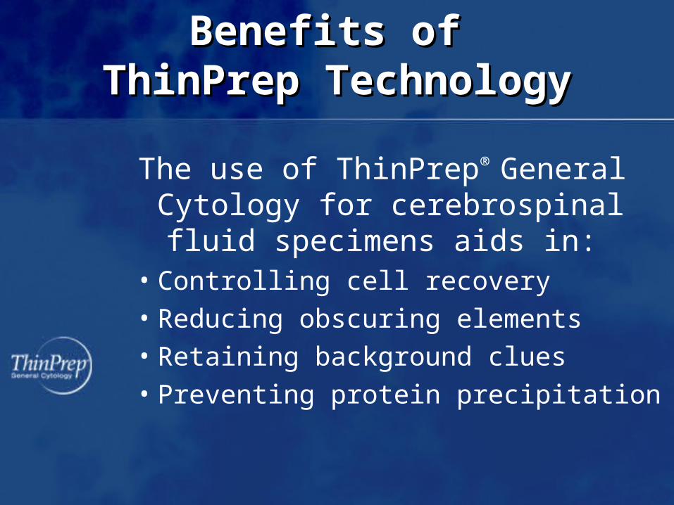
Benefits of Benefits of ThinPrep TechnologyThinPrep Technology
The use of ThinPrep® General Cytology for cerebrospinal fluid specimens aids in:
• Controlling cell recovery• Reducing obscuring elements• Retaining background clues• Preventing protein precipitation
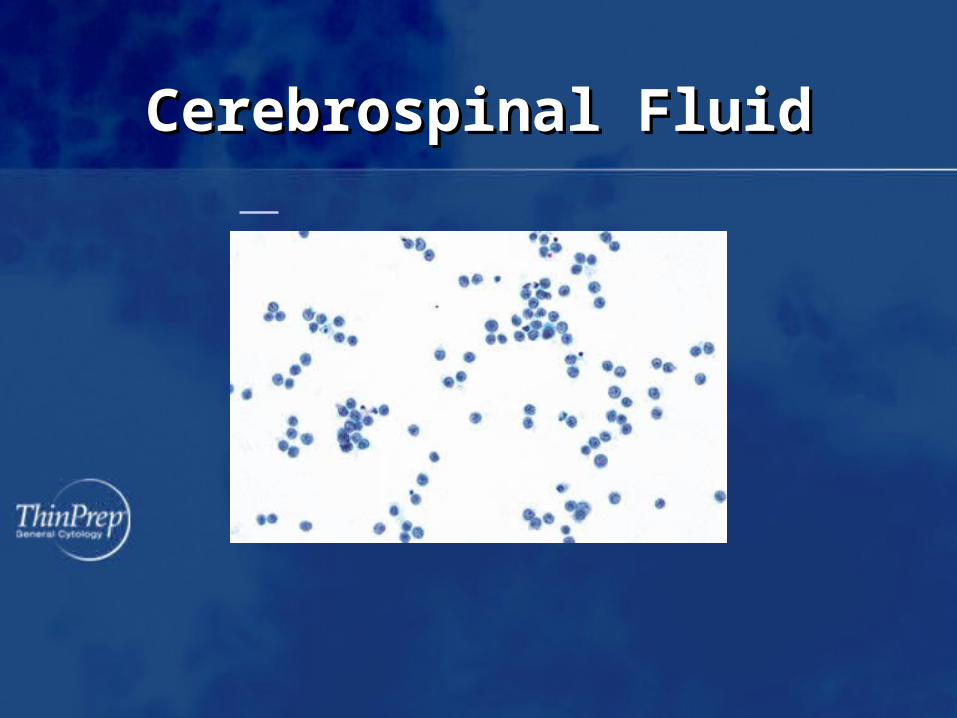
Cerebrospinal FluidCerebrospinal Fluid
AnatomyAnatomy
• Subarachnoid space– The space that surrounds the brain and
spinal cord – Contains approximately 80-100 ml of
cerebrospinal fluid (CSF)– Lined internally by the pia mater and
externally by the arachnoid membrane
Biological Nature of CSFBiological Nature of CSF
• Created mainly by filtration of plasma through the choroid plexus
• Low specific gravity
• Contains proteins, inorganic salts and dextrose
• Is normally acellular
Normal Components and Normal Components and FindingsFindings
• Lumbar puncture– May appear more cellular with ThinPrep due
to better cell retrieval– Rare lymphocytes, monocytes and PMN’s– Cells from surrounding tissue
• Ependymal cells• Arachnoidal cells• Choroid plexus cells
Normal Components and Normal Components and FindingsFindings
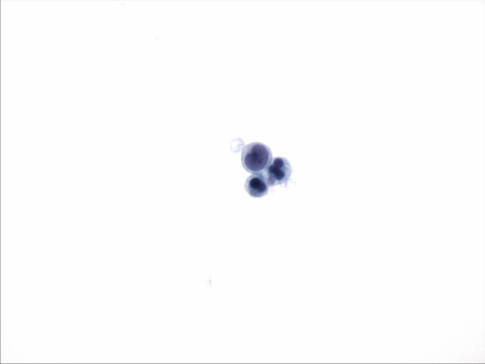
• Ventricular fluid – Abundant choroid plexus cells– Neurons– Capillaries– May see multinucleated giant cells
Normal Components and Normal Components and FindingsFindings
• Contaminants– Cellular
• Squamous cells• Chondrocytes• Red blood cells
– Non-cellular• Talc
• Causes of nonmalignant meningitis/encephalitis– Bacterial– Viral– Fungal
Benign EntitiesBenign Entities
Cytology of Benign EntitiesCytology of Benign Entities
• Acute inflammatory process– Bacterial
• Predominance of PMN’s
– Viral• Predominance of active lymphocytes
– Fungal• Cellular pattern may depend on immune status
of patient
• May be mixed inflammatory cell infiltrate
Cytology of Benign EntitiesCytology of Benign Entities
• Chronic inflammatory process– Lymphocytes typically predominate in most
chronic infections– Monocytes– Histiocytes
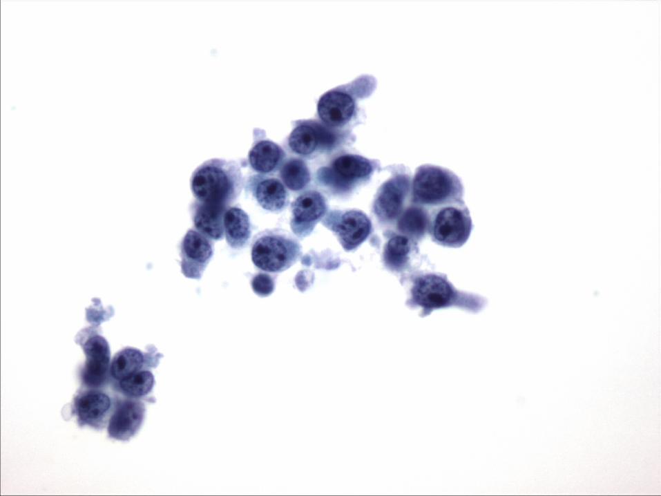
Primary Malignant Disease Primary Malignant Disease
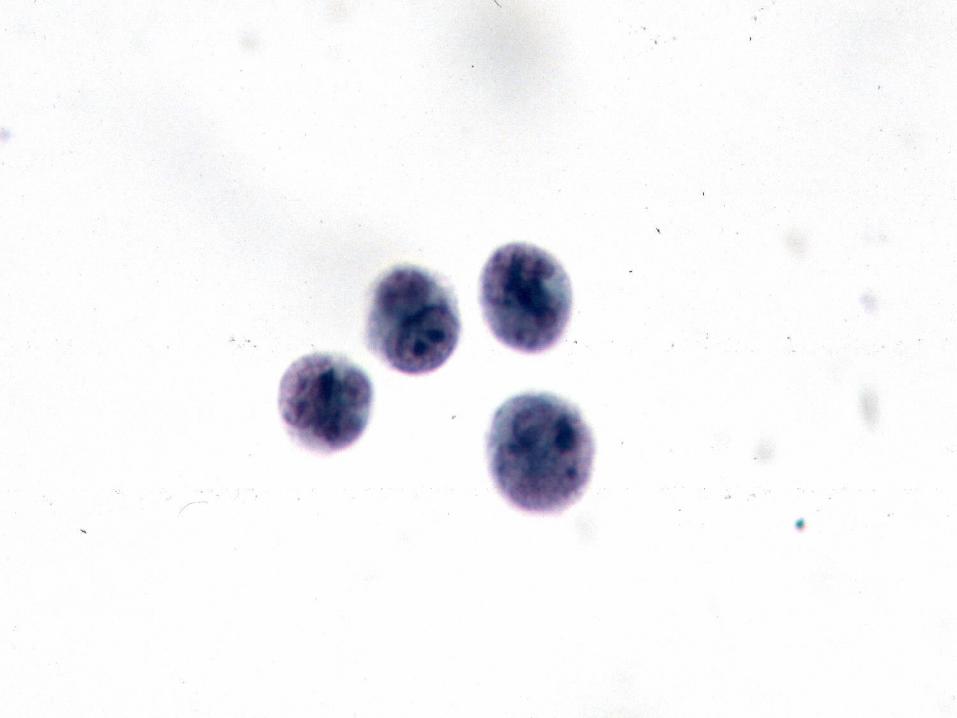
• Leukemia– Leukemic cells are larger than normal
lymphocytes– Nuclei are irregular and three dimensional– Mitotic figures can be seen– Nucleoli may be prominent
Primary Malignant Disease Primary Malignant Disease
• Lymphoma– Singly distributed usually monomorphic
population of cells with high N:C ratio– Nuclei are irregular with clumpy chromatin– Macronucleoli may be present– Mitotic activity may be evident
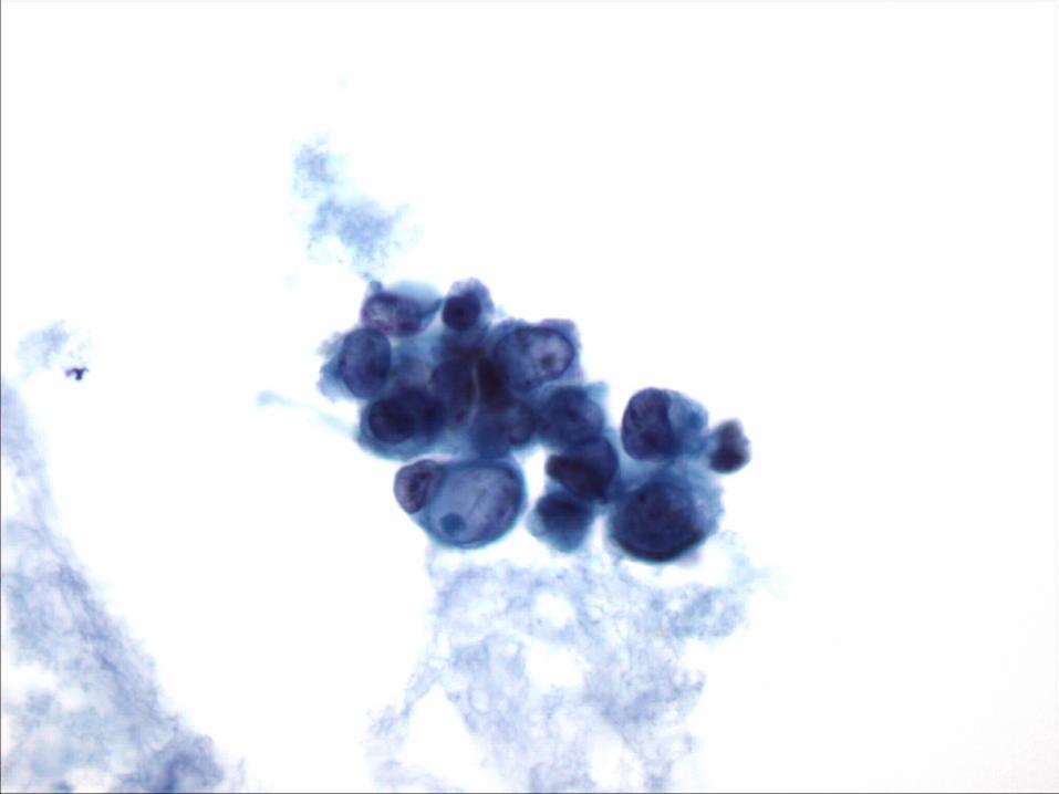
Metastatic Malignant Disease Metastatic Malignant Disease
• Adenocarcinoma– Cells often present singly or in small clusters– Nuclei are irregular, three dimensional and
eccentrically located– Nucleoli are often present– There may be cytoplasmic vacuolization
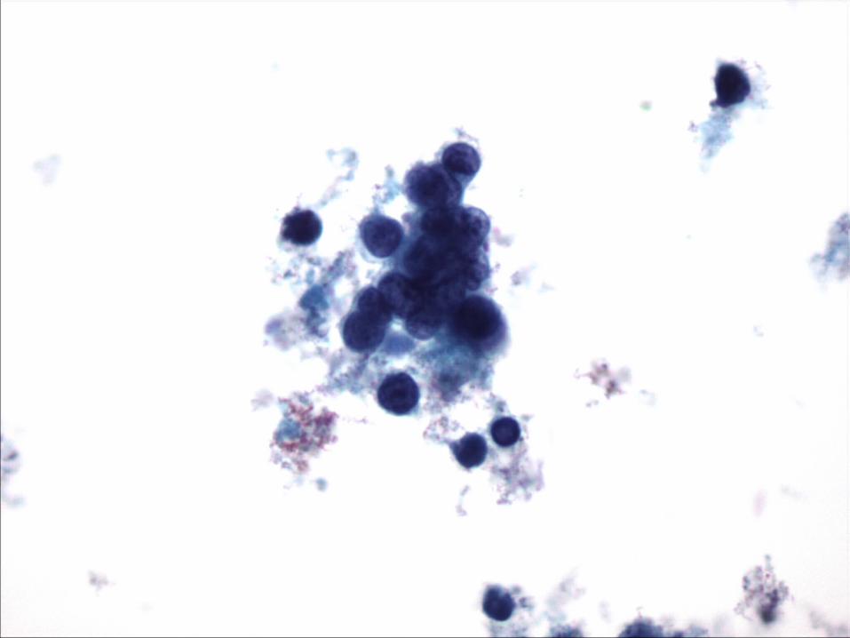
Metastatic Malignant Disease Metastatic Malignant Disease
• Small cell carcinoma– Cells are present in small, molded groups– Nuclei exhibit classic salt and pepper
chromatin pattern and may be angular– Cells have only a scant rim of fragile
cytoplasm
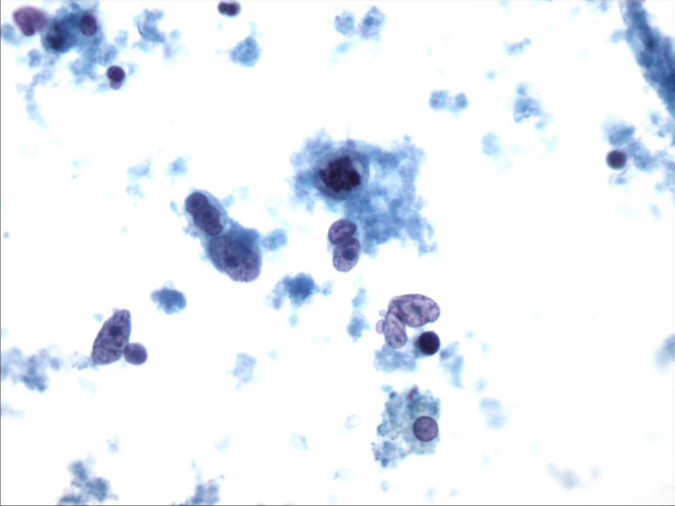
Metastatic Malignant Disease Metastatic Malignant Disease
• Malignant melanoma– Cells are usually singly distributed with
occasional loose clusters– Nuclei are round to oval, centrally or
eccentrically located and may be multiple– Nuclear chromatin is vesicular with
eosinophilic macronucleoli– Coarse brown melanin granules may be
present within the cytoplasm

For more information…For more information…
• Refer to your ThinPrep 2000 Operator’s Manual
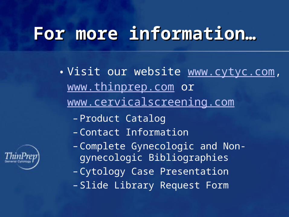
For more information…For more information…
• Visit our website www.cytyc.com, www.thinprep.com or www.cervicalscreening.com
– Product Catalog
– Contact Information
– Complete Gynecologic and Non-gynecologic Bibliographies
– Cytology Case Presentation
– Slide Library Request Form
BibliographyBibliography
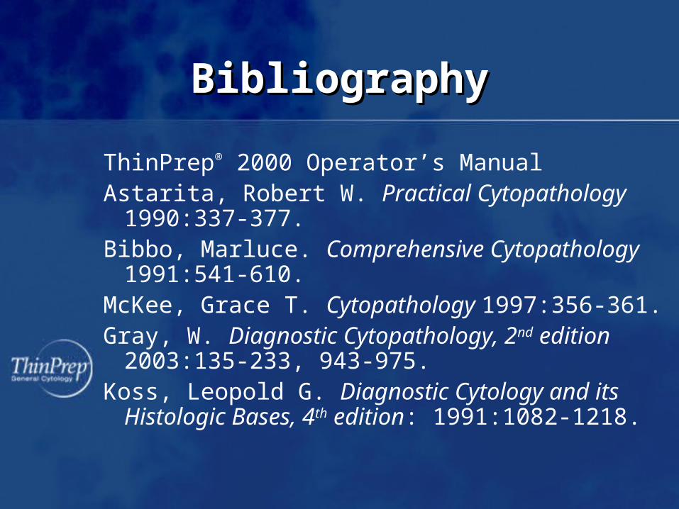
ThinPrep® 2000 Operator’s ManualAstarita, Robert W. Practical Cytopathology
1990:337-377.Bibbo, Marluce. Comprehensive Cytopathology
1991:541-610.McKee, Grace T. Cytopathology 1997:356-361.Gray, W. Diagnostic Cytopathology, 2nd edition
2003:135-233, 943-975.Koss, Leopold G. Diagnostic Cytology and its
Histologic Bases, 4th edition: 1991:1082-1218.