Title Hologic Proprietary © 2012 ThinPrep ® Non-Gyn Lecture Series Body Fluid Cytology.

ThinPrep® Non-Gyn Lecture Series
Urinary Cytology
ADS-00622 Rev. 003 © 2019 Hologic, Inc. All rights reserved.

Benefits of ThinPrep® Technology
The use of ThinPrep Non-Gyn for specimens from the urinary tract: • Optimizes cell preservation• Standardizes specimen preparation• Simplifies slide screening• Offers the versatility to perform ancillary testing
ThinPrep 2000 Processor [operator's manual]. MAN-02585-001. Marlborough, MA: Hologic, Inc.; 2018. ThinPrep 5000 Processor [operator's manual]. MAN-02203-001. Marlborough, MA: Hologic, Inc.; 2018.

Materials
• ThinPrep® 2000 Processor or ThinPrep® 5000 Processor
• ThinPrep Microscope Slides (Non-Gyn)• ThinPrep UroCyte® Microscope Slides• ThinPrep Non-Gyn Filters (Blue)• ThinPrep UroCyte Filters (Yellow)• Multi-Mix™ Racked Vortex• CytoLyt® and PreservCyt® Solutions• Optional - UroCyte® Urine Collection Kit
ThinPrep 2000 Processor [operator's manual]. MAN-02585-001. Marlborough, MA: Hologic, Inc.; 2018. ThinPrep 5000 Processor [operator's manual]. MAN-02203-001. Marlborough, MA: Hologic, Inc.; 2018.

Materials
• 50 mL capacity “swing arm” centrifuge • 50 mL centrifuge tubes• Slide staining system and reagents • 1 mL plastic transfer pipettes• 95% alcohol• Coverslips and mounting media• Optional - glacial acetic acid and saline for
troubleshooting
ThinPrep 2000 Processor [operator's manual]. MAN-02585-001. Marlborough, MA: Hologic, Inc.; 2018. ThinPrep 5000 Processor [operator's manual]. MAN-02203-001. Marlborough, MA: Hologic, Inc.; 2018.

Hologic® Solutions
• CytoLyt®Solution
• PreservCyt®Solution
MAN-01322-4750 Rev. 002, CytoLyt IFU for the US, Canada, and Europe, Marlborough, MA: Hologic, Inc.; 2019.MAN-01320-4750 Rev. 005, NonGYN PreservCyt IFU for US, Canada, and Europe, Marlborough, MA: Hologic, Inc.; 2019.
Copyright © 2019 Hologic, All rights reserved.
Hologic® SolutionsCytoLyt® Solution
• Methanol-based, buffered preservative solution • Lyses red blood cells• Prevents protein precipitation• Dissolves mucus• Cells are preserved for 8 days at room
temperature• Intended as transport medium• Used in specimen preparation prior to processing
MAN-01322-4750 Rev. 002, CytoLyt IFU for the US, Canada, and Europe, Marlborough, MA: Hologic, Inc.; 2019.

Hologic® SolutionsPreservCyt® Solution
• Methanol-based, buffered preservative solution designed to support cells during transport and processing
• Specimens must be stored in PreservCyt Solution prior to processing
• PreservCyt Solution cannot be substituted with any other reagents
• Cells in PreservCyt Solution are preserved for up to 3 weeks in a temperature range of 4°-37°C
MAN-01320-4750 Rev. 005, NonGYN PreservCyt IFU for US, Canada, and Europe, Marlborough, MA: Hologic, Inc.; 2019.

Hologic® SuppliesUroCyte® Collection Kit, Filters and Slides
Copyright © 2019 Hologic, All rights reserved.
MAN-01324-4750 Rev 002 UroCyte PreservCyt IFU in the UroCyte vial kit for the US, Canada, and Europe, Marlborough, MA: Hologic, Inc.; 2019.MAN-01323-4750 Rev 002 UroCyte PreservCyt IFU in the UroCyte urine collection kit for the US, Canada, and Europe, Marlborough, MA: Hologic, Inc; 2019

Hologic® Supplies UroCyte® Filters and Slides
Relocation ringCell deposition
area
ThinPrep
C8967-04
UroCyte Filter• ThinPrep® 2000 processor
compatible with software card• ThinPrep 5000 processor run
on Sequence UroCyte• Specialized filter: 10 mm
diameter filter area, 8.5 micron pore size
• Specialized microscope slides• 16 mm diameter ring for easy
cell spot location

Sample Collection
• Routine Urine Collection:– Fresh – recommended– CytoLyt® Solution
• If fresh collection is not possible, collect samples directly into CytoLyt Solution
• The minimum CytoLyt to sample ratio should be 1:3 and is not considered a wash step, but only a collection step
– PreservCyt® Solution• Fresh urine can be mixed with a 2:1 urine-to-
PreservCyt Solution ratioMAN-01322-4750 Rev. 002, CytoLyt IFU for the US, Canada, and Europe, Marlborough, MA: Hologic, Inc.; 2019.MAN-01320-4750 Rev. 005, NonGYN PreservCyt IFU for US, Canada, and Europe, Marlborough, MA: Hologic, Inc.; 2019.ThinPrep 2000 Processor [operator's manual]. MAN-02585-001. Marlborough, MA: Hologic, Inc.; 2018. ThinPrep 5000 Processor [operator's manual]. MAN-02203-001. Marlborough, MA: Hologic, Inc.; 2018.

Sample Collection
• UroCyte ® Collection– ThinPrep® UroCyte Urine collection kit – recommended
or process fresh urine
Note: Fresh urine can be mixed with a 2:1 urine-to-PreservCyt ® Solution ratio and stored for up to 48 hours before processing.
Note - If using the UroCyte Urine Collection Kit, do not exceed a 2:1 ratio of urine to PreservCyt Solution. If the urine volume exceeds 60 mL, pour off excess. A minimum volume of 33 mL of urine is required to perform the UroVysion™ assay.
MAN-01323-4750 Rev 002 UroCyte PreservCyt IFU in the UroCyte urine collection kit for the US, Canada, and Europe, Marlborough, MA: Hologic, Inc; 2019ThinPrep 2000 Processor [operator's manual]. MAN-02585-001. Marlborough, MA: Hologic, Inc.; 2018. ThinPrep 5000 Processor [operator's manual]. MAN-02203-001. Marlborough, MA: Hologic, Inc.; 2018.
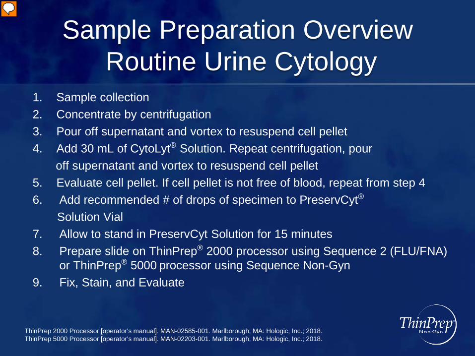
Sample Preparation Overview Routine Urine Cytology
1. Sample collection2. Concentrate by centrifugation 3. Pour off supernatant and vortex to resuspend cell pellet4. Add 30 mL of CytoLyt® Solution. Repeat centrifugation, pour
off supernatant and vortex to resuspend cell pellet5. Evaluate cell pellet. If cell pellet is not free of blood, repeat from step 46. Add recommended # of drops of specimen to PreservCyt®
Solution Vial 7. Allow to stand in PreservCyt Solution for 15 minutes8. Prepare slide on ThinPrep® 2000 processor using Sequence 2 (FLU/FNA)
or ThinPrep® 5000 processor using Sequence Non-Gyn9. Fix, Stain, and Evaluate
ThinPrep 2000 Processor [operator's manual]. MAN-02585-001. Marlborough, MA: Hologic, Inc.; 2018. ThinPrep 5000 Processor [operator's manual]. MAN-02203-001. Marlborough, MA: Hologic, Inc.; 2018.

Sample Preparation OverviewUroCyte®
1. Sample collection.2. Transfer the sample evenly into two labeled 50 mL centrifuge tubes, concentrate by
centrifugation.3. Pour off supernatant and vortex to resuspend cell pellet.4. Add 30 mL of CytoLyt® Solution to one 50 mL tube and vortex. Transfer the contents
of this tube into the second 50 mL tube and vortex. The specimen is now combined into one 50 mL tube. The empty tube can be discarded. Centrifuge, pour off supernatant and vortex to resuspend cell pellet.
5. Evaluate cell pellet appearance. If pellet is not free of blood, add 30 mL of CytoLyt Solution and repeat from step 4.
6. Add entire specimen to PreservCyt® Solution Vial and allow to stand in PreservCyt Solution for 15 minutes.
7. Prepare slide on ThinPrep® 2000 processor using Sequence 5 (UroCyte) or ThinPrep5000 processor using Sequence UroCyte.
8. Fix, stain, and evaluate cytology OR perform molecular diagnostic testing according to manufacturer’s instructions for use.
ThinPrep 2000 Processor [operator's manual]. MAN-02585-001. Marlborough, MA: Hologic, Inc.; 2018. ThinPrep 5000 Processor [operator's manual]. MAN-02203-001. Marlborough, MA: Hologic, Inc.; 2018.

Sample Preparation Techniques
• Centrifugation 600g for 10 minutes or 1200g for 5 minutes- Concentrate cellular material in order to separate the cellular components from the supernatant
Refer to Centrifuge Speed Chart in the ThinPrep®
2000 or ThinPrep 5000 Owners Manual, Non-Gynecologic section to determine the correct speed for your centrifuge to obtain force of 600g or 1200g.
ThinPrep 2000 Processor [operator's manual]. MAN-02585-001. Marlborough, MA: Hologic, Inc.; 2018. ThinPrep 5000 Processor [operator's manual]. MAN-02203-001. Marlborough, MA: Hologic, Inc.; 2018.

Sample Preparation Techniques
• Pour off supernatant• Invert the centrifuge tube 180° in one smooth
movement, pour off all supernatant and return tube to its original position
Note: Failure to completely pour off the supernatant may result in a sparsely cellular sample (due to dilution of the cell pellet).
ThinPrep 2000 Processor [operator's manual]. MAN-02585-001. Marlborough, MA: Hologic, Inc.; 2018. ThinPrep 5000 Processor [operator's manual]. MAN-02203-001. Marlborough, MA: Hologic, Inc.; 2018.

Sample Preparation Techniques
• Vortex to re-suspend cell pellet• Purpose of this step is to randomize the cell pellet and
to improve the results of the subsequent CytoLyt®
solution washing procedure• Place the centrifuge tube onto a vortexor and agitate
the cell pellet for 3 seconds or vortex manually by syringing the pellet back and forth with a plastic pipette
ThinPrep 2000 Processor [operator's manual]. MAN-02585-001. Marlborough, MA: Hologic, Inc.; 2018. ThinPrep 5000 Processor [operator's manual]. MAN-02203-001. Marlborough, MA: Hologic, Inc.; 2018.

Sample Preparation Techniques
• CytoLyt® Solution Wash• Preserve cellular morphology while lysing red blood
cells, dissolving mucus and reducing protein precipitation
• Add 30 mL of CytoLyt Solution to a cell pellet, concentrate by centrifugation, pour off the supernatant, vortex and evaluate cell pellet
ThinPrep 2000 Processor [operator's manual]. MAN-02585-001. Marlborough, MA: Hologic, Inc.; 2018. ThinPrep 5000 Processor [operator's manual]. MAN-02203-001. Marlborough, MA: Hologic, Inc.; 2018.

Sample Preparation Techniques
• Evaluate cell pellet• If cell pellet is white, pale pink, tan or not visible.
Calculate number of drops of specimen to be added to the PreservCyt® Solution Vial (will be discussed in detail on future slides)
• If cell pellet is distinctly red or brown indicating the presence of remaining blood conduct a second CytoLyt® Wash
ThinPrep 2000 Processor [operator's manual]. MAN-02585-001. Marlborough, MA: Hologic, Inc.; 2018. ThinPrep 5000 Processor [operator's manual]. MAN-02203-001. Marlborough, MA: Hologic, Inc.; 2018.

Sample Preparation Techniques
• Calculate how many drops of specimen to add to PreservCyt® vial:– If pellet volume is > 1 mL (if not consider next 2 slides)
• Add 1 mL of CytoLyt® Solution into the tube and vortex briefly to resuspend the cell pellet
• Transfer 1 drop of the specimen to a fresh PreservCyt Solution Vial
ThinPrep 2000 Processor [operator's manual]. MAN-02585-001. Marlborough, MA: Hologic, Inc.; 2018. ThinPrep 5000 Processor [operator's manual]. MAN-02203-001. Marlborough, MA: Hologic, Inc.; 2018.

Sample Preparation Techniques
• Calculate how many drops of specimen to add to PreservCyt® vial:– If pellet is clearly visible and pellet volume is < 1 mL
(if not consider next slide) • Vortex pellet and transfer 2 drops to a fresh
PreservCyt solution vial
ThinPrep 2000 Processor [operator's manual]. MAN-02585-001. Marlborough, MA: Hologic, Inc.; 2018. ThinPrep 5000 Processor [operator's manual]. MAN-02203-001. Marlborough, MA: Hologic, Inc.; 2018.

Sample Preparation Techniques
• Calculate how many drops of specimen to add to PreservCyt® vial:– If pellet is not visible or scant,
• Add contents of a fresh PreservCyt Solution Vial into the tube and vortex briefly to mix the solution
• Pour entire sample back into the vial
ThinPrep 2000 Processor [operator's manual]. MAN-02585-001. Marlborough, MA: Hologic, Inc.; 2018. ThinPrep 5000 Processor [operator's manual]. MAN-02203-001. Marlborough, MA: Hologic, Inc.; 2018.

Troubleshooting Sample Preparation
• Due to the biological variability among samples and variability in collection methods, standard processes may not always yield a satisfactory and uniformly distributed preparation on the first slide.
ThinPrep 2000 Processor [operator's manual]. MAN-02585-001. Marlborough, MA: Hologic, Inc.; 2018. ThinPrep 5000 Processor [operator's manual]. MAN-02203-001. Marlborough, MA: Hologic, Inc.; 2018.
Troubleshooting Sample Preparation
• After staining, you may observe the following irregularities:• Non-uniform distribution of cells in the cell spot without
a “sample is dilute” message• Uneven distribution in the form of a ring or halo of
cellular material and/or white blood cells• A sparse cell spot lacking in cellular component and
containing blood, protein and debris – may be accompanied by a “sample is dilute” message
ThinPrep 2000 Processor [operator's manual]. MAN-02585-001. Marlborough, MA: Hologic, Inc.; 2018. ThinPrep 5000 Processor [operator's manual]. MAN-02203-001. Marlborough, MA: Hologic, Inc.; 2018.

Techniques Used in Troubleshooting
• Diluting the Sample 20 to 1• Glacial Acetic Acid Wash for Blood and Non-
Cellular Debris• Saline Wash for Protein
ThinPrep 2000 Processor [operator's manual]. MAN-02585-001. Marlborough, MA: Hologic, Inc.; 2018. ThinPrep 5000 Processor [operator's manual]. MAN-02203-001. Marlborough, MA: Hologic, Inc.; 2018.

Techniques Used in Troubleshooting
• Diluting the sample 20 to 1• Add 1 mL of the sample that is suspended in
PreservCyt® solution to a new PreservCyt solution vial (20 mL). This is most accurately done with a calibrated pipette
ThinPrep 2000 Processor [operator's manual]. MAN-02585-001. Marlborough, MA: Hologic, Inc.; 2018. ThinPrep 5000 Processor [operator's manual]. MAN-02203-001. Marlborough, MA: Hologic, Inc.; 2018.

Techniques Used in Troubleshooting
• Glacial acetic acid wash for blood and non-cellular debris
• If sample is bloody, it can be further washed using a solution of 9 parts CytoLyt® solution and 1 part glacial acetic acid
ThinPrep 2000 Processor [operator's manual]. MAN-02585-001. Marlborough, MA: Hologic, Inc.; 2018. ThinPrep 5000 Processor [operator's manual]. MAN-02203-001. Marlborough, MA: Hologic, Inc.; 2018.

Techniques Used in Troubleshooting
• Saline wash for protein• If sample contains protein, it can be further washed
with saline solution in place of CytoLyt® solution
ThinPrep 2000 Processor [operator's manual]. MAN-02585-001. Marlborough, MA: Hologic, Inc.; 2018. ThinPrep 5000 Processor [operator's manual]. MAN-02203-001. Marlborough, MA: Hologic, Inc.; 2018.

Troubleshooting Bloody or Proteinaceous Specimens
“Sample is Dilute” message
No, continue to next slide.
Yes
Check to see if cellularity is adequate. If not, use more of the pellet, if available and
prepare new slide.
ThinPrep 2000 Processor [operator's manual]. MAN-02585-001. Marlborough, MA: Hologic, Inc.; 2018. ThinPrep 5000 Processor [operator's manual]. MAN-02203-001. Marlborough, MA: Hologic, Inc.; 2018.

Troubleshooting Bloody or Proteinaceous Specimens
Does the slide have a “halo” of cellular
material and/or white blood cells?
No, continue to next slide.
Yes
Dilute 20:1 by adding 1 mL of residual sample to a new PreservCyt®
solution vial and prepare new slide.
If halo is present on the new slide, contact Hologic® Technical
Service.
ThinPrep 2000 Processor [operator's manual]. MAN-02585-001. Marlborough, MA: Hologic, Inc.; 2018. ThinPrep 5000 Processor [operator's manual]. MAN-02203-001. Marlborough, MA: Hologic, Inc.; 2018.

Troubleshooting Bloody or Proteinaceous Specimens
Is the slide sparse and does it contain blood, protein or
non-cellular debris?
Yes-protein
Centrifuge remaining specimen from PreservCyt vial, pour off and vortex. Add 30 mL of saline to
sample, centrifuge, pour off and vortex. Add appropriate number of drops to PreservCyt vial
and prepare new slide. If resulting slide is sparse, contact Hologic Technical Service.
No
Contact Hologic®
Technical Service
Yes-blood or non-cellular debris
Centrifuge remaining specimen from PreservCyt® vial, pour off and vortex. Add 30 mL of a 9:1 CytoLyt®to glacial acetic acid solution to the sample, centrifuge, pour off and vortex. Add appropriate number of drops to PreservCyt vial and prepare new slide. If the resulting slide is sparse, contact Hologic Technical Service.
ThinPrep 2000 Processor [operator's manual]. MAN-02585-001. Marlborough, MA: Hologic, Inc.; 2018. ThinPrep 5000 Processor [operator's manual]. MAN-02203-001. Marlborough, MA: Hologic, Inc.; 2018.

Troubleshooting Common Artifacts
• Smudged nuclear detail• Compression artifact• Staining artifact• Edge of the cylinder artifact
ThinPrep 2000 Processor [operator's manual]. MAN-02585-001. Marlborough, MA: Hologic, Inc.; 2018. ThinPrep 5000 Processor [operator's manual]. MAN-02203-001. Marlborough, MA: Hologic, Inc.; 2018.

Troubleshooting Common Artifacts
• Smudged nuclear detail• May occur if specimen is collected in saline, PBS or
RPMI• To avoid this, collect either fresh, or in CytoLyt® or in
PreservCyt® solution
ThinPrep 2000 Processor [operator's manual]. MAN-02585-001. Marlborough, MA: Hologic, Inc.; 2018. ThinPrep 5000 Processor [operator's manual]. MAN-02203-001. Marlborough, MA: Hologic, Inc.; 2018.

Troubleshooting Common Artifacts
• Compression Artifact• Appears as “air-dry” artifact on the perimeter of the cell
spot• Due to a compression of cells between the edge of the
filter and the glass slide
ThinPrep 2000 Processor [operator's manual]. MAN-02585-001. Marlborough, MA: Hologic, Inc.; 2018. ThinPrep 5000 Processor [operator's manual]. MAN-02203-001. Marlborough, MA: Hologic, Inc.; 2018.

Troubleshooting Common Artifacts
• Staining Artifact• Mimics air drying• Appears as a red or orange central staining primarily in
cells clusters or groups• Due to the incomplete rinsing of the counter stains• To eliminate this artifact, fresh alcohol baths or an
additional rinse step after the cytoplasmic stains is required
ThinPrep 2000 Processor [operator's manual]. MAN-02585-001. Marlborough, MA: Hologic, Inc.; 2018. ThinPrep 5000 Processor [operator's manual]. MAN-02203-001. Marlborough, MA: Hologic, Inc.; 2018.

Troubleshooting Common Artifacts
• Edge of the Cylinder Artifact• Narrow rim of cellular material just beyond the
circumference of the cell spot• Results from cells from the outer edge of the wet filter
cylinder being transferred to the glass slide
ThinPrep 2000 Processor [operator's manual]. MAN-02585-001. Marlborough, MA: Hologic, Inc.; 2018. ThinPrep 5000 Processor [operator's manual]. MAN-02203-001. Marlborough, MA: Hologic, Inc.; 2018.

Specimen Types
• Voided Urine• Catheterized Bladder Urine• Cystoscopy specimens:
a. Bladder Urineb. Bladder Washc. Ureter or Renal Wash
• Ileal Conduit Urine• Retrograde Brushing

Voided Urine
• Easily obtained2
• Often contaminated with cells from the perineum or the genital tract2
• Samples entire urinary tract2
• Cytology remains one of the best ways to diagnose a variety of bladder lesions, most importantly HGUC1
• Cells can show degeneration1,2
• Low numbers of urothelial cells may be present21. Cibas ES, Ducatman BS. Urine and Bladder Washings - Ancillary Techniques. In: Cytology - Diagnostic Principles and Clinical Correlates. 3rd ed.
Philadelphia, PA: Saunders Elsevier; 2009:123-124.2. DeMay RM. Urine. In: The Art & Science of Cytopathology. 2nd ed. Chicago, IL: American Society for Clinical Pathology; 2012:435-488.

Catheterized Urine
• May see instrumentation effect (lithiasis) and/or lubricant material1
• Lesions in the urethra may be missed2
• Invasive, uncomfortable and risk of urinary tract infection to the patient1,2
• If catheter is indwelling cellular degeneration can be pronounced due to possible extended length of time at room temperature1
• Urothelial cell clusters may be scraped off by the tip of the catheter, mimicking the appearance of a low-grade papillary neoplasm1
1. Cibas ES, Ducatman BS. Urine and Bladder Washings - Ancillary Techniques. In: Cytology - Diagnostic Principles and Clinical Correlates. 3rd ed. Philadelphia, PA: Saunders Elsevier; 2009:123-124.
2. DeMay RM. Urine. In: The Art & Science of Cytopathology. 2nd ed. Chicago, IL: American Society for Clinical Pathology; 2012:435-488.

Bladder Urine & Washing• Bladder urine is collected using a catheter followed by an irrigation of sterile
isotonic saline– Diagnostic sensitivity is enhanced if both the washing and the urine present in the bladder at
cystoscopy are submitted for evaluation
• Used to diagnose and monitor urothelial neoplasia • Highly cellular specimens containing well preserved cells• Method of choice when bladder malignancy is suspected• Almost no extraneous cellular contamination with excellent preservation if
processed quickly, however, contamination from lubricant can render specimens unsatisfactory
• Does not sample the upper urinary tract• Risks include infection, spread of tumor and limited sample area• Large clusters of urothelial cells (deep and superficial), large multi-nucleated
umbrella cells, squamous cells and rare red blood cells are common expected findings
DeMay RM. Urine. In: The Art & Science of Cytopathology. 2nd ed. Chicago, IL: American Society for Clinical Pathology; 2012:435-488.

Retrograde Catheterization with Washings or Brushings
• Sampling and diagnosis of upper urinary tumors can be achieved using this method
• Able to sample inaccessible sites with the ureteropyeloscope and take direct sampling.
• Specimens are similar to those collected from the bladder– Urothelial cells from the upper urinary tract have a more
atypical appearance than from the lower urinary tract– Often see tight pseudopapillary clusters, due to
instrumentation or lithiasisDeMay RM. Urine. In: The Art & Science of Cytopathology. 2nd ed. Chicago, IL: American Society for Clinical Pathology; 2012:435-488.

Ileal Conduit/Loop Urine
• During a cystectomy, a portion of the bowel is used as a urine conduit1,2
• Commonly contains:– Abundant, intestinal glandular epithelial cells1,2
• Mostly seen as single, very degenerated cells resembling macrophages1,2
– Urothelial cells are sparse or absent2
• Important to screen thoroughly as patients with a history of bladder cancer are at increased risk for developing tumors of the ureters and kidneys1
1. Cibas ES, Ducatman BS. Urine and Bladder Washings - Ancillary Techniques. In: Cytology - Diagnostic Principles and Clinical Correlates. 3rd ed. Philadelphia, PA: Saunders Elsevier; 2009:123-124.
2. DeMay RM. Urine. In: The Art & Science of Cytopathology. 2nd ed. Chicago, IL: American Society for Clinical Pathology; 2012:435-488.
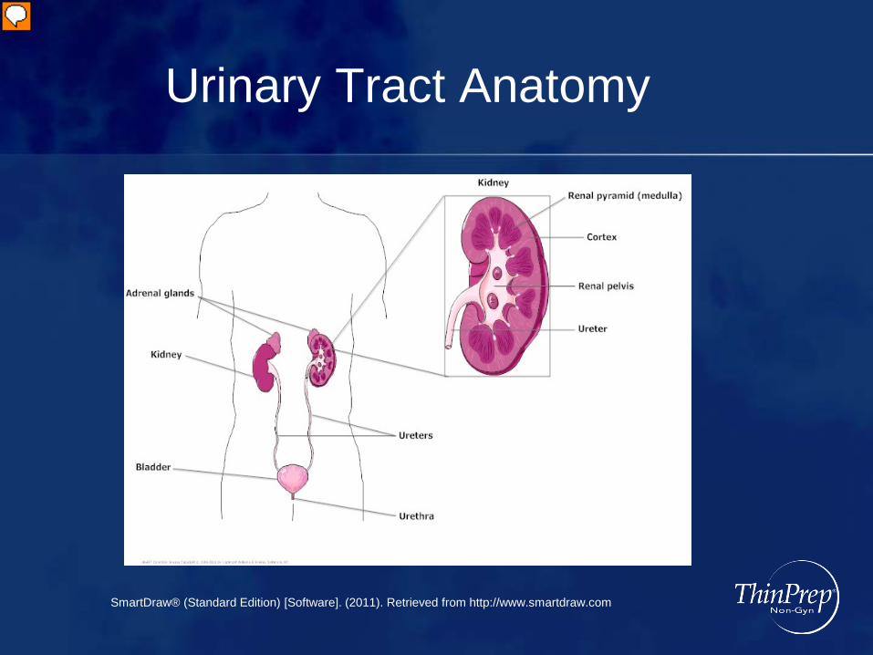
Urinary Tract Anatomy
SmartDraw® (Standard Edition) [Software]. (2011). Retrieved from http://www.smartdraw.com

Histology of EpitheliumUreter
Cou
rtesy
of B
rian
Bich
at L
ake
Supe
rior C
olle
ge
Bich, Brian. Ureter, Bladder . 2008. Photograph. Lake Superior College, Duluth, MN. Web. Lake Superior College. [http://employee.lsc.edu/faculty/BrianBich/Picture%20Library/Form AllItems.aspx?RootFolder=%2Ffaculty%2FBrianBich%2FPicture%20Library%2FAnat%2DPhys%20II%20%28Biol%201141%29&View={997D7559-348B-48F0-A9FE-07741C9C8B65}
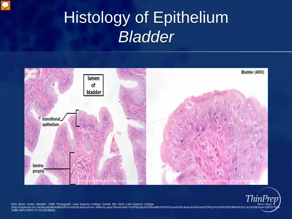
Histology of EpitheliumBladder
Courtesy of Brian Bich at Lake Superior College Courtesy of Brian Bich at Lake Superior College
Bich, Brian. Ureter, Bladder . 2008. Photograph. Lake Superior College, Duluth, MN. Web. Lake Superior College. [http://employee.lsc.edu/faculty/BrianBich/Picture%20Library/Form AllItems.aspx?RootFolder=%2Ffaculty%2FBrianBich%2FPicture%20Library%2FAnat%2DPhys%20II%20%28Biol%201141%29&View={997D7559-348B-48F0-A9FE-07741C9C8B65}

Biological Nature of Urine
• Waste product of metabolism, made in the kidneys and is formed by filtration, reabsorption, and tubular secretion
• Chiefly comprised of water (91 to 96%) but also contains inorganic salts, protein, hormones and other metabolites
• Specific gravity range of 1.003 to 1.035• pH range of 5.5 to 7.0
Biology Online Dictionary. Urine. https://www.biology-online.org/dictionary/Urine. Accessed June 3, 2019.

Normal Components and Findings
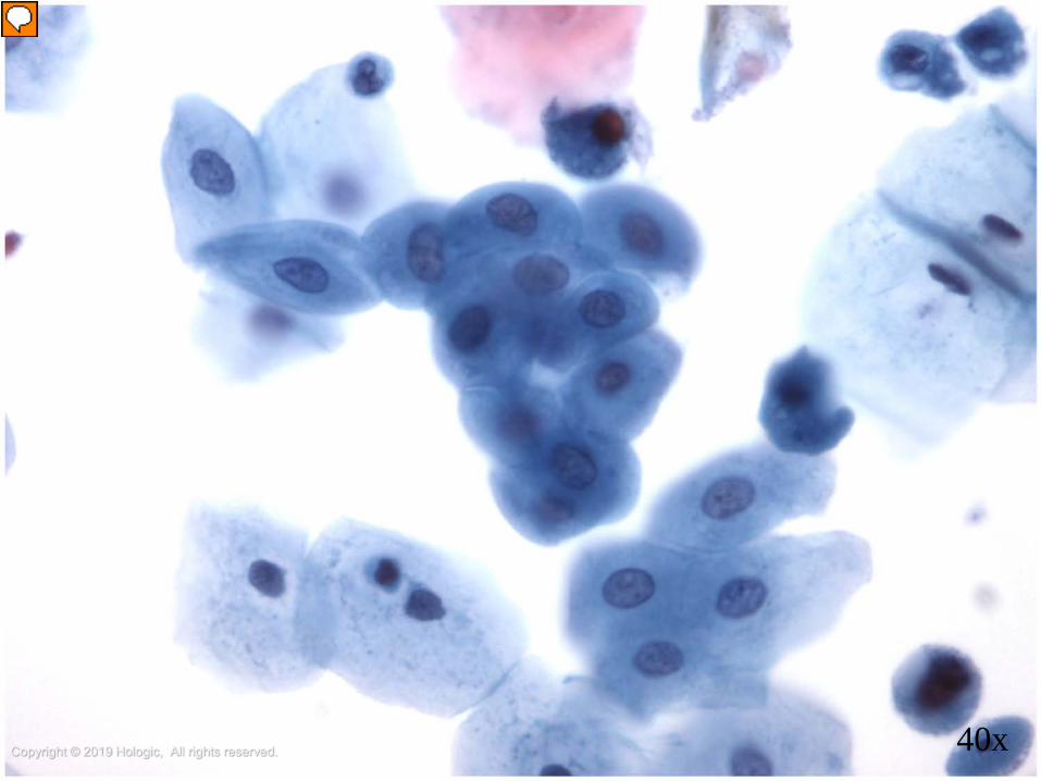
• Benign urothelial cells• Cytoplasm is abundant and may be foamy to dense• Chromatin is finely granular and nucleoli may be present
with multinucleation (umbrella cells)
ThinPrep Non-GYN Morphology Reference Atlas: Urinary 119-147

Copyright © 2019 Hologic, All rights reserved. 40x
Copyright © 2019 Hologic, All rights reserved. 40x

Copyright © 2019 Hologic, All rights reserved. 60x

Copyright © 2019 Hologic, All rights reserved. 60x

Copyright © 2019 Hologic, All rights reserved. 60x

• Benign squamous and glandular cells• Benign squamous cells may be shed from the trigone
or be present as contaminant• Glandular cells may be shed from many sites
including the paraurethral and prostate glands and found in loop urines
Normal Components and Findings
ThinPrep Non-Gyn Morphology Reference Atlas Urinary: 119-147

Copyright © 2019 Hologic, All rights reserved. 20x

Copyright © 2019 Hologic, All rights reserved. 40x

Copyright © 2019 Hologic, All rights reserved. 40x

Copyright © 2019 Hologic, All rights reserved. 40x

Normal Components and Findings
• Crystals• Contaminants
• Bacteria and yeast• Pollen and talc• Spermatozoa and seminal vesicle cells• Lubricant• Corpora amylacea
ThinPrep Non-Gyn Morphology Reference Atlas Urinary: 119-147

Copyright © 2019 Hologic, All rights reserved. 20x

Copyright © 2019 Hologic, All rights reserved. 60x

Copyright © 2019 Hologic, All rights reserved. 60x

Copyright © 2019 Hologic, All rights reserved. 40x
Copyright © 2019 Hologic, All rights reserved. 40x

Copyright © 2019 Hologic, All rights reserved. 60x

Copyright © 2019 Hologic, All rights reserved. 20x

Copyright © 2019 Hologic, All rights reserved. 60x

Other Disease FindingsRenal Tubular Cells• Small columnar cells occurring singly, in small sheets or as a granular
cast.• Their presence is associated with kidney disease.
Casts• Their presence may be associated with kidney disease, infection and/or
bleeding in the kidney.• May be filled with RBCs, WBCs, degenerated renal tubular cells
(granular) or amorphous, eosinophilic proteinaceous material (hyaline)
Inflammation and RBCs• May represent trauma, infection or tumor. Eosinophils may be
associated with drug induced interstitial cystitis.
ThinPrep Non-Gyn Morphology Reference Atlas Urinary: 119-147

Copyright © 2019 Hologic, All rights reserved. 40x

Copyright © 2019 Hologic, All rights reserved. 40x

Copyright © 2019 Hologic, All rights reserved. 60x

Copyright © 2019 Hologic, All rights reserved. 20x

Benign Entities and Changes
• Reactive changes are very common findings in urinary cytology and may be due to:• Instrumentation• Infection/Inflammation• Drug therapy• Calculi
ThinPrep Non-Gyn Morphology Reference Atlas Urinary: 119-147

Reactive Changes
• Features of reactive urothelial cells may include:• Marked cellular and nuclear enlargement• Prominent nucleoli• Coarser chromatin pattern• Multinucleation• Abundant cytoplasm remains• Large honeycombed sheets (especially with
instrumentation)
ThinPrep Non-Gyn Morphology Reference Atlas Urinary: 119-147

Copyright © 2019 Hologic, All rights reserved. 40x

Copyright © 2019 Hologic, All rights reserved. 40x

Copyright © 2019 Hologic, All rights reserved. 60x

Copyright © 2019 Hologic, All rights reserved. 40x

Benign Entities and Changes
• Infectious agents that may be seen in specimens from the urinary tract may include:• Bacteria (most commonly E. Coli or
streptococcus)• Candida• Polyoma Virus• CMV• Trichomonads
ThinPrep Non-Gyn Morphology Reference Atlas Urinary: 119-147

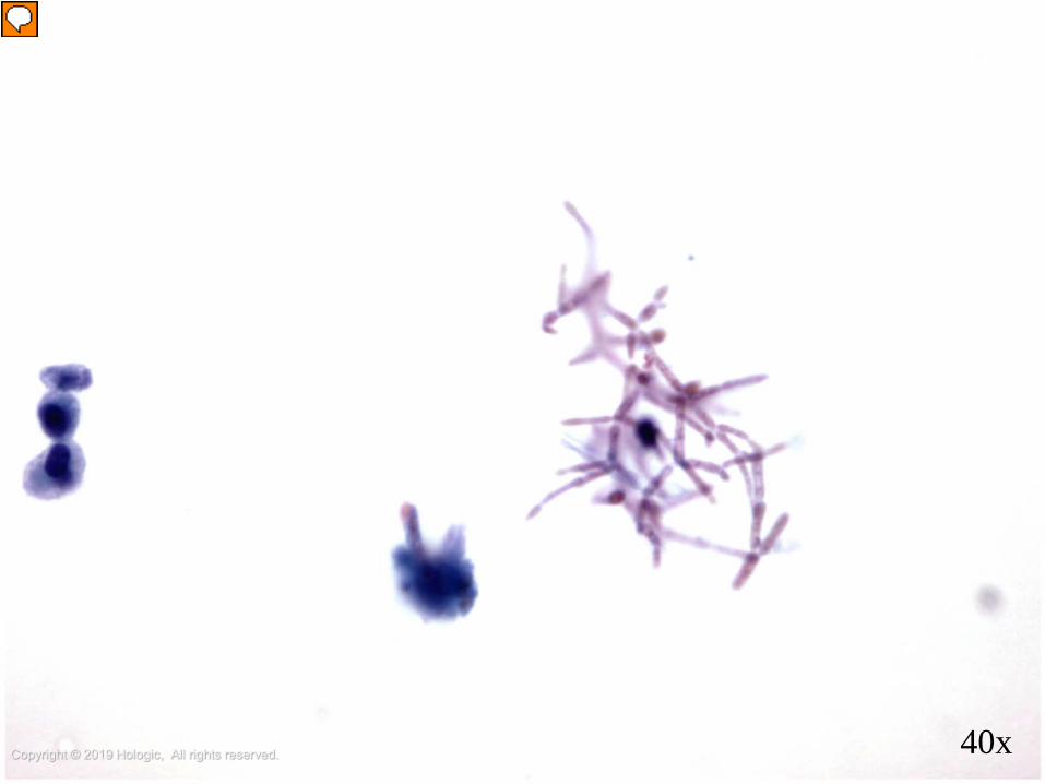
Candida
• Candida may be in the form of spores and/or the traditional septate, branching filaments.
• It is very often seen as a contaminant from the female genital tract or external genitalia.
• It can be the source of an infection, especially in an immune compromised patient
ThinPrep Non-Gyn Morphology Reference Atlas Urinary: 119-147

Copyright © 2019 Hologic, All rights reserved. 20x

Polyoma Virus
• Polyoma virus can present a diagnostic challenge, as cells infected with the virus (decoy cells) can mimic malignancy
• Decoy cells are commonly plasmacytoid cells with eccentrically placed nuclei
• The virus causes a basophilic intranuclear inclusion, which often appears very dense and dark with a smooth nuclear membrane
ThinPrep Non-Gyn Morphology Reference Atlas Urinary: 119-147

Copyright © 2019 Hologic, All rights reserved. 60x

Copyright © 2019 Hologic, All rights reserved. 40x

Cytomegalovirus (CMV)
• This virus usually appears in patients with compromised immune systems or it may be transmitted from a mother to her fetus at birth.
• CMV infected cells typically have a large eosinophilic or basophilic intranuclear inclusion that causes margination of the nuclear chromatin, resulting in a “bull’s eye” appearance
ThinPrep Non-Gyn Morphology Reference Atlas Urinary: 119-147

Trichomonas
• Trichomonads are an uncommon finding in urine and should be carefully discerned from degenerated PMNs.
• In a urinary specimen, the organism is usually round and bears its diagnostic eye spot, similar to that seen in Gyn samples.
ThinPrep Non-Gyn Morphology Reference Atlas Urinary: 119-147

Copyright © 2019 Hologic, All rights reserved. 10x

Diagnosis and Criteria
We will be utilizing The Paris System for Reporting Urinary Cytology for the sections covering diagnostic criteria and categorization

Bladder Cancer in the USStatistics
• The 4th most common cancer in men1
• Estimated that 81,190 people will be diagnosed with bladder cancer in 2018 with an average age of 73 and an estimated 17,240 deaths1
• Men are 4 times more likely than women to be diagnosed with the disease. In addition, incidence rates in white men are double those of black men.1
• 5 year survival rates can range from 98% with stage 0 to 15% with stage IV1
• 708,444 survivors in need of screening in 20152
• About 70% bladder cancers are superficial3
1. Cancer.net. ASCO.org sponsored. https://www.cancer.net/cancer-types/bladder-cancer/treatments-stage. Accessed June 25, 20182. National Cancer Institute. Surveillance, Epidemiology and End Results Program. Cancer Stat Facts: Bladder Cancer.
https://seer.cancer.gov/statfacts/html/urinb.html. Accessed June 25, 2018.3. UpToDate. Patient Educations Bladder Cancer treatment. https://www.uptodate.com/contents/bladder-cancer-treatment-non-muscle-invasive-superficial-
cancer-beyond-the-basics. Accessed June 25, 2018

Bladder Cancer in the USTreatment
• Early-stage bladder cancer is treated locally with surgical excision during cystoscopy
• Bacillus Calmette-Guerin (BCG) is a standard immunotherapy drug for high grade non-invasive bladder cancer
• Cystectomy is the removal of the bladder, used for invasive or recurrent tumors
• Advanced bladder cancer is often treated with intravesical or systemic chemotherapy
Cancer.net. ASCO.org sponsored. https://www.cancer.net/cancer-types/bladder-cancer/treatments-stage. Accessed June 25, 2018
Bladder Cancer in the USPrognosis
• Low grade bladder tumors are more likely to be cured1
• High grade bladder tumors are more likely to recur andprogress1
• Taking all superficial bladder tumors together, about 65% will recur in the first 2 years but mainly as early non-invasive bladder cancer2
• About 30% will recur as more serious disease with less chance of cure or control2
• Treated patients are followed with cystoscopy at regular (typically 3 month) intervals and any new tumors visualized are removed surgically3
1. Rosenthal DL, Wojcik EM, Kurtycz DF. Pathogenesis of Urothelial Carcinoma In: The Paris System for Reporting Urinary Cytology. Springer International Publishing Switzerland; 2016.
2. Rosenthal DL, et al, eds. High Grade Urothelial Carcinoma (HGUC). In: The Paris System for Reporting Urinary Cytology. Cham, Switzerland: Springer International Publishing; 2016:61-74 :1-4. . Rosenthal DL, et al, eds. Clinical Management. In: The Paris System for Reporting Urinary Cytology. Cham, Switzerland: Springer International Publishing; 2016:143-151.
3. Rosenthal DL, et al, eds. Clinical Management. In: The Paris System for Reporting Urinary Cytology. Cham, Switzerland: Springer International Publishing; 2016:143-151.

Bladder Cancer Staging
TNM Classification
Jewett-Strong Marshall Definition
Tis 0 Limited to mucosa, flat insitu
Ta 0 Limited to mucosa, papillary
T1 A Lamina propria invaded
T2a B1 < halfway through muscularis
T2b B2 > halfway through muscularis
T3 C Perivesical fat
T4a C Prostate, uterus or vagina
T4b C Pelvic wall or abdominal wall
N1-N3 D1 Pelvic lymph node(s) involved
M1 D2 Distant metastases
Copyright © 2019 Hologic, All rights reserved.

Diagnosis and MonitoringCystoscopy: The gold standard for diagnosis and monitoring
Post treatment Frequency0-2 years: Every 3 months2-4 years: Every 6 months5-or more years: Yearly
Cytology: Performed as an adjunct test to cystoscopy, often on bladder washings
http://www.wpclipart.comAldousari S, Kassouf W. Update on the management of non-muscle invasive bladder cancer. Can Urol Assoc J. 2010 Feb; 4(1): 56–64

The Paris System for Reporting Urinary Cytology: Rationale
• Main goal of urinary cytology is to find and diagnose high grade lesions
• Current lack of reproducibility and lack of sensitivity for low grade lesions
• There is a need to standardize and educate clinicians on terminology
• Wide interobserver variability for atypical cells-national atypical rates vary among institutions
Rosenthal DL, et al, eds. The Paris System for Reporting Urinary Cytology. Cham, Switzerland: Springer International Publishing; 2016.

The Paris System for Reporting Urinary Cytology: Pathogenesis - Two Pathways
The current understanding is that there are two different pathogenetic pathways:
• Hyperplasia• Dysplasia
The molecular abnormalities associated with these two pathways indicate that these pathways are distinct and separate, and confer separate risks of progression to malignancy
Rosenthal DL, et al, eds. Pathogenesis of Urothelial Carcinoma. In: The Paris System for Reporting Urinary Cytology. Cham, Switzerland: Springer International Publishing; 2016:1-4.

The Paris System for Reporting Urinary Cytology: Pathogenesis - Two Pathways
Hyperplasia• 80% of urothelial carcinomas use this pathway• Starts as hyperplasia that progresses to low-grade papillary urothelial
carcinoma (LGUC)• Genetically stable pathway• Characterized by FGFR3 alterations (fibroblast growth factor
receptor 3) • High recurrence rate• Nonaggressive behavior• Currently accepted progression rate to HGUC is at 10% and new
studies have pointed to the rate being much lower (< 1-6%)
Rosenthal DL, et al, eds. Pathogenesis of Urothelial Carcinoma. In: The Paris System for Reporting Urinary Cytology. Cham, Switzerland: Springer International Publishing; 2016:1-4.

The Paris System for Reporting Urinary Cytology: Pathogenesis - Two Pathways
Dysplasia• Found to be the pathway for 20% of urothelial
carcinomas• Genetically unstable pathway associated with multiple
mutations including TP53• Dysplasia progresses to a high-grade papillary tumor or
less often to a flat urothelial carcinoma (carcinoma in situ)
• High recurrence rate and a high risk of muscle-invasive, stage T2, T3, and T4 tumors with lymph node and systemic metastasis
Rosenthal DL, et al, eds. Pathogenesis of Urothelial Carcinoma. In: The Paris System for Reporting Urinary Cytology. Cham, Switzerland: Springer International Publishing; 2016:1-4.
The Paris System for Reporting Urinary Cytology: Adequacy Algorithm
Source of disagreement and controversyNumerous variables can effect adequacy. This algorithm considers the variables that effect the usefulness of the specimen to diagnose the suspicion of urothelial carcinoma
• Collection type• Cellularity• Volume• Cytomorphological findings (considered first)
Rosenthal DL, et al, eds. Adequacy of Urine Specimens (Adequacy). In: The Paris System for Reporting Urinary Cytology. Cham, Switzerland: Springer International Publishing; 2016:5-11.

The Paris System for Reporting Urinary Cytology: Adequacy Algorithm (cont.)
Cytomorphologic features: • Presence of atypical or malignant cellsSpecimen type:• Voided urine- greater than 30 mL more likely
adequate• Instrumented- cellularity 2600 cells- 20 urothelial
cells/10 high power fields• Presence of excessive and obscuring lubricant,
inflammatory cells, or red blood cells should be interpreted as unsatisfactory/nondiagnostic
Rosenthal DL, et al, eds. Adequacy of Urine Specimens (Adequacy). In: The Paris System for Reporting Urinary Cytology. Cham, Switzerland: Springer International Publishing; 2016:5-11.

The Paris System for Reporting Urinary Cytology: Adequacy Algorithm (cont.)
“*”: Cut-offs for appropriate benign urothelial cellularity should be validated for both instrumented and non-instrumented sources
Reproduced from: Rosenthal DL, et al, eds. Adequacy of Urine Specimens (Adequacy). In: The Paris System for Reporting Urinary Cytology. Cham, Switzerland: Springer International Publishing; 2016:5-11.

The Paris System for Reporting Urinary Cytology Criteria: Categories
• Negative for High-Grade Urothelial Carcinoma (NHGUC)
• Atypical Urothelial Cells (AUC)• Suspicious for High-Grade Urothelial Carcinoma
(SHGUC)• High-Grade Urothelial Carcinoma (HGUC)• Low-Grade Urothelial Neoplasia (LGUN)• Other Malignancies Primary and Metastatic and
Miscellaneous Lesions
Rosenthal DL, et al, eds. Contents. In: The Paris System for Reporting Urinary Cytology. Cham, Switzerland: Springer International Publishing; 2016:xiii.

Diagram by Guliz Barkan, MD. Permission Granted by Eva M. Wojcik, MDWojcik EM. The Paris System (TPS) for Reporting Urinary Cytology: A New Paradigm. ASCP/ASC – Advanced Cytopathology Education. 2016

Nuclear:Cytoplasmic Ratio
Diagram by Lisa Zhang, MD. Permission Granted by Eva M. Wojcik, MD
Wojcik EM. The Paris System (TPS) for Reporting Urinary Cytology: A New Paradigm. ASCP/ASC – Advanced Cytopathology Education. 2016

The Paris System for Reporting Urinary Cytology: Diagnostic Categories & Criteria
Benign Findings – either voided or instrumented:• Benign urothelial, glandular, and squamous cells• Benign urothelial tissue fragments (BUTF) and urothelial
sheets or clusters• Changes associated with lithiasis• Viral cytopathic effect: polyoma virus (BK virus- decoy
cells)• Post therapy effect, including epithelial cells from urinary
diversions
Rosenthal DL, et al, eds. Negative for High Grade Carcinoma (Negative). In: The Paris System for Reporting Urinary Cytology. Cham, Switzerland: Springer International Publishing; 2016:13-38.
Copyright © 2019 Hologic, All rights reserved. 40x

Copyright © 2019 Hologic, All rights reserved. 60x

Copyright © 2019 Hologic, All rights reserved. 60x

Atypical Urothelial Cells (AUC)The abnormal urothelial cells must be non-superficial and non-degenerated as well as display:
• Increased nuclear cytoplasmic ratio of >0.5 (if this is the only finding, the case should not be reported in the AUC category)
In addition, one of the minor criteria below must also be displayed:• Nuclear hyperchromasia (without being so pronounced as that of cells in the
SHGUC or HGUC category)• Irregular nuclear membranes (typically displaying an irregular nuclear shape and
variably thickened chromatinic rim, while remaining round, not oval in shape)• Irregular, coarse, clumped chromatin
The Paris System for Reporting Urinary Cytology: Diagnostic Categories & Criteria
Rosenthal DL, et al, eds. Atypical Urothelial Cells (AUC). In: The Paris System for Reporting Urinary Cytology. Cham, Switzerland: Springer International Publishing; 2016:39-48.

Copyright © 2019 Hologic, All rights reserved. 40x
Copyright © 2019 Hologic, All rights reserved. 40x

Copyright © 2019 Hologic, All rights reserved. 60x

Copyright © 2019 Hologic, All rights reserved. 60x

Suspicious for High Grade Urothelial Carcinoma (SHGUC)
Must display both required criteria below:• Increased N/C ratio of at least 0.5-0.7• Moderate to severe hyperchromasia
In addition to above criteria, must also display either or both of the criteria below:• Irregular clumpy chromatin• Marked irregular nuclear membranes
The number of abnormal cells, previous history of HGUC and collection method should all be taken into consideration. 5-10 cells are recommended for the diagnosis of HGUC with 10 being the recommended number in instrumented specimens. SHGUC can be used restrictively for cases that do not meet that quantity, being careful not to render a diagnosis by applying criteria to degenerated cells.
The Paris System for Reporting Urinary Cytology: Diagnostic Categories & Criteria
Rosenthal DL, et al, eds. Suspicious for High Grade Urothelial Carcinoma (Suspicious). In: The Paris System for Reporting Urinary Cytology. Cham, Switzerland: Springer International Publishing; 2016:49-60.
40xCopyright © 2019 Hologic, All rights reserved.

40xCopyright © 2019 Hologic, All rights reserved.

40xCopyright © 2019 Hologic, All rights reserved.

High Grade Urothelial Carcinoma (HGUC)Malignancy criteria:
• At least 5-10 abnormal cells• N/C ratio is > 0.7 (majority of HGUC cells with some showing a range of 0.5-0.7)• Moderate to severe hyperchromasia• Markedly irregular nuclear membrane• Coarse/clumped chromatin
Other features:• Pleomorphism with marked variation in cellular size and shape• Scant, pale, or dense cytoplasm• Prominent nucleoli• Mitoses• Necrotic debris• Inflammation
The Paris System for Reporting Urinary Cytology: Diagnostic Categories & Criteria
Rosenthal DL, et al, eds. High Grade Urothelial Carcinoma(HGUC). In: The Paris System for Reporting Urinary Cytology. Cham, Switzerland: Springer International Publishing; 2016:61-74.

60xCopyright © 2019 Hologic, All rights reserved.

Copyright © 2019 Hologic, All rights reserved. 40x

Copyright © 2019 Hologic, All rights reserved. 20x

Copyright © 2019 Hologic, All rights reserved. 60x

60xCopyright © 2019 Hologic, All rights reserved.

Copyright © 2019 Hologic, All rights reserved. 60x

• The distinction between low-grade lesions and normal epithelium is extremely difficult
• This diagnosis can be rarely made and should be based only on the presence of well-defined fibrovascular cores in the absence of cellular atypia.
• LGUN is a cytologic term which combines:– Low grade papillary lesions:
• urothelial papilloma• PUNLMP• LGPUC
– Flat, low-grade intraurothelial neoplasia
The Paris System for Reporting Urinary Cytology: Low-Grade Urothelial Neoplasia (LGUN) Facts
Rosenthal DL, et al, eds. Low Grade Urothelial Carcinoma (LGUN). In: The Paris System for Reporting Urinary Cytology. Cham, Switzerland: Springer International Publishing; 2016:75-86.

The Paris System for Reporting Urinary Cytology: Diagnostic Categories & Criteria
Low Grade Urothelial Neoplasm (LGUN)Regardless of the specimen type (voided or instrumented):
• Three-dimensional papillary clusters, defined as clusters of cells with nuclear overlapping, forming papillae WITH fibrovascular cores including capillaries. The presence of this feature is the only time a definitive diagnosis of LGUN is possible. This finding is exceedingly rare.
LGUN may be considered (particularly with correlation in biopsy or cystoscopic findings which should be reported as a note on the report whenever possible) BUT be categorized as NHGUC in the presence of these cytologic features:
• Three-dimensional cellular clusters without fibrovascular cores• Increased numbers of monotonous single (non-umbrella) cells
The features below were previously reported as criteria for LGPUC and may also be associated with HGUC. Without other HGUC features these changes may suggest a LGUN lesion BUT be categorized as NHGUC:
• Cytoplasmic homogeneity• Nuclear border irregularity• Increased N/C ratio
Rosenthal DL, et al, eds. Low Grade Urothelial Carcinoma (LGUN). In: The Paris System for Reporting Urinary Cytology. Cham, Switzerland: Springer International Publishing; 2016:75-86.

Copyright © 2019 Hologic, All rights reserved. 20x

Copyright © 2019 Hologic, All rights reserved. 40x

Copyright © 2019 Hologic, All rights reserved.
Squamous Cell Carcinoma
• Relatively rare in the United States• Strong association with Schistosomiasis• Marked nuclear pleomorphism with dense hyperchromatic
nuclei and macronucleoli. Abundant cytoplasm that may form spindle and tadpole shapes. Keratin pearls, intercellular bridges and keratohyalin granules may be seen.
• To help distinguish from HGUC with squamous differentiation, look for the presence of abnormal urothelial cells
• Non-keratinized squamous carcinomas may be more difficult to distinguish from HGUC.
ThinPrep Non-Gyn Morphology Reference Atlas Urinary: 119-147

Copyright © 2019 Hologic, All rights reserved. 40x

Adenocarcinoma
• Bladder• Rare, accounting for <2% of bladder cancers• Features include three-dimensional clusters of round,
vacuolated cells with large, eccentrically placed, irregular nuclei with open chromatin and prominent nucleoli
ThinPrep Non-Gyn Morphology Reference Atlas Urinary: 119-147

Adenocarcinoma
• Kidney• Only sheds into urine in end stage disease• Typically, cells are round with vacuolated cytoplasm
and may be degenerated• Nuclei are large and round with prominent nucleoli• Appearance may vary according to differentiation of
tumor
ThinPrep Non-Gyn Morphology Reference Atlas Urinary: 119-147

Copyright © 2019 Hologic, All rights reserved. 40x

Adenocarcinoma
• Prostate:• May shed after instrumentation (especially of prostate)
or when disease has invaded bladder• May present as clusters or single cells• Typically is characterized by loose clusters of
glandular cells with eccentric nuclei and prominent nucleoli
• May show cytoplasmic mucin vacuoles
ThinPrep Non-Gyn Morphology Reference Atlas Urinary: 119-147

Copyright © 2019 Hologic, All rights reserved. 40x

Copyright © 2019 Hologic, All rights reserved. 60x

Fluorescence In Situ Hybridization (FISH)
• Used for the Detection and Monitoring of Urothelial Carcinoma
• Technique which maps the presence or absence of targeted genomic sequences in chromosomes using fluorescent probes
• The targeted genes within the chromosomes are then detected using a fluorescent microscope
Cibas ES, Ducatman BS. Urine and Bladder Washings - Ancillary Techniques. In: Cytology - Diagnostic Principles and Clinical Correlates. 3rd ed. Philadelphia, PA: Saunders Elsevier; 2009:123-124.

UroVysion Bladder Cancer FISH Assay
• Additional testing method for the initial detection of bladder cancer in patients with hematuria suspected of having bladder cancer and monitoring for recurrence
• Detects aneuploidy in chromosomes 3, 7, 17 and the loss of 9p21 locus using Fluorescence In Situ Hybridization
Cibas ES, Ducatman BS. Urine and Bladder Washings - Ancillary Techniques. In: Cytology - Diagnostic Principles and Clinical Correlates. 3rd ed. Philadelphia, PA: Saunders Elsevier; 2009:123-124.

Urine Processed by UroCyte® Method
Copyright © 2019 Hologic, All rights reserved.Copyright © 2019 Hologic, All rights reserved.
ThinPrep Non-Gyn Morphology Reference Atlas Urinary: 119-147
For More Information
Visit our Websites:• www.healthdxs.com• www.cytologystuff.com• www.hologic.com
To View:• Product Catalogs• Online Operators Manuals• Complete Gynecologic & Non-gynecologic bibliographies• Cytology Case Presentation and Unknowns