Resolution - sfed.org · Naveen Arya, M.D. Gastroenterology and Therapeutic Endoscopy Department,...
7
Naveen Arya, M.D. Halton Healthcare Services, Oakville, Ontario, Canada Marco Carniel, M.D. Landesklinikum Wiener Neustadt, Wiener Neustadt, Austria Nestor Chopita, M.D., Bárbara Agustina Amendolara, M.D., Francisco Tufare, M.D. and Nelson Condado, M.D. Hospital San Martín de La Plata, Buenos Aires, Argentina Robert S. Dean, M.D. Vassar Brothers Medical Center Poughkeepsie, NY, USA Robert D. Fanelli, M.D., F.A.C.S. Berkshire Medical Center and Surgical Specialists of Western New England, P.C., USA David A. Florez, M.D. Elms Endoscopy Center/ Trident Gastroenterology, Charlestown, South Carolina, USA Francisco Igea, M.D. Hospital Rio Carrion, Palencia, Spain Houssam Kharrat, M.D. Covenant Medical Center, Lubbock, TX, USA Luis F. Lara, M.D. The University of Texas Southwestern Medical Center at Dallas, USA Adolfo Parra-Blanco, M.D. Canarias University Hospital, Santa Cruz de Tenerife, Spain CASES PRESENTED BY: Resolution ™ Clip Global Technique Spotlight
-
Upload
truongtruc -
Category
Documents
-
view
214 -
download
0
Transcript of Resolution - sfed.org · Naveen Arya, M.D. Gastroenterology and Therapeutic Endoscopy Department,...

Naveen Arya, M.D.Halton Healthcare Services, Oakville, Ontario, Canada
Marco Carniel, M.D.Landesklinikum Wiener Neustadt, Wiener Neustadt, Austria
Nestor Chopita, M.D., Bárbara Agustina Amendolara, M.D., Francisco Tufare, M.D. and Nelson Condado, M.D.Hospital San Martín de La Plata, Buenos Aires, Argentina
Robert S. Dean, M.D.Vassar Brothers Medical CenterPoughkeepsie, NY, USA
Robert D. Fanelli, M.D., F.A.C.S.Berkshire Medical Center and Surgical Specialists of Western New England, P.C., USA
David A. Florez, M.D.Elms Endoscopy Center/ Trident Gastroenterology,Charlestown, South Carolina, USA
Francisco Igea, M.D.Hospital Rio Carrion, Palencia, Spain
Houssam Kharrat, M.D.Covenant Medical Center, Lubbock, TX, USA
Luis F. Lara, M.D. The University of Texas Southwestern Medical Center at Dallas, USA
Adolfo Parra-Blanco, M.D.Canarias University Hospital, Santa Cruz de Tenerife, Spain
C A S E S P R E S E N T E D B Y :
Resolution™ ClipGlobal Technique Spotlight

Naveen Arya, M.D.Gastroenterology and Therapeutic Endoscopy Department, Halton Healthcare Services, Oakville, Ontario, Canada
CLOSURE OF A PERFORATION AFTER A SALINE-ASSISTED POLYPECTOMY
HISTORY
An 87-year-old male presented with a three month history of rectal bleeding. Thepatient had no weight loss, change in bowel habit or abdominal pain. His pastmedical history was relevant for coronary artery disease with coronary bypass,hypertension and hyperlipidemia. He was on Coumadin™, Altace™, Lipitor™ andAtenolol. Upon examination, the abdomen was found to be obese.
PROCEDURE
The patient had a colonoscopy carried out by a general surgeon and was found tohave an 8cm rectal mass. The biopsy proved to be a tublovillous adenoma with highgrade dysplasia and the patient was referred for a polypectomy. The risks of theprocedure such as perforation, bleeding or serious side effects were discussed withthe patient and he consented to the procedure.
The polyp was endoscopically removed with saline-assisted injection in a piecemealfashion (Figure 1). The edges were coagulated with Argon plasma coagulation at40W (Figure 2). During the procedure it was noted that the middle of thepolypectomy base was open and the plane was obviously through the submucosa(Figure 3). The base of the polyp was then clipped closed with 16 Resolution™ Clips(Figures 4, 5 and 6).
After the procedure the patient was taken to recovery, given antibiotics IV and threeviews of the abdomen were taken with no free air seen. The patient was stable andwanted to go home. He was put on PO antibiotics for seven days. The patient wasseen within one week and had no symptoms or signs of perforation and was stable.
POST PROCEDURE
Six months later, the patient had a flexible sigmoidoscopy and there was noevidence of residual polyp. A scar was found and biopsies taken from it revealednormal mucosa.
This case demonstrates the utility of clips and gives endoscopists another modality to increase their therapeutic arsenal.
Figure 1
Figure 2
Figure 3
Figure 4 Figure 5 Figure 6
HISTORY
A 72-year-old female was referred to our centre for an endoscopic mucosalresection. The patient has a history of coronary heart disease, st.p. myocardialinfarction in 2000, arterial hypertension, hyperlipidaemia and osteoporosis.
In February 2005, the referring hospital performed a partial rectal polypectomy. Inthe last pre-colonoscopy examination, a recurrent flat polypoid formation wasdiscovered in the rectum.
PROCEDURE
An approximately 2.5cm flat adenoma could be seen in the rectum at 8cm. In thecentre of the lesion, scar tissue after the partial polypectomy in 2005 was readily distinguishable (Figure 1).
Fluid infiltration of the polyp revealed good lifting sign. Utilising a straight suctioncap, the lesion was totally resected in four fragments using a piecemeal techniquewith a cautery snare.
A 12mm perforation was revealed upon retrieval of the last fragment (Figure 2). By applying 7 Resolution™ Clips, the perforation edges could be properly aligned step-by-step and the perforation successfully closed (Figures 3, 4 and 5).
HISTOLOGICAL FINDINGS
Flat tubular partially tubulovillous adenoma with low to moderate grade intraep-ithelial neoplasia, the muscularis mucosae overall intact – resection in toto.
POST PROCEDURE
The patient was kept under clinical observation for several days. There was noincrease of inflammatory parameters upon serological examination and no clinicalsigns of peritonitis. After two days, the patient was introduced to a dietary regimenwhich was well tolerated. A surgical intervention was not necessary and after four days the patient was discharged.
Marco Carniel, M.D.Head of Endoscopy, Landesklinikum Wiener Neustadt, Wiener Neustadt, Austria
Figure 1
Figure 2
Figure 3
Figure 4Figure 5
CLOSURE OF AN IATROGENIC PERFORATION AFTERAN ENDOSCOPIC MUCOSAL RESECTION
Coumadin™ is a trademark of Bristol-Myers Squibb. Altace™ is a trademark of King Pharmaceuticals. Lipitor™ is a trademark of Pfizer.

Nestor Chopita, M.D., Bárbara Agustina Amendolara, M.D., Francisco Tufare, M.D., Nelson Condado, M.D.Gastroenterology Department, Hospital San Martín de La Plata, Buenos Aires, Argentina
TREATMENT OF POST-EMR BLEEDING WITH ENDOSCOPIC CLIPS
HISTORY
A 62-year-old male with cirrhosis (Child B) and a history of variceal bleeding wasadmitted to our hospital for gastric polyposis resection.
PROCEDURE
The endoscopy showed gastric polyposis and there was an abnormal area ofmucosa on the gastric incisura. Biopsy taken from the site revealed intra-mucosalcarcinoma and an endoscopic mucosal resection was performed (Figure 1).
The patient had haemodynamic instability, haematemesis and haematocritdropped at 22% 12 hours after the procedure. Another endoscopy showed activebleeding in the EMR site (Figure 2). The bleeding was successfully managed withthe use of two Resolution™ Clips (Figures 3 and 4).
POST PROCEDURE
During the follow up, the patient was in good condition and there were nocomplications (Figure 5). Histological results confirmed early gastric cancer (EGC).
Figure 1
Figure 2
Figure 3
Figure 4 Figure 5
HISTORY
A 91-year-old man presented with a history of anaemia and haematochezia. Thepatient was on chronic anticoagulation therapy with Coumadin™ due to porcinemitral valve replacement. Other past medical history includes a two vesselCABG, TIA, and seizure disorder. A colonoscopy was deemed necessary toexplore potential sources of hematochezia. Concern was given in particular to theneed for continuing anticoagulation therapy. The patient was counselled todiscontinue his Coumadin therapy five days prior to the scheduled colonoscopy.Low-molecular-weight heparin was used to maintain anticoagulation until theevening prior to the procedure.
PROCEDURE
A colonoscopy was performed, during which time a 2cm polyp, on a thick stalk,was observed in the descending colon. This polyp was removed using aCaptivator™ II Single - use Snare and cautery techniques. Reinspection of theresection margin revealed pooling of fresh blood at the polyp stalk base (Figure 1).Two Resolution™ Clips were places at the polyp stalk base with goodhaemostasis (Figures 2 and 3).
POST PROCEDURE
Low-molecular-weight heparin was resumed the morning after the procedure.There was no residual bleeding from the colon noted. Coumadin therapy was resumed 14 days post procedure and the patient has not had any further haematochezia.
Robert S. Dean, M.D.Vassar Brothers Medical Center, Poughkeepsie, NY
Figure 1
Figure 2
Figure 3
PREVENTION OF POST-POLYPECTOMY HAEMORRHAGE USING THE RESOLUTION CLIP DEVICE
Coumadin™ is a trademark of Bristol-Myers Squibb.
Robert D. Fanelli, M.D., F.A.C.S.Director of Surgical Endoscopy, Berkshire Medical Center and Surgical Specialists of Western New England, P.C.
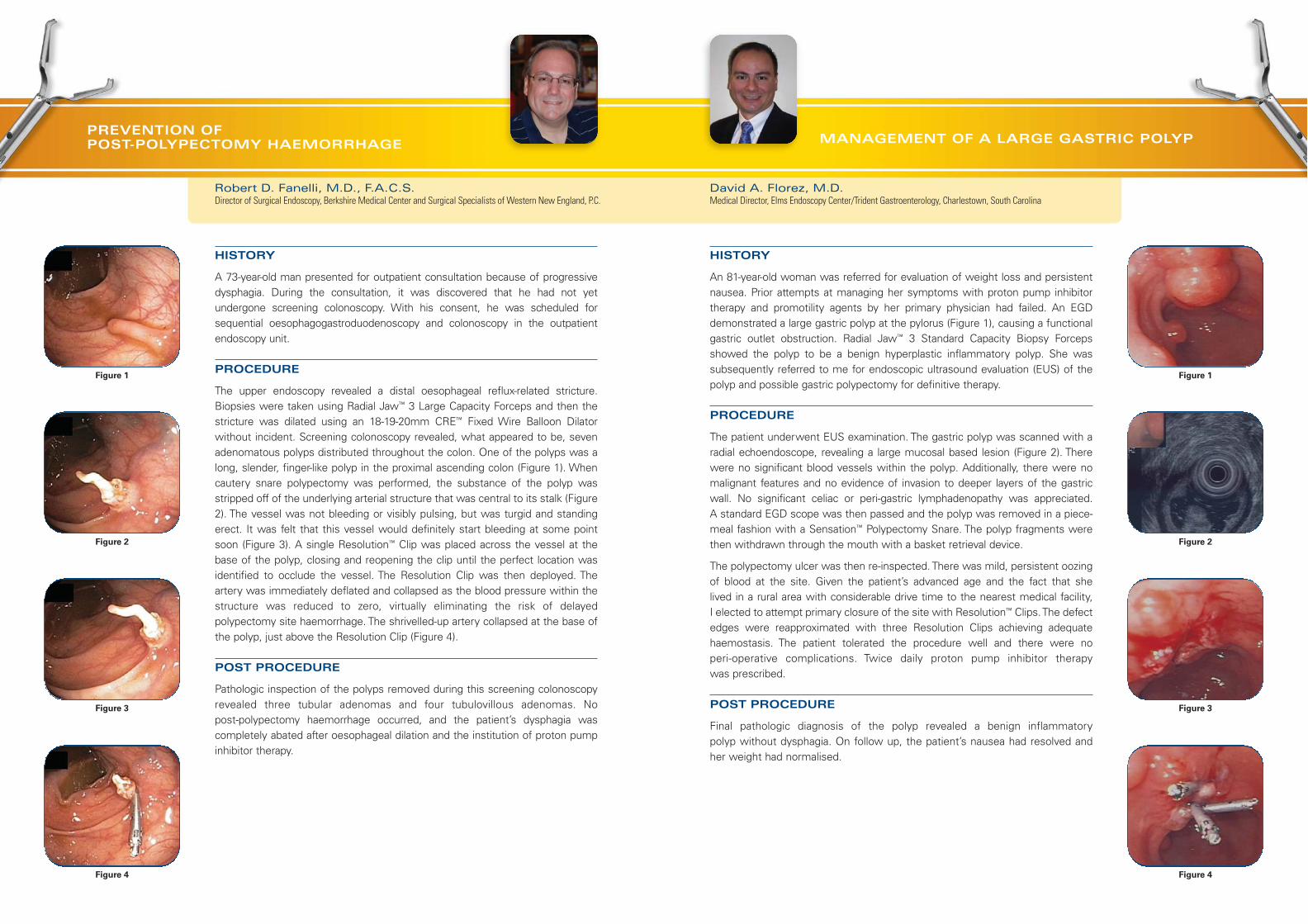
PREVENTION OF POST-POLYPECTOMY HAEMORRHAGE
HISTORY
A 73-year-old man presented for outpatient consultation because of progressivedysphagia. During the consultation, it was discovered that he had not yet undergone screening colonoscopy. With his consent, he was scheduled forsequential oesophagogastroduodenoscopy and colonoscopy in the outpatientendoscopy unit.
PROCEDURE
The upper endoscopy revealed a distal oesophageal reflux-related stricture.Biopsies were taken using Radial Jaw™ 3 Large Capacity Forceps and then thestricture was dilated using an 18-19-20mm CRE™ Fixed Wire Balloon Dilatorwithout incident. Screening colonoscopy revealed, what appeared to be, sevenadenomatous polyps distributed throughout the colon. One of the polyps was along, slender, finger-like polyp in the proximal ascending colon (Figure 1). Whencautery snare polypectomy was performed, the substance of the polyp wasstripped off of the underlying arterial structure that was central to its stalk (Figure2). The vessel was not bleeding or visibly pulsing, but was turgid and standingerect. It was felt that this vessel would definitely start bleeding at some pointsoon (Figure 3). A single Resolution™ Clip was placed across the vessel at thebase of the polyp, closing and reopening the clip until the perfect location wasidentified to occlude the vessel. The Resolution Clip was then deployed. Theartery was immediately deflated and collapsed as the blood pressure within thestructure was reduced to zero, virtually eliminating the risk of delayedpolypectomy site haemorrhage. The shrivelled-up artery collapsed at the base ofthe polyp, just above the Resolution Clip (Figure 4).
POST PROCEDURE
Pathologic inspection of the polyps removed during this screening colonoscopyrevealed three tubular adenomas and four tubulovillous adenomas. No post-polypectomy haemorrhage occurred, and the patient’s dysphagia wascompletely abated after oesophageal dilation and the institution of proton pumpinhibitor therapy.
Figure 1
Figure 2
Figure 3
Figure 4
HISTORY
An 81-year-old woman was referred for evaluation of weight loss and persistentnausea. Prior attempts at managing her symptoms with proton pump inhibitortherapy and promotility agents by her primary physician had failed. An EGDdemonstrated a large gastric polyp at the pylorus (Figure 1), causing a functionalgastric outlet obstruction. Radial Jaw™ 3 Standard Capacity Biopsy Forcepsshowed the polyp to be a benign hyperplastic inflammatory polyp. She wassubsequently referred to me for endoscopic ultrasound evaluation (EUS) of thepolyp and possible gastric polypectomy for definitive therapy.
PROCEDURE
The patient underwent EUS examination. The gastric polyp was scanned with aradial echoendoscope, revealing a large mucosal based lesion (Figure 2). Therewere no significant blood vessels within the polyp. Additionally, there were nomalignant features and no evidence of invasion to deeper layers of the gastricwall. No significant celiac or peri-gastric lymphadenopathy was appreciated. A standard EGD scope was then passed and the polyp was removed in a piece-meal fashion with a Sensation™ Polypectomy Snare. The polyp fragments werethen withdrawn through the mouth with a basket retrieval device.
The polypectomy ulcer was then re-inspected. There was mild, persistent oozingof blood at the site. Given the patient’s advanced age and the fact that she lived in a rural area with considerable drive time to the nearest medical facility, I elected to attempt primary closure of the site with Resolution™ Clips. The defectedges were reapproximated with three Resolution Clips achieving adequatehaemostasis. The patient tolerated the procedure well and there were no peri-operative complications. Twice daily proton pump inhibitor therapy was prescribed.
POST PROCEDURE
Final pathologic diagnosis of the polyp revealed a benign inflammatory polyp without dysphagia. On follow up, the patient’s nausea had resolved andher weight had normalised.
David A. Florez, M.D.Medical Director, Elms Endoscopy Center/Trident Gastroenterology, Charlestown, South Carolina
Figure 1
Figure 2
Figure 3
Figure 4
MANAGEMENT OF A LARGE GASTRIC POLYP
Francisco Igea, M.D.Gastroenterology Department, Hospital Rio Carrion, Palencia, Spain
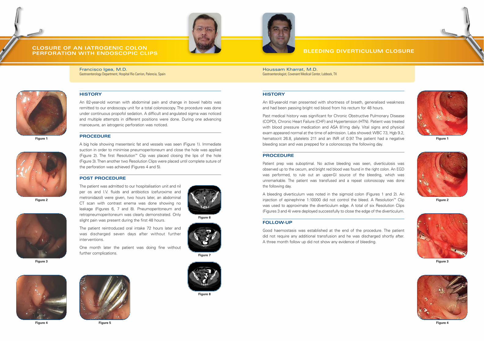
CLOSURE OF AN IATROGENIC COLON PERFORATION WITH ENDOSCOPIC CLIPS
HISTORY
An 82-year-old woman with abdominal pain and change in bowel habits wasremitted to our endoscopy unit for a total colonoscopy. The procedure was doneunder continuous propofol sedation. A difficult and angulated sigma was noticedand multiple attempts in different positions were done. During one advancingmanoeuvre, an iatrogenic perforation was noticed.
PROCEDURE
A big hole showing mesenteric fat and vessels was seen (Figure 1). Immediatesuction in order to minimise pneumoperitoneum and close the hole was applied(Figure 2). The first Resolution™ Clip was placed closing the lips of the hole (Figure 3). Then another two Resolution Clips were placed until complete suture ofthe perforation was achieved (Figures 4 and 5).
POST PROCEDURE
The patient was admitted to our hospitalisation unit and nilper os and I.V. fluids and antibiotics (cefuroxime andmetronidazol) were given, two hours later, an abdominalCT scan with contrast enema was done showing noleakage (Figures 6, 7 and 8). Pneumoperitoneum andretropneumoperitoneum was clearly demonstrated. Onlyslight pain was present during the first 48 hours.
The patient reintroduced oral intake 72 hours later andwas discharged seven days after without furtherinterventions.
One month later the patient was doing fine without further complications.
Figure 1
Figure 2
Figure 3
Figure 4
HISTORY
An 83-year-old man presented with shortness of breath, generalised weakness and had been passing bright red blood from his rectum for 48 hours.
Past medical history was significant for Chronic Obstructive Pulmonary Disease(COPD), Chronic Heart Failure (CHF) and Hypertension (HTN). Patient was treatedwith blood pressure medication and ASA 81mg daily. Vital signs and physical exam appeared normal at the time of admission. Labs showed: WBC 7.3, Hgb 9.2,hematocrit 26.8, platelets 211 and an INR of 0.97. The patient had a negative bleeding scan and was prepped for a colonoscopy the following day.
PROCEDURE
Patient prep was suboptimal. No active bleeding was seen, diverticulosis wasobserved up to the cecum, and bright red blood was found in the right colon. An EGDwas performed, to rule out an upper-GI source of the bleeding, which wasunremarkable. The patient was transfused and a repeat colonoscopy was done the following day.
A bleeding diverticulum was noted in the sigmoid colon (Figures 1 and 2). Aninjection of epinephrine 1:10000 did not control the bleed. A Resolution™ Clip was used to approximate the diverticulum edge. A total of six Resolution Clips(Figures 3 and 4) were deployed successfully to close the edge of the diverticulum.
FOLLOW-UP
Good haemostasis was established at the end of the procedure. The patient did not require any additional transfusion and he was discharged shortly after. A three month follow up did not show any evidence of bleeding.
Houssam Kharrat, M.D.Gastroenterologist, Covenant Medical Center, Lubbock, TX
Figure 1
Figure 2
Figure 3
Figure 4
BLEEDING DIVERTICULUM CLOSURE
Figure 5
Figure 6
Figure 7
Figure 8

Luis F. Lara, M.D.Assistant Professor of Internal MedicineDivision of Digestive and Liver Diseases, The University of Texas, Southwestern Medical Center at Dallas
BLEEDING FROM A COLON TUMOUR AFTER BIOPSY WITH FORCEPS
HISTORY
A 42-year-old white male diagnosed with cystic fibrosis, who had a bilateral lungtransplant in 1997 with a single previous episode of rejection and diabetesmellitus, was referred to evaluate episodic mild haematochezia and new onsetconstipation requiring intermittent laxative use. The patient had a previousspontaneous diverticular perforation for which he had a diverting colostomy thathad been repaired three years earlier.
PROCEDURE
The colonic anastomosis appeared normal. A circumferential, friable massoccluding the lumen to about 12mm and measuring 4cm in length was seen in the distal transverse colon. A standard biopsy forceps was used to biopsy the mass (Figure 1). Bleeding continued from one of the biopsy sites (estimatedto be 8mm in size), which did not stop after five minutes of irrigation andobservation (Figure 2). A Resolution™ Clip was used to approximate the sides of the biopsy site, achieving haemostasis, which was confirmed before scope withdrawal (Figure 3). Follow-up confirmed there was no further bleeding(Figure 4).
Figure 1
Figure 2
Figure 3
Figure 4
HISTORY
A 73-year-old male underwent an upper endoscopy for the study of long-standingreflux symptoms. The examination was done under conscious sedation with poortolerance, in spite of the administration of 100 µg of fentanyl and 7 mg ofMidazolam, and nausea occured repeatedly during the procedure.
During the initial passage of the endoscope in the oesophagus, a long segment of Barrett’s oesophagus was detected, with no other gross abnormalities. Onwithdrawal, two tears were found in the lower end of the columnar epithelium,proximally to the cardia: a shallow 1cm tear (located at 6 o’clock – Figure 1) and adeep 3cm tear (located at 3 o’clock – Figure 1).
PROCEDURE
The affected area was flushed with water in order to have a clear view of the lesion.The bottom of the tear was inspected in detail and no signs of perforation wereevident. There was some oozing from the tear and a decision was made to suturethe mucosal defect in order to prevent further complications such as delayedbleeding or perforation.
Five Resolution™ Clips were applied and attached consecutively in a distal toproximal order (Figure 2). The reason for this is that the clips initially applied mayhamper the precise application of further clips, and it is easier to check the mucosaldefect remaining to be sutured if the direction is from distal to proximal. This aspectis important, because although clipping is considered to be almost devoid of anyrisks, there is a possibility that a clip incorrectly placed at the bottom of a mucosaldefect instead of grasping normal mucosa at the edge of the lesion can result in a perforation.
The tear was successfully sutured with five Resolution Clips, which were placedeasily (Figure 3). The extent of the Barrett’s oesophagus was C5M8 according to thePrague Classification. No biopsies were taken to confirm the diagnosis. The patientremained asymptomatic and was discharged after observation.
POST PROCEDURE
The patient was on high dose PPI. Four months later in a second endoscopy, notrace of the tear was found, but random biopsies revealed high-grade dysplasia inthe Barrett’s epithelium. A third endoscopy was performed in order to try to locateany dysplastic areas, and two small erosions were found in a Barrett’s tongue(Figure 4). Biopsy taken from both areas revealed high-grade dysplasia and anendoscopic mucosectomy is planned.
Mucosal tears in the oesophagus may occur during diagnostic or therapeuticendoscopy, but they are very infrequent and underlying conditions such aseosinophilic oesophagitis should be ruled out. Resolution Clips are a valuable toolfor the management of intraprocedural complications (bleeding, and perforations).
Adolfo Parra-Blanco, M.D.Department of Gastroenterology, Canarias University Hospital, Santa Cruz de Tenerife, Spain
Figure 1
Figure 2
Figure 3
Figure 4
TREATMENT OF MUCOSAL TEARS IN THE OESOPHAGUS WITH ENDOSCOPIC CLIPS

All cited trademarks are the property of their respective owners. CAUTION: The law restricts these devices to sale by or on the order of a physician. Indications, contraindications, warnings and instructions for usecan be found in the product labelling supplied with each device. Information for the use only in countries with applicable health authority product registrations.
PSST 5136 Printed in the UK by Gosling.
European Distribution Centre –
The N etherlands
T: +31 45 54 67 700 F: +31 45 54 67 800
European Headquarters –
Paris
T: +33 1 57 66 80 00 F: +33 1 57 66 84 99
Argentina (Freephone)T: 0800 555 2678 F: +5411 4896 8517
Australia/New Zealand
T: +61 2 8063 8100 F: +61 2 9330 1404
Austria
T: +43 1 60 810 F: +43 1 60 810 60
Belgium (Freephone)T: 0800 94 494 F: 0800 93 343
Brazil
T: +55 11 5502 8500 F: +55 11 5103 2212
Canada
T: +1 888 359 9691 F: +1 888 575 7396
Chile
T: +562 445 4904 F: +562 445 4915
China – Beijing
T: +86 10 8525 1588 F: +86 10 8525 1566
China – Guangzhou
T: +86 20 8767 9791 F: +86 20 8767 9789
China – Shanghai
T: +86 21 6141 5959 F: +86 21 6141 5900
Colombia
T: +57 1 629 5045 F: +57 1 612 4761
Czech Republic
T: +420 296 331 901 F: +420 296 331 935
Denmark (Freephone)T: 80 30 80 02 F: 80 30 80 05
Eire (Freephone)T: 1800 882 969 F: 1800 882 968
Finland
T: +358 20 762 8882 F: +358 20 762 8883
France
T: +33 1 39 30 49 00 F: +33 1 39 30 49 01
Germany (Freephone)T: 08000 723300 F: 08000 723319
Greece
T: +30 210 9542 300 F: +30 210 9542 310
Hong Kong
T: +852 2960 7100 F: +852 2563 5276
Hungary
T: +36 1 456 30 40 F: +36 1 456 30 41
India – Bangalore
T: +91 80 2212 4928/9 F: +91 80 2207 5153
India – Chennai
T: +91 44 2220 1879 F: +91 44 2220 0716
India – Delhi
T: +91 11 4243 2222 F: +91 11 2610 0808
India – Mumbai
T: +91 22 4030 9165 F: +91 22 4040 9199
Italy
T: +39 010 60 60 1 F: +39 010 60 60 200
Korea
T: +82 2 3476 2121 F: +82 2 3476 1776
Mexico
T: +52 55 5687 63 90 F: +52 55 5687 62 28
Middle East/Gulf/North Africa
T: +961 1 805 410 F: +961 1 805 445
The Netherlands
T: +31 30 602 55 44 F: +31 30 602 55 05
Norway (Freephone)T: 800 104 04 F: 800 101 90
Poland
T: +48 22 435 14 14 F: +48 22 435 14 10
Portugal
T: +351 1 381 25 40 F: +351 21 381 25 58
South Africa
T: +27 11 840 8600 F: +27 11 463 6077
South East Asia – Malaysia
T: +60 3 2283 3813 F: +60 3 2284 3813
South East Asia – Philippines
T: +63 2 687 6994 F: +63 2 687 3047
South East Asia – Singapore
T: +65 6418 8888 F: +65 6418 8899
South East Asia – Thailand
T: +66 2 6380 100 F: +66 2 6380 400
South East Europe
T: +30 210 9542 300 F: +30 210 9542 310
Spain – Barcelona
T: +34 93 444 72 00 F: +34 93 405 90 45
Spain – Madrid
T: +34 901 11 12 15 F: +34 91 319 50 03
Sweden
T: +46 42 25 69 00 F: +46 42 25 69 69
Switzerland
T: +41 32 626 57 00 F: +41 32 626 57 01
Taiwan
T: +886 2 2747 7278 F: +886 2 2747 7270
Turkey – Istanbul
T: +90 216 464 36 66 F: +90 216 464 36 67
UK (Freephone)T: 0844 800 4512 F: 0844 800 4513
Uruguay
T: +59 82 900 6212 F: +59 82 900 6212
Venezuela
T: +58 212 959 6275 F: +58 212 959 5328
RCS Nanterre B420 668 420© 2008 Boston Scientific Corporation or its affiliates. All rights reserved. DINEND2208EA
www.bostonscientific.com
www.bostonscientific-international.com
These cases reflect the opinion of and are the responsibility of their respective authors.