Research Establishment and Characterization of a Panel of
13
Cancer Therapy: Preclinical Establishment and Characterization of a Panel of Human Uveal Melanoma Xenografts Derived from Primary and/or Metastatic Tumors Fariba Némati 1 , Xavier Sastre-Garau 2 , Cécile Laurent 3,4,14 , Jérôme Couturier 6 , Pascale Mariani 7 , Laurence Desjardins 8 , Sophie Piperno-Neumann 9 , Olivier Lantz 2 , Bernard Asselain 3,4,10,14 , Corine Plancher 10 , Delphine Robert 2 , Isabelle Péguillet 2 , Marie-Hélène Donnadieu 2 , Ahmed Dahmani 1 , Marie-Andrée Bessard 1 , David Gentien 11 , Cécile Reyes 12 , Simon Saule 5 , Emmanuel Barillot 3,4,14 , Sergio Roman-Roman 12 , and Didier Decaudin 1,13 Abstract Purpose: Uveal melanoma is the most common primary intraocular malignant tumor in adults and is de- fined by a poor natural outcome, as 50% of patients die from metastases. The aim of this study was to develop and characterize a panel of human uveal melanoma xenografts transplanted into immunodeficient mice. Experimental Design: Ninety tumor specimens were grafted into severe combined immunodeficient mice, and 25 transplantable xenografts were then established (28%). Relationship between tumor graft and clinical, biological, and therapeutic features of the patients included were investigated. Characterization of 16 xenografts included histology, molecular analyses by immunohistochemistry, genetic alteration anal- ysis (single-nucleotide polymorphism), and specific tumor antigen expression by quantitative reverse tran- scription-PCR. Pharmacologic characterization (chemosensitivity) was also done in four models using two drugs, temozolomide and fotemustine, currently used in the clinical management of uveal melanoma. Results: Take rate of human uveal melanoma was 28% (25 of 90). Tumor take was independent of size, histologic parameters, or chromosome 3 monosomy but was significantly higher in metastatic tumors. Interestingly, in vivo tumor growth was prognostic for a lower metastasis-free survival in patients with primary tumors. A high concordance between the patients' tumors and their corresponding xeno- grafts was found for all parameters tested (histology, genetic profile, and tumor antigen expression). Finally, the four xenografts studied displayed different response profiles to chemotherapeutic agents. Conclusions: Based on these results, this panel of 16 uveal melanoma xenografts represents a useful pre- clinical tool for both pharmacologic and biological assessments. Clin Cancer Res; 16(8); 2352–62. ©2010 AACR. Uveal melanoma is the most common primary intraoc- ular malignant tumor in adults. Despite the increased di- agnostic accuracy and the development of conservative and effective treatments on primary tumor sites, such as plaque radiotherapy and photon beam therapy, the mor- tality remains stable and 50% of patients die from metas- tases that frequently involve the liver. Chemotherapy, such as oral temozolomide and intra-arterial fotemustine used at the metastatic stage, induces very low response rates, 14.3% and 36%, respectively, and a median survival time of 6.7 and 15 months (1–3). No postoperative adjuvant therapies are currently available to decrease the risk of metastases. Several prognostic factors of dissemi- nated relapse after initial ophthalmologic treatment have been determined, including location with respect to the equator, monosomy 3, and retinal detachment (4). However, no effect of these prognostic markers on patient care can be envisaged in the absence of effective systemic therapies. The growing body of knowledge about molecular and genetics events involved in oncogenesis and tumor pro- gression has led to the identification of new therapeutic targets and therapeutic agents. Preclinical investigation in relevant models is therefore mandatory to select therapeu- tic agents before their assessment in clinical trials. To ob- tain preclinical results with high predictive value for Authors' Affiliations: 1 Laboratory of Preclinical Investigation, Translational Research Department, Institut Curie; 2 Department of Tumor Biology, Institut Curie; 3 Institut Curie; 4 Institut National de la Sante et de la Recherche Medicale, U900; 5 Centre National de la Recherche Scientifique UMR146, Institut Curie; 6 Department of Genetics, Institut Curie; 7 Department of Visceral Surgery, Institut Curie; 8 Department of Ophthalmological Oncology, Institut Curie; 9 Department of Medical Oncology, Institut Curie; 10 Department of Statistics, Institut Curie; 11 Affymetrix Platform, Translational Research Department, Institut Curie; 12 Translational Research Department, Institut Curie; 13 Department of Clinical Hematology, Institut Curie, Paris, France and 14 Ecole des Mines de Paris, Fontainebleau, France Note: Supplementary data for this article are available at Clinical Cancer Research Online (http://clincancerres.aacrjournals.org/). Corresponding Author: Didier Decaudin, Laboratoire d'Investigation Pré-clinique/Service d'Hématologie Clinique, Institut Curie, 26 rue d'Ulm, 75.248 Paris cedex 05, France. Phone: 33-1-44-32-46-90; Fax: 33-1-53-10-40-11; E-mail: [email protected]. doi: 10.1158/1078-0432.CCR-09-3066 ©2010 American Association for Cancer Research. Clinical Cancer Research Clin Cancer Res; 16(8) April 15, 2010 2352 Research. on January 6, 2019. © 2010 American Association for Cancer clincancerres.aacrjournals.org Downloaded from Published OnlineFirst April 14, 2010; DOI: 10.1158/1078-0432.CCR-09-3066 Research. on January 6, 2019. © 2010 American Association for Cancer clincancerres.aacrjournals.org Downloaded from Published OnlineFirst April 14, 2010; DOI: 10.1158/1078-0432.CCR-09-3066 Research. on January 6, 2019. © 2010 American Association for Cancer clincancerres.aacrjournals.org Downloaded from Published OnlineFirst April 14, 2010; DOI: 10.1158/1078-0432.CCR-09-3066
Transcript of Research Establishment and Characterization of a Panel of

2352
Published OnlineFirst April 14, 2010; DOI: 10.1158/1078-0432.CCR-09-3066 Published OnlineFirst April 14, 2010; DOI: 10.1158/1078-0432.CCR-09-3066 Published OnlineFirst April 14, 2010; DOI: 10.1158/1078-0432.CCR-09-3066
Cancer Therapy: Preclinical Clinical
CancerResearch
Establishment and Characterization of a Panel of HumanUveal Melanoma Xenografts Derived from Primaryand/or Metastatic Tumors
Fariba Némati1, Xavier Sastre-Garau2, Cécile Laurent3,4,14, Jérôme Couturier6, Pascale Mariani7, Laurence Desjardins8,Sophie Piperno-Neumann9, Olivier Lantz2, Bernard Asselain3,4,10,14, Corine Plancher10, Delphine Robert2,Isabelle Péguillet2, Marie-Hélène Donnadieu2, Ahmed Dahmani1, Marie-Andrée Bessard1, David Gentien11,Cécile Reyes12, Simon Saule5, Emmanuel Barillot3,4,14, Sergio Roman-Roman12, and Didier Decaudin1,13
Abstract
Authors'TranslationTumor BioSante et dRechercheGenetics, I8Departmeof MedicalCurie; 11AInstitut Cu13Departme14Ecole des
Note: SuppResearch O
CorresponPré-cliniqud'Ulm, 75.233-1-53-10
doi: 10.115
©2010 Am
Clin Canc
DowDowDow
Purpose:Uveal melanoma is the most common primary intraocular malignant tumor in adults and is de-fined by a poor natural outcome, as 50%of patients die frommetastases. The aim of this studywas to developand characterize a panel of human uveal melanoma xenografts transplanted into immunodeficient mice.Experimental Design: Ninety tumor specimens were grafted into severe combined immunodeficient
mice, and 25 transplantable xenografts were then established (28%). Relationship between tumor graftand clinical, biological, and therapeutic features of the patients includedwere investigated. Characterizationof 16 xenografts included histology, molecular analyses by immunohistochemistry, genetic alteration anal-ysis (single-nucleotide polymorphism), and specific tumor antigen expression by quantitative reverse tran-scription-PCR. Pharmacologic characterization (chemosensitivity) was also done in four models using twodrugs, temozolomide and fotemustine, currently used in the clinical management of uveal melanoma.Results: Take rate of human uveal melanoma was 28% (25 of 90). Tumor take was independent of
size, histologic parameters, or chromosome 3 monosomy but was significantly higher in metastatictumors. Interestingly, in vivo tumor growth was prognostic for a lower metastasis-free survival in patientswith primary tumors. A high concordance between the patients' tumors and their corresponding xeno-grafts was found for all parameters tested (histology, genetic profile, and tumor antigen expression).Finally, the four xenografts studied displayed different response profiles to chemotherapeutic agents.Conclusions: Based on these results, this panel of 16 uveal melanoma xenografts represents a useful pre-
clinical tool for both pharmacologic and biological assessments. Clin Cancer Res; 16(8); 2352–62. ©2010 AACR.
Uveal melanoma is the most common primary intraoc-ular malignant tumor in adults. Despite the increased di-agnostic accuracy and the development of conservativeand effective treatments on primary tumor sites, such as
Affiliations: 1Laboratory of Preclinical Investigation,al Research Department, Institut Curie; 2Department oflogy, Institut Curie; 3Institut Curie; 4Institut National de lae la Recherche Medicale, U900; 5Centre National de laScientifique UMR146, Institut Curie; 6Department of
nstitut Curie; 7Department of Visceral Surgery, Institut Curie;nt of Ophthalmological Oncology, Institut Curie; 9DepartmentOncology, Institut Curie; 10Department of Statistics, Institutffymetrix Platform, Translational Research Department,rie; 12Translational Research Department, Institut Curie;nt of Clinical Hematology, Institut Curie, Paris, France andMines de Paris, Fontainebleau, France
lementary data for this article are available at Clinical Cancernline (http://clincancerres.aacrjournals.org/).
ding Author: Didier Decaudin, Laboratoire d'Investigatione/Service d'Hématologie Clinique, Institut Curie, 26 rue48 Paris cedex 05, France. Phone: 33-1-44-32-46-90; Fax:-40-11; E-mail: [email protected].
8/1078-0432.CCR-09-3066
erican Association for Cancer Research.
er Res; 16(8) April 15, 2010
Researcon Januaclincancerres.aacrjournals.org nloaded from Researc
on Januaclincancerres.aacrjournals.org nloaded from Researc
on Januaclincancerres.aacrjournals.org nloaded from
plaque radiotherapy and photon beam therapy, the mor-tality remains stable and 50% of patients die from metas-tases that frequently involve the liver. Chemotherapy, suchas oral temozolomide and intra-arterial fotemustine usedat the metastatic stage, induces very low response rates,14.3% and 36%, respectively, and a median survivaltime of 6.7 and 15 months (1–3). No postoperativeadjuvant therapies are currently available to decrease therisk of metastases. Several prognostic factors of dissemi-nated relapse after initial ophthalmologic treatment havebeen determined, including location with respect to theequator, monosomy 3, and retinal detachment (4).However, no effect of these prognostic markers on patientcare can be envisaged in the absence of effective systemictherapies.The growing body of knowledge about molecular and
genetics events involved in oncogenesis and tumor pro-gression has led to the identification of new therapeutictargets and therapeutic agents. Preclinical investigation inrelevant models is therefore mandatory to select therapeu-tic agents before their assessment in clinical trials. To ob-tain preclinical results with high predictive value for
h. ry 6, 2019. © 2010 American Association for Cancerh. ry 6, 2019. © 2010 American Association for Cancerh. ry 6, 2019. © 2010 American Association for Cancer

Translational Relevance
The prognosis of uveal melanoma patients remainsgenerally poor, with a risk of metastatic relapse andrelative inefficacy of conventional chemotherapies.New therapies, differing from classic treatments, aretherefore warranted to improve the outcome of thesepatients. The treatment of cancer is continually im-proving due to growing knowledge of oncogenesisand the development of new targeted compounds.Early clinical trials evaluating such candidates requirea large number of patients, are expensive, are time con-suming, and expose patients to certain risks. In vivopreclinical assessment of antitumor agents in relevantanimal models is a crucial step in the drug develop-ment process. We have therefore developed and char-acterized a panel of uveal melanoma xenograftsobtained from human primary tumors to allow pre-clinical pharmacologic assessment and to explore thebiology of this human cancer.
Primary Human Uveal Melanoma Xenografts
Published OnlineFirst April 14, 2010; DOI: 10.1158/1078-0432.CCR-09-3066
clinical trials, the choice of the tumor models on whichnew compounds and novel drug combinations are evalu-ated is critical. Human tumor fragments obtained from pa-tients and directly transplanted into immunodeficientmice, known as primary xenografts or tumor grafts (5),are one category of recognized models used as tools forpreclinical assays. Xenografts are known to reproduce themarked heterogeneity of human tumors and generally veryclosely resemble the patient's tumor in terms of histopath-ologic and molecular features, as well as response to ther-apy (6, 7). Furthermore, procedures for assessment oftherapeutic efficacy are now well standardized and facili-tate evaluation of combined therapies, particularly interms of biostatistical analysis.This study was designed to establish a panel of primary
human uveal melanoma xenografts obtained from patienttumor samples transplanted into severe combined immu-nodeficient (SCID) mice. The resulting xenografts werethen characterized and compared with the patient's origi-nal tumors, particularly in terms of molecular prognosticmarkers previously identified in human uveal melanomasand response to standard chemotherapies. Our dataindicate that these models constitute a useful and rele-vant tool for preclinical assessment of new therapeuticapproaches.
Materials and Methods
Patients and tumor samples. Ninety tumor specimenswere obtained after enucleation of uveal melanoma, 73from primary tumors (enucleation), and 17 from metasta-ses. All patients had previously given their informed con-sent for experimental research on residual tumor tissueavailable after histopathologic and cytogenetic analyses.
www.aacrjournals.org
Researcon Januaclincancerres.aacrjournals.org Downloaded from
Some specimens were fixed in acetic acid, buffered forma-lin, alcohol solution (AFA) for further morphologic andimmunohistochemical analysis, and others were storedin liquid nitrogen for further genomic analyses.Data collection and statistical analysis. To define prog-
nostic factors of in vivo tumor engraftment, the followingpatient characteristics were collected for the 90 cases in-cluded: gender, age, intraocular tumor location, thicknessand diameter, retinal detachment, ciliary body and extra-scleral tumor infiltration, primary treatment, histology,mitotic index, monosomy 3 (for available cases), primarytumor or metastatic sample, interval between diagnosis ofprimary tumor and first metastasis, and overall survival. Aχ2 test was used for univariate analysis. Survival curveswere estimated using the Kaplan-Meier method (8). Statis-tical tests were two-sided and done using a 5% level of sig-nificance using the log-rank test. Metastasis-free survivalwas defined as the duration from initial diagnosis to firstmetastasis, and overall survival was measured from thedate of initial diagnosis to the date of death irrespectiveof the cause.Establishment of uveal melanoma xenografts. Fresh tumor
samples obtained from pathologists were immediatelytransplanted into the interscapular fat pad of two to fournon-preirradiated immunodeficient female SCID mice, 5to 7 wk old, without any extracellular matrix preparationunder total xylazine/ketamine anesthesia. Mice were main-tained in a specific pathogen-free animal housing (InstitutCurie) and regularly observed for tumor growth. Micewithout growing tumors 1 y after initial transplantationwere sacrificed. Animal care and housing were in accor-dance with the institutional guidelines of the French EthicsCommittee (Ministère de l'Alimentation, de l'Agricultureet de la Pêche, Direction de la Santé et de la ProtectionAnimale, Paris, France) and under the supervision of au-thorized investigators.At a volume of ∼1 cm3, tumors were removed and sub-
sequently transplanted to naive SCID mice. Samples wereconcomitantly stored frozen in DMSO-FCS solution or di-rectly in liquid nitrogen and fixed in AFA for furtherstudies. After three consecutive mouse-to-mouse passages,the xenograft was considered to be stabilized and was sub-mitted to the process of standard characterization, includ-ing histopathologic, molecular, and genetic features, all ofwhich were compared with the patient's tumor, and in vivotherapeutic assessments.Histopathologic analyses. Morphologic examination was
done on each xenograft and compared with the histologicfindings of the corresponding patient's tumor. For lightmicroscopy examination, 4-μm-thick AFA-fixed paraffin-embedded sections were stained with H&E safran. c-kit,Bcl-2, and β-catenin expressions were determined by im-munohistochemistry. Antigen retrieval was done by incu-bating tissue sections for 20 min in 10 mmol/L citratebuffer (pH 6.1) in an 850 W microwave oven. Tissue sec-tions were then incubated for 30 min with anti–c-kit (poly-clonal rabbit, CD117; DAKO), anti–Bcl-2 (monoclonalmouse, clone 124; DAKO), or anti–β-catenin (monoclonal
Clin Cancer Res; 16(8) April 15, 2010 2353
h. ry 6, 2019. © 2010 American Association for Cancer

Némati et al.
2354
Published OnlineFirst April 14, 2010; DOI: 10.1158/1078-0432.CCR-09-3066
mouse, 14/β-catenin; BD Biosciences) antibodies. Antibo-dies were diluted to 1:100 except for c-kit, which was dilut-ed to 1:200. Staining was revealed by using the VectastainElite ABC peroxidase mouse IgG kit (Vector Laboratories)and diaminobenzidine (DAKO) as chromogen. Stainingfor the various tumor samples was assessed semiquantita-tively. An isotypic control was done by using IgG1α FITC(1:1250 dilution; DAKO). Semiquantitative assessmentwas done by estimating at ×200magnification, the percent-age of positive neoplastic cytoplasms or nuclei definedwithin the area of highest positivity chosen after scanningthe entire tumor surface at low power (10× objective).Genomic analyses. To define the chromosome 3 copy
number and loss of heterozygosity status and to detect otherabnormalities, genetic analyses of the patients' tumors andthe corresponding xenografts were done using AffymetrixGenome-Wide SNP Arrays 6.0. DNA was purified asdescribed (9), and loss of heterozygosity of chromosome3 was detected with single-nucleotide polymorphisms(SNP) as described (10). Data were analyzed using PartekGenomic Suite software, version 6.4, build 6.09.0129(Partek, Inc.) using Partek's default parameters. Fluores-cence in situ hybridization (FISH) and array comparativegenomic hybridization (array-CGH) were done in twosamples. FISH was done on intact nuclei after dissociationof the tumor fragment using a labeled centromeric probespecific for chromosome 3 (Vysis, AbbottMolecular) accord-ing to the supplier's protocol. For array-CGH, DNA extrac-tion, labeling, and hybridization were done as previouslydescribed (11).Expression of tumor-specific antigens. Expression of
tumor-specific antigens was assessed by reverse transcrip-tion-PCR on RNA extracted from both frozen patientand tumor graft samples. Total RNA extraction wasdone by centrifugation on a CsCl cushion to totally eli-minate melanin contamination. PCR amplification wascarried out on cDNA (60 ng equivalent RNA) in the pres-ence of 0.025 units/μL of Platinum Taq (Life Technolo-gies), 200 μmol/L of deoxynucleotide triphosphate, and0.4 μmol/L of each primer (12–14) in a final volume of30 μL. After a denaturation cycle of 4 min at 94°C, ampli-ficationwas run for 21 to 33 cycles (94°C for 1min, anneal-ing temperature of 1 to 2 min, 72°C for 1 min) accordingto Table 1 and followed by an extension cycle of 15 minat 72°C. Serial dilutions of 60 ng of equivalent RNAfrom melanoma cell lines were used for semiquantitativedetermination: SK23 for Melan-A, tyrosinase, and NA17;MZ2 cell line for MAGE1, MAGE2, MAGE3, MAGE6, andMAGE10; LB23 cell line for MAGE4; and LB373 cell linefor LAGE1, LAGE2, andMAGE-C2. Annealing temperaturesand amplification cycles were as follows: β-actin, 65°C for1 min for 21 cycles; NA17, 62°C for 1 min for 33 cycles;tyrosinase, 65°C for 1 min for 25 cycles; Melan-A, 60°Cfor 1 min for 24 cycles; MAGE1, 72°C for 1 min for 31 cy-cles; MAGE2, 67°C for 1 min for 31 cycles; MAGE3, 72°Cfor 1 min for 31 cycles; MAGE4, 68°C for 1 min for 31cycles; MAGE6, 70°C for 2 min for 31 cycles; MAGE10,65°C for 1 min for 31 cycles; LAGE1, 62°C for 1 min for
Clin Cancer Res; 16(8) April 15, 2010
Research. on January clincancerres.aacrjournals.org Downloaded from
Table 1. Clinical characteristics of all uvealmelanoma patients (n = 90; univariate analysis)and in vivo tumor take rate (%)
Patients and tumorcharacteristics
Pa(n)
6, 2019. © 2010 Ame
tients
Clini
rican Assoc
Tumor takerate (%)
P
cal Cancer R
iation for Can
Gender
Male 47 23.4 N S Female 43 32.6Age at diagnosis (y)
<60 48 50.7 N S ≥60 42 49.2History of previous cancer
No 86 25.6 N S(0.06)
Yes 4 75.0 Primary tumor locationAnterior to equator
3 0 On equator 75 30.7 N S Posterior to equator 11 18.2Origin of tumor sample
Primary tumor 73 21.9 0 .02 Metastatic tumor 17 52.9Primary tumor diameter (NA = 4)
≤15 mm 18 16.7 N S 15-18 mm 27 37.0 >18 mm 41 26.8Primary tumor thickness
≤7 mm 17 29.4 N S 7-10 mm 14 42.8 >10 mm 59 23.7Retinal detachment (NA = 2)
No 31 38.7 N S(0.07)
Yes 57 1.0 Extrascleral invasion (NA = 5)No
78 26.9 N S Yes 7 28.6Primary tumor treatment
No enucleation 17 35.3 N S Enucleation 73 26.1Histology (n = 73)
Epithelioid 29 31.0 Spindle 19 36.8 N S Mixed 23 13.0Ciliary body involvement (NA = 1)
No 53 22.6 N S Yes 19 36.8Extrascleral involvement (NA = 1)
No 66 27.3 N S Yes 6 16.7Mitotic index (NA = 9)
Nil-low 72 24.2 N S Medium-high 7 42.8Monosomy 3 of primary tumors (NA = 10)
No 30 20.0 N S Yes 33 33.3Initial treatment
Proton therapy 73 35.3 N S Enucleation 17 26.1Abbreviations: NA, not available; NS, not significant.
esearch
cer

Primary Human Uveal Melanoma Xenografts
Published OnlineFirst April 14, 2010; DOI: 10.1158/1078-0432.CCR-09-3066
30 cycles; LAGE2, 62°C for 1 min for 30 cycles; and MAGE-C2, 60°C for 1 min for 30 cycles.In vivo tumor growth and antitumor efficacy of standard
chemotherapeutic drugs. For each new tumor graft model,in vivo spontaneous tumor growth was determined by thegrowth delay after transplantation, the time to reach a tu-mor volume of 40 to 200 mm3, and the doubling time.For experimental therapeutic assays, female mice were xe-nografted with a tumor fragment of 15 mm3. Mice bearing
www.aacrjournals.org
Researcon Januaclincancerres.aacrjournals.org Downloaded from
growing tumors with a volume of 40 to 200 mm3 wereindividually identified and randomly assigned to the con-trol or treatment groups (6-10 animals per group, as de-tailed in the tables and legends of the figures), andtreatment was started on day 1. Animals with tumorvolumes outside this range were excluded. Mice wereweighed twice a week. Xenografted mice were sacrificedwhen their tumor reached a volume of 2,500 mm3. Tumorvolumes were calculated by measuring two perpendicular
Fig. 1. Prognostic effect of in vivo tumor growth on survival of uveal melanoma patients. A, overall survival of the overall study population (90 cases)according to xenograft take. B, overall survival of patients with primary tumors according to xenograft take. C, overall survival of patients with metastatictumors according to xenograft take. D, metastasis-free survival for patients with primary tumors according to xenograft take.
Clin Cancer Res; 16(8) April 15, 2010 2355
h. ry 6, 2019. © 2010 American Association for Cancer
Némati et al.
2356
Published OnlineFirst April 14, 2010; DOI: 10.1158/1078-0432.CCR-09-3066
diameters with calipers. Each tumor volume (V) was cal-culated according to the following formula: V = a × b2 / 2,where a and b are the largest and smallest perpendiculartumor diameters. Relative tumor volumes (RTV) were cal-culated from the following formula: RTV = (Vx/V1), whereVx is the tumor volume on day x and V1 is the tumor vol-ume at initiation of therapy (day 1). Growth curves wereobtained by plotting the mean values of RTV on the Y axisagainst time (X axis, expressed as days after start of treat-ment). Antitumor activity was evaluated according to tu-mor growth inhibition (TGI), calculated according to thefollowing formula: percent GI = 100 − (RTVt / RTVc ×100), where RTVt is the median RTV of treated mice andRTVc is the median RTV of controls, both at a given timepoint when the antitumor effect was optimal. Fifty percentTGI was considered to be the limit for a meaningful bio-logical effect. Statistical significance of differences ob-served between the individual RTVs corresponding to thetreated mice and control groups was calculated by the two-tailed Student's t test. Growth delay index was calculatedas the time required to reach the same RTV in the treatedand control groups, at a RTV of 4.Two cytotoxic drugs considered to be standard treat-
ment for uveal melanomas were tested, namely, fotemus-tine (Muphoran, Servier) and temozolomide (Temodal,Schering Plough). Fotemustine was reconstituted in the
Clin Cancer Res; 16(8) April 15, 2010
Researcon Januaclincancerres.aacrjournals.org Downloaded from
appropriate solution according to the supplier, diluted in5% dextrose, and administered i.p. at a dose of 30 mg/kgevery 3 wk. Temozolomide was reconstituted in water anddiluted in PBS/5% dextrose/Tween 80 (2/1/1‰) and ad-ministered orally in a 0.3 mL volume on day 1 every 4 wk.Mice were treated for three cycles.
Results
Establishment of xenografts. A total of 90 uveal melano-ma samples obtained from primary tumors or metastaseswere implanted s.c. into SCID mice as described in Mate-rials and Methods. Of the 90 tumors transplanted in im-munodeficient mice, XX gave rise to viable tumors (takerate, 28%). Twenty-five of these tumors were shown to dis-play uveal melanoma characteristics on histopathology.Molecular and genetic characterization was done on16 models obtained from 10 primary ocular tumors(MP34/38/41/42/46/47/55/71/77/80), 5 liver metastases(MM26/28/52/66/74), and 1 skin metastasis (MM33).In vivo therapeutic evaluation was done on four models:MP77, MM66, MM26, and MP38.Clinical characteristic of the 90 patients and their effect
on the growth of the corresponding xenografted tumors arepresented in Table 1. Briefly, gender, age, history of previ-ous cancer, and tumor parameters such as tumor site,
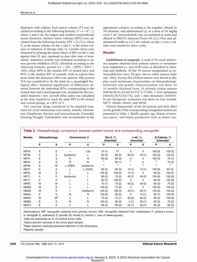
Table 2. Histopathologic comparison between patient tumors and corresponding xenografts
Models
Histopathology Chromosome 3 Bcl-2, %(intensity)*h. ry 6, 2019. © 2010
c-kit, %(intensity)*
Clinica
American Associat
β-Catenin, %(intensity)*
P
X P X P X P X Pl Cancer R
ion for Can
X
MP34
E E — L3p 70 (1) 0† 0 0 100 (3) 100 (3) MP38 S E Isodisomy L 80 (2) 80 (2) 60 (2) 50 (1) 100 (3) 100 (3) MP41 E E N N 90 (3) 90 (2) 0 0 100 (3) 70 (1) MP42 S S N N ‡ 60 (1) ‡ 0 ‡ 70 (2) MP46 E M L (FISH) Isodisomy ‡ ‡ ‡ ‡ ‡ ‡MP47
E E L L (CGH) 80 (2) 90 (2) 10 (1) 10 (1) 100 (3) 90 (3) MP55 M E L L 100 (3) 100 (2) 15 (1) 0 60 (2) 100 (2) MP71 E E Isodisomy L 80 (2) 70 (2) 40 (1) 30 (2) 100 (3) 100 (3) MP77 E E L L 60 (1) 100 (2) 0 0 90 (2) 100 (3) MP80 M M L L 70 (1) 70 (2) 60 (2) 40 (2) 80 (2) 70 (2) MM26 M E L L 100 (2) 75 (2) 0 0 100 (2) 100 (3) MM28 M E — Isodisomy 100 (3) 100 (3) 20 (1) 60 (1) 100 (3) 100 (3) MM33 E E N N 100 (2) 80 (2) 0 10 (1) 90 (1) 100 (3) MM52 M M§ L L 70 (2) 70 (1) 80 (2) 80 (2) 80 (2) 100 (3) MM66 E E N N 100 (2) 90 (2) 5 (1) 30 (1) 80 (2) 70 (2) MM74 E E L L 90 (2) 90 (2) 20 (1) 40 (1) 90 (3) 80 (3)Abbreviations: MP, xenografts obtained from primary tumors; MM, xenografts obtained from metastases; P, patient's tumor;X, xenograft; E, epithelioid; S, spindle; M, mixed; N, normal; L, loss of heterozygosity.*Data are expressed as % of positive tumor cells.†Eighty percent necrosis in the tumor graft sample.‡Major pigment overload prevented definition of the phenotype.§Majority spindle.
esearch
cer

Primary Human Uveal Melanoma Xenografts
Published OnlineFirst April 14, 2010; DOI: 10.1158/1078-0432.CCR-09-3066
diameter and thickness, retinal detachment, and tumorexternalization were not predictive of in vivo tumor take.Tumor growth was also independent of histologic param‐eters, such as epithelioid or spindle cell morphology,ciliary body involvement, mitotic index, and the presenceof monosomy 3. Inversely, the tumor take rate was signif-icantly increased when the tumor tissues were derived from
www.aacrjournals.org
Researcon Januaclincancerres.aacrjournals.org Downloaded from
metastases versus primary intraocular tumors, withtake rates of 52.9% and 21.9%, respectively (P = 0.02).The 5-year overall survival of all patients included accord-ing to the in vivo tumor growth (growth/no growth)was 26% and 77%, respectively (P = 0.01; Fig. 1A). Tumortake inmice was predictive of a short overall survival in me-tastatic patients but not in primary tumor patients (Fig. 1B
Fig. 2. Histopathologic featuresof four patient tumors andcorresponding xenografts. MM26:patient (A) and xenograft (B);MM28: patient (C) and xenograft(D); MM66: patient (E) andxenograft (F); MP77: patient (G)and xenograft (H). H&E sections,×200.
Clin Cancer Res; 16(8) April 15, 2010 2357
h. ry 6, 2019. © 2010 American Association for Cancer

Némati et al.
2358
Published OnlineFirst April 14, 2010; DOI: 10.1158/1078-0432.CCR-09-3066
and C). Interestingly, a significant correlation was shownbetween in vivo tumor growth and the 5-year metastasis-free survival of patients with a primary tumor (see Fig. 1D).Histopathologic analyses. As shown in Table 2, the 16
uveal melanoma xenografts analyzed were composed ofpure epithelioid cell in 9 cases, mixed with a predomi-nance of epithelioid cells in 5 cases, and spindle cells in1 case. In all but one case (MP38), histopathologic analy-ses showed a concordance between xenografts and thecorresponding patient's tumor. For xenograft MP38, tu-mor cells were of the epithelioid type, whereas the patienthad a spindle cell tumor. This apparent discrepancy couldbe explained by the presence of a small epithelioid contin-gent not observed on the histologic section. In three othercases of patient tumors with mixed cellularity, xenografts(MP55, MM26, and MM28) were defined as epithelioidcells, and in one case of a patient's epithelioid cell tumor,the corresponding xenograft (MP46) showed epithelioidcells, suggesting that these tumor cells may have a highercapacity for in vivo engraftment than spindle tumor cells.Four examples of patient tumors and corresponding xeno-grafts are presented in Fig. 2.To establish comparisons between xenografts and pri-
mary tumors, patient tumors, and their corresponding xe-nografts, the expression of three proteins was determinedby immunohistochemistry for all patients and thecorresponding xenografts (i.e., Bcl-2, c-kit, and β-catenin).The expression of these proteins was not affected by thelocalized or metastatic status of the tumors. Bcl-2 expres-sion was similar in all 14 patient tumors and theircorresponding xenografts, with a range of positive tumorcells from 60% (MP77) to 100% (MP55 and MM28) andan immunostaining intensity of 1 (14%), 2 (79%), and 3(7%). A major pigment overload prevented Bcl-2 pheno-typing in two cases. c-kit expression was heterogeneousand varied between 0% (four patients and theircorresponding xenografts) and 80% (MM52) with a stain-ing intensity of 1 (67%) and 2 (33%). Three xenograft andpatient tumor couples presented slight variations in c-kitexpression (MP55, MM33, and MM66). All 14 patient tu-mors and their corresponding xenografts presented a highlevel of β-catenin expression, between 70% and 100% oftumor cells, and a staining intensity of 1 (7%), 2 (27%),and 3 (66%), with complete concordance between tumorgrafts and primary tumors in all cases.Genomic analyses. To validate the concordance between
patient tumors and the corresponding xenografts, genomicanalyses focused on chromosome 3 status. As shown in
Clin Cancer Res; 16(8) April 15, 2010
Researcon Januaclincancerres.aacrjournals.org Downloaded from
Table 2, chromosome 3 status was determined by SNP ar-ray analyses in all 16 tumor grafts and in 14 correspondingpatient tumors due to insufficient material in 2 cases.Moreover, except for patient tumor MP49 and xenograftMP47 analyzed by FISH and array-CGH, respectively, allanalyses were done by SNP analysis. Of the 14 cases forwhich both xenograft and patient tumor were studied,10 tumors presented loss of heterozygosity of chromosome3 (either monosomy or isodisomy) and 4 had a normalchromosome 3 status. A good concordance was observedbetween patient tumors and their corresponding xeno-grafts. The four tumors with disomic and heterozygouschromosome 3 led to xenografts with a similar chromo-some 3 status. All chromosome 3 monosomic tumors ledto monosomic xenografts. Interestingly, the two chromo-some 3 isodisomic tumors led to monosomic xenografts.Minor differences in the genomic profiles of a tumor andits corresponding xenograft were also revealed by SNP ar-rays: in xenograft MP77 (derived from a chromosome 3monosomic tumor), a homozygous loss was observed in3p14.2 over ∼250 kb (containing the FHIT gene, whichshows aberrant transcripts in about one half of all esopha-geal, stomach, and colon carcinomas) and in 3q13.31 over360 kb (containing a noncoding RNA). In xenograft MM26,a normal DNA copy number (two copies) was observed in3p14.1 over 260 kb (containing the microphthalmia-associated transcription factor, which regulates differentia-tion of melanocytes in retinal pigment epithelium).Expression of tumor-specific antigens. To compare xeno-
grafts and primary tumors, patient tumors and theircorresponding xenografts were tested for 12 tumor-specificantigens (i.e., MAGE1, MAGE2, MAGE3, MAGE4, MAGE6,MAGE10, MAGE-C2, LAGE1, LAGE2, NA17, tyrosinase,and Melan-A). As shown in Supplementary Table S1, noMAGE and LAGE antigens were significantly expressed ineither the patient tumors or their correspondingxenografts, except in two cases (patient tumor MP41 ex-pressed LAGE2 <4% in comparison with 20-100% forthe corresponding xenograft; patient tumor MP55 ex-pressed MAGE1 to MAGE6 and no MAGE10, MAGE-C2,and LAGE1, whereas the corresponding xenograft ex-pressed all MAGE and LAGE antigens except for MAGE-C2). Melan-A was expressed in all patient tumors andcorresponding xenografts, with variations in the expres-sion level in four cases (MP34, MP41, MP42, andMP46). NA17 was expressed in all cases. Finally, the levelof expression of tyrosinase was similar between patienttumors and their corresponding xenografts, except in
Fig. 3. Effects of fotemustine and temozolomide in the four human uveal melanoma xenografts: MP77 (A and B), MM26 (C and D), MM66 (E and F), andMP38 (G and H). A, C, E, and G, fotemustine (•) was administered i.p. at a dose of 30 mg/kg every 3 wk. Mice in the control groups (○) received 0.2 mLof the drug-formulating vehicle with the same schedule as the treated animals. B, D, F, and H, temozolomide (▪) was administered orally at a dose of40 mg/kg day 1 to day 5 every 28 d. Mice in the control groups (□) received 0.3 mL of the drug-formulating vehicle with the same schedule as the treatedanimals. Treatments started when subcutaneous growing tumor volumes were 63 to 400 mm3. Tumor growth was calculated by measuring twoperpendicular diameters with calipers. Tumor volume and RTV were calculated as described in Materials and Methods. Growth curves were obtainedby plotting mean RTV against time. Bars, SD. Fotemustine (n = 8-9 mice) and corresponding fotemustine control group (n = 8-10 mice); Temodal(n = 6-10 mice) and corresponding Temodal control group (n = 6-10 mice).
Clinical Cancer Research
h. ry 6, 2019. © 2010 American Association for Cancer

Primary Human Uveal Melanoma Xenografts
Clin Cancer Res; 16(8) April 15, 2010www.aacrjournals.org 2359
Research. on January 6, 2019. © 2010 American Association for Cancerclincancerres.aacrjournals.org Downloaded from
Published OnlineFirst April 14, 2010; DOI: 10.1158/1078-0432.CCR-09-3066

Némati et al.
2360
Published OnlineFirst April 14, 2010; DOI: 10.1158/1078-0432.CCR-09-3066
four cases (MP46, MP77, MM66, and MM74). A verymarked difference was observed in one case (i.e., MM66)for which the patient's tumor did not express tyrosinase,whereas the xenograft expressed a level of 100%. However,it should be noted that for this tumor, derived from aliver metastasis, the patient's corresponding primarytumor expressed a high level of tyrosinase antigen (i.e.,100%), as in the metastasis-derived xenograft (data notshown).In vivo tumor growth and antitumor efficacy of standard
chemotherapeutic drugs. The characteristics of spontaneoustumor growth of the 16 characterized xenografts areshown in Supplementary Table S2. The main features wereas follows: (a) the growth delay after initial transplanta-tion ranged from 1 to 18 months; (b) after in vivo stabili-zation and serial transplantations, the time fromtransplantation to a tumor size of 60 to 200 mm3 rangedfrom 20 days (MP77) to 132 days (MP38); and (c) thedoubling time measured between 500 and 1,000 mm3
ranged from 5 days (MP77) to 100 days (MP47).Finally, to further characterize the established xenograft
models, extensive therapeutic experiments with fotemus-tine and temozolomide, currently used in the treatmentof metastatic uveal melanoma, were done. Four xenograftswere chosen for pharmacologic characterization (Fig. 3;Table 3): xenograft MP77 obtained from a patient's prima-ry tumor and displaying monosomy 3, xenograft MM26obtained from a liver metastasis also displayed monosomy3, xenograft MM66 obtained from a liver metastasis anddefined by a disomy 3 status, and xenograft MP38 ob-tained from primary tumor with monosomy 3. In vivo re-sponses to chemotherapy are shown in Fig. 3. For xenograftMP77, fotemustine and temozolomide were both cytotox-ic, with optimal TGI of 100% and 97% and a growth delayindex of 21.3 and 13.8 days, respectively (Fig. 3A and B).Both cytotoxic agents induced complete regression of thetumors in 89% and 87% of treated mice, respectively.However, in both situations, tumor relapse occurred aftera median time of 58 days after fotemustine administrationand 36 days after temozolomide treatment. Similarly, fote-
Clin Cancer Res; 16(8) April 15, 2010
Researcon Januaclincancerres.aacrjournals.org Downloaded from
mustine and temozolomide were both highly effective onxenograft MP38, with an optimal GI of 100% and 97%, re-spectively (Fig. 3C and D). Optimal GIs induced by fote-mustine and temozolomide on xenograft MM26 were86% and 95%, respectively (Fig. 3E and F). Finally, nocomplete remission was observed after either treatmenton xenograft MM66: fotemustine and temozolomide wereless effective in this model, with an optimal GI of 65% and86% and a growth delay index of 1.2 and 1.8, respectively(Fig. 3G and H). No complete remission was observed aftereither treatment.
Discussion
The aim of this study was to establish a panel of repre-sentative xenografts obtained from human uveal melano-mas and to constitute a pharmacologic tool for drugefficacy evaluation. These tumor grafts were obtained bytransplantation of human primary tumors or metastasesinto immunodeficient mice. Such validated tumor graftscould then be useful to test the antitumor efficacy ofnew agents or drug combinations to improve the clinicaloutcome of uveal melanoma patients. Various uveal mel-anoma models have been developed, mainly by inocula-tion into mice, rats, or rabbits of various establishedhuman cell lines obtained from primary or metastatic tu-mors, such as 92-1, SP-6.5, OCM-1, OCM-2, OCM-3,OCM-8, IPC227, OM431, C918, and M619 cells (15). Var-ious injection modalities have also been used in orthoto-pic (16–18) and nonorthotopic situations (19). Similarly,a few genetic engineering models have been obtained bytransforming uveal cells with oncogenic viruses (20). Pri-mary tumor growth was observed in the retinal pigmentepithelium and in metastatic lesions. One therapeutic ex-periment (dacarbazine and external beam irradiation) hasalready been reported (21). However, the prevalence ofmice developing tumors remains low (i.e., ∼20%) and in-sufficient for preclinical pharmacologic screening. Finally,two research teams have each developed a primary humantumor xenograft (16, 22). Establishment of a large panel
Table 3. In vivo efficacy of fotemustine and temozolomide in the four human melanoma xenografts tested
Models
Treatments GDI (d) TGI (%)h. ry 6, 20
CR/total mice (%)
C
19. © 2010 American As
Median CR duration (d)
MP77
Fotemustine 13.8 100 (15) 8/9 (89) 58 Temozolomide 21.3 97 (14) 7/8 (87) 36MP38
Fotemustine >3.75 100 (73) 7/9 (66) (>48-30) Temozolomide >2 97 (66) 0/9 (0) —MM26
Fotemustine 2.8 86 (44) 0/8 (0) — Temozolomide 5.1 95 (59) 0/10 (0) —MM66
Fotemustine 1.2 65 (49) 0/8 (0) — Temozolomide 1.8 86 (15) 2/6 (33) 5Abbreviations: GDI, growth delay inhibition defined as the time required to reach the same RTV in the treated group and the controlgroup (usually at a RTV of 4); CR, complete remission.
linical Cancer Research
sociation for Cancer

Primary Human Uveal Melanoma Xenografts
Published OnlineFirst April 14, 2010; DOI: 10.1158/1078-0432.CCR-09-3066
of uveal melanoma models therefore constitutes an essen-tial step for preclinical experiments.Ninety human uveal melanoma fresh tumors were
grafted into SCID immunodeficient mice, and 25 tumorgrafts were obtained (28%). Univariate analyses of prog-nostic factors for in vivo tumor growth showed that the or-igin of the patient's tumor samples was the only parametersignificantly correlated with tumor take. Heegaard et al.(22) obtained only one tumor after eight grafts (13%).This tumor was histologically a mixed cell tumor, as wasthe patient tumor from whom the xenograft was estab-lished. The authors considered that the low tumor graftcould be due to the stromal content of the tumor thatmight influence the take rate. The tumors investigated allhad very scant stroma, which may have affected tumor nu-trition immediately after transplantation and may have re-sulted in the low take rate.In other human cancer types (i.e., breast cancers), pejo-
rative clinical and biological factors are associated with anincreased in vivo tumor take (6, 23, 24). As reported withother cancers (25), in vivo tumor growth in this study pop-ulation constituted a predictive factor for overall survivaland, in nonmetastatic patients, for metastasis-free survival.This observation could therefore be the basis for molecularstudies in the two groups of tumors discriminated by theircapacity to grow in immunodeficient mice.Tumor graft characterization constitutes the first step of
validation of the model and comprises histopathologicanalyses done concomitantly in both tumor grafts andthe corresponding patient tumors by a pathologist experi-enced in the field of human cancer. A very good concor-dance was observed between the histologic features of thepatient's tumor and the corresponding xenografts. However,in a few cases of mixed or spindle cell uveal melanoma, anepithelioid uveal melanoma was diagnosed in the xeno-graft, suggesting that these tumor cells have a bettercapacity to survive and grow in mice, as reported by Liggettet al. (26). This observation is concordant with the factthat epithelioid uveal melanoma has a poorer prognosisthan other forms (4, 27, 28). Immunohistochemical stud-ies showed that xenografts preserved the characteristicproperties of the patient's tumor. Bcl-2, previously shownto be highly expressed in uveal melanoma (29, 30), wasexpressed in all cases studied. The expression of c-kit,β-catenin, and tumor-specific antigens, studied in patienttumors and the corresponding tumor grafts, showed veryfew discordances, suggesting relative stability of tumor cellcharacteristics during the in vivo transplantation process.β-Catenin, described as a biomarker involved in class 2
www.aacrjournals.org
Researcon Januaclincancerres.aacrjournals.org Downloaded from
tumor metastasis signature (27, 31, 32), was stronglyexpressed in all uveal melanoma samples and may explainthe relatively high risk of metastatic disease for uveal mel-anoma patients.Biological characterization of the models was completed
by evaluating the response of four tumor grafts to conven-tional chemotherapy used in metastatic uveal melanomapatients (i.e., temozolomide and fotemustine). Three ofthe four models studied showed high sensitivity to treat-ment, with complete remissions but constant and rapidprogression after stopping treatment. A similar situationwas observed in uveal melanoma patients for whom theoverall response rate after temozolomide and fotemustineadministered in the metastatic setting was 14.3% and36%, respectively, with a median response duration of1.84 and 11 months (1–3, 33, 34). All these data thereforesuggest that these uveal melanoma tumor grafts correlatewith the clinical outcome of metastatic uveal melanomapatients and can be used for preclinical pharmacologic as-sessments. Moreover, the high complete remission rate inone model (MP77) is of particular interest to evaluatetherapeutic compounds that could be concomitantly com-bined with chemotherapy or administered as adjuvanttreatment, with a main readout defined as the relapse ratein both situations.In conclusion, the present study describes a new panel
of uveal melanoma tumor xenografts, corresponding tothe clinical outcome observed in patients. Such a panelcould therefore be useful for preclinical therapeutic experi-ments and for screening of new molecular markers of re-sponse and resistance. To improve the accuracy of ourestablished models, as previously reported with other tu-mor cell lines (18, 35), we are currently developing ortho-topic and/or liver metastatic tumors that might moreclosely mimic the natural characteristics and natural histo-ry of human uveal melanoma.
Disclosure of Potential Conflicts of Interest
No potential conflicts of interest were disclosed.
Acknowledgments
The costs of publication of this article were defrayed in part by thepayment of page charges. This article must therefore be hereby markedadvertisement in accordance with 18 U.S.C. Section 1734 solely toindicate this fact.
Received 11/19/2009; revised 01/15/2010; accepted 01/18/2010;published OnlineFirst 04/06/2010.
References
1. Peters S, Voelter V, Zografos L, et al. Intra-arterial hepatic fotemus-tine for the treatment of liver metastases from uveal melanoma:experience in 101 patients. Ann Oncol 2006;17:578–83.
2. Middleton MR, Grob JJ, Aaronson N, et al. Randomized phase IIIstudy of temozolomide versus dacarbazine in the treatment of pa-tients with advanced metastatic malignant melanoma. J Clin Oncol2000;18:158–66.
3. Bedikian AY, Papadopoulos N, Plager C, et al. Phase II evaluation oftemozolomide in metastatic choroidal melanoma. Melanoma Res2003;13:303–6.
4. Desjardins L, Levy-Gabriel C, Lumbroso-Lerouic L, et al. Prognosticfactors for malignant uveal melanoma. Retrospective study on2,241 patients and recent contribution of monosomy-3 research.J Fr Ophtalmol 2006;29:741–9.
Clin Cancer Res; 16(8) April 15, 2010 2361
h. ry 6, 2019. © 2010 American Association for Cancer

Némati et al.
2362
Published OnlineFirst April 14, 2010; DOI: 10.1158/1078-0432.CCR-09-3066
5. Garber K. From human to mouse and back: ‘tumorgraft’ modelssurge in popularity. J Natl Cancer Inst 2009;101:6–8.
6. Marangoni E, Vincent-Salomon A, Auger N, et al. A new modelof patient tumor-derived breast cancer xenografts for preclinicalassays. Clin Cancer Res 2007;13:3989–98.
7. Arvelo F, Poupon MF, Goguel AF, et al. Response of a multidrug-resistant human small-cell lung cancer xenograft to chemotherapy.J Cancer Res Clin Oncol 1993;120:17–23.
8. Kaplan EL. Nonparametric estimation from incomplete observations.J Am Stat Assoc 1958;53:457–81.
9. Marty B, Maire V, Gravier E, et al. Frequent PTEN genomic alterationsand activated phosphatidylinositol 3-kinase pathway in basal-likebreast cancer cells. Breast Cancer Res 2008;10:R101.
10. Tuefferd M, De Bondt A, Van Den Wyngaert I, et al. Genome-widecopy number alterations detection in fresh frozen and matched FFPEsamples using SNP 6.0 arrays. Genes Chromosomes Cancer 2008;47:957–64.
11. Trolet J, Hupe P, Huon I, et al. Genomic profiling and identification ofhigh-risk uveal melanoma by array CGH analysis of primary tumorsand liver metastases. Invest Ophthalmol Vis Sci 2009;50:2572–80.
12. Chambost H, van Baren N, Brasseur F, Olive D. MAGE-A genes arenot expressed in human leukemias. Leukemia 2001;15:1769–71.
13. Jacobs JF, Brasseur F, Hulsbergen-van de Kaa CA, et al. Cancer-germline gene expression in pediatric solid tumors using quantitativereal-time PCR. Int J Cancer 2007;120:67–74.
14. Chambost H, Van Baren N, Brasseur F, et al. Expression of geneMAGE-A4 in Reed-Sternberg cells. Blood 2000;95:3530–3.
15. Beliveau A, Berube M, Carrier P, et al. Tumorigenicity of the mixedspindle-epithelioid SP6.5 and epithelioid TP17 uveal melanoma celllines is differentially related to α5β1 integrin expression. InvestOphthalmol Vis Sci 2001;42:3058–65.
16. Cheng H, Wu ZY, Zheng JL, et al. A preliminary study in establish-ment of mice model of experimental uveal melanoma. Zhonghua YanKe Za Zhi 2006;42:733–7.
17. Braun RD, Vistisen KS. Measurement of human choroidal melanomaxenograft volume in rats using high-frequency ultrasound. InvestOphthalmol Vis Sci 2008;49:16–22.
18. Wang S, Coleman EJ, Pop LM, et al. Effect of an anti-CD54 (ICAM-1)monoclonal antibody (UV3) on the growth of human uveal melanomacells transplanted heterotopically and orthotopically in SCID mice. IntJ Cancer 2006;118:932–41.
19. Verin P, Meunier J, Gendre P, et al. Graft of a uveal melanoma onhamster kidney; ultrastructure of the original tissue and of cultivatedfragments. Bull Soc Ophtalmol Fr 1971;71:170–4.
Clin Cancer Res; 16(8) April 15, 2010
Researcon Januaclincancerres.aacrjournals.org Downloaded from
20. Albert DM, Shadduck JA, Liu HS, et al. Animal models for the studyof uveal melanoma. Int Ophthalmol Clin 1980;20:143–60.
21. Syed NA, Windle JJ, Darjatmoko SR, et al. Transgenic mice withpigmented intraocular tumors: tissue of origin and treatment. InvestOphthalmol Vis Sci 1998;39:2800–5.
22. Heegaard S, Spang-Thomsen M, Prause JU. Establishment andcharacterization of human uveal malignant melanoma xenografts innude mice. Melanoma Res 2003;13:247–51.
23. Sharkey FE, Fogh J. Considerations in the use of nude mice forcancer research. Cancer Metastasis Rev 1984;3:341–60.
24. Fogh J, Orfeo T, Tiso J, Sharkey FE. Establishment of human coloncarcinoma lines in nude mice. Exp Cell Biol 1979;47:136–44.
25. John T, Li M, Panchal D, et al. Correlation of primary tumor engraft-ment in immune deficient mice and relapse rate in patients with early-stage non-small cell lung carcinoma (NSCLC) [abstract 11082]. J ClinOncol 2009;27.
26. Liggett PE, Lo G, Pince KJ, et al. Heterotransplantation of humanuveal melanoma. Graefes Arch Clin Exp Ophthalmol 1993;231:15–20.
27. Chang SH, Worley LA, Onken MD, Harbour JW. Prognostic bio-markers in uveal melanoma: evidence for a stem cell-like phenotypeassociated with metastasis. Melanoma Res 2008;18:191–200.
28. Damato B. Developments in the management of uveal melanoma.Clin Experiment Ophthalmol 2004;32:639–47.
29. Triozzi PL, Eng C, Singh AD. Targeted therapy for uveal melanoma.Cancer Treat Rev 2008;34:247–58.
30. Sulkowska M, Famulski W, Bakunowicz-Lazarczyk A, et al. Bcl-2expression in primary uveal melanoma. Tumori 2001;87:54–7.
31. Zuidervaart W, Pavey S, van Nieuwpoort FA, et al. Expression ofWnt5a and its downstream effector β-catenin in uveal melanoma.Melanoma Res 2007;17:380–6.
32. Conway RM, Cursiefen C, Behrens J, et al. Biomolecular markers ofmalignancy in human uveal melanoma: the role of the cadherin-catenin complex and gene expression profiling. Ophthalmologica2003;217:68–75.
33. Voelter V, Diserens AC, Moulin A, et al. Infrequent promoter methyl-ation of the MGMT gene in liver metastases from uveal melanoma.Int J Cancer 2008;123:1215–8.
34. Leyvraz S, Spataro V, Bauer J, et al. Treatment of ocular melanomametastatic to the liver by hepatic arterial chemotherapy. J Clin Oncol1997;15:2589–95.
35. Yang H, Fang G, Huang X, et al. In-vivo xenograft murine humanuveal melanoma model develops hepatic micrometastases.Melanoma Res 2008;18:95–103.
Clinical Cancer Research
h. ry 6, 2019. © 2010 American Association for Cancer

Correction
Correction: Establishment andCharacterization of a Panel of HumanUveal Melanoma Xenografts Derived fromPrimary and/or Metastatic Tumors
In this article (Clin Cancer Res 2010;16:2352–62), which was published in theApril 15, 2010, issue of Clinical Cancer Research (1), there was an error in thelegend of Fig. 3. The corrected legend appears below.
Reference1. Némati F, Sastre-Garau X, Laurent C, Couturier J, Mariani P, Desjardins L, Piperno-Neumann S,
Lantz O, Asselain B, Plancher C, Robert D, Péguillet I, Donnadieu MH, Dahmani A, Bessard MA,Gentien D, Reyes C, Saule S, Barillot E, Roman-Roman S, Decaudin D. Establishment andcharacterization of a panel of human uveal melanoma xenografts derived from primary and/ormetastatic tumors. Clin Cancer Res 2010;16:2352–62.
Published OnlineFirst 07/13/2010.©2010 American Association for Cancer Research.doi: 10.1158/1078-0432.CCR-10-1531
ClinicalCancer
Research
Fig. 3. Effects of fotemustine and temozolomide in the four human uveal melanoma xenografts: MP77 (A and B), MP38 (C and D), MM26 (E and F),and MM66 (G and H). A, C, E, and G, fotemustine (•) was administered i.p. at a dose of 30 mg/kg every 3 wk. Mice in the control groups (○) received 0.2 mLof the drug-formulating vehicle with the same schedule as the treated animals. B, D, F, and H, temozolomide (▪) was administered orally at a doseof 40 mg/kg day 1 to day 5 every 28 d. Mice in the control groups (□) received 0.3 mL of the drug-formulating vehicle with the same schedule as the treatedanimals. Treatments started when subcutaneous growing tumor volumes were 63 to 400 mm3. Tumor growth was calculated by measuring twoperpendicular diameters with calipers. Tumor volume and RTV were calculated as described in Materials and Methods. Growth curves were obtainedby plotting mean RTV against time. Bars, SD. Fotemustine (n = 8-9 mice) and corresponding fotemustine control group (n = 8-10 mice); Temodal(n = 6-10 mice) and corresponding Temodal control group (n = 6-10 mice).
www.aacrjournals.org 3807

2010;16:2352-2362. Published OnlineFirst April 14, 2010.Clin Cancer Res Fariba Némati, Xavier Sastre-Garau, Cécile Laurent, et al. TumorsMelanoma Xenografts Derived from Primary and/or Metastatic Establishment and Characterization of a Panel of Human Uveal
Updated version
10.1158/1078-0432.CCR-09-3066doi:
Access the most recent version of this article at:
Material
Supplementary
http://clincancerres.aacrjournals.org/content/suppl/2010/04/06/1078-0432.CCR-09-3066.DC1
Access the most recent supplemental material at:
Cited articles
http://clincancerres.aacrjournals.org/content/16/8/2352.full#ref-list-1
This article cites 34 articles, 8 of which you can access for free at:
Citing articles
http://clincancerres.aacrjournals.org/content/16/8/2352.full#related-urls
This article has been cited by 11 HighWire-hosted articles. Access the articles at:
E-mail alerts related to this article or journal.Sign up to receive free email-alerts
Subscriptions
Reprints and
To order reprints of this article or to subscribe to the journal, contact the AACR Publications
Permissions
Rightslink site. Click on "Request Permissions" which will take you to the Copyright Clearance Center's (CCC)
.http://clincancerres.aacrjournals.org/content/16/8/2352To request permission to re-use all or part of this article, use this link
Research. on January 6, 2019. © 2010 American Association for Cancerclincancerres.aacrjournals.org Downloaded from
Published OnlineFirst April 14, 2010; DOI: 10.1158/1078-0432.CCR-09-3066