Establishment and characterization of gemcitabine ...
13
INTERNATIONAL JOURNAL OF ONCOLOGY 47: 398-410, 2015 Abstract. To establish and characterize the gemcitabine- resistant cholangiocarcinoma (CCA) cell lines, CCA KKU-M139 and KKU-M214 cell lines were exposed stepwisely to increasing gemcitabine (GEM). The resultant drug-resistant cell lines, KKU-M139/GEM and KKU-M214/ GEM, retained the resistant phenotype in drug-free medium at least for 2 months. Sulforhodamine B assay demonstrated that KKU-M139/GEM and KKU-M214/GEM were 25.88- and 62.31-fold more resistant to gemcitabine than their parental cells. Both gemcitabine-resistant cell lines were cross- resistant to 5-fluorouracil (5-FU), doxorubicin and paclitaxel indicating their multidrug-resistant nature. Using reverse transcriptase-polymerase chain reaction (RT-PCR), real-time PCR and western blot analyses, gemcitabine-resistant cells showed upregulation of RRM1 and downregulation of hENT1 and dCK. In relation to multidrug resistance, these cell lines showed upregulation of multidrug resistance protein 1 (MRP1) leading to an increase of drug efflux. Using cell adhesion and Boyden chamber transwell assays, these cell lines also showed higher cell adhesion, migration and invasion capabilities via the activations of protein kinase C (PKC), focal adhesion kinase (FAK), extracellular signal-regulated kinase-1/2 (ERK1/2) and nuclear factor-κ B (NF-κ B). Higher activity of matrix metalloproteinase-9 (MMP-9) and urokinase plasminogen activator (uPA) was also observed by a gelatin zymography assay and a casein-plasminogen zymography assay. Flow cytometry analysis indicated the G2/M arrest regulated by downregulation of cyclin B1 and cyclin-dependent kinase 1 (Cdk1) resulted in an extended population doubling time. Using Annexin V/propidium iodide staining, evasion of apop- tosis via an intrinsic pathway was observed in both cell lines in association with upregulation of Bcl-2 and downregulation of Bax. Interestingly, Fas was additionally downregulated in KKU-M214/GEM supporting the view of its higher GEM resistant characteristics. These findings indicate that long-term exposure of CCA cell lines to gemcitabine induce not only multidrug resistance but also enhance their invasiveness. Introduction Cholangiocarcinoma (CCA) is a cancer originating from the biliary epithelial cells. Its incidence and mortality rate are increasing worldwide (1). Surgical resection of the tumor is the most effective treatment, but only 25% of patients are resectable at presentation (2). Unresectable CCA patients are recom- mended for chemotherapy, using either gemcitabine alone or gemcitabine-based regimens (3). Unfortunately, the response rate and median survival time of gemcitabine treatment are only 17.5-30% over 7.6-14 months (4). The combinations of gemcitabine and cisplatin or oxaliplatin give a response rate of ~35%, and median survival times of 11-15 months (4). A major problem of chemotherapy for human carcinoma, including CCA, is the development of chemoresistance, especially multi- drug resistance (MDR) (5). Gemcitabine (2', 2'-difluorodeoxycytidine, dFdC) is a deoxy- cytidine analogue that has been used widely in the treatment of various solid tumors including CCA (6,7). The expression level of human equilibrative nucleoside transporter 1 (hENT1), the major gemcitabine transporter in human cancer cells, has been reported to correlate with sensitivity to gemcitabine (8). Inside the cell, gemcitabine is phosphorylated by the rate-limiting enzyme deoxycytidine kinase (dCK) to its monophosphate form (dFdCMP) and subsequential to its di- and triphosphate forms (dFdCTP) (9). The incorporation of an active metabolite, dFdCTP, into the DNA or RNA strand, causes chain termina- Establishment and characterization of gemcitabine-resistant human cholangiocarcinoma cell lines with multidrug resistance and enhanced invasiveness WAREEPORN WATTANAWONGDON 1,7,8 , CHARIYA HAHNVAJANAWONG 1,7,8 , NISANA NAMWAT 2 , SIRIMAS KANCHANAWAT 3 , THIDARUT BOONMARS 4 , PATCHAREE JEARANAIKOON 6 , CHANWIT LEELAYUWAT 6 , ANCHALEE TECHASEN 6 and WUNCHANA SEUBWAI 5 Departments of 1 Microbiology, 2 Biochemistry, 3 Pharmacology, 4 Parasitology and 5 Forensic Medicine, Faculty of Medicine, 6 Center for Research and Development of Medical Diagnostic Laboratories, Faculty of Associated Medical Sciences, 7 Center of Excellence for Innovation in Chemistry, and 8 Liver Fluke and Cholangiocarcinoma Research Center, Faculty of Medicine, Khon Kaen University, Khon Kaen 40002, Thailand Received March 17, 2015; Accepted May 4, 2015 DOI: 10.3892/ijo.2015.3019 Correspondence to: Dr Chariya Hahnvajanawong, Department of Microbiology, Faculty of Medicine, Khon Kaen University, Khon Kaen 40002, Thailand E-mail: [email protected] Key words: cholangiocarcinoma, drug-resistant cell line, gemcitabine, multidrug resistance, evasion apoptosis, invasiveness
Transcript of Establishment and characterization of gemcitabine ...

INTERNATIONAL JOURNAL OF ONCOLOGY 47: 398-410, 2015398
Abstract. To establish and characterize the gemcitabine-resistant cholangiocarcinoma (CCA) cell lines, CCA KKU-M139 and KKU-M214 cell lines were exposed stepwisely to increasing gemcitabine (GEM). The resultant drug-resistant cell lines, KKU-M139/GEM and KKU-M214/GEM, retained the resistant phenotype in drug-free medium at least for 2 months. Sulforhodamine B assay demonstrated that KKU-M139/GEM and KKU-M214/GEM were 25.88- and 62.31-fold more resistant to gemcitabine than their parental cells. Both gemcitabine-resistant cell lines were cross-resistant to 5-fluorouracil (5-FU), doxorubicin and paclitaxel indicating their multidrug-resistant nature. Using reverse transcriptase-polymerase chain reaction (RT-PCR), real-time PCR and western blot analyses, gemcitabine-resistant cells showed upregulation of RRM1 and downregulation of hENT1 and dCK. In relation to multidrug resistance, these cell lines showed upregulation of multidrug resistance protein 1 (MRP1) leading to an increase of drug efflux. Using cell adhesion and Boyden chamber transwell assays, these cell lines also showed higher cell adhesion, migration and invasion capabilities via the activations of protein kinase C (PKC), focal adhesion kinase (FAK), extracellular signal-regulated kinase-1/2 (ERK1/2) and nuclear factor-κB (NF-κB). Higher activity of matrix metalloproteinase-9 (MMP-9) and urokinase plasminogen activator (uPA) was also observed by a gelatin zymography assay and a casein-plasminogen zymography assay. Flow cytometry analysis indicated the G2/M arrest regulated by
downregulation of cyclin B1 and cyclin-dependent kinase 1 (Cdk1) resulted in an extended population doubling time. Using Annexin V/propidium iodide staining, evasion of apop-tosis via an intrinsic pathway was observed in both cell lines in association with upregulation of Bcl-2 and downregulation of Bax. Interestingly, Fas was additionally downregulated in KKU-M214/GEM supporting the view of its higher GEM resistant characteristics. These findings indicate that long-term exposure of CCA cell lines to gemcitabine induce not only multidrug resistance but also enhance their invasiveness.
Introduction
Cholangiocarcinoma (CCA) is a cancer originating from the biliary epithelial cells. Its incidence and mortality rate are increasing worldwide (1). Surgical resection of the tumor is the most effective treatment, but only 25% of patients are resectable at presentation (2). Unresectable CCA patients are recom-mended for chemotherapy, using either gemcitabine alone or gemcitabine-based regimens (3). Unfortunately, the response rate and median survival time of gemcitabine treatment are only 17.5-30% over 7.6-14 months (4). The combinations of gemcitabine and cisplatin or oxaliplatin give a response rate of ~35%, and median survival times of 11-15 months (4). A major problem of chemotherapy for human carcinoma, including CCA, is the development of chemoresistance, especially multi-drug resistance (MDR) (5).
Gemcitabine (2', 2'-difluorodeoxycytidine, dFdC) is a deoxy-cytidine analogue that has been used widely in the treatment of various solid tumors including CCA (6,7). The expression level of human equilibrative nucleoside transporter 1 (hENT1), the major gemcitabine transporter in human cancer cells, has been reported to correlate with sensitivity to gemcitabine (8). Inside the cell, gemcitabine is phosphorylated by the rate-limiting enzyme deoxycytidine kinase (dCK) to its monophosphate form (dFdCMP) and subsequential to its di- and triphosphate forms (dFdCTP) (9). The incorporation of an active metabolite, dFdCTP, into the DNA or RNA strand, causes chain termina-
Establishment and characterization of gemcitabine-resistant human cholangiocarcinoma cell lines with multidrug
resistance and enhanced invasivenessWAREEPORN WATTANAWONGDON1,7,8, CHARIYA HAHNVAJANAWONG1,7,8, NISANA NAMWAT2,
SIRIMAS KANCHANAWAT3, THIDARUT BOONMARS4, PATCHAREE JEARANAIKOON6, CHANWIT LEELAYUWAT6, ANCHALEE TECHASEN6 and WUNCHANA SEUBWAI5
Departments of 1Microbiology, 2Biochemistry, 3Pharmacology, 4Parasitology and 5Forensic Medicine, Faculty of Medicine, 6Center for Research and Development of Medical Diagnostic Laboratories, Faculty of Associated Medical Sciences,
7Center of Excellence for Innovation in Chemistry, and 8Liver Fluke and Cholangiocarcinoma Research Center, Faculty of Medicine, Khon Kaen University, Khon Kaen 40002, Thailand
Received March 17, 2015; Accepted May 4, 2015
DOI: 10.3892/ijo.2015.3019
Correspondence to: Dr Chariya Hahnvajanawong, Department of Microbiology, Faculty of Medicine, Khon Kaen University, Khon Kaen 40002, ThailandE-mail: [email protected]
Key words: cholangiocarcinoma, drug-resistant cell line, gemcitabine, multidrug resistance, evasion apoptosis, invasiveness

WATTANAWONGDON et al: MULTIDRUG RESISTANCE OF GEM-RESISTANT CHOLANGIOCARCINOMA CELLS 399
tion, leading to apoptosis (6). Ribonucleotide reductase (RR) is mainly responsible for the conversion of ribonucleoside diphosphate to deoxyribonucleoside diphosphate, which is essential for DNA synthesis. dFdCDP is an inhibitor of RR, thereby causing a decrease in dNTP pools and decrease feed-back inhibition of dCK, leading to increase phosphorylation of gemcitabine (10). RR consists of two subunits, RRM1 and RRM2 (10). Overexpression of RRM1 was reported to asso-ciate with gemcitabine resistance in non-small cell lung cancer cell lines (11). Gemcitabine kills cancer cells by triggering apoptosis through both intrinsic and extrinsic pathways and the defective apoptotic pathway is considered to be associated with resistance to a wide range of chemotherapeutic drugs of cancer cells (12,13). In the mitochondrial (intrinsic) apoptosis pathway, the Bcl-2 family proteins of both anti-apoptotic proteins (i.e., Bcl-2) and pro-apoptotic proteins (i.e., Bax) play a critical role (14). In the death receptor (extrinsic) apoptosis pathway, the Fas (CD95) system plays a crucial role (12). Increased expression of Bcl-2 protein while decreased expression of Bax and Fas proteins have been reported in chemotherapeutic drug-resistant cancers (12,15).
The mechanism of gemcitabine resistance is well char-acterized in pancreatic cancer cells, it involves alteration of gemcitabine metabolism, decreased intracellular drug accumulation, inhibition of apoptosis pathways and aberrant activity in signaling pathways that modulate the cell cycle and apoptosis (16).
The mechanism of gemcitabine resistance in CCA is not studied well and the available information is still limited (17). Therefore, better understanding of gemcitabine resistance mechanisms would allow us to improve therapeutic strate-gies for CCA. In this study, we established high and stable gemcitabine-resistant CCA cell lines (KKU-M139/GEM and KKU-M214/GEM) and investigated gene expressions associ-ated with gemcitabine transport, metabolism and characterized the biological properties compared with their parental cells. Moreover, the molecular mechanisms and signaling pathways of both cell lines were also evaluated.
Materials and methods
Cell culture. Two human intrahepatic CCA cell lines, KKU-M139 and KKU-M214 were established in the Liver Fluke and Cholangiocarcinoma Research Center, Khon Kaen University, Thailand (18-20). Both cell types were cultured in RPMI-1640 medium (Gibco BRL, Grand Island, NY, USA) containing 10% fetal bovine serum, 100 U/ml of penicillin and 100 mg/ml streptomycin (Gibco BRL) at 37˚C in 5% CO2 humidified atmosphere.
Cytotoxicity assay. The parental CCA cells (KKU-M139 and KKU-M214) and their drug-resistant cells (KKU-M139/GEM and KKU-M214/GEM) were seeded at 1x10⁴ cells/well in triplicate into 96-well culture plates (Costar, Cambridge, MA, USA) for 24 h. KKU-M139 and KKU-M139/GEM were treated with 0.025-16 µM of gemcitabine (Lilly Pharmaceuticals, Fegersheim, France), 0.5-64 µM of 5-FU (Boryung Pharm, Seoul, Korea), 0.5-100 µM of doxorubicin (Dabur Pharma Ltd., New Delhi, India), and 0.1-8 µM of paclitaxel (Bristol-Myers-Squibb Co., Princeton, NJ, USA) while KKU-M214
and KKU-M214/GEM were treated with 0.025-64 µM of gemcitabine, 0.5-128 µM of 5-FU, 0.05-16 µM of doxorubicin, and 0.01-0.8 µM of paclitaxel, in triplicate for 72 h. Cells treated with 0.9% normal saline was used as the solvent-control cells. Cytotoxicity was determined using the sulforhodamine B (SRB) assay (21). Drug concentrations required to inhibit 50% cell growth (IC50) were calculated from concentration-effect curves after linear regression analysis. The extent of the resis-tance of drug-resistant cells to chemotherapeutic drugs was determined as the resistance ratio (RR), which is a ratio of the IC50 value of drug-resistant cells to parental cells.
Induction of gemcitabine-resistant CCA cell lines. KKU-M139 and KKU-M214 cell lines were exposed to the graded concentrations of gemcitabine as described previously (22). In brief, cells (1x10⁵) were cultured in 25-cm2 flasks (SPL Life Sciences, Pocheon, Korea) for 24 h and then exposed to gemcitabine at a concentration of 1X IC50 value (0.41 µM for KKU-M139, 0.45 µM for KKU-M214) for 72 h. Surviving cells were cultured in drug-free medium to allow cells to attain 80% confluence. These cells were cultured at this drug concentra-tion until they grew steadily and then the IC50 values of these two CCA cell lines were determined by the SRB assay (21). These surviving cells were then exposed to gemcitabine at 2-fold increase of IC50 concentration for 6 rounds. Two human gemcitabine-resistant CCA cell lines, KKU-M139/GEM and KKU-M214/GEM were obtained after 14 months of culture. KKU-M139/GEM and KKU-M214/GEM grew steadily under the presence of gemcitabine at 6 and 14 µM, respectively. Both drug-resistant CCA cell lines were grown in drug-free medium for two weeks then harvested, frozen in the liquid nitrogen, and stored at -80˚C until analyzed. These drug-resistant cells were cultured in drug-free medium for 2 weeks before performing the experiments.
Doubling time. Doubling time of both parental and gemcitabine-resistant CCA cell lines was determined using the trypan blue dye exclusion test. In brief, cells were plated in triplicate at a density of 1x10⁵ cells/well in 6-well plates (SPL Life Sciences) in drug-free medium for 24, 48 and 72 h. At each time-point, cells were harvested, stained with trypan blue and counted on a hemocytometer. Doubling time was calculated using the formula [(T-T0) x log2]/(logN-logN0) where T is the total time at mid log-phase, T0 is the time zero, N is the total number of cells at mid log-phase, and N0 is the total number of cells at the day of plating (19).
Cell cycle analysis. Cells were plated at a density of 1x10⁵ cells/well in 6-well plates for 24 h. Cells were harvested, washed twice with cold phosphate buffered saline (PBS), fixed overnight in 70% ethanol at 4˚C, incubated for 30 min in the dark with RNase A (final concentration 2 µg/ml) and propidium iodide (PI) (final concentration 2.4 µg/ml) at room temperature. The cell cycle distribution was examined using a FACScanto™ II flow cytometer (Becton-Dickinson, San Jose, CA, USA) and the data were analyzed using FACS Diva™ software (Becton-Dickinson).
Apoptosis assay. Cells (1x10⁵ cells/well) were plated in triplicate into 6-well plates for 24 h. KKU-M139 and

INTERNATIONAL JOURNAL OF ONCOLOGY 47: 398-410, 2015400
KKU-M139/GEM were then treated with 9 µM gemcitabine, whereas KKU-M214 and KKU-M214/GEM were treated with 16 µM gemcitabine for 48 h. Apoptotic cells were determined by the Annexin V-FLUOS staining kit (Roche Diagnostic, Mannheim, Germany) according to the manufacturer's instruc-tions. After treatment, floating and adherent cells were collected, washed twice with cold PBS, resuspended in 100 µl binding buffer containing 2 µl Annexin V-FITC and 2 µl PI (50 µg/ml) and incubated at room temperature for 15 min in the dark. Then, flow cytometric analysis was performed. Data acquisition and analysis were performed using a FACScanto II flow cytometer and the data were analyzed using FACS Diva software.
Cell adhesion assay. Flat-bottom 96-well plates were coated overnight at 37˚C with 30 µg/well of Matrigel, then washed twice with PBS and blocked at room temperature for 90 min with 3% bovine serum albumin (BSA) (Pierce Biotechnology Inc., Rockford, IL, USA) in serum-free medium. Cells (1x10⁴ cells/well) were plated and allowed to adhere for 1 h. The non-adherent cells were removed by washing with PBS and the adherent cells were fixed for 30 min in 20% trichloroacetic acid (TCA) (Sigma Chemical Co., St. Louis, MO, USA). The adherent cells were stained with an SRB dye. The cells were counted under a light microscope, the mean value of 5 random low-power fields (x10 magnification) was determined. Data were presented as the percentage of adherent cells to total cells. The experiments were performed in triplicate with three independent experiments.
Cell migration and invasion assays. A cell migration and invasion assay were performed using the modified Boyden chamber system. In brief, cells (1x10⁵ cells/well) in serum-free medium were plated onto the upper chamber of Transwell® pre-coated Matrigel-culture inserts (8-µm pore size polycarbonate membrane filters; Becton-Dickinson). The lower chamber contained RPMI-1640 medium supplemented with 10% FBS as a chemo-attractant. After incubation for 48 h, cells on the upper surface of the membrane were wiped away. The invaded cells on the undersurface of the membrane were fixed with 20% TCA, and stained with SRB dye (Sigma Chemical Co.). Cells were counted under a light microscope, and the mean value of 5 randomly selected low-power fields (x20 magnifi-cation) was determined. The migration assay was performed similar to the invasion assay without Matrigel coating on the membrane. The experiments were performed in triplicate using two independent experiments.
Zymographic analysis. MMP-9 activity was determined by gelatin zymography (23). Cells (1x10⁵ cells/well) were plated in 6-well plates in RPMI-1640 medium containing 10% FBS for 24 h. Cells at 70% confluence were incubated in serum-free RPMI-1640 medium for 24 h, and the supernatants were collected. The protein concentration of the supernatants was determined by the Coomassie protein assay kit (Pierce Biotechnology). The supernatant protein (20 µg) was electro-phoresed in 7.5% polyacrylamide gels containing 0.1% gelatin and sodium dodecyl sulfate (SDS) under non-reducing conditions. Gels were washed twice at room temperature in 2.5% Triton X-100 with gentle agitation and subsequently incubated in developing buffer (50 mM Tris-HCl, pH 8.0; 5 mM CaCl2 and 0.02% NaN3) at 37˚C for 20 h. Gels were stained with staining solution (0.5% Coomassie brilliant blue, 30% methanol and 10% acetic acid) and were destained in the same solution without Coomassie brilliant blue. Gelatinase activity of MMPs appeared as clear bands on blue background. Digestion bands were quantitated using the Scion Image program (Scion Corp., Frederick, MD, USA).
The uPA activity was determined by casein-plasminogen zymography (23). In brief, the supernatant was electropho-resed in 7.5% polyacrylamide gels containing 2% casein (w/v) and 20 µg/ml plasminogen, and then processed as described in the gelatin zymography.
RNA extraction, reverse transcription (RT) and real-time polymerase chain reaction (PCR). Total RNA was extracted from parental and drug-resistant cells using TRIzol® reagent (Invitrogen, Carlsbad, CA, USA) according to the manufac-turer's instructions. The first-strand cDNA was synthesized from RNA by reverse transcription at 42˚C for 60 min in a 20-µl reaction mixture containing 3 µg of total RNA, 0.5 µg of oligo(dT)15 primer (Amersham Pharmacia Biotechnology Inc., Piscataway, NJ, USA), 20 U RNasin ribonuclease inhibitor and 20 U of Moloney murine leukemia virus (MMLV) reverse transcriptase (Fermentas, Ontario, Canada) in 5X polymerase chain reaction (PCR) buffer, 10 mM dNTPs and 3 mM MgCl2.
Real-time PCR was conducted according to the manu-facturer's instruction in a final volume of 25 µl containing 2X SYBR Green PCR Master Mix (Roche Applied Science, Mannheim, Germany), cDNA template and 5 pmol of each primer using a LightCycler® 480 Instrument (Idaho Technology, Inc., UT, USA). The PCR primers for hENT1, dCK, RRM1 and glyceraldehyde-3-phosphate dehydrogenase (GAPDH) (Integrated DNA Technologies, Coralville, IA,
Table I. Primer sequences used for real-time PCR.
Gene Forward Reverse Refs.
hENT1 5'-TGTGCTTCGGGCCCAAGAA-3' 5'-TTGCCCGGAACAGGAAGGA-3' (19)dCK 5'-GATGATGTATGAGAAACCTGAACG-3' 5'-CCAGTCTTGATAAATTGTCCACTC-3' (24)RRM1 5'-CACATCAGAACACACATACGAC-3' 5'-GCACTCTCAAAAGGGTATCTCA-3' (24)GAPDH 5'-TCATCAGCAATGCCTCCTGCA-3' 5'-TGGGTGGCAGTGATGGCA-3' (19)
hENT1, human equilibrative nucleoside transporter-1; dCK, deoxycytidine kinase; RRM1, ribonucleotide reductase M1; GAPDH, glyceralde-hyde-3-phosphate dehydrogenase.

WATTANAWONGDON et al: MULTIDRUG RESISTANCE OF GEM-RESISTANT CHOLANGIOCARCINOMA CELLS 401
USA) are given in Table I (19,24). Real-time PCR started with a denaturation step at 95˚C for 10 min, followed by 45 cycles of denaturation at 95˚C for 15 sec, annealing at 60˚C for 30 sec and extension at 72˚C for 1 min, then final extension at 72˚C for 10 min. To verify amplification products, a melting curve was constructed from 60 to 99˚C. The comparative Ct method was used to calculate a relative expression of target mRNA as described previously (25). The mRNA expressions of target genes were normalized to the GAPDH gene and given as fold change in gene expression level between parental and drug-resistant cells. All data were analyzed using LightCycler 480 software, version 1.5 (Roche Applied Science).
Preparation of total cell, cytosol or nuclear lysates or membrane proteins. To prepare total cell lysate, cells were rinsed twice with PBS and lysed by vigorous shaking at 4˚C for 20 min with cold RIPA buffer (50 mM Tris-HCl, pH 7.5, 0.5% Nonidet P-40, 150 mM NaCl, 1 mM dithiothreitol, 1 mM EDTA, 0.1% sodium dodesyl sulfate, 0.5% deoxycholate) containing protease inhibitors cocktail (Pierce Biotechnology) and phosphatase inhibitors cocktail (Pierce Biotechnology). Cell lysates were centrifuged at 13,000 g at 4˚C for 30 min, and the supernatants were collected. To prepare cytosol lysate, cells were lysed with ice-cold buffer A (10 mM HEPES, 0.1 mM EDTA, 10 mM KC1, 0.2% NP40, 1.5 mM MgCl2, 1 mM DTT, and 0.5 mM phenylmethylsulfonyl fluoride), followed by vortexing and centrifugation at 13,000 g at 4˚C for
30 min. Supernatants were collected as cytosolic fraction. The nuclear pellets were extracted with ice-cold buffer B (20 mM HEPES, 1.5 mM MgCl2, 25% glycerol, 0.1 mM EDTA, 1 mM DTT, 420 mM NaCl, and 0.5 mM phenylmethylsulfonyl fluoride). Nuclear proteins in the supernatant were collected after centrifugation at 13,000 g for 30 min at 4˚C. The final pellets were resuspended in buffer C (20 mM HEPES pH 7.9, 1.5 mM MgCl2, 10 mM KCl, 1 mM EDTA, 250 mM sucrose, 10% glycerol, 0.1% sodium deoxycholate, 0.5% NP-40, 1 mM sodium orthovanadate) and membrane proteins in the super-natant were collected after centrifugation at 13,000 g at 4˚C for 30 min. All lysates were stored at -80˚C until use. Protein concentration was determined using the Coomassie protein assay kit (Pierce Biotechnology).
Western blotting. Equal amounts of proteins were separated in a 7.5% or 12% SDS-polyacrylamide gel electrophoresis and electroblotted onto nitrocellulose membranes (Millipore, Billerica, MA, USA). After blocking with Tris-buffered saline containing 0.1% Tween-20 and 5% skimmed milk at room temperature for 1 h, the blots were incubated at 4˚C overnight with primary antibodies against Cyclin B1, Cdk1, p21, Bcl-2, Bax, FAK, p-FAK, MMP-9, NF-κB/p65, PKC, Fas, FADD and MRP1 (Santa Cruz Biotechnology, Santa Cruz, CA, USA) or p53 (Upstate, CA, USA) or ERK1/2, p-ERK1/2 (Cell Signaling Technology Inc., Beverly, MA, USA) or β-actin (Sigma Chemical Co.) or Histone H1 (Abcam, Cambridge, UK). These
Table II. Primary antibody used in western blot analysis.
Target Type Host species/targeted species Dilution Second antibody
Cyclin B1 Polyclonal Rabbit anti-human 1:400 Goat anti-rabbitCdk1 Polyclonal Rabbit anti-human 1:400 Goat anti-rabbitp21 Polyclonal Rabbit anti-human 1:400 Goat anti-rabbitBcl-2 Polyclonal Rabbit anti-human 1:200 Goat anti-rabbitBax Polyclonal Rabbit anti-human 1:200 Goat anti-rabbitFAK Polyclonal Rabbit anti-human 1:400 Goat anti-rabbitp-FAK Polyclonal Rabbit anti-human 1:200 Goat anti-rabbitMMP-9 Monoclonal Mouse anti-human 1:1,000 Goat anti-mouseNF-κB/p65 Polyclonal Rabbit anti-human 1:500 Goat anti-rabbitPKC Polyclonal Rabbit anti-human 1:500 Goat anti-rabbitFas Polyclonal Rabbit anti-human 1:400 Goat anti-rabbitFADD Polyclonal Rabbit anti-human 1:400 Goat anti-rabbitMRP1 Polyclonal Rabbit anti-human 1:200 Goat anti-rabbitp53 Monoclonal Mouse anti-human 1:1,000 Goat anti-mouseERK1/2 Polyclonal Rabbit anti-human 1:400 Goat anti-rabbitp-ERK1/2 Polyclonal Rabbit anti-human 1:400 Goat anti-rabbitβ-actin Monoclonal Mouse anti-human 1:3,000 Goat anti-mouseHistone H1 Monoclonal Mouse anti-human 1:500 Goat anti-mouse
Cdk1, cyclin-dependent kinase 1; p21, protein 21; Bcl-2, B-cell lymphoma protein 2; Bax, Bcl-2 associated X protein; FAK, focal adhesion kinase; p-FAK, phosphorylated FAK; MMP-9, matrix metalloproteinase-9; NF-κB/p65, nuclear factor-κB/p65; PKC, protein kinase C; Fas, fatty acid synthetase; FADD, Fas-associated death domain; MRP1, multidrug resistance-associated protein 1; p53, protein 53; ERK1/2, extra-cellular signal-regulated kinase-1/2; p-ERK1/2, phosphorylated ERK1/2.

INTERNATIONAL JOURNAL OF ONCOLOGY 47: 398-410, 2015402
primary antibodies are listed and characterized in Table II. Blots were incubated at room temperature for 1 h with the horseradish peroxidase-conjugated goat anti-mouse IgG and goat anti-rabbit IgG antibodies (Santa Cruz Biotechnology), washed, and were visualized by an enhanced chemilumi-nescence kit (Pierce Biotechnology). Band intensity was visualized and captured using the Imagequant™ LAS 4000 (GE Healthcare, Piscataway, NY, USA) and analyzed using the Scion Image program. Relative intensity was determined and normalized to β-actin (total cell, cytosolic and membrane lysates) or Histone H1 (nuclear lysate).
Statistical analysis. Statistical analyses of parental and gemcitabine-resistant cells were performed using Student's t-test, *p<0.05 indicates a significant difference. Comparison between two CCA cell lines were analyzed by Mann-Whitney test, #p<0.05 indicates a significant difference.
Results
Establishment of gemcitabine resistance in cholangiocarci-noma cell lines. Gemcitabine-resistant cell lines were established from parental KKU-M139 and KKU-M214 cell lines by expo-sure to stepwisely increasing concentrations of gemcitabine over a period of 14 months. The increment of gemcitabine concentration was from 0.40 to 12.80 µM for KKU-M139 cells, and from 0.45 to 14.40 µM for KKU-M214 cells. The estab-lished resistant cells were designated as KKU-M139/GEM and KKU-M214/GEM. No apparent morphological differences were observed between parental and gemcitabine-resistant cells. The growth rate of both gemcitabine-resistant cells was slightly slower than that of their parental cells. The population doubling time of parental KKU-M139 and resitant KKU-M139/GEM cell lines were 17.36±1.26 and 20.32±2.79 h, and that of parental KKU-M214 and resistant KKU-M214/GEM cell lines were 22.68±2.20 and 25.21±1.88 h, respectively.
The sensitivity of the parental and gemcitabine-resistant cells to gemcitabine, 5-FU, doxorubicin and paclitaxel was examined. The IC50 values and the resistance ratios for the gemcitabine-resistant cells and the parental cells are shown in Table III. Both KKU-M139/GEM and KKU-M214/GEM were remarkably resistant to gemcitabine compared with their parental cells. KKU-M214/GEM was more resistant to gemcitabine with resistant ratio (RR) of 62.31 compared with KKU-M139/GEM with RR of 25.88 (Table III). In addition,
Table III. IC50 values and resistant ratio in parental and gemcitabine-resistant cell lines to various chemotherapeutic drugs.
IC50 value (µM) -----------------------------------------------------------------------------------------------------------------------------------------------------------------------------------------------------------------Drugs KKU-M139 KKU-M139/GEM RR p-value KKU-M214 KKU-M214/GEM RR p-value
Gemcitabine 0.41±0.006 10.16±0.10 25.88 <0.001 0.45±0.006 28.04±0.68 62.31 <0.015-FU 3.31±0.08 26.12±0.17 7.89 <0.01 3.76±0.16 79.73±2.15 21.20 <0.001Doxorubicin 3.59±0.40 27.69±0.40 7.71 <0.01 0.74±0.06 13.48±1.3 18.22 <0.05Paclitaxel 0.45±0.06 3.13±0.35 6.96 <0.05 0.05±0.006 0.36±0.04 7.2 <0.01
NS, not significant difference. RR, resistant ratio.
Figure 1. Decreased mRNA expression of (A) hENT1 and (B) dCK while increased mRNA expression of (C) RRM1 in KKU-M139/GEM and KKU-M214/GEM compared with the parental cells by real-time PCR. GAPDH was amplified as an internal control. Data are expressed as the mean ± SD of three independent experiments. Statistical analyses of parental and gemcitabine-resistant cells were performed using Student's t-test, *p<0.05, **p<0.01, ***p<0.001 indicate a significant difference. Comparison between two CCA cell lines were analyzed by Mann-Whitney test, ##p<0.01 indicates a significant difference.

WATTANAWONGDON et al: MULTIDRUG RESISTANCE OF GEM-RESISTANT CHOLANGIOCARCINOMA CELLS 403
both resistant cell lines showed cross-resistance against 5-FU, doxorubicin and paclitaxel, suggesting their acquisition of a potential multidrug-resistant (MDR) phenotype. IC50 values of gemcitabine of both resistant cells remained unchanged after sequential passages in gemcitabine-free medium for 2 months with the IC50 value of 10.26±0.17 µM for KKU-M139/GEM, and that of 26.62±0.16 µM for KKU-M214/GEM. Similar stability was also observed after storage of the cells at -80˚C for 3 months with the IC50 values of 10.27±0.04 µM for KKU-M139/GEM and that of 27.08±0.41 µM for KKU-M214/GEM.
Alteration of hENT1, dCK and RRM1 mRNA expression in gemcitabine-resistant CCA cell lines. The expression levels of hENT1, dCK and RRM1 mRNA in the parental and gemcitabine-resistant cells were determined using real-time RT-PCR. The expression levels of hENT1 and dCK mRNA in both gemcitabine-resistant cell lines were significantly lower than that of their parental cells (p<0.05, Fig. 1). In contrast, the expression levels of RRM1 mRNA in the gemcitabine-resistant cells were significantly higher than that of their respective parental cells (p<0.01, Fig. 1). KKU-M214/GEM showed lower expression levels of hENT1 and dCK mRNA (p<0.01, Fig. 1B) and significantly higher expression level of RRM1 mRNA (p<0.01, Fig. 1C) than KKU-M139/GEM.
Figure 2. Expression level of MRP1 protein was higher in KKU-M139/GEM and KKU-M214/GEM compared with their parental cells. (A) Western blot analysis for MRP1 protein was performed in KKU-M139, KKU-M139/GEM, KKU-M214, KKU-M214/GEM. (B) The band intensity of MRP1 protein was quantified by densitometry and normalized to β-actin. Data are expressed as mean ± SD of three independent experiments. Statistical analyses of parental and gemcitabine-resistant cells were performed using Student's t-test, *p<0.05, ***p<0.001 indicate a significant difference. Comparison between two CCA cell lines were analyzed by Mann-Whitney test, ###p<0.001 indicates a significant difference.
Figure 3. Change in cell cycle distribution and downregulation of G2/M phase-regulated proteins in KKU-M139/GEM and KKU-M214/GEM cells. (A) KKU-M139 and KKU-M139/GEM (top panel) and KKU-M214 and KKU-M214/GEM (bottom panel) were placed onto the 6-well plates in drug-free medium for 24 h and flow cytometry was performed. (B) Western blot analysis of G2/M phase regulated protein expression and subsequent densito-metry are shown. The densities of cyclin B1, Cdk1, p53 and p21 were decreased in KKU-M139/GEM and KKU-M214/GEM compared with their parental cells. (C) Densitometric scanning of bands after normalization to β-actin demon-strated cyclin B1, Cdk1, p53 and p21 protein expressions in KKU-M139/GEM and KKU-M214/GEM. All data are expressed as mean ± SD of three separated experiments. Statistical analyses of parental and gemcitabine-resistant cells were performed using Student's t-test, *p<0.05, **p<0.01, ***p<0.001 indicate a significant difference.

INTERNATIONAL JOURNAL OF ONCOLOGY 47: 398-410, 2015404
MRP1 protein expression is increased in gemcitabine-resistant CCA cell lines. The MRP1 protein expression in parental and gemcitabine-resistant cell lines were evaluated using western blot analysis. Compared to the parental cells, the MRP1 protein expression was significantly higher in gemcitabine-resistant cells (p<0.05, Fig. 2).
Change in cell cycle distribution in gemcitabine-resistant CCA cell lines. Cell cycle distributions in parental and gemcitabine-resistant cells were determined using flow cytometry. The proportion of the cells in S and G2/M phases were significantly higher in drug-resistant KKU-M139/GEM (19.7±0.44 and 29.37±1.74%) compared with those of parental cells (12.67±1.23 and 11.5±0.40%) (p<0.01, Fig. 3A). In KKU-M214/GEM cells, the cells in G2/M phase (20.53±3.95%) was significantly higher than that (13.13±1.01%) in KKU-M214 cells, but the proportions of the cells in G0/G1 in KKU-M139/GEM and KKU-M214/GEM cells (43.33±2.87 and 47.60±4.87%) were significantly lower than those of KKU-M139 and KKU-M214 cells (68.60±5.89 and 63.23±1.16%) (p<0.01, Fig. 3A).
Downregulation of G2/M phase-regulated proteins in gemcitabine-resistant CCA cell lines. Western blot analyses showed that the protein expression of cyclin B1, cyclin-depen-dent kinase 1 (Cdk1), p53 and p21 was significantly lower in gemcitabine-resistant cells compared to the parental cells (p<0.01, Fig. 3B and C), suggesting the possible role of cyclin B1, Cdk1, p53 and p21 proteins in causing G2/M phase cell cycle arrest in the drug resitant cell lines.
Enhancement of cell adhesion in gemcitabine-resistant CCA cell lines. The ability to adhere to basement membrane is an important characteristic of cancer metastasis. In the cell adhe-sion assay, the numbers of drug-resistant KKU-M139/GEM and KKU-M214/GEM cells adhered to Matrigel was, respec-tively, 2.10- and 1.72-fold higher than that of their parental cell lines (p<0.01, Fig. 4).
Apoptotic evasion in gemcitabine-resistant CCA cell lines. Apoptosis induction in parental and gemcitabine-resistant CCA cell lines after treatment with gemcitabine was evaluated using Annexin V-FITC/PI staining. The percentage of apoptotic cells in KKU-M139/GEM cells after 9 µM gemcitabine treatment was 0.45-fold lower than that in parental KKU-M139 cells
(p<0.001, Fig. 5A and B). Similarly, the percentage of apop-totic cells in KKU-M214/GEM cells after 16 µM gemcitabine treatment was 0.39-fold lower than that in KKU-M214 cells (p<0.001, Fig. 5A and B). These results indicate apoptotic evasion in KKU-M139/GEM and KKU-M214/GEM.
Aberration of apoptotic regulation in gemcitabine-resistant CCA cell lines. To gain insight into the possible mechanisms of apoptotic evasion in drug-resistant cells, the apoptotic regulated protein expressions of both intrinsic and extrinsic pathways were determined using western blot analyses. In the intrinsic pathway, p53 (Fig. 3B and C) and Bax were downreg-ulated, while Bcl-2 was upregulated in both KKU-M139/GEM and KKU-M214/GEM compared with the parental cells (p<0.05, Fig. 5C and D). In the extrinsic pathway, the FADD protein expression was not different between the parental and gemcitabine-resistant cells. Interestingly, Fas was downregulated in KKU-M214/GEM (p<0.001) but not in KKU-M139/GEM cells (Fig. 5C and D). These results suggest that KKU-M214/GEM evades apoptosis via both intrinsic and extrinsic pathways, whereas KKU-M139/GEM evades apop-tosis through an intrinsic pathway.
Enhancement of migration and invasion in gemcitabine-resistant CCA cell lines. The ability of CCA cells to migrate and invade the basement membrane and surrounding tissues is an important characteristics of cancer metastasis. These properties of gemcitabine-resistant cell lines and their parental cell lines were determined using a Boyden chamber assay system. Using the Transwell system, the number of migrated cells of KKU-M139/GEM was 1.89-fold higher than that of the parental cells, whereas the KKU-M214/GEM was 1.98-fold higher than those of parental cells (p<0.05, Fig. 6B). Using the Matrigel invasion assay, the number of invaded cells of KKU-M139/GEM and KKU-M214/GEM were 1.82-fold and 1.72-fold higher than that in the respective parental cells (p<0.05, Fig. 6D).
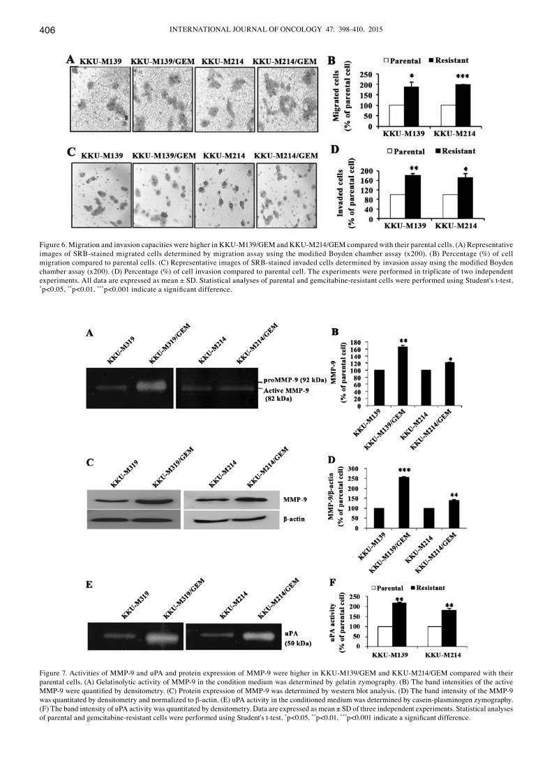
Increased MMP-9 and uPA activities in gemcitabine-resistant CCA cell lines. The proteolytic activity of MMP-9 of the parental and gemcitabine-resistant cell lines were measured by gelatin zymography. The MMP-9 activity in KKU-M139/GEM and KKU-M214/GEM was 1.55- and 1.21-fold higher than that in the parental cells (p<0.05, Fig. 7A and B). Using western blot
Figure 4. Cell adhesion was increased in KKU-M139/GEM and KKU-M214/GEM cells compared with their parental cells. (A) Representative images of SRB-stained adhesive cells determined by adhesion assay. (B) Percentage (%) of cell adhesion compared to the parental cells. Statistical analyses of parental and gemcitabine-resistant cells were performed using Student's t-test, **p<0.01 indicates a significant difference.

WATTANAWONGDON et al: MULTIDRUG RESISTANCE OF GEM-RESISTANT CHOLANGIOCARCINOMA CELLS 405
analyses, MMP-9 protein expression in KKU-M139/GEM and KKU-M214/GEM was 2.54- and 1.39-fold higher, respectively, than that in the parental cells (p<0.01, Fig. 7C and D). These results show that an increased enzymatic activity of MMP-9 was due to increased protein expression. The uPA activity in KKU-M139/GEM and KKU-M214/GEM is, respectively, 2.17-fold and 1.81-fold higher than that of the parental cells (p<0.01, Fig. 7E and F), as determined by casein-plasminogen zymography. These findings suggest that higher activities of MMP-9 and uPA may play a role in higher migration and inva-
sion capacities in KKU-M139/GEM and KKU-M214/GEM compared to the parental cells.
PKC signaling pathway plays an important role in apoptosis evasion, cell adhesion, migration and invasion. Cell lysates from the parental and gemcitabine-resistant cell lines were analyzed for protein expression by western blot analyses. Protein levels of all PKC fractions (cytosolic and membrane), FAK and p-FAK were significantly higher in KKU-M139/GEM and KKU-M214/GEM cells compared to those in the respec-
Figure 5. Apoptosis induction was decreased in KKU-M139/GEM and KKU-M214/GEM cells compared with their parental cells. KKU-M139 and KKU-M139/GEM were treated with 9 µM gemcitabine, whereas KKU-M214 and KKU-M214/GEM were treated with 16 µM gemcitabine. (A) Apoptotic cells in gemcitabine-treated parental and gemcitabine-resistant cells were determined by Annexin V-FITC/PI staining visualized: viable and non-apoptotic cells (Q3), early apoptotic cells (Q4), late apoptotic cells (Q2) and necrotic cells (Q1). (B) Percentages of apoptotic cells (Q2+Q4) in KKU-M139/GEM and KKU-M214/GEM were significantly lower than KKU-M139 and KKU-M214 cells. (C) Western blot analysis showed apoptosis regulated protein expression levels. Bax expression was decreased while Bcl-2 expression was increased in KKU-M139/GEM and KKU-M214/GEM compared with their parental cells. Fas expression was decreased in KKU-M214/GEM but was not changed in KKU-M139/GEM. (D) The band intensity of the proteins was quantitated by densitom-etry and normalized to β-actin. Data are expressed as mean ± SD of three independent experiments. Statistical analyses of parental and gemcitabine-resistant cells were performed using Student t-test, *p<0.05, **p<0.01, ***p<0.001 indicate a significant difference.
INTERNATIONAL JOURNAL OF ONCOLOGY 47: 398-410, 2015406
Figure 6. Migration and invasion capacities were higher in KKU-M139/GEM and KKU-M214/GEM compared with their parental cells. (A) Representative images of SRB-stained migrated cells determined by migration assay using the modified Boyden chamber assay (x200). (B) Percentage (%) of cell migration compared to parental cells. (C) Representative images of SRB-stained invaded cells determined by invasion assay using the modified Boyden chamber assay (x200). (D) Percentage (%) of cell invasion compared to parental cell. The experiments were performed in triplicate of two independent experiments. All data are expressed as mean ± SD. Statistical analyses of parental and gemcitabine-resistant cells were performed using Student's t-test, *p<0.05, **p<0.01, ***p<0.001 indicate a significant difference.
Figure 7. Activities of MMP-9 and uPA and protein expression of MMP-9 were higher in KKU-M139/GEM and KKU-M214/GEM compared with their parental cells. (A) Gelatinolytic activity of MMP-9 in the condition medium was determined by gelatin zymography. (B) The band intensities of the active MMP-9 were quantified by densitometry. (C) Protein expression of MMP-9 was determined by western blot analysis. (D) The band intensity of the MMP-9 was quantitated by densitometry and normalized to β-actin. (E) uPA activity in the conditioned medium was determined by casein-plasminogen zymography. (F) The band intensity of uPA activity was quantitated by densitometry. Data are expressed as mean ± SD of three independent experiments. Statistical analyses of parental and gemcitabine-resistant cells were performed using Student's t-test, *p<0.05, **p<0.01, ***p<0.001 indicate a significant difference.

WATTANAWONGDON et al: MULTIDRUG RESISTANCE OF GEM-RESISTANT CHOLANGIOCARCINOMA CELLS 407
tive parental cells (p<0.05, Fig. 8). In the PKC downstream signaling pathway, protein levels of ERK1/2, p-ERK1/2 and fractions of NF-κB/p65 (cytosolic and nuclear) were signifi-cantly higher in KKU-M139/GEM and KKU-M214/GEM cells compared to those of the respective parental cells (p<0.05, Fig. 8). These results suggest that higher expression of PKC membrane fraction, p-FAK, p-ERK1/2 and NF-κB nuclear fraction may play role in higher protein level of Bcl-2, MMP-9 and uPA in KKU-M139/GEM and KKU-M214/GEM cells compared to the parental cells. These findings suggest the signaling pathways involved in apoptosis evasion, cell adhe-sion, migration and invasion of gemcitabine-resistant CCA cell lines as proposed in Fig. 9.
Discussion
In this study, we successfully established highly gemcitabine-resistant CCA cell lines, from the respective parental CCA cell lines, KKU-M139 and KKU-M214. The IC50 values of KKU-M139/GEM and KKU-M214/GEM were 10.16 and 28.04 µM, respectively, which was far higher than those of previously reported gemcitabine-resistant cell lines, HuCCT1 and YSCCC, for which IC50 was 0.26 and 0.04 µM, respec-tively (17). The gemcitabine-resistance remained stable after continuous culture in drug-free medium for 2 months, and was also stable even following storage at -80˚C for 3 months.
Acquired gemcitabine resistance involved the alteration of gemcitabine transporter and disruption of gemcitabine metabolism to convert its active form. In our results, RRM1 was upregulated and hENT1 and dCK were downregulated, suggesting that hENT1, dCK and RRM1 play an important role in gemcitabine resistance. In non-small cell lung cancer cells, expression of hENT1 and dCK was decreased whereas RRM1 was increased in association with acquired gemcitabine-resistance (8,11).
The importance of ATP-binding cassette (ABC) trans-porters in mediating drug efflux is well documented (26). MRP expression was associated with decreased intracellular accu-mulation of various chemotherapeutic agents (27). Our results demonstrated that both of the gemcitabine-resistant cells overexpressed MRP1 protein compared to their parental cells. Overexpression of MRP1-3 has been reported in CCA (18,28). Thus, upregulation of MRP1 led to an increase of drug efflux resulted in multidrug resistance. Even though these anticancer drugs are different in their structure and anticancer mecha-nisms, the disruption of drug transporter function contribute to an acquired MDR in CCA cells. Hence, KKU-M139/GEM and KKU-M214/GEM showed cross-resistance to several anti-cancer drugs simultaneously, for example; 5-FU, doxorubicin and paclitaxel. 5-FU and gemcitabine share similar molecular structure, mechanisms of action and metabolic characteristics (15). This might explain why gemcitabine-resistant CCA cells can become cross-resistant to 5-FU. Paclitaxel is primarily an M-phase-specific drug that stabilize microtubules, resulting in an arrest of the cell cycle in M-phase and apoptosis (29). The G2/M arrest of gemcitabine-resistant CCA cells prevents cells entering M-phase where paclitaxel is mostly active and gain an advantage for acquired paclitaxel resistance. Doxorubicin can only target cell cycling and proliferation. Therefore, doxorubicin may lose anticancer activity if the gemcitabine-resistant CCA cells are prevented from entering cell cycle by G2/M-phase arrest. The association of decreased expression of cyclin B1 with doxorubicin resistance was reported in breast cancer cells (30).
Extension of the population doubling time in chemore-sistance has been reported in a variety of cancer cells (5,31). The prolonged proliferating time of the gemcitabine-resistant CCA cell lines reported here seems to have resulted from cell cycle arrest at G2/M phase because of decreased cyclin B1 and Cdk1 expression. Similar findings have been reported in adriamycin-resistant CCA cell line (QBC939/ADM) (32). Transition from G2 to M phase of the cell cycle is regulated by the Cdk1/cyclin B1 complex and CDKIs including p21 and p27 (33). P53 is a key tumor suppressor protein which is known
Figure 8. Activation of PKC signaling pathways in KKU-M139/GEM and KKU-M214/GEM. (A) Cell lysates from the parental and gemcitabine-resis-tant cell lines were analyzed for protein expression by western blot analyses. The protein expression levels of PKC (C, cytosolic), PKC (M, membrane), FAK, p-FAK, ERK1/2, p-ERK1/2, NF-κB/p65 (C, cytosolic) and NF-κB/p65 (N, nucleus) were higher in KKU-M139/GEM and KKU-M214/GEM com-pared with their parental cells. (B) The band intensity of the proteins was quantitated by densitometry and normalized to β-actin (total cell, cytosolic and membrane lysates) and Histone H1 (nuclear lysate). Data are expressed as mean ± SD of three independent experiments. Statistical analyses of parental and gemcitabine-resistant cells were performed using Student's t-test, *p<0.05, **p<0.01, ***p<0.001 indicate a significant difference.

INTERNATIONAL JOURNAL OF ONCOLOGY 47: 398-410, 2015408
as a transcriptional regulator of p21 and Bax (34). Our results also suggested the downregulation of p21 via decreased p53. Loss of p21 expression after chemotherapy was observed in human resistant tumor models harboring wild-type p53 (35). Taken together, the altered cell cycle profile of gemcitabine-resistant CCA cell lines may provide some advantages from killing by anticancer drugs. G2 arrest allows damage repair and eventual resumption of the cell cycle progression, preventing cells from anticancer activity of chemotherapeutic drugs.
Evasion of apoptosis via intrinsic and extrinsic pathway has been evident as the usual mechanism for chemoresis-tance (13). In this study, after treatment with gemcitabine, the percentage of apoptotic cells in both of the gemcitabine-resistant CCA cells were significantly decreased compared to their parental cells. In the mitochondrial (intrinsic) apoptosis pathway, the Bcl-2 family proteins including anti-apoptotic proteins (i.e., Bcl-2, Mcl-1 and Bcl-XL) and pro-apoptotic proteins (i.e., Bax) played an important role (14). An increase in the ratio of anti- to pro-apoptotic Bcl-2 proteins has been detected in various cancers and has been correlated to tumor cell survival and apoptosis resistance (14). Acquired resistance of pancreatic cancer cell line, Capan-1, against gemcitabine was associated with upregulation of the anti-apoptotic genes bcl-XL and mcl-1 and a higher ratio of Bcl-2/Bax expression (15). In this study, downregulation of p53 expression may cause decreased Bax protein expression which is the target gene product induced by p53. Our present data indicate that
resistance to chemotherapeutic drugs of two CCA cell lines is at least partly mediated via the increase of Bcl-2 protein expression and decrease of p53 and Bax protein expression. These changes lead to increased Bcl-2/Bax ratio and evasion of the intrinsic apoptosis pathway.
In the death receptor (extrinsic) apoptosis pathway, ligation of death receptors to the tumor necrosis factor (TNF) receptor super-family (i.e., CD95 or Fas) by the corresponding ligands (i.e., CD95 ligand or Fas ligand) leads to the recruitment of proteins such as Fas-associated death domain (FADD) and caspase-8 and subsequently activation of caspase-8, caspase-3 leading to apoptotic cell death (36). In this study, Fas expres-sion was significantly decreased in KKU-M214/GEM but was unaltered in KKU-M139/GEM compared with their parental cells. In contrast, the FADD expression was not changed in either of the gemcitabine-resistant CCA cells. These results indicate that decreased expression of Fas in KKU-M214/GEM may prevent the transmission of the death signal from the cell surface to intracellular signaling cascades leading to evasion of the extrinsic apoptosis pathway. Decreased expression of Fas has been reported in chemotherapeutic drug-resistant human renal cell carcinoma (37). It is interesting to note that KKU-M214/GEM may evade apoptosis through both intrinsic and extrinsic pathways, whereas, KKU-M139/GEM may evade apoptosis only through the intrinsic pathway. These results may explain the higher IC50 values of gemcitabine in KKU-M214/GEM (28.04±0.68 µM) than in KKU-M139/GEM (10.16±0.10 µM). Additionally, decrease of Fas expression
Figure 9. A possible mechanism by which CCA mediates gemcitabine resistance and invasiveness.

WATTANAWONGDON et al: MULTIDRUG RESISTANCE OF GEM-RESISTANT CHOLANGIOCARCINOMA CELLS 409
would also allow cells to evade immune responses mediated by both T and NK cells.
Invasion of cancer cells into basement membranes is a critical step in metastasis. The key markers that are involved in degradation of the ECM are the serine protease (plasmin), urokinase plasminogen activator (uPA) system, cysteine protease (cathepsin B and L) and matrix metalloproteinases (MMPs) (38). In our study, the established resistant cell lines demonstrated the relationship between drug resistance and cancer invasion. Both KKU-M139/GEM and KKU-M214/GEM exhibited an aggressive phenotype, i.e., higher migration and invasion capabilities with higher MMP-9 and uPA activities, similar to those in various cancer cell lines reported previously (39,40). The exposure of the human nasal carcinoma cell line RPMI-2650 to melphalan, an alkylating agent, resulted in not only an MDR phenotype but also enhancement of cell invasion in vitro. However, paclitaxel (Taxol) exposure promoted MDR without altered cell invasiveness (39). Osmak et al reported increased cathepsin D, uPA and plasminogen activator inhib-itor-1 (PAI-1) in a drug-resistant cervical and laryngeal cell line, although they did not investigate the invasive/metastatic potential of these cells (40).
Environmental-mediated drug resistance (EMDR) is thought to be triggered by the adhesion of integrins from tumor cells to stromal fibroblast or to components of the surrounding extracellular matrix (ECM) (41). The signaling could be due to degradation of apoptotic proteins or enhanced stability or altered subcellular distribution of anti-apoptotic proteins and cell cycle regulators (42,43). During adhesion, ligation of integrin with ECM ligands trigger PKC transloca-tion from cytosol to the intracellular membrane (44). PKC is a family of serine/threonine kinases associated with the regulation of cell cycle progression either during the G1/S or G2/M transition, cell adhesion, migration, invasion and drug resistance (45). Elevated levels of PKC and its downstream, FAK, have been shown in various cancer cells (44,45). FAK is a cytoplasmic tyrosine kinase that plays an important role in integrin-mediated signaling. It is the key mediator of several signaling pathways, especially ERK1/2, which is in the mitogen-activated protein kinases (MAPK) pathway (46). ERK1/2 also regulates transcription factor NF-κB indirectly by phosphorylating inhibitor of NF-κB kinase-α (IKK-α), IKK-α subsequently phosphorylates inhibitor of NF-κB (IκB), leading to ubiquitination and degradation of IκB-α. This promotes the translocation of NF-κB from the cytoplasm to the nucleus driving the transcription of its target genes (47,48). Our results confirm previous finding that all fractions of PKC expression, FAK phosphorylation, ERK1/2 phosphorylation, two fractions of NF-κB/p65 expression were increased in both gemcitabine-resistant CCA cells suggesting the involvement of the PKC and MAPK signaling pathways. Additionally, ERK1/2 signaling is responsible for resistance to gemcitabine in other CCA cells (49). Also in CCA (SCKR) cells, the activation of AKT and ERK1/2 signaling induced by cisplatin is likely to be the mechanism of cisplatin resistance, while the activation of ERK1/2 signaling is involved in 5-FU and gemcitabine resistance (50). The possible signaling pathways involved in the mediation of gemcitabine resistance of CCA are shown in Fig. 9.
In conclusion, the establishment of CCA cell lines highly resistant to gemcitabine was achieved using stepwise long-term exposure to the ascending series of gemcitabine. The established cell lines harbor multidrug resistance and invasiveness. The mechanism of enhanced drug resistance may include MRP1 expression, activation of the PKC signaling pathway, NF-κB activation and its downstream targets resulting in prolongation of proliferation time, evasion from apoptosis, and an increase of cancer invasiveness. An increased understanding of relation-ship between drug resistance and cancer cell invasiveness will ultimately lead to a consideration of means by which induc-tion of the aggressive phenotype should be avoided. Finally, molecular targets for circumvention of multidrug resistance and invasion are revealed for possible targeted therapy for CCA.
Acknowledgements
This study was supported in part by grants from the Center of Excellence for Innovation in Chemistry, Commission on Higher Education (grant no. 48-03-3-00-144), Khon Kaen University (grant nos. 563903, 573302, 582702) Research Fund, and Khon Kaen University under the Incubation Researcher Project, Khon Kaen University, Thailand. The authors sincerely thank Professor Yukifumi Nawa, invited Professor/Consultant, KKU, for reviewing the manuscript.
References
1. Khan SA, Taylor-Robinson SD, Toledano MB, Beck A, Elliott P and Thomas HC: Changing international trends in mortality rates for liver, biliary and pancreatic tumours. J Hepatol 37: 806-813, 2002.
2. Valverde A, Bonhomme N, Farges O, Sauvanet A, Flejou JF and Belghiti J: Resection of intrahepatic cholangiocarcinoma: A Western experience. J Hepatobiliary Pancreat Surg 6: 122-127, 1999.
3. Skipworth JR, Olde Damink SW, Imber C, Bridgewater J, Pereira SP and Malagó M: Review article: Surgical, neo-adjuvant and adjuvant management strategies in biliary tract cancer. Aliment Pharmacol Ther 34: 1063-1078, 2011.
4. Macias R: Cholangiocarcinoma: Biology, clinical management, and phamacological perspectives. ISRN Hematol 2014: 1-13, 2014.
5. Chen SY, Hu SS, Dong Q, Cai JX, Zhang WP, Sun JY, Wang TT, Xie J, He HR, Xing JF, et al: Establishment of paclitaxel-resistant breast cancer cell line and nude mice models, and underlying multidrug resistance mechanisms in vitro and in vivo. Asian Pac J Cancer Prev 14: 6135-6140, 2013.
6. Voutsadakis IA: Molecular predictors of gemcitabine response in pancreatic cancer. World J Gastrointest Oncol 3: 153-164, 2011.
7. Hertel LW, Boder GB, Kroin JS, Rinzel SM, Poore GA, Todd GC and Grindey GB: Evaluation of the antitumor activity of gemcitabine (2',2'-difluoro-2'-deoxycytidine). Cancer Res 50: 4417-4422, 1990.
8. Achiwa H, Oguri T, Sato S, Maeda H, Niimi T and Ueda R: Determinants of sensitivity and resistance to gemcitabine: The roles of human equilibrative nucleoside transporter 1 and deoxy-cytidine kinase in non-small cell lung cancer. Cancer Sci 95: 753-757, 2004.
9. Plunkett W, Huang P, Xu YZ, Heinemann V, Grunewald R and Gandhi V: Gemcitabine: Metabolism, mechanisms of action, and self-potentiation. Semin Oncol 22 (Suppl 11): S3-S10, 1995.
10. Goan YG, Zhou B, Hu E, Mi S and Yen Y: Overexpression of ribonucleotide reductase as a mechanism of resistance to 2,2-difluorodeoxycytidine in the human KB cancer cell line. Cancer Res 59: 4204-4207, 1999.
11. Davidson JD, Ma L, Flagella M, Geeganage S, Gelbert LM and Slapak CA: An increase in the expression of ribonucleotide reductase large subunit 1 is associated with gemcitabine resis-tance in non-small cell lung cancer cell lines. Cancer Res 64: 3761-3766, 2004.

INTERNATIONAL JOURNAL OF ONCOLOGY 47: 398-410, 2015410
12. Meli M, Tolomeo M, D'Alessandro N, Grimaudo S, Notarbartolo M, Papoff G, Ruberti G, Rausa L and Dusonchet L: Resistance to gemcitabine in a lymphoma cell line resistant to Fas-mediated apoptosis. Anticancer Res 24B: 851-857, 2004.
13. Akada M, Crnogorac-Jurcevic T, Lattimore S, Mahon P, Lopes R, Sunamura M, Matsuno S and Lemoine NR: Intrinsic chemore-sistance to gemcitabine is associated with decreased expression of BNIP3 in pancreatic cancer. Clin Cancer Res 11: 3094-3101, 2005.
14. Fulda S: Tumor resistance to apoptosis. Int J Cancer 124: 511-515, 2009.
15. Shi X, Liu S, Kleeff J, Friess H and Büchler MW: Acquired resistance of pancreatic cancer cells towards 5-Fluorouracil and gemcitabine is associated with altered expression of apoptosis-regulating genes. Oncology 62: 354-362, 2002.
16. Bergman AM, Pinedo HM and Peters GJ: Determinants of resis-tance to 2',2'-difluorodeoxycytidine (gemcitabine). Drug Resist Updat 5: 19-33, 2002.
17. Sato J, Kimura T, Saito T, Anazawa T, Kenjo A, Sato Y, Tsuchiya T and Gotoh M: Gene expression analysis for predicting gemcitabine resistance in human cholangiocarcinoma. J Hepatobiliary Pancreat Sci 18: 700-711, 2011.
18. Tepsiri N, Chaturat L, Sripa B, Namwat W, Wongkham S, Bhudhisawasdi V and Tassaneeyakul W: Drug sensitivity and drug resistance profiles of human intrahepatic cholangiocarci-noma cell lines. World J Gastroenterol 11: 2748-2753, 2005.
19. Namwat N, Amimanan P, Loilome W, Jearanaikoon P, Sripa B, Bhudhisawasdi V and Tassaneeyakul W: Characterization of 5-fluorouracil-resistant cholangiocarcinoma cell lines. Chemotherapy 54: 343-351, 2008.
20. Thanasai J, Limpaiboon T, Jearanaikoon P, Sripa B, Pairojkul C, Tantimavanich S and Miwa M: Effects of thymidine phosphory-lase on tumor aggressiveness and 5-fluorouracil sensitivity in cholangiocarcinoma. World J Gastroenterol 16: 1631-1638, 2010.
21. Skehan P, Storeng R, Scudiero D, Monks A, McMahon J, Vistica D, Warren JT, Bokesch H, Kenney S and Boyd MR: New colorimetric cytotoxicity assay for anticancer-drug screening. J Natl Cancer Inst 82: 1107-1112, 1990.
22. De Angelis PM, Kravik KL, Tunheim SH, Haug T and Reichelt WH: Comparison of gene expression in HCT116 treatment derivatives generated by two different 5-fluorouracil exposure protocols. Mol Cancer 3: 11, 2004.
23. Yodkeeree S, Chaiwangyen W, Garbisa S and Limtrakul P: Curcumin, demethoxycurcumin and bisdemethoxycurcumin differentially inhibit cancer cell invasion through the down-regulation of MMPs and uPA. J Nutr Biochem 20: 87-95, 2009.
24. Zhang S, Furugawa T, Takahashi H, et al: Analysis of gene expression in gemcitabine-resistant cells derived from human pancreatic cancer cell. International Conference on Human Health and Biomedical Engineering pp182-185, 2011.
25. Gerard CJ, Olsson K, Ramanathan R, Reading C and Hanania EG: Improved quantitation of minimal residual disease in multiple myeloma using real-time polymerase chain reaction and plasmid-DNA complementarity determining region III standards. Cancer Res 58: 3957-3964, 1998.
26. Modok S, Mellor HR and Callaghan R: Modulation of multidrug resistance efflux pump activity to overcome chemoresistance in cancer. Curr Opin Pharmacol 6: 350-354, 2006.
27. Zaman GJ, Flens MJ, van Leusden MR, de Haas M, Mülder HS, Lankelma J, Pinedo HM, Scheper RJ, Baas F and Broxterman HJ: The human multidrug resistance-associated protein MRP is a plasma membrane drug-efflux pump. Proc Natl Acad Sci USA 91: 8822-8826, 1994.
28. Courtois A, Payen L, Lagadic D, Guillouzo A and Fardel O: Evidence for a multidrug resistance-associated protein 1 (MRP1)-related transport system in cultured rat liver biliary epithelial cells. Life Sci 64: 763-774, 1999.
29. Shah MA and Schwartz GK: Cell cycle-mediated drug resis-tance: An emerging concept in cancer therapy. Clin Cancer Res 7: 2168-2181, 2001.
30. Smith L, Watson MB, O'Kane SL, Drew PJ, Lind MJ and Cawkwell L: The analysis of doxorubicin resistance in human breast cancer cells using antibody microarrays. Mol Cancer Ther 5: 2115-2120, 2006.
31. Guo X, Goessl E, Jin G, Collie-Duguid ES, Cassidy J, Wang W and O'Brien V: Cell cycle perturbation and acquired 5-fluoro-uracil chemoresistance. Anticancer Res 28A: 9-14, 2008.
32. Liu ZH, He YP, Zhou Y, Zhang P and Qin H: Establishment and identification of the human multi-drug-resistant cholangiocar-cinoma cell line QBC939/ADM. Mol Biol Rep 38: 3075-3082, 2011.
33. Wu ZZ, Chien CM, Yang SH, Lin YH, Hu XW, Lu YJ, Wu MJ and Lin SR: Induction of G2/M phase arrest and apoptosis by a novel enediyne derivative, THDA, in chronic myeloid leukemia (K562) cells. Mol Cell Biochem 292: 99-105, 2006.
34. Reinke V and Lozano G: Differential activation of p53 targets in cells treated with ultraviolet radiation that undergo both apoptosis and growth arrest. Radiat Res 148: 115-122, 1997.
35. Siddik ZH, Mims B, Lozano G and Thai G: Independent pathways of p53 induction by cisplatin and X-rays in a cisplatin-resistant ovarian tumor cell line. Cancer Res 58: 698-703, 1998.
36. Strasser A, Puthalakath H, O'Reilly LA and Bouillet P: What do we know about the mechanisms of elimination of autoreactive T and B cells and what challenges remain. Immunol Cell Biol 86: 57-66, 2008.
37. Ramp U, Dejosez M, Mahotka C, Czarnotta B, Kalinski T, Wenzel M, Lorenz I, Müller M, Krammer P, Gabbert HE, et al: Deficient activation of CD95 (APO-1/Fas)-mediated apoptosis: A potential factor of multidrug resistance in human renal cell carcinoma. Br J Cancer 82: 1851-1859, 2000.
38. Liotta LA, Tryggvason K, Garbisa S, Hart I, Foltz CM and Shafie S: Metastatic potential correlates with enzymatic degra-dation of basement membrane collagen. Nature 284: 67-68, 1980.
39. Liang Y, Meleady P, Cleary I, McDonnell S, Connolly L and Clynes M: Selection with melphalan or paclitaxel (Taxol) yields variants with different patterns of multidrug resistance, integrin expression and in vitro invasiveness. Eur J Cancer 37: 1041-1052, 2001.
40. Liang Y, McDonnell S and Clynes M: Examining the relation-ship between cancer invasion/metastasis and drug resistance. Curr Cancer Drug Targets 2: 257-277, 2002.
41. Zahreddine H and Borden KL: Mechanisms and insights into drug resistance in cancer. Front Pharmacol 4: 28, 2013.
42. Hazlehurst LA, Argilagos RF and Dalton WS: Beta1 integrin mediated adhesion increases Bim protein degradation and contributes to drug resistance in leukaemia cells. Br J Haematol 136: 269-275, 2007.
43. Lwin T, Hazlehurst LA, Dessureault S, Lai R, Bai W, Sotomayor E, Moscinski LC, Dalton WS and Tao J: Cell adhesion induces p27Kip1-associated cell-cycle arrest through down-regulation of the SCFSkp2 ubiquitin ligase pathway in mantle-cell and other non-Hodgkin B-cell lymphomas. Blood 110: 1631-1638, 2007.
44. Goel HL and Dey CS: Role of protein kinase C during insulin mediated skeletal muscle cell spreading. J Muscle Res Cell Motil 23: 269-277, 2002.
45. Kang J: Protein kinase C (PKC) isozymes and cancer. New J Sci 36: 2014, 2014.
46. Liao CH, Sang S, Ho CT and Lin JK: Garcinol modulates tyrosine phosphorylation of FAK and subsequently induces apoptosis through down-regulation of Src, ERK, and Akt survival signaling in human colon cancer cells. J Cell Biochem 96: 155-169, 2005.
47. Perkins ND: The Rel/NF-kappa B family: Friend and foe. Trends Biochem Sci 25: 434-440, 2000.
48. Ghosh S and Karin M: Missing pieces in the NF-kappaB puzzle. Cell 109 (Suppl): S81-S96, 2002.
49. Matsumoto K, Nagahara T, Okano J and Murawaki Y: The growth inhibition of hepatocellular and cholangiocellular carcinoma cells by gemcitabine and the roles of extracellular signal-regulated and checkpoint kinases. Oncol Rep 20: 863-872, 2008.
50. Yoon H, Min JK, Lee JW, Kim DG and Hong HJ: Acquisition of chemoresistance in intrahepatic cholangiocarcinoma cells by activation of AKT and extracellular signal-regulated kinase (ERK)1/2. Biochem Biophys Res Commun 405: 333-337, 2011.