Research Article Resolving Contributions of Oxygen...
8
Research Article Resolving Contributions of Oxygen-Consuming and ROS-Generating Enzymes at the Synapse Engy A. Abdel-Rahman, Ali M. Mahmoud, Abdullah Aaliya, Yasmine Radwan, Basma Yasseen, Abdelrahman Al-Okda, Ahmed Atwa, Eslam Elhanafy, Moaaz Habashy, and Sameh S. Ali Center for Aging and Associated Diseases, Helmy Institute of Medical Sciences, Zewail City of Science and Technology, Giza, Egypt Correspondence should be addressed to Sameh S. Ali; [email protected] Received 23 June 2016; Revised 10 October 2016; Accepted 6 November 2016 Academic Editor: Andr´ es Trostchansky Copyright © 2016 Engy A. Abdel-Rahman et al. is is an open access article distributed under the Creative Commons Attribution License, which permits unrestricted use, distribution, and reproduction in any medium, provided the original work is properly cited. Disruption of cellular redox homeostasis is implicated in a wide variety of pathologic conditions and aging. A fundamental factor that dictates such balance is the ratio between mitochondria-mediated complete oxygen reduction into water and incomplete reduction into superoxide radical by mitochondria and NADPH oxidase (NOX) enzymatic activity. Here we determined mitochondrial as well as NOX-dependent rates of oxygen consumption in parallel with H 2 O 2 generation in freshly isolated synaptosomes using high resolution respirometry combined with fluorescence or electrochemical sensory. Our results indicate that although synaptic mitochondria exhibit substantially higher respiratory activities (8–82-fold greater than NOX oxygen consumption depending on mitochondrial respiratory state), NADPH-dependent oxygen consumption is associated with greater H 2 O 2 production (6-7-fold higher NOX-H 2 O 2 ). We also show that, in terms of the consumed oxygen, while synaptic mitochondria “leaked” 0.71% ± 0.12 H 2 O 2 during NAD + -linked resting, 0.21% ± 0.04 during NAD + -linked active respiration, and 0.07% ± 0.02 during FAD + -linked active respiration, NOX converted 38% ± 13 of O 2 into H 2 O 2 . Our results indicate that NOX rather than mitochondria is the major source of synaptic H 2 O 2 . e present approach may assist in the identification of redox-modulating synaptic factors that underlie a variety of physiological and pathological processes in neurons. 1. Introduction Substantial evidence indicates that the synapse is a center stage for brain physiology and pathology [1]. Synaptic activity is now known to produce ROS that are essential regulators of multitudes of normal physiological processes in neurons including cognition and memory. e high levels of ROS gen- eration in synapses, alongside their high-energy demands, make them more vulnerable to stressful insults encountered in aging, neurodegenerative, neuropsychological, and neu- rodevelopmental disorders [2]. e relative importance of specific enzymatic sources of ROS in synapses is not fully understood. Mitochondria are one source of cellular ROS. A portion of oxygen consumed by mitochondria escapes the aerobic ATP production pathway and forms oxygen radicals primarily in the form of superoxide anions (O 2 ∙− ) that are instantaneously dismutated to hydrogen peroxide (H 2 O 2 ) by mitochondrial superoxide dismutase (SOD) [3–5]. Since the brain is highly metabolically active organ that exhibits robust oxygen consumption [6], mitochondria respiratory activity was oſten considered the prime source of brain ROS. However, recent data indicate that NADPH oxidases (NOX), the enzyme family known to generate ROS as their only and primary function, are widely expressed in the CNS where they considerably contribute to ROS generation [7, 8]. While NOX2 is found principally in phagocytes, recent reports showed that NOX2 and homologs (NOX1, NOX3–5, Duox1, and 2) are expressed in a miscellaneous array of tissues and cell types. NOX2 and NOX4 have been characterized in the neurons of adult mouse nervous system, potentially contributing to wide range of physiologic functions and to several neurological disorders [9]. Although it is likely that the initial product of all NOX enzymes is O 2 ∙− , which spontaneously dismutates to H 2 O 2 via superoxide dismutase (SOD), it is now clear that H 2 O 2 is predominantly produced by several NOX isoforms, particularly NOX4, Duox1, and Hindawi Publishing Corporation Oxidative Medicine and Cellular Longevity Volume 2016, Article ID 1089364, 7 pages http://dx.doi.org/10.1155/2016/1089364
Transcript of Research Article Resolving Contributions of Oxygen...

Research ArticleResolving Contributions of Oxygen-Consuming andROS-Generating Enzymes at the Synapse
Engy A. Abdel-Rahman, Ali M. Mahmoud, Abdullah Aaliya,Yasmine Radwan, Basma Yasseen, Abdelrahman Al-Okda, Ahmed Atwa,Eslam Elhanafy, Moaaz Habashy, and Sameh S. Ali
Center for Aging and Associated Diseases, Helmy Institute of Medical Sciences, Zewail City of Science and Technology, Giza, Egypt
Correspondence should be addressed to Sameh S. Ali; [email protected]
Received 23 June 2016; Revised 10 October 2016; Accepted 6 November 2016
Academic Editor: Andres Trostchansky
Copyright © 2016 Engy A. Abdel-Rahman et al.This is an open access article distributed under the Creative Commons AttributionLicense, which permits unrestricted use, distribution, and reproduction in anymedium, provided the originalwork is properly cited.
Disruption of cellular redox homeostasis is implicated in a wide variety of pathologic conditions and aging. A fundamentalfactor that dictates such balance is the ratio between mitochondria-mediated complete oxygen reduction into water andincomplete reduction into superoxide radical bymitochondria andNADPHoxidase (NOX) enzymatic activity. Herewe determinedmitochondrial as well as NOX-dependent rates of oxygen consumption in parallel with H
2O2generation in freshly isolated
synaptosomes using high resolution respirometry combined with fluorescence or electrochemical sensory. Our results indicatethat although synaptic mitochondria exhibit substantially higher respiratory activities (8–82-fold greater than NOX oxygenconsumption depending on mitochondrial respiratory state), NADPH-dependent oxygen consumption is associated with greaterH2O2production (6-7-fold higher NOX-H
2O2). We also show that, in terms of the consumed oxygen, while synaptic mitochondria
“leaked” 0.71% ± 0.12 H2O2during NAD+-linked resting, 0.21% ± 0.04 during NAD+-linked active respiration, and 0.07% ± 0.02
during FAD+-linked active respiration, NOX converted 38% ± 13 of O2into H
2O2. Our results indicate that NOX rather than
mitochondria is the major source of synaptic H2O2. The present approach may assist in the identification of redox-modulating
synaptic factors that underlie a variety of physiological and pathological processes in neurons.
1. Introduction
Substantial evidence indicates that the synapse is a centerstage for brain physiology and pathology [1]. Synaptic activityis now known to produce ROS that are essential regulatorsof multitudes of normal physiological processes in neuronsincluding cognition andmemory.The high levels of ROS gen-eration in synapses, alongside their high-energy demands,make them more vulnerable to stressful insults encounteredin aging, neurodegenerative, neuropsychological, and neu-rodevelopmental disorders [2]. The relative importance ofspecific enzymatic sources of ROS in synapses is not fullyunderstood. Mitochondria are one source of cellular ROS.A portion of oxygen consumed by mitochondria escapes theaerobic ATP production pathway and forms oxygen radicalsprimarily in the form of superoxide anions (O
2
∙−) that areinstantaneously dismutated to hydrogen peroxide (H
2O2)
by mitochondrial superoxide dismutase (SOD) [3–5]. Since
the brain is highly metabolically active organ that exhibitsrobust oxygen consumption [6], mitochondria respiratoryactivity was often considered the prime source of brain ROS.However, recent data indicate that NADPH oxidases (NOX),the enzyme family known to generate ROS as their only andprimary function, are widely expressed in the CNS wherethey considerably contribute to ROS generation [7, 8]. WhileNOX2 is found principally in phagocytes, recent reportsshowed that NOX2 and homologs (NOX1, NOX3–5, Duox1,and 2) are expressed in a miscellaneous array of tissuesand cell types. NOX2 and NOX4 have been characterizedin the neurons of adult mouse nervous system, potentiallycontributing to wide range of physiologic functions andto several neurological disorders [9]. Although it is likelythat the initial product of all NOX enzymes is O
2
∙−, whichspontaneously dismutates to H
2O2via superoxide dismutase
(SOD), it is now clear that H2O2is predominantly produced
by several NOX isoforms, particularly NOX4, Duox1, and
Hindawi Publishing CorporationOxidative Medicine and Cellular LongevityVolume 2016, Article ID 1089364, 7 pageshttp://dx.doi.org/10.1155/2016/1089364

2 Oxidative Medicine and Cellular Longevity
Doux2. This apparent H2O2generation may be attributed
to the rapid dismutation of O2
∙−. However, recent reportsshowed that for, NOX4, H
2O2generation is mediated by the
third extracellular loop of the enzyme (reviewed in [10]).Synaptic localization of mitochondria [11, 12] and some
NOX isoforms have been documented [8, 13]. Synaptosomes(isolated nerve terminals), which have been extensively usedfor studying brain synaptic physiology, were found to containmitochondria with distinct biophysical properties from thoseof neuronal bodymitochondria [11, 12]. NOX2 andNOX4 arealso expressed in synaptosomal plasmamembrane [8, 13] andwe previously reported that synaptosomes exhibit NADPH-dependent oxygen consumption [13]. Given the critical roleof NOX and mitochondria in cellular ROS production andthe presence of some NOX isoforms and distinctive mito-chondria in synaptosomes, it is of paramount importance tocharacterize the interplay between their respiratory functionsand ROS generation in synapses. Our previous study usingspin-trapping electron paramagnetic resonance spectroscopyshowed that NOX rather than mitochondria was the maincontributor of synaptic superoxide generation [8]. How-ever, the relationship between mitochondria and/or NOX-dependent O
2consumption and resulting ROS generation is
still ambiguous, despite the long-held idea that augmentedenergy expenditure will result in higher ROS generation[14]. Much vagueness exists over the relationship betweenthe rate of oxygen consumption and the production ofreactive oxygen species (ROS) such as hydrogen peroxide bymitochondria and NOX in synapses. Here, we use combinedhigh resolution respirometry and fluorometry to simultane-ously monitor oxygen consumption and H
2O2production
by synaptosomal mitochondria and NOX. We will describefor the first time how the well-established Amplex Redassay or an electrochemical sensor can be used to quantifyH2O2production by NOX combined with the simultaneous
measurement of NOX-dependent oxygen consumption byhigh resolution respirometry.
2. Materials and Methods
2.1. Animals. C57BL6/C males (6 weeks old) were purchasedfrom Misr University for Science and Technology (Cairo,Egypt) and were housed for at least one month in ZewailCity animal facility until sacrificed. All animals weremaintained in pathogen-free, individually ventilated cagesin 12 h light/12 h dark cycles at 24∘C and 50% relativehumidity, with free access to water and standard laboratoryrodent chow. Animals were decapitated following quickcervical dislocation which is an approved and consideredhumanemethod of small animal euthanasia by the AmericanVeterinaryMedical Association (AVMA) (https://www.avma.org/KB/Policies/Documents/euthanasia.pdf). All experi-ments were conducted in adherence to the NIH Institu-tional Animal Care and Use Committee guidelines https://grants.nih.gov/grants/olaw/GuideBook.pdf.
2.2. Isolation of Synaptosomes. Isolation of synaptosomeswasperformed as previously described [13]. Briefly, brains were
quickly removed; forebrainswere dissected andhomogenizedusing a Dounce homogenizer in ice-cold isolation buffer(0.32M sucrose, 1mM EDTA, 10mM Tris-HCl buffer, pH7.4, 10mM glucose). The homogenate was then centrifugedat 3,100×g for 3min at 4∘C. The supernatant was removedand the pellet was resuspended in half the volume of iso-lation buffer, then homogenized again, and recentrifuged.The supernatant was collected and mixed with percoll toa final concentration of 15% by volume. The mixture wasthen layered onto a step gradient of 23% and 40% percoll.Centrifugationwas then performed at 16,000 rpm for 5min at4∘C. The band at the interface of the two layers was collectedand rinsed in isolation buffer, followed by centrifugation andresuspension in synaptosomal buffer (120mMNaCl, 4.7mMKcl, 2.2mM CaCl
2, 1.2mM MgCl
2, 25mM HEPES, 1.2mM
MgSO4, 1.2mm KH
2PO4, and 10mM glucose).
2.3. Determination of NOX Activity in Synaptosomes byOroboros� High Resolution O2k Oxygraph. NOX activity insynaptosomal preparations was determined by measuring,in the same sample, NADPH-induced oxygen consumptionand the associated rate of hydrogen peroxide formationsimultaneously using Amplex Red fluorescence/or H
2O2
electrochemical HPO-ISO-2mm sensor (WPI, Sarasota,USA) which is compatible with the O2k-NO Amp-Module(Oroboros�).
2.4. Measurements of NADPH Oxidase Respiratory Rates.NADPH oxidase respiratory assessments (and hydrogen per-oxide determinations) were carried out at 37∘Cusing the highresolution respirometry system Oxygraph-2K (OroborosInstruments, Innsbruck, Austria) in 2mL chambers. Beforestarting the experiment, calibration at air saturation wasperformed by allowing the respiration medium, MIR05, toequilibrate with air in the oxygraph chambers and be stirredat 540 to 560 rpm for 30 to 40min, until a stable signalwas detected. Synaptosomal protein (0.2mg) was added tothe respiration medium in the chamber. Activation of NOXwas evoked by the addition of 200 𝜇M NADPH (3 doses).The rates of oxygen consumption were calculated as thenegative time derivative of oxygen concentration. The rate ofNOX-dependent hydrogen peroxide formation was detectedin parallel to oxygen consumption in the same sample byusing two different approaches. In the fluorometric method,horseradish peroxidase (1 U per mL) and Amplex Ultra-Red fluorescent dye (10 𝜇M) were utilized. The excitationwavelength was 525 nm and fluorescence detection was at587 nm. For determination of NOX-dependent hydrogenperoxide production by amperometric detection in parallelwith oxygen consumption, the fluorescent dye was substi-tuted by inserting an HPO-ISO-2 WPI electrode in theO2k chamber. Signals were calibrated using known amountsof hydrogen peroxide that were exogenously added by theend of each run. At the peak of oxygen-consuming, HPO-producing NADPH activity, specific NOX inhibitor VAS-2870 (10 𝜇M) or ebselen (10 𝜇M) was added to confirm thatthe activity recorded was mediated by NOX. Data acquisitionand analysis were performed with the DatLab� software,version 4.3 (Oroboros Instruments).This enables continuous

Oxidative Medicine and Cellular Longevity 3
monitoring and recording of the oxygen concentration in thechambers as well as of the derived oxygen flux over time,normalized for the amount of homogenized tissue acquiredat rates of 0.5–1Hz.
2.5. Measurements of Synaptic Mitochondrial Oxygen Con-sumption and Hydrogen Peroxide Production. After blockingNOX activity, synaptosomes were permeabilized by the addi-tion of saponin (25 𝜇g/mL). State 4 respiration was triggeredby adding the following substrates: 10mM pyruvate + 10mMmalate + 10mM glutamate. State 3 respiration was theninduced by adding ADP (1mM) for measuring OXPHOS Ifollowed by 10mM succinate to assess OXPHOS I+II. Parallelassessment of oxygen consumption and hydrogen peroxideformation by synaptosomal mitochondria was performed asdescribed above. In a control experiment, we confirmed thatouter mitochondrial membrane integrity was not affected bythe saponification process as exogenously added cytochromec (10 𝜇M) did not impact mitochondrial OCR (data notshown).
3. Results
3.1. NADPH Oxidases Are Minor Oxygen Consumers butMajor Hydrogen Peroxide Producers in Synaptosomes. Wehave previously reported the detection of oxygen consump-tion by synaptosomal NOX using Oxygraphy [8, 13]. Here,we utilized high resolution respirometry (Oroboros O2k-Station) to follow NADPH-induced oxygen consumptionin isolated C57BL6/C male synaptosomes. Additionally, wesimultaneously used the O2k-Fluorometer or the HPO sen-sor, for the first time, to monitor the rate of hydrogenperoxide production in the same sample. In the fluorometricassay the highly sensitive AR/HRP system has been usedfor assessment of H
2O2production by recording changes in
resorufin fluorescence [15]. Since HRP has been shown tocatalyze the oxidation of NADPH with subsequent gener-ation of H
2O2[16], we compared NOX-dependent oxygen
consumption and hydrogen peroxide production obtained byusing the AR/HRP system and the HPO sensor. In Figures1(a)–1(f), we show representative traces of NADPH-induced,VAS-2870-inhibitable (a, c, e), or ebselen-inhibitable (b, d,f) synaptosomal oxygen consumption (a, b) and parallelhydrogen peroxide production obtained using AR/HRP flu-orometric assay (c and d), or Oroboros-compatible amper-ometric HPO sensor (e, f). Using the AR/HRP assay, wecould continuously monitor H
2O2generated during NOX
activation. We calibrated our H2O2fluorescence signal using
high resolution measurements of oxygen production due toselective decomposition of H
2O2by exogenous catalase (not
shown). This allowed us to quantify the O2proportions that
are converted to H2O2in real time. Previous studies showed
that NADPH can generate H2O2nonenzymatically through
its interaction with HRP [17]. In this regard, we foundthat, in the absence of added synaptosomes, the addition ofNADPH alone in our AR/HRP assay resulted in enhancedfluorescence. However, under our experimental conditions,this increase in background fluorescence was far less than the
resorufin fluorescence detected in the presence of synapto-somes. Our results are in tune with another study utilizingthe AR/HRP system for microsomal enzymes activity, whichshowed that increased resorufin fluorescence resulting fromthe interaction between HRP and NADPH was less than 2–5%of the fluorescencemonitored in the presencemicrosomalenzymes [17].
After subtraction of background fluorescence, there wereno significant differences in oxygen consumption by synap-tosomal NOX between fluorometric (𝑛 = 5) and electro-chemical (𝑛 = 5) experiments. As shown in Figure 1(g), thehydrogen peroxide production by synaptosomalNOXdid notsignificantly differ between the two experimental approaches.UsingO2k combinedwith theAR/HRP fluorescencemodule,the addition of 200𝜇M NADPH (3 doses) to synaptosomalproteins (0.2mg) in our study resulted in absolute ratesof oxygen consumption and H
2O2production of 0.0055 ±
0.0011 𝜇M/s and 0.0018±0.0005 𝜇M/s H2O2. Similar rates of
H2O2production were obtained when the AR/HRP system
was replaced by HPO electrode 0.0023 ± 0.0005 𝜇M/s H2O2,
confirming that there was minor or no interference fromcomponents of the reaction mix.
Both VAS-2870 and ebselen significantly inhibitedNADPH-induced oxygen consumption by about 50–75%(𝑁 = 5–8, 𝑝 < 0.05). However, while VAS-2870 reducedH2O2production proportionately by ∼60% (𝑁 = 8,
𝑝 < 0.05), we observed a strong elimination of hydrogenperoxide by ebselen, which is in tune with reports showingthat the later possesses glutathione peroxidase activity(reviewed in [18]).
3.2. Relative to NOX, Mitochondria Are Major Oxygen Con-sumers but Minor Hydrogen Peroxide Producers in Synapto-somes. To investigate the relative importance of NOX andmitochondria in energy expenditure and H
2O2produc-
tion at the synapse, we employed isolated nerve terminals(synaptosomes) that contain both pre- and postsynapticvesicles. Synaptosomal preparations are populated with NOXisoforms and mitochondria of characteristic biophysicalproperties. Experiments typically involved parallel mea-surements of oxygen consumption and hydrogen peroxideproduction using high resolution respirometry combinedwith the HRP/Amplex Red fluorescence assay.This approachallows one to follow both NOX and mitochondrial activitiesin synaptosomes and estimate resultant hydrogen peroxideproduction in the same sample. When NADPHwas added tosynaptosomes, oxygen consumption and hydrogen peroxideproduction due to NOX activity were triggered as describedabove. This activity was abolishable by the specific NOXinhibitor VAS-2870 (10 𝜇M) or ebselen (10 𝜇M). Subsequentaddition of pyruvate, malate, and glutamate to synaptosomeselicited oxygen consumption due to NAD+-linked restingmitochondrial metabolic activity (Figure 2(a)) while also“leaking” hydrogen peroxide in parallel (Figure 2(b)). Ina separate experiment, we confirmed the involvement ofsynaptosomal mitochondria in the substrate-triggered oxy-gen consumption (Figure 2(c)). As can be seen in Figure 2(c),both NAD+- and FAD+-linked respiratory activities werecompletely inhibitable by oligomycin (complex V), rotenone
4 Oxidative Medicine and Cellular Longevity
0.000
0.002
0.004
0.006
0.008
FluorElectro
20 25 30 35160
170
180
190
200
Time (min)
NADPH
−10
0
10
20
30
VAS-2870
0 500 1000 1500 2000 25000
2000
4000
6000
8000
Time (seconds)
NADPH
VAS-2870
(a)
(b)
(g) (h) (i)
10 15 20 25 30160
170
180
190
200
210
Time (min)
−20
−10
0
10
20
30
Ebselen
NADPH
0 500 1000 1500 20000
2000
4000
6000
8000
Ebselen
NADPH
Time (seconds)15 20 25 30 35 40
12345678
Ebselen
Time (min)
NADPH 01234567
130 140 150 160 1700.00.51.01.52.02.53.0
Time (min)
NADPH
VAS-2870
0.0
0.4
0.8
1.2
1.6
−0.020
−0.015
−0.002
0.000
0.002
NADPH
Ebselen
Vas-2870
NADPH Vas-2870 Ebselen0.000
0.002
0.004
0.006
0.008
(c)
(d)
(e)
(f)
∗
∗
O2
flux
(𝜇M
/sec
)
O2 H2O2 H2O2
[O2] (
𝜇M
)[O
2] (
𝜇M
)
O2
flux
(nm
ol/s
ec)
O2
flux
(nm
ol/s
ec)
Flux
(𝜇M
/sec
)
[H2O
2](𝜇
M)
[H2O
2](𝜇
M)
H2O
2flu
x (n
mol
/sec
)H
2O
2flu
x (n
mol
/sec
)
H2O
2sig
nal (
PA)
H2O
2sig
nal (
PA)
H2O
2flu
x (𝜇
M/s
ec)
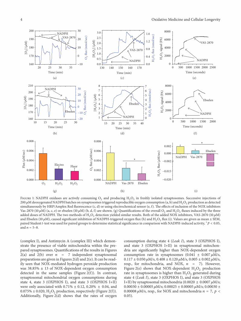
Figure 1: NADPH oxidases are actively consuming O2and producing H
2O2in freshly isolated synaptosomes. Successive injections of
200 𝜇MdeoxygenatedNADPHbatches on synaptosomes triggered reproducible oxygen consumption (a, b) andH2O2production as detected
simultaneously by HRP/Amplex Red fluorescence (c, d) or using electrochemical sensor (e, f). The effects of inclusion of the NOX inhibitorsVas-2870 (10 𝜇M) (a, c, e) or ebselen (10𝜇M) (b, d, f) are shown. (g) Quantifications of the overall O
2and H
2O2fluxes induced by the three
added doses of NADPH. The two methods of H2O2detection yielded similar results. Both of the added NOX inhibitors, VAS-2870 (10𝜇M)
and Ebselen (10𝜇M), caused significant inhibition of NADPH-triggered oxygen flux (h) and H2O2flux (i). Values are given as mean ± SEM,
paired Student 𝑡-test was used for paired groups to determine statistical significance in comparison with NADPH-induced activity, ∗𝑝 < 0.05,and 𝑛 = 5–8.
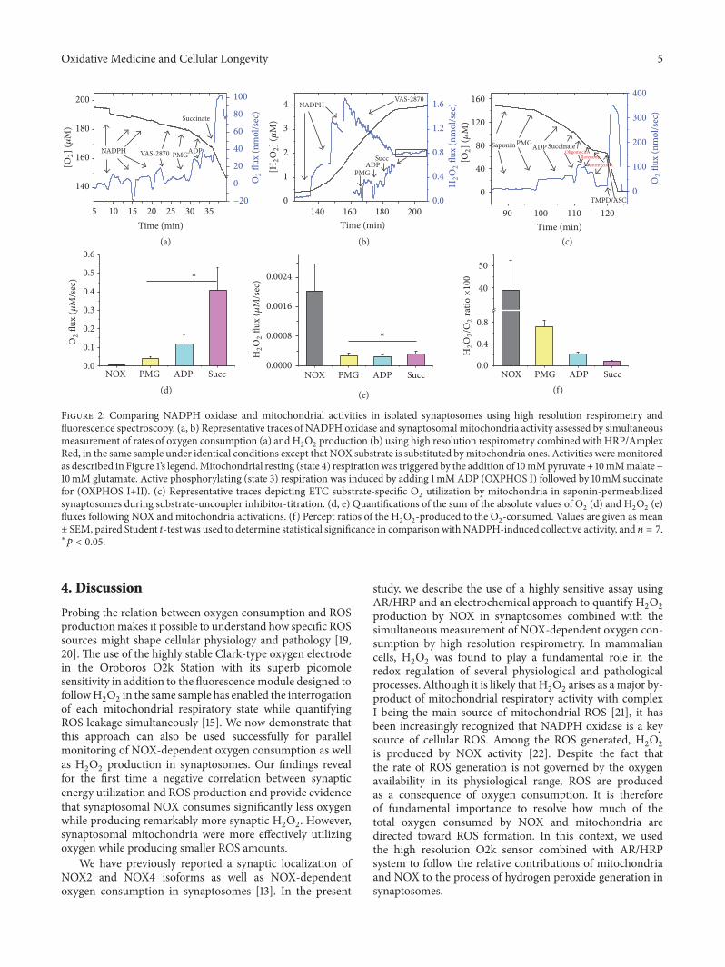
(complex I), and Antimycin A (complex III) which demon-strate the presence of viable mitochondria within the pre-pared synaptosomes. Quantifications of the results in Figures2(a) and 2(b) over 𝑛 = 7 independent synaptosomalpreparations are given in Figures 2(d) and 2(e). It can be read-ily seen that NOX-mediated hydrogen peroxide productionwas 38.85% ± 13 of NOX-dependent oxygen consumptiondetected in the same samples (Figure 2(f)). In contrast,synaptosomal mitochondrial oxygen consumptions duringstate 4, state 3 (OXPHOS I), and state 3 (OXPHOS I+II)were only associated with 0.71% ± 0.12, 0.20% ± 0.04, and0.075% ± 0.020, H
2O2production, respectively (Figure 2(f)).
Additionally, Figure 2(d) shows that the rates of oxygen
consumption during state 4 (Leak I), state 3 (OXPHOS I),and state 3 (OXPHOS I+II) in synaptosomal mitochon-dria are significantly higher than NOX-dependent oxygenconsumption rate in synaptosomes (0.041 ± 0.007 𝜇M/s,0.117 ± 0.050 𝜇M/s, 0.408 ± 0.120 𝜇M/s, 0.005 ± 0.002 𝜇M/s,resp., for mitochondria, and NOX, 𝑛 = 7). However,Figure 2(e) shows that NOX-dependent H
2O2production
rate in synaptosomes is higher than H2O2generated during
state 4 (Leak I), state 3 (OXPHOS I), and state 3 (OXPHOSI+II) by synaptosomal mitochondria (0.0020 ± 0.0007 𝜇M/s;0.00030 ± 0.00005 𝜇M/s; 0.00025 ± 0.00005 𝜇M/s; 0.00030 ±0.00008 𝜇M/s, resp., for NOX and mitochondria 𝑛 = 7, 𝑝 <0.05).
Oxidative Medicine and Cellular Longevity 5
NOX PMG ADP Succ0.0
0.4
0.8
40
50
NOX PMG ADP Succ0.0000
0.0008
0.0016
0.0024
NOX PMG ADP Succ0.0
0.1
0.2
0.3
0.4
0.5
0.6
140 160 180 2000
1
2
3
4
Time (min)
0.0
0.4
0.8
1.2
1.6
PMGADP
Succ
NADPHVAS-2870
5 10 15 20 25 30 35
140
160
180
200
Time (min)
NADPH
−20
0
20
40
60
80
100
VAS-2870 PMGADP
Succinate
(a)
(d) (e) (f)
(b) (c)
90 100 110 120
0
40
80
120
160
Time (min)
0
100
200
300
400
Saponin PMGADP SuccinateOligomycin
RotenoneAntimycin-A
TMPD/ASC
[O2]
(𝜇M
)
O2
flux
(nm
ol/s
ec)
O2
flux
(𝜇M
/sec
) ∗
∗
[H2O
2] (
𝜇M
)
H2O
2flu
x (n
mol
/sec
)
O2
flux
(nm
ol/s
ec)
[O2] (
𝜇M
)
H2O
2flu
x (𝜇
M/s
ec)
H2O
2/O
2ra
tio×10
0
Figure 2: Comparing NADPH oxidase and mitochondrial activities in isolated synaptosomes using high resolution respirometry andfluorescence spectroscopy. (a, b) Representative traces of NADPH oxidase and synaptosomal mitochondria activity assessed by simultaneousmeasurement of rates of oxygen consumption (a) and H
2O2production (b) using high resolution respirometry combined with HRP/Amplex
Red, in the same sample under identical conditions except that NOX substrate is substituted bymitochondria ones. Activities weremonitoredas described in Figure 1’s legend.Mitochondrial resting (state 4) respirationwas triggered by the addition of 10mMpyruvate + 10mMmalate +10mM glutamate. Active phosphorylating (state 3) respiration was induced by adding 1mMADP (OXPHOS I) followed by 10mM succinatefor (OXPHOS I+II). (c) Representative traces depicting ETC substrate-specific O
2utilization by mitochondria in saponin-permeabilized
synaptosomes during substrate-uncoupler inhibitor-titration. (d, e) Quantifications of the sum of the absolute values of O2(d) and H
2O2(e)
fluxes following NOX and mitochondria activations. (f) Percept ratios of the H2O2-produced to the O
2-consumed. Values are given as mean
± SEM, paired Student 𝑡-test was used to determine statistical significance in comparison with NADPH-induced collective activity, and 𝑛 = 7.∗𝑝 < 0.05.
4. Discussion
Probing the relation between oxygen consumption and ROSproductionmakes it possible to understand how specific ROSsources might shape cellular physiology and pathology [19,20]. The use of the highly stable Clark-type oxygen electrodein the Oroboros O2k Station with its superb picomolesensitivity in addition to the fluorescencemodule designed tofollowH
2O2in the same sample has enabled the interrogation
of each mitochondrial respiratory state while quantifyingROS leakage simultaneously [15]. We now demonstrate thatthis approach can also be used successfully for parallelmonitoring of NOX-dependent oxygen consumption as wellas H2O2production in synaptosomes. Our findings reveal
for the first time a negative correlation between synapticenergy utilization and ROS production and provide evidencethat synaptosomal NOX consumes significantly less oxygenwhile producing remarkably more synaptic H
2O2. However,
synaptosomal mitochondria were more effectively utilizingoxygen while producing smaller ROS amounts.
We have previously reported a synaptic localization ofNOX2 and NOX4 isoforms as well as NOX-dependentoxygen consumption in synaptosomes [13]. In the present
study, we describe the use of a highly sensitive assay usingAR/HRP and an electrochemical approach to quantify H
2O2
production by NOX in synaptosomes combined with thesimultaneous measurement of NOX-dependent oxygen con-sumption by high resolution respirometry. In mammaliancells, H
2O2was found to play a fundamental role in the
redox regulation of several physiological and pathologicalprocesses. Although it is likely that H
2O2arises as amajor by-
product of mitochondrial respiratory activity with complexI being the main source of mitochondrial ROS [21], it hasbeen increasingly recognized that NADPH oxidase is a keysource of cellular ROS. Among the ROS generated, H
2O2
is produced by NOX activity [22]. Despite the fact thatthe rate of ROS generation is not governed by the oxygenavailability in its physiological range, ROS are producedas a consequence of oxygen consumption. It is thereforeof fundamental importance to resolve how much of thetotal oxygen consumed by NOX and mitochondria aredirected toward ROS formation. In this context, we usedthe high resolution O2k sensor combined with AR/HRPsystem to follow the relative contributions of mitochondriaand NOX to the process of hydrogen peroxide generation insynaptosomes.
6 Oxidative Medicine and Cellular Longevity
Our study revealed that NOX considerably contributes tothe levels of hydrogen peroxide in synaptosomes. However,the higher level of NOX-dependent H
2O2was associated
with a lower oxygen consumption rate. We also found that38.85% of oxygen consumed by synaptic NOX is convertedto H2O2. The initial product of all NOX enzymes is O
2
∙−,which spontaneously dismutates toH
2O2, (reviewed in: [10]).
H2O2is predominantly detected for several NOX isoforms,
particularly NOX4, Duox1, and Doux2. This apparent directH2O2generation may be attributed to the rapid dismuta-
tion of O2
∙−. The ∼0.4 stoichiometry of H2O2formation
relative to NOX-dependent oxygen consumption obtainedin our study is in accordance with the previously suggestedsuperoxide dismutase-like mechanism involving two oxygenbinding/reduction steps for every H
2O2generated in NOX4
active site [23]. This is inconsistent with a proposed mech-anism involving single oxygen molecule binding, followedwith reduction by heme center in two sequential electrontransfer steps, to produce superoxide intermediate [24].Therefore, while either mechanism could participate in thegeneration of small amounts of superoxide [24], our resultsare in agreement with a mechanism involving two oxygenbinding/reduction steps. However, since it is not possible todismiss that the detected H
2O2is resulting from the dismu-
tation of O2
∙− by synaptosomal SOD, both NOX2 and NOX4might be contributing to the generation of NADPH-inducedH2O2signals. We attempted to disentangle NOX isoforms
contributions using the established NOX inhibitors VAS-2870 (preassembled NOX2 and NOX4 inhibitor) and ebselen(proposed as a potent NOX2 inhibitor), recently reviewedin [25]. Interestingly, 10 𝜇M VAS-2870 inhibited ∼50% ofNADPH-induced activity whether it is recorded as oxygenconsumption or as H
2O2production (Figures 1(h) and 1(i)).
Meanwhile, 10 𝜇M ebselen quenched ∼75% of NADPH-induced oxygen consumption while completely reversingH2O2signal (Figures 1(h) and 1(i)). This is consistent with
previous reports that selenium-containing ebselen is ableto consume hydrogen peroxide in a catalytic cycle thatutilizes thiol-containing compounds, such as glutathione, asa substrate (reviewed in [18]). Although not sufficient toquantify individual contributions, these results indicate thatboth NOX2 and NOX4 are important contributors to theobserved NADPH-induced activities in synaptosomes.
Finally, we evaluated in the same synaptosomal samplethe proportion of the mitochondrially utilized oxygen thatconverts into hydrogen peroxide during complex I-mediatedresting respiration and complex I and complex I+II-mediatedactive respiration. Mitochondria residing at synapses playcrucial role in synaptic function and failure. Synaptic mito-chondria have biophysical properties that are distinct fromthat of their siblings in the soma [11, 12]. Our results revealedthat only 0.71% of oxygen consumed by synaptic mitochon-dria during complex I resting respiration was converted toH2O2, which is not markedly different from that previously
reported for isolated rat brain mitochondria. That is, previ-ous report showed that 0.79% percent of the total oxygenconsumption by isolated rat brain mitochondria producedhydrogen peroxide during complex I resting respiration [26].The underlying mechanism of the observed low rates of
hydrogen peroxide production by mitochondria comparedto NOX may involve a respiratory protection conferred byslips (intrinsic decoupling) in mitochondrial redox protonpumps. In fact, consensus from several studies demonstratingthat intrinsic decoupling between the flow of electrons andproton translocation prevents excessive electronegativity ofredox carriers in complexes I and III, which lowers free [O
2]
and retards the generation of O2
∙−. In addition, respiratoryprotection conveyed by “mild” uncoupling that is causedby H+ leakage across the mitochondrial membrane couldcontribute to the observed lower rates of mitochondrialhydrogen peroxide production in synaptosomes. In line withthis reasoning, it has been shown that a minor reductionin ΔΨ, due to mild uncoupling, would, in fact, preventO2
∙− formation. Therefore intrinsic decoupling and milduncoupling were suggested to have a natural antioxidanteffect, contributing to lower rates of mitochondrial hydrogenperoxide production [reviewed in [27]]. No previous studyhas investigated the relationship between the rate of oxygenconsumption and the production of reactive oxygen species(ROS) such as hydrogen peroxide by synaptosomal mito-chondria and NOX.Therefore, we believe that this is the firstreport that addressed in detail the interplay between synapticROS generation by NADPH oxidases and mitochondria andtheir respiratory functions.
5. Conclusion
We employed high resolution respirometry equipped witha fluorescence detection module to simultaneously monitoroxygen consumption and H
2O2production by NADPH oxi-
dase andmitochondria in synaptosomes. Using this assay, weshowed that NOX consumes less oxygen and produces moreROS, contributing considerably to synaptic H
2O2generation.
However, mitochondria at synapses were utilizing oxygenmore efficiently while producing smaller ROS amounts. Ourresults may eventually assist in understanding the synapticmechanisms by which specific ROS sources are implicated inneuronal physiological as well as pathological processes.
Disclosure
Engy A. Abdel-Rahman is on leave from the PharmacologyDepartment, School of Medicine, Assiut University, Assiut,Egypt. Sameh S. Ali is on leave from the Department ofAnesthesiology, The University of California, San Diego.
Competing Interests
No competing interests are declared for any of the contribut-ing authors and received funding did not lead to any conflictof interests regarding the publication of this manuscript.
Acknowledgments
The authors thank Mr. Mahmoud Aboulsauod and Mr.Abdelrahman Khalifa for logistical and technical help duringthis study. This work was supported by a National Research
Oxidative Medicine and Cellular Longevity 7
Grant no. 6364 by the Science and Technology DevelopmentFund, Egypt, awarded to SSA.
References
[1] C. Luscher and J. T. Isaac, “The synapse: center stage for manybrain diseases,” Journal of Physiology, vol. 587, no. 4, pp. 727–729,2009.
[2] M. P. Mattson and D. Liu, “Energetics and oxidative stress insynaptic plasticity and neurodegenerative disorders,” Neuro-Molecular Medicine, vol. 2, no. 2, pp. 215–231, 2002.
[3] S. Drose and U. Brandt, “Molecular mechanisms of superoxideproduction by the mitochondrial respiratory chain,” Advancesin Experimental Medicine and Biology, vol. 748, pp. 145–169,2012.
[4] A. J. Kowaltowski, N. C. de Souza-Pinto, R. F. Castilho, andA. E. Vercesi, “Mitochondria and reactive oxygen species,” FreeRadical Biology and Medicine, vol. 47, no. 4, pp. 333–343, 2009.
[5] E. B. Tahara, F. D. T. Navarete, and A. J. Kowaltowski, “Tissue-, substrate-, and site-specific characteristics of mitochondrialreactive oxygen species generation,” Free Radical Biology andMedicine, vol. 46, no. 9, pp. 1283–1297, 2009.
[6] C. A. Massaad and E. Klann, “Reactive oxygen species in theregulation of synaptic plasticity andmemory,”Antioxidants andRedox Signaling, vol. 14, no. 10, pp. 2013–2054, 2011.
[7] Q.-G. Zhang, M. D. Laird, D. Han et al., “Critical role of nadphoxidase in neuronal oxidative damage and microglia activationfollowing traumatic brain injury,” PLoS ONE, vol. 7, no. 4,Article ID e34504, 2012.
[8] S. S. Ali, J. W. Young, C. K. Wallace et al., “Initial evidencelinking synaptic superoxide production with poor short-termmemory in aged mice,” Brain Research, vol. 1368, pp. 65–70,2011.
[9] D. W. Infanger, R. V. Sharma, and R. L. Davisson, “NADPHoxidases of the brain: distribution, regulation, and function,”Antioxidants andRedox Signaling, vol. 8, no. 9-10, pp. 1583–1596,2006.
[10] Z.Nayernia, V. Jaquet, andK.-H.Krause, “New insights onNOXenzymes in the central nervous system,”Antioxidants andRedoxSignaling, vol. 20, no. 17, pp. 2815–2837, 2014.
[11] M. O. Breckwoldt, A. A. Armoundas, M. A. Aon et al.,“Mitochondrial redox and pH signaling occurs in axonal andsynaptic organelle clusters,” Scientific Reports, vol. 6, article2023251, 2016.
[12] M. R. Brown, P. G. Sullivan, and J. W. Geddes, “Synapticmitochondria are more susceptible to Ca2+overload than non-synapticmitochondria,”The Journal of Biological Chemistry, vol.281, no. 17, pp. 11658–11668, 2006.
[13] M. M. Behrens, S. S. Ali, D. N. Dao et al., “Ketamine-inducedloss of phenotype of fast-spiking interneurons is mediated byNADPH-oxidase,” Science, vol. 318, no. 5856, pp. 1645–1647,2007.
[14] Y. Li, Y. Xia, J. Li et al., “Prognostic nomograms for pre- andpostoperative predictions of long-term survival for patientswho underwent liver resection for huge hepatocellular carci-noma,” Journal of the American College of Surgeons, vol. 221, no.5, pp. 962–974.e4, 2015.
[15] G. Krumschnabel, M. Fontana-Ayoub, Z. Sumbalova et al.,“Simultaneous high-resolution measurement of mitochondrialrespiration and hydrogen peroxide production,” Methods inMolecular Biology, vol. 1264, pp. 245–261, 2015.
[16] J. L. Michot, A. Virion, D. Deme, S. De Prailaune, and J. Pom-mier, “NADPH oxidation catalyzed by the peroxidase/H
2O2
system. Guaiacol-mediated and scopoletin-mediated oxidationof NADPH to NADP+,” European Journal of Biochemistry, vol.148, no. 3, pp. 441–445, 1985.
[17] V. Mishin, J. P. Gray, D. E. Heck, D. L. Laskin, and J. D.Laskin, “Application of the Amplex red/horseradish peroxidaseassay tomeasure hydrogen peroxide generation by recombinantmicrosomal enzymes,” Free Radical Biology and Medicine, vol.48, no. 11, pp. 1485–1491, 2010.
[18] M. J. ParnhamandH. Sies, “The early research and developmentof ebselen,” Biochemical Pharmacology, vol. 86, no. 9, pp. 1248–1253, 2013.
[19] G. S. Shadel andT. L.Horvath, “Mitochondrial ROS signaling inorganismal homeostasis,”Cell, vol. 163, no. 3, pp. 560–569, 2015.
[20] E. Abdel-Rahman, A.M.Mahmoud, A.M.Khalifa, and S. S. Ali,“Physiological and pathophysiological ROS as probed by EPRspectroscopy: the underutilized research window on muscleaging,”The Journal of Physiology, vol. 594, no. 16, pp. 4591–4613,2016.
[21] D. F. Stowe and A. K. S. Camara, “Mitochondrial reactiveoxygen species production in excitable cells: modulators ofmitochondrial and cell function,” Antioxidants and Redox Sig-naling, vol. 11, no. 6, pp. 1373–1414, 2009.
[22] G. Groeger, C. Quiney, and T. G. Cotter, “Hydrogen peroxideas a cell-survival signaling molecule,” Antioxidants and RedoxSignaling, vol. 11, no. 11, pp. 2655–2671, 2009.
[23] I. Takac, K. Schroder, L. Zhang et al., “The E-loop is involvedin hydrogen peroxide formation by the NADPH oxidase Nox4,”The Journal of Biological Chemistry, vol. 286, no. 15, pp. 13304–13313, 2011.
[24] Y. Nisimoto, B. A. Diebold, D. Constentino-Gomes, and J.D. Lambeth, “Nox4: a hydrogen peroxide-generating oxygensensor,” Biochemistry, vol. 53, no. 31, pp. 5111–5120, 2014.
[25] E. Cifuentes-Pagano, D. N. Meijles, and P. J. Pagano, “Thequest for selective nox inhibitors and therapeutics: challenges,triumphs andpitfalls,”Antioxidants andRedox Signaling, vol. 20,no. 17, pp. 2741–2754, 2014.
[26] A. A. Starkov, “The role of mitochondria in reactive oxygenspecies metabolism and signaling,” Annals of the New YorkAcademy of Sciences, vol. 1147, pp. 37–52, 2008.
[27] S. Papa and V. P. Skulachev, “Reactive oxygen species, mito-chondria, apoptosis and aging,” Molecular and Cellular Bio-chemistry, vol. 174, no. 1-2, pp. 305–319, 1997.

Submit your manuscripts athttp://www.hindawi.com
Stem CellsInternational
Hindawi Publishing Corporationhttp://www.hindawi.com Volume 2014
Hindawi Publishing Corporationhttp://www.hindawi.com Volume 2014
MEDIATORSINFLAMMATION
of
Hindawi Publishing Corporationhttp://www.hindawi.com Volume 2014
Behavioural Neurology
EndocrinologyInternational Journal of
Hindawi Publishing Corporationhttp://www.hindawi.com Volume 2014
Hindawi Publishing Corporationhttp://www.hindawi.com Volume 2014
Disease Markers
Hindawi Publishing Corporationhttp://www.hindawi.com Volume 2014
BioMed Research International
OncologyJournal of
Hindawi Publishing Corporationhttp://www.hindawi.com Volume 2014
Hindawi Publishing Corporationhttp://www.hindawi.com Volume 2014
Oxidative Medicine and Cellular Longevity
Hindawi Publishing Corporationhttp://www.hindawi.com Volume 2014
PPAR Research
The Scientific World JournalHindawi Publishing Corporation http://www.hindawi.com Volume 2014
Immunology ResearchHindawi Publishing Corporationhttp://www.hindawi.com Volume 2014
Journal of
ObesityJournal of
Hindawi Publishing Corporationhttp://www.hindawi.com Volume 2014
Hindawi Publishing Corporationhttp://www.hindawi.com Volume 2014
Computational and Mathematical Methods in Medicine
OphthalmologyJournal of
Hindawi Publishing Corporationhttp://www.hindawi.com Volume 2014
Diabetes ResearchJournal of
Hindawi Publishing Corporationhttp://www.hindawi.com Volume 2014
Hindawi Publishing Corporationhttp://www.hindawi.com Volume 2014
Research and TreatmentAIDS
Hindawi Publishing Corporationhttp://www.hindawi.com Volume 2014
Gastroenterology Research and Practice
Hindawi Publishing Corporationhttp://www.hindawi.com Volume 2014
Parkinson’s Disease
Evidence-Based Complementary and Alternative Medicine
Volume 2014Hindawi Publishing Corporationhttp://www.hindawi.com