MJ died on 25thJune, 09. Propofol Conrad Murray What is Propofol ?
� LABORATORY INVESTIGATIONSAnesthesiology 2004; 100:1419–26 © 2004 American Society of Anesthesiologists, Inc. Lippincott Williams & Wilkins, Inc.
Recovery from Sleep Deprivation Occurs during PropofolAnesthesiaAvery Tung, M.D.,* Bernard M. Bergmann, Ph.D.,† Stacy Herrera, B.A.,‡ Dingcai Cao, Ph.D.,§Wallace B. Mendelson, M.D.�
Background: Some neurophysiologic similarities betweensleep and anesthesia suggest that an anesthetized state mayreverse effects of sleep deprivation. The effect of anesthesia onsleep homeostasis, however, is unknown. To test the hypothe-sis that recovery from sleep deprivation occurs during anesthe-sia, the authors followed 24 h of sleep deprivation in the ratwith a 6-h period of either ad libitum sleep or propofol anes-thesia, and compared subsequent sleep characteristics.
Methods: With animal care committee approval, electroen-cephalographic/electromyographic electrodes and intrajugularcannulae were implanted in 32 rats. After a 7-day recovery and24-h baseline electroencephalographic/electromyographic re-cording period, rats were sleep deprived for 24 h by the disk-over-water method. Rats then underwent 6 h of either propofolanesthesia (n � 16) or ad libitum sleep with intralipid admin-istration (n � 16), followed by electroencephalographic/electromyographic monitoring for 72 h.
Results: In control rats, increases above baseline in non–rapideye movement sleep, rapid eye movement sleep, and non–rapideye movement delta power persisted for 12 h after 24 h of sleepdeprivation. Recovery from sleep deprivation in anesthetized ratswas similar in timing to that of controls. No delayed reboundeffects were observed in either group for 72 h after deprivation.
Conclusion: These data show that a recovery process similar tothat occurring during naturally occurring sleep also takes placeduring anesthesia and suggest that sleep and anesthesia sharecommon regulatory mechanisms. Such interactions betweensleep and anesthesia may allow anesthesiologists to better under-stand a potentially important source of variability in anestheticaction and raise the possibility that anesthetics may facilitate sleepin environments where sleep deprivation is common.
SLEEP deprivation is common in patients in the intensivecare unit1 and can result in worsened agitation and respi-
ratory, immune, and endocrine system dysfunction.2–4 Al-though naturally occurring sleep readily reverses conse-quences of sleep deprivation,5 such sleep can be difficult toobtain in an intensive care environment. In sleep-deprivedcritically ill patients, behavioral similarities between levelsof sedation approaching general anesthesia and naturallyoccurring sleep have raised the possibility that the anesthe-tized state may substitute for sleep and may thus allowrecovery from sleep deprivation.6,7
Although general anesthesia differs electroencephalo-graphically from naturally occurring sleep, anesthetics mayact partly by duplicating activities of brain regions impor-tant in initiating or maintaining sleep.8 Effects of anesthet-ics on regional neuronal activity suggest activation of en-dogenous sleep-promoting pathways.9,10 Sleep deprivationpotentiates anesthetic-induced loss of righting reflex,11 andanesthetic agents increase sleep when administered intobrain regions known to regulate sleep.12 In addition, theneurotransmitter adenosine increases sleep,13 enhances an-esthetic potency,14 and delays recovery from halothaneanesthesia.15 These observations imply that sleep and an-esthesia are neurophysiologically related and suggest thatanesthesia and sleep may have similar effects on the sleep-deprived state.
During recovery from sleep deprivation, increases inthe intensity and amount of non–rapid eye movement(NREM) and rapid eye movement (REM) sleep are ob-served that are related to the extent of deprivation andrepresent a homeostatic recovery response.16 To test thehypothesis that recovery from sleep deprivation occursduring the anesthetized state, we followed a 24-h periodof sleep deprivation in a rat model with 6 h of eitherpropofol anesthesia or ad libitum sleep. We then com-pared NREM and REM sleep characteristics of rats givenanesthesia to those of rats allowed to sleep ad lib. Ifrecovery from sleep deprivation were to occur during an-esthesia, the duration and degree of the recovery responseafter emergence should be similar to that observed in con-trols. If no recovery were to occur, however, the sleep-deprived state should continue throughout the anesthetic.A larger recovery response (manifested as increased NREMand REM intensity and duration) than in controls wouldthen be expected on emergence from anesthesia.
Materials and Methods
This study was performed with approval from theanimal care committee at our institution (Institutional
This article is accompanied by an Editorial View. Please see:Nelson LE, Franks NP, Maze M: Rested and refreshed afteranesthesia? Overlapping neurobiologic mechanisms of sleepand anesthesia. ANESTHESIOLOGY 2004; 100:1341–2.
�
* Associate Professor of Anesthesia and Critical Care, ‡ Research Technician,Department of Anesthesia and Critical Care, † Researcher, Sleep Research Labo-ratory, � Professor of Psychiatry and Clinical Pharmacology, Department ofPsychiatry, § Biostatistician, Department of Health Studies, The University ofChicago.
Received from the Departments of Anesthesia and Critical Care and Psychiatry,The University of Chicago, Chicago, Illinois. Submitted for publication Septem-ber 5, 2003. Accepted for publication January 26, 2004. Supported by the BrainResearch Foundation and the Department of Anesthesia and Critical Care, TheUniversity of Chicago, Chicago, Illinois, and grant No. K08-GM000697-01A1 fromthe National Institutes of Health, Bethesda, Maryland. Presented in part at themeeting of the Associated Professional Sleep Societies, Chicago, Illinois, June 7,2003.
Address reprint requests to Dr. Tung: Department of Anesthesia and Critical Care,The University of Chicago, 5801 South Ellis Avenue, Chicago, Illinois 60637. Addresselectronic mail to: [email protected]. Individual article reprints may bepurchased through the Journal Web site, www.anesthesiology.org.
Anesthesiology, V 100, No 6, Jun 2004 1419

Animal Care and Use Committee, University of Chicago,Chicago, Illinois) for the care and use of laboratoryanimals. Male Sprague-Dawley rats (Harlan Industries,Indianapolis, IN) weighing 250–300 g were anesthetizedwith intraperitoneal ketamine (70 mg/kg) and xylazine(6 mg/kg). A silastic PE-10 intravenous catheter (IITC,Woodland Hills, CA) was implanted into the internaljugular vein and tunneled to exit through the neck.During the same surgery, five stainless steel screws(Small Parts Inc., Miami Lakes, FL) were implantedthrough the skull to serve as dural electroencephalo-graphic electrodes, and two electromyographic elec-trodes were implanted in the neck musculature. Afterthe surgical procedure, rats recovered for 7 days in atemperature- (21°–24°C) and light-controlled room withad libitum access to food and water. Lights were turnedon at 6:00 AM and off at 6:00 PM, and the intravenouscatheter was flushed every other day with 0.2 ml salineto maintain patency. Including the preoperative phase,rats were adapted to this light cycle for 10–14 daysbefore initiating electrophysiologic monitoring.
After the 7-day recovery period, rats were acclimatedto the disk-over-water sleep deprivation apparatus for 24 hin preparation for the deprivation protocol. In this para-digm, rats are placed on a 45-cm-diameter disk suspendedhorizontally over a pan of water with continuous comput-erized electroencephalographic/electromyographic moni-toring. When sleep onset is detected, the computer rotatesthe disk at a rate of 3 revolutions/min, causing the rat towake up and walk to avoid falling into the water. When therat awakens, rotation stops. This method has been previ-ously validated as able to produce near-total sleep depriva-tion without excessive physical exertion.17
During the initial 24-h acclimation period, rats wereplaced in the disk-over-water apparatus with a platformover the wheel to eliminate the water hazard and toallow ad libitum activity. After acclimation, electroen-cephalographic/electromyographic recordings were ob-tained for a 24-h period beginning and ending at 12:00noon to establish baseline values. Immediately after thebaseline period, the platform was removed and rats un-derwent a 24-h period of total sleep deprivation. Tem-perature and lighting conditions in the apparatus duringbaseline and deprivation were the same as for adaptation(21°–24°C and lights on from 6:00 AM to 6:00 PM).
When the sleep-deprivation period ended, the plat-form was replaced, and 16 rats were anesthetized with acontinuous infusion of propofol (Zeneca Pharmaceuti-cals, Wilmington, DE) diluted to 5 mg/ml with 0.9%saline and administered via syringe pump (Baxter AS50;Baxter Healthcare Corp., Round Lake, IL). Sedation wasbegun at a propofol dose of 500 �g · kg�1 · min�1 andcontinued until the righting reflex was lost and the ratwas able to tolerate clip-type pulse oximetry withoutmoving. Sedation was then titrated downward at 5-minintervals to the lowest level required to maintain loss of
righting reflex and tolerance of pulse oximetry and toprevent spontaneous movement with gentle prodding.Rats were allowed to breathe spontaneously, rectal tem-perature was maintained higher than 36°C via heatlamp, and continuous pulse oximetry (Ohmeda Biox3740; Ohmeda, Madison, WI) was used to verify oxygensaturation greater than 90%. Vital signs, infusion rates,and rat behavior were continuously monitored and re-corded every 15 min. The infusion was continued for atotal of 5.5 h (12:00 PM–5:30 PM). Control rats for whichad libitum sleep was allowed (n � 16) underwent thesame protocol but received an infusion of 5% intralipid(Baxter Healthcare, Deerfield, IL) at 1 ml/h, a rate equalto the average ml/h rate for rats receiving propofol. At5:30 PM, all infusions were discontinued, and the electro-encephalogram/electromyogram was monitored contin-uously in both groups for 72 h to determine the timecourse and characteristics of NREM and REM recoverysleep.
Electroencephalographic and electromyographic datafor all rats were recorded on a Grass model 78 polygraph(Grass-Telefactor, West Warwick, RI) with a paper speedof 10 mm/s and also relayed to a computer for digitalrecording and spectral analysis. Electroencephalo-graphic and electromyographic data were divided into30-s epochs and were scored as waking, NREM, or REMsleep using an automated scoring system previously val-idated against visual and behavioral methods.17,18 Por-tions of the electroencephalogram/electromyogramwere also scored visually to verify the reliability of theautomated system. Definitions of sleep stages have beenpresented in detail previously.18
In addition to sleep staging, spectral analysis was per-formed on the midline electroencephalogram. After am-plification and filtering (0.5–18 Hz [3 dB points,12 dB/oct]), the electroencephalogram was sampled at64 Hz. Using a 4-s window with a Hanning windowvector to minimize artifact at the borders of each win-dow, six equally-spaced fast Fourier transforms wereperformed for each 30-s epoch to calculate the fre-quency distribution of electroencephalographic power.Low-frequency delta (2–4 Hz) power was extracted foreach window, averaged over the epoch, and divided bybandwidth to obtain power density.
At the conclusion of all electroencephalographic/elec-tromyographic recording, all rats were killed by intra-peritoneal injection of 300 mg/kg pentobarbital.
Statistical AnalysisAll statistical analyses were performed using SAS ver-
sion 8 (SAS Institute, Cary, NC). NREM sleep stage datawere collected for each rat, averaged in 3 h blocks, andexpressed as a percent of recording time. Because REMsleep comprised less than 6% of total recording time andwas absent during much of the active phase, REM sleepdata (expressed as number of epochs) was averaged into
1420 TUNG ET AL.
Anesthesiology, V 100, No 6, Jun 2004
larger, 6-h blocks to ensure that all blocks containedsome REM sleep. To correct for differences in mean deltapower density due to variability in electrode placementor skull shape, delta power during epochs scored asNREM sleep was normalized by dividing the NREM val-ues for each rat for each block by the mean delta powerdensity during REM sleep for the same block. To preventdivision by zero in blocks with no REM sleep, deltapower was thus averaged into 6-h blocks. This approachhas previously been reported to minimize within-groupvariance in rodents.19
Baseline sleep for propofol and intralipid groups werecompared using a repeated-measures analysis of variance(ANOVA). For each group, comparisons between post-deprivation recovery and baseline sleep were also per-formed using a repeated-measures ANOVA.
To determine the magnitude and extent of postdepri-vation rebound sleep, each 3-h block of postdeprivationrecovery sleep was first normalized by subtracting base-line sleep at the same time point. This step served toremove the normal circadian variation in sleep withtime. Each 3-h block of normalized postdeprivation sleepwas then compared to 0 using a one-sided t test withBonferroni adjustments for multiple comparisons.
Between-groups comparisons of postdeprivation sleepcharacteristics was performed by subtracting baseline
values from postdeprivation sleep in both groups andcomparing the resulting differences using a block-by-block repeated-measures ANOVA.
Results
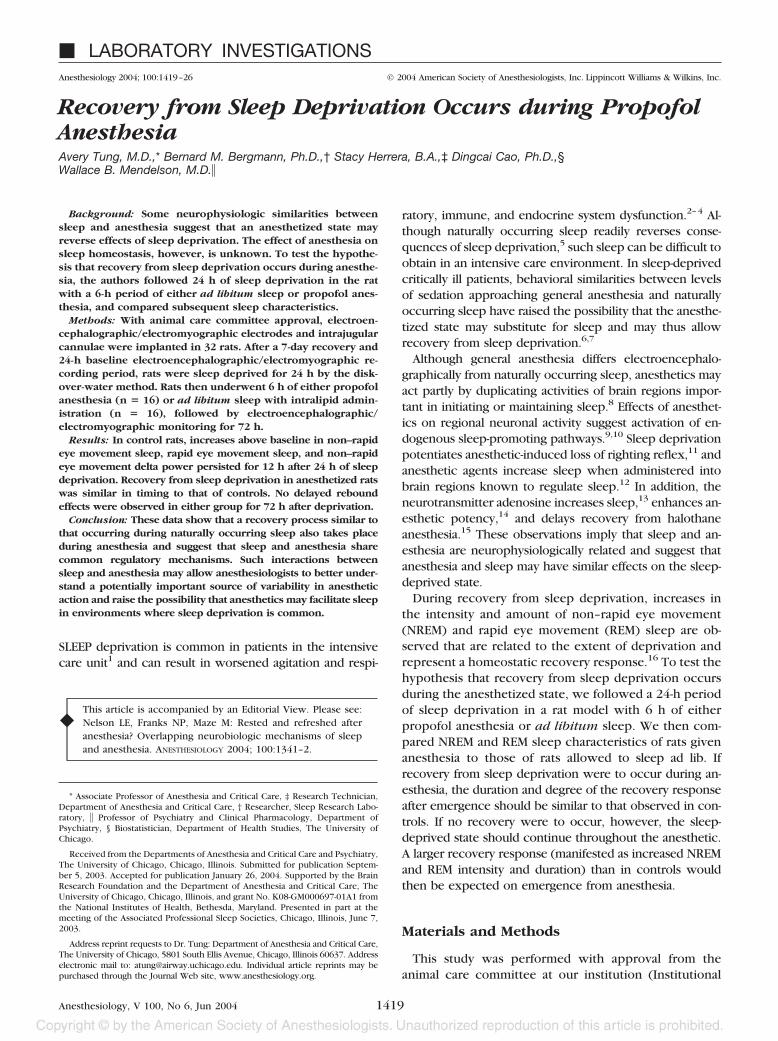
For both intralipid and propofol groups, averageweights (266.1 � 19 vs. 279.8 � 22 g), ages (71.3 � 6.3vs. 70 � 6.1 days), and degrees of sleep deprivation(92.9 � 6.3 vs. 90.5 � 9.2% wake/24 h) were similar.During deprivation, there was no significant differencein disk rotation between rats in the intralipid (18.3 �9.4%/24-h period) and propofol (15.4 � 13%) groups.The average infusion rate for rats sedated with propofolover the 6-h period was 297 � 38 �g · kg�1 · min�1,corresponding to a total infusion volume of 5.95 �0.6 ml over 6 h. Control rats received a total infusionvolume of 6 ml over 6 h (1 ml/h). Visual inspection ofthe electroencephalogram during propofol anesthesiarevealed a continuous, high-amplitude pattern withoutnoticeable NREM or REM stages for the entire 6-h period(fig. 1). During the 6-h intervention period, sleep incontrol rats (NREM � REM) totaled 4.27 � 0.33 h. Ratsanesthetized with propofol took less than 10 min toreach their behavioral endpoint, defined as a loss of
Fig. 1. Sample electroencephalographic/electromyographic recordings from rats anesthetized with propofol (A) and control ratsgiven intralipid (B). Paper speed was 10 mm/s. In intralipid rat recording, sleep-to-wake transition is marked. EEG � electroen-cephalogram; EMG � electromyogram.
1421RECOVERY FROM SLEEP DEPRIVATION DURING ANESTHESIA
Anesthesiology, V 100, No 6, Jun 2004

righting reflex and tolerance of clip-style pulse oximetry.The average time from discontinuing the infusion to firstmovement was 30 � 18 min.
NREM SleepBaseline values for NREM sleep in control rats given
intralipid and anesthetized rats given propofol were sim-ilar (fig. 2). Both groups demonstrated normal circadianvariability (less sleep during the lights-off period be-tween 6:00 PM and 6:00 AM) and appropriate synchroni-zation to the experimental lighting cycle. In controls,NREM sleep after deprivation was significantly increasedabove baseline values (obtained at the same time point)for 12 h before returning to baseline (fig. 3). Specifically,NREM sleep was increased above baseline by (mean �SD) 19.3 � 3.0% in the first 3-h block (12:00 PM–3:00 PM,t(15) � 6.38, P � 0.001), 18.1 � 8.3% in the secondblock (3:00 PM–6:00 PM, t(15) � 8.76, P � 0.001), 14.0 �10.6% in the third block (6:00 PM–9:00 AM, t(15) � 5.30,P � 0.001), and 9.3 � 11.7% in the fourth block (9:00AM–12:00 AM, t(15) � 3.17, P � 0.025). NREM sleep
returned to baseline levels during the fifth block and didnot differ from baseline for the remainder of the 72-hrecording period. In particular, no delayed rebound wasobserved.
In rats given propofol, conventional scoring of theelectroencephalogram/electromyogram during the 6-hanesthetic was not performed. No comparisons to base-line were therefore made for the first two blocks afterdeprivation. After emergence, NREM sleep was in-creased by 22.3 � 18.0% in the first 3-h block only(t(15) � 4.45, P � 0.002; fig. 3). NREM sleep returned tobaseline during the second 3-h block after emergencefrom anesthesia and was not different from baseline forthe subsequent 72-h recording period. The overall dura-tion of increased sleep after deprivation (including the6-h anesthetic) was thus 9 h. As with control rats, nodelayed increase in postdeprivation NREM sleep wasobserved.
When NREM sleep had returned to baseline levels inboth groups, no delayed increases were observed, andcircadian variation appeared normal for the duration of the
Fig. 2. Non–rapid eye movement (NREM)sleep frequency during baseline record-ing. NREM sleep was scored in 30-s ep-ochs, collected and averaged in 3-hblocks, and expressed as percent of totalblock time � SD. Shaded areas depictdark phase (6:00 PM–6:00 AM) recording.AM and PM are denoted as a and p,respectively.
Fig. 3. Rebound non–rapid eye movement(NREM) sleep after deprivation forpropofol and control animals. Valueswere calculated by collecting and averag-ing NREM sleep in 3-h blocks before andafter deprivation, subtracting postdepri-vation NREM sleep from baseline valuesobtained at the same circadian time pointand expressed as mean change in abso-lute percentage from baseline values �SD. Note that NREM behavior was notplotted for propofol rats during anesthe-sia. Shaded areas depict dark phase (6:00PM–6:00 AM) recording. *, † P < 0.05 whencompared with baseline values forpropofol and intralipid groups, respec-tively. AM and PM are denoted as a and p,respectively.
1422 TUNG ET AL.
Anesthesiology, V 100, No 6, Jun 2004
72-h recording period. When postdeprivation NREM sleepwas directly compared between propofol and intralipidgroups, a repeated-measures ANOVA showed more NREMsleep in controls than in anesthetized rats during only one3-h block after deprivation, with no other differences be-tween the two groups for the entire recording period. Inno postdeprivation block did NREM sleep for propofol ratssignificantly exceed that of controls.
REM SleepAs with baseline values for NREM sleep, baseline val-
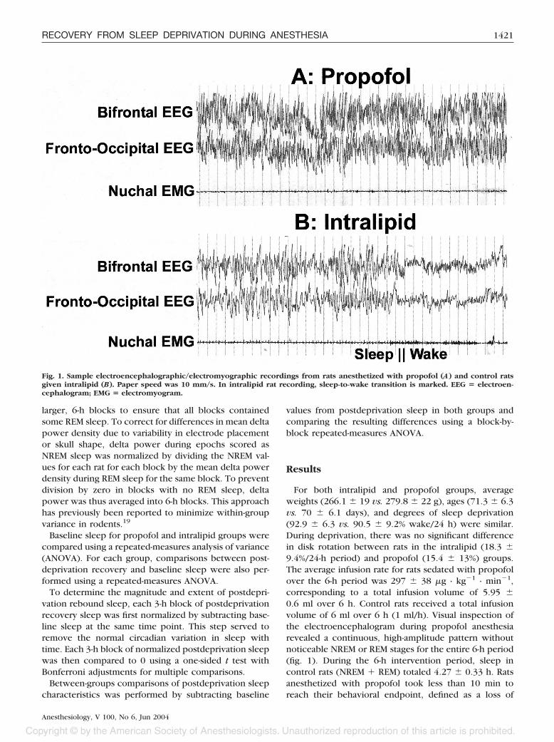
ues for REM sleep in propofol and intralipid groups weresimilar by repeated-measures ANOVA. In both groups, acharacteristic circadian pattern with REM sleep greatestin the second block (6:00 PM–12:00 AM) was observed(fig. 4). In control rats, sleep deprivation significantlyincreased REM sleep for the first two 6-h blocks beforereturning to baseline (37.4 � 27.6 epochs, t(15) � 5.41,P � 0.001 for the first block, 12:00 PM–6:00 PM; 40.2 � 40.1epochs, t(15) � 3.95, P � 0.001 for the second block, 6:00PM–12:00 AM) (fig. 4). The duration of REM sleep reboundwas thus 12 h. After deprivation, REM sleep for each re-covery day was greatest during the first block (12:00 PM–6:00 PM), suggesting residual sleep deprivation.
In rats given propofol, REM sleep was not evaluatedduring the 6-h propofol anesthetic. After emergence,REM sleep was increased for 6 h (6:00 PM–12:00 AM, 20.8 �40.1, t(15) � 2.45, P � 0.05) before returning to baseline.The REM rebound response in propofol rats was thuscomplete 12 h after the end of deprivation. As with controlrats, rats given propofol demonstrated the same altereddaily pattern of REM sleep after deprivation, suggesting asimilar degree of residual sleep deprivation (fig. 4).
When postdeprivation REM sleep was normalized inboth groups by subtracting baseline values, repeated-measures ANOVA showed slightly higher REM sleep incontrol rats during the fourth block after deprivation butno other differences between intralipid and propofolrats. At no time after deprivation was REM sleep inpropofol rats increased above that in controls.
NREM Delta PowerDuring baseline monitoring, delta power did not differ
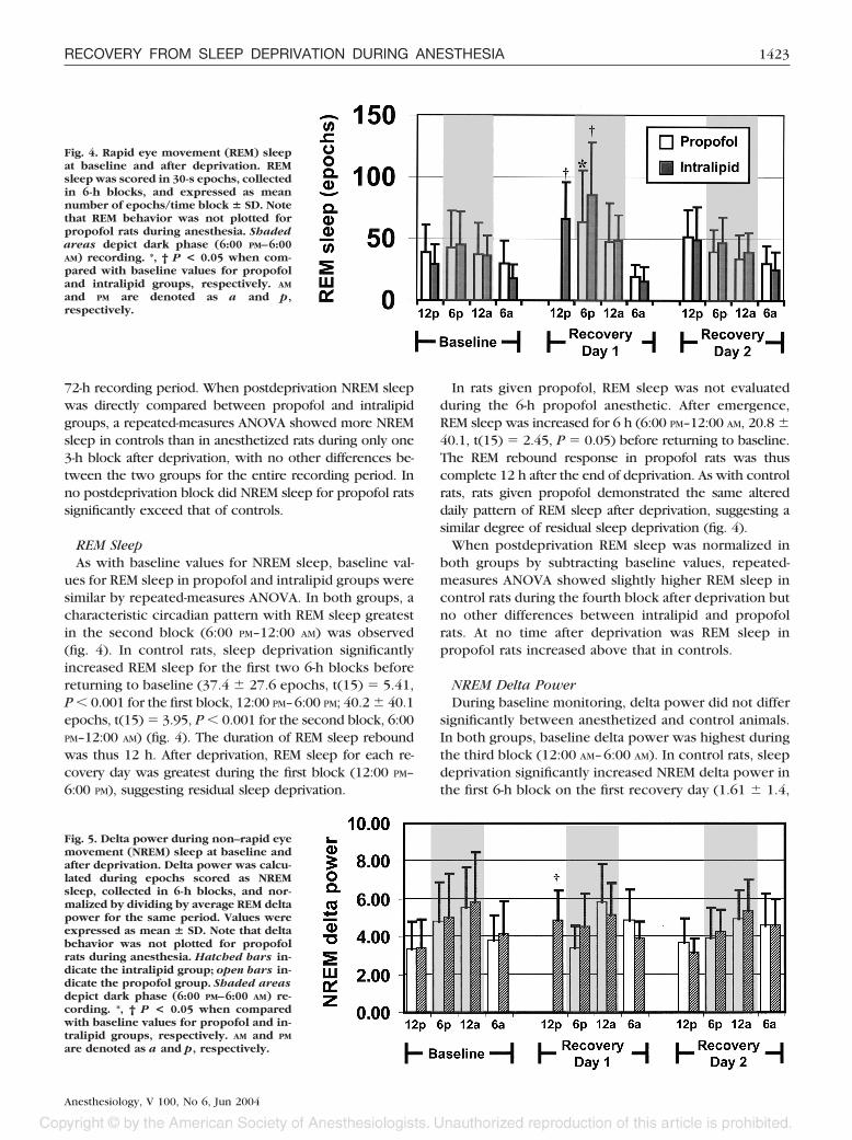
significantly between anesthetized and control animals.In both groups, baseline delta power was highest duringthe third block (12:00 AM–6:00 AM). In control rats, sleepdeprivation significantly increased NREM delta power inthe first 6-h block on the first recovery day (1.61 � 1.4,
Fig. 5. Delta power during non–rapid eyemovement (NREM) sleep at baseline andafter deprivation. Delta power was calcu-lated during epochs scored as NREMsleep, collected in 6-h blocks, and nor-malized by dividing by average REM deltapower for the same period. Values wereexpressed as mean � SD. Note that deltabehavior was not plotted for propofolrats during anesthesia. Hatched bars in-dicate the intralipid group; open bars in-dicate the propofol group. Shaded areasdepict dark phase (6:00 PM–6:00 AM) re-cording. *, † P < 0.05 when comparedwith baseline values for propofol and in-tralipid groups, respectively. AM and PM
are denoted as a and p, respectively.
Fig. 4. Rapid eye movement (REM) sleepat baseline and after deprivation. REMsleep was scored in 30-s epochs, collectedin 6-h blocks, and expressed as meannumber of epochs/time block � SD. Notethat REM behavior was not plotted forpropofol rats during anesthesia. Shadedareas depict dark phase (6:00 PM–6:00AM) recording. *, † P < 0.05 when com-pared with baseline values for propofoland intralipid groups, respectively. AM
and PM are denoted as a and p,respectively.
1423RECOVERY FROM SLEEP DEPRIVATION DURING ANESTHESIA
Anesthesiology, V 100, No 6, Jun 2004

t(15) � 3.61, P � 0.005). NREM delta subsequentlyreturned to baseline during the second block and wasindistinguishable from baseline for the duration of the72-h monitoring period (fig. 5).
In rats anesthetized with propofol, delta power wasnot calculated during the anesthetic. Absolute deltapower, however, was negligible for the entire anes-thetic, a finding consistent with propofol anesthesia inrats.20 After emergence, delta power was not increasedabove baseline in any block for the duration of the 72-hrecording period.
When postdeprivation delta power was normalized tobaseline and compared between groups, no differenceswere found. Absolute theta power, characteristically in-creased during REM sleep and some waking periods,18
was negligible in rats receiving propofol.
Discussion
We found that after 24 h of sleep deprivation, recoverysleep behavior in rats for which 6 h of ad libitum sleepwas allowed was no different from that of rats subjectedto a 6-h propofol anesthetic. After emergence frompropofol anesthesia, increases in NREM sleep, REMsleep, and delta power suggesting recovery from sleepdeprivation persisted for 9, 12, and 6 h, respectively.These increases were equal to or slightly less than thosefor control rats for which ad libitum recovery wasallowed. In addition, when sleep had returned to base-line in rats given propofol, no delayed increase in anyform of sleep was seen for the duration of the 72-hmonitoring period.
In principle, a period of anesthesia might modify thehomeostatic regulation of sleep debt in three ways. Forexample, general anesthesia might be a permissive statethat allows normal sleep homeostatic processes to oc-cur. An anesthetized organism would thus repay sleepdebt built up during previous wakefulness and emergeless sleep-deprived after an anesthetic than before. Alter-natively, anesthesia might progressively increase sleepdebt in a fashion similar to wakefulness. Prolonged an-esthetics would then induce a sleep-deprived state. Fi-nally, anesthesia might represent a state unlike eithersleep or waking, in which sleep debt neither accumu-lates nor dissipates. Organisms emerging from anesthesiawould then have the same degree of sleep deprivation aswhen they were initially anesthetized.
Under normal conditions, robust sleep homeostaticmechanisms act to preserve adequate sleep after sleepdeprivation. Even mild sleep loss increases the propen-sity to sleep,5 and sustained deprivation can cause theorganism to sleep when doing so would be life-threaten-
ing.# Recovery from sleep deprivation is characterized byrebound increases in NREM and REM sleep and changes inthe power spectrum of NREM sleep that persist until ho-meostatic mechanisms have been satisfied.16
Our findings are consistent with the presence of anactive sleep homeostatic process during anesthesia withpropofol. If the anesthetized state had resembled wake-fulness in its effect on sleep homeostasis, additionalsleep “debt” would have accrued throughout the 6-hanesthetic. This increased debt, combined with a delayin the initiation of recovery until after emergence, wouldhave resulted in a greater amount and intensity of NREMand REM sleep when compared with controls, a higherdelta power during NREM sleep, and a correspondingdelay in the return of NREM and REM sleep to baseline.If sleep debt had remained static during anesthesia, re-covery from deprivation would also have been delayeduntil after the anesthetic and would have manifested ashigher levels of sleep and NREM delta power comparedwith controls and a longer duration of recovery. Ourobservation that rats anesthetized with propofol hadrecovery characteristics nearly identical to rats for whichunrestricted sleep was allowed strongly suggests that arecovery process occurred during the 6-h anesthetic andimplies that anesthesia with propofol affects sleep ho-meostasis in a fashion similar to naturally occurring sleep.
Although the mechanisms by which sleep reversesbehavioral manifestations of sleep deprivation are poorlyunderstood, plausible links between anesthesia andsleep regulatory mechanisms support an ability of thebrain to recover from sleep deprivation during anesthe-sia. Sleep deprivation increases extracellular adenosineconcentrations in the basal forebrain of rats and cats, asite known to modulate central nervous system arous-al.13 Evidence that administration of adenosine reuptakeinhibitors into the basal forebrain increases sleep21 andthat adenosine delays recovery from halothane anesthe-sia15 suggests that extracellular adenosine concentra-tions plays a role in control mechanisms for both sleepand anesthesia. Increases in basal forebrain adenosinemay potentiate anesthetic action, for example, and re-duced metabolic demands during anesthesia may allowadenosine concentrations built up during wakefulness todissipate. Alternatively, anesthetic-induced changes inbrain activity may directly decrease adenosine release.
Neurophysiologic similarities between anesthesia andsleep may also allow the anesthetized state to reversebehavioral effects of sleep deprivation. Positron electrontomography/metabolic scanning9 and microelectrode re-cordings of thalamic relay neuronal activity22 both dem-onstrate reductions in thalamic activity during anesthe-sia, an important characteristic of naturally occurringsleep.23 Histologic studies suggest that dexmedetomi-dine anesthesia increases activity in the ventrolateralpreoptic nucleus and reduces activity in the locus cer-uleus10 in a fashion similar to sleep. Although the mo-
# National Heart, Lung and Blood Institute/National Center for Sleep DisorderResearch: Drowsy driving and automobile crashes. Available at: http://www.nhl-bi.nih.gov/health/prof/sleep/drsy_drv.htm. Accessed December 31, 2003.
1424 TUNG ET AL.
Anesthesiology, V 100, No 6, Jun 2004

lecular mechanisms governing sleep homeostasis are un-known, some feature of brain activity during anesthesiamay duplicate the aspect of naturally occurring sleepthat modulates homeostatic control of sleep.
We did not test other anesthetic agents for two rea-sons. Because our central finding was that no differenceexisted between anesthetized and control groups, webelieved that to include another group would haveweakened the statistical finding of no difference. In ad-dition, other anesthetics, such as isoflurane, may havehad delayed effects on sleep patterns, preventing usfrom interpreting postdeprivation behavior. We also tar-geted a single, behaviorally defined endpoint. Becausesleep deprivation can affect anesthetic potency,11 fixeddoses of anesthetic would have resulted in changes inthe depth of anesthesia as the degree of deprivationchanged over time. We therefore titrated our anestheticto loss of righting reflex and tolerance of clip-style pulseoximetry to maximize behavioral similarities to sleep.Although a dose–response curve involving a differentbehavioral endpoint (such as electroencephalographicsilence) was possible, cardiorespiratory depressionwould have necessitated intubation and mechanical ven-tilation for the duration of the anesthetic.
Delayed, propofol-induced effects on sleep may alsohave altered the interpretation of our results. In humans,the combination of inhaled anesthetics and surgery re-sults in initial suppression of REM sleep, followed by arebound increase on the second or third postoperativeday.24 We thought it unlikely in our study that a delayedeffect of propofol anesthesia suppressed subsequent sleepand obscured detection of a recovery response. No delayedrebound suggestive of an initial suppressive effect was seenin anesthetized rats, and block-by-block comparisons be-tween baseline and postanesthesia sleep after the end ofrecovery revealed no suppressive effect in any sleep mea-sure. It is possible that the shorter recovery duration inanesthetized animals actually indicated a more efficientrecovery process during anesthesia than during ad libitumconditions. This possibility might plausibly be explained bydifferences in the time spent in the unresponsive state (6 hfor anesthetized rats vs. 4.27 h for controls).
Finally, lighting conditions during our study deservemention. Rats are nocturnal animals, normally asleepduring daylight hours.25 At baseline, rats in both groupsdemonstrated appropriately less sleep during the darkperiod. In our study design, the 6-h intervention oc-curred during the light phase (12:00 PM–6:00 PM), withthe next 12 h spent in darkness (6:00 PM–6:00 AM). Wechose this strategy because recovery during the lightphase is subject to a “ceiling effect” on total sleep,26
which may have limited the size of the rebound weobserved. Locating the infusion period in the last 6 h ofthe light phase thus allowed us to contrast high light-phase recovery sleep levels in controls to near-zero overtsleep in anesthetized animals and examine recovery dur-
ing darkness when between-groups comparisons wouldbe optimally sensitive to differences in recovery sleep.Although previous studies in our laboratory have exam-ined recovery from sleep deprivation under constantlighting conditions,25,26 removing such a circadian cueintroduces the possibility of circadian drift over time. Wetherefore chose to monitor recovery under baseline,12:12-h lighting conditions to minimize this possibility.Because propofol and control rat recovery data werecompared at the same point in their diurnal cycle andtime after deprivation, phase shifts and diurnal variationwere unlikely to affect our results.
In summary, we report that after a 24-h period of sleepdeprivation, rats anesthetized for 6 h with propofol re-covered to the same degree as rats allowed 6 h of adlibitum sleep. This observation suggests that sleep andanesthesia may share common control mechanisms andraises the possibility that understanding anesthetic ef-fects on known correlates of sleep homeostasis mayfacilitate knowledge regarding effects and consequencesof sleep deprivation. Clinically, such interactions be-tween sleep and anesthesia may allow anesthesiologiststo better understand how sleep deprivation and anesthe-sia interact and may potentially allow anesthetics tofacilitate sleep in environments where sleep deprivationis common.
The authors thank Martin J. Szafran, B.A. (Department of Anesthesia andCritical Care, The University of Chicago, Chicago, Illinois), for his valuabletechnical assistance with this study.
References
1. Cooper AB, Thornley KS, Young GB, Slutsky AS, Stewart TE, Hanly PJ: Sleepin critically ill patients requiring mechanical ventilation. Chest 2000; 117:809–18
2. Chen HI, Tang YR: Sleep loss impairs inspiratory muscle endurance. Am RevRespir Dis 1989; 140:907–9
3. Irwin M: Effects of sleep and sleep loss on immunity and cytokines. BrainBehav Immun 2002; 16:503–12
4. Spiegel K, Leproult R, Van Cauter E: Impact of sleep debt on metabolic andendocrine function. Lancet 1999; 354:1435–9
5. Bonnet MH: Sleep deprivation, Principles and Practice of Sleep Medicine,2nd edition. Edited by Kryger MH, Roth T, Dement W. Philadelphia, Saunders,1994, pp 50–67
6. Manthous CA, Schmidt GA, Hall JB: Liberation from mechanical ventilation:A decade of progress. Chest 1998; 114:886–901
7. Treggiari-Venzi M, Borgeat A, Fuchs-Buder T, Gachoud JP, Suter PM: Over-night sedation with midazolam or propofol in the ICU: Effects on sleep quality,anxiety and depression. Intensive Care Med 1996; 22:1186–90
8. Lydic R, Biebuyck JF: Sleep neurobiology: Relevance for mechanistic studiesof anaesthesia. Br J Anaesth 1994; 72:506–8
9. Alkire MT, Pomfrett CJ, Haier RJ, Gianzero MV, Chan CM, Jacobsen BP,Fallon JH: Functional brain imaging during anesthesia in humans: Effects ofhalothane on global and regional cerebral glucose metabolism. ANESTHESIOLOGY
1999; 90:701–910. Nelson LE, Lu J, Guo T, Saper CB, Franks NP, Maze M: The �2-adrenoceptor
agonist dexmedetomidine converges on an endogenous sleep-promoting path-way to exert its sedative effects. ANESTHESIOLOGY 2003; 98:428–36
11. Tung A, Szafran MJ, Bluhm B, Mendelson WB: Sleep deprivation potenti-ates the onset and duration of loss of righting reflex induced by propofol andisoflurane. ANESTHESIOLOGY 2002; 97:906–11
12. Tung A, Bluhm B, Mendelson WB: The hypnotic effect of propofol in themedial preoptic area of the rat. Life Sci 2001; 69:855–62
13. Porkka-Heiskanen T, Strecker RE, Thakkar M, Bjorkum AA, Greene RW,McCarley RW: Adenosine: A mediator of the sleep-inducing effects of prolongedwakefulness. Science 1997; 276:1265–8
14. Kaputlu I, Sadan G, Ozdem S: Exogenous adenosine potentiates hypnosisinduced by intravenous anaesthetics. Anaesthesia 1998; 53:496–500
1425RECOVERY FROM SLEEP DEPRIVATION DURING ANESTHESIA
Anesthesiology, V 100, No 6, Jun 2004

15. Tanase D, Baghdoyan HA, Lydic R: Dialysis delivery of an adenosine A1receptor agonist to the pontine reticular formation decreases acetylcholinerelease and increases anesthesia recovery time. ANESTHESIOLOGY 2003; 98:912–20
16. Borbely AA, Tobler I, Hanagasioglu M: Effect of sleep deprivation on sleepand EEG power spectra in the rat. Behav Brain Res 1984; 14:171–82
17. Bergmann BM, Kushida CA, Everson CA, Gilliland MA, Obermeyer W,Rechtschaffen A: Sleep deprivation in the rat: II. Methodology. Sleep 1989;12:5–12
18. Bergmann BM, Winter JB, Rosenberg RS, Rechtschaffen A: NREM sleepwith low-voltage EEG in the rat. Sleep 1987; 10:1–11
19. Naylor E, Bergmann BM, Krauski K, Zee PC, Takahashi JS, Vitaterna MH,Turek FW: The circadian clock mutation alters sleep homeostasis in the mouse.J Neurosci 20000; 20:8138–43
20. Tan PPC, Shyr MH, Yang CH, Kuo TBJ, Pan WHT, Chan SHH. Powerspectral analysis of the electroencephalographic and hemodynamic correlates ofpropofol anesthesia in the rat: Intravenous infusion. Neurosci Lett 1993; 160:205–8
21. Strecker RE, Morairty S, Thakkar MM, Porkka-Heiskanen T, Basheer R,
Dauphin LJ, Rainnie DG, Portas CM, Greene RW, McCarley RW: Adenosinergicmodulation of basal forebrain and preoptic/anterior hypothalamic neuronal ac-tivity in the control of behavioral state. Behav Brain Res 2000; 115:183–204
22. Vahle-Hinz C, Detsch O, Siemers M, Kochs E, Bromm B: Local GABA(A)receptor blockade reverses isoflurane’s suppressive effects on thalamic neuronsin vivo. Anesth Analg 2001; 92:1578–84
23. Jones BE: Basic mechanisms of sleep-wake states, Principles and Practiceof Sleep Medicine, 2nd edition. Edited by Kryger MH, Roth T, Dement W.Philadelphia, Saunders, 1994, pp 145–62
24. Knill RL, Moote CA, Skinner MI, Rose EA: Anesthesia with abdominalsurgery leads to intense REM sleep during the first postoperative week. ANESTHE-SIOLOGY 1990; 73:52–61
25. Bergmann BM, Mistlberger RE, Rechtschaffen A: Period-amplitude analysisof rat electroencephalogram: Stage and diurnal variations and effects of supra-chiasmatic nuclei lesions. Sleep 1987; 10:523–36
26. Mistlberger RE, Bergmann BM, Waldenar W, Rechtschaffen A: Recoverysleep following sleep deprivation in intact and suprachiasmatic nuclei-lesionedrats. Sleep 1983; 6:217–33
1426 TUNG ET AL.
Anesthesiology, V 100, No 6, Jun 2004