Recent advances in emulsion-based delivery approaches for ...

“fncel-07-00272” — 2013/12/20 — 21:17 — page 1 — #1
REVIEW ARTICLEpublished: 24 December 2013doi: 10.3389/fncel.2013.00272
Recent molecular approaches to understanding astrocytefunction in vivoDavid Davila1, KarineThibault 1,Todd A. Fiacco 2* and Cendra Agulhon 1*
1 Glia-Glia and Glia-Neuron Interactions Group, National Center for Scientific Research, UFR Biomedicale, Paris Descartes University, Paris, France2 Department of Cell Biology and Neuroscience, and Center for Glial-Neuronal Interactions and Program in Cellular, Molecular and Developmental Biology,University of California at Riverside, Riverside, CA, USA
Edited by:
Carole Escartin, Molecular ImagingResearch Center, France
Reviewed by:
Keith Murai, McGill University, CanadaFrank W. Pfrieger, National Center forScientific Research, France
*Correspondence:
Cendra Agulhon, Glia-Glia andGlia-Neuron Interactions Group,National Center for ScientificResearch, UFR Biomedicale, ParisDescartes University, 45 rue desSaints Pères, 75006 Paris, Francee-mail: [email protected];Todd A. Fiacco, Department of CellBiology and Neuroscience, andCenter for Glial-Neuronal Interactionsand Program in Cellular, Molecularand Developmental Biology,University of California Riverside, 1109Biological Sciences Building,Riverside, CA 92521, USAe-mail: [email protected]
Astrocytes are a predominant glial cell type in the nervous systems, and are becomingrecognized as important mediators of normal brain function as well as neurodevelopmental,neurological, and neurodegenerative brain diseases. Although numerous potential mecha-nisms have been proposed to explain the role of astrocytes in the normal and diseased brain,research into the physiological relevance of these mechanisms in vivo is just beginning. Inthis review, we will summarize recent developments in innovative and powerful molecularapproaches, including knockout mouse models, transgenic mouse models, and astrocyte-targeted gene transfer/expression, which have led to advances in understanding astrocytebiology in vivo that were heretofore inaccessible to experimentation. We will examine therecently improved understanding of the roles of astrocytes – with an emphasis on astrocytesignaling – in the context of both the healthy and diseased brain, discuss areas where therole of astrocytes remains debated, and suggest new research directions.
Keywords: astrocytes, knockout mice, transgenic mice, chemogenetics, viral gene transduction, glial cell
progenitors, GPCR, neuron–glia interactions
INTRODUCTIONAlthough astrocytes are the most abundant glial cell type in themammalian nervous system and have emerged as crucial regula-tors of nervous system development, function, and health, ourunderstanding of the physiology of astrocytes remains limited.How do astrocytes interact with neurons and other cell types of thenervous system? What are the primary functions of astrocytes inbrain development, health, and disease? One challenge to addressthese questions is identifying the function of individual astro-cytic molecules that regulate brain function. A unique approachto investigate the molecular basis of astrocyte activity consists ofmanipulating the genome of higher organisms. The mouse rep-resents a great animal model that has been used extensively forgenetic manipulation in neuroscience, leading to understandingof neuronal functions in unprecedented detail. A concerted effortin recent years to develop genetic approaches to study astrocytesis guiding the field into new territory and improving our under-standing of interactions between neurons and astrocytes. Herewe will review techniques for genetic manipulation of astrocytesand highlight the most recent innovative and elegant approachesthat are providing insight into fundamental roles of astrocytes inpathophysiology in vivo. In particular, we will describe the mainmolecular approaches used in this field, including knockout (KO)mouse models, transgenic mouse models, and astrocyte-targeted
gene transfer and expression using adeno-associated viral (AAV)or in utero electroporation (IUE) approaches. We will also touchon a remarkable recent study involving engraftment of geneticallymodified human glial progenitors into the mouse brain, providinginsight into the role of human astrocytes in the unique cognitiveabilities of the human brain. The present review does not attemptto be comprehensive; rather it highlights certain major themes andareas of recent progress on the roles of astrocytes in brain func-tion, with an emphasis on astrocyte signaling and in vivo studies.For complementary reviews on astrocyte function in health anddisease with an emphasis on molecular approaches, readers aredirected to (Fiacco et al., 2009b; Figueiredo et al., 2011; Agulhonet al., 2012; Nedergaard and Verkhratsky, 2012; Clarke and Barres,2013; Freeman and Rowitch, 2013; Tong et al., 2013), as well as theother contributions to this special topic.
STUDYING ASTROCYTE FUNCTION THROUGH GENEDISRUPTIONThe elimination of one or more specific genes in an animal modelis a reliable and widespread approach to discovering the func-tion of specific proteins and the cell types expressing them. Manygenes have been identified, isolated, and subsequently manipu-lated to fully or conditionally suppress their expression (Sikorskiand Peters, 1997; Grimm, 2006; Vogel, 2007). These molecular
Frontiers in Cellular Neuroscience www.frontiersin.org December 2013 | Volume 7 | Article 272 | 1

“fncel-07-00272” — 2013/12/20 — 21:17 — page 2 — #2
Davila et al. Molecular approaches to understanding astrocyte function
developments represent powerful tools that can be used in vivo toaid in discovering the role of astrocytes in the healthy and diseasedbrain.
FULL (CONSTITUTIVE) KNOCKOUT MOUSE MODELSAs research into astroglia physiology in vivo is still a newly develop-ing field, much information can be gleaned from the eliminationof a gene or the deletion of a functional domain of a protein inastrocytes to shed light on the function of both the targeted geneand astrocytes in general. The process of generating a new line ofKO mice is laborious, but has been refined to maximize efficiency(reviewed in Hall et al., 2009; Limaye et al., 2009). Once the desiredgene is identified, gene targeting can be used to generate a KOmouse. A targeting vector containing a neomycin-resistant markeris inserted into embryonic stem (ES) cells via electroporation andis introduced into the DNA through homologous recombination,allowing complete removal of one or more exons from the gene ofinterest (Figure 1A). This results in the production of a mutatedor truncated protein or, more often, no protein at all. ES cells thatdo not take up the foreign construct are killed through exposureto neomycin, and those that have successfully replaced the gene orthe exons of this gene survive and are subsequently microinjectedinto mouse blastocysts, which are then grown in surrogate mouseuteri. Strategic mating of the chimeric mice will ultimately resultin a mouse with the gene globally eliminated (Capecchi, 1989; Hallet al., 2009; Limaye et al., 2009; Figure 1A).
Role of GFAP in the healthy and diseased brainOne of the first genes that was removed in astrocytes is the geneencoding glial fibrillary acidic protein (GFAP; Lewis et al., 1984;Reeves et al., 1989; Masood et al., 1993; McCall et al., 1996; Enget al., 2000). GFAP is a member of the family of intermediatefilament structural proteins, which, in the mature nervous system,is found predominantly in protoplasmic and specialized astrocytesof the central nervous system (CNS) as well as in satellite cells,non-myelinating Schwann cells, and enteric glia in the peripheralnervous system (PNS; Jessen et al., 1984; Eng, 1985; Kato et al.,1990). Outside the nervous system, GFAP has also been detectedin some rare non-glial cells of the salivary glands (Achstatter et al.,1986; Gustafsson et al., 1989), fibroblasts (Hainfellner et al., 2001),myoepithelial cells (Viale et al., 1991), liver stellate cells (Gard et al.,1985), and lymphocytes (Riol et al., 1997). This protein is oneof the key elements of the cytoskeleton that contributes to themorphology and motility of astrocyte processes (Fuchs and Weber,1994; Pekny and Pekna, 2004; Gomi et al., 2010; Middeldorp andHol, 2011) and is upregulated in reactive astrocytes (astrogliosis)in essentially any CNS pathology (Eng and Ghirnikar, 1994; Enget al., 2000; Pekny and Nilsson, 2005; Sofroniew, 2009; Sosunovet al., 2013). During development, GFAP is expressed widely in anumber of progenitor cell types giving rise to both neurons andglia. For example, GFAP-expressing radial glia in the ventricularzone (VZ) give rise to mature astrocytes, oligodendrocytes andneurons, as well as guiding subsequent migration of neurons (Gotzet al., 2002; Malatesta et al., 2003; Anthony et al., 2004; Merkleet al., 2004).
The seminal studies reporting the findings obtained with theGFAP KO mouse models were made by targeted deletion of the
GFAP gene in ES cells (Pekny et al., 1995; Liedtke et al., 1996;McCall et al., 1996) or by targeted disruption of the GFAP gene byinsertion of a LacZ cassette (Gomi et al., 1995). These GFAP KOmouse lines have been used to investigate whether changes in astro-cyte processes and their morphological structure can influencebrain morphogenesis and function, the physiology of adjacentsynapses, and functional recovery after CNS insults. The over-all appearance of the GFAP KO mice is indistinguishable fromwild-type mice; they develop normally and display no gross alter-ations in behavior or CNS morphology. This observation suggestsat first that GFAP is not essential for normal brain morphogen-esis and function. However, closer analyses have indicated theinvolvement of GFAP in a wide variety of processes. First, thesemice display enhanced hippocampal long-term potentiation (LTP)and deficient cerebellar long-term depression (LTD) in acute brainslices from adult mice, suggesting that GFAP intermediate filamentprotein is important for astrocyte–neuronal interactions and thatastrocyte processes play a vital role in modulating synaptic efficacyin the CNS (McCall et al., 1996; Shibuki et al., 1996). In agreementwith the cerebellar ex vivo findings, a significant impairment ofeye blink conditioning was found in the GFAP KO mice, suggest-ing that GFAP is required for normal communication betweenBergmann glia (specialized astrocytes) and Purkinje cells duringinduction and maintenance of cerebellar LTD in vivo (Shibukiet al., 1996). Second, morphological and functional alterations inthe blood–brain barrier (BBB), disorganization of white matterarchitecture and vascularization, as well as hydrocephalus werereported in 18–24 month GFAP KO mice, suggesting an involve-ment of GFAP in the long-term maintenance of normal BBB andCNS myelination (Liedtke et al., 1996). Third, when challenged bydiverse brain injuries in vivo, the GFAP KO mice were more vulner-able to: (i) CNS mechanical trauma (Nawashiro et al., 1998; Otaniet al., 2006), (ii) cerebral ischemia (Nawashiro et al., 2000; Tanakaet al., 2002), (iii) kainic acid-induced neurotoxicity (Otani et al.,2006), and (iv) autoimmune encephalomyelitis (Liedtke et al.,1998). This was indicated by: (i) increased brain hemorrhage andmortality of mice; (ii) larger cortical infarct volume and profounddecrease in cerebral blood flow; (iii) neurodegeneration; and (iv)enhanced clinical course of autoimmune encephalomyelitis andlesions, respectively. Conversely, other investigators have foundthat suppressing astrocytic GFAP expression in reactive astrocytesincreases their basal levels of glial cell derived neurotrophic fac-tor (GDNF), leading to improvement in neuronal survival frommetabolic and excitotoxic insults (Hanbury et al., 2003). Beneficialneuroprotective and regenerative effects have also been reportedafter hippocampal and spinal cord injuries in mice lacking bothGFAP and vimentin, another astrocyte intermediate filament pro-tein (Kinouchi et al., 2003; Menet et al., 2003; Wilhelmsson et al.,2004). Collectively, these findings suggest that the GFAP com-ponent of the astrocyte cytoskeleton plays an important role inthe physiology and pathology of the nervous system. However,because GFAP is expressed in progenitor cells during develop-ment giving rise to neurons, oligodendrocytes and astrocytes,these findings have to be viewed carefully. Inducible GFAP KOstrategies can be used to circumvent this problem by removingGFAP only from astrocytes after this developmental window hasclosed (see below).
Frontiers in Cellular Neuroscience www.frontiersin.org December 2013 | Volume 7 | Article 272 | 2

“fncel-07-00272” — 2013/12/20 — 21:17 — page 3 — #3
Davila et al. Molecular approaches to understanding astrocyte function
FIGURE 1 | Schematic representation of the main genetic manipulations
to make knockout (KO) mice. (A) Conventional strategy for full (constitutive)gene knockout. The targeted gene is inactivated following insertion of anantibiotic (neomycin or neo) resistance gene within, or in place, of one ofseveral essential exons(s) through electroporation of a targeting vector inmurine embryonic stem (ES) cells and subsequent homologousrecombination. Totipotent embryonic ES cells with successful recombinationwill survive neomycin treatment to be selectively chosen and injected inmouse blastocysts. Blastocytes are then transferred to the uterus of
pseudopregnant females where they can differentiate into all cell types of achimeric mouse (red circles). After breeding the chimeric mice, the resultingoffspring will derive from the ES cells – as seen with the transmission of coatcolor (red) – if the introduced ES cells become established into the germlineof the chimeric mouse. In the full KO, the gene of interest is knocked out in allcell types (not only astrocytes) of the offspring. (B) Classical Cre-LoxPstrategy for conditional gene knockout. The creation of two mouse lines isnecessary. First, a floxed mouse line (green) is obtained by homologous
(Continued)
Frontiers in Cellular Neuroscience www.frontiersin.org December 2013 | Volume 7 | Article 272 | 3

“fncel-07-00272” — 2013/12/20 — 21:17 — page 4 — #4
Davila et al. Molecular approaches to understanding astrocyte function
FIGURE 1 | Continued
recombination in ES cells. Two LoxP sites are positioned in intronic regionsthat flank one or several essential exons of the targeted gene. In this mouseline the targeted gene is normally expressed. Second, a Cre mouse line(orange) created by classical transgenesis, following pronuclear injection ofthe cDNA encoding the Cre-recombinase under the control of anastrocyte-specific promoter. This line gives astrocyte selectivity to thesystem. Breeding of the floxed mouse with the Cre mouse leads to thegeneration of a new mouse line (green/orange-striped) in which the floxedexon(s) are excised only in astrocyte-expressing Cre, while the targetedgene remains functional in other cell types. In this case theCre-recombinase (orange squares) is constitutively expressed, i.e.,inactivation of the targeted gene occurs as soon as the astrocyte-specificpromoter driving Cre-recombinase is active. (C) Inducible CreERT2-LoxPstrategy for temporal control of gene knockout. As in (B), a floxed mouseline (green) and a CreERT2 mouse line (yellow) are necessary to obtain athird mouse line (green/yellow-striped) in which the floxed exon(s) areexcised only in astrocyte-expressing CreERT2. In this mouse line, theCreERT2-recombinase (yellow squares) is expressed in astrocytes but iskept in the cytoplasmic compartment and is inactive. To achieve temporalcontrol of the gene knockout, CreERT2 activity (red/yellow squares) isinduced by synthetic steroid ligand (tamoxifen or 4-hydroxy-tamoxifencapable of crossing the blood brain barrier) administrated systemically at anychosen time. Binding of steroid to CreERT2 allows translocation of CreERT2
to the nucleus where recombination of floxed genes can occur selectively inastrocytes while the targeted gene remains functional in other cell types.
In light of more recent evidence suggesting that astroglio-sis is not a uniform process but rather a multifaceted responsewith context-dependent reactions (reviewed in Sofroniew, 2009;Sofroniew and Vinters, 2010), the GFAP KO mouse models willcontinue to be valuable tools in future studies seeking to furtherunravel the function of GFAP and astrocytes in the variety ofhuman brain challenges or diseases in vivo. These mouse modelswill assist in understanding the stages when astrocytes are engagedin beneficial or detrimental functions. Furthermore, a number ofconsiderations can be taken into account which may lead to both are-evaluation of earlier findings using these mice as well as open upnew research directions. These include the following: (i) the het-erogeneity of astrocyte morphology and physiology (Aley et al.,2006; Emsley and Macklis, 2006; Lee et al., 2006; Matyash and Ket-tenmann, 2010; Zhang and Barres, 2010; Oberheim et al., 2012;Freeman and Rowitch, 2013; Schreiner et al., 2013); (ii) the vari-ability of GFAP expression levels among this heterogeneous cellpopulation (Bernal and Peterson, 2011; Daniel-Christoph et al.,2013); (iii) the fluctuation of GFAP expression during circadianlight–dark cycles, hormonal cycles, developmental, or pathologicalstages (Hajos, 2008); (iv) the existence of about eight alternativelyspliced GFAP isoforms that may execute distinct functions in spe-cific subsets of astrocytes (Middeldorp and Hol, 2011); and (v) thepost-translational modification of these different isoforms suchas phosphorylation and glycosylation that may influence GFAPassembly into intermediate filaments (Middeldorp and Hol, 2011).Suitable GFAP isoform-specific KO models will be required infuture studies to address these issues. One way to investigate theimpact of alternative splicing of the GFAP gene in a mouse modelwould be to selectively delete a single isoform. This can be accom-plished by deleting a coding exon from the genome, introducing astop codon, inactivating the splice sites responsible for generatinga specific isoform, or overexpressing a dominant negative versionof a certain isoform (Moroy and Heyd, 2007). An alternative way
to investigate the impact of alternative GFAP splicing would be touse antisense oligonucleotides to prevent the inclusion of a par-ticular exon in the mature mRNA (Gebski et al., 2003; Dori et al.,2005). Isoform-specific KO mouse models provide a compellingapproach to study how alternative splicing of the GFAP gene maycontribute to the regulation of pathophysiological CNS processesin vivo.
Role of IP3R2 in normal synaptic transmission and plasticityAnother global KO of a gene in astrocytes is the gene encod-ing the inositol-1,4,5-trisphosphate type 2 receptor (IP3R2). Twoconstitutive IP3R2-deficient mouse models have been obtainedthrough a variation of the gene targeting strategy. In one, thetargeting construct was generated by flanking exon 3 of IP3R2with two LoxP sites and flanking the neo-cassette by FRT sites(Li et al., 2005). In the other, exon 1 of the gene was fused witha LacZ cassette (Futatsugi et al., 2005). Deletion of the IP3R2subtype is a valuable tool in astrocyte research, as it is the onlyfunctional IP3R subtype expressed by astrocytes and the mainmechanism by which astrocytes elevate intracellular Ca2+ lev-els (Petravicz et al., 2008; Agulhon et al., 2010; Di Castro et al.,2011; Takata et al., 2011). This mouse line has been critical toaddress the concept of gliotransmission, which stipulates thatneuroactive molecules including neurotransmitters (called glio-transmitters) are released from mature passive astrocytes in aneuronally-induced and Ca2+-dependent manner downstream ofGq protein-coupled receptors (Gq GPCRs) to quickly modulatesynaptic transmission and plasticity (illustrated and compre-hensively discussed in Agulhon et al., 2008, 2012; Hamilton andAttwell, 2010). Upon Gq GPCR activation, IP3 is producedintracellularly, leading to astrocytic IP3R2 activation and Ca2+release from the endoplasmic reticulum. Ex vivo and in vivostudies demonstrate that Ca2+ transients in mature passive astro-cytes are driven by metabotropic Gq GPCRs, which can beactivated by the spillover of neurotransmitters released frompresynaptic terminals (Porter and McCarthy, 1996; Wang et al.,2006). Therefore, astrocytic Gq GPCRs are considered to bethe physiological link between neuronal activity and detectableCa2+ increases in mature astrocytes. However, it is importantto keep in mind that future studies using improved imag-ing methods may reveal alternate sources of activity-drivenCa2+ transients in astrocytes. For example, new membrane-tethered genetically encoded Ca2+ indicators have allowed fordetection of previously unreported constitutive Ca2+ transients(Shigetomi et al., 2013b; Tong et al., 2013).
The use of the IP3R2 KO mouse model was done to selec-tively obliterate the endogenous Gq GPCR/IP3R2-mediated Ca2+elevations. The initial studies using this mouse model reportedthat removing astrocytic Gq GPCR/IP3R2-mediated Ca2+ fluxesdid not affect basal and evoked excitatory synaptic transmission(EPSCs), or short- and long-term plasticity (NMDA receptor-dependent LTP) in the hippocampus ex vivo (Fiacco et al., 2007;Petravicz et al., 2008; Agulhon et al., 2010). The implicationsof these findings have already been well documented and dis-cussed (Agulhon et al., 2008, 2010, 2012; Petravicz et al., 2008;Fiacco et al., 2009a; Hamilton and Attwell, 2010; Kirchhoff, 2010;Nedergaard and Verkhratsky, 2012). Recently, an ex vivo study
Frontiers in Cellular Neuroscience www.frontiersin.org December 2013 | Volume 7 | Article 272 | 4
“fncel-07-00272” — 2013/12/20 — 21:17 — page 5 — #5
Davila et al. Molecular approaches to understanding astrocyte function
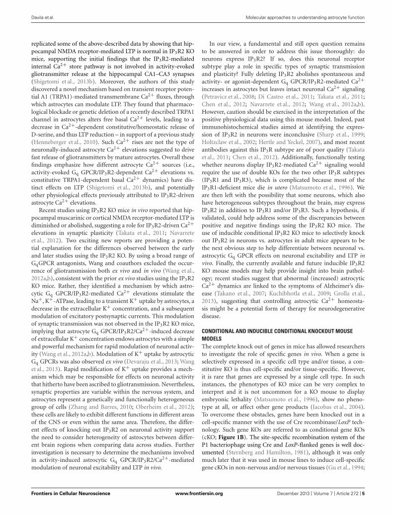
replicated some of the above-described data by showing that hip-pocampal NMDA receptor-mediated LTP is normal in IP3R2 KOmice, supporting the initial findings that the IP3R2-mediatedinternal Ca2+ store pathway is not involved in activity-evokedgliotransmitter release at the hippocampal CA1–CA3 synapses(Shigetomi et al., 2013b). Moreover, the authors of this studydiscovered a novel mechanism based on transient receptor poten-tial A1 (TRPA1)-mediated transmembrane Ca2+ fluxes, throughwhich astrocytes can modulate LTP. They found that pharmaco-logical blockade or genetic deletion of a recently described TRPA1channel in astrocytes alters free basal Ca2+ levels, leading to adecrease in Ca2+-dependent constitutive/homeostatic release ofD-serine, and thus LTP reduction – in support of a previous study(Henneberger et al., 2010). Such Ca2+ rises are not the type ofneuronally-induced astrocyte Ca2+ elevations suggested to drivefast release of gliotransmitters by mature astrocytes. Overall thesefindings emphasize how different astrocyte Ca2+ sources (i.e.,activity-evoked Gq GPCR/IP3R2-dependent Ca2+ elevations vs.constitutive TRPA1-dependent basal Ca2+ dynamics) have dis-tinct effects on LTP (Shigetomi et al., 2013b), and potentiallyother physiological effects previously attributed to IP3R2-drivenastrocyte Ca2+ elevations.
Recent studies using IP3R2 KO mice in vivo reported that hip-pocampal muscarinic or cortical NMDA receptor-mediated LTP isdiminished or abolished, suggesting a role for IP3R2-driven Ca2+elevations in synaptic plasticity (Takata et al., 2011; Navarreteet al., 2012). Two exciting new reports are providing a poten-tial explanation for the differences observed between the earlyand later studies using the IP3R2 KO. By using a broad range ofGqGPCR antagonists, Wang and coauthors excluded the occur-rence of gliotransmission both ex vivo and in vivo (Wang et al.,2012a,b), consistent with the prior ex vivo studies using the IP3R2KO mice. Rather, they identified a mechanism by which astro-cytic Gq GPCR/IP3R2-mediated Ca2+ elevations stimulate theNa+, K+-ATPase, leading to a transient K+ uptake by astrocytes, adecrease in the extracellular K+ concentration, and a subsequentmodulation of excitatory postsynaptic currents. This modulationof synaptic transmission was not observed in the IP3R2 KO mice,implying that astrocyte Gq GPCR/IP3R2/Ca2+-induced decreaseof extracellular K+ concentration endows astrocytes with a simpleand powerful mechanism for rapid modulation of neuronal activ-ity (Wang et al., 2012a,b). Modulation of K+ uptake by astrocyticGq GPCRs was also observed ex vivo (Devaraju et al., 2013; Wanget al., 2013). Rapid modification of K+ uptake provides a mech-anism which may be responsible for effects on neuronal activitythat hitherto have been ascribed to gliotransmission. Nevertheless,synaptic properties are variable within the nervous system, andastrocytes represent a genetically and functionally heterogeneousgroup of cells (Zhang and Barres, 2010; Oberheim et al., 2012);these cells are likely to exhibit different functions in different areasof the CNS or even within the same area. Therefore, the differ-ent effects of knocking out IP3R2 on neuronal activity supportthe need to consider heterogeneity of astrocytes between differ-ent brain regions when comparing data across studies. Furtherinvestigation is necessary to determine the mechanisms involvedin activity-induced astrocytic Gq GPCR/IP3R2/Ca2+-mediatedmodulation of neuronal excitability and LTP in vivo.
In our view, a fundamental and still open question remainsto be answered in order to address this issue thoroughly: doneurons express IP3R2? If so, does this neuronal receptorsubtype play a role in specific types of synaptic transmissionand plasticity? Fully deleting IP3R2 abolishes spontaneous andactivity- or agonist-dependent Gq GPCR/IP3R2-mediated Ca2+increases in astrocytes but leaves intact neuronal Ca2+ signaling(Petravicz et al., 2008; Di Castro et al., 2011; Takata et al., 2011;Chen et al., 2012; Navarrete et al., 2012; Wang et al., 2012a,b).However, caution should be exercised in the interpretation of thepositive physiological data using this mouse model. Indeed, pastimmunohistochemical studies aimed at identifying the expres-sion of IP3R2 in neurons were inconclusive (Sharp et al., 1999;Holtzclaw et al., 2002; Hertle and Yeckel, 2007), and most recentantibodies against this IP3R subtype are of poor quality (Takataet al., 2011; Chen et al., 2012). Additionally, functionally testingwhether neurons display IP3R2-mediated Ca2+ signaling wouldrequire the use of double KOs for the two other IP3R subtypes(IP3R1 and IP3R3), which is complicated because most of theIP3R1-deficient mice die in utero (Matsumoto et al., 1996). Weare then left with the possibility that some neurons, which alsohave heterogeneous subtypes throughout the brain, may expressIP3R2 in addition to IP3R1 and/or IP3R3. Such a hypothesis, ifvalidated, could help address some of the discrepancies betweenpositive and negative findings using the IP3R2 KO mice. Theuse of inducible conditional IP3R2 KO mice to selectively knockout IP3R2 in neurons vs. astrocytes in adult mice appears to bethe next obvious step to help differentiate between neuronal vs.astrocytic Gq GPCR effects on neuronal excitability and LTP invivo. Finally, the currently available and future inducible IP3R2KO mouse models may help provide insight into brain pathol-ogy; recent studies suggest that abnormal (increased) astrocyticCa2+ dynamics are linked to the symptoms of Alzheimer’s dis-ease (Takano et al., 2007; Kuchibhotla et al., 2009; Grolla et al.,2013), suggesting that controlling astrocytic Ca2+ homeosta-sis might be a potential form of therapy for neurodegenerativedisease.
CONDITIONAL AND INDUCIBLE CONDITIONAL KNOCKOUT MOUSEMODELSThe complete knock out of genes in mice has allowed researchersto investigate the role of specific genes in vivo. When a gene isselectively expressed in a specific cell type and/or tissue, a con-stitutive KO is thus cell-specific and/or tissue-specific. However,it is rare that genes are expressed by a single cell type. In suchinstances, the phenotypes of KO mice can be very complex tointerpret and it is not uncommon for a KO mouse to displayembryonic lethality (Matsumoto et al., 1996), show no pheno-type at all, or affect other gene products (Iacobas et al., 2004).To overcome these obstacles, genes have been knocked out in acell-specific manner with the use of Cre recombinase/LoxP tech-nology. Such gene KOs are referred to as conditional gene KOs(cKO; Figure 1B). The site-specific recombination system of theP1 bacteriophage using Cre and LoxP-flanked genes is well doc-umented (Sternberg and Hamilton, 1981), although it was onlymuch later that it was used in mouse lines to induce cell-specificgene cKOs in non-nervous and/or nervous tissues (Gu et al., 1994;
Frontiers in Cellular Neuroscience www.frontiersin.org December 2013 | Volume 7 | Article 272 | 5

“fncel-07-00272” — 2013/12/20 — 21:17 — page 6 — #6
Davila et al. Molecular approaches to understanding astrocyte function
Morozov et al., 2003). In order to accomplish this, two mouse linesare required: one that has been genetically engineered to expressthe Cre site-specific DNA recombinase of bacteriophage P1 undera cell-specific promoter (transgenic mice; Figure 2A), and a sec-ond that has been made via homologous recombination in EScells expressing the 34-base-pair LoxP site (recognition site for Crerecombinase) flanking the gene of interest or one or more exonsof this gene. When these two mouse lines are crossed, the geneor exons of interest are excised to obtain the desired cKO mouseline (reviewed in Sauer, 1998; Figure 1B). While cKO mice arevery valuable tools, their use has been limited in the astrocyte fieldbecause the promoters that are known to be astrocyte specific in theadult are also expressed in progenitor cells of the developing brain.As a consequence, not only astrocytes, but also a large percentage
of neurons and oligodendrocytes, exhibit recombination whenusing astrocyte-specific promoters to drive the expression of Crerecombinase (Gotz et al., 2002; Malatesta et al., 2003; Anthonyet al., 2004; Merkle et al., 2004; Casper and McCarthy, 2006). Thislimitation prompted investigators in the field to develop trans-genic mice that enable inducible cell-specific gene KOs in orderto recombine astrocytic genes postdevelopmentally (referred toas inducible cKO mice; Figure 1C). To this end, mice have beendeveloped which express Cre recombinase fused to a mutated formof the human estrogen receptor (ERT2) that restricts the fusionprotein to the cytoplasm unless exposed to the estrogen analogtamoxifen (or 4-hydroxy-tamoxifen; Feil et al., 1997). This formof the Cre recombinase, called CreERT2, enters the nucleus to causecell-specific recombination only when tamoxifen is given to mice
FIGURE 2 | Schematic representation of the genetic modification
systems to make astrocyte-specific transgenic mice. (A) Conventionalstrategy for conditional expression of a transgene selectively in astrocytes. Atransgene of interest is placed under the control of an astrocyte-specificpromoter; this cDNA construct is purified and microinjected into the malepronucleus of fertilized murine eggs. Several copies of the DNA will berandomly inserted in the mouse genome. Then, the eggs are transferred tothe oviduct of pseudopregnant females. The resulting offspring (green)deriving from the eggs will display constitutive transgene expressionselectively in astrocytes as soon as the astrocyte-specific promoter is active,while it will not be expressed in other cell types. (B) Tet-Off strategy forinducible and temporal control of transgene expression selectively inastrocytes. The TeT-Off system has been used more frequently than theTet-On system, and is based on the tetracycline (tet) bacterial resistance geneoperon. The creation of two transgenic lines is required. The first transgenicline (orange) contains the tTA under the control of an astrocyte-specific
promoter: this line provides astrocyte selectivity to the system. The secondtransgenic line (green) contains the transgene of interest under the control ofthe tet operon (TetO) DNA-binding element fused to a minimal promoter fromcytomegalovirus. The TetO promoter element can be activated upon bindingof the tetracycline transactivator (tTA) to drive ubiquitous expression. When atetracycline derivative, such as doxycycline, is given to the bigenic mice(green/orange-striped – obtained from breeding the tTA line with the TetOline) in the drinking water, doxycycline binds to tTA to prevent its interactionwith the TetO minimal promoter and thereby turn off transgene expressionselectively in astrocytes. Removing doxycycline from the drinking water ofthe bigenic mice leads to binding of tTA to the TetO minimal promoter andtranscription of the transgene of interest within a week. This allows desiredtemporal expression of the transgene selectively in astrocytes while it is notexpressed in other cell types. Additionally, this system allows testing whetherpotential physiological or behavioral effects of the transgene are reversible byputting the bigenic mice back on doxycycline.
Frontiers in Cellular Neuroscience www.frontiersin.org December 2013 | Volume 7 | Article 272 | 6
“fncel-07-00272” — 2013/12/20 — 21:17 — page 7 — #7
Davila et al. Molecular approaches to understanding astrocyte function
(Hayashi and McMahon, 2002). Thus, genetically engineered miceexpressing CreERT2 under the control of an astrocyte-specific pro-moter (e.g., GFAP,glutamate transporter GLAST,Aldh1l1 or Cx43)allows study of the effect of gene deletion specifically in astrocytesif tamoxifen is given to mice at later development stages (Eckardtet al., 2004; Hirrlinger et al., 2006; Mori et al., 2006; Casper et al.,2007; Figure 1C). One limitation of the CreERT2 inducible systemand inducible systems in general is that recombination efficiencymay be low and therefore the phenotypes generated may be moresubtle, as has been reported for Cx43 compared to floxed reportergenes (Casper et al., 2007).
Role of CB1R in working memoryMultiple inducible cKO mice have been recently developed tostudy astrocytic function in vivo. One example is a tamoxifen-inducible cKO mouse specifically lacking cannabinoid type-1receptor (CB1R) expression in astrocytes (Han et al., 2012). Amouse line carrying a LoxP-flanked CB1R (Marsicano et al., 2003)was crossed with a line expressing CreERT2 under the control ofthe human GFAP promoter (Hirrlinger et al., 2006). The resultingnew line of mice (GFAP-CB1R-KO) has been used to investigate themechanisms underlying impairment of working memory, which isone of the most important deleterious effects of marijuana intox-ication in humans (Ranganathan and D’Souza, 2006) and animals(Lichtman and Martin, 1996; Wise et al., 2009). Understandingthe side-effects associated with the use of marijuana has impor-tant clinical implications because the derivatives of marijuana orsynthetic cannabinoids represent promising therapeutic moleculesfor several human conditions, including pain, nausea, seizures,ischemia, cerebral trauma, and tumors (Lemberger, 1980; Carlini,2004; Hall et al., 2005). The endocannabinoid system has recentlyemerged as an important neuromodulatory system, and the CB1R(GPCR predominantly coupling to Gi proteins) is highly abun-dant in the CNS (Herkenham et al., 1990; Matsuda et al., 1990). Inparticular, the CB1R is expressed in glutamatergic and GABAergicneurons (Herkenham et al., 1990; Kawamura et al., 2006) as well asin astrocytes (Navarrete and Araque, 2008; Han et al., 2012) of thehippocampal CA1 area, an area that contributes to spatial work-ing memory (SWM). Because past studies have established thatCB1Rs mediate retrograde inhibition of neurotransmitter release,control neuronal excitability, and regulate short- and long-termplasticity (Di Marzo et al., 1994; Kreitzer and Regehr, 2001; Alger,2002; Wilson and Nicoll, 2002; Freund et al., 2003; Chevaleyreet al., 2006; Maldonado et al., 2006; Heifets and Castillo, 2009;Kano et al., 2009), research on the function of CB1R signaling inpain (Kato et al., 2012), aversive memory (Marsicano et al., 2002),epilepsy (Monory et al., 2006), food intake (Bellocchio et al., 2010),analgesia (Zimmer et al., 1999), or development (Jin et al., 2004)has focused mainly on neuronal CB1Rs using neuronal-specificCB1R KO mouse models. However, Han et al. (2012) report anunappreciated, yet major, role of astrocytic CB1Rs in SWM. Theirresults show that acute cannabinoid exposure in vivo elicits apreviously unreported form of LTD at CA3–CA1 hippocampalsynapses, which is associated with an impairment of SWM; botheffects are abolished in tamoxifen-treated GFAP-CB1R-KO, butconserved in mice lacking CB1R in glutamatergic or GABAergicneurons (Han et al., 2012). These findings strongly suggest that
astrocytic CB1R is a primary mediator of cannabinoid-inducedLTD. Based on further in vivo pharmacological studies, the authorsspeculated that their findings are consistent with the possibilitythat acute exogenous cannabinoid exposure leads to glutamaterelease from astrocytes, which in turn could activate extrasynap-tic NR2B-containing NMDA receptors to trigger AMPA receptorinternalization at CA3–CA1 synapses. These events could eventu-ally induce cannabinoid-mediated LTD at these synapses, alteringhippocampal SWM. However, several points should to be keptin mind when considering this interpretation of the data, whichmight imply a substantially more complex mechanism at theCA1–CA3 synapses: (i) the apparent mechanistic discrepancybetween this in vivo cannabinoid-induced LTD and previous exvivo “gliotransmission” studies suggesting that astrocytic CB1R-mediated Ca2+ increases trigger the release of glutamate to activatepre-synaptic mGluRs that leads to glutamate presynaptic releaseand subsequent potentiation of postsynaptic NMDA receptor-mediated currents (Navarrete and Araque, 2008, 2010); (ii) thecurrent debate as to whether astrocytes actually release glutamatein a GPCR/Ca2+-dependent manner in vivo (Wang et al., 2012acompared to Navarrete et al., 2012); (iii) the emerging evidencethat astrocyte GPCR/Ca2+-dependent release of glutamate occursin the early steps of inflammatory processes rather than duringnormal physiology (Agulhon et al., 2012; Pascual et al., 2012); (iv)the recent findings suggesting that astrocytic GPCR/Ca2+ trig-gers an increase of K+ uptake (Wang et al., 2012a,b; Devarajuet al., 2013); and (v) a past study reporting that LTD is modulatedby astrocytic K+ uptake (Janigro et al., 1997). Further inves-tigation should help determine whether cannabinoid-inducedLTD is due to an increase of extracellular glutamate, a decreaseof extracellular K+, a combination of both, or some othermechanism.
Role of DRD2 in neuroinflammationAstrocytes become reactive in nearly all brain pathologies, andplay important roles in neuroinflammation, a common fea-ture of the aging brain and most neurological disorders andneurodegenerative diseases (Sofroniew, 2009; Sofroniew andVinters, 2010). Changes in reactive astrocytes include upregula-tion of GFAP expression (Pekny and Nilsson, 2005), secretionof inflammatory mediators from astrocytes (Lucas et al., 2006;Peng et al., 2006; Farina et al., 2007; Brambilla et al., 2009;Steele and Robinson, 2012), and altered expression of astrocyticGPCRs (Aronica et al., 2003; Hamby et al., 2012). One suchGPCR is the dopamine D2 receptor (DRD2) that couples to Gi
(Missale et al., 1998) and is expressed not only in neurons, butalso in astrocytes (Bal et al., 1994; Khan et al., 2001; Luo et al.,2009). Downregulation of DRD2 expression has been reportedin the brain of the elderly (Kaasinen et al., 2000), suggestingthat astrocytic DRD2 signaling may be involved in neuroinflam-mation that occurs in the CNS during aging and disease. Arecent study has investigated this question using both astrocyte-specific cKO and tamoxifen-inducible cKO mouse models (Shaoet al., 2013). Several complementary methodological approacheshave been used, including immunohistochemistry, biochemistry,and molecular biology to show that DRD2-deficient astrocytesshow robust GFAP upregulation in the substantia nigra of aged
Frontiers in Cellular Neuroscience www.frontiersin.org December 2013 | Volume 7 | Article 272 | 7

“fncel-07-00272” — 2013/12/20 — 21:17 — page 8 — #8
Davila et al. Molecular approaches to understanding astrocyte function
mice. Moreover, DRD2-deficient astrocytes were found to producemore proinflammatory mediators than their wild-type counter-parts, suggesting that astrocytic DRD2 is a key negative regulatorof neuroinflammation. To identify the downstream effectors ofDRD2 that might be involved in the regulation of inflamma-tory mediator production, microarray analysis was carried outand showed a pronounced decrease of the small heat-shockprotein αB-crystallin (CRYAB) in DRD2-deficient astrocytes, aprotein known to display anti-inflammatory and neuroprotec-tive activities (Ousman et al., 2007; Bhat and Steinman, 2009).These findings suggest that activation of astrocytic DRD2 con-trols CRYAB expression to suppress inflammatory responses inastrocytes and thus contribute to the maintenance of the immunestate balance under normal conditions (Shao et al., 2013). Theimplications of these findings are profound in age-related dis-eases, where a decrease of DRD2 signaling in astrocytes may leadto an abnormal increase of proinflammatory mediator release,and underlie the progression of cognitive and motor functionimpairments. Therefore, the astrocyte DRD2 signaling pathwaycould be a potential target for therapy of several neurological,neuroinflammatory, and neurodegenerative disorders, in whichproduction of inflammatory mediators is a common element(Lucas et al., 2006).
It is interesting, however, to note that activation of anotherGi GPCR, called CXCR4 in cultured cortical astrocytes has pre-viously been reported to increase release of the proinflammatorycytokine tumor necrosis factor α (TNFα; Bezzi et al., 2001), andnot to negatively control the release of proinflammatory moleculesas reported by Shao et al. (2013). Several obvious possibilitiesmay explain these differences: (i) astrocytic Gi signaling triggersdifferent cellular mechanisms depending on whether the stud-ies are performed in vivo vs. ex vivo cultures; (ii) astrocytic Gi
signaling leads to distinct responses depending on the area ofthe brain (e.g., substantia nigra vs. cortex); or (iii) astrocyticGi GPCRs are involved in different functions depending on thedevelopmental stage (mature vs. immature astrocytes). Addition-ally, another possibility that has not been addressed thoroughlyin the field when investigating the role of astrocyte signaling ingeneral is that different astrocytic Gi GPCRs may activate dis-tinct signaling pathways even though they are known to coupleto Gi proteins. In other words, depending on the endogenous orsynthetic agonist used to active a specific Gi GPCR, this recep-tor may show a different bias toward other G proteins over Gi
or different functional selectivity for β-arrestin recruitment overGi activation (Allen et al., 2011; Zidar, 2011; Audet et al., 2012;Blattermann et al., 2012; Hiller et al., 2013). The depth of thecomplexity of GPCR signaling is now becoming familiar terri-tory to receptor biologists, yet the application of this knowledgeto the astrocyte field remains extremely limited. Therefore, find-ings relative to astrocytic GPCR signaling (coupling to Gq, Gi,or Gs proteins) in physiology and pathology likely reflect thecomplex interactions of multiple signaling pathways and pro-vides an explanation for seemingly contradictory observations.Examples of this have already been observed relative to astrocyteCa2+ signaling which is traditionally ascribed to Gq GPCR sig-naling pathways, yet astrocytic Gi-coupled GABAB and CB1Rsalso have been reported to evoke Ca2+ elevations in astrocytes
(Serrano et al., 2006; Navarrete and Araque, 2008). Future studiesusing biased ligands in combination with astrocyte-specific cKOsof specific G proteins or β-arrestins is one approach to elucidatethe role of astrocyte signaling molecules in brain function anddysfunction.
STUDYING ASTROCYTE FUNCTION THROUGH GENEEXPRESSION OR CELL-SPECIFIC RESCUE OF GLOBALGENE KNOCKOUTAs methods for generating full or inducible conditional geneKO have been useful in deciphering some of the astrocyte genefunctions in vivo, constitutive, inducible, and/or reversible regu-lation of gene expression represent other powerful approaches forunderstanding astrocyte function. To this end, four molecularapproaches can be used: (i) expression of a genetically modi-fied gene (transgene); (ii) reversible regulation of an endogenousgene; (iii) viral transduction of genes; or (iv) in utero geneelectroporation (IUE).
TRANSGENE EXPRESSIONConventional (Figure 2A) and inducible (Figure 2B) transgenesisallowing for astrocyte-specific expression of transgenes, consti-tutively or at specific times, have been used in the field. Theseapproaches employ a small transcriptional unit derived fromastrocyte-specific promoters. Of importance for the topic ofthis review, mapping of the transcriptional regulatory elementsof the GFAP promoter has been critical to develop regulatoryunits (promoters) of small size to direct transgene expression inthe majority of astrocytes in vivo without significant expressionin other cell types in the brain (Masood et al., 1993; Brenner,1994; Brenner et al., 1994). Other astrocyte-specific promotershave also been used successfully to drive transgene expressionin astrocytes, such as Cx30, Cx43, S100b, Aldh1l1, or GLASTpromoters (reviewed in Pfrieger and Slezak, 2012). The DNAconstruct containing a promoter fused to the transgene of inter-est is microinjected into the male pronucleus of fertilized eggsfor random insertion in the mouse genome, followed by transferof the fertilized eggs to the oviduct of pseudopregnant recipientfemales (Cho et al., 2009; Figure 2A). The resulting pups are iden-tified as transgenic by polymerase chain reaction and checked forastrocyte-specific transgene expression. This constitutive gain-of-function approach however does not provide temporal control ofthe transgene expression, which can be problematic when studyingthe function of a transgene during developmental stages as men-tioned above (Gotz et al., 2002; Malatesta et al., 2003; Anthonyet al., 2004; Merkle et al., 2004; Casper and McCarthy, 2006).To overcome this limitation, inducible transgenic mouse mod-els have been developed. The most commonly used method totemporally control gene expression in mouse models is based onthe tet-operon/repressor and the estrogen (tetracycline) recep-tor ligand-binding domain (Gossen and Bujard, 1992; Baronand Bujard, 2000; Mansuy and Bujard, 2000; Saunders, 2011;Figure 2B). This system is bi-transgenic, which means that itinvolves the mating of two different transgenic mouse lines inorder to produce a new tetracycline-regulated transgenic mouseline designed to activate the expression of a transgene in a spe-cific cell type at a specific time point. The first line contains an
Frontiers in Cellular Neuroscience www.frontiersin.org December 2013 | Volume 7 | Article 272 | 8

“fncel-07-00272” — 2013/12/20 — 21:17 — page 9 — #9
Davila et al. Molecular approaches to understanding astrocyte function
astrocyte-specific promoter driving the expression of the tetra-cycline (tet) transactivator (tTA). The second line carries thetransgene of interest driven by the tet operon (TetO) DNA-bindingelement fused to a minimal promoter from cytomegalovirus. Inthis system, bigenic mice are maintained on tetracycline (doxycy-cline) to block transgene expression. When doxycycline is removedat a given time, tTA then binds to the TetO minimal pro-moter leading to targeted expression of the transgene of interest(Figure 2B).
Role of astrocytic Gq GPCR signaling in physiology and behaviorOne important limitation in addressing the role of astrocyticGPCR signaling in neurophysiology has been the inability to phar-macologically activate astrocytic GPCRs in a cell type-specificmanner. Indeed, astrocytes in vivo express members of most ofthe different families of GPCRs linked to the diverse array ofintracellular signaling cascades (Porter and McCarthy, 1997) thatare also known to be expressed by neurons. Therefore, the useof pharmacological approaches consisting of agonist applicationex vivo or in vivo does not allow cell-specific GPCR activation.Therefore, interpretation of the findings is made difficult by directactivation of neuronal (but also other cell type) receptors by theapplied agonist in addition to the intended astrocytic targets. As aconsequence, it has been difficult to determine the effect of selec-tively stimulating astrocytic Gq GPCR-mediated Ca2+ signalingcascades on physiological processes such as synaptic transmissionand plasticity. This complication has led investigators to considerprevious reports of gliotransmission with caution.
Thus, advances in this field depend upon the developmentof novel tools to better address the physiological relevance ofastrocytic Gq GPCR Ca2+ signaling. In order to overcome someof the limitations associated with traditional pharmacologicalapproaches, two novel transgenic mouse models were developed(Fiacco et al., 2007; Agulhon et al., 2013). In the first model, anovel Gq GPCR is expressed selectively in astrocytes that is notexpressed by other cell types in the brain, is not activated byendogenous ligands released in brain, and whose ligand, thepeptide FMRF, does not activate endogenous brain Gq GPCRs(Fiacco et al., 2007). The novel receptor, the so-called Mas-relatedgene A1 (MrgA1), is a member of a family of GPCRs normallyexpressed in specific subsets of nociceptive sensory neurons inthe spinal cord (Dong et al., 2001), but is not found in thebrain. The MrgA1 receptor is targeted to astrocytes using theinducible Tet-Off system (Figure 2B). In one transgenic mouseline the green fluorescent protein (GFP)-tagged MrgA1 recep-tor is transcribed from the TetO promoter. When crossed witha second transgenic line in which tTA is targeted to astrocytesusing the human GFAP promoter, a bigenic line (referred toas the MrgA1 mice) is obtained, in which MgrA1-GFP is selec-tively expressed in the vast majority of astrocytes in the absenceof doxycycline (Fiacco et al., 2007). Several studies using thisnovel transgenic MrgA1 mouse model have shown that increasingastrocytic MrgA1-mediated Ca2+ fluxes does not lead to glio-transmission ex vivo and in vivo, suggesting that astrocytes do notrelease gliotransmitters in a Gq GPCR/Ca2+-dependent manner(Fiacco et al., 2007; Agulhon et al., 2010; Wang et al., 2012a,b).Rather, it has been reported that astrocytic MrgA1R-mediated
Ca2+ elevations potentiate astrocyte glutamate and K+ uptake(Wang et al., 2012a,b, 2013; Devaraju et al., 2013), suggesting thatthe mechanisms by which agonist-induced astrocyte Gq GPCRactivation modulates neuronal activity may be different than pre-viously thought, in agreement with previous and most recentfindings (Fiacco et al., 2007; Agulhon et al., 2010; Wang et al.,2013). It is important to bear in mind that agonist-evoked stimula-tion of Gq GPCRs, including MrgA1Rs, generates Ca2+ elevationsthat may not recapitulate some endogenous Ca2+ elevations inastrocytes which often remain confined to fine processes. Onestrategy in future studies could be to employ a caged version ofFMRF to locally stimulate astrocyte Ca2+ elevations by activa-tion of a Gq signaling pathway, which may more closely resemblethe subset of microdomain astrocytic Ca2+ elevations which havebeen observed in astrocytes in vivo.
More recently, another transgenic mouse line has been devel-oped in order to facilitate investigation of the role of astrocytic Gq
GPCR signaling in vivo (Agulhon et al., 2013). One of the limi-tations of the MrgA1 mice is that the MrgA1 FMRF ligand doesnot cross the BBB, making in vivo studies more invasive as surg-eries would be required in order to infuse FMRF into the brain offreely moving mice. Additionally, endogenous MrgA1 is expressedin sensory nerve terminals, preventing the use of MrgA1 miceto study spinal cord astrocytes (as FMRF may diffuse and activateendogenous MrgA1 in nociceptive sensory neurons). To overcomesome of these obstacles, a new GFAP-hM3Dq mouse line was cre-ated (Agulhon et al., 2013). The innovation of this mouse lineis based on the use of a novel genetically engineered Gq GPCR(called hM3Dq) that does not respond to endogenous ligands,but instead responds to an inert synthetic ligand (clozapine-N-oxide, CNO) that crosses the BBB and activates signaling cascadesin a similar fashion as endogenous Gq GPCRs (Armbruster et al.,2007). Such new chemogenetic technology (called designer recep-tors exclusively activated by a designer drug, or DREADD) hasrecently led to several important discoveries in neuronal function(e.g., Alexander et al., 2009; Ferguson et al., 2011; Atasoy et al.,2012; Garner et al., 2012), and is now being applied to astro-cytes for the first time (Agulhon et al., 2013). This mouse linewas made through conventional transgenesis using a constructcontaining the human GFAP promoter driving the expressionof hM3Dq (Figure 2A). Inducible and reversible expression ofthe transgene is not a constraint when using the DREADD tech-nology, as hM3Dq can be activated postdevelopmentally throughintraperitoneal CNO injection. Immunohistochemical screeningin adult mice indicates that expression of hM3Dq is restrictedto astrocytes within the CNS, and in non-myelinating Schwanncells of sympathetic, sensory, and sciatic nerves, as well as satel-lite cells in sympathethic, parasympathetic, and sensory gangliawithin the PNS. Such expression is expected for a GFAP-driventransgene and allows CNO-induced global stimulation in GFAP+glial cells within the CNS and PNS. This provides a power-ful approach, as it can reveal most of the physiological andbehavioral phenotypes mediated by GFAP+ glial cell Gq GPCRsignaling. Acute CNO intraperitoneal injections of GFAP-hM3Dqmice resulted in previously unreported and long-lasting (minutesto hours) modulation of autonomic nervous system (ANS) func-tion, including increased heart rate, blood pressure, and saliva
Frontiers in Cellular Neuroscience www.frontiersin.org December 2013 | Volume 7 | Article 272 | 9
“fncel-07-00272” — 2013/12/20 — 21:17 — page 10 — #10
Davila et al. Molecular approaches to understanding astrocyte function
formation, as well as decreased body temperature. Furthermore,changes in activity-related behavior and motor coordinationwere observed in CNO-treated GFAP-hM3Dq mice. To addresswhether ANS and activity-related effects were due to Ca2+ orother signaling pathways downstream of Gq GPCR activation,the GFAP-hM3Dq mice were crossed with IP3R2 KO mice inorder to generate GFAP-hM3Dq mice in which IP3R2-dependentCa2+ increases were abolished. Interestingly, CNO-treated GFAP-hM3Dq/IP3R2-deficient mice exhibited similar autonomic andmotor modulation as CNO-treated GFAP-hM3Dq mice. Thesefindings suggest that CNO-induced phenotypes are not dependenton IP3R2-dependent Ca2+ increases. Thus, other (non-Ca2+) sig-naling molecules activated by Gq GPCRs in GFAP+ glial cellsmay be important contributors to the functional effects of CNO.Collectively, these findings open new avenues into investiga-tion of GFAP+ glial cell (astrocyte, non-myelinating Schwanncell, satellite cell) function in animal physiology. Further stud-ies employing local CNO infusion into specific regions of the CNSor PNS, and specific blockers of Gq GPCR signaling molecules(e.g., protein kinase C or βγ-dependent activation of signalingcascades), will help dissect out the areas of the nervous sys-tem and the signaling pathways that are responsible for eachof the CNO-induced phenotypes. Viral transduction of hM3Dqin wild-type mice is also a potential strategy to help deter-mine what areas of the CNS vs. PNS are involved in the effectsobserved in CNO-treated GFAP-hM3Dq mice. Finally, whenspecific markers of different subpopulations of CNS astrocytesbecome available, it will be possible to use new promoters to createtransgenic mice expressing hM3Dq in distinct subsets of astro-cytes. This will allow determination of whether a specific subsetof astrocytes is responsible for all of the phenotypes observed inCNO-treated GFAP-hM3Dq mice, or whether different subsets ofastrocytes are responsible for distinct phenotypes. Clearly, thereis much to be learned with regard to the role of astrocytes, andmore generally of GFAP+ glial cells, in complex physiology andbehavior. Collectively, the findings of this study point to astro-cytes as potential therapeutic targets for some ANS or motordysfunctions.
CELL-SPECIFIC RESCUE OF A GENE KNOCKOUTAnother powerful molecular approach is to constitutively knockout a particular gene product and then rescue it in a specific cellpopulation. This provides information on the contribution of aspecific cell type to the phenotype produced by the full KO ofthe protein. This technique employs a LoxP-flanked selectableneo marker and transcriptional/translational stop cassette (calledneostop) located in the endogenous gene of interest to generatea null mutant that can be activated by Cre-mediated recombi-nation. This construct is placed into a targeting vector, whichis inserted into ES cells and introduced into the mouse genomethrough homologous recombination (Müller, 1999; Figure 1A) toeventually generate a mouse line in which expression of the tar-geted endogenous gene is suppressed. When this Cre respondingmouse line is crossed with a transgenic line expressing Cre recom-binase under a cell type-specific promoter, recombination of LoxPsites excises the neostop cassette, thus re-activating expression ofthe endogenous gene conditionally (Figure 1B) and with temporal
control in a specific cell population (Figure 1C). This approach ispowerful in the sense that it enables study of important questionsregarding the phenotype reversibility of certain diseases involv-ing the loss of a single gene function, and thus has the potentialto open doors for future therapeutic approaches (Dragatsis andZeitlin, 2001). By providing gain- and loss-of-function informa-tion, this approach has led to recent important steps forward inthe understanding of the role of astrocytes in autism spectrumdisorders.
ROLE OF ASTROCYTE MeCP2 IN RETT’S SYNDROMERett’s syndrome (RTT) is an X-chromosome-linked autism spec-trum disorder caused by the loss of function of the epigeneticfactor methyl-CpG-binding protein 2 (MeCP2; Amir et al., 1999;Chahrour and Zoghbi, 2007). MeCP2 aberrations result in aconstellation of neuropsychiatric, neuroanatomical, and neuro-physiological abnormalities as well as autonomic dysfunctions,such as respiratory abnormalities. Delayed neuronal maturationand synaptogenesis, sparse and short dendritic spines (Fukudaet al., 2005), impaired synaptic transmission and plasticity (Daniet al., 2005; Asaka et al., 2006; Moretti et al., 2006), and alterednumber of glutamatergic synapses and expression of excitatoryglutamate transporter VGLUT1 (Chao et al., 2007) were detectedin global MeCP2 KO mouse lines (also called RTT mouse mod-els). Although no unifying principle on MeCP2 function has yetemerged, it has been reported that MeCP2 acts as a transcrip-tional repressor, activator or RNA-binding protein (Nan et al.,1998; Colantuoni et al., 2001; Chahrour et al., 2008). Most studieshave been directed toward understanding the in vivo mecha-nisms of neuronal MeCP2 (Akbarian et al., 2001) using differentneuronal-specific cKO mouse models, which led to the thoughtthat the primary cause of RTT is cell autonomous, i.e., result-ing from a lack of functional MeCP2 in neurons (Chen et al.,2001; Guy et al., 2001; Luikenhuis et al., 2004). However, morerecent studies have shown that MeCP2 is also expressed in allglial cell types, including astrocytes, oligodendrocyte progenitorcells, oligodendrocytes, and microglia (Ballas et al., 2009; Dereckiet al., 2012). In particular, in vitro studies have shown that astro-cytic MeCP2 supports normal neuronal morphology, indicating anon-cell autonomous influence of MeCP2 on neuronal function(Ballas et al., 2009; Maezawa et al., 2009). Global re-expressionof the MeCP2 gene postnatally in full MeCP2 KO mice demon-strated disease reversibility in this RTT mouse model, suggestingthat the neurological defects in MeCP2 disorders are not per-manent (Guy et al., 2007). Based on these studies, Lioy et al.(2011) asked whether astrocytic MeCP2 may also have a rolein rescuing RTT neuropathological symptoms in vivo in orderto determine the contribution of astrocytes to the symptoms ofRTT. They used a genetically engineered mouse line in which theendogenous MeCP2 gene is globally silenced by insertion of aLoxP-flanked stop cassette, but can be conditionally activated bystop cassette deletion via Cre-mediated excision (MeCP2lox−Stop
mice; Guy et al., 2007). When these MeCP2lox−Stop mice werecrossed with mice expressing CreERT2 under a human GFAPpromoter (hGFAP–CreERT2), (Hirrlinger et al., 2006), the result-ing new mouse line (MeCP2lox−Stop::hGFAP–CreERT2) was thusspecifically designed for the investigation of astrocytic function
Frontiers in Cellular Neuroscience www.frontiersin.org December 2013 | Volume 7 | Article 272 | 10

“fncel-07-00272” — 2013/12/20 — 21:17 — page 11 — #11
Davila et al. Molecular approaches to understanding astrocyte function
in RTT (Lioy et al., 2011). The MeCP2lox−Stop::hGFAP–CreERT2
mice are globally deficient in MeCP2 until injected with tamox-ifen, which induces the excision of the Lox-stop cassette in theendogenous MeCP2 gene, restoring the expression of MeCP2in an astrocyte-specific manner. Strikingly, it was found thatre-expression of MeCP2 specifically in astrocytes significantlyimproved locomotion, anxiety levels, breathing patterns, andaverage life span, indicating that astrocytes are involved in theneuropathology of RTT and that restoring astrocyte MeCP2 canameliorate four consistent and robust RTT-like symptoms. Addi-tionally, restoration of MeCP2 in astrocytes in the global KO miceexerted a non-cell-autonomous positive effect on KO neurons invivo, restoring normal dendritic arborization and increasing lev-els of VGLUT1. Altogether, these findings suggest that astrocyticMeCP2 gene replacement is well-suited as a therapeutic strat-egy. Therefore, these findings have major implications not onlyfor improving the understanding of astrocyte function in patho-physiology but also have valuable clinical implications (Gadallaet al., 2013; Garg et al., 2013). Deciphering the cellular (astro-cyte vs. neuron) and molecular underpinnings of RTT is likelyto contribute to the understanding of the pathogenesis of abroader class of autism spectrum disorders. Finally, the use ofconditional endogenous gene repair mutations has a clear appli-cation for studying astrocytic contributions to other single genediseases.
VIRAL GENE TRANSDUCTION STRATEGIESA new technique that is gaining momentum in the study of astro-cyte function is viral-mediated delivery of transgenic constructs toexpress or perturb a protein or molecule of interest. This strategyoffers an attractive alternative to generation of transgenic mouselines, which can be very time consuming and expensive due tocosts associated with maintenance of animal lines and genotyp-ing. Viral mediated gene delivery is reviewed in detail in this specialissue (Merienne et al., 2013) and therefore this section will focusmainly on a particular application of this approach for the studyof astrocyte Ca2+ activity.
Briefly, in these experiments, a transgenic construct is insertedinto a recombinant AAV vector which is then injected into thebrain of wild-type mice. Approximately 2 weeks is sufficient forthe viral vector to be incorporated into the host genome and forhigh expression of the transgenic construct to occur (Ortinskiet al., 2010; Xie et al., 2010; Shigetomi et al., 2013a). Several AAVserotypes have been generated that show tropism preferentiallyfor neurons or astrocytes or a combination of cells (Ortinski et al.,2010). The AAV 2/5 pseudotype shows the strongest tropism forastrocytes and therefore appears to have emerged as the AAV con-struct of choice for viral vector targeting of astrocytes. Substitutionof the CMV minimal promoter with an astrocyte-specific pro-moter derived from GFAP results in astrocyte-specific expressionof the transgene.
An example of an application of this technique in the studyof astrocyte biochemistry and function is recording Ca2+ fluctua-tions in astrocytes. Monitoring astrocyte Ca2+ activity continuesto be a primary technique in the study of astrocytes ever sinceit was discovered in vitro that astrocyte Ca2+ elevations mod-ulate neuronal excitability (Parpura et al., 1994; Araque et al.,
1998). Early approaches used commercially available membrane-permeable AM ester forms of organic Ca2+ indicator dyes to“bulk-load” and monitor changes in cytosolic Ca2+ concentra-tion in astrocytes (Porter and McCarthy, 1996; Nett et al., 2002).This technique offered the advantage of monitoring Ca2+ activityin many astrocytes simultaneously, but at the cost of poor resolu-tion of astrocyte processes that are the first responders to neuronalactivity and which may operate autonomously from other astro-cytic compartments. A derivative of this technique developedmore recently is “bolus-loading” in which the AM Ca2+ indi-cator is pressure-ejected to load astrocytes with Ca2+ indicatordeeper in the tissue where cells and their connections are moreintact (Garaschuk et al., 2006). While this technique increases vis-ibility of the larger astrocyte processes, it still suffers from lowsignal-to-noise and difficulty differentiating between domains ofindividual astrocytes due to high background labeling. Further-more, a secondary label, such as sulforhodamine 101, is oftenrequired to confirm the loaded cells as astrocytes (Nimmerjahnet al., 2004), but this indicator has been shown to alter neu-ronal activity (Kang et al., 2010). Moreover, cell-impermeant Ca2+indicators delivered via patch pipette offer excellent signal-to-noise and detection of Ca2+ events in small, bulk fine processesof astrocytes (Fiacco et al., 2007; Xie et al., 2012), but there issome concern about dialysis of the cellular contents by the patchpipette, especially in cases where the pipette is left in whole-cell patch clamp mode during measurements which can dampenCa2+ signals. Last, while patch clamp delivery of Ca2+ indica-tor to astrocytes is relatively straightforward in brain slices, thisapproach is not as practical for recording astrocyte Ca2+ activityin vivo.
Viral-mediated delivery of GCaMP genetically encoded calciumindicators (GECIs) overcomes many of the limitations associatedwith the traditional approaches described above. It can be thoughtof as“high resolution bulk-loading” in that many astrocytes can bemonitored at once and with the excellent signal-to-noise offeredby the latest GCaMPs. Because the GECIs are delivered usingan astrocyte-specific promoter, no secondary labeling is neces-sary to confirm the cells as astrocytes. Studies have indicated veryspecific transfection of astrocytes using this technique (Ortinskiet al., 2010; Xie et al., 2010; Shigetomi et al., 2013a). Recently,Shigetomi et al. (2013a) used the AAV 2/5 vector to deliver aLck membrane tethered form of GCaMP3 to astrocytes. Notonly do the transfected astrocytes show significantly more fre-quent Ca2+ activity in fine processes compared to bulk-loadingprotocols (Shigetomi et al., 2013a), but because the indicator ismembrane-tethered, it can monitor Ca2+ activity in the smallest“branchlets” and “leaflets” (Tong et al., 2013). Because Ca2+ activ-ity monitored using the Lck-GCaMP3 indicator was diminishedby an inhibitor of TRPA1 channels, the data provide evidencefor astrocyte Ca2+ signals produced by a mechanism other thanIP3R2-mediated internal stores. It will be important to follow-upthis interesting finding in future studies by determining the rela-tive ability of the Lck-GCaMP3 to detect TRPA1 vs. IP3R-mediatedCa2+ elevations or mitochondrial Ca2+ dynamics (Parnis et al.,2013) in astrocytes. For example, how much of the Ca2+ activitydetected by Lck-GCaMP3 is diminished by IP3R inhibitors? Thiswould help rule out possible non-specific effects of HC 030031,
Frontiers in Cellular Neuroscience www.frontiersin.org December 2013 | Volume 7 | Article 272 | 11

“fncel-07-00272” — 2013/12/20 — 21:17 — page 12 — #12
Davila et al. Molecular approaches to understanding astrocyte function
the drug used to block TRPA1 channels. Firmer evidence of theTRPA1-mediated Ca2+ elevations could be provided by trans-fecting IP3R2 KO mice with the Lck-GCaMP3 Ca2+ indicatorand determining the extent to which any remaining Ca2+ ele-vations are blocked by the TRPA1 inhibitor. In summary, themembrane-tethered version of the new astrocytic GECIs may bespecialized to distinguish among different sources of astrocyteCa2+, due to a combination of its location, excellent signal-to-noise, and sensitivity to changes in Ca2+ concentration near themembrane.
Like any technique, viral delivery methods are not without lim-itations. GECIs have yet to be fully characterized. While they offerthe advantage of increasing understanding of Ca2+ microdomainsand conditions under which Ca2+ elevations occur, high levelsof expression may result in significant calcium buffering withunintended consequences. Perhaps a more significant concernassociated with the use of AAV vectors is the possibility of induc-ing reactive gliosis. Astrocytic Ca2+ signaling is disrupted inreactive astrocytes, with typically exaggerated and more widelypropagating Ca2+ elevations and with increased propensity forgliotransmitter release (reviewed in Agulhon et al., 2012). There-fore, great care has been taken to determine the amount ofreactive astrocytosis (if any) apparent after viral transfection byimmunostaining for the astrocytic markers GFAP and vimentinto look for astrocytic hypertrophy (Xie et al., 2010; Shigetomiet al., 2013a). The AAV 2/5 vector was actually used purpose-fully to induce astrogliosis to determine the effect on astrocyteglutamine synthetase (GS) and neuronal excitability (Ortinskiet al., 2010). Ortinski et al. (2010) found a significant increasein neuronal excitability caused by reduced GABA synthesis inneurons following loss of GS in reactive astrocytes. These datawere confirmed by recovery of GABA transmission with exoge-nous application of glutamine. High titers of AAV 2/5 causedthe astrogliosis and effects on neurons while low titers did not.Subsequent studies have actually used even higher titers thanOrtinski et al. (2010), but reported normal astrocytic morphol-ogy suggesting that the astrocytes were not reactive (Xie et al.,2010; Shigetomi et al., 2013a). It is unclear what is behind thesedisparate findings but one possibility is that Ortinski et al. usedthe 638 bp GFAP Gfa104 promoter to target astrocytes, whileXie et al. (2010) and Shigetomi et al. (2013a,b) used the 681 bpGFAP gfaABC1D promoter. This is an important issue to con-tinue to explore as there may be reactive changes induced by AAVin astrocytes that are underway prior to overt gliosis and hyper-trophy. Because astrogliosis in itself significantly alters neuronalexcitability, care needs to be taken interpreting the role of astro-cyte Ca2+ on neuronal activity using virally transfected GECIs.Overall, the new astrocytic GECIs offer a tantalizing approachto record astrocyte Ca2+ activity in vivo. Future studies willhelp define limitations of viral-mediated GECI expression, lead-ing to refinement in both methodology and interpretation ofdata.
IN UTERO GENE ELECTROPORATION STRATEGIESOne emerging methodology to manipulate gene expression inastrocytes in vivo is by IUE. IUE is a method of gene deliv-ery into mouse embryos (Fukuchi-Shimogori and Grove, 2001;
Saito and Nakatsuji, 2001; Takahashi et al., 2002), which hasbecome a method of choice for gain and loss of functionstudies in embryonic CNS cell progenitors. The IUE methodinvolves injecting a plasmid DNA vector into the ventricles ofthe embryonic brain, and then using electrical pulses to facil-itate the transfer of the DNA into the progenitor cells of theVZ/subventricular zone (SVZ). The optimal development stagefor using this method in mice is between embryonic day (E)10.5 and E16.5. In order to obtain stable expression of a trans-gene of interest in highly proliferative neural precursors andtheir progeny, a combination of transposon-mediated gene trans-fer into the host genome (Cary et al., 1989) with IUE has beenused. This approach enables the expression of a transgene stablyand efficiently in mitotic neural precursors (radial glia) duringdevelopment, and in both cortical astrocytes and oligodendro-cytes, after birth (Ding et al., 2005; Cadinanos and Bradley,2007; Wilson et al., 2007; VandenDriessche et al., 2009; Woltjenet al., 2009; Yoshida et al., 2010; Chen and LoTurco, 2012). Thetransposon system involves a transgene of interest from a donorplasmid and a helper plasmid that expresses a transposase underthe control of a cell-specific promoter (Cadinanos and Bradley,2007; Chen and LoTurco, 2012). For instance, by combining aplasmid containing a transposase under the control of the GLASTpromoter with a donor plasmid containing the green fluorescentmarker eGFP, astrocytes originating from GLAST positive pro-genitors were labeled with eGFP in the cortex of P27 mice (Chenand LoTurco, 2012). Using an alternate transposon system andthe GFAP and S100β promoters, stable postnatal expression ofeGFP was obtained in astrocytes of juvenile mice (generated fromGFAP- and S100β-expressing progenitors; Yoshida et al., 2010).The relatively low number of astrocytes expressing eGFP allowedthe authors to trace the long-term lineage of glial progenitorsin vivo.
Tracing the long-term lineage of glial progenitor is of greatimportance in the field because of the growing evidence for astro-cytic morphological, molecular, and functional heterogeneity.Whether this heterogeneity is specified during brain develop-ment is not clear. A recent study has addressed this questionand analyzed cell lineages thoroughly using IUE (Garcia-Marquesand Lopez-Mascaraque, 2013). Twelve plasmids containing thesequences of six fluorescent proteins, whose expression was drivenby the GFAP promoter in either the cytosol or the nucleus, wereused. After co-electroporation of the plasmid mixture with a plas-mid encoding a transposase under the control of the ubiquitousCMV promoter into ventricles of E14 embryos, the sequencescoding the fluorescent markers were randomly inserted intothe genome of progenitor cells to generate many colors by acombination of the fluorescent proteins. This stochastic andcombinatorial expression of six fluorescent proteins producesinheritable markers for in vivo long-term tracing of glial pro-genitor lineages (Garcia-Marques and Lopez-Mascaraque, 2013).Unanticipated and highly specific clonal distribution in spe-cific domains was revealed in the cortex of adult mice. More-over, the authors found that different classes of astrocytesemerge from different clones, reinforcing the view that lin-eage origin defines astrocyte heterogeneity. The positional iden-tity of these clones represents an additional level of astrocytic
Frontiers in Cellular Neuroscience www.frontiersin.org December 2013 | Volume 7 | Article 272 | 12

“fncel-07-00272” — 2013/12/20 — 21:17 — page 13 — #13
Davila et al. Molecular approaches to understanding astrocyte function
heterogeneity, which is likely associated with specific regionalfunctions. Interestingly, astrocytes of the same clone often(but not always) responded equally to cortical injury, sug-gesting the dependence of their genetic information. Whilesome clones exhibited strong morphological alteration follow-ing injury, other clones located similar distances from the lesionwere unresponsive, suggesting that the developmentally deter-mined features of different astrocytic clones should not beoverlooked when developing brain therapies (Martin-Lopez et al.,2013).
In addition to the use of astrocyte-specific promoters to drivetransgene expression in postnatal astrocytes, conditional CreERT2
systems have also been combined with IUE to induce cell- andtime-specific expression of transgenes (Matsuda and Cepko, 2007;LoTurco et al., 2009), widening the set of possible genetic manipu-lations using IUE. Therefore, although the IUE technology hasbeen used mainly for descriptive analysis so far, it is likely tobecome an important tool more commonly used in the field.The relative ease of implementation and inherent flexibility of aplasmid-based system should make this method valuable to manyinvestigators interested in marking and manipulating glial progen-itor lineages in the CNS of species for which Cre reporter lines arenot available (e.g., rats). Additionally, one of the advantages of theIUE approach over the generation of transgenic mice is that it isboth cheaper and less time consuming. Furthermore, comparedto surgical viral transfer performed postnatally, it is less invasive.The introduction of transgenes in astrocytes through IUE, whenthe immune system is immature, does not produce overt reactivegliosis thereby enabling study of astrocytes in a more physiologicalcontext compared to viral-based approaches. However, limitationsof IUE include variable electroporation efficiency, promoter leak-iness, and difficulty in precisely controlling the number of labeledastrocytes.
STUDYING ASTROCYTE FUNCTION THROUGH GENETICMANIPULATION OF HUMAN GLIAL PROGENITORSAlthough the molecular approaches described above have led toimportant steps forward in deciphering some of the functionsof astrocytes in the pathophysiology of the mammalian brain,an important question remains: can the findings obtained fromstudies of the rodent brain be extrapolated to human astro-cytes? There is mounting evidence that rodent astrocytes havemultiple key functions in the developing and adult nervous sys-tem, including roles in synapse formation and function, workingmemory, autonomic and locomotor functions, and cognition(Barres, 2008; Halassa and Haydon, 2010; Lioy et al., 2011; Hanet al., 2012; Molofsky et al., 2012; Wang et al., 2012a; Agulhonet al., 2013; Clarke and Barres, 2013). However, human astro-cytes are both morphologically and functionally distinct fromthose of rodents (Colombo, 1996; Oberheim et al., 2009). There-fore, do human astrocytes have unique properties compared totheir rodent counterparts, which could explain the higher cogni-tive abilities of humans? This fascinating question in the field hasbeen recently explored by the groups of Maiken Nedergaard andSteven Goldman (Han et al., 2013).
In this study, the authors used human glial chimeric mousebrains to ask whether human astrocytes have unique properties
that can influence activity-dependent synaptic plasticity ex vivoand learning and memory in vivo. To do so, they used human glialprogenitor cells (GPCs) obtained by magnetic-assisted cell sortingusing antibodies against PSA-NCAM and A5B5 cell surface anti-gens. The A2B5+/PSA-NCAM− glial cells were expanded via a cellcultured protocol that promoted differentiation into astrocytes.Prior to transplantation into brains of neonatal immune-deficientmice, GPCs were transduced using aVSVg-pseudotyped lentiviral-CMC-EGFP in order to label them with a GFP. Once transplanted,mice matured to become adult chimeras for both mouse andhuman astrocytes (Windrem et al., 2004, 2008), and their brainswere analyzed in adult mice. Human-derived cells survived inthe rodent host brains and infiltrated the cortex and hippocam-pus to give rise to some EGFP+ astrocytes. Human astrocytesretained the large size and complex morphology that was previ-ously reported in human brains (Oberheim et al., 2009), indicatingthat they matured in a cell-autonomous fashion. Additionally,human astrocytes extended processes contacting blood vesselsand displayed long and tortuous processes, a phenotype typicallyobserved in a specialized subpopulation of interlaminar astrocytesin the cortical white matter of adult human brains (Oberheimet al., 2009). Another type of engrafted human astroglial cell exhib-ited varicosity-studded processes (Kettenmann and Ransom, 2005;Oberheim et al., 2009). Finally, human astrocytes occupied dis-tinct non-overlapping domains and formed gap junctions withrodent host astrocytes. At the physiological level, the input resis-tance of human astrocytes was twice as large as those of mouseastrocytes and Ca2+ signals propagated threefold faster than inmouse astrocytes. Notably, using acute hippocampal slices, Hanet al. (2013) observed that the slope of fEPSPs was steeper andLTP was stronger and longer-lasting in mice grafted with humanastrocytes compared to mice grafted with mouse astrocytes, indi-cating that excitatory synaptic transmission was enhanced in thepresence of human astrocytes. Thus, enhancement of LTP wasa specific characteristic of human glial cells. Interestingly, thepotentiation of fEPSPs in human glial chimeric mice and theenhancement of LTP did not result from higher expression ofNMDA receptors, increased synaptic release of glutamate, alteredadenosine tone or increased glial release of D-serine. Rather itwas found that human astrocytes facilitated synaptic insertionof the GluR1 subunit in host murine neurons through a TNFα-dependent, PKC/CaMKII-mediated pathway, consistent with thepotentiation of AMPA receptor-mediated currents, thus lower-ing the threshold for induction of LTP in human glial chimericmice. Strikingly, human glial chimeric mice performed better inhippocampus-mediated learning tasks compared to their untrans-planted littermates. Together, these findings suggest that humanastrocytes may have some unique properties to enhance cognition.This study represents an important scientific and technologicalstep forward to study the function of human astrocytes in liveadult brains, which has not been possible so far. It also illustratesthe power of combining different technological advancementstogether in live animals. It was unclear in the study if vari-cose projection astrocytes were present only in cortical layers 5and 6 as discovered previously in human temporal neocortex,or if these cells were present throughout the cortex and hip-pocampus in the chimeric mice. Abnormal expression of varicose
Frontiers in Cellular Neuroscience www.frontiersin.org December 2013 | Volume 7 | Article 272 | 13
“fncel-07-00272” — 2013/12/20 — 21:17 — page 14 — #14
Davila et al. Molecular approaches to understanding astrocyte function
projection astrocytes in the hippocampus may in part explainsome of the unique cognitive abilities in these animals. In addi-tion, further investigation will be needed to determine whether theenhanced cognitive performance of the humanized mice is specif-ically due to human astrocytes, or whether the progenitor cellsthat did not differentiate into mature astrocytes contributed to thephenotype.
SUMMARY AND FUTURE DIRECTIONSImportant advances continue to be made in development ofmolecular tools to understand astrocyte function and the effectof manipulating individual astrocytic signaling molecules on neu-ronal activity and pathophysiology of the brain in vivo. Severalexamples have been provided in this review, including generationof conditional and inducible genetically engineered mouse linesfor selective expression or removal of specific astrocyte proteins,as well as viral- or IUE-mediated gene delivery techniques forthe study of astrocyte function. Study of astrocyte Ca2+ activitycontinues to be heavily emphasized, and while there is certainlymore to discover in this area, there is an incredibly diverse arrayof other astrocytic GPCRs, ion channels, transporters, and sig-naling molecules to explore. Recent development of new linesusing chemogenetics is one such example to further understandthe importance of astrocytic signaling pathways in health and dis-ease in vivo. The effect of selectively perturbing or stimulatingother signaling molecules in astrocytes, such as G proteins, proteinkinases, cAMP, diacylglycerol, phospholipase C, and phosphoinos-itol isoforms may open up new areas of research on astrocytes.Furthermore, a greater appreciation of astrocyte heterogene-ity in different brain areas or even between adjacent synapsescalls for the need to develop selective markers to identify astro-cyte subtypes and molecular approaches to manipulate specificastrocyte subpopulations. Another recent molecular approachonly just underway is understanding the effects of enhancedDNA methylation or de-methylation (epigenetic mechanisms) onastrocyte protein expression and function in development anddisease. Finally, there has been a strong tendency to focus on theimmediate, short-term consequences of manipulating astrocytereceptors and signaling molecules on neuronal activity and brainfunction. Based on the predominance of metabotropic signalingcascades in astrocytes and the role of astrocytes in homeostasis ofthe synaptic microenvironment, the role of astrocyte-to-neuroncommunication may be tuned toward long-term regulatory func-tions. Therefore, another area of future study is to examine theoutcome of stimulating astrocytic signaling cascades on genetranscription, synthesis of new proteins, and long-term regula-tion of cellular processes in neurons, astrocytes, and other glialcell types.
ACKNOWLEDGMENTSResearch in the authors’ laboratories is supported by a grant (Chairof Excellence) from the Paris School of Neuroscience (Ecole desNeurosciences de Paris, ENP), a Marie Curie Career Integrationgrant, and an Ile-de-France Regional Council grant to CendraAgulhon, as well as NIH NINDS R01NS082570 to Todd A. Fiacco.The content is solely the responsibility of the authors and does notnecessarily represent the official views of the National Institutes of
Health. We apologize to those authors whose work we were unableto cite due to topic and space constraints.
REFERENCESAchstatter, T., Moll, R., Anderson, A., Kuhn, C., Pitz, S., Schwechheimer, K.,
et al. (1986). Expression of glial filament protein (GFP) in nerve sheaths andnon-neural cells re-examined using monoclonal antibodies, with special empha-sis on the co-expression of GFP and cytokeratins in epithelial cells of humansalivary gland and pleomorphic adenomas. Differentiation 31, 206–227. doi:10.1111/j.1432-0436.1986.tb00401.x
Agulhon, C., Boyt, K. M., Xie, A. X., Friocourt, F., Roth, B. L., and McCarthy, K.(2013). Modulation of the autonomic nervous system by acute glial cell Gq-GPCRactivation in vivo. J. Physiol. doi:10.1113/jphysiol.2013.261289
Agulhon, C., Fiacco, T. A., and McCarthy, K. D. (2010). Hippocampal short- andlong-term plasticity are not modulated by astrocyte Ca2+ signaling. Science 327,1250–1254. doi: 10.1126/science.1184821
Agulhon, C., Petravicz, J., McMullen, A. B., Sweger, E. J., Minton, S. K., Taves, S. R.,et al. (2008). What is the role of astrocyte calcium in neurophysiology? Neuron59, 932–946. doi: 10.1016/j.neuron.2008.09.004
Agulhon, C., Sun, M. Y., Murphy, T., Myers, T., Lauderdale, K., and Fiacco, T. A.(2012). Calcium signaling and gliotransmission in normal vs. reactive astrocytes.Front. Pharmacol. 3:139. doi:10.3389/fphar.2012.00139
Akbarian, S., Chen, R. Z., Gribnau, J., Rasmussen, T. P., Fong, H., Jaenisch,R., et al. (2001). Expression pattern of the Rett syndrome gene MeCP2 inprimate prefrontal cortex. Neurobiol. Dis. 8, 784–791. doi: 10.1006/nbdi.2001.0420
Alexander, G. M., Rogan, S. C., Abbas, A. I., Armbruster, B. N., Pei, Y.,Allen, J. A., et al. (2009). Remote control of neuronal activity in transgenicmice expressing evolved G protein-coupled receptors. Neuron 63, 27–39. doi:10.1016/j.neuron.2009.06.014
Aley, P. K., Murray, H. J., Boyle, J. P., Pearson, H. A., and Peers, C. (2006). Hypoxiastimulates Ca2+ release from intracellular stores in astrocytes via cyclic ADPribose-mediated activation of ryanodine receptors. Cell Calcium 39, 95–100. doi:10.1016/j.ceca.2005.09.009
Alger, B. E. (2002). Retrograde signaling in the regulation of synaptic transmission:focus on endocannabinoids. Prog. Neurobiol. 68, 247–286. doi: 10.1016/S0301-0082(02)00080-1
Allen, J. A., Yost, J. M., Setola, V., Chen, X., Sassano, M. F., Chen, M., et al. (2011).Discovery of beta-arrestin-biased dopamine D2 ligands for probing signal trans-duction pathways essential for antipsychotic efficacy. Proc. Natl. Acad. Sci. U.S.A.108, 18488–18493. doi: 10.1073/pnas.1104807108
Amir, R. E., Van den Veyver, I. B., Wan, M., Tran, C. Q., Francke, U., and Zoghbi, H.Y. (1999). Rett syndrome is caused by mutations in X-linked MECP2, encod-ing methyl-CpG-binding protein 2. Nat. Genet. 23, 185–188. doi: 10.1038/13810
Anthony, T. E., Klein, C., Fishell, G., and Heintz, N. (2004). Radial glia serve asneuronal progenitors in all regions of the central nervous system. Neuron 41,881–890. doi: 10.1016/S0896-6273(04)00140-0
Araque, A., Parpura, V., Sanzgiri, R. P., and Haydon, P. G. (1998). Glutamate-dependent astrocyte modulation of synaptic transmission between culturedhippocampal neurons. Eur. J. Neurosci. 10, 2129–2142. doi: 10.1046/j.1460-9568.1998.00221.x
Armbruster, B. N., Li, X., Pausch, M. H., Herlitze, S., and Roth, B. L. (2007). Evolvingthe lock to fit the key to create a family of G protein-coupled receptors potentlyactivated by an inert ligand. Proc. Natl. Acad. Sci. U.S.A. 104, 5163–5168. doi:10.1073/pnas.0700293104
Aronica, E., Gorter, J. A., Jansen, G. H., van Veelen, C. W., van Rijen, P.C., Ramkema, M., et al. (2003). Expression and cell distribution of groupI and group II metabotropic glutamate receptor subtypes in Taylor-typefocal cortical dysplasia. Epilepsia 44, 785–795. doi: 10.1046/j.1528-1157.2003.54802.x
Asaka, Y., Jugloff, D. G., Zhang, L., Eubanks, J. H., and Fitzsimonds, R. M. (2006).Hippocampal synaptic plasticity is impaired in the Mecp2-null mouse model ofRett syndrome. Neurobiol. Dis. 21, 217–227. doi: 10.1016/j.nbd.2005.07.005
Atasoy, D., Betley, J. N., Su, H. H., and Sternson, S. M. (2012). Deconstruction of aneural circuit for hunger. Nature 488, 172–177. doi: 10.1038/nature11270
Audet, N., Charfi, I., Mnie-Filali, O., Amraei, M., Chabot-Dore, A. J., Mil-lecamps, M., et al. (2012). Differential association of receptor-Gbetagamma
Frontiers in Cellular Neuroscience www.frontiersin.org December 2013 | Volume 7 | Article 272 | 14
“fncel-07-00272” — 2013/12/20 — 21:17 — page 15 — #15
Davila et al. Molecular approaches to understanding astrocyte function
complexes with beta-arrestin2 determines recycling bias and potential for tol-erance of delta opioid receptor agonists. J. Neurosci. 32, 4827–4840. doi:10.1523/JNEUROSCI.3734-11.2012
Bal, A., Bachelot, T., Savasta, M., Manier, M., Verna, J. M., Benabid, A. L., et al.(1994). Evidence for dopamine D2 receptor mRNA expression by striatal astro-cytes in culture: in situ hybridization and polymerase chain reaction studies. BrainRes. Mol. Brain Res. 23, 204–212. doi: 10.1016/0169-328X(94)90227-5
Ballas, N., Lioy, D. T., Grunseich, C., and Mandel, G. (2009). Non-cell autonomousinfluence of MeCP2-deficient glia on neuronal dendritic morphology. Nat.Neurosci. 12, 311–317. doi: 10.1038/nn.2275
Baron, U., and Bujard, H. (2000). Tet repressor-based system for regulated geneexpression in eukaryotic cells: principles and advances. Methods Enzymol. 327,401–421. doi: 10.1016/S0076-6879(00)27292-3
Barres, B. A. (2008). The mystery and magic of glia: a perspective on their roles inhealth and disease. Neuron 60, 430–440. doi: 10.1016/j.neuron.2008.10.013
Bellocchio, L., Lafenetre, P., Cannich, A., Cota, D., Puente, N., Grandes, P., et al.(2010). Bimodal control of stimulated food intake by the endocannabinoidsystem. Nat. Neurosci. 13, 281–283. doi: 10.1038/nn.2494
Bernal, G. M., and Peterson, D. A. (2011). Phenotypic and gene expression mod-ification with normal brain aging in GFAP-positive astrocytes and neural stemcells. Aging Cell 10, 466–482. doi: 10.1111/j.1474-9726.2011.00694.x
Bezzi, P., Domercq, M., Brambilla, L., Galli, R., Schols, D., De Clercq, E., et al.(2001). CXCR4-activated astrocyte glutamate release via TNFalpha: amplificationby microglia triggers neurotoxicity. Nat. Neurosci. 4, 702–710. doi: 10.1038/89490
Bhat, R., and Steinman, L. (2009). Innate and adaptive autoimmunity directed to thecentral nervous system. Neuron 64, 123–132. doi: 10.1016/j.neuron.2009.09.015
Blattermann, S., Peters, L., Ottersbach, P. A., Bock, A., Konya, V., Weaver, C. D., et al.(2012). A biased ligand for OXE-R uncouples Galpha and Gbetagamma signalingwithin a heterotrimer. Nat. Chem. Biol. 8, 631–638. doi: 10.1038/nchembio.962
Brambilla, R., Persaud, T., Hu, X., Karmally, S., Shestopalov, V. I., Dvoriantchikova,G., et al. (2009). Transgenic inhibition of astroglial NF-kappa B improves func-tional outcome in experimental autoimmune encephalomyelitis by suppressingchronic central nervous system inflammation. J. Immunol. 182, 2628–2640. doi:10.4049/jimmunol.0802954
Brenner, M. (1994). Structure and transcriptional regulation of the GFAP gene.Brain Pathol. 4, 245–257. doi: 10.1111/j.1750-3639.1994.tb00840.x
Brenner, M., Kisseberth, W. C., Su, Y., Besnard, F., and Messing, A. (1994). GFAPpromoter directs astrocyte-specific expression in transgenic mice. J. Neurosci. 14,1030–1037.
Cadinanos, J., and Bradley, A. (2007). Generation of an inducible and optimizedpiggyBac transposon system. Nucleic Acids Res. 35, e87. doi: 10.1093/nar/gkm446
Capecchi, M. R. (1989). Altering the genome by homologous recombination. Science244, 1288–1292. doi: 10.1126/science.2660260
Carlini, E. A. (2004). The good and the bad effects of (-) trans-delta-9-tetrahydrocannabinol (delta 9-THC) on humans. Toxicon 44, 461–467. doi:10.1016/j.toxicon.2004.05.009
Cary, L. C., Goebel, M., Corsaro, B. G., Wang, H. G., Rosen, E., and Fraser, M.J. (1989). Transposon mutagenesis of baculoviruses: analysis of Trichoplusia nitransposon IFP2 insertions within the FP-locus of nuclear polyhedrosis viruses.Virology 172, 156–169. doi: 10.1016/0042-6822(89)90117-7
Casper, K. B., Jones, K., and McCarthy, K. D. (2007). Characterization of astrocyte-specific conditional knockouts. Genesis 45, 292–299. doi: 10.1002/dvg.20287
Casper, K. B., and McCarthy, K. D. (2006). GFAP-positive progenitor cells produceneurons and oligodendrocytes throughout the CNS. Mol. Cell. Neurosci. 31, 676–684. doi: 10.1016/j.mcn.2005.12.006
Chahrour, M., Jung, S. Y., Shaw, C., Zhou, X., Wong, S. T., Qin, J., et al.(2008). MeCP2, a key contributor to neurological disease, activates and repressestranscription. Science 320, 1224–1229. doi: 10.1126/science.1153252
Chahrour, M., and Zoghbi, H. Y. (2007). The story of Rett syndrome: fromclinic to neurobiology. Neuron 56, 422–437. doi: 10.1016/j.neuron.2007.10.001
Chao, H. T., Zoghbi, H. Y., and Rosenmund, C. (2007). MeCP2 controls excitatorysynaptic strength by regulating glutamatergic synapse number. Neuron 56, 58–65.doi: 10.1016/j.neuron.2007.08.018
Chen, F., and LoTurco, J. (2012). A method for stable transgenesis of radial glialineage in rat neocortex by piggyBac mediated transposition. J. Neurosci. Methods207, 172–180. doi: 10.1016/j.jneumeth.2012.03.016
Chen, N., Sugihara, H., Sharma, J., Perea, G., Petravicz, J., Le, C., et al. (2012).Nucleus basalis-enabled stimulus-specific plasticity in the visual cortex is medi-ated by astrocytes. Proc. Natl. Acad. Sci. U.S.A. 109, E2832–E2841. doi:10.1073/pnas.1206557109
Chen, R. Z., Akbarian, S., Tudor, M., and Jaenisch, R. (2001). Deficiency of methyl-CpG binding protein-2 in CNS neurons results in a Rett-like phenotype in mice.Nat. Genet. 27, 327–331. doi: 10.1038/85906
Chevaleyre, V., Takahashi, K. A., and Castillo, P. E. (2006). Endocannabinoid-mediated synaptic plasticity in the CNS. Annu. Rev. Neurosci. 29, 37–76. doi:10.1146/annurev.neuro.29.051605.112834
Cho, A., Haruyama, N., and Kulkarni, A. B. (2009). Generation of transgenic mice.Curr. Protoc. Cell Biol. Chapter 19, Unit 19 11. doi:10.1002/0471143030.cb1911s42
Clarke, L. E., and Barres, B. A. (2013). Emerging roles of astrocytes in neural circuitdevelopment. Nat. Rev. Neurosci. 14, 311–321. doi: 10.1038/nrn3484
Colantuoni, C., Jeon, O. H., Hyder, K., Chenchik, A., Khimani, A. H., Narayanan,V., et al. (2001). Gene expression profiling in postmortem Rett syndrome brain:differential gene expression and patient classification. Neurobiol. Dis. 8, 847–865.doi: 10.1006/nbdi.2001.0428
Colombo, J. A. (1996). Interlaminar astroglial processes in the cerebral cortexof adult monkeys but not of adult rats. Acta Anat. (Basel) 155, 57–62. doi:10.1159/000147790
Dani, V. S., Chang, Q., Maffei, A., Turrigiano, G. G., Jaenisch, R., and Nelson, S. B.(2005). Reduced cortical activity due to a shift in the balance between excitationand inhibition in a mouse model of Rett syndrome. Proc. Natl. Acad. Sci. U.S.A.102, 12560–12565. doi: 10.1073/pnas.0506071102
Daniel-Christoph, W., Scheibe, J., Glocke, I., Weise, G., Deten, A., Boltze, J.,et al. (2013). Object-based analysis of astroglial reaction and astrocyte subtypemorphology after ischemic brain injury. Acta Neurobiol. Exp. (Wars) 73, 79–87.
Derecki, N. C., Cronk, J. C., Lu, Z., Xu, E., Abbott, S. B., Guyenet, P. G., et al. (2012).Wild-type microglia arrest pathology in a mouse model of Rett syndrome. Nature484, 105–109. doi: 10.1038/nature10907
Devaraju, P., Sun, M. Y., Myers, T. L., Lauderdale, K., and Fiacco, T. A.(2013). Astrocytic group I mGluR-dependent potentiation of astrocytic gluta-mate and potassium uptake. J. Neurophysiol. 109, 2404–2414. doi: 10.1152/jn.00517.2012
Di Castro, M. A., Chuquet, J., Liaudet, N., Bhaukaurally, K., Santello, M., Bouvier,D., et al. (2011). Local Ca2+ detection and modulation of synaptic release byastrocytes. Nat. Neurosci. 14, 1276–1284. doi: 10.1038/nn.2929
Di Marzo, V., Fontana, A., Cadas, H., Schinelli, S., Cimino, G., Schwartz, J. C., et al.(1994). Formation and inactivation of endogenous cannabinoid anandamide incentral neurons. Nature 372, 686–691. doi: 10.1038/372686a0
Ding, S., Wu, X., Li, G., Han, M., Zhuang, Y., and Xu, T. (2005). Efficient transpo-sition of the piggyBac (PB) transposon in mammalian cells and mice. Cell 122,473–483. doi: 10.1016/j.cell.2005.07.013
Dong, X., Han, S., Zylka, M. J., Simon, M. I., and Anderson, D. J. (2001). A diversefamily of GPCRs expressed in specific subsets of nociceptive sensory neurons.Cell 106, 619–632. doi: 10.1016/S0092-8674(01)00483-4
Dori, A., Cohen, J., Silverman, W. F., Pollack, Y., and Soreq, H. (2005). Functionalmanipulations of acetylcholinesterase splice variants highlight alternative splicingcontributions to murine neocortical development. Cereb. Cortex 15, 419–430. doi:10.1093/cercor/bhh145
Dragatsis, I., and Zeitlin, S. (2001). A method for the generation of conditional generepair mutations in mice. Nucleic Acids Res. 29, e10. doi: 10.1093/nar/29.3.e10
Eckardt, D., Theis, M., Degen, J., Ott, T., van Rijen, H. V., Kirchhoff, S., et al.(2004). Functional role of connexin43 gap junction channels in adult mouseheart assessed by inducible gene deletion. J. Mol. Cell. Cardiol. 36, 101–110. doi:10.1016/j.yjmcc.2003.10.006
Emsley, J. G., and Macklis, J. D. (2006). Astroglial heterogeneity closely reflectsthe neuronal-defined anatomy of the adult murine CNS. Neuron Glia Biol. 2,175–186. doi: 10.1017/S1740925X06000202
Eng, L. F. (1985). Glial fibrillary acidic protein (GFAP): the major protein of glialintermediate filaments in differentiated astrocytes. J. Neuroimmunol. 8, 203–214.doi: 10.1016/S0165-5728(85)80063-1
Eng, L. F., and Ghirnikar, R. S. (1994). GFAP and astrogliosis. Brain Pathol. 4,229–237. doi: 10.1111/j.1750-3639.1994.tb00838.x
Eng, L. F., Ghirnikar, R. S., and Lee, Y. L. (2000). Glial fibrillary acidic pro-tein: GFAP-thirty-one years (1969–2000). Neurochem. Res. 25, 1439–1451. doi:10.1023/A:1007677003387
Frontiers in Cellular Neuroscience www.frontiersin.org December 2013 | Volume 7 | Article 272 | 15

“fncel-07-00272” — 2013/12/20 — 21:17 — page 16 — #16
Davila et al. Molecular approaches to understanding astrocyte function
Farina, C., Aloisi, F., and Meinl, E. (2007). Astrocytes are active players in cerebralinnate immunity. Trends Immunol. 28, 138–145. doi: 10.1016/j.it.2007.01.005
Feil, R., Wagner, J., Metzger, D., and Chambon, P. (1997). Regulation of Cre recom-binase activity by mutated estrogen receptor ligand-binding domains. Biochem.Biophys. Res. Commun. 237, 752–757. doi: 10.1006/bbrc.1997.7124
Ferguson, S. M., Eskenazi, D., Ishikawa, M., Wanat, M. J., Phillips, P. E., Dong,Y., et al. (2011). Transient neuronal inhibition reveals opposing roles of indirectand direct pathways in sensitization. Nat. Neurosci. 14, 22–24. doi: 10.1038/nn.2703
Fiacco, T. A., Agulhon, C., and McCarthy, K. D. (2009a). Sorting out astrocytephysiology from pharmacology. Annu. Rev. Pharmacol. Toxicol. 49, 151–174. doi:10.1146/annurev.pharmtox.011008.145602
Fiacco, T. A., Casper, K., Sweger, E., Agulhon, C., Taves, S., Kurtzer-Minton, S.,et al. (2009b). “Molecular approaches for studying astrocytes,” in Astrocytes in(Patho)physiology of the Nervous System, eds V. Parpura and P. G. Haydon (NewYork, NY: Springer), 383–405.
Fiacco, T. A., Agulhon, C., Taves, S. R., Petravicz, J., Casper, K. B., Dong,X., et al. (2007). Selective stimulation of astrocyte calcium in situ doesnot affect neuronal excitatory synaptic activity. Neuron 54, 611–626. doi:10.1016/j.neuron.2007.04.032
Figueiredo, M., Lane, S., Tang, F., Liu, B. H., Hewinson, J., Marina, N., et al.(2011). Optogenetic experimentation on astrocytes. Exp. Physiol. 96, 40–50. doi:10.1113/expphysiol.2010.052597
Freeman, M. R., and Rowitch, D. H. (2013). Evolving concepts of gliogenesis:a look way back and ahead to the next 25 years. Neuron 80, 613–623. doi:10.1016/j.neuron.2013.10.034
Freund, T. F., Katona, I., and Piomelli, D. (2003). Role of endogenous cannabi-noids in synaptic signaling. Physiol. Rev. 83, 1017–1066. doi: 10.1152/phys-rev.00004.2003
Fuchs, E., and Weber, K. (1994). Intermediate filaments: structure, dynam-ics, function, and disease. Annu. Rev. Biochem. 63, 345–382. doi:10.1146/annurev.bi.63.070194.002021
Fukuchi-Shimogori, T., and Grove, E. A. (2001). Neocortex patterning by thesecreted signaling molecule FGF8. Science 294, 1071–1074. doi: 10.1126/sci-ence.1064252
Fukuda, T., Itoh, M., Ichikawa, T., Washiyama, K., and Goto, Y. (2005). Delayedmaturation of neuronal architecture and synaptogenesis in cerebral cortex ofMecp2-deficient mice. J. Neuropathol. Exp. Neurol. 64, 537–544.
Futatsugi, A., Nakamura, T., Yamada, M. K., Ebisui, E., Nakamura, K., Uchida, K.,et al. (2005). IP3 receptor types 2 and 3 mediate exocrine secretion underlyingenergy metabolism. Science 309, 2232–2234. doi: 10.1126/science.1114110
Gadalla, K. K., Bailey, M. E., Spike, R. C., Ross, P. D., Woodard, K. T., Kalburgi, S.N., et al. (2013). Improved survival and reduced phenotypic severity followingAAV9/MECP2 gene transfer to neonatal and juvenile male Mecp2 knockout mice.Mol. Ther. 21, 18–30. doi: 10.1038/mt.2012.200
Garaschuk, O., Milos, R. I., and Konnerth, A. (2006). Targeted bulk-loading offluorescent indicators for two-photon brain imaging in vivo. Nat. Protoc. 1, 380–386. doi: 10.1038/nprot.2006.58
Garcia-Marques, J., and Lopez-Mascaraque, L. (2013). Clonal identity deter-mines astrocyte cortical heterogeneity. Cereb. Cortex 23, 1463–1472. doi:10.1093/cercor/bhs134
Gard, A. L., White, F. P., and Dutton, G. R. (1985). Extra-neural glial fibrillary acidicprotein (GFAP) immunoreactivity in perisinusoidal stellate cells of rat liver. J.Neuroimmunol. 8, 359–375. doi: 10.1016/S0165-5728(85)80073-4
Garg, S. K., Lioy, D. T., Cheval, H., McGann, J. C., Bissonnette, J. M., Murtha, M. J.,et al. (2013). Systemic delivery of MeCP2 rescues behavioral and cellular deficitsin female mouse models of Rett syndrome. J. Neurosci. 33, 13612–13620. doi:10.1523/JNEUROSCI.1854-13.2013
Garner, A. R., Rowland, D. C., Hwang, S. Y., Baumgaertel, K., Roth, B. L., Kentros,C., et al. (2012). Generation of a synthetic memory trace. Science 335, 1513–1516.doi: 10.1126/science.1214985
Gebski, B. L., Mann, C. J., Fletcher, S., and Wilton, S. D. (2003). Morpholinoantisense oligonucleotide induced dystrophin exon 23 skipping in mdx mousemuscle. Hum. Mol. Genet. 12, 1801–1811. doi: 10.1093/hmg/ddg196
Gomi, H., Yokoyama, T., Fujimoto, K., Ikeda, T., Katoh, A., Itoh, T., et al.(1995). Mice devoid of the glial fibrillary acidic protein develop normally andare susceptible to scrapie prions. Neuron 14, 29–41. doi: 10.1016/0896-6273(95)90238-4
Gomi, H., Yokoyama, T., and Itohara, S. (2010). Role of GFAP in morphologicalretention and distribution of reactive astrocytes induced by scrapie encephalopa-thy in mice. Brain Res. 1312, 156–167. doi: 10.1016/j.brainres.2009.11.025
Gossen, M., and Bujard, H. (1992). Tight control of gene expression in mammaliancells by tetracycline-responsive promoters. Proc. Natl. Acad. Sci. U.S.A. 89, 5547–5551. doi: 10.1073/pnas.89.12.5547
Gotz, M., Hartfuss, E., and Malatesta, P. (2002). Radial glial cells as neuronalprecursors: a new perspective on the correlation of morphology and lineagerestriction in the developing cerebral cortex of mice. Brain Res. Bull. 57, 777–788.doi: 10.1016/S0361-9230(01)00777-8
Grimm, D. (2006). Mouse genetics. A mouse for every gene. Science 312, 1862–1866.doi: 10.1126/science.312.5782.1862
Grolla, A. A., Fakhfouri, G., Balzaretti, G., Marcello, E., Gardoni, F., Canon-ico, P. L., et al. (2013). Abeta leads to Ca(2+) signaling alterations andtranscriptional changes in glial cells. Neurobiol. Aging 34, 511–522. doi:10.1016/j.neurobiolaging.2012.05.005
Gu, H., Marth, J. D., Orban, P. C., Mossmann, H., and Rajewsky, K. (1994). Deletionof a DNA polymerase beta gene segment in T cells using cell type-specific genetargeting. Science 265, 103–106. doi: 10.1126/science.8016642
Gustafsson, H., Virtanen, I., and Thornell, L. E. (1989). Glial fibrillary acidic proteinand desmin in salivary neoplasms. Expression of four different types of interme-diate filament proteins within the same cell type. Virchows Arch. B Cell Pathol.Incl. Mol. Pathol. 57, 303–313. doi: 10.1007/BF02899095
Guy, J., Gan, J., Selfridge, J., Cobb, S., and Bird, A. (2007). Reversal of neurolog-ical defects in a mouse model of Rett syndrome. Science 315, 1143–1147. doi:10.1126/science.1138389
Guy, J., Hendrich, B., Holmes, M., Martin, J. E., and Bird, A. (2001). A mouseMecp2-null mutation causes neurological symptoms that mimic Rett syndrome.Nat. Genet. 27, 322–326. doi: 10.1038/85899
Hainfellner, J. A., Voigtlander, T., Strobel, T., Mazal, P. R., Maddalena, A. S., Aguzzi,A., et al. (2001). Fibroblasts can express glial fibrillary acidic protein (GFAP) invivo. J. Neuropathol. Exp. Neurol. 60, 449–461.
Hajos, F. (2008). Changes in glial fibrillary acidic protein (GFAP) immonureactivityreflect neuronal states. Neurochem. Res. 33, 1643–1650. doi: 10.1007/s11064-008-9745-2
Halassa, M. M., and Haydon, P. G. (2010). Integrated brain circuits: astrocyticnetworks modulate neuronal activity and behavior. Annu. Rev. Physiol. 72, 335–355. doi: 10.1146/annurev-physiol-021909-135843
Hall, B., Limaye, A., and Kulkarni, A. B. (2009). Overview: generation of geneknockout mice. Curr. Protoc. Cell Biol. Chapter 19, Unit 19.12. 19.12.1–17. doi:10.1002/0471143030.cb1912s44
Hall, W., Christie, M., and Currow, D. (2005). Cannabinoids and cancer: causation,remediation, and palliation. Lancet Oncol. 6, 35–42. doi: 10.1016/S1470-2045(04)01711-5
Hamby, M. E., Coppola, G., Ao,Y., Geschwind, D. H., Khakh, B. S., and Sofroniew, M.V. (2012). Inflammatory mediators alter the astrocyte transcriptome and calciumsignaling elicited by multiple G-protein-coupled receptors. J. Neurosci. 32, 14489–14510. doi: 10.1523/JNEUROSCI.1256-12.2012
Hamilton, N. B., and Attwell, D. (2010). Do astrocytes really exocytose neurotrans-mitters? Nat. Rev. Neurosci. 11, 227–238. doi: 10.1038/nrn2803
Han, J., Kesner, P., Metna-Laurent, M., Duan, T., Xu, L., Georges, F.,et al. (2012). Acute cannabinoids impair working memory through astroglialCB1 receptor modulation of hippocampal LTD. Cell 148, 1039–1050. doi:10.1016/j.cell.2012.01.037
Han, X., Chen, M., Wang, F., Windrem, M., Wang, S., Shanz, S., et al.(2013). Forebrain engraftment by human glial progenitor cells enhances synap-tic plasticity and learning in adult mice. Cell Stem Cell 12, 342–353. doi:10.1016/j.stem.2012.12.015
Hanbury, R., Ling, Z. D., Wuu, J., and Kordower, J. H. (2003). GFAP knockout micehave increased levels of GDNF that protect striatal neurons from metabolic andexcitotoxic insults. J. Comp. Neurol. 461, 307–316. doi: 10.1002/cne.10667
Hayashi, S., and McMahon, A. P. (2002). Efficient recombination in diversetissues by a tamoxifen-inducible form of Cre: a tool for temporally regu-lated gene activation/inactivation in the mouse. Dev. Biol. 244, 305–318. doi:10.1006/dbio.2002.0597
Heifets, B. D., and Castillo, P. E. (2009). Endocannabinoid signaling andlong-term synaptic plasticity. Annu. Rev. Physiol. 71, 283–306. doi:10.1146/annurev.physiol.010908.163149
Frontiers in Cellular Neuroscience www.frontiersin.org December 2013 | Volume 7 | Article 272 | 16
“fncel-07-00272” — 2013/12/20 — 21:17 — page 17 — #17
Davila et al. Molecular approaches to understanding astrocyte function
Henneberger, C., Papouin, T., Oliet, S. H., and Rusakov, D. A. (2010). Long-termpotentiation depends on release of D-serine from astrocytes. Nature 463, 232–236.doi: 10.1038/nature08673
Herkenham, M., Lynn, A. B., Little, M. D., Johnson, M. R., Melvin, L. S., de Costa B.R., et al. (1990). Cannabinoid receptor localization in brain. Proc. Natl. Acad. Sci.U.S.A. 87, 1932–1936. doi: 10.1073/pnas.87.5.1932
Hertle, D. N., and Yeckel, M. F. (2007). Distribution of inositol-1,4,5-trisphosphate receptor isotypes and ryanodine receptor isotypes duringmaturation of the rat hippocampus. Neuroscience 150, 625–638. doi:10.1016/j.neuroscience.2007.09.058
Hiller, C., Kling, R. C., Heinemann, F. W., Meyer, K., Hubner, H., and Gmeiner, P.(2013). Functionally selective dopamine D2/D3 receptor agonists comprising anenzyme moiety. J. Med. Chem. 56, 5130–5141. doi: 10.1021/jm400520c
Hirrlinger, P. G., Scheller, A., Braun, C., Hirrlinger, J., and Kirchhoff, F. (2006).Temporal control of gene recombination in astrocytes by transgenic expressionof the tamoxifen-inducible DNA recombinase variant CreERT2. Glia 54, 11–20.doi: 10.1002/glia.20342
Holtzclaw, L. A., Pandhit, S., Bare, D. J., Mignery, G. A., and Russell, J. T. (2002).Astrocytes in adult rat brain express type 2 inositol 1,4,5-trisphosphate receptors.Glia 39, 69–84. doi: 10.1002/glia.10085
Iacobas, D. A., Scemes, E., and Spray, D. C. (2004). Gene expression alterations inconnexin null mice extend beyond the gap junction. Neurochem. Int. 45, 243–250.doi: 10.1016/j.neuint.2003.12.008
Janigro, D., Gasparini, S., D’Ambrosio, R., McKhann, G. II., and DiFrancesco,D. (1997). Reduction of K+ uptake in glia prevents long-term depressionmaintenance and causes epileptiform activity. J. Neurosci. 17, 2813–2824.
Jessen, K. R., Thorpe, R., and Mirsky, R. (1984). Molecular identity, distribution andheterogeneity of glial fibrillary acidic protein: an immunoblotting and immuno-histochemical study of Schwann cells, satellite cells, enteric glia and astrocytes. J.Neurocytol. 13, 187–200. doi: 10.1007/BF01148114
Jin, K., Xie, L., Kim, S. H., Parmentier-Batteur, S., Sun, Y., Mao, X. O., et al. (2004).Defective adult neurogenesis in CB1 cannabinoid receptor knockout mice. Mol.Pharmacol. 66, 204–208. doi: 10.1124/mol.66.2.204
Kaasinen, V., Vilkman, H., Hietala, J., Nagren, K., Helenius, H., Olsson, H., et al.(2000). Age-related dopamine D2/D3 receptor loss in extrastriatal regions ofthe human brain. Neurobiol. Aging 21, 683–688. doi: 10.1016/S0197-4580(00)00149-4
Kang, J., Kang, N., Yu, Y., Zhang, J., Petersen, N., Tian, G. F., et al. (2010). Sulforho-damine 101 induces long-term potentiation of intrinsic excitability and synapticefficacy in hippocampal CA1 pyramidal neurons. Neuroscience 169, 1601–1609.doi: 10.1016/j.neuroscience.2010.06.020
Kano, M., Ohno-Shosaku, T., Hashimotodani, Y., Uchigashima, M., and Watanabe,M. (2009). Endocannabinoid-mediated control of synaptic transmission. Physiol.Rev. 89, 309–380. doi: 10.1152/physrev.00019.2008
Kato, A., Punnakkal, P., Pernia-Andrade, A. J., von Schoultz, C., Sharopov, S., Nyi-las, R., et al. (2012). Endocannabinoid-dependent plasticity at spinal nociceptorsynapses. J. Physiol. 590, 4717–4733. doi: 10.1113/jphysiol.2012.234229
Kato, H., Yamamoto, T., Yamamoto, H., Ohi, R., So, N., and Iwasaki, Y. (1990).Immunocytochemical characterization of supporting cells in the enteric nervoussystem in Hirschsprung’s disease. J. Pediatr. Surg. 25, 514–519. doi: 10.1016/0022-3468(90)90563-O
Kawamura, Y., Fukaya, M., Maejima, T., Yoshida, T., Miura, E., Watanabe, M.,et al. (2006). The CB1 cannabinoid receptor is the major cannabinoid receptor atexcitatory presynaptic sites in the hippocampus and cerebellum. J. Neurosci. 26,2991–3001. doi: 10.1523/JNEUROSCI.4872-05.2006
Kettenmann, H., and Ransom, B. R. (2005). Neuroglia. New York: Oxford UniversityPress.
Khan, Z. U., Koulen, P., Rubinstein, M., Grandy, D. K., and Goldman-Rakic, P. S.(2001). An astroglia-linked dopamine D2-receptor action in prefrontal cortex.Proc. Natl. Acad. Sci. U.S.A. 98, 1964–1969. doi: 10.1073/pnas.98.4.1964
Kinouchi, R., Takeda, M., Yang, L., Wilhelmsson, U., Lundkvist, A., Pekny, M., et al.(2003). Robust neural integration from retinal transplants in mice deficient inGFAP and vimentin. Nat. Neurosci. 6, 863–868. doi: 10.1038/nn1088
Kirchhoff, F. (2010). Neuroscience. Questionable calcium. Science 327, 1212–1213.doi: 10.1126/science.1187420
Kreitzer, A. C., and Regehr, W. G. (2001). Retrograde inhibition of presynapticcalcium influx by endogenous cannabinoids at excitatory synapses onto Purkinjecells. Neuron 29, 717–727. doi: 10.1016/S0896-6273(01)00246-X
Kuchibhotla, K. V., Lattarulo, C. R., Hyman, B. T., and Bacskai, B. J.(2009). Synchronous hyperactivity and intercellular calcium waves in astro-cytes in Alzheimer mice. Science 323, 1211–1215. doi: 10.1126/science.1169096
Lee, Y., Su, M., Messing, A., and Brenner, M. (2006). Astrocyte heterogene-ity revealed by expression of a GFAP-LacZ transgene. Glia 53, 677–687. doi:10.1002/glia.20320
Lemberger, L. (1980). Potential therapeutic usefulness of marijuana. Annu. Rev.Pharmacol. Toxicol. 20, 151–172. doi: 10.1146/annurev.pa.20.040180.001055
Lewis, S. A., Balcarek, J. M., Krek, V., Shelanski, M., and Cowan, N. J. (1984).Sequence of a cDNA clone encoding mouse glial fibrillary acidic protein: struc-tural conservation of intermediate filaments. Proc. Natl. Acad. Sci. U.S.A. 81,2743–2746. doi: 10.1073/pnas.81.9.2743
Li, X., Zima, A. V., Sheikh, F., Blatter, L. A., and Chen, J. (2005). Endothelin-1-induced arrhythmogenic Ca2+ signaling is abolished in atrial myocytes ofinositol-1,4,5-trisphosphate(IP3)-receptor type 2-deficient mice. Circ. Res. 96,1274–1281. doi: 10.1161/01.RES.0000172556.05576.4c
Lichtman, A. H., and Martin, B. R. (1996). Delta 9-tetrahydrocannabinol impairsspatial memory through a cannabinoid receptor mechanism. Psychopharmacology(Berl.) 126, 125–131. doi: 10.1007/BF02246347
Liedtke, W., Edelmann, W., Bieri, P. L., Chiu, F. C., Cowan, N. J., Kucherlapati,R., et al. (1996). GFAP is necessary for the integrity of CNS white matter archi-tecture and long-term maintenance of myelination. Neuron 17, 607–615. doi:10.1016/S0896-6273(00)80194-4
Liedtke, W., Edelmann, W., Chiu, F. C., Kucherlapati, R., and Raine, C. S. (1998).Experimental autoimmune encephalomyelitis in mice lacking glial fibrillary acidicprotein is characterized by a more severe clinical course and an infiltrative centralnervous system lesion. Am. J. Pathol. 152, 251–259.
Limaye, A., Hall, B., and Kulkarni, A. B. (2009). Manipulation of mouse embryonicstem cells for knockout mouse production. Curr. Protoc. Cell Biol. Chapter 19,Unit 19. 13. 19. 13.1–24. doi: 10.1002/0471143030.cb1913s44
Lioy, D. T., Garg, S. K., Monaghan, C. E., Raber, J., Foust, K. D., Kaspar, B. K., et al.(2011). A role for glia in the progression of Rett’s syndrome. Nature 475, 497–500.doi: 10.1038/nature10214
LoTurco, J., Manent, J. B., and Sidiqi, F. (2009). New and improved tools for in uteroelectroporation studies of developing cerebral cortex. Cereb. Cortex 19 (Suppl. 1),i120–i125. doi: 10.1093/cercor/bhp033
Lucas, S. M., Rothwell, N. J., and Gibson, R. M. (2006). The role of inflammationin CNS injury and disease. Br. J. Pharmacol. 147(Suppl. 1), S232–S240. doi:10.1038/sj.bjp.0706400
Luikenhuis, S., Giacometti, E., Beard, C. F., and Jaenisch, R. (2004). Expression ofMeCP2 in postmitotic neurons rescues Rett syndrome in mice. Proc. Natl. Acad.Sci. U.S.A. 101, 6033–6038. doi: 10.1073/pnas.0401626101
Luo, X., Zhang, X., Shao, W., Yin, Y., and Zhou, J. (2009). Crucial roles of MZF-1in the transcriptional regulation of apomorphine-induced modulation of FGF-2expression in astrocytic cultures. J. Neurochem. 108, 952–961. doi: 10.1111/j.1471-4159.2008.05845.x
Maezawa, I., Swanberg, S., Harvey, D., LaSalle, J. M., and Jin, L. W. (2009).Rett syndrome astrocytes are abnormal and spread MeCP2 deficiency throughgap junctions. J. Neurosci. 29, 5051–5061. doi: 10.1523/JNEUROSCI.0324-09.2009
Malatesta, P., Hack, M. A., Hartfuss, E., Kettenmann, H., Klinkert, W., Kirchhoff,F., et al. (2003). Neuronal or glial progeny: regional differences in radial glia fate.Neuron 37, 751–764. doi: 10.1016/S0896-6273(03)00116-8
Maldonado, R., Valverde, O., and Berrendero, F. (2006). Involvement of theendocannabinoid system in drug addiction. Trends Neurosci. 29, 225–232. doi:10.1016/j.tins.2006.01.008
Mansuy, I. M., and Bujard, H. (2000). Tetracycline-regulated gene expression in thebrain. Curr. Opin. Neurobiol. 10, 593–596. doi: 10.1016/S0959-4388(00)00127-6
Marsicano, G., Goodenough, S., Monory, K., Hermann, H., Eder, M., Cannich,A., et al. (2003). CB1 cannabinoid receptors and on-demand defense againstexcitotoxicity. Science 302, 84–88. doi: 10.1126/science.1088208
Marsicano, G., Wotjak, C. T., Azad, S. C., Bisogno, T., Rammes, G., Cascio, M. G.,et al. (2002). The endogenous cannabinoid system controls extinction of aversivememories. Nature 418, 530–534. doi: 10.1038/nature00839
Martin-Lopez, E., Garcia-Marques, J., Nunez-Llaves, R., and Lopez-Mascaraque,L. (2013). Clonal astrocytic response to cortical injury. PLoS ONE 8:e74039.doi:10.1371/journal.pone.0074039
Frontiers in Cellular Neuroscience www.frontiersin.org December 2013 | Volume 7 | Article 272 | 17

“fncel-07-00272” — 2013/12/20 — 21:17 — page 18 — #18
Davila et al. Molecular approaches to understanding astrocyte function
Masood, K., Besnard, F., Su, Y., and Brenner, M. (1993). Analysis of a segment ofthe human glial fibrillary acidic protein gene that directs astrocyte-specific tran-scription. J. Neurochem. 61, 160–166. doi: 10.1111/j.1471-4159.1993.tb03551.x
Matsuda, L. A., Lolait, S. J., Brownstein, M. J., Young, A. C., and Bonner, T. I.(1990). Structure of a cannabinoid receptor and functional expression of thecloned cDNA. Nature 346, 561–564. doi: 10.1038/346561a0
Matsuda, T., and Cepko, C. L. (2007). Controlled expression of transgenes intro-duced by in vivo electroporation. Proc. Natl. Acad. Sci. U.S.A. 104, 1027–1032.doi: 10.1073/pnas.0610155104
Matsumoto, M., Nakagawa, T., Inoue, T., Nagata, E., Tanaka, K., Takano, H.,et al. (1996). Ataxia and epileptic seizures in mice lacking type 1 inositol1,4,5-trisphosphate receptor. Nature 379, 168–171. doi: 10.1038/379168a0
Matyash, V., and Kettenmann, H. (2010). Heterogeneity in astrocyte morphologyand physiology. Brain Res. Rev. 63, 2–10. doi: 10.1016/j.brainresrev.2009.12.001
McCall, M. A., Gregg, R. G., Behringer, R. R., Brenner, M., Delaney, C. L., Galbreath,E. J., et al. (1996). Targeted deletion in astrocyte intermediate filament (Gfap)alters neuronal physiology. Proc. Natl. Acad. Sci. U.S.A. 93, 6361–6366. doi:10.1073/pnas.93.13.6361
Menet, V., Prieto, M., Privat, A., and Gimenez y Ribotta, M. (2003). Axonal plasticityand functional recovery after spinal cord injury in mice deficient in both glialfibrillary acidic protein and vimentin genes. Proc. Natl. Acad. Sci. U.S.A. 100,8999–9004. doi: 10.1073/pnas.1533187100
Merienne, N., Le Douce, J., Faivre, E., Deglon, N., and Bonvento, G. (2013). Efficientgene delivery and selective transduction of astrocytes in the mammalian brainusing viral vectors. Front. Cell Neurosci. 7:106. doi:10.3389/fncel.2013.00106
Merkle, F. T., Tramontin, A. D., Garcia-Verdugo, J. M., and Alvarez-Buylla, A. (2004).Radial glia give rise to adult neural stem cells in the subventricular zone. Proc.Natl. Acad. Sci. U.S.A. 101, 17528–17532. doi: 10.1073/pnas.0407893101
Middeldorp, J., and Hol, E. M. (2011). GFAP in health and disease. Prog. Neurobiol.93, 421–443. doi: 10.1016/j.pneurobio.2011.01.005
Missale, C., Nash, S. R., Robinson, S. W., Jaber, M., and Caron, M. G. (1998).Dopamine receptors: from structure to function. Physiol. Rev. 78, 189–225.
Molofsky, A. V., Krencik, R., Ullian, E. M., Tsai, H. H., Deneen, B., Richardson, W.D., et al. (2012). Astrocytes and disease: a neurodevelopmental perspective. GenesDev. 26, 891–907. doi: 10.1101/gad.188326.112
Monory, K., Massa, F., Egertova, M., Eder, M., Blaudzun, H., Westenbroek, R., et al.(2006). The endocannabinoid system controls key epileptogenic circuits in thehippocampus. Neuron 51, 455–466. doi: 10.1016/j.neuron.2006.07.006
Moretti, P., Levenson, J. M., Battaglia, F., Atkinson, R., Teague, R., Antalffy,B., et al. (2006). Learning and memory and synaptic plasticity are impairedin a mouse model of Rett syndrome. J. Neurosci. 26, 319–327. doi:10.1523/JNEUROSCI.2623-05.2006
Mori, T., Tanaka, K., Buffo, A., Wurst, W., Kuhn, R., and Gotz, M. (2006). Induciblegene deletion in astroglia and radial glia: a valuable tool for functional and lineageanalysis. Glia 54, 21–34. doi: 10.1002/glia.20350
Moroy, T., and Heyd, F. (2007). The impact of alternative splicing in vivo: mousemodels show the way. RNA 13, 1155–1171. doi: 10.1261/rna.554607
Morozov, A., Kellendonk, C., Simpson, E., and Tronche, F. (2003). Using con-ditional mutagenesis to study the brain. Biol. Psychiatry 54, 1125–1133. doi:10.1016/S0006-3223(03)00467-0
Müller, U. (1999). Ten years of gene targeting: targeted mouse mutants, fromvector design to phenotype analysis. Mech. Dev. 82, 3–21. doi: 10.1016/S0925-4773(99)00021-0
Nan, X., Ng, H. H., Johnson, C. A., Laherty, C. D., Turner, B. M., Eisenman, R.N., et al. (1998). Transcriptional repression by the methyl-CpG-binding pro-tein MeCP2 involves a histone deacetylase complex. Nature 393, 386–389. doi:10.1038/30764
Navarrete, M., and Araque, A. (2008). Endocannabinoids mediate neuron-astrocytecommunication. Neuron 57, 883–893. doi: 10.1016/j.neuron.2008.01.029
Navarrete, M., and Araque, A. (2010). Endocannabinoids potentiate synap-tic transmission through stimulation of astrocytes. Neuron 68, 113–126. doi:10.1016/j.neuron.2010.08.043
Navarrete, M., Perea, G., Fernandez de Sevilla, D., Gomez-Gonzalo, M., Nunez,A., Martin, E. D., et al. (2012). Astrocytes mediate in vivo cholinergic-inducedsynaptic plasticity. PLoS Biol. 10:e1001259. doi:10.1371/journal.pbio.1001259
Nawashiro, H., Brenner, M., Fukui, S., Shima, K., and Hallenbeck, J. M. (2000).High susceptibility to cerebral ischemia in GFAP-null mice. J. Cereb. Blood FlowMetab. 20, 1040–1044. doi: 10.1097/00004647-200007000-00003
Nawashiro, H., Messing, A., Azzam, N., and Brenner, M. (1998). Mice lacking GFAPare hypersensitive to traumatic cerebrospinal injury. Neuroreport 9, 1691–1696.doi: 10.1097/00001756-199806010-00004
Nedergaard, M., and Verkhratsky, A. (2012). Artifact versus reality: how astrocytescontribute to synaptic events. Glia 60, 1013–1023. doi: 10.1002/glia.22288
Nett, W. J., Oloff, S. H., and McCarthy, K. D. (2002). Hippocampal astrocytes insitu exhibit calcium oscillations that occur independent of neuronal activity. J.Neurophysiol. 87, 528–537.
Nimmerjahn, A., Kirchhoff, F., Kerr, J. N., and Helmchen, F. (2004). Sulforhodamine101 as a specific marker of astroglia in the neocortex in vivo. Nat. Methods 1,31–37. doi: 10.1038/nmeth706
Oberheim, N. A., Goldman, S. A., and Nedergaard, M. (2012). Heterogeneity ofastrocytic form and function. Methods Mol. Biol. 814, 23–45. doi: 10.1007/978-1-61779-452-0_3
Oberheim, N. A., Takano, T., Han, X., He, W., Lin, J. H., Wang, F., et al. (2009).Uniquely hominid features of adult human astrocytes. J. Neurosci. 29, 3276–3287.doi: 10.1523/JNEUROSCI.4707-08.2009
Ortinski, P. I., Dong, J., Mungenast, A., Yue, C., Takano, H., Watson, D. J., et al.(2010). Selective induction of astrocytic gliosis generates deficits in neuronalinhibition. Nat. Neurosci. 13, 584–591. doi: 10.1038/nn.2535
Otani, N., Nawashiro, H., Fukui, S., Ooigawa, H., Ohsumi, A., Toyooka, T.,et al. (2006). Enhanced hippocampal neurodegeneration after traumatic orkainate excitotoxicity in GFAP-null mice. J. Clin. Neurosci. 13, 934–938. doi:10.1016/j.jocn.2005.10.018
Ousman, S. S., Tomooka, B. H., van Noort, J. M., Wawrousek, E. F., O’Connor, K. C.,Hafler, D. A., et al. (2007). Protective and therapeutic role for alphaB-crystallin inautoimmune demyelination. Nature 448, 474–479. doi: 10.1038/nature05935
Parnis, J., Montana, V., Delgado-Martinez, I., Matyash, V., Parpura, V., Ketten-mann, H., et al. (2013). Mitochondrial exchanger NCLX plays a major role in theintracellular Ca2+ signaling, gliotransmission, and proliferation of astrocytes. J.Neurosci. 33, 7206–7219. doi: 10.1523/JNEUROSCI.5721-12.2013
Parpura, V., Basarsky, T. A., Liu, F., Jeftinija, K., Jeftinija, S., and Haydon, P. G.(1994). Glutamate-mediated astrocyte-neuron signalling. Nature 369, 744–747.doi: 10.1038/369744a0
Pascual, O., Ben Achour, S., Rostaing, P., Triller, A., and Bessis, A. (2012).Microglia activation triggers astrocyte-mediated modulation of excitatory neu-rotransmission. Proc. Natl. Acad. Sci. U.S.A. 109, E197–E205. doi: 10.1073/pnas.1111098109
Pekny, M., Leveen, P., Pekna, M., Eliasson, C., Berthold, C. H., Westermark, B.,et al. (1995). Mice lacking glial fibrillary acidic protein display astrocytes devoidof intermediate filaments but develop and reproduce normally. EMBO J. 14,1590–1598.
Pekny, M., and Nilsson, M. (2005). Astrocyte activation and reactive gliosis. Glia 50,427–434. doi: 10.1002/glia.20207
Pekny, M., and Pekna, M. (2004). Astrocyte intermediate filaments in CNSpathologies and regeneration. J. Pathol. 204, 428–437. doi: 10.1002/path.1645
Peng, H., Erdmann, N., Whitney, N., Dou, H., Gorantla, S., Gendelman, H. E., et al.(2006). HIV-1-infected and/or immune activated macrophages regulate astrocyteSDF-1 production through IL-1beta. Glia 54, 619–629. doi: 10.1002/glia.20409
Petravicz, J., Fiacco, T. A., and McCarthy, K. D. (2008). Loss of IP3 receptor-dependent Ca2+ increases in hippocampal astrocytes does not affect baselineCA1 pyramidal neuron synaptic activity. J. Neurosci. 28, 4967–4973. doi:10.1523/JNEUROSCI.5572-07.2008
Pfrieger, F. W., and Slezak, M. (2012). Genetic approaches to study glial cells in therodent brain. Glia 60, 681–701. doi: 10.1002/glia.22283
Porter, J. T., and McCarthy, K. D. (1996). Hippocampal astrocytes in situ respondto glutamate released from synaptic terminals. J. Neurosci. 16, 5073–5081.
Porter, J. T., and McCarthy, K. D. (1997). Astrocytic neurotransmitter receptorsin situ and in vivo. Prog. Neurobiol. 51, 439–455. doi: 10.1016/S0301-0082(96)00068-8
Ranganathan, M., and D’Souza, D. C. (2006). The acute effects of cannabinoidson memory in humans: a review. Psychopharmacology (Berl.) 188, 425–444. doi:10.1007/s00213-006-0508-y
Reeves, S. A., Helman, L. J., Allison, A., and Israel, M. A. (1989). Molecular cloningand primary structure of human glial fibrillary acidic protein. Proc. Natl. Acad.Sci. U.S.A. 86, 5178–5182. doi: 10.1073/pnas.86.13.5178
Riol, H., Tardy, M., Rolland, B., Levesque, G., and Murthy, M. R. (1997).Detection of the peripheral nervous system (PNS)-type glial fibrillary acidic
Frontiers in Cellular Neuroscience www.frontiersin.org December 2013 | Volume 7 | Article 272 | 18

“fncel-07-00272” — 2013/12/20 — 21:17 — page 19 — #19
Davila et al. Molecular approaches to understanding astrocyte function
protein (GFAP) and its mRNA in human lymphocytes. J. Neurosci. Res.48, 53–62. doi: 10.1002/(SICI)1097-4547(19970401)48:1<53::AID-JNR5>3.0.CO;2-D
Saito, T., and Nakatsuji, N. (2001). Efficient gene transfer into the embryonicmouse brain using in vivo electroporation. Dev. Biol. 240, 237–246. doi:10.1006/dbio.2001.0439
Sauer, B. (1998). Inducible gene targeting in mice using the Cre/lox system. Methods14, 381–392. doi: 10.1006/meth.1998.0593
Saunders, T. L. (2011). Inducible transgenic mouse models. Methods Mol. Biol. 693,103–115. doi: 10.1007/978-1-60761-974-1_7
Schreiner, A. E., Durry, S., Aida, T., Stock, M. C., Ruther, U., Tanaka, K., et al.(2013). Laminar and subcellular heterogeneity of glast and GLT-1 immunore-activity in the developing postnatal mouse hippocampus. J. Comp. Neurol.doi:10.1002/cne.23450 [Epub ahead of print].
Serrano, A., Haddjeri, N., Lacaille, J. C., and Robitaille, R. (2006). GABAergicnetwork activation of glial cells underlies hippocampal heterosynaptic depression.J. Neurosci. 26, 5370–5382. doi: 10.1523/JNEUROSCI.5255-05.2006
Shao, W., Zhang, S. Z., Tang, M., Zhang, X. H., Zhou, Z., Yin, Y. Q., et al. (2013).Suppression of neuroinflammation by astrocytic dopamine D2 receptors viaalphaB-crystallin. Nature 494, 90–94. doi: 10.1038/nature11748
Sharp, A. H., Nucifora, F. C. Jr., Blondel, O., Sheppard, C. A., Zhang, C.,Snyder, S. H., et al. (1999). Differential cellular expression of isoforms ofinositol 1,4,5-triphosphate receptors in neurons and glia in brain. J. Comp.Neurol. 406, 207–220. doi: 10.1002/(SICI)1096-9861(19990405)406:2<207::AID-CNE6>3.0.CO;2-7
Shibuki, K., Gomi, H., Chen, L., Bao, S., Kim, J. J., Wakatsuki, H., et al. (1996).Deficient cerebellar long-term depression, impaired eyeblink conditioning, andnormal motor coordination in GFAP mutant mice. Neuron 16, 587–599. doi:10.1016/S0896-6273(00)80078-1
Shigetomi, E., Bushong, E. A., Haustein, M. D., Tong, X., and Jackson-Weaver, O.,and Kracun, S. (2013a). Imaging calcium microdomains within entire astrocyteterritories and endfeet with GCaMPs expressed using adeno-associated viruses.J. Gen. Physiol. 141, 633–647. doi: 10.1085/jgp.201210949
Shigetomi, E., Jackson-Weaver, O., Huckstepp, R. T., O’Dell T. J., and Khakh, B. S.(2013b). TRPA1 channels are regulators of astrocyte basal calcium levels and long-term potentiation via constitutive D-serine release. J. Neurosci. 33, 10143–10153.doi: 10.1523/JNEUROSCI.5779-12.2013
Sikorski, R., and Peters, R. (1997). Transgenics on the internet. Nat. Biotechnol. 15,289. doi: 10.1038/nbt0397-289
Sofroniew, M. V. (2009). Molecular dissection of reactive astrogliosis and glial scarformation. Trends Neurosci. 32, 638–647. doi: 10.1016/j.tins.2009.08.002
Sofroniew, M. V., and Vinters, H. V. (2010). Astrocytes: biology and pathology. ActaNeuropathol. 119, 7–35. doi: 10.1007/s00401-009-0619-8
Sosunov, A. A., Guilfoyle, E., Wu, X., McKhann, G. M. II., and Goldman, J.E. (2013). Phenotypic conversions of “protoplasmic” to “reactive” astrocytes inAlexander disease. J. Neurosci. 33, 7439–7450. doi: 10.1523/JNEUROSCI.4506-12.2013
Steele, M. L., and Robinson, S. R. (2012). Reactive astrocytes give neurons lesssupport: implications for Alzheimer’s disease. Neurobiol. Aging 33, 423, e1–e13.doi: 10.1016/j.neurobiolaging.2010.09.018
Sternberg, N., and Hamilton, D. (1981). Bacteriophage P1 site-specific recombi-nation. I. Recombination between loxP sites. J. Mol. Biol. 150, 467–486. doi:10.1016/0022-2836(81)90375-2
Takahashi, M., Sato, K., Nomura, T., and Osumi, N. (2002). Manipulating geneexpressions by electroporation in the developing brain of mammalian embryos.Differentiation 70, 155–162. doi: 10.1046/j.1432-0436.2002.700405.x
Takano, T., Han, X., Deane, R., Zlokovic, B., and Nedergaard, M. (2007).Two-photon imaging of astrocytic Ca2+ signaling and the microvasculature inexperimental mice models of Alzheimer’s disease. Ann. N.Y. Acad. Sci. 1097,40–50. doi: 10.1196/annals.1379.004
Takata, N., Mishima, T., Hisatsune, C., Nagai, T., Ebisui, E., Mikoshiba, K., et al.(2011). Astrocyte calcium signaling transforms cholinergic modulation to corticalplasticity in vivo. J. Neurosci. 31, 18155–18165. doi: 10.1523/JNEUROSCI.5289-11.2011
Tanaka, H., Katoh, A., Oguro, K., Shimazaki, K., Gomi, H., Itohara, S., et al.(2002). Disturbance of hippocampal long-term potentiation after transientischemia in GFAP deficient mice. J. Neurosci. Res. 67, 11–20. doi: 10.1002/jnr.10004
Tong, X., Shigetomi, E., Looger, L. L., and Khakh, B. S. (2013). Genetically encodedcalcium indicators and astrocyte calcium microdomains. Neuroscientist 19, 274–291. doi: 10.1177/1073858412468794
VandenDriessche, T., Ivics, Z., Izsvak, Z., and Chuah, M. K. (2009). Emerg-ing potential of transposons for gene therapy and generation of inducedpluripotent stem cells. Blood 114, 1461–1468. doi: 10.1182/blood-2009-04-210427
Viale, G., Gambacorta, M., Coggi, G., Dell’Orto, P., Milani, M., and Doglioni, C.(1991). Glial fibrillary acidic protein immunoreactivity in normal and diseasedhuman breast. Virchows Arch. A Pathol. Anat. Histopathol. 418, 339–348. doi:10.1007/BF01600164
Vogel, G. (2007). Nobel prizes. A knockout award in medicine. Science 318, 178–179.doi: 10.1126/science.318.5848.178
Wang, F., Smith, N. A., Xu, Q., Fujita, T., Baba, A., Matsuda, T., et al. (2012a).Astrocytes modulate neural network activity by Ca(2)+-dependent uptake ofextracellular K+. Sci. Signal. 5, ra26. doi: 10.1126/scisignal.2002334
Wang, F., Xu, Q., Wang, W., Takano, T., and Nedergaard, M. (2012b).Bergmann glia modulate cerebellar Purkinje cell bistability via Ca2+-dependentK+ uptake. Proc. Natl. Acad. Sci. U.S.A. 109, 7911–7916. doi: 10.1073/pnas.1120380109
Wang, F., Smith, N. A., Xu, Q., Goldman, S., Peng, W., Huang, J. H., et al.(2013). Photolysis of caged Ca2+ but not receptor-mediated Ca2+ signal-ing triggers astrocytic glutamate release. J. Neurosci. 33, 17404–17412. doi:10.1523/JNEUROSCI.2178-13.2013
Wang, X., Lou, N., Xu, Q., Tian, G. F., Peng, W. G., Han, X., et al. (2006). AstrocyticCa2+ signaling evoked by sensory stimulation in vivo. Nat. Neurosci. 9, 816–823.doi: 10.1038/nn1703
Wilhelmsson, U., Li, L., Pekna, M., Berthold, C. H., Blom, S., Eliasson, C., et al.(2004). Absence of glial fibrillary acidic protein and vimentin prevents hypertro-phy of astrocytic processes and improves post-traumatic regeneration. J. Neurosci.24, 5016–5021. doi: 10.1523/JNEUROSCI.0820-04.2004
Wilson, M. H., Coates, C. J., and George, A. L. Jr. (2007). PiggyBac transposon-mediated gene transfer in human cells. Mol. Ther. 15, 139–145. doi:10.1038/sj.mt.6300028
Wilson, R. I., and Nicoll, R. A. (2002). Endocannabinoid signaling in the brain.Science 296, 678–682. doi: 10.1126/science.1063545
Windrem, M. S., Nunes, M. C., Rashbaum, W. K., Schwartz, T. H., Goodman, R. A.,McKhann, G. II., et al. (2004). Fetal and adult human oligodendrocyte progenitorcell isolates myelinate the congenitally dysmyelinated brain. Nat. Med. 10, 93–97.doi: 10.1038/nm974
Windrem, M. S., Schanz, S. J., Guo, M., Tian, G. F., Washco, V., Stan-wood, N., et al. (2008). Neonatal chimerization with human glial progenitorcells can both remyelinate and rescue the otherwise lethally hypomyeli-nated shiverer mouse. Cell Stem Cell 2, 553–565. doi: 10.1016/j.stem.2008.03.020
Wise, L. E., Thorpe, A. J., and Lichtman, A. H. (2009). Hippocampal CB(1) recep-tors mediate the memory impairing effects of delta(9)-tetrahydrocannabinol.Neuropsychopharmacology 34, 2072–2080. doi: 10.1038/npp.2009.31
Woltjen, K., Michael, I. P., Mohseni, P., Desai, R., Mileikovsky, M., Hamalainen,R., et al. (2009). piggyBac transposition reprograms fibroblasts to inducedpluripotent stem cells. Nature 458, 766–770. doi: 10.1038/nature07863
Xie, A. X., Sun, M. Y., Murphy, T., Lauderdale, K., Tiglao, E., and Fiacco, T. A.(2012). Bidirectional scaling of astrocytic metabotropic glutamate receptor sig-naling following long-term changes in neuronal firing rates. PLoS ONE 7:e49637.doi:10.1371/journal.pone.0049637
Xie, Y., Wang, T., Sun, G. Y., and Ding, S. (2010). Specific disruption of astro-cytic Ca2+ signaling pathway in vivo by adeno-associated viral transduction.Neuroscience 170, 992–1003. doi: 10.1016/j.neuroscience.2010.08.034
Yoshida, A., Yamaguchi, Y., Nonomura, K., Kawakami, K., Takahashi, Y., and Miura,M. (2010). Simultaneous expression of different transgenes in neurons and gliaby combining in utero electroporation with the Tol2 transposon-mediated genetransfer system. Genes Cells 15, 501–512. doi: 10.1111/j.1365-2443.2010.01397.x
Zhang, Y., and Barres, B. A. (2010). Astrocyte heterogeneity: an underap-preciated topic in neurobiology. Curr. Opin. Neurobiol 20, 588–594. doi:10.1016/j.conb.2010.06.005
Zidar, D. A. (2011). Endogenous ligand bias by chemokines: implications at thefront lines of infection and leukocyte trafficking. Endocr. Metab. Immune Disord.Drug Targets 11, 120–131. doi: 10.2174/187153011795564160
Frontiers in Cellular Neuroscience www.frontiersin.org December 2013 | Volume 7 | Article 272 | 19

“fncel-07-00272” — 2013/12/20 — 21:17 — page 20 — #20
Davila et al. Molecular approaches to understanding astrocyte function
Zimmer, A., Zimmer, A. M., Hohmann, A. G., Herkenham, M., and Bonner, T.I. (1999). Increased mortality, hypoactivity, and hypoalgesia in cannabinoidCB1 receptor knockout mice. Proc. Natl. Acad. Sci. U.S.A. 96, 5780–5785. doi:10.1073/pnas.96.10.5780
Conflict of Interest Statement: The authors declare that the research was conductedin the absence of any commercial or financial relationships that could be construedas a potential conflict of interest.
Received: 07 October 2013; accepted: 06 December 2013; published online: 24 December2013.
Citation: Davila D, Thibault K, Fiacco TA and Agulhon C (2013) Recent molecularapproaches to understanding astrocyte function in vivo. Front. Cell. Neurosci. 7:272.doi: 10.3389/fncel.2013.00272This article was submitted to the journal Frontiers in Cellular Neuroscience.Copyright © 2013 Davila, Thibault, Fiacco and Agulhon. This is an open-accessarticle distributed under the terms of the Creative Commons Attribution License(CC BY). The use, distribution or reproduction in other forums is permitted,provided the original author(s) or licensor are credited and that the original pub-lication in this journal is cited, in accordance with accepted academic practice. Nouse, distribution or reproduction is permitted which does not comply with theseterms.
Frontiers in Cellular Neuroscience www.frontiersin.org December 2013 | Volume 7 | Article 272 | 20